appendix a: background information for atrazine and - atsdr
TRANSCRIPT

34
Appendix A: Background Information for Atrazine and Deethylatrazine
Atrazine is a triazine herbicide (an herbicide containing the s-triazine ring) that inhibits photosynthesis in
plants. Deethylatrazine is a metabolite and environmental degradation product of atrazine. The structures
of these chemicals are depicted in Appendix E, and also in the metabolic scheme presented later in this
appendix.
A.1 Toxicokinetics
Atrazine is rapidly absorbed from the gastrointestinal tract, based on tissue distribution in case reports of
atrazine ingestion and on plasma concentrations and urinary and fecal excretion in single dose studies in
rats (ATSDR 2003; EPA 2002c). Absorption of atrazine, based on excretion of atrazine and its
metabolites in the urine of rats during 72–96 hours after dosing, ranged from at least 37% (one study;
high dose) to at least 66% (three studies; lower doses) (EPA 2002c). Fecal excretion of atrazine and
metabolites accounted for 14% of the dose in 24 hours and 19% of the dose in 72 hours after dosing
(Timchalk et al. 1990). Based on the fecal excretion data, at least 81% of the dose of atrazine was
absorbed.
In experimental animals and humans, atrazine is metabolized by (ATSDR 2003; EPA 2002c):
• successive N-dealkylation to deethylatrazine (desethylatrazine) or deisopropylatrazine
(desisopropyl atrazine), and didealkylatrazine (commonly called diaminochlorotriazine or
DACT), the major urinary metabolite;
• glutathione conjugation of atrazine and the above-listed metabolites, followed by conversion to
mercapturic acid derivatives (atrazine mercapturate, deethylatrazine mercapturate, and so forth);
The dealkylation of atrazine is carried out by microsomal cytochrome P450 enzymes (ATSDR 2003; EPA
2002c). Studies with human liver microsomes indicated that CYP1A2 is the primary isozyme involved in
this Phase 1 metabolism (ATSDR 2003). Studies in rat liver microsomes, conducted by a different group
of investigators, initially indicated that CYP2B1 and 2C11 were the primary isozymes for atrazine
metabolism in the rat (ATSDR 2003), but further in vitro studies by the same group concluded that
CYP1A1/w is the primary isozyme involved in the dealkylation of atrazine, and that CYP 2B1/2 may be
involved in hydroxylation of the isopropyl group (Hanioka et al. 1999).
35
Oral studies with radiolabeled atrazine in rats indicate extensive tissue distribution of radioactivity,
including to the brain (EPA 2002c).
The major route of excretion is urinary (ATSDR 2003; EPA 2002c).
Studies of the toxicokinetics of deethylatrazine do not appear to be available (ATSDR 2003; EPA 2002a,
2002b, 2002c). The metabolism of deethylatrazine can be inferred from the metabolism of atrazine.
Deethylatrazine is expected to be conjugated with glutathione or further dealkylated to
diaminochlorotriazine, followed by conjugation with glutathione.
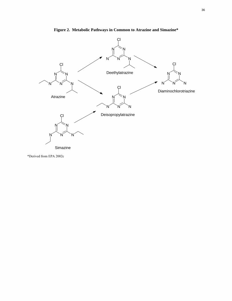
Deisopropylatrazine and diaminochlorotriazine also are metabolites of simazine. Figure 2 summarizes
the metabolic dealkylation pathways that are common to atrazine and simazine, and includes deethyl
atrazine.
A.2 Health Effects
Based on results of studies in experimental animals, to be reviewed later in this section, concerns for the
potential impacts of atrazine on human health include reproductive and carcinogenic effects. The
epidemiological studies, however, provide little evidence of such impacts.
Three related survey studies of farm couples in Ontario investigated the potential impact of atrazine
exposure (primarily direct exposure of the men) on reproductive and developmental endpoints including
time to pregnancy, spontaneous abortion, preterm delivery, sex ratio, and small for gestational age
(Arbuckle et al. 2001; Curtis et al. 1999; Savitz et al. 1997). These studies controlled for potential
reproductive confounders, but relied entirely on self reporting of exposure and pregnancy outcome. The
only significant reported association was an elevated odds ratio for preterm delivery with atrazine
exposure through its use as an herbicide in the yard (but not with use on crops). Similar results were
reported for triazine use in the yard (but not on crops) in the same study. The preterm delivery odds ratios
were not adjusted for exposure to other pesticides (ATSDR 2003). A study of low birth weight,
prematurity, and intrauterine growth retardation in Iowa communities with herbicide-contaminated
municipal water supply reported an elevated relative risk for intrauterine growth retardation (adjusted for
N N N ClCl
Deethylatrazine N NN N
N N NN N N Cl
Diaminochlorotriazine Atrazine N N
N N N
Deisopropylatrazine Cl
N N
N N N
Simazine
*Derived from EPA 2002c
N N
36
Figure 2. Metabolic Pathways in Common to Atrazine and Simazine*
Cl
37
mother’s age) as compared with communities served by other water supplies (Munger et al. 1997).
Results for low birth weight and prematurity were not significant. Multiple linear regression analyses
revealed that, after controlling for potential confounding factors including maternal smoking, atrazine was
more strongly correlated with intrauterine growth retardation than were the other herbicides, but the
herbicides (atrazine, cyanazine, metolachlor) were intercorrelated. In addition, estimates of exposure and
confounding factors were made on the community rather than individual level. Thus, these studies do not
provide adequate evidence of reproductive effects in humans, but may indicate a need for further study.
Atrazine causes neuroendocrine, reproductive, and reproductive developmental effects in experimental
animals. Animal studies have shown that atrazine disrupts estrus cyclicity (i.e., irregular ovarian cycling
and changes in the number and/or percentage of days in estrus and diestrus) and alters plasma hormone
levels in rats and pigs. These effects appear to be mediated by changes in the hypothalamic-pituitary
ovary axis that are species-, and even strain-, specific. In Sprague-Dawley rats, atrazine accelerates the
normal process of reproductive senescence, which is initiated by a failure of the hypothalamus to release
levels of gonadotropin releasing hormone (GnRH) that are adequate to stimulate the pituitary to release
LH. Without sufficient LH, ovulation does not occur, estrogen levels remain high, and persistent estrus
results. In other strains of rats, atrazine causes elevated progesterone levels, which leads to pseudo-
pregnancy and persistent diestrus (ATSDR 2003).
The mechanism of reproductive senescence in humans does not involve disruption of hormonal
regulation, but is initiated by depletion of ova in the ovaries, which ultimately results in decreased plasma
estrogen levels. Therefore, disruption of the menstrual cycle or acceleration of reproductive senescence is
not anticipated to occur in humans as a result of atrazine exposure. However, it is not known whether
atrazine will cause other perturbations in the hypothalamus-pituitary-gonad axis resulting in reproductive
effects in human (ATSDR 2003).
Developmental effects have been observed following pregestational, gestational, and lactational oral
exposure of rat and rabbit dams and peripubertal oral exposure of rats to atrazine. The observed effects
included impaired development of the reproductive system, postimplantation losses, decreases in fetal
body weight, incomplete ossification, and neurodevelopmental effects (ATSDR 2003).
A number of epidemiology studies have investigated the carcinogenic potential of atrazine or triazine
herbicides (ATSDR 2003; IARC 1999a). These studies include cohort studies of triazine manufacturing
workers, case-control studies of farmers using atrazine or triazines, and ecological studies of populations

38
in agricultural areas with high atrazine or triazine use and populations of areas with atrazine-contaminated
drinking water. Results of these studies were inconclusive. Odds ratios, standardized mortality ratios
(SMRs), or relative risks generally were not elevated or were not statistically significantly elevated after
adjustment for exposure to other pesticides. A few studies reported statistically significant correlations or
elevated odds ratios for cancer of the prostate (Mills 1998), breast (Kettles et al. 1997), ovary (Donna et
al. 1989), or stomach (Van Leeuwen et al. 1999) and triazine or atrazine exposure. These studies,
however, had no individual measures of exposure and/or no accounting for exposure to other pesticides,
and are not confirmed by the other available epidemiological studies on the same chemicals.
Statistically significant earlier onset or increased incidences of mammary tumors were observed in female
Sprague-Dawley rats, but not in female F344 rats or in mice (ATSDR 2003). The early onset of
mammary tumors in female Sprague-Dawley rats is believed to be the result of atrazine-induced
acceleration of reproductive senescence, as further explained under mechanisms of action.
Deethylatrazine was not explicitly considered in the epidemiology studies. Because it is frequently
detected in surface and groundwaters that contain atrazine (Gilliom et al. 1999; Squillace et al. 2002),
studies that involved exposure to atrazine or triazines through drinking water probably included exposure
to deethylatrazine.
A few studies of deethylatrazine have been performed in animals. In these studies, deethylatrazine
generally produced the same effects as atrazine. Diaminochlorotriazine, a metabolite of both atrazine and
deethylatrazine, has been tested more extensively and caused similar reproductive function and
reproductive developmental effects, and carcinogenic effects (mammary gland tumors in Sprague-Dawley
female rats) affects as did atrazine (EPA 2002c). Therefore, it is reasonable to assume that deethyl
atrazine will do so as well.
A.3 Mechanisms of Action
The primary target of atrazine in some animal species is the female reproductive system. Altered estrus
cyclicity has been observed in Sprague-Dawley, Long-Evans, and Donryu rats following exposure to
≥5 mg/kg/day atrazine for intermediate or chronic durations and to a single dose of 300 mg/kg/day.
Atrazine does not appear to have estrogenic activity. Atrazine is thought to disrupt endocrine function,
and the estrus cycle, primarily through its action on the central nervous system in a manner very similar to
the known mechanism of reproductive senescence in some strains of rats. In certain strains of rats,
39
including Sprague-Dawley and Long-Evans, reproductive senescence begins by 1 year of age, and results
from inadequate stimulation of the pituitary by the hypothalamus to release LH; low serum levels of LH
lead to anovulation, persistent high plasma levels of estrogen, and persistent estrus. Atrazine apparently
accelerates the process of reproductive senescence in these strains of rats (ATSDR 2003).
Atrazine has been shown to induce mammary tumor formation in female Sprague-Dawley rats, but not
male Sprague-Dawley or male or female F344 rats. This effect is also thought to be the result of
acceleration of reproductive senescence, as described above. Both the failure to ovulate and the state of
persistent estrus lead to constant elevated serum levels of endogenous estrogen, which may result in
tumor formation in estrogen-sensitive tissues. The rat does not appear to be an adequate model for
potential atrazine carcinogenicity in women because reproductive senescence in women involves ovarian
depletion and decreased serum estrogen levels instead of decreasing hypothalamic function and increased
serum estrogen levels (ATSDR 2003; EPA 2002a, 2002b, 2002c).
As previously stated, atrazine has been shown to alter serum LH and prolactin levels in Sprague-Dawley
rats by altering the hypothalamic control of these hormones (Cooper et al. 2000). LH and prolactin are
released from the pituitary in response to GnRH from the hypothalamus. One proposed mechanism is
that atrazine decreases the hypothalamic secretion of norepinephrine, which in turn decreases the release
of GnRH (EPA 2002a, 2002c). Another proposed mechanism is that atrazine disrupts hypothalamic
release of GnRH by interfering with the binding of some ligands, but not others, to the GABAA receptors
in a noncompetitive manner (ATSDR 2003).
A.4 Health Guidelines
ATSDR (2003) did not derive inhalation MRLs for atrazine because of the lack of suitable data.
ATSDR (2003) derived an acute oral MRL of 0.01 mg/kg/day based on a no-observed-adverse-effect
level (NOAEL) of 1 mg/kg/day for decreased body weight gain in rabbits administered atrazine by
gavage on gestation days 7–19, and using an uncertainty factor of 100. The LOAEL was 5 mg/kg/day;
slight but statistically significant reductions in food consumption and body weight gain were seen at this
dose level.
ATSDR (2003) derived an intermediate oral MRL of 0.003 mg/kg/day based on a LOAEL for delayed
onset of estrus in pigs using an uncertainty factor of 300.
40
EPA derived an oral RfD of 0.035 mg/kg/day based on a NOAEL of 3.5 mg/kg/day in a chronic dietary
study in rats, and using an uncertainty factor of 100 (IRIS 2003). The LOAEL was 25 mg/kg/day. The
critical effects were decreased body weight gain in the rat study and cardiac toxicity in a 1-year dietary
study in dogs. This RfD was verified by EPA in 1993; significant new studies have been published since
that time (IRIS 2003).
More recently, the EPA (2002b) Office of Pesticide Programs derived an acute RfD of 0.10 mg/kg/day
based on a weight-of-evidence analysis of four developmental studies, and a chronic RfD of
0.018 mg/kg/day based on attenuation of the LH surge and estrus cycle disruptions in female Sprague
Dawley rats. Although not on IRIS, these derivations include a consideration of toxicological and
mechanistic data that have become available since the RfD on IRIS was derived. They have been
subjected to extensive review, including public comment, and are available online (EPA 2002b). A
FQPA default safety factor of 10 (EPA 2003) was applied to protect infants and children (and other
populations) when assessing dietary (food + drinking water) exposures, resulting in a acute population
adjusted dose (PAD) of 0.01 mg/kg/day and a chronic PAD of 0.0018 mg/kg/day (EPA 2002b). These
RfDs and PADs are for atrazine together with its chlorinated metabolites (including deethylatrazine),
which are considered to have equivalent toxicity to atrazine.
The EPA (2002c) Office of Pesticide Programs has concluded that atrazine, deethylatrazine, diamino
chlorotriazine, deisopropylatrazine, simazine, and propazine should be considered a Common Mechanism
Group for cumulative risk assessment due to their ability to suppress the pituitary LH surge resulting in
effects on reproductive function and reproductive development.
The National Toxicology Program (NTP 2003) does not include atrazine in its listings.
The International Agency for Research on Cancer (IARC 1999a) classified atrazine as not classifiable as
to its carcinogenicity to humans (Group 3) based on inadequate evidence in humans and sufficient
evidence in experimental animals.
EPA has not published a cancer assessment of atrazine on IRIS (2003). The EPA (2002a, 2002b) Office
of Pesticide Programs classified atrazine and its chlorinated metabolites (including deethylatrazine) as not
likely to be carcinogenic to humans.
41
A.5 Derivation of Target Organ Toxicity Dose (TTD) Values
It is recommended that the chronic PAD of 0.0018 mg/kg/day for atrazine and its chlorinated metabolites
(EPA 2002b) be adopted as a provisional TTD for reproductive effects. Details of the derivation of this
guidance value are as follows: A chronic RfD of 0.018 mg/kg/day was based on an oral NOAEL for
atrazine of 1.8 mg/kg/day for attenuation of the LH surge and estrus cycle disruptions in female Sprague
Dawley rats (EPA 2002a, 2002b). An uncertainty factor of 100 was applied to the NOAEL (10 for
interspecies extrapolation and 10 for intraspecies variations). The LOAEL for these effects was
3.65 mg/kg/day. A FQPA safety factor of 10 was applied to protect infants and children when assessing
dietary (food + drinking water) exposures, resulting in chronic PAD of 0.0018 mg/kg/day. The RfD and
PAD are for atrazine together with its chlorinated metabolites (including deethylatrazine), which are
considered to have equivalent toxicity to atrazine (EPA 2002b).
Summary (TTD for Atrazine and Deethylatrazine)
TTDREPRO = 0.0018 mg/kg/day
A.6 References
Arbuckle TE, Lin Z, Mery LS. 2001. An exploratory analysis of the effect of pesticide exposure on the risk of spontaneous abortion in an Ontario farm population. Environ Health Perspect 109(8):851–857.
ATSDR. 2003. Toxicological profile for atrazine. Post-public comment draft (version 1). Atlanta, GA: Agency for Toxic Substances and Disease Registry.
Cooper RL, Stoker TE, Tyrey L, et al. 2000. Atrazine disrupts the hypothalamic control of pituitary-ovarian function. Toxicol Sci 53:297–307.
Curtis KM, Savitz DA, Weinberg CR, et al. 1999. The effect of pesticide exposure on time to pregnancy. Epidemiology 10:112–117.
Donna A, Crosignani P, Robutti F, et al. 1989. Triazine herbicides and ovarian epithelial neoplasms. Scand J Work Environ Health 15:47–53.
EPA. 2002a. Memorandum: Atrazine/DACT- fourth report of the hazard identification assessment review committee. Document attached. U.S. Environmental Protection Agency. Office of Pesticide Programs. http://www.epa.gov/oppsrrd1/reregistration/atrazine/hed_hiarc_atrazine_5april02.PDF.
EPA. 2002b. Revised human health risk assessment: Atrazine: Memorandum attached. Washington, DC: U.S. Environmental Protection Agency. Office of Pesticide Programs. http://www.epa.gov/oppsrrd1/reregistration/atrazine/hed_redchap_16apr02.PDF.

42
EPA. 2002c. The grouping of a series of triazine pesticides based on a common mechanism of toxicity. Washington, DC: U.S. Environmental Protection Agency. Office of Pesticides Programs. http://www.epa.gov/oppsrrd1/cumulative/triazines/triazinescommonmech.pdf.
EPA. 2003. Pesticides. Regulating pesticides. The Food Quality Protection Act (FQPA). http://www.epa.gov/oppfead1/fqpa/backgrnd.htm.
Gilliom RJ, Barbash JE, Kolpin DW, et al. 1999. Testing water quality for pesticide pollution: U.S. Geological Survey investigations reveal widespread contamination of the nation's water resources. Environ Sci Technol 33(7):164A–169A.
Hanioka N, Jinno H, Tanaka-Kagawa T, et al. 1999. In vitro metabolism of chlorotriazines: Characterization of simazine, atrazine, and propazine metabolism using liver microsomes for rats treated with various cytochrome P450 inducers. Toxicol Appl Pharmacol 156:195–205.
IARC. 1999a. Atrazine. International Agency for Research on Cancer. IARC Monogr Eval Carcinog Risks Hum 73:59–113.
IRIS. 2003. Integrated Risk Information System. U.S. Environmental Protection Agency. http://www.epa.gov/iris.
Kettles MA, Browning SR, Prince TS, et al. 1997. Triazine herbicide exposure and breast cancer incidence: An ecologic study of Kentucky counties. Environ Health Perspect 105(11):1222–1227.
Mills PK. 1998. Correlation analysis of pesticide use data and cancer incidence rates in California Counties. Arch Environ Health 53(6):410–413.
Munger R, Isacson P, Hu S, et al. 1997. Intrauterine growth retardation in Iowa communities with herbicide-contaminated drinking water supplies. Environ Health Perspect 105(3):308–314.
NTP. 2003. 10th report on carcinogens. U.S. Department of Health and Human Services. National Toxicology Program. http://ehp.niehs.nih.gov/roc/toc10.htm.
Savitz DA, Arbuckle T, Kaczor D, et al. 1997. Male pesticide exposure and pregnancy outcome. Am J Epidemiol 146(12):1025–1036.
Squillace PJ, Scott JC, Moran MJ, et al. 2002. VOCs, pesticides, nitrate, and their mixtures in groundwater used for drinking water in the United States. Environ Sci Technol 36:1923–1930. Timchalk C, Dryzga MD, Langvardt PW, et al. 1990. Determination of the effect of tridiphane on the pharmacokinetics of [14C]-atrazine following oral administration to male Fischer 344 rats. Toxicology 61:27–40.
Van Leeuwen JA, Waltner-Toews D, Abernathy T, et al. 1999. Associations between stomach cancer incidence and drinking water contamination. Int J Epidemiol 28(5):836–840.
43
Appendix B: Background Information for Simazine
Simazine is a triazine herbicide (an herbicide containing the s-triazine ring) that inhibits photosynthesis in
plants. The structure of simazine is depicted in Appendix E, and also in the metabolic scheme presented
previously in Figure 2.
B.1 Toxicokinetics
Less information is available regarding the toxicokinetics of simazine than was available for atrazine.
The percent of administered radiolabel excreted in urine during 96 hours after oral dosing of rats with
radiolabeled simazine was 49.3%, indicating that absorption was at least 49.3% (EPA 2002c).
In experimental animals, simazine is metabolized by (EPA 2002c; Guddewar and Dauterman 1979; IARC
1999b):
• successive N-dealkylation to deisopropylatrazine (desisopropyl atrazine), and didealkylatrazine
(commonly called diaminochlorotriazine or DACT);
• glutathione conjugation of simazine and the above-listed metabolites (probably followed by
conversion to mercapturic acid derivatives).
Deisopropylatrazine and diaminochlorotriazine also are metabolites of atrazine. Figure 2 summarizes the
metabolic dealkylation pathways that are common to atrazine and simazine
The dealkylation of simazine is carried out by microsomal cytochrome P450 enzymes (EPA 2002c).
Studies with rat liver microsomes indicate that the specific isozymes involved in this dealkylation are
CYP1A1/2 (Hanioka et al. 1999).
An oral study with radiolabeled simazine in rats indicates extensive tissue distribution of radioactivity,
including to the brain (EPA 2002c).
B.2 Health Effects
Some of the epidemiological studies reviewed in Appendix A were on agricultural exposure to trazines in
Midwestern states, and did not specify whether exposure to simazine occurred. Because atrazine and
cyanazine are the main triazines used as herbicides in the corn belt of the Midwest, it is likely that
44
exposures were mainly to atrazine and cyanazine (Snedeker and Clark 1998). The Ontario farm survey
studies reviewed in Appendix A listed atrazine and cyanazine, but not simazine. IARC (1999b) stated
that no human reproductive and developmental effects data were available for simazine, and no human
cancer data were available for simazine alone.
Studies in rats indicate that simazine has effects on reproductive function and reproductive development
similar to those of atrazine, as do its metabolites deisopropylatrazine and diaminochlorotriazine (EPA
2002c). Also, simazine and diaminochlorotriazine cause mammary gland tumors in Sprague-Dawley
female rats (EPA 2002c). As explained previously for atrazine, this carcinogenic effect of simazine is not
considered relevant to humans (see Section A.2 and A.3).
B.3 Mechanisms of Action
The mechanism of action of simazine and its metabolites deisopropylatrazine and diaminochlorotriazine
is considered to be the same as for atrazine as described in Section A.3 with regard to neuroendocrine,
reproductive, and carcinogenic effects (EPA 2002c).
B.4 Health Guidelines
ATSDR has not developed a toxicological profile or MRLs for simazine.
EPA derived an oral RfD of 0.005 mg/kg/day based on a NOAEL of 0.52 mg/kg/day in a chronic dietary
study in rats, and using an uncertainty factor of 100 (IRIS 2003). The LOAEL was 5.3 mg/kg/day. The
critical effects were reduction in weight gain and hematological changes (mainly depression of red cell
parameters. This RfD was verified by EPA in 1993; significant new studies have been published since
that time (IRIS 2003).
More recently than the 1993 assessment that is on IRIS (2003), the EPA (2002c) Office of Pesticide
Programs concluded that atrazine, deethylatrazine, diaminochlorotriazine, deisopropylatrazine, simazine,
and propazine should be considered a Common Mechanism Group for cumulative risk assessment due to
their ability to suppress the pituitary LH surge resulting in effects on reproductive function and
reproductive development. These effects were considered the critical effects. Taking into account newer
toxicological and mechanistic data, EPA (2002b) developed new RfDs for atrazine and its chlorinated
metabolites (see Section A.4), but has not yet developed new RfDs for simazine.
45
NTP (2003) does not include simazine in its listings.
IARC (1999b) classified simazine as not classifiable as to its carcinogenicity to humans (Group 3) based
on inadequate evidence in humans and sufficient evidence in experimental animals.
EPA has not published a cancer assessment of simazine on IRIS (2003).
B.5 Derivation of Target Organ Toxicity Dose (TTD) Values
It is recommended that the chronic PAD of 0.0018 mg/kg/day for atrazine and its chlorinated metabolites
(EPA 2002b) also be adopted as a provisional TTD for reproductive effects for simazine. The derivation
of this guidance value is described in Section A.5. The structure, molecular weights, metabolism,
toxicity, and mechanisms of action of these chemicals are similar, and they are considered to belong to a
Common Mechanism Group for cumulative risk assessment due to their ability to suppress the pituitary
LH surge resulting in effects on reproductive function and reproductive development (EPA 2002c). This
value is recommended as an interim measure until an up-to-date guidance value is developed specifically
for simazine.
Summary (TTD for Simazine)
TTDREPRO = 0.0018 mg/kg/day
B.6 References
EPA. 2002b. Revised human health risk assessment: Atrazine: Memorandum attached. Washington, DC: U.S. Environmental Protection Agency. Office of Prevention, Pesticides and Toxic Substances. http://www.epa.gov/oppsrrd1/reregistration/atrazine/hed_redchap_16apr02.PDF.
EPA. 2002c. The grouping of a series of triazine pesticides based on a common mechanism of toxicity. Washington, DC: U.S. Environmental Protection Agency. Office of Pesticides Program. http://www.epa.gov/oppsrrd1/cumulative/triazines/triazinescommonmech.pdf.
Guddewar MB, Dauterman WC. 1979. Studies on glutathione S-transferase preparation from mouse liver which conjugates chloro-s-triazine herbicides. Pestic Biochem Physiol 12(1):1–9.
Hanioka N, Jinno H, Tanaka-Kagawa T, et al. 1999. In vitro metabolism of chlorotriazines: Characterization of simazine, atrazine, and propazine metabolism using liver microsomes for rats treated with various cytochrome P450 inducers. Toxicol Appl Pharmacol 156:195–205.
46
IARC. 1999b. Simazine. International Agency for Research on Cancer. IARC Monogr Eval Carcinog Risks Hum 73:625–640.
IRIS. 2003. Integrated Risk Information System. U.S. Environmental Protection Agency. http://www.epa.gov/iris.
NTP. 2003. 10th report on carcinogens. U.S. Department of Health and Human Services. National Toxicology Program. http://ehp.niehs.nih.gov/roc/toc10.htm.
Snedeker SM, Clark H. 1998. Critical evaluation of simazine's breast cancer risk. Cornell University. Program on Breast Cancer and Environmental Risk Factors in New York State (BCERF). http://www.cfe.cornell.edu/bcerf/.
47
Appendix C: Background Information for Diazinon
Diazinon is an organophosphorus insecticide. The structure of diazinon and its toxic metabolite,
diazoxon, are provided in Appendix E.
C.1 Toxicokinetics
Diazinon is rapidly absorbed from the gastrointestinal tract, based on case reports of ingestion of diazinon
formulation or solution, on single oral dose studies in rats and dogs, and on repeated oral dose studies in
rats. Absorption in rats and dogs was at least 85% of the dose (ATSDR 1996; WHO 1998). The main
features of diazinon metabolism are:
• activation of diazinon through conversion of the P=S moiety to P=O, resulting in the toxic
intermediate, diazoxon;
• cleavage of the ester bonds of diazinon and diazoxon resulting in 2-isopropyl-4-methyl-6-hydro
pyrimidine (from both), diethylphosphorothioc acid (from diazinon), and diethylphosphoric acid
(from diazoxon);
• oxidation of the isopropyl substituent of 2-isopropyl-4-methyl-6-hydropyrimidine to the
corresponding primary and tertiary alcohols;
• glutathione-mediated cleavage of the ester bond with the formation of a glutathione conjugate
(minor pathway).
The resulting metabolites are excreted primarily in the urine (ATSDR 1996; WHO 1998).
The metabolic activation of diazinon to diazoxon is carried out by microsomal cytochrome P450
monooxygenases. A single study of diazinon in rat hepatic microsomes has reported that CYP2B1/2 are
the major P450 isozymes that catalyze the production of diazoxon (Fabrizi et al. 1999).
C.2 Health Effects
The principal toxic effect of diazinon in humans, experimental animals, and insects is acetylcholinesterase
inhibition. Acetylcholine is a neurotransmitter in the central and peripheral neurons. Inhibition of acetyl
cholinesterase, the enzyme that breaks down and terminates the action of acetylcholine, results in the
accumulation of acetylcholine at acetylcholine receptors leading to continued stimulation.

48
In humans and experimental animals, the accumulation of acetylcholine results in cholinergic responses in
the peripheral (muscarinic and nicotinic) and central nervous system and neuromuscular junctions. These
cholinergic responses, seen in severe acetylcholinesterase inhibition, include excessive glandular
secretions (salivation, lacrimation, rhinitis), miosis, bronchoconstriction, vasodilation, hypotension,
diarrhea, nausea, vomiting, urinary incontinence, and bradycardia associated with muscarinic receptor
stimulation. Tachycardia, mydriasis (dilation of the pupil), muscle fasciculations, cramping, twitching,
muscle weakness, muscle paralysis, and hypertension are associated with nicotinic receptor stimulation.
Central nervous system toxicity includes respiratory depression, anxiety, insomnia, headache, apathy,
drowsiness, dizziness, loss of concentration, confusion, tremors, convulsions, and coma. These effects
usually appear within a few minutes to 24 hours after exposure, depending on the extent and route of
exposure. In nonfatal exposures, the effects are usually transient, with rapid and complete recovery
following cessation of exposure. Recovery from diazinon poisoning results from increased availability of
active acetylcholinesterase either from synthesis of new enzyme, the spontaneous hydrolysis of the
enzyme-phosphate ester complex, or treatment with atropine, a competitive antagonist of acetylcholine at
muscarinic and central nervous system receptors, and with pralidoxime (2-PAM), a drug that regenerates
inhibited acetylcholinesterase enzyme by displacing the diethylphosphoester bond that diazoxon forms at
the active site (Aaron and Howland 1998; ATSDR 1996).
In some cases, however, diazinon may cause a condition known as the intermediate syndrome (Aaron and
Howland 1998; WHO 1998). This syndrome occurs during apparent recovery about 24–96 hours after
severe cholinergic crisis, and includes paralysis of the respiratory muscles, upper extremity muscles, neck
flexors, and motor cranial nerves. Diazinon has been tested for organophosphate-induced delayed
neurotoxicity in chickens; results were negative (ATSDR 1996). No cases of delayed neuropathy from
diazinon exposure have been reported (ATSDR 1996; WHO 1998).
Acetylcholinesterase activity is also present in erythrocytes where it is known as erythrocyte acetyl
cholinesterase. Both forms of acetylcholinesterase are produced by the same gene and are kinetically
identical. In in vitro assays, erythrocyte and neural acetylcholinesterase are inhibited to roughly the same
extent by exposure to diazinon and many other organophosphorus compounds with insecticidal activity;
measurement of erythrocyte acetylcholinesterase can be used as a surrogate indicator of the extent of
inhibition of neural acetylcholinesterase (ATSDR 1996).
A cholinesterase capable of hydrolyzing acetylcholine and butyrlcholine is produced by the liver and
circulates in the blood. This enzyme, referred to as serum cholinesterase, plasma cholinesterase, pseudo
49
cholinesterase, or butyrylcholinesterase, is also inhibited by diazinon and is often used as a marker for
exposure (ATSDR 1996). This enzyme is present in some nonneural cells in the central and peripheral
nervous systems as well as in plasma and serum, the liver, and other organs. Its physiologic function is
not known, but is hypothesized to be the hydrolysis of esters ingested from plants (Lefkowitz et al. 1996).
Plasma cholinesterases are also inhibited by organophosphate compounds through irreversible binding;
this binding can act as a detoxification mechanism as it affords some protection to acetylcholinesterase in
the nervous system (Parkinson 1996; Taylor 1996). In general, this enzyme is inhibited by diazinon at
lower levels of exposure than required to inhibit neural or erythrocyte acetylcholinesterase (ATSDR
1996).
A few case reports of diazinon ingestion or dermal exposure have reported acute pancreatitis as a
component of severe diazinon intoxication (ATSDR 1996; WHO 1998). Diazinon at sublethal doses also
caused pancreatic ductal hypertension in dogs, and acute pancreatitis in dogs and guinea pigs but not in
cats (Dressel et al. 1980; Frick et al. 1987). Effects on the pancreas appear to be a high-dose
phenomenon.
Epidemiological studies provide no specific evidence of carcinogenicity for diazinon, and the available
animal studies do not suggest that diazinon would be likely to cause cancer in humans (ATSDR 1996;
EPA 2000).
C.3 Mechanisms of Action
Diazinon and diazoxon inhibit acetylcholinesterase by reacting with the active site to form a stable
dialkylphosphorylated enzyme that cannot hydrolyze acetylcholine. Diazoxon, the active metabolic
intermediate of diazinon, is much more potent than diazinon in inhibiting acetylcholinesterase (ATSDR
1996; WHO 1998).
The mechanism of action with regard to pancreatic toxicity in dogs and guinea pigs appears to be
inhibition of butyrylcholinesterase in the pancreas and its smooth muscle sphincters, leading to ductal
hypertension and cholinergic hyperstimulation of the acinar cells (Dressel et al. 1980; Frick et al. 1987).
50
C.4 Health Guidelines
ATSDR (1996) derived an intermediate inhalation MRL of 0.009 mg/m3 for brain acetylcholinesterase
inhibition diazinon based on a NOAEL of 0.46 mg/m3 in a 21-day study in rats. An uncertainty factor of
30 was applied. The LOAEL (20% decrease in brain acetylcholinesterase) was 1.57 mg/m3.
ATSDR (1996) derived an intermediate oral MRL of 0.0002 mg/kg/day based on a NOAEL of
0.021 mg/kg/day for brain acetylcholinesterase inhibition in dogs given diazinon in their food daily for
13 weeks. An uncertainty factor of 100 was used. The LOAEL (31% decrease in erythrocyte and brain
acetylcholinesterase) was 5.9 mg/kg/day. A chronic oral MRL was not derived because the chronic
NOAEL (0.05 mg/kg/day for brain cholinesterase inhibition in rats) that was considered as the basis for
the MRL would have resulted in an MRL (0.0005 mg/kg/day) that was slightly higher than the
intermediate oral MRL. Since the intermediate-duration MRL would be more protective, it was the only
one derived, and was considered protective for individuals living near hazardous waste sites.
EPA (IRIS 2003) does not have an online file for diazinon.
The EPA (2000) Office of Pesticide Programs derived acute and chronic RfDs of 0.0025 and
0.0002 mg/kg/day based on NOAELs for cholinesterase inhibition of 2.5 mg/kg/day (in rats) and
0.02 mg/kg/day in seven feeding studies (in rats and dogs), respectively. An additional 10-fold FQPA
safety factor (EPA 2003) was not used for special sensitivity in infants and children because the EPA
concluded that the data indicated that this factor could be reduced to 1-fold. The PADs are therefore the
same as the RfDs. Although this RfD and PAD are not on IRIS, they have been subjected to extensive
review, including public comment, and are available online (EPA 2000).
NTP (2003) and IARC (2003) do not include diazinon in their listings. The EPA (2000) Office of
Pesticide Programs classified diazinon as a not likely human carcinogen based on the lack of evidence of
carcinogenicity in mice and rats.
C.5 Derivation of Target-Organ Toxicity Dose (TTD) Values
The intermediate oral MRL of 0.0002 mg/kg/day for neurological effects (brain acetylcholinesterase
inhibition in dogs for 13-week oral exposure) is appropriate for use as a chronic guidance value as well
(ATSDR 1996), and is the same as the chronic oral RfD developed by EPA (2000). An uncertainty factor

51
of 100 was applied to a NOAEL of 0.021 mg/kg/day, as described in the previous section. The LOAEL
(31% decrease in erythrocyte and brain acetylcholinesterase) was 5.9 mg/kg/day.
Summary (TTD for Diazinon)
MRLNEURO =0.0002 mg/kg/day
C.6 References
Aaron CK, Howland MA. 1998. Insecticides: Organophosphates and carbamates. In: Goldfrank LR, Flomenbaum NE, Lewin NA, et al., eds. Goldfrank's toxicologic emergencies. Stamford, CT: Appleton & Lange, 1429–1449.
ATSDR. 1996. Toxicological profile for diazinon. Atlanta, GA: Agency for Toxic Substances and Disease Registry.
Dressel TD, Goodale RL, Borner JW, et al. 1980. A study of the cholinesterases of the canine pancreatic sphincters and the relationship between reduced butyrylcholinesterase activity and pancreatic ductal hypertension. Ann Surg 192(5):614–619.
EPA. 2000. Human health risk assessment: Diazinon: Memorandum attached. Washington, DC: U.S. Environmental Protection Agency. Office of Pesticide Programs. http://www.epa.gov/pesticides/op/diazinon/final_red.pdf.
EPA. 2003. Pesticides. Regulating pesticides. The Food Quality Protection Act (FQPA). http://www.epa.gov/oppfead1/fqpa/backgrnd.htm.
Fabrizi L, Gemma S, Testai E, et al. 1999. Identification of the cytochrome P450 isoenzymes involved in the metabolism of diazinon in the rat liver. J Biochem Mol Toxicol 13(1):53–61.
Frick TW, Dalo S, O'Leary JF, et al. 1987. Effects of insecticide, diazinon, on pancreas of dog, cat and guinea pig. J Environ Pathol Toxicol Oncol 7(4):1–11.
IARC. 2003. Overall evaluations of carcinogenicity to humans: As evaluated in IARC Monographs volumes 1–82 (at total of 885 agents, mixtures and exposures). International Agency for Research on Cancer. http://193.51.164.11/moneeval/crthall.html.
IRIS. 2003. Integrated Risk Information System. U.S. Environmental Protection Agency. http://www.epa.gov/iris.
Lefkowitz RJ, Hoffman BB, Taylor P. 1996. Neurotransmission: The autonomic and somatic motor nervous systems. In: Goodman LS, Gilman A, Hardman JG, et al., eds., Goodman & Gilman's the pharmacological basis of therapeutics. New York, NY: McGraw-Hill: Health Professions Division, 105–139.
NTP. 2003. 10th report on carcinogens. U.S. Department of Health and Human Services. National Toxicology Program. http://ehp.niehs.nih.gov/roc/toc10.htm.
52
Parkinson A. 1996. Biotransformation of xenobiotics. In: Klassen CD, ed. Casarett and Doull's Toxicology: The basic science of poisons. New York: McGraw-Hill, 115–118, 145–146.
Taylor P. 1996. Anticholinesterase agents. In: Goodman LS, Gilman A, Hardman JG, et al., eds. Goodman & Gilman's the pharmacological basis of therapeutics. New York, NY: McGraw-Hill: Health Professions Division, 161–176.
WHO. 1998. Environmental health criteria 198: Diazinon. World Health Organization. International Programme on Chemical Safety. http://www.inchem.org/documents/ehc/ehc/ehc198.htm.

53
Appendix D: Background Information for Nitrate
Nitrate occurs naturally in foods, particularly in vegetables. Inorganic fertilizers, livestock waste, and
septic tank discharges are primary contributors to nitrate contamination of drinking water (NRC 1995).
The structures of nitrate and its metabolite nitrite are shown in Appendix E.
D.1 Toxicokinetics
Available studies indicate that oral absorption of nitrate is nearly 100% (for reviews, see EPA 1990 and
WHO 1978). Witter (1979, cited in EPA 1990) administered oral radioactive nitrate ion to two male
volunteers; one received the nitrate 1 hour after a large meal, the other about 10 hours after eating. In the
subject who had recently eaten, the radioactivity had a disappearance half-life from the stomach of about
30 minutes, but the radioactivity in the pylorus remained constant, suggesting that the nitrate had moved
to the small intestine rather than being absorbed through the stomach. In the second subject, the
disappearance half-life was 10 minutes. Studies in animals have also demonstrated that the bulk of an
orally-administered nitrate is absorbed through the small intestine, likely through the upper portion of that
organ. Absorbed nitrate is distributed throughout the body, but does not appear to accumulate in any
organ (EPA 1990).
The major metabolic pathway for nitrate is conversion to nitrite, and then to ammonia. Small amounts of
nitrate, perhaps 5–10% of the total exposure, are converted to nitrite by bacteria in the saliva, stomach,
and small intestine. This reaction is pH dependent, with no nitrate reduction occurring below pH 4 and
above pH 9, and the presence of oxygen inhibits the reduction of nitrite to ammonia. Absorbed nitrite
rapidly reacts with hemoglobin in the blood to form methemoglobin, which in adults, is rapidly converted
to oxyhemoglobin, then back to hemoglobin. In infants, particularly those under 3 months old, these
reducing systems are not fully developed, which may result in a buildup of methemoglobin in the blood.
Due to the higher stomach pH typically found in infants, it is believed that they also convert more nitrate
to nitrite in the stomach than adults. There are large species differences in the rate of reaction of nitrite
with hemoglobin, paralleled by similar differences in the rates of reduction of methemoglobin, making
extrapolation of results from animal data to humans problematic. Another potential metabolic pathway,
though less prevalent than the reaction with hemoglobin, is the reaction of nitrite with endogenous
molecules to form N-nitroso compounds, many of which have toxic effects, including carcinogenicity.
Available data in humans have demonstrated that excretion of ingested nitrate is rapid, with excretion
almost exclusively in the urine (EPA 1990; WHO 1978). Animal data support this observation. In both
54
humans and animals, considerably more nitrate is eliminated in the urine than is ingested in a normal diet,
implying that there is significant endogenous nitrate formation.
Parks et al. (1981, cited in EPA 1990) reported that following intratracheal instillation of trace amounts of
nitrate to BALB/C mice, absorption from the lungs was complete within a 10-minute period. Additional
studies of the toxicokinetics of inhaled nitrate are not available; however, the behavior of absorbed nitrate
following inhalation exposure is not expected to differ from nitrate absorbed following oral exposure.
D.2 Health Effects
The most sensitive known effects of exposure to nitrate result from increased levels of methemoglobin
arising from the nitrite-hemoglobin reaction. In healthy adults, methemoglobin formation and reduction
is continuous, with steady-state methemoglobin levels in healthy adults being 2.5% of the total hemo
globin content or lower (EPA 1990). Due to the large excess capacity of the blood to carry oxygen, levels
of methemoglobin up to 10% typically do not cause significant clinical signs. Levels above 10% may
result in cyanosis, weakness, rapid pulse, and, at levels exceeding 50%, death. Other reported effects of
nitrate in animals include altered thyroid function, amyloidosis of the liver, kidney, spleen, and adrenal
glands, and altered lung and liver weights.
Because of greater numbers of nitrate-reducing bacteria in the gastrointestinal tract and diminished
methemoglobin-reducing capacity, infants, especially those 3 months and younger, are particularly
susceptible to nitrate/nitrite-induced methemoglobinemia. A study by Bosch et al. (1950) examined
139 cases of methemoglobinemia in young children (90% of these cases occurred in children <2 months
of age). Examination of the wells used to supply water to the children revealed that none of the wells
supplied <10 mg/L nitrate-nitrogen, with all but two of the wells containing >25 mg/L. Walton (1951)
presented the results of a survey on morbidity and mortality among infants due to methemoglobinemia.
The results of the survey revealed 239 cases of infant methemoglobinemia, 39 of them fatal. Of the
214 cases where quantitative data were available on nitrate levels in water, none occurred in infants
consuming water with <10 mg/L nitrate-nitrogen, 5 cases occurred in infants exposed to 11–20 mg/L
nitrate-nitrogen, 36 cases in infants exposed to 21–50 mg/L nitrate-nitrogen, and 173 cases in infants
exposed to >50 mg/L nitrate-nitrogen. Many other studies have examined the effects of high (>20 mg/L)
levels of nitrate in the drinking water of infants, and have found increased methemoglobin levels and
signs of clinical methemoglobinemia in exposed infants (for reviews, see EPA 1990 and WHO 1978).
55
The Nuclear Regulatory Commission (NRC 1995), in its evaluation of the drinking water maximum
contaminant level goals (MCLGs) and MCLs for nitrate and nitrite, discussed the possible contributions
of infection and inflammatory reactions (particularly diarrhea in infants) to methemoglobinemia.
Infection increases the production of nitric oxide, which can be converted to nitrate. Avery (1999)
reviewed the evidence for gastrointestinal infection and inflammation as a cause of methemoglobinemia
in infants. Most of the studies of nitrate and infant methemoglobinemia are not adequate to clarify this
issue. A recent nested case-control study of methemoglobinemia risk factors, however, reported a
stronger association of infant methemoglobinemia with nitrate exposure (from formula and tea made with
nitrate-contaminated water) than with diarrhea (Zeman et al. 2002).
The nitrite ion and various organic nitrate compounds (e.g., nitroglycerin) cause vasodilation and
hypotension, but inorganic nitrate ion does not (EPA 1990).
D.3 Mechanisms of Action
The known toxic effects of nitrate exposure result from the conversion of nitrate to nitrite. The
conversion is mainly the result of bacterial oxidation reactions within the gastrointestinal tract. Exposure
of hemoglobin to nitrite results in the oxidation of the Fe2+ ion in the heme of hemoglobin to Fe3+,
resulting in the formation of methemoglobin. Methemoglobinemia results in the majority of the
symptoms seen following high-dose acute nitrate exposure in humans. Under normal conditions, healthy
adults will have <2.5% methemoglobin in the blood. Methemoglobin can be reduced back to hemoglobin
by both spontaneous (nicotinamide adenine dinucleotide phosphate [NADH]-dependent) and dormant
(NADPH-dependent) methemoglobin reductase enzymes.
Infants are particularly susceptible to methemoglobinemia due to their high gut content of nitrate-
reducing bacteria, their lower enzymatic capacity to reduce methemoglobin to hemoglobin, and the
presence of hemoglobin F, which is more susceptible to oxidation by nitrite. The high pH of the infant
gastrointestinal system favors the growth of nitrate-reducing bacteria, particularly in the stomach and
especially after ingestion of contaminated waters, since the ingested bacteria are likely to flourish in the
stomach. The stomach of adults is typically too acidic to allow for significant bacterial growth and the
resulting conversion of nitrate to nitrite. Additionally, the enzymes involved in the conversion of
methemoglobin to hemoglobin do not fully develop in humans until between 3 and 6 months after birth,
resulting in an increased susceptibility to methemoglobinemia.
56
As mentioned in Section D.1, the reaction rates for the nitrite-hemoglobin reaction vary considerably
across species (many animal species lack nitrate-reducing bacteria), as do the rates of the reactions
reducing methemoglobin back to functional hemoglobin. In addition, since the rates of conversion of
nitrate to nitrite by bacteria can vary within individuals, the extent of nitrate toxicity can vary greatly
depending on age and other factors within both humans and animals.
D.4 Health Guidelines
ATSDR has not published a toxicological profile for nitrates. No MRL values are available.
EPA (IRIS 2003) has derived an oral RfD of 1.6 mg/kg/day for nitrate, based on a NOAEL of
1.6 mg/kg/day for methemoglobinemia in exposed infants (Bosch et al. 1950; Walton 1951). An
uncertainty factor of 1 was applied to the NOAEL since the study was performed in a sensitive population
of humans (infants age 0–3 months).
NTP (1993) and IARC (2003) do not include nitrate in their listings. Nitrate has not undergone an
evaluation of carcinogenic potential by EPA (IRIS 2003).
D.5 Derivation of Target Organ Toxicity Dose (TTD) Values
In the absence of a toxicological profile and MRLs for nitrate, the chronic oral RfD of 1.6 mg/kg/day for
nitrate (IRIS 2003) can be adopted as the TTD for hematological effects.
Summary (TTD for Nitrate)
TTDHEMATO = 1.6 mg/kg/day
D.6 References
Avery AA. 1999. Infantile methemoglobinemia: Reexamining the role of drinking water nitrates. Environ Health Perspect 107(7):583–586.
Bosch HM, Rosefield AB, Huston R, et al. 1950. Methemoglobinemia and Minnesota well supplies. J Am Water Works Assoc 42:161–170.
EPA. 1990. Criteria document for nitrate/nitrite. Office of Drinking Water, U.S. Environmental Protection Agency, Washington, DC.
57
IARC. 2003. Overall evaluations of carcinogenicity to humans: As evaluated in IARC Monographs volumes 1–82 (at total of 885 agents, mixtures and exposures). International Agency for Research on Cancer. http://193.51.164.11/moneeval/crthall.html.
IRIS. 2003. Integrated Risk Information System. U.S. Environmental Protection Agency. http://www.epa.gov/iris.
NRC. 1995. Nitrate and nitrate in drinking water. National Research Council. Washington, DC: National Academy Press. PB95267092.
NTP. 2003. 10th report on carcinogens. U.S. Department of Health and Human Services. National Toxicology Program. http://ehp.niehs.nih.gov/roc/toc10.htm.
Parks NJ, Krohn KA, Mathis CA, et al. 1981. Nitrogen-13-labeled nitrite and nitrate: Distribution and metabolism after intratracheal administration. Science 212:58–61. (As cited in EPA 1990.)
Walton, G. 1951. Survey of literature relating to infant methemoglobinemia due to nitrate-contaminated water. Am J Public Health 41:986–996.
WHO. 1978. Environmental Health Criteria 5: Nitrates, nitrites and N-nitroso compounds. Geneva: World Health Organization.
Witter JP, Gatley SJ, Balish E. 1979. Distribution of nitrogen-13 from labeled nitrate (13NO3-) in humans and rats. Science 204:411–413. (As cited in EPA 1990.)
Zeman CL, Kross B, Vlad M. 2002. A nested case-control study of methemoglobinemia risk factors in children of Transylvania, Romania. Environ Health Perspect 110(8):817–822.
N
N
N
N
N N
N
N
N
N
Atrazine CAS No. 1912-24-9
Cl
Deethylatrazine CAS No. 6190-65-4
N N
N N N
Simazine CAS No. 122-34-9
Cl Cl
Cl
N N
N N N N O
N N
N N N N O
O O N
P O
S N
O O N
P O
O N
CAS No. 333-41-5
O
N+
O O
Nitrate CAS No. 14797-55-8
Diazinon
Diazoxon CAS No. 962-58-3
N O O
Nitrite CAS No. 14797-65-0
58
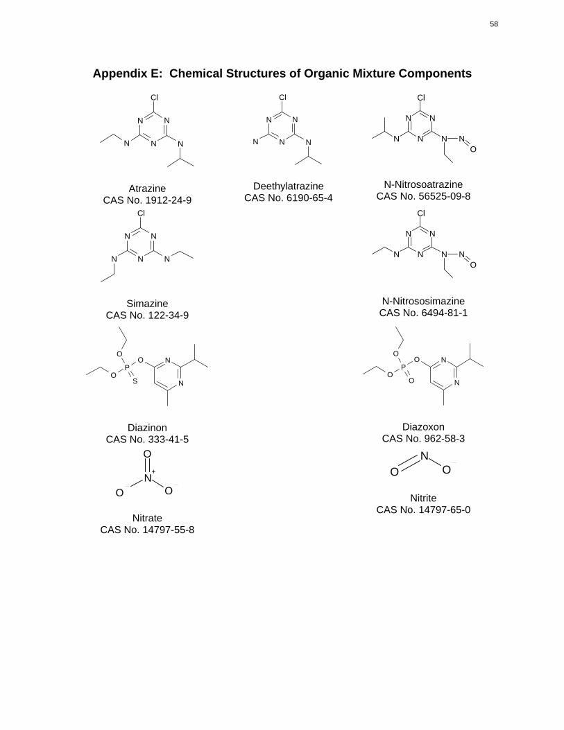
Appendix E: Chemical Structures of Organic Mixture Components
N-Nitrosoatrazine CAS No. 56525-09-8
Cl
N-Nitrososimazine CAS No. 6494-81-1