appendiceal mucinous neoplasms -...
TRANSCRIPT

Appendiceal Mucinous Neoplasms
Mark Bettington
Envoi Specialist Pathologists

A brief history
• Appendiceal mucinous neoplasms have a checkered and confusing past
• Appendiceal mucocoele was first described by Rokitansky in 1842
• Mucinous cystadenoma was the preferred term in the 1950s to 1980s
• The “uncertain malignant potential” of these lesions was first raised by Norman Carr et al, in 1995 to reflect the uncertain behaviour of cases with mucin dissecting the wall of the appendix or in cases with rupture


Further progress
• Over the last 20 years there has been a lot of progress in better characterising appendicealmucinous neoplasms
• Lots of papers, sometimes contradictory
• However a much clearer picture is now emerging from the literature
Some of the landmark papers
• Young et al; AJSP 1991 15:415-429
• Carr et al; Cancer 1995 75:757-768
• Misdraji et al; AJSP 2003 27(8):1089-1103
• Pai et al; AJSP 2009 33(10):1425-1439

What are the major issues?
• In actual fact there is only one issue we are trying to resolve:
Is there a risk of recurrence as pseudomyxomaperitonei?
How great is the risk?

Where are we now?
• Earlier this year this paper was published in AJSP
• This is a very useful consensus statement and provides lots of helpful guidelines and checklists for diagnosing appendiceal lesions (and pseudomyxoma peritonei)

Back to the beginning - What are the common epithelial lesions of the
appendix?
• Conventional adenomas
• Serrated polyps
• LAMNs
• HAMNs – not common

Conventional adenoma or serrated polyp
• Use the same definitions as for the colorectum
• The key is the muscularis mucosa has to be intact (with a few provisos to follow)

Low grade appendiceal mucinous neoplasms
• Defined as a mucinous neoplasm with low grade cytologic atypia and ANY of:1. Loss of muscularis mucosae2. Fibrosis of submucosa3. ‘Pushing invasion’ (expansile or diverticulum-like)4. Dissection of acellular mucin in the wall (what about cellular mucin?)5. Undulating or flattened epithelial growth6. Rupture of the appendix7. Mucin and/or cells outside the appendix

High grade appendiceal mucinous neoplasms
• Same as above except the cytology is high grade
Mucinous adenocarcinoma
• Conventional type
• Signet ring cell (must exclude a goblet cell carcinoid)

Approach to the LAMN
• Ideally this begins at the cut-up
- for any unusually dilated appendix, encourage the registrar to stop and think ‘could this be a LAMN?’
- careful macroscopic assessment (in particular is there any evidence of rupture? Any mucin on the serosal surface? Any diverticula?)
- entirely embed the specimen
- know what to look for histologically

Scenario one – the lesion with intact muscularis mucosa
• Very easy
• This is probably not a LAMN
• It is usually either a conventional adenoma or (more often) a serrated polyp of the appendix
• Provided the base is clear, it is adequately treated by appendicectomy
• Examples to follow

Scenario two – the lesion with no muscularis mucosa but confined to the
appendix
• This is almost always a LAMN
• Rarely a HAMN (high grade)
• Rarely an adenocarcinoma (infiltrative, desmoplastic)
• If it is a LAMN it has effectively no risk of recurrence
• These cases can incite some anxiety, especially if there is mucin (especially cellular mucin) getting deep into the wall of the appendix
• As long as it is all embedded and it doesn’t get to the serosal surface it is okay


Scenario 3a – Acellular mucin breaches the serosal surface
• This is still a LAMN
• These lesions have a remarkably limited risk of recurrence as PMP
• These lesions incite a great deal of anxiety (is there epithelium around that is just hiding a few more microns into the block? Did the surgeon have a good look around the peritoneal cavity?)
• All we can do is report what is in front of us

Scenario 3b – Cellular mucin breaches the serosal surface
• This is still a LAMN
• This lesion has a substantial risk of recurrence as PMP
• The concept of confined to the right lower quadrant is being abandoned (thankfully) because the more recent reports are that these cases still have a high risk of recurrence (although the number of cases studied is small)

Risk of progression from different series
Study
Progression of acellular mucin to PMP Follow up
Progression of cellular mucin to PMP
Higa 0/7 (0%) 3-6 years* 4 /7 (57%)
Qizilbash 0/6 (0%) ? ?
Carr 0/14 (0%) ≤ 11 years ?
Pai 1/12 (8%) 2-14 years 3 /4 (75%)
Young 1/5 (20%) 1-8 years 8 /12 (75%)
Yantiss 2/50 (4%) 0.5-15 years 5/15 (33%)

What constitutes adequate treatment for these lesions?
• If there is no extra-appendiceal mucin evident to the surgeon then many will stop at appendicectomy
• Four scenario 3b many would offer right hemicolectomy but there may not be any benefit to this strategy
• Watchful waiting is often all that can be done

Demonstrative cases

Case 1 (E12-08828)
• Female
• 45 years
• Incidental detection of abnormal appendix by CT scan for abdominal pain

Case 2 (E12-31287)
• Female
• 82 years
• No history given
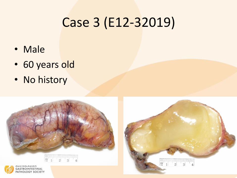
Case 3 (E12-32019)
• Male
• 60 years old
• No history

Case 4 (E13-32575)
• Female
• 76 years old
• No history
Case 5 (E14-31343)
• Male
• 75 years old
• No history

Case 5
• Recurred one year later as acellular mucin
• No free mucin seen at time of appendicectomy
• Multiple levels and no epithelium
• ?Must assume that it is sampling error

Case 6 (E15-12565)
• Female
• 77 years old
• No history

Case 7 (E14-35559)
• Female
• 62 years
• No history

Case 7
• Massive recurrence at one year