apoe signaling in neurodegenerative diseases: an
TRANSCRIPT
APOE signaling in neurodegenerative diseases:an integrative approach targeting APOE coding andnoncoding variants for disease interventionXiaopu Zhou1,2,3, Amy KY Fu1,2,3 and Nancy Y Ip1,2,3
Available online at www.sciencedirect.com
ScienceDirect
APOE (apolipoprotein E) is a key regulator of lipid metabolism
and a leading genetic risk factor for Alzheimer’s disease. While
APOE participates in multiple biological pathways, its roles in
diseases are largely due to the mutant protein encoded by
APOE-e4. However, emerging evidence suggests that some
noncoding Alzheimer’s disease risk variants residing in APOE
and its nearby regions exert APOE-e4-independent risks and
modify APOE gene expression. Moreover, intervention
strategies targeting APOE are being explored. In this review, we
summarize the literature on the genetic risks and roles of APOE
in biological systems. Moreover, we propose an integrative
approach to evaluate disease risk and tailor interventions to aid
research on APOE-associated diseases.
Addresses1Division of Life Science, State Key Laboratory of Molecular Neu-
roscience, Molecular Neuroscience Center, The Hong Kong University of
Science and Technology, Hong Kong, China
2Hong Kong Center for Neurodegenerative Diseases, Hong Kong
Science Park, Hong Kong, China3Guangdong Provincial Key Laboratory of Brain Science, Disease and
Drug Development, Hong Kong University of Science and Technology
Shenzhen Research Institute, Shenzhen–Hong Kong Institute of Brain
Science, 518057 Shenzhen, Guangdong, China
Corresponding author: Ip, Nancy Y ([email protected])
Current Opinion in Neurobiology 2021, 69:58–67
This review comes from a themed issue on Molecular neuroscience
Edited by Frank Bradke and Yukiko Goda
https://doi.org/10.1016/j.conb.2021.02.001
0959-4388/ã 2021 The Authors. Published by Elsevier Ltd. This is an
open access article under the CC BY-NC-ND license (http://creative-
commons.org/licenses/by-nc-nd/4.0/).
IntroductionApoE (apolipoprotein E), encoded by APOE, was first
described as an arginine-rich polypeptide [1–4]. ApoE is a
key regulator of triglyceride and cholesterol metabolism
in the peripheral system and brain [5]. In the peripheral
system, plasma ApoE mainly resides in lipoprotein
particles including very-low-density lipoproteins, chylo-
microns, and subsets of high-density lipoproteins,
Current Opinion in Neurobiology 2021, 69:58–67
facilitating the hepatic endocytosis of intestinal chylomi-
crons and the lipid redistribution across tissues [6–8].
ApoE is also highly expressed in the brain, which contains
the largest proportion of cholesterol in the body (�20% of
the total) [9]. In the brain, ApoE mainly exists in high-
density, lipoprotein-like particles and maintains lipid
homeostasis and proper neuronal functioning [10].
Human and mice lacking APOE exhibit functional def-
icits in both the peripheral system and brain. In particular,
APOE-deficient mice exhibit elevated plasma cholesterol
levels, early onset of atherosclerotic lesions, and elevated
inflammation [11]. Moreover, compared to wild-type
mice, APOE-deficient mice exhibit fewer dendrites and
lower synapse density in neurons of the neocortex and
hippocampus as well as greater age-dependent neurode-
generation in the neocortex [12]. Meanwhile, humans
lacking APOE exhibit exceptionally high cholesterol
levels; mild atherosclerosis; normal cognitive function
with subtle deficits in memory, language, visuospatial
abilities and executive functions [13�]. Hence, ApoE
has important roles in maintaining normal physiological
functions in both the brain and peripheral system.
APOE-associated pathways in the brain andperipheral systemApoE affects diverse biological pathways in both the
nervous and immune systems that are associated with
neurodegenerative diseases. Biochemical analysis sug-
gests that APOE is predominantly expressed by the liver,
adipose tissue, arteries, and brain [6]. Single-cell RNA
sequencing technique enable high-throughput assays of
comprehensive gene expression profiles at single-cell
resolution. Accordingly, reanalysis of publicly available
data with mouse tissues revealed that APOE is predomi-
nately expressed in the liver, skin, brain, adipose tissue,
heart, aorta, and lungs, particularly in Kupffer cells,
hepatocytes, and leukocytes [14]. Kupffer cells (a type
of resident hepatic macrophage) along with hepatocytes
are the major sources of peripheral APOE expression,
although the role of APOE expressed by Kupffer cells
is not well understood [15]. Meanwhile, in the human
brain, APOE is mainly expressed by astrocytes, microglia,
oligodendrocyte progenitors and oligodendrocytes, endo-
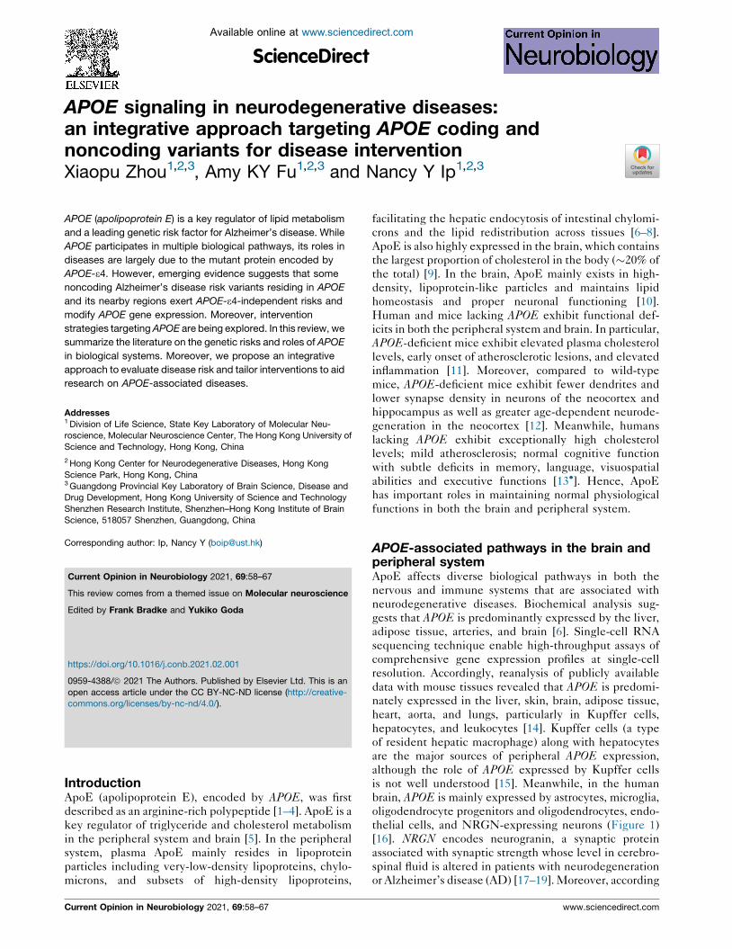
thelial cells, and NRGN-expressing neurons (Figure 1)
[16]. NRGN encodes neurogranin, a synaptic protein
associated with synaptic strength whose level in cerebro-
spinal fluid is altered in patients with neurodegeneration
or Alzheimer’s disease (AD) [17–19]. Moreover, according
www.sciencedirect.com

Genetics and biology of apolipoprotein E in neurodegenerative diseases Zhou, Fu and Ip 59
Figure 1
(a)
(d)
(f) (g)
(b)
(c)
(e)
Mouse tissue (single-cell)
Human brain (single-cell)
Mouse brain (bulk) Human brain (bulk)
Current Opinion in Neurobiology
APOE expression profiles at the single-cell and tissue levels.
(a–c) APOE expression in mouse tissues and cells. APOE expression profiles in selected mouse tissues at the (a) single-cell level and (b) tissue
level. (c) APOE expression profiles in selected cell types. (d, e) APOE expression in human brain cells. (d) APOE expression profiles at the single-
cell level in the human brain prefrontal cortex and anterior cingulate cortex. (e) APOE expression profiles in selected cell types. (f, g) APOE
expression in (f) the mouse and (g) human brain. Histogram showing the ranking of the APOE transcript levels across all detected genes in the
www.sciencedirect.com Current Opinion in Neurobiology 2021, 69:58–67

60 Molecular neuroscience
to the Brain RNA-Seq database (https://www.
brainrnaseq.org/) that integrates bulk RNA-Seq data from
mouse and human brains [20,21], APOE expression is
higher in the mouse brain than the human brain
(Figure 1). These lines of evidence corroborate the find-
ings of biochemical studies on APOE expression profiles,
providing a basis for further characterization of the roles of
APOE and associated pathways in neurodegenerative
diseases.
Most studies on APOE in the brain are restricted to neurons,
astrocytes, and microglia, which have revealed specific
function of APOE signaling in those cells (Table 1). Astro-
cytes are the major source of the APOE in the brain, which
can modulate neurite outgrowth [22]. The activation of
APOE signaling in neurons triggers phosphorylation
cascades that activate several downstream kinases [23].
Specifically, APOE signaling modulates neurite out-
growth/extension, synaptogenesis, axon remodeling
[24–26], and calcium homeostasis [27] and also prevents
neuronal apoptosis [28]. Furthermore, as a protective
mechanism induced upon injury or stress, neurons increase
their APOE expression to recruit lipids from the surround-
ing environment for repair [8]. Moreover, upon activation,
microglia exhibit elevated APOE expression [29], which
modulates their molecular phenotypes and regulates their
cell state transition from a homeostatic state to a disease/
neurodegeneration-associated state by influencing gene
expression in a TREM2-dependent manner [30�]. Further-
more, ApoE4, a mutated form of ApoE protein that is
known to associate with AD pathogenesis, can bind to
DNA and act as a transcription factor to modulate cytokine
production in neuronal cell lines, further indicating that
APOE plays important roles in transcriptomic regulation of
immune-related pathways [31,32]. Notably, a recent
human postmortem brain study revealed the presence of
APOE circular RNA, which accounts for approximately
one-third of the total brain APOE RNA, although its
function and cellular expression profile are unknown
[33�]. Therefore, further investigation is required to under-
stand the roles of APOE expression and its associated
pathways in the brain.
Emerging studies also suggest that APOE plays roles in
the vascular system. The breakdown of blood–brain
barrier (BBB) might account for the neurodegeneration
and neuroinflammation in AD [34]; the APOE-deficientmice exhibit age-dependent BBB disruption [35]. In
addition, disease-associated ApoE4 protein can also lead
to BBB dysfunction [36,37]. Moreover, a recent study
using an induced pluripotent stem cell-derived BBB
(Figure 1 Legend Continued) corresponding tissues. Data obtained from th
individual cells was visualized by t-distributed stochastic neighbor embeddi
individual cells. Astro, astrocytes; Astro-FB, fibrous astrocytes; Astro-PP, pr
kilobase of transcript per million mapped reads; Neu, neurons; Neu-NRGN,
oligodendrocyte progenitor cells; Macro, macrophages; Micro, microglia.
Current Opinion in Neurobiology 2021, 69:58–67
model suggests that ApoE4 expressed by pericytes might
modulate amyloid deposition near the BBB, making it a
possible trigger of microvasculature injury [38�]. Interest-
ingly, although peripheral ApoE cannot effectively dif-
fuse into the brain owing to its limited BBB permeability
[39–41], manipulating peripheral ApoE level can poten-
tially affect cognitive functions [42�]. Given the modula-
tory effects of both vascular and peripheral APOE on brain
functions, an intervention strategy targeting these sources
of APOE could be effective for certain neurodegenerative
diseases including AD.
APOE coding variants in human diseasesGenetics and biochemical studies revealed the existence
of 3 APOE isoforms in the general population—APOE-e2(encoding ApoE2; Cys112–Cys158), APOE-e3 (the most
common form; Cys112–Arg158), and APOE-e4 (encoding
ApoE4; Arg112–Arg158)—which are defined by combi-
nations of 2 coding mutations in exon 4 of APOE [43–47]
(Table 2). These mutations of APOE modify its protein
function by altering internal domain–domain interactions
[48]. In ApoE4, the cysteine-to-arginine substitution at
residue 112 renders the formation of a new salt bridge
between Glu109 and Arg112, and modifies the orientation
of Arg61 (a key residue), resulting in ApoE4 having higher
affinity for very-low-density lipoproteins [49,50]. Mean-
while, in ApoE2, the arginine-to-cysteine substitution at
residue 158 changes the salt bridge within and between
the helical structures, which also modifies the charges of
the receptor-binding region [50]. Accordingly, ApoE4
does not efficiently interact with high-density lipopro-
teins and preferentially binds low-density lipoproteins,
whereas ApoE2 exhibits greatly reduced affinity for low-
density lipoprotein receptor [49,51].
An early genetics study revealed an association between
APOE and abnormal lipid metabolism [8]. Specifically,
APOE-e2 is one of the genetic risk factors of type III
hyperlipoproteinemia, a familial lipoprotein metabolic
disorder characterized by elevated blood cholesterol
and triglyceride levels along that incurs risks of severe
atherosclerotic vascular diseases [52]. In contrast, APOE-e2 is also associated with longevity, reduced age-
associated cognitive decline, and reduced AD risk [53].
Meanwhile, ApoE-e4 is associated with neurodegenera-
tive diseases including AD, vascular dementia, Lewy
body dementia, frontotemporal dementia, Parkinson’s
disease, and multiple sclerosis; some cerebrovascular
disorders such as cerebral amyloid angiopathy and stroke;
and poor outcomes after traumatic brain injury [54].
Given that these diseases are predominately associated
e Brain RNA-Seq database. (a, d right panels) APOE expression in
ng (t-SNE) plots; color denotes normalized APOE expression in
otoplasmic astrocytes; Endo, Endothelieal cells; FPKM, fragments per
NRGN-expressing neurons; Oligo, oligodendrocytes; OPCs,
www.sciencedirect.com

Genetics and biology of apolipoprotein E in neurodegenerative diseases Zhou, Fu and Ip 61
Table 1
Roles of APOE-associated pathways in the brain and peripheral system
Tissue or cell type Function References
Peripheral system � Redistribute lipids [6–8]
� Modify cognitive function [42�]Astrocytes � Produce majority of the ApoE protein in the brain [8]
� Modulate neurite outgrowth [22]
Neurons � Modulate neurite outgrowth and extension, synaptogenesis, and axon remodeling [24–26]
� Regulate calcium homeostasis [27]
� Confer neuroprotection [28]
� Repair damaged neurons by recruiting lipids from the surrounding environment [8]
� Induce microglial activation
Microglia � Regulate transcriptome profile [30�]
Table 2
Summary of APOE isoforms and associated phenotypes
Isoform (or alias) Mutations Phenotypes/Notes References
e1 (Weisgraber allele)
Asp127–Cys158
Obesity
[64]Non insulin-dependent diabetes
Hypertension
Moderate lipid disturbances
e1y (e3r) Arg112–Cys158 No obvious phenotypic change [65]
e2
Cys112–Cys158
Longer lifespan
[52,53]
Type III hyperlipoproteinemia (elevated blood cholesterol and
triglyceride levels)
Atherosclerotic vascular diseases Reduced risk of Alzheimer’s
disease
e3 Cys112–Arg158 Most frequent isoform in the general population
e4Arg112–Arg158
Neurodegenerative diseases including Alzheimer’s disease[54]Cerebrovascular disorders
e5 Lys3 Mild hypertriglyceridemia [66]
e5Arg84–Arg112
Elevated blood cholesterol level[67]Triple-vessel disease
e7Lys244–Lys245
Diabetes mellitus[68]Hypertriglyceridemia
Christchurch allele (R136S)
Ser136
Significant enrichment of mutated ApoE in very low-density
lipoprotein [69,72��]Type III hyperlipoproteinemia
with the nervous and vascular systems and involve altered
immune status, APOE might have broad roles in modu-
lating specific pathways associated with neuronal, vascu-
lar, and immune functions.
Of note, APOE-e4 is a well-recognized genetic risk
factor for AD, which is one of the most common neuro-
degenerative diseases [55]. APOE-e4 modulates various
AD-associated endophenotypes covering a broad spec-
trum of disease signatures including synaptic plasticity
[56], neuronal activity [57], brain volume [58], cognitive
performance [59], onset age [60], and brain immune
status [61]. Compared to APOE-e2 and APOE-e3,APOE-e4 can also modify the pathological hallmarks
of AD, including amyloid-beta (Ab), tau neurofibrillary
tangles, and neurodegeneration by promoting Abaggregation, inhibiting Ab clearance, and exacerbating
tau-mediated neurodegeneration in tau transgenic
mice [62,63]. Hence, APOE-e4 plays critical roles in the
www.sciencedirect.com
pathogenesis of AD by modulating a plethora of biological
pathways involved in the disease pathogenesis.
Multiple rare APOE coding risk variants have also been
identified, including the APOE-e1 Weisgraber allele
(Asp127–Cys158) [64], APOE-e1y (Arg112–Cys158)
[65], several forms of APOE-e5 (including Lys3 and
Arg84–Arg112) [66,67], APOE-e7 (Lys244–Lys245) [68],
and the Christchurch mutation (R136S or Ser136) [69],
which potentially modify disease risk by altering ApoE
function (Table 2). For instance, ApoE with the Christch-
urch mutation tends to exist in a higher-order oligomeri-
zation state with a larger aggregate size when compared
with the normal ApoE protein [70], which consequently
reduces the binding affinity of APOE for low-density
lipoprotein receptor by 60% [71]. In particular, in a case
report of a homozygous carrier who exhibited high brain
Ab load, the Christchurch mutation elicited a strong
neuronal protective effect against the causal PSEN1
Current Opinion in Neurobiology 2021, 69:58–67

62 Molecular neuroscience
E280A coding risk variant, as indicated by low tau tangle
load, low plasma neurofilament light chain level, and mild
cognitive issues [72��]. This implies that appropriately
manipulating the APOE signaling pathway alone might be
sufficient for AD intervention.
Notably, besides altering protein function, mutations of
APOE-e2 and APOE-e4 are located in a well-defined
genomic region with DNA methylation modification
(i.e., a CpG island) [73]. Moreover, an APOE-e4 variant
introduces a cytosine base that is highly methylated
(65–90%) across human tissues [74]. Concordantly, the
APOE-e4 allele is associated with reduced brain APOEexpression [75,76��]. In addition, compared to noncar-
riers, APOE-e4 heterozygous carriers exhibit decreased
APOE-e4 mRNA expression in the brain and blood
[76��,77]. Therefore, APOE-e4 potentially exerts disease
risk effects by encoding a mutated protein with altered
APOE expression and functions, which leads to the
dysregulation of APOE-associated signaling pathways
and consequently modifies disease risk.
Contributions of APOE noncoding riskvariants to Alzheimer’s diseaseAPOE coding risk variants unequivocally impact associ-
ated signaling pathways, while the dysregulation of APOEexpression might also trigger or modulate AD onset and
progression. The expression of APOE is controlled in a
tissue-specific manner by multiple enhancers in nearby
regions [74,78], which might also be modified by variants
located in noncoding regions [79–83]. Association analy-
ses have revealed numerous AD-associated noncoding
variants and haplotypes residing in APOE and its nearby
regions [84–86]. Interestingly, several studies also show
that a specific haplotype structure with an ethnic-specific
distribution might account for the variable AD risk of
APOE-e4 carriers among populations [87�,88�]. Neverthe-
less, it remains controversial whether those noncoding
variants exert risk effects for AD.
Accordingly, our group conducted a comprehensive
fine-mapping analysis of APOE genetic risk in APOEand its nearby regions based on whole-genome sequenc-
ing data [76��]. Our analysis enabled the identification of
all genomic variants and their haplotype structures in the
study cohort for association analysis, which identified
noncoding risk variants and haplotypes residing in the
PVRL2 and APOC1 regions that exert AD risk effects
independent of APOE-e4. Further analysis revealed their
potential regulatory functions in the modification of brain
APOE expression; specifically, carriers of PVRL2 or
APOC1 haplotypes exhibited higher brain APOE expres-
sion than noncarriers. Moreover, if the noncoding risk
variants and APOE-e4 reside in the same chromosome,
those noncoding variants might modify the disease pene-
trance of APOE-e4 via enhanced expression of ApoE4.
Meanwhile, the identified risk haplotypes are more
Current Opinion in Neurobiology 2021, 69:58–67
prevalent in populations of European descent than those
of African descent, corroborating the lower AD risk of
APOE-e4 observed in the latter [89]. Taken together, the
discoveries of these noncoding risk factors and their
putative regulatory functions provide a genetic basis for
the variable APOE-e4 AD risk across ethnic groups and
suggests that changes in APOE expression alone might
also confer AD risk.
Notably, the AD-associated APOE noncoding variants span
the PVRL2–TOMM40–APOE–APOC1 region, potentially
affecting the expressions of those genes and influencing
several other AD-associated mechanisms in parallel to the
Ab cascade, including viral infection, mitochondrial dys-
function, and neuroinflammation [90–92]. Specifically,
PVRL2 encodes a surface protein that abets herpes and
pseudorabies viruses entering cells—a process potentially
associated with AD pathogenesis [93,94]. Meanwhile,
TOMM40 encodes a mitochondrial membrane protein that
facilitates protein transport into mitochondria, potentially
accounting for the observed mitochondrial dysfunction in
the AD brain [95]. Furthermore, APOC1 encodes a protein
involved in lipid metabolism that is implicated in athero-
sclerosis, which is a possible trigger of neuroinflammation
[96,97]. Hence, the identified noncoding APOE risk factors
might elicit additional effects on neurodegenerative dis-
eases in addition to modifying APOE expression.
Proposed model for APOE-associatedintervention strategiesCorroborating the critical involvement of APOE in AD
pathogenesis, intervention strategies targeting APOEexpression or ApoE4 mutant proteins can ameliorate
AD-associated symptoms. For instance, administration
of the RXR agonist bexarotene, which can trigger
APOE expression, rapidly enhances Ab clearance and
improves neuronal and cognitive functioning in AD
mouse models [98]. Meanwhile, PH002, a small mole-
cule structure corrector that can reshape ApoE4 protein
into an ApoE3–like structure, can ameliorate the neu-
ronal toxicity of ApoE4 and reduce p-tau, Ab40, and
Ab42 levels in induced pluripotent stem cell-derived
neurons [99�]. Moreover, other intervention strategies
including ApoE-neutralizing antibody [100], APOEantisense oligonucleotides [101], and virus-mediated
ApoE2 overexpression [102] can also elicit protective
effects against AD by modulating ApoE2 expression
and its accompanying signaling pathways. Thus, such
APOE-targeting therapeutic strategies could be devel-
oped as treatments for neurodegenerative diseases.
Nevertheless, considering the complexity of APOEsignaling, which involves multiple cell types, APOE sig-
naling must be manipulated in a cell-type-specific and
genotype-aware manner. Moreover, applying the afore-
mentioned intervention strategies at a population scale
might yield inconsistent effects owing to variations in
www.sciencedirect.com

Genetics and biology of apolipoprotein E in neurodegenerative diseases Zhou, Fu and Ip 63
Figure 2
APOE andnearby regions
Genotyping
Strategies
Genotypes/Groups ApoE Intervention strategies
APOE coding risk factor
APOE noncoding risk factor
ApoE Medications
IndividualsApoE-ε4
Current Opinion in Neurobiology
Integrative approaches targeting APOE-related diseases.
The coding and noncoding genotypes of APOE and its nearby regions are detected by array or sequencing methods. Individuals harboring
different combinations of coding and noncoding mutations are segregated and subjected to different intervention paradigms using drugs targeting
APOE expression and ApoE mutant proteins. TF, transcription factor.
genetic background. For instance, among AD patients
treated with bexarotene, APOE-e4 carriers exhibit
no significant change in Ab load, whereas APOE-e3homozygous patients exhibit a regional reduction in Abload [103�]. On the other hand, the presence of certain
APOE noncoding genetic risk variants might modify the
chromatin state of nearby regions and consequently inter-
fere with the APOE expression-inducing effect of bexar-
otene [104]. Therefore, a rigorous investigation of the
genomic contexts of APOE and nearby regions may
facilitate the development of more-effective and geno-
type-specific intervention strategies. For instance, a mul-
tistage screening assay can be applied to individuals
identified as having APOE-e4, both APOE-e4 and non-
coding risk variants, or only noncoding risk variants—
each of whom may be submitted to a different treatment
paradigm for AD.
www.sciencedirect.com
Accordingly, by combining coding and noncoding genetic
information, a polygenic risk score model can be designed
to stratify patients and assess an individual’s risk of
developing a given disease [105–107]. Furthermore,
long-read sequencing technology can directly capture
all genomic contexts of the APOE and its nearby regions,
including single nucleotide polymorphisms, insertions,
deletions, structural variations, and haplotype informa-
tion, which would more accurately stratify individuals for
subsequent preclinical or clinical investigation [76��].The results of such research might yield tailored inter-
vention strategies applicable to a broad range of diseases
associated with APOE (Figure 2).
ConclusionAPOE is expressed in various cell types and involved in
diverse biological functions. The contributions of APOEcoding and noncoding genetic variants to human diseases
Current Opinion in Neurobiology 2021, 69:58–67

64 Molecular neuroscience
through the modulation of protein functions and gene
expression highlight the roles of the dysregulation of
APOE signaling in modifying disease risk. Meanwhile,
APOE-targeting intervention strategies have demon-
strated beneficial effects in decreasing disease-associated
phenotypes, which are influenced by genetic contexts.
Thus, properly integrating genetic information and
APOE-targeting interventions will enable early disease
risk prediction for individuals and help stratify individuals
for appropriate intervention strategies.
Conflict of interest statementNothing declared.
AcknowledgementsThis work was supported in part by the Research Grants Council of HongKong (Theme-Based Research Scheme [T13-607/12R], and theCollaborative Research Fund [C6027-19GF]), the National Key R&DProgram of China (2017YFE0190000 and 2018YFE0203600), the Areas ofExcellence Scheme of the University Grants Committee (AoE/M-604/16),the Innovation and Technology Commission (ITCPD/17-9), theGuangdong Provincial Key S&T Program (2018B030336001), the ShenzhenKnowledge Innovation Program (JCYJ20180507183642005 andJCYJ20170413173717055), and the HKUST-SIAT Joint Laboratory forBrain Science.
References and recommended readingPapers of particular interest, published within the period of review,have been highlighted as:
� of special interest�� of outstanding interest
1. Shore VG, Shore B: Heterogeneity of human plasma very lowdensity lipoproteins. Separation of species differing in proteincomponents. Biochemistry 1973, 12:502-507.
2. Shelburne FA, Quarfordt SH: A new apoprotein of humanplasma very low density lipoproteins. J Biol Chem 1974,249:1428-1433.
3. Utermann G: Isolation and partial characterization of anarginine-rich apolipoprotein from human plasma very-low-density lipoproteins: apolipoprotein E. Hoppe Seylers Z PhysiolChem 1975, 356:1113-1122.
4. Utermann G, Menzel HJ, Langer KH: On the polypeptidecomposition of an abnormal high density lipoprotein (LP-E)occurring in LCAT-deficient plasma. FEBS Lett 1974, 45:29-32.
5. Mahley RW: Central nervous system lipoproteins: ApoE andregulation of cholesterol metabolism. Arterioscler Thromb VascBiol 2016, 36:1305-1315.
6. Getz GS, Reardon CA: Apoprotein E as a lipid transport andsignaling protein in the blood, liver, and artery wall. J Lipid Res2009, 50:S156-S161.
7. Cooper AD: Hepatic uptake of chylomicron remnants. J LipidRes 1997, 38:2173-2192.
8. Huang Y, Mahley RW, Apolipoprotein E: Structure and function inlipid metabolism, neurobiology, and Alzheimer’s diseases.Neurobiol Dis 2014, 72:3-12.
9. Bjorkhem I, Meaney S, Fogelman AM: Brain cholesterol: longsecret life behind a barrier. Arterioscler Thromb Vasc Biol 2004,24:806-815.
10. de Chaves EP, Narayanaswami V: Apolipoprotein E andcholesterol in aging and disease in the brain. Future Lipidol2008, 3:505-530.
11. Lo Sasso G, Schlage WK, Boue S, Veljkovic E, Peitsch MC,Hoeng J: The Apoe-/- mouse model: a suitable model to studycardiovascular and respiratory diseases in the context of
Current Opinion in Neurobiology 2021, 69:58–67
cigarette smoke exposure and harm reduction. J Transl Med2016, 14:146.
12. Masliah E, Mallory M, Ge N, Alford M, Veinbergs I, Roses AD:Neurodegeneration in the central nervous system of apoE-deficient mice. Exp Neurol 1995, 136:107-122.
13.�
Mak ACY, Pullinger CR, Tang LF, Wong JS, Deo RC, Schwarz JM,Gugliucci A, Movsesyan I, Ishida BY, Chu C et al.: Effects of theabsence of apolipoprotein E on lipoproteins, neurocognitivefunction, and retinal function. JAMA Neurol 2014, 71:1228-1236
By characterizing an individual harboring the homozygous APOE frame-shift mutation (c.291del, p.E97fs) who is devoid of APOE expression, theauthors suggest that APOE is not essential for normal brain functioning.This work provides hints about the roles of APOE in the human system.
14. Schaum N, Karkanias J, Neff NF, May AP, Quake SR, Wyss-Coray T, Darmanis S, Batson J, Botvinnik O, Chen MB et al.:Single-cell transcriptomics of 20 mouse organs creates aTabula Muris. Nature 2018, 562:367-372.
15. Gaudreault N, Kumar N, Olivas VR, Eberle D, Rapp JH, Raffai RL:Macrophage-specific apoE gene repair reduces diet-inducedhyperlipidemia and atherosclerosis in hypomorphic apoEmice. PLoS One 2012, 7.
16. Velmeshev D, Schirmer L, Jung D, Haeussler M, Perez Y, Mayer S,Bhaduri A, Goyal N, Rowitch DH, Kriegstein AR: Single-cellgenomics identifies cell type–specific molecular changes inautism. Science (80-) 2019, 364:685-689.
17. Casaletto KB, Elahi FM, Bettcher BM, Neuhaus J, Bendlin BB,Asthana S, Johnson SC, Yaffe K, Carlsson C, Blennow K et al.:Neurogranin, a synaptic protein, is associated with memoryindependent of Alzheimer biomarkers. Neurology 2017,89:1782-1788.
18. De Vos A, Jacobs D, Struyfs H, Fransen E, Andersson K,Portelius E, Andreasson U, De Surgeloose D, Hernalsteen D,Sleegers K et al.: C-terminal neurogranin is increased incerebrospinal fluid but unchanged in plasma in Alzheimer’sdisease. Alzheimers Dement 2015, 11:1461-1469.
19. Zhong L, Cherry T, Bies CE, Florence MA, Gerges NZ:Neurogranin enhances synaptic strength through itsinteraction with calmodulin. EMBO J 2009, 28:3027-3039.
20. Zhang Y, Chen K, Sloan SA, Bennett ML, Scholze AR, O’Keeffe S,Phatnani HP, Guarnieri P, Caneda C, Ruderisch N et al.: An RNA-sequencing transcriptome and splicing database of glia,neurons, and vascular cells of the cerebral cortex. J Neurosci2014, 34:11929-11947.
21. Zhang Y, Sloan SA, Clarke LE, Caneda C, Plaza CA,Blumenthal PD, Vogel H, Steinberg GK, Edwards MSB, Li G et al.:Purification and characterization of progenitor and maturehuman astrocytes reveals transcriptional and functionaldifferences with mouse. Neuron 2016, 89:37-53.
22. Sun Y, Wu S, Bu G, Onifade MK, Patel SN, LaDu MJ, Fagan AM,Holtzman DM: Glial fibrillary acidic protein–apolipoprotein E(apoE) transgenic mice: astrocyte-specific expression anddiffering biological effects of astrocyte-secreted apoE3 andapoE4 lipoproteins. J Neurosci 1998, 18:3261-3272.
23. Hoe HS, Harris DC, Rebeck GW: Multiple pathways ofapolipoprotein E signaling in primary neurons. J Neurochem2005, 93:145-155.
24. Nathan BP, Jiang Y, Wong GK, Shen F, Brewer GJ, Struble RG:Apolipoprotein E4 inhibits, and apolipoprotein E3 promotesneurite outgrowth in cultured adult mouse cortical neuronsthrough the low-density lipoprotein receptor-related protein.Brain Res 2002, 928:96-105.
25. Huang YWA, Zhou B, Nabet AM, Wernig M, Sudhof TC:Differential signaling mediated by ApoE2, ApoE3, and ApoE4in human neurons parallels Alzheimer’s disease risk. JNeurosci 2019, 39:7408-7427.
26. Huang ZJ, Cao F, Wu Y, Peng JH, Zhong JJ, Jiang Y, Yin C,Guo ZD, Sun XC, Jiang L et al.: Apolipoprotein E promotes whitematter remodeling via the Dab1-dependent pathway aftertraumatic brain injury. CNS Neurosci Ther 2020, 26:698-710.
www.sciencedirect.com

Genetics and biology of apolipoprotein E in neurodegenerative diseases Zhou, Fu and Ip 65
27. Veinbergs I, Everson A, Sagara Y, Masliah E: Neurotoxic effectsof apolipoprotein E4 are mediated via dysregulation of calciumhomeostasis. J Neurosci Res 2002, 67:379-387.
28. Hayashi H, Campenot RB, Vance DE, Vance JE: Protection ofneurons from apoptosis by apolipoprotein e-containinglipoproteins does not require lipoprotein uptake and involvesactivation of phospholipase Cg1 and inhibition of calcineurin.J Biol Chem 2009, 284:29605-29613.
29. Srinivasan K, Friedman BA, Etxeberria A, Huntley MA, van derBrug MP, Foreman O, Paw JS, Modrusan Z, Beach TG,Serrano GE et al.: Alzheimer’s patient microglia exhibitenhanced aging and unique transcriptional activation. Cell Rep2020, 31.
30.�
Krasemann S, Madore C, Cialic R, Baufeld C, Calcagno N, ElFatimy R, Beckers L, O’Loughlin E, Xu Y, Fanek Z et al.: TheTREM2-APOE pathway drives the transcriptional phenotype ofdysfunctional microglia in neurodegenerative diseases.Immunity 2017, 47:566-581.e9
By conducting transcriptomic analysis in microglia from human brainsaffected by Alzheimer’s disease and microglia from multiple mousemodels of diseases, the authors identified genes associated with neu-rodegenerative microglia phenotypes that are potentially modifiedby TREM2–APOE signaling. This work suggests that APOE plays rolesin the transcriptional regulation of disease-associated gene expression inneurodegenerative diseases.
31. Theendakara V, Peters-Libeu CA, Spilman P, Poksay KS,Bredesen DE, Rao RV: Direct transcriptional effects ofapolipoprotein E. J Neurosci 2016, 36:685-700.
32. Theendakara V, Peters-Libeu CA, Bredesen DE, Rao RV:Transcriptional effects of ApoE4: relevance to Alzheimer’sdisease. Mol Neurobiol 2018, 55:5243-5254.
33.�
Lee EG, Tulloch J, Chen S, Leong L, Saxton AD, Kraemer B,Darvas M, Keene CD, Shutes-David A, Todd K et al.: Redefiningtranscriptional regulation of the APOE gene and itsassociation with Alzheimer’s disease. PLoS One 2020, 15
By studying APOE transcripts in human postmortem brains, the authorsidentified the presence of a circular form of APOE transcript whoseexpression is altered in the brain in Alzheimer’s disease. This is the firstreport of the presence of APOE circular RNA in the human system.
34. Sweeney MD, Sagare AP, Zlokovic BV: Blood-brain barrierbreakdown in Alzheimer disease and other neurodegenerativedisorders. Nat Rev Neurol 2018, 14:133-150.
35. Hafezi-Moghadam A, Thomas KL, Wagner DD: ApoE deficiencyleads to a progressive age-dependent blood-brain barrierleakage. Am J Physiol Physiol 2007, 292:C1256-C1262.
36. Bell RD, Winkler EA, Singh I, Sagare AP, Deane R, Wu Z,Holtzman DM, Betsholtz C, Armulik A, Sallstrom J et al.:Apolipoprotein e controls cerebrovascular integrity viacyclophilin A. Nature 2012, 485:512-516.
37. Montagne A, Nation DA, Sagare AP, Barisano G, Sweeney MD,Chakhoyan A, Pachicano M, Joe E, Nelson AR, D’Orazio LM et al.:APOE4 leads to blood–brain barrier dysfunction predictingcognitive decline. Nature 2020, 581:71-76.
38.�
Blanchard JW, Bula M, Davila-Velderrain J, Akay LA, Zhu L,Frank A, Victor MB, Bonner JM, Mathys H, Lin YT et al.:Reconstruction of the human blood–brain barrier in vitroreveals a pathogenic mechanism of APOE4 in pericytes. NatMed 2020, 26:952-963
By establishing an induced pluripotent stem cell-derived culture systemmimicking the human blood–brain barrier, the authors investigated themechanisms by which APOE-e4 affects vascular function—specificallyhow it modulates amyloid accumulation via the blood–brain barrier. Thisstudy provides hints about how APOE-e4 affects the blood–brain barrierin neurogenerative diseases.
39. Kraft HG, Menzel HJ, Hoppichler F, Vogel W, Utermann G:Changes of genetic apolipoprotein phenotypes caused byliver transplantation. Implications for apolipoproteinsynthesis. J Clin Invest 1989, 83:137-142.
40. Linton MF, Gish R, Hubl ST, Butler E, Esquivel C, Bry WI,Boyles JK, Wardell MR, Young SG: Phenotypes ofapolipoprotein B and apolipoprotein E after livertransplantation. J Clin Invest 1991, 88:270-281.
www.sciencedirect.com
41. Zlokovic BV, Martel CL, Mackic JB, Matsubara E, Wisniewski T,Mccomb JG, Frangione B, Ghiso J: Brain uptake of circulatingapolipoproteins J and E complexed to Alzheimer0s amyloid b.Biochem Biophys Res Commun 1994, 205:1431-1437.
42.�
Lane-Donovan C, Wong WM, Durakoglugil MS, Wasser CR,Jiang S, Xian X, Herz J: Genetic restoration of plasma apoeimproves cognition and partially restores synapticdefects in ApoE-deficient mice. J Neurosci 2016, 36:10141-10150
By comparing the cognition and synaptic activity between ApoE-generalknockout mice and ApoE brain-specific—knockout mice, the authorsobserved that restoring plasma ApoE improves learning, memory, andsynaptic function. This work suggests that peripheral APOE plays a role inmodifying brain cognition and neuronal functions.
43. Zannis VI, Breslow JL: Human very low density lipoproteinapolipoprotein E isoprotein polymorphism is explained bygenetic variation and posttranslational modification.Biochemistry 1981, 20:1033-1041.
44. Zannis VI, Breslow JL, Utermann G, Mahley RW, Weisgraber KH,Havel RJ, Goldstein JL, Brown MS, Schonfeld G, Hazzard WRet al.: Proposed nomenclature of apoE isoproteins, apoEgenotypes, and phenotypes. J Lipid Res 1982, 23:911-914.
45. Utermann G, Steinmetz A, Weber W: Genetic control of humanapolipoprotein E polymorphism: comparison of one-and two-dimensional techniques of isoprotein analysis. Hum Genet1982, 60:344-351.
46. Weisgraber KH, Rall SC, Mahley RW: Human E apoproteinheterogeneity. Cysteine-arginine interchanges in the aminoacid sequence of the apo-E isoforms. J Biol Chem 1981,256:9077-9083.
47. Hatters DM, Peters-Libeu CA, Weisgraber KH: Apolipoprotein Estructure: insights into function. Trends Biochem Sci 2006,31:445-454.
48. Fernandez CG, Hamby ME, McReynolds ML, Ray WJ: The role ofapoE4 in disrupting the homeostatic functions of astrocytesand microglia in aging and Alzheimer’s disease. Front AgingNeurosci 2019, 10.
49. Dong LM, Wilson C, Wardell MR, Simmons T, Mahley RW,Weisgraber KH, Agard DA: Human apolipoprotein E. Role ofarginine 61 in mediating the lipoprotein preferences of the E3and E4 isoforms. J Biol Chem 1994, 269:22358-22365.
50. Mahley RW, Rall SC, Apolipoprotein E: Far more than a lipidtransport protein. Annu Rev Genomics Hum Genet 2000, 1:507-537.
51. Dong LM, Weisgraber KH: Human apolipoprotein E4 domaininteraction. Arginine 61 and glutamic acid 255 interact todirect the preference for very low density lipoproteins. J BiolChem 1996, 271:19053-19057.
52. Utermann G, Hees M, Steinmetz A: Polymorphism ofapolipoprotein E and occurrence of dysbetalipoproteinaemiain man. Nature 1977, 269:604-607.
53. Suri S, Heise V, Trachtenberg AJ, Mackay CE: The forgottenAPOE allele: a review of the evidence and suggestedmechanisms for the protective effect of APOE e2. NeurosciBiobehav Rev 2013, 37:2878-2886.
54. Verghese PB, Castellano JM, Holtzman DM: Apolipoprotein E inAlzheimer’s disease and other neurological disorders. LancetNeurol 2011, 10:241-252.
55. Liu CC, Kanekiyo T, Xu H, Bu G: Apolipoprotein e and Alzheimerdisease: risk, mechanisms and therapy. Nat Rev Neurol 2013,9:106-118.
56. Kim J, Yoon H, Basak J, Kim J: Apolipoprotein E in synapticplasticity and alzheimer’s disease: potential cellular andmolecular mechanisms. Mol Cells 2014, 37:833-840.
57. Najm R, Jones EA, Huang Y: Apolipoprotein E4, inhibitorynetwork dysfunction, and Alzheimer’s disease. MolNeurodegener 2019, 14.
Current Opinion in Neurobiology 2021, 69:58–67

66 Molecular neuroscience
58. Piers R: Structural brain volume differences betweencognitively intact ApoE4 carriers and non-carriers across thelifespan. Neural Regen Res 2018, 13:1309-1312.
59. Rawle MJ, Davis D, Bendayan R, Wong A, Kuh D, Richards M:Apolipoprotein-E (Apoe) e4 and cognitive decline over theadult life course. Transl Psychiatry 2018, 8.
60. Blacker D, Haines JL, Rodes L, Terwedow H, Go RCP, Harrell LE,Perry RT, Bassett SS, Chase G, Meyers D et al.: ApoE-4 and ageat onset of Alzheimer’s disease: the NIMH genetics initiative.Neurology 1997, 48:139-147.
61. Keene CD, Cudaback E, Li X, Montine KS, Montine TJ:Apolipoprotein E isoforms and regulation of the innateimmune response in brain of patients with Alzheimer’sdisease. Curr Opin Neurobiol 2011, 21:920-928.
62. Kanekiyo T, Xu H, Bu G: ApoE and Ab in Alzheimer’s disease:accidental encounters or partners? Neuron 2014, 81:740-754.
63. Shi Y, Yamada K, Liddelow SA, Smith ST, Zhao L, Luo W, Tsai RM,Spina S, Grinberg LT, Rojas JC et al.: ApoE4 markedlyexacerbates tau-mediated neurodegeneration in a mousemodel of tauopathy. Nature 2017, 549:523-527.
64. Iron A, Richard P, Pascual de Zulueta M, Thomas G, Thomas M:Genotyping of a patient homozygous for a rare apolipoproteinE1 [Gly127!Asp; Arg158!Cys] (Weisgraber allele). J InheritMetab Dis 1995, 18:723-726.
65. Murrell JR, Price BM, Baiyewu O, Gureje O, Deeg M, Hendrie H,Ogunniyi A, Hall K: The fourth apolipoprotein E haplotype foundin the Yoruba of Ibadan. Am J Med Genet Part B NeuropsychiatrGenet 2006, 141:426-427.
66. Maeda H, Nakamura H, Kobori S, Okada M, Niki H, Ogura T,Hiraga S: Molecular cloning of a human apolipoprotein Evariant: E5 (Glu3——Lys3). J Biochem 1989, 105:491-493.
67. Wardell MR, Rall SC, Schaefer EJ, Kane JP, Weisgraber KH: Twoapolipoprotein E5 variants illustrate the importance of theposition of additional positive charge on receptor-bindingactivity. J Lipid Res 1991, 32:521-528.
68. Maeda H, Nakamura H, Kobori S, Okada M, Mori H, Niki H,Ogura T, Hiraga S: Identification of human apolipoprotein Evariant gene: apolipoprotein E7 (Glu244,245"Lys244,245). JBiochem 1989, 105:51-54.
69. Wardell MR, Brennan SO, Janus ED, Fraser R, Carrell RW:Apolipoprotein E2-Christchurch (136 Arg——Ser). New variantof human apolipoprotein E in a patient with type IIIhyperlipoproteinemia. J Clin Invest 1987, 80:483-490.
70. Georgiadou D, Chroni A, Vezeridis A, Zannis VI, Stratikos E:Biophysical analysis of apolipoprotein E3 variants linkedwith development of type III hyperlipoproteinemia. PLoSOne 2011, 6.
71. Lalazar A, Weisgraber KH, Rall SC, Giladi H, Innerarity TL,Levanon AZ, Boyles JK, Amit B, Gorecki M, Mahley RW et al.: Site-specific mutagenesis of human apolipoprotein E. Receptorbinding activity of variants with single amino acidsubstitutions. J Biol Chem 1988, 263:3542-3545.
72.��
Arboleda-Velasquez JF, Lopera F, O’Hare M, Delgado-Tirado S,Marino C, Chmielewska N, Saez-Torres KL, Amarnani D,Schultz AP, Sperling RA et al.: Resistance to autosomaldominant Alzheimer’s disease in an APOE3 Christchurchhomozygote: a case report. Nat Med 2019, 25:1680-1683
The authors characterized an individual harboring the pathogenic PSEN1E280A mutation and 2 copies of the APOE-e3 Christchurch (R136S)mutation. The individual exhibited a high amyloid burden accompaniedby mild tau deposition and neurodegeneration in the brain. This worksuggests that APOE plays roles in disease pathogenesis and progression,and is a potential target for disease intervention.
73. Foraker J, Millard SP, Leong L, Thomson Z, Chen S, Keene CD,Bekris LM, Yu CE, Fischer A: The APOE gene is differentiallymethylated in Alzheimer’s disease. J Alzheimer’s Dis 2015,48:745-755.
74. Yu CE, Cudaback E, Foraker J, Thomson Z, Leong L, Lutz F,Gill JA, Saxton A, Kraemer B, Navas P et al.: Epigenetic signature
Current Opinion in Neurobiology 2021, 69:58–67
and enhancer activity of the human APOE gene. Hum MolGenet 2013, 22:5036-5047.
75. Bertrand P, Poirier J, Oda T, Finch CE, Pasinetti GM: Associationof apolipoprotein E genotype with brain levels ofapolipoprotein E and apolipoprotein J (clusterin) in Alzheimerdisease. Mol Brain Res 1995, 33:174-178.
76.��
Zhou X, Chen Y, Mok KY, Kwok TCY, Mok VCT, Guo Q, Ip FC,Chen Y, Mullapudi N, Weiner MW et al.: Non-coding variability atthe APOE locus contributes to the Alzheimer’s risk. NatCommun 2019, 10
By conducting fine-mapping analysis using whole-genome sequencingdata of APOE and nearby regions, the authors identified noncodingvariants and haplotypes that exert disease risk effects independent ofAPOE-e4. Genotype–expression and genotype–endophenotype associa-tion analyses further suggest the roles of those noncoding risk variants inmodifying APOE expression and Alzheimer’s disease-associated endo-phenotypes. This work resolves the debate on the roles of noncoding riskvariants in APOE and nearby regions, and suggests an alternative diseasemechanism in parallel with APOE-e4 coding risk.
77. Lambert JC, Perez-Tur J, Dupire MJ, Galasko D, Mann D,Amouyel P, Hardy J, Delacourte A, Chartier-Harlin MC: Distortionof allelic expression of apolipoprotein E in Alzheimer’sdisease. Hum Mol Genet 1997, 6:2151-2154.
78. Najm R, Jones EA, Huang Y: Apolipoprotein E4, inhibitorynetwork dysfunction, and Alzheimer’s disease. MolNeurodegener 2019, 14:24.
79. Bekris LM, Millard SP, Galloway NM, Vuletic S, Albers JJ, Li G,Galasko DR, DeCarli C, Farlow MR, Clark CM et al.: Multiple SNPswithin and surrounding the apolipoprotein E gene influencecerebrospinal fluid apolipoprotein E protein levels. JAlzheimer’s Dis 2008, 13:255-266.
80. Bekris LM, Galloway NM, Montine TJ, Schellenberg GD, Yu CE:APOE mRNA and protein expression in postmortem brain aremodulated by an extended haplotype structure. Am J MedGenet Part B Neuropsychiatr Genet 2010, 153:409-417.
81. Laws SM, Hone E, Gandy S, Martins RN: Expanding theassociation between the APOE gene and the risk ofAlzheimer’s disease: possible roles for APOE promoterpolymorphisms and alterations in APOE transcription. JNeurochem 2003, 84:1215-1236.
82. Bullido MJ, Artiga MJ, Recuero M, Sastre I, Garcıa MA, Aldudo J,Lendon C, Han SW, Morris JC, Frank A et al.: A polymorphism inthe regulatory region of APOE associated with risk forAlzheimer’s dementia. Nat Genet 1998, 18:69-71.
83. Bratosiewicz-Wasik J, Liberski PP, Peplonska B, Styczynska M,Smolen-Dzirba J, Cycon M, Wasik TJ: Regulatory region singlenucleotide polymorphisms of the apolipoprotein E gene asrisk factors for Alzheimer’s disease. Neurosci Lett 2018,684:86-90.
84. Zhou X, Chen Y, Mok KY, Zhao Q, Chen K, Chen Y, Hardy J, Li Y,Fu AKY, Guo Q et al.: Identification of genetic risk factors in theChinese population implicates a role of immune system inAlzheimer’s disease pathogenesis. Proc Natl Acad Sci U S A2018, 115:1697-1706.
85. Jansen IE, Savage JE, Watanabe K, Bryois J, Williams DM,Steinberg S, Sealock J, Karlsson IK, Hagg S, Athanasiu L et al.:Genome-wide meta-analysis identifies new loci and functionalpathways influencing Alzheimer’s disease risk. Nat Genet2019, 51:404-413.
86. Takei N, Miyashita A, Tsukie T, Arai H, Asada T, Imagawa M,Shoji M, Higuchi S, Urakami K, Kimura H et al.: Geneticassociation study on in and around the APOE in late-onsetAlzheimer disease in Japanese. Genomics 2009, 93:441-448.
87.�
Babenko VN, Afonnikov DA, Ignatieva EV, Klimov AV, Gusev FE,Rogaev EI: Haplotype analysis of APOE intragenic SNPs. BMCNeurosci 2018, 19
By conducting genetic and haplotype analyses in different ethnic groups,the authors identified Alzheimer’s disease-associated haplotypes whoseprevalence varies among ethnic groups. This work suggests that ethnicbackground might modify the risk effects of APOE by influencing thegenomic content of APOE and nearby genomic regions.
www.sciencedirect.com

Genetics and biology of apolipoprotein E in neurodegenerative diseases Zhou, Fu and Ip 67
88.�
Rajabli F, Feliciano BE, Celis K, Hamilton-Nelson KL,Whitehead PL, Adams LD, Bussies PL, Manrique CP, Rodriguez A,Rodriguez V et al.: Ancestral origin of ApoE e4 Alzheimerdisease risk in Puerto Rican and African Americanpopulations. PLoS Genet 2018, 14
By conducting genetic analyses with 2 different ethnic groups, theauthors revealed the roles of regions near APOE in modulatingAlzheimer’s disease risk associated with APOE-e4. The study presentsan explanation for the observed variations in Alzheimer’s disease riskassociated with APOE-e4 across populations.
89. Tang MX, Stern Y, Marder K, Bell K, Gurland B, Lantigua R,Andrews H, Feng L, Tycko B, Mayeux R: The APOE-e4 allele andthe risk of Alzheimer disease among African Americans,whites, and Hispanics. J Am Med Assoc 1998, 279:751-755.
90. Heneka MT, Carson MJ, El Khoury J, Landreth GE, Brosseron F,Feinstein DL, Jacobs AH, Wyss-Coray T, Vitorica J, Ransohoff RMet al.: Neuroinflammation in Alzheimer’s disease. Lancet Neurol2015, 14:388-405.
91. Sochocka M, Zwoli�nska K, Leszek J: The infectious etiology ofAlzheimer’s disease. Curr Neuropharmacol 2017, 15:996-1009.
92. Yoo S-M, Park J, Kim S-H, Jung Y-K: Emerging perspectives onmitochondrial dysfunction and inflammation in Alzheimer’sdisease. BMB Rep 2020, 53:35-46.
93. Balin BJ, Hudson AP: Herpes viruses and Alzheimer’s disease:new evidence in the debate. Lancet Neurol 2018, 17:839-841.
94. Cairns DM, Rouleau N, Parker RN, Walsh KG, Gehrke L,Kaplan DL: A 3D human brain–like tissue model of herpes-induced Alzheimer’s disease. Sci Adv 2020, 6.
95. Wang W, Zhao F, Ma X, Perry G, Zhu X: Mitochondriadysfunction in the pathogenesis of Alzheimer’s disease:recent advances. Mol Neurodegener 2020, 15.
96. Fuior EV, Gafencu AV: Apolipoprotein c1: its pleiotropic effectsin lipid metabolism and beyond. Int J Mol Sci 2019, 20.
97. Yin C, Ackermann S, Ma Z, Mohanta SK, Zhang C, Li Y,Nietzsche S, Westermann M, Peng L, Hu D et al.: ApoE attenuatesunresolvable inflammation by complex formation withactivated C1q. Nat Med 2019, 25:496-506.
98. Cramer PE, Cirrito JR, Wesson DW, Lee CYD, Karlo JC, Zinn AE,Casali BT, Restivo JL, Goebel WD, James MJ et al.: ApoE-directed therapeutics rapidly clear b-amyloid and reversedeficits in AD mouse models. Science (80-) 2012, 335:1503-1506.
99.�
Wang C, Najm R, Xu Q, Jeong DE, Walker D, Balestra ME,Yoon SY, Yuan H, Li G, Miller ZA et al.: Gain of toxicapolipoprotein E4 effects in human iPSC-derived neurons is
www.sciencedirect.com
ameliorated by a small-molecule structure corrector article.Nat Med 2018, 24:647-657
By studying induced pluripotent stem cell-derived human neurons har-boring the APOE-e3 or APOE-e4 genotype, the authors revealed howAPOE-e4 modifies neuronal functions. Specifically, the authors demon-strate the beneficial effects of an ApoE4–targeting small-molecule struc-ture corrector, which alleviates the phenotypes in neurons harboringAPOE-e4. Thus, this study suggests a possible intervention strategy thatinvolves targeting ApoE4 proteins.
100. Liao F, Li A, Xiong M, Bien-Ly N, Jiang H, Zhang Y, Finn MB,Hoyle R, Keyser J, Lefton KB et al.: Targeting of nonlipidated,aggregated apoE with antibodies inhibits amyloidaccumulation. J Clin Invest 2018, 128:2144-2155.
101. Huynh TPV, Liao F, Francis CM, Robinson GO, Serrano JR,Jiang H, Roh J, Finn MB, Sullivan PM, Esparza TJ et al.: Age-dependent effects of apoe reduction using antisenseoligonucleotides in a model of b-amyloidosis. Neuron 2017,96:1013-1023.e4.
102. Hu J, Liu CC, Chen XF, Zhang YW, Xu H, Bu G: Opposing effectsof viral mediated brain expression of apolipoprotein E2(apoE2) and apoE4 on apoE lipidation and Ab metabolism inapoE4-targeted replacement mice. Mol Neurodegener 2015, 10.
103.�
Cummings JL, Zhong K, Kinney JW, Heaney C, Moll-Tudla J,Joshi A, Pontecorvo M, Devous M, Tang A, Bena J: Double-blind,placebo-controlled, proof-of-concept trial of bexarotene Xinmoderate Alzheimer’s disease. Alzheimers Res Ther 2016, 8:4
The authors examined the brain amyloid load in patients receiving theretinoid X receptor agonist bexarotene and observed variable effects ofbexarotene on brain amyloid load between APOE-e4 carriers and non-carriers. Specifically, they did not observe changes in amyloid burdenamong APOE-e4 carriers after bexarotene treatment. The study suggeststhat certain genotypes modify drug responses.
104. Luo W, Schork NJ, Marschke KB, Ng SC, Hermann TW, Zhang J,Sanders JM, Tooker P, Malo N, Zapala MA et al.: Identification ofpolymorphisms associated with hypertriglyceridemia andprolonged survival induced by bexarotene in treating non-small cell lung cancer. Anticancer Res 2011, 31:2303-2311.
105. Escott-Price V, Myers A, Huentelman M, Shoai M, Hardy J:Polygenic risk score analysis of Alzheimer’s disease in caseswithout APOE4 or APOE2 alleles. J Prev Alzheimers Dis 2019,6:16-19.
106. Escott-Price V, Shoai M, Pither R, Williams J, Hardy J: Polygenicscore prediction captures nearly all common genetic risk forAlzheimer’s disease. Neurobiol Aging 2017, 49:214.e7.
107. Zhou X, Chen Y, Ip FCF, Lai NCH, Li YYT, Jiang Y, Zhong H,Chen Y, Zhang Y, Ma S et al.: Genetic and polygenic risk scoreanalysis for Alzheimer’s disease in the Chinese population.Alzheimers Dement 2020, 12:e12074.
Current Opinion in Neurobiology 2021, 69:58–67