antigen cross-presentation of immune complexes
TRANSCRIPT

Antigen Cross-Presentation of Immune Complexes
CitationPlatzer, Barbara, Madeleine Stout, and Edda Fiebiger. 2014. “Antigen Cross-Presentation of Immune Complexes.” Frontiers in Immunology 5 (1): 140. doi:10.3389/fimmu.2014.00140. http://dx.doi.org/10.3389/fimmu.2014.00140.
Published Versiondoi:10.3389/fimmu.2014.00140
Permanent linkhttp://nrs.harvard.edu/urn-3:HUL.InstRepos:12153050
Terms of UseThis article was downloaded from Harvard University’s DASH repository, and is made available under the terms and conditions applicable to Other Posted Material, as set forth at http://nrs.harvard.edu/urn-3:HUL.InstRepos:dash.current.terms-of-use#LAA
Share Your StoryThe Harvard community has made this article openly available.Please share how this access benefits you. Submit a story .
Accessibility

REVIEW ARTICLEpublished: 01 April 2014
doi: 10.3389/fimmu.2014.00140
Antigen cross-presentation of immune complexesBarbara Platzer , Madeleine Stout and Edda Fiebiger*
Department of Pediatrics, Division of Gastroenterology and Nutrition, Boston Children’s Hospital, Harvard Medical School, Boston, MA, USA
Edited by:Marianne Boes, University MedicalCentre Utrecht, Netherlands
Reviewed by:Andrew Mark Lew, Walter and ElizaHall Institute of Medical Research,AustraliaXinjian Chen, University of Utah, USA
*Correspondence:Edda Fiebiger , Department ofPediatrics, Division ofGastroenterology and Nutrition,Boston Children’s Hospital, HarvardMedical School, 300 LongwoodAvenue, Enders 630, Boston, MA02115, USAe-mail: [email protected]
The ability of dendritic cells (DCs) to cross-present tumor antigens has long been a focus ofinterest to physicians, as well as basic scientists, that aim to establish efficient cell-basedcancer immune therapy. A prerequisite for exploiting this pathway for therapeutic purposesis a better understanding of the mechanisms that underlie the induction of tumor-specificcytotoxic T-lymphocyte (CTL) responses when initiated by DCs via cross-presentation. Theability of humans DC to perform cross-presentation is of utmost interest, as this celltype is a main target for cell-based immunotherapy in humans. The outcome of a cross-presentation event is guided by the nature of the antigen, the form of antigen uptake, andthe subpopulation of DCs that performs presentation. Generally, CD8α+ DCs are consid-ered to be the most potent cross-presenting DCs.This paradigm, however, only applies tosoluble antigens. During adaptive immune responses, immune complexes form when anti-bodies interact with their specific epitopes on soluble antigens. Immunoglobulin G (IgG)immune complexes target Fc-gamma receptors on DCs to shuttle exogenous antigensefficiently into the cross-presentation pathway. This receptor-mediated cross-presentationpathway is a well-described route for the induction of strong CD8+ T cell responses. IgG-mediated cross-presentation is intriguing because it permits the CD8− DCs, which arecommonly considered to be weak cross-presenters, to efficiently cross-present. Engag-ing multiple DC subtypes for cross-presentation might be a superior strategy to boostCTL responses in vivo. We here summarize our current understanding of how DCs useIgG-complexed antigens for the efficient induction of CTL responses. Because of its impor-tance for human cell therapy, we also review the recent advances in the characterizationof cross-presentation properties of human DC subsets.
Keywords: anti-tumor immune responses, DC subset functions, cell type-specific cross-presentation, IgG-complexed antigens, Fc receptor-mediated antigen uptake, CD8+ T cell priming
INTRODUCTIONThe mechanism of cross-presentation allows exogenous antigensto access the processing and presentation machinery of a cell sothat exogenous antigenic peptides are displayed on MHC class Imolecules for T cell recognition, which consequently leads to thepriming of CD8+ T cell responses. As such, the cross-presentationpathway is essential for inducing cytotoxic T-lymphocyte (CTL)responses against viruses as well as intracellular bacteria, whichdo not infect the APC (1–4). Additionally, cross-presentation isthought to be crucial in mounting immune responses againsttumor antigens. Indeed, cross-priming of tumor reactive cytotoxicCD8+ T cells through cell-based tumor vaccines is a major goalin cancer immunotherapy (5, 6). Induction, the so called prim-ing, of tumor-specific CD8+ T cells is an appealing therapeuticstrategy because the generated CTLs not only mediate antigen-specific killing of the targeted tumor via cell–cell contacts, but alsoprovide the host with long-lasting memory responses that mayprevent cancer recurrence.
Dendritic cells (DCs) have been proven to be superior in rout-ing exogenous protein antigen toward cross-presentation; how-ever, they comprise a heterogeneous cell population, and signifi-cant differences in the cross-presentation capacity of different DCsubsets have been reported (4). Importantly, cross-presentation
of antigen does not result solely in the priming of CTLs butcan also lead to the induction of cross-tolerance (7). The latterimmunological outcome should by all means be avoided duringcancer therapy. Thus, to take full advantage of the therapeu-tic potential of antigen cross-presentation by DCs, significanteffort was made to delineate precisely how cross-presentation isinitiated and regulated. By now, many mechanistic details of anti-gen cross-presentation have been discovered whereas others stillremain enigmatic. In contrast to MHC class II-restricted antigenpresentation, the default pathway for the display of exogenousantigens for immune recognition and the induction of CD4+ Tcell responses, cross-presentation in vivo is thought to be con-trolled rather strictly by the type of DCs used as antigen-presentingcells. In this review, we summarize the current knowledge onhow immune complexes facilitate antigen cross-presentation andexpand the cross-presentation capacity of specific DC subsets. Wealso discuss the therapeutic potential of this cross-presentationpathway.
IgG IMMUNE-COMPLEXED ANTIGENS ENTER THECROSS-PRESENTATION PATHWAY THROUGH Fc RECEPTORSOur immune system has to respond to a variety of different formsof antigens and thus has developed an array of mechanisms to
www.frontiersin.org April 2014 | Volume 5 | Article 140 | 1
Platzer et al. Cross-presentation through Fc receptors
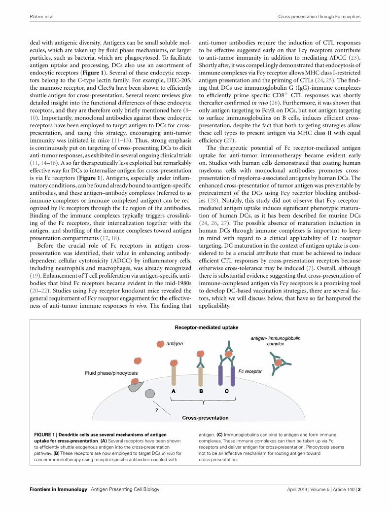
deal with antigenic diversity. Antigens can be small soluble mol-ecules, which are taken up by fluid phase mechanisms, or largerparticles, such as bacteria, which are phagocytosed. To facilitateantigen uptake and processing, DCs also use an assortment ofendocytic receptors (Figure 1). Several of these endocytic recep-tors belong to the C-type lectin family. For example, DEC-205,the mannose receptor, and Clec9a have been shown to efficientlyshuttle antigen for cross-presentation. Several recent reviews givedetailed insight into the functional differences of these endocyticreceptors, and they are therefore only briefly mentioned here (8–10). Importantly, monoclonal antibodies against these endocyticreceptors have been employed to target antigen to DCs for cross-presentation, and using this strategy, encouraging anti-tumorimmunity was initiated in mice (11–13). Thus, strong emphasisis continuously put on targeting of cross-presenting DCs to elicitanti-tumor responses, as exhibited in several ongoing clinical trials(11, 14–16). A so far therapeutically less exploited but remarkablyeffective way for DCs to internalize antigen for cross-presentationis via Fc receptors (Figure 1). Antigens, especially under inflam-matory conditions, can be found already bound to antigen-specificantibodies, and these antigen–antibody complexes (referred to asimmune complexes or immune-complexed antigen) can be rec-ognized by Fc receptors through the Fc region of the antibodies.Binding of the immune complexes typically triggers crosslink-ing of the Fc receptors, their internalization together with theantigen, and shuttling of the immune complexes toward antigenpresentation compartments (17, 18).
Before the crucial role of Fc receptors in antigen cross-presentation was identified, their value in enhancing antibody-dependent cellular cytotoxicity (ADCC) by inflammatory cells,including neutrophils and macrophages, was already recognized(19). Enhancement of T cell proliferation via antigen-specific anti-bodies that bind Fc receptors became evident in the mid-1980s(20–22). Studies using Fcγ receptor knockout mice revealed thegeneral requirement of Fcγ receptor engagement for the effective-ness of anti-tumor immune responses in vivo. The finding that
anti-tumor antibodies require the induction of CTL responsesto be effective suggested early on that Fcγ receptors contributeto anti-tumor immunity in addition to mediating ADCC (23).Shortly after, it was compellingly demonstrated that endocytosis ofimmune complexes via Fcγ receptor allows MHC class I-restrictedantigen presentation and the priming of CTLs (24, 25). The find-ing that DCs use immunoglobulin G (IgG)-immune complexesto efficiently prime specific CD8+ CTL responses was shortlythereafter confirmed in vivo (26). Furthermore, it was shown thatonly antigen targeting to FcγR on DCs, but not antigen targetingto surface immunoglobulins on B cells, induces efficient cross-presentation, despite the fact that both targeting strategies allowthese cell types to present antigen via MHC class II with equalefficiency (27).
The therapeutic potential of Fc receptor-mediated antigenuptake for anti-tumor immunotherapy became evident earlyon. Studies with human cells demonstrated that coating humanmyeloma cells with monoclonal antibodies promotes cross-presentation of myeloma-associated antigens by human DCs. Theenhanced cross-presentation of tumor antigen was preventable bypretreatment of the DCs using Fcγ receptor blocking antibod-ies (28). Notably, this study did not observe that Fcγ receptor-mediated antigen uptake induces significant phenotypic matura-tion of human DCs, as it has been described for murine DCs(24, 26, 27). The possible absence of maturation induction inhuman DCs through immune complexes is important to keepin mind with regard to a clinical applicability of Fc receptortargeting. DC maturation in the context of antigen uptake is con-sidered to be a crucial attribute that must be achieved to induceefficient CTL responses by cross-presentation receptors becauseotherwise cross-tolerance may be induced (7). Overall, althoughthere is substantial evidence suggesting that cross-presentation ofimmune-complexed antigen via Fcγ receptors is a promising toolto develop DC-based vaccination strategies, there are several fac-tors, which we will discuss below, that have so far hampered theapplicability.
FIGURE 1 | Dendritic cells use several mechanisms of antigenuptake for cross-presentation. (A) Several receptors have been shownto efficiently shuttle exogenous antigen into the cross-presentationpathway. (B) These receptors are now employed to target DCs in vivo forcancer immunotherapy using receptor-specific antibodies coupled with
antigen. (C) Immunoglobulins can bind to antigen and form immunecomplexes. These immune complexes can then be taken up via Fcreceptors and deliver antigen for cross-presentation. Pinocytosis seemsnot to be an effective mechanism for routing antigen towardcross-presentation.
Frontiers in Immunology | Antigen Presenting Cell Biology April 2014 | Volume 5 | Article 140 | 2

Platzer et al. Cross-presentation through Fc receptors
CROSS-PRESENTATION OF IMMUNE COMPLEXES AND THEDIVERSITY OF Fc RECEPTORSA major difficulty for studying and determining the therapeuticapplicability of cross-presentation of immune complexes is thecomplexity of the Fcγ receptor family [Table 1; Ref. (29)]. Severaltypes of Fc receptors have been found in addition to species-dependent differences. In mice, four different classes of Fcγ recep-tors comprising FcγRI, FcγRIIB, FcγRIII, and FcγRIV have beendescribed. The activating Fc receptors FcγRI, FcγRIII, and FcγRIVconsist of an immunoglobulin binding α-chain and a signaltransducing γ-chain, which carries an immunoreceptor tyrosine-based activation motif (ITAM). In contrast, FcγRIIB is a singlechain inhibitory receptor with an immunoreceptor tyrosine-basedinhibitory motif (ITIM). The human FcγR system seems to be farmore complex as exemplified by the presence of gene families forFcγRI and FcγRII, as well as the presence of several allelic forms forFcγRIIIA, FcγRIIIB, and FcγRIIB. Mouse FcγRIV is most closelyrelated to human FcγRIIIA whereas mouse FcγRIII is most similarto human FcγRIIA. FcγRIIIB is unique for the human system, butboth species have the inhibitory function of FcγRIIB in common.
Dendritic cells simultaneously express activating and inhibitoryFc receptors [reviewed in Ref. (18)]. The conserved expressionof an inhibitory Fc receptor along with activating Fc recep-tors among species suggests that Fc receptor-mediated cross-presentation is tightly regulated in vivo. The requirement ofstrictly controlling Fc receptor-mediated cross-presentation wasdemonstrated by studies that show that antibody-mediated cross-presentation of self-antigens contributes to autoimmune disease(34, 35). The authors looked at the development of autoim-mune diabetes in RIP-OVA mice. In this model, the transferof OVA-specific naïve CD8+ T cells induces peripheral toler-ance. Importantly, the co-administration of anti-OVA IgG leadsto CD8+ T cell-driven diabetes through the activating Fcγ recep-tors on DCs. The disease pathogenesis in this model was furtheraugmented in FcγRIIB knockout mice, suggesting a tolerogenicfunction of FcγRIIB in vivo. In line with a tolerogenic func-tion of this receptor, it was shown that DCs from FcγRIIBknockout mice generate overall stronger immune responses andthat blocking immune complex binding to FcγRIIB promotesDC maturation, which is considered one of the most impor-tant factors for efficient priming of CTL responses (36–39). Thissuggests that expression of inhibitory FcγRIIB, which restrictsDC maturation under non-inflammatory conditions and thus
probably prevents autoimmunity, may hamper immunotherapeu-tic approaches against tumors and microbial infections (29, 40).Hence, it is important to be aware of the expression patterns andratios of activating versus inhibitory Fc receptors on murine andhuman DCs when studying the effects of immune complexes.
Additionally, IgG subclass composition of immune complexeshas been shown to influence binding affinity resulting in differentbinding properties to individual Fc receptors (41). For example,immune complexes composed of human IgG1 bind with relativelyhigh affinities to all Fc receptors, whereas IgG2 immune complexesseem to bind primarily to human FcγRIIA and FcγRIIIA (42).Furthermore, disparities in the binding affinities of immunoglob-ulin isotypes for specific Fcγ receptors exist between mice andhumans. Thus, predictions of immune complex functions drawnfrom wild-type mouse models might be inadequate. A prominentexample of the failure of previous studies in accurately recapit-ulating the specificity and diversity of Fcγ receptor interactionsis the outcome of a clinical trial using a CD28-specific superag-onistic antibody; this led to severe side effects including severepain and extreme swelling, as well as one individual sufferingfrom heart, liver, and kidney failure (43). To address this prob-lem, an FcγR humanized mouse strain was recently generatedthrough transgenic expression of the entire human FcγR fam-ily under the control of their human regulatory elements on agenetic background lacking all mouse FcγRs (44). The animalsdemonstrate normal lymphoid tissue development and gener-ate normal immune responses. Thus, this mouse strain offersa greatly improved model to study immune complex-mediatedcross-presentation, although it addresses only the species-specificdifferences regarding Fcγ receptors. Humans and mice also dis-play differences in the expression patterns of Fc receptors forIgE and IgA, which might contribute to cross-presentation ofimmune-complexed antigen in vivo (45–48).
Increasing evidence suggests that allelic isoforms and polymor-phisms of Fc receptors are shaping immune responses in humans.FcγRIIA (CD32A), the major phagocytic FcγR in humans, exhibitsa polymorphism in the ligand-binding domain (49). Individ-uals homozygous for the R allelic form of CD32A (CD32ARallele) have been described as more susceptible to bacterialinfections and autoimmune diseases compared to individualshomozygous for the H allelic form of CD32A (CD32AH) andCD32AR/H heterozygous individuals (50, 51). A binding studyusing two-dimensional affinity measurements also demonstrated
Table 1 | Overview of human and murine Fcγ receptors.
Human/mouse IgG receptor CD Function Affinity Structure
Human (30–33) FcγRIIA CD32A Activation Low to medium α-Chain with ITAM
FcγRIIC CD32C Activation Low to medium α-Chain with ITAM
FcγRIIIA CD16A Activation Low to medium α-Chain and γ2-chains with ITAM
FcγRIIIB CD16B Activation Low to medium GPI-linked α-chain
Human and mouse (30–33) FcγRI CD64 Activation High α-Chain and γ2-chains with ITAM
FcγRIIB CD32B Inhibition Low to medium α-Chain with ITIM
Mouse (30–33) FcγRIII CD16 Activation Low to medium α-Chain and γ2-chains with ITAM
FcγRIV Activation Low to medium α-Chain and γ2-chains with ITAM
www.frontiersin.org April 2014 | Volume 5 | Article 140 | 3

Platzer et al. Cross-presentation through Fc receptors
that compared to CD32AH, CD32AR has significantly lower affin-ity toward IgG2, as well as to IgG1 and IgG3, suggesting that thelower binding of CD32AR to IgGs might be responsible for thelack of immune complex clearance, which leads to increased sus-ceptibility to bacterial infections and autoimmune diseases (52).Genetic variations in Fc receptors have also been linked to cancersusceptibility (53–55). However, less efficient immune complexbinding might also be reflected in less efficient antigen uptake andpresentation via this receptor, and thus consequences for immunecomplexes cross-presentation should be expected. Of note, glyco-sylations in the IgG–Fc region can also affect Fc receptor-bindingproperties as discussed in detail in a recent review (56). How anti-gen cross-presentation of immune complexes and T cell primingis altered by differences in IgG subclass composition, IgG–Fc gly-cosylation, and Fc receptor polymorphisms is currently unknown,but is important to address. In conclusion, the complexity of inter-actions of IgG with the Fc receptor system in addition to concernsabout species specificity presents a major hurdle that needs to beovercome for successful therapeutic applications.
CROSS-PRESENTATION OF IMMUNE COMPLEXES AND THEDIVERSITY OF DC SUBPOPULATIONSWhether it would be beneficial to target a specific DC subset thatdisplays a superior capacity to cross-present antigen for thera-peutic approaches is currently a field of extensive investigation(4, 57). We will first focus on what we know so far about thecross-presentation capacity of DC subsets in general and then dis-cuss our current understanding of cross-presentation of immunecomplexes in regard to DC subsets. DCs are a heterogeneouscell population, and substantial effort was made to characterizedifferent subsets in mice and identify their human counterparts[reviewed in Ref. (58–60)]. In principal, murine and human DCscan be divided into two major subsets, classical/conventional DCs(cDCs) and plasmacytoid DCs (pDCs). In mice, cDCs compriseCD8α+ and CD8α− lineages, which have been found to differ intheir ontogeny and display functional specializations. Since theexpression of surface markers on human and murine DCs is notconserved, only recently has gene expression profiling allowed forthe identification of human CD141+ DCs as functional equiva-lents of the mouse CD8α+ DCs, while human CD1c+ DCs appearto be comparable to mouse CD8- DCs (61, 62).
In mice, the CD8α+ DC subset is considered to be more effi-cient at antigen cross-presentation than other DC subsets (63–66).The corresponding human subset, CD141+ DCs, is also potent atinducing CD8+ T cell responses in vitro, although their superi-ority to other human DC subsets is uncertain (67–73). Severalgroups have now reported that all human DC subsets can effi-ciently cross-present several forms of antigen [reviewed by Ref.(57)]. Initially, CD141+ DCs isolated from human blood weredescribed to better cross-present CMV protein pp65 in compar-ison to CD1c+ DCs and pDCs from the same donor (67). It isimportant to note, however, that cross-presentation in vivo occursrather in secondary lymphoid organs. A recent study has overcomethe difficulties in isolating sufficient amounts of human DCs fromlymphoid tissue and characterized in detail the cross-presentationproperties of tonsil-resident DCs (73). An important finding ofthis study was that all tonsillar DC subsets (i.e., pDCs and the
two populations of cDCs, CD1c+ DCs and CD141+ DCs) dis-played comparable capacities to cross-present soluble antigens incontrast to macrophages, which lacked this ability. Interestingly,necrotic cells were phagocytosed and cross-presented by CD1c+
DCs and CD141+ DCs with similar efficiency, while pDCs werepoor at taking up necrotic particles, consequently resulting in inef-ficient cross-presentation. Tonsillar macrophages were found to bethe most efficient at taking up dead cells, but despite this fact theycompletely failed to cross-present necrotic cells. Collectively, theability to efficiently cross-present in humans seems less restrictedto a specific DC subpopulation than as observed in mice. Alongthese lines, it has been shown that the cross-presentation proper-ties of human DCs depend on the antigen uptake pathway and theability of the pathway to route the antigen into an early endoso-mal compartment rather than on a specific DC subset (74, 75).CD141+ DCs are superior cross-presenters compared to CD1c+
DCs only when the antigen is delivered via CD205, a receptorthat preferentially targets antigens to late endo/lysosomal com-partments. If antigen is targeted through CD40, CD1c+ DCs areas efficient as CD141+ DCs. These findings argue that targetingone specific DC subset for the design of DC-based vaccines maynot offer the presumed advantage.
The cross-presentation studies discussed above focused pri-marily on soluble antigen uptake and targeting antigen via severalendocytic receptors. How does cross-presentation of immunecomplexes fit into this picture? Targeting DCs through IgGimmune complexes has been proven to be superior to solubleimmune complexes for inducing CD8+ T cell responses and asanti-tumor vaccines by utilizing murine bone marrow-derivedDCs (76, 77). In addition, circulating specific antibodies have beenshown to enhance systemic cross-priming by delivering immune-complexed antigen to murine DCs in vivo (78). Notably in mice,immune-complexed antigen allows the CD8α− DC subset, whichhas been proven to be very poor at presenting soluble antigen,to become potent cross-presenting cells (79). Interestingly, cross-presentation by CD8α− DCs depends on activating Fcγ receptors.Lack of the signal transducing γ-chain specifically abolishes pre-sentation of immune-complexed antigen on MHC class I mole-cules but not on MHC class II molecules (79). Another remarkablefeature regarding cross-presentation of immune complexes is theirreliance on FcRn, an IgG binding receptor that is primarily locatedintracellularly and binds IgG independently from their Fcγ recep-tor interaction sites (80). How FcRn promotes cross-presentationof immune complex is discussed later in more detail.
Our knowledge regarding cross-presentation of immune-complexed antigen by human DC subsets is still very lim-ited. The effects of Fcγ receptor antigen targeting on theefficiency of cross-presentation in human DCs were recentlyinvestigated using human cytomegalovirus (HCMV) pp65 asa protein antigen (81). In line with the data obtained frommurine models, immune-complexed antigen is more efficientlycross-presented than comparable amounts of soluble antigen byhuman DCs. The enhanced cross-presentation capacity observedwas not mediated by increased antigen uptake or inductionof DC maturation through the immune-complexed antigen.The authors also demonstrated that both of the two majorintracellular cross-presentation pathways (4), the cytosolic and
Frontiers in Immunology | Antigen Presenting Cell Biology April 2014 | Volume 5 | Article 140 | 4

Platzer et al. Cross-presentation through Fc receptors
the vacuolar/endosomal pathway, are involved in Fcγ receptor-mediated uptake of immune complexes and their processing.Notably, monocyte-derived DCs as well as CD141+ DCs requiredantigen processing by both intracellular pathways. The finding thatCD141+ DCs, which are the human equivalent to CD8α+ DCs,use both processing pathways for immune complexes points tounique features of human DCs. Murine CD8α+ DCs mainly usethe cytosolic pathway to process antigen for cross-presentation,including the processing of immune complexes (82). Another dif-ference to murine DCs is that the CD141+ DC subset proved to besuperior to CD1c+ DCs in cross-presenting pp65 immune com-plexes (81). These findings point to obvious differences betweenmurine and human DC subsets regarding immune complex-mediated cross-presentation. Since the human DCs were isolatedfrom blood (81) and the murine DCs were isolated from the spleen(79, 80), it is possible that DCs from blood and lymphoid tissuegenerally differ in their cross-presentation capacities of immunecomplexes,which have similarly been observed for human DC sub-sets in response to soluble antigen as described above. In any case,the study by Flinsenberg et al. found that Fcγ receptor targetingincreases cross-presentation of HCMV antigen by human bloodand tonsillar CD141+DCs, which suggest that targeting of this DCsubset with immune complexes might improve DC-based vacci-nation strategies. Another very important aspect of this study isthe detailed characterization of Fcγ receptor expression on humanDC subsets. Although CD1c+ DCs expressed overall higher lev-els of FcγRII, CD141+ DCs seem to express higher levels of theactivating FcγRIIA relative to the inhibitory FcγRIIB. Thus, thisstudy clearly demonstrates that the overall expression level of onespecific Fcγ receptor does not determine the functional outcome,and that we need to consider the diversity of Fcγ receptor expres-sion by distinct DC subsets to evaluate the therapeutic potentialof immune complex-mediated cross-presentation.
A further difference between mice and humans seems to be thecross-presentation capacity of pDCs. Several studies have reportedthat murine pDCs do not possess the ability to cross-present (83–86) or that their capacity is insignificant when compared to cDCs(87). In contrast to mouse pDCs, human pDCs can efficientlycross-present antigen and induce CD8+ T cell responses (88–90).Human pDCs also express FcγRIIA, and this receptor has beenshown to mediate internalization of immunoglobulins bound tochromatin (91), Coxsackie virus (92), the model antigen KLH (93),and the tumor antigen NY-ESO-1 (94). In addition, the group ofde Vries described that pDCs can use several receptor-targetedantigen uptake pathways, including the activating FcγRIIA recep-tor, to target antibody-coated nanoparticles for cross-presentation.Although this study did not use classical immune complexes,together with a vaccination study in which pDCs significantlyprolonged overall survival in melanoma patients (95), it sup-ports the notion that pDCs are interesting targets for DC-basedimmunotherapeutic strategies.
Collectively, we should keep in mind that some of the observeddifferences between human and murine DC subsets regardingcross-presentation of immune complexes most likely stem fromdifferences in their Fc receptor expression and from different bind-ing affinities for IgG isotypes. Recently, various published andpublicly available microarray data were compiled, and this mRNA
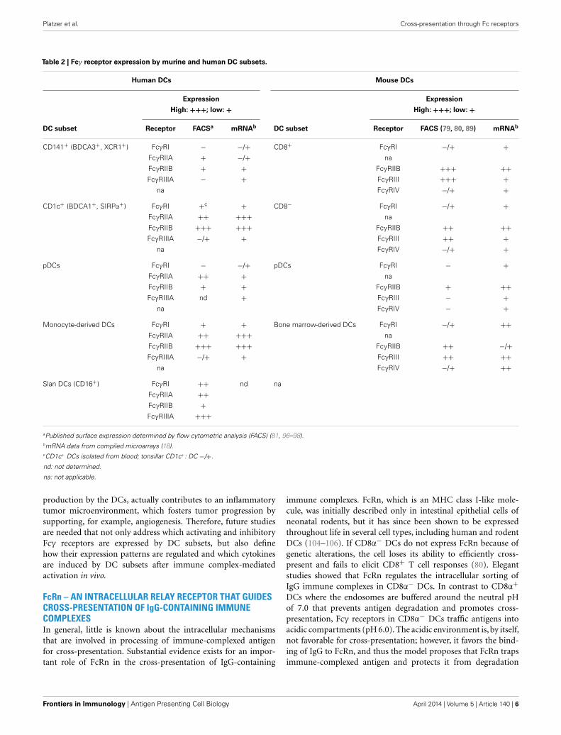
collection provides an excellent overview of mouse and human Fcγreceptor expression by DC subsets, monocytes, and macrophages(18). Overall, the Fcγ receptor expression levels obtained bymRNA analysis correspond well with the surface expression lev-els acquired by flow cytometric analysis (FACS) (Table 2). Forthe future, it will be important to determine whether the Fcγreceptor expression of human DC subsets isolated from bloodalso matches the expression on tissue-resident DCs from differentorgans.
REGULATION OF Fcγ RECEPTOR EXPRESSION IMPACTSCROSS-PRESENTATION OF IMMUNE COMPLEXESEfficient cross-presentation for inducing protective immuneresponses against tumors or viruses is strongly governed by theratio of activating versus inhibitory Fcγ receptors expressed onDCs. In addition to the DC subset, the maturation/activation stateof DCs likely impacts their Fcγ receptors expression pattern. Thematuration/activation state of DCs is in general strongly influ-enced by the cytokine milieu of the microenvironment, and aconsiderable number of cytokines have been shown to regulateFcγ receptor expression in vitro. TGF-β1 down-regulates surfaceexpression FcγRI and FcγRIII on monocytes (99). IL-4, a cytokineassociated with Th2-type immune responses, increases the expres-sion of inhibitory FcγRIIB. In contrast, the Th1-cytokine IFN-γ increases expression of activating Fc receptors on monocytes(100). Monocytes also have been shown to respond to IFN-γand TNF-α treatment with enhanced immune complex bindingvia FcγRI, even when saturated with pre-bound monomeric IgG(101). Cytokine-induced changes in Fcγ receptor expression werealso found using monocyte-derived DCs (96). Immature DCsgenerated with GM-CSF and IL-4 from monocytes express highamounts of inhibitory FcγRIIB, which is down-regulated uponDC maturation induced by TNF-α. The authors also showed thatblood DCs activated with a cytokine cocktail containing TNF-α, IL-1β, IL-6, and PGE2 induce more influenza-specific CD8+
T memory cells via targeting of FcγRI and FcγRIIA. Interest-ingly, crosslinking of inhibitory FcγRIIB only reduced the cross-presentation ability of immature DCs but not of mature DCs.Treatment of mature blood DCs with IL-10, or a combinationof IL-10 and IL13, was found to increase expression of FcγRIIAand FcγRIIB (96). To sum up, although we know that cytokinescan modulate Fcγ receptor expression, and that tumors createcytokine-rich microenvironments that involve the production ofimmunosuppressive as well as inflammatory cytokines to drivetumor progression (102, 103), our knowledge is very limitedas to how cytokines from the tumor microenvironment affectcross-presentation of immune complexes by DCs. Thus, regard-ing anti-tumor therapy, this gap in knowledge might explainwhy the long-term therapeutic outcomes of immune complex-based strategies were not more successful, although efficientcross-presentation is induced by IgG-complexed antigens. Oneexplanation could be that the tumor microenvironment pro-motes the induction of cross-tolerance by keeping the DCs inan immature state, which is associated with high expression levelsof inhibitory FcγRIIB. Another possible scenario would be thatimmune complex-mediated cross-presentation via activating Fcγreceptors, which is known to result in inflammatory cytokine
www.frontiersin.org April 2014 | Volume 5 | Article 140 | 5
Platzer et al. Cross-presentation through Fc receptors
Table 2 | Fcγ receptor expression by murine and human DC subsets.
Human DCs Mouse DCs
Expression Expression
High: +++; low: + High: +++; low: +
DC subset Receptor FACSa mRNAb DC subset Receptor FACS (79, 80, 89) mRNAb
CD141+ (BDCA3+, XCR1+) FcγRI − −/+ CD8+ FcγRI −/+ +
FcγRIIA + −/+ na
FcγRIIB + + FcγRIIB +++ ++
FcγRIIIA − + FcγRIII +++ +
na FcγRIV −/+ +
CD1c+ (BDCA1+, SIRPα+) FcγRI +c
+ CD8− FcγRI −/+ +
FcγRIIA ++ +++ na
FcγRIIB +++ +++ FcγRIIB ++ ++
FcγRIIIA −/+ + FcγRIII ++ +
na FcγRIV −/+ +
pDCs FcγRI − −/+ pDCs FcγRI − +
FcγRIIA ++ + na
FcγRIIB + + FcγRIIB + ++
FcγRIIIA nd + FcγRIII − +
na FcγRIV − +
Monocyte-derived DCs FcγRI + + Bone marrow-derived DCs FcγRI −/+ ++
FcγRIIA ++ +++ na
FcγRIIB +++ +++ FcγRIIB ++ −/+
FcγRIIIA −/+ + FcγRIII ++ ++
na FcγRIV −/+ ++
Slan DCs (CD16+) FcγRI ++ nd na
FcγRIIA ++
FcγRIIB +
FcγRIIIA +++
aPublished surface expression determined by flow cytometric analysis (FACS) (81, 96–98).bmRNA data from compiled microarrays (18).cCD1c+ DCs isolated from blood; tonsillar CD1c+: DC −/+.
nd: not determined.
na: not applicable.
production by the DCs, actually contributes to an inflammatorytumor microenvironment, which fosters tumor progression bysupporting, for example, angiogenesis. Therefore, future studiesare needed that not only address which activating and inhibitoryFcγ receptors are expressed by DC subsets, but also definehow their expression patterns are regulated and which cytokinesare induced by DC subsets after immune complex-mediatedactivation in vivo.
FcRn – AN INTRACELLULAR RELAY RECEPTOR THAT GUIDESCROSS-PRESENTATION OF IgG-CONTAINING IMMUNECOMPLEXESIn general, little is known about the intracellular mechanismsthat are involved in processing of immune-complexed antigenfor cross-presentation. Substantial evidence exists for an impor-tant role of FcRn in the cross-presentation of IgG-containing
immune complexes. FcRn, which is an MHC class I-like mole-cule, was initially described only in intestinal epithelial cells ofneonatal rodents, but it has since been shown to be expressedthroughout life in several cell types, including human and rodentDCs (104–106). If CD8α− DCs do not express FcRn because ofgenetic alterations, the cell loses its ability to efficiently cross-present and fails to elicit CD8+ T cell responses (80). Elegantstudies showed that FcRn regulates the intracellular sorting ofIgG immune complexes in CD8α− DCs. In contrast to CD8α+
DCs where the endosomes are buffered around the neutral pHof 7.0 that prevents antigen degradation and promotes cross-presentation, Fcγ receptors in CD8α− DCs traffic antigens intoacidic compartments (pH 6.0). The acidic environment is, by itself,not favorable for cross-presentation; however, it favors the bind-ing of IgG to FcRn, and thus the model proposes that FcRn trapsimmune-complexed antigen and protects it from degradation
Frontiers in Immunology | Antigen Presenting Cell Biology April 2014 | Volume 5 | Article 140 | 6
Platzer et al. Cross-presentation through Fc receptors
within an acidic loading compartment. The study also showed thatin parallel to antigen entry into the FcRn-positive compartment,key components of the phagosome-to-cytosol cross-presentationmachinery are rapidly recruited to the endo/lysosome. Vesiclesthat contained IgG-opsonized particles or IgG immune complexesrapidly acquired greater quantities of vacuolar ATPase (V-ATPase),gp91phox, and Rab27a than those that resulted from internal-ization of IgG mutants that cannot interact with FcRn. Con-sistent with this concept, it was described that the presence ofFcRn also affects the oxidation state as well as the acidificationof vesicles. Inhibitor studies demonstrated that FcRn-mediatedcross-presentation depends on the proteasome as well as Sec61α,which is indicative for the cytosolic cross-presentation pathway.Since insulin-regulated amino peptidase (IRAP) enrichment wasnot depicted in FcRn-positive IgG immune complex-containingvesicles, and cathepsin inhibitors did not abrogate IgG immunecomplex cross-presentation, the authors concluded that the alter-native vacuolar pathway was not involved. In summary, this studysuggests that FcRn binding of IgG immune complexes enables aslower and more controlled antigenic degradation in CD8α− DCs,thereby permitting this population of DCs to become efficientcross-presenting cells.
The most compelling evidence for the exceptional importanceof FcRn for cross-presentation of IgG immune complexes and IgG-opsonized particles is derived from in vivo studies that analyzedthe effects of FcRn-deficiency on chronic intestinal inflammationand colonic cancer (107, 108). In a chemically induced chroniccolitis model, which is associated with generating high levelsof anti-bacterial antibodies that enter the host as IgG immunecomplexes, Baker et al. demonstrated that FcRn-dependent cross-presentation is carried out by CD8α− DCs in vivo, leading togreater levels of cytotoxic T cell activation during the course ofcolitis. In a recent study, the same group focused on the impactof FcRn on tumor development, clearly demonstrating the impor-tance of this molecule for anti-tumor immune surveillance (108).The authors found that the DC-specific deletion of FcRn leadsto increased tumor burden in experimental models of colon can-cer and lung metastasis. Strikingly, this study also demonstratedthat colon cancer patients with higher numbers of FcRn-positiveDCs in the adjacent tumor tissue had significantly better prog-noses, confirming the crucial role of FcRn and demonstratingthe vital role of cross-presentation of IgG immune complexes inanti-tumor immunity in general. It will now be of utmost impor-tance to elucidate the details of the intracellular mechanism ofthis process to evaluate whether the pathway can be employed forcancer immunotherapy.
CONCLUSIONAlthough ample evidence suggests that Fcγ receptor targetingthrough immune complexes allows for more efficient cross-presentation compared to soluble antigen, it still needs to beproven which advantages it may have over targeting of otherendocytic receptors on DCs, especially in vivo. In this respect,it is very important to continue developing better murine mod-els which more accurately reflect the human immune system. Therecently published humanized FcγR mouse strain is here a promis-ing step in the right direction. For therapeutic manipulations, we
also need to better understand how Fcγ receptor expression byDCs is regulated. Can we use cytokines and/or TLR ligands tomodulate the ratio of inhibitory versus activating Fcγ receptorsexpressed by DC subsets to improve therapeutic strategies? TLR-2 ligands, for example, have been shown to increase expressionof inhibitory FcγRIIB in macrophages (109), a consequence notdesirable in the context of viral or tumor vaccine development.Furthermore, how does the size of immune complexes influencecross-presentation? How does the antibody to antigen ratio inimmune complexes influence cross-presentation? Indeed, it hasbeen shown that immune complex size and glycosylation on IgGimpact the binding to human Fcγ receptors (110). In summary, itis fair to conclude that many important questions remain openand need to be addressed. Irrespectively, cross-presentation ofimmune complexes represents an exciting potential pathway toimprove DC-based vaccination strategies for anti-viral as well asanti-tumor therapy.
ACKNOWLEDGMENTSThis work was supported by grants from the National Institutes ofHealth: K01DK093597 (to Barbara Platzer) and AI075037 (to EddaFiebiger). This work was also supported by the Harvard DigestiveDiseases Center Grant P30DK034854.
REFERENCES1. Sigal LJ, Crotty S, Andino R, Rock KL. Cytotoxic T-cell immunity to virus-
infected non-haematopoietic cells requires presentation of exogenous antigen.Nature (1999) 398:77–80. doi:10.1038/18038
2. Jung S, Unutmaz D, Wong P, Sano G, de Los Santos K, Sparwasser T, et al.In vivo depletion of CD11c+ dendritic cells abrogates priming of CD8+T cells by exogenous cell-associated antigens. Immunity (2002) 17:211–20.doi:10.1016/S1074-7613(02)00365-5
3. Winau F, Weber S, Sad S, de Diego J, Hoops SL, Breiden B, et al. Apoptotic vesi-cles crossprime CD8 T cells and protect against tuberculosis. Immunity (2006)24:105–17. doi:10.1016/j.immuni.2005.12.001
4. Joffre OP, Segura E, Savina A, Amigorena S. Cross-presentation by dendriticcells. Nat Rev Immunol (2012) 12:557–69. doi:10.1038/nri3254
5. Apetoh L, Locher C, Ghiringhelli F, Kroemer G, Zitvogel L. Harnessing den-dritic cells in cancer. Semin Immunol (2011) 23:42–9. doi:10.1016/j.smim.2011.01.003
6. Andersen BM, Ohlfest JR. Increasing the efficacy of tumor cell vaccines byenhancing cross priming. Cancer Lett (2012) 325:155–64. doi:10.1016/j.canlet.2012.07.012
7. Bonifaz L, Bonnyay D, Mahnke K, Rivera M, Nussenzweig MC, Steinman RM.Efficient targeting of protein antigen to the dendritic cell receptor DEC-205in the steady state leads to antigen presentation on major histocompatibilitycomplex class I products and peripheral CD8+ T cell tolerance. J Exp Med(2002) 196:1627–38. doi:10.1084/jem.20021598
8. Tacken PJ, Figdor CG. Targeted antigen delivery and activation of dendritic cellsin vivo: steps towards cost effective vaccines. Semin Immunol (2011) 23:12–20.doi:10.1016/j.smim.2011.01.001
9. Caminschi I, Maraskovsky E, Heath WR. Targeting dendritic cells in vivo forcancer therapy. Front Immunol (2012) 3:13. doi:10.3389/fimmu.2012.00013
10. Kreutz M, Tacken PJ, Figdor CG. Targeting dendritic cells – why bother? Blood(2013) 121:2836–44. doi:10.1182/blood-2012-09-452078
11. de Vries IJ, Krooshoop DJ, Scharenborg NM, Lesterhuis WJ, Diepstra JH, VanMuijen GN, et al. Effective migration of antigen-pulsed dendritic cells to lymphnodes in melanoma patients is determined by their maturation state. CancerRes (2003) 63:12–7.
12. Sancho D, Mourao-Sa D, Joffre OP, Schulz O, Rogers NC, Pennington DJ, et al.Tumor therapy in mice via antigen targeting to a novel, DC-restricted C-typelectin. J Clin Invest (2008) 118:2098–110. doi:10.1172/JCI34584
13. Hemmi H, Zaidi N, Wang B, Matos I, Fiorese C, Lubkin A, et al. Treml4, anIg superfamily member, mediates presentation of several antigens to T cells
www.frontiersin.org April 2014 | Volume 5 | Article 140 | 7

Platzer et al. Cross-presentation through Fc receptors
in vivo, including protective immunity to HER2 protein. J Immunol (2012)188:1147–55. doi:10.4049/jimmunol.1102541
14. Tacken PJ, de Vries IJ, Torensma R, Figdor CG. Dendritic-cell immunotherapy:from ex vivo loading to in vivo targeting. Nat Rev Immunol (2007) 7:790–802.doi:10.1038/nri2173
15. Eubel J, Enk AH. Dendritic cell vaccination as a treatment modalityfor melanoma. Expert Rev Anticancer Ther (2009) 9:1631–42. doi:10.1586/era.09.139
16. Robson NC, Hoves S, Maraskovsky E, Schnurr M. Presentation of tumourantigens by dendritic cells and challenges faced. Curr Opin Immunol (2010)22:137–44. doi:10.1016/j.coi.2010.01.002
17. Baker K, Rath T, Lencer WI, Fiebiger E, Blumberg RS. Cross-presentationof IgG-containing immune complexes. Cell Mol Life Sci (2013) 70:1319–34.doi:10.1007/s00018-012-1100-8
18. Guilliams M, Bruhns P, Saeys Y, Hammad H, Lambrecht BN. The functionof Fcgamma receptors in dendritic cells and macrophages. Nat Rev Immunol(2014) 14:94–108. doi:10.1038/nri3582
19. Clynes R, Takechi Y, Moroi Y, Houghton A, Ravetch JV. Fc receptors are requiredin passive and active immunity to melanoma. Proc Natl Acad Sci U S A (1998)95:652–6. doi:10.1073/pnas.95.2.652
20. Celis E, Chang TW. HBsAg-serum protein complexes stimulate immune T lym-phocytes more efficiently than do pure HBsAg. Hepatology (1984) 4:1116–23.doi:10.1002/hep.1840040604
21. Celis E, Zurawski VR Jr, Chang TW. Regulation of T-cell function by antibod-ies: enhancement of the response of human T-cell clones to hepatitis B surfaceantigen by antigen-specific monoclonal antibodies. Proc Natl Acad Sci U S A(1984) 81:6846–50. doi:10.1073/pnas.81.21.6846
22. Snider DP, Segal DM. Targeted antigen presentation using crosslinked antibodyheteroaggregates. J Immunol (1987) 139:1609–16.
23. Vasovic LV, Dyall R, Clynes RA, Ravetch JV, Nikolic-Zugic J. Synergy between anantibody and CD8+ cells in eliminating an established tumor. Eur J Immunol(1997) 27:374–82. doi:10.1002/eji.1830270206
24. Regnault A, Lankar D, Lacabanne V, Rodriguez A, Thery C, Rescigno M,et al. Fcgamma receptor-mediated induction of dendritic cell maturationand major histocompatibility complex class I-restricted antigen presenta-tion after immune complex internalization. J Exp Med (1999) 189:371–80.doi:10.1084/jem.189.2.371
25. Rodriguez A, Regnault A, Kleijmeer M, Ricciardi-Castagnoli P, Amigorena S.Selective transport of internalized antigens to the cytosol for MHC class I pre-sentation in dendritic cells. Nat Cell Biol (1999) 1:362–8. doi:10.1038/14058
26. Schuurhuis DH, Ioan-Facsinay A, Nagelkerken B, Van Schip JJ, Sedlik C, MeliefCJ, et al. Antigen-antibody immune complexes empower dendritic cells toefficiently prime specific CD8+ CTL responses in vivo. J Immunol (2002)168:2240–6.
27. Machy P,Serre K,Leserman L. Class I-restricted presentation of exogenous anti-gen acquired by Fcgamma receptor-mediated endocytosis is regulated in den-dritic cells. Eur J Immunol (2000) 30:848–57. doi:10.1002/1521-4141(200003)30:3<848::AID-IMMU848>3.0.CO;2-Q
28. Dhodapkar KM, Krasovsky J, Williamson B, Dhodapkar MV. Antitumor mon-oclonal antibodies enhance cross-presentation of cellular antigens and the gen-eration of myeloma-specific killer T cells by dendritic cells. J Exp Med (2002)195:125–33. doi:10.1084/jem.20011097
29. Nimmerjahn F, Ravetch JV. FcgammaRs in health and disease. Curr Top Micro-biol Immunol (2011) 350:105–25. doi:10.1007/82_2010_86
30. Hulett MD, Hogarth PM. Molecular basis of Fc receptor function. Adv Immunol(1994) 57:1–127. doi:10.1016/S0065-2776(08)60671-9
31. Ravetch JV. Fc receptor. In: Paul WE, editor. Fundamental Immunology.Philadelphia, PA: Lippincott Williams & Wilkins (2003). p. 685–700.
32. Nimmerjahn F, Ravetch JV. Fcγ receptors: old friends and new family members.Immunity (2006) 24:19–28. doi:10.1016/j.immuni.2005.11.010
33. Nimmerjahn F, Ravetch JV. Fcγ receptors as regulators of immune responses.Nat Rev Immunol (2008) 8:34–47. doi:10.1038/nri2206
34. Desai DD, Harbers SO, Flores M, Colonna L, Downie MP, Bergtold A, et al. Fcgamma receptor IIB on dendritic cells enforces peripheral tolerance by inhibit-ing effector T cell responses. J Immunol (2007) 178:6217–26.
35. Harbers SO, Crocker A, Catalano G, D’Agati V, Jung S, Desai DD, et al.Antibody-enhanced cross-presentation of self antigen breaks T cell tolerance.J Clin Invest (2007) 117:1361–9. doi:10.1172/JCI29470
36. Kalergis AM, Ravetch JV. Inducing tumor immunity through the selectiveengagement of activating Fcgamma receptors on dendritic cells. J Exp Med(2002) 195:1653–9. doi:10.1084/jem.20020338
37. Boruchov AM, Heller G, Veri MC, Bonvini E, Ravetch JV, Young JW. Activatingand inhibitory IgG Fc receptors on human DCs mediate opposing functions.J Clin Invest (2005) 115:2914–23. doi:10.1172/JCI24772
38. Dhodapkar KM, Dhodapkar MV. Recruiting dendritic cells to improve anti-body therapy of cancer. Proc Natl Acad Sci U S A (2005) 102:6243–4.doi:10.1073/pnas.0502547102
39. van Montfoort NT, Hoen PA, Mangsbo SM, Camps MG, Boross P, Melief CJ,et al. Fcgamma receptor IIb strongly regulates Fcgamma receptor-facilitated Tcell activation by dendritic cells. J Immunol (2012) 189:92–101. doi:10.4049/jimmunol.1103703
40. Boross P, Arandhara VL, Martin-Ramirez J, Santiago-Raber ML, CarlucciF, Flierman R, et al. The inhibiting Fc receptor for IgG, FcgammaRIIB, isa modifier of autoimmune susceptibility. J Immunol (2011) 187:1304–13.doi:10.4049/jimmunol.1101194
41. Bruhns P. Properties of mouse and human IgG receptors and their contribu-tion to disease models. Blood (2012) 119:5640–9. doi:10.1182/blood-2012-01-380121
42. Bruhns P, Iannascoli B, England P, Mancardi DA, Fernandez N, Jorieux S,et al. Specificity and affinity of human Fcgamma receptors and their poly-morphic variants for human IgG subclasses. Blood (2009) 113:3716–25.doi:10.1182/blood-2008-09-179754
43. Vitetta ES, Ghetie VF. Immunology. Considering therapeutic antibodies. Sci-ence (2006) 313:308–9. doi:10.1126/science.1130482
44. Smith P, Dilillo DJ, Bournazos S, Li F, Ravetch JV. Mouse model recapitulatinghuman Fcgamma receptor structural and functional diversity. Proc Natl AcadSci U S A (2012) 109:6181–6. doi:10.1073/pnas.1203954109
45. van Egmond M, Van Vuuren AJ, Morton HC, Van Spriel AB, Shen L, HofhuisFM, et al. Human immunoglobulin A receptor (FcalphaRI, CD89) functionin transgenic mice requires both FcR gamma chain and CR3 (CD11b/CD18).Blood (1999) 93:4387–94.
46. Dehlink E, Baker AH, Yen E, Nurko S, Fiebiger E. Relationships between levelsof serum IgE, cell-bound IgE, and IgE-receptors on peripheral blood cells ina pediatric population. PLoS One (2010) 5:e12204. doi:10.1371/journal.pone.0012204
47. Platzer B, Dehlink E, Turley SJ, Fiebiger E. How to connect an IgE-drivenresponse with CTL activity? Cancer Immunol Immunother (2012) 61:1521–5.doi:10.1007/s00262-011-1127-y
48. Vasudev M, Cheung DS, Pincsak H, Li SH, Yan K, Simpson P, et al.Expression of high-affinity IgE receptor on human peripheral blooddendritic cells in children. PLoS One (2012) 7:e32556. doi:10.1371/journal.pone.0032556
49. Salmon JE, Edberg JC, Brogle NL, Kimberly RP. Allelic polymorphisms ofhuman Fc gamma receptor IIA and Fc gamma receptor IIIB. Independentmechanisms for differences in human phagocyte function. J Clin Invest (1992)89:1274–81. doi:10.1172/JCI115712
50. Sanders LA, van de Winkel JG, Rijkers GT, Voorhorst-Ogink MM, de Haas M,Capel PJ, et al. Fc gamma receptor IIa (CD32) heterogeneity in patients withrecurrent bacterial respiratory tract infections. J Infect Dis (1994) 170:854–61.doi:10.1093/infdis/170.4.854
51. van Sorge NM, van der Pol WL, van de Winkel JG. FcgammaR polymorphisms:implications for function, disease susceptibility and immunotherapy. TissueAntigens (2003) 61:189–202. doi:10.1034/j.1399-0039.2003.00037.x
52. Shashidharamurthy R, Zhang F, Amano A, Kamat A, Panchanathan R, Ezek-wudo D, et al. Dynamics of the interaction of human IgG subtype immunecomplexes with cells expressing R and H allelic forms of a low-affinityFc gamma receptor CD32A. J Immunol (2009) 183:8216–24. doi:10.4049/jimmunol.0902550
53. Pandey JP, Kistner-Griffin E, Namboodiri AM, Black L, Jobim M. Sugges-tive evidence that Fc variants of IgG2 and FcgammaRIIa loci interact tocontribute to the risk of prostate cancer. Hum Immunol (2013) 74:1656–8.doi:10.1016/j.humimm.2013.08.280
54. Pandey JP, Namboodiri AM, Kistner-Griffin E. A genetic variant of Fcgam-maRIIIa is strongly associated with humoral immunity to cyclin B1 in AfricanAmerican patients with prostate cancer. Immunogenetics (2013) 65:91–6.doi:10.1007/s00251-012-0660-y
Frontiers in Immunology | Antigen Presenting Cell Biology April 2014 | Volume 5 | Article 140 | 8

Platzer et al. Cross-presentation through Fc receptors
55. Pandey JP, Namboodiri AM, Kistner-Griffin E, Iwasaki M, Kasuga Y, HamadaGS, et al. Racially restricted contribution of immunoglobulin Fcgamma andFcgamma receptor genotypes to humoral immunity to human epidermalgrowth factor receptor 2 in breast cancer. Clin Exp Immunol (2013) 171:273–7.doi:10.1111/cei.12018
56. Anthony RM, Wermeling F, Ravetch JV. Novel roles for the IgG Fc glycan. AnnN Y Acad Sci (2012) 1253:170–80. doi:10.1111/j.1749-6632.2011.06305.x
57. Segura E, Amigorena S. Cross-presentation by human dendritic cell subsets.Immunol Lett (2013) 158:73–8. doi:10.1016/j.imlet.2013.12.001
58. Liu K, Nussenzweig MC. Origin and development of dendritic cells. ImmunolRev (2010) 234:45–54. doi:10.1111/j.0105-2896.2009.00879.x
59. Haniffa M, Collin M, Ginhoux F. Ontogeny and functional specializationof dendritic cells in human and mouse. Adv Immunol (2013) 120:1–49.doi:10.1016/B978-0-12-417028-5.00001-6
60. Merad M, Sathe P, Helft J, Miller J, Mortha A. The dendritic cell lineage:ontogeny and function of dendritic cells and their subsets in the steady stateand the inflamed setting. Annu Rev Immunol (2013) 31:563–604. doi:10.1146/annurev-immunol-020711-074950
61. Robbins SH, Walzer T, Dembele D, Thibault C, Defays A, Bessou G, et al. Novelinsights into the relationships between dendritic cell subsets in human andmouse revealed by genome-wide expression profiling. Genome Biol (2008)9:R17. doi:10.1186/gb-2008-9-1-r17
62. Crozat K, Guiton R, Guilliams M, Henri S, Baranek T, Schwartz-Cornil I, et al.Comparative genomics as a tool to reveal functional equivalences betweenhuman and mouse dendritic cell subsets. Immunol Rev (2010) 234:177–98.doi:10.1111/j.0105-2896.2009.00868.x
63. den Haan JM, Lehar SM, Bevan MJ. CD8(+) but not CD8(-) dendriticcells cross-prime cytotoxic T cells in vivo. J Exp Med (2000) 192:1685–96.doi:10.1084/jem.192.12.1685
64. Iyoda T, Shimoyama S, Liu K, Omatsu Y, Akiyama Y, Maeda Y, et al. The CD8+dendritic cell subset selectively endocytosis dying cells in culture and in vivo.J Exp Med (2002) 195:1289–302. doi:10.1084/jem.20020161
65. Schulz O, Reis E, Sousa C. Cross-presentation of cell-associated antigens byCD8alpha+ dendritic cells is attributable to their ability to internalize deadcells. Immunology (2002) 107:183–9. doi:10.1046/j.1365-2567.2002.01513.x
66. Schnorrer P, Behrens GM, Wilson NS, Pooley JL, Smith CM, El-Sukkari D,et al. The dominant role of CD8+ dendritic cells in cross-presentation is notdictated by antigen capture. Proc Natl Acad Sci U S A (2006) 103:10729–34.doi:10.1073/pnas.0601956103
67. Bachem A, Guttler S, Hartung E, Ebstein F, Schaefer M, Tannert A, et al.Superior antigen cross-presentation and XCR1 expression define humanCD11c+CD141+ cells as homologues of mouse CD8+ dendritic cells. J ExpMed (2010) 207:1273–81. doi:10.1084/jem.20100348
68. Jongbloed SL, Kassianos AJ, Mcdonald KJ, Clark GJ, Ju X, Angel CE, et al.Human CD141+ (BDCA-3)+ dendritic cells (DCs) represent a unique myeloidDC subset that cross-presents necrotic cell antigens. J Exp Med (2010)207:1247–60. doi:10.1084/jem.20092140
69. Poulin LF, Salio M, Griessinger E, Anjos-Afonso F, Craciun L, Chen JL, et al.Characterization of human DNGR-1+ BDCA3+ leukocytes as putative equiv-alents of mouse CD8alpha+ dendritic cells. J Exp Med (2010) 207:1261–71.doi:10.1084/jem.20092618
70. Mittag D, Proietto AI, Loudovaris T, Mannering SI, Vremec D, Shortman K,et al. Human dendritic cell subsets from spleen and blood are similar in phe-notype and function but modified by donor health status. J Immunol (2011)186:6207–17. doi:10.4049/jimmunol.1002632
71. van de Ven R, van den Hout MF, Lindenberg JJ, Sluijter BJ, Van Leeuwen PA,Lougheed SM, et al. Characterization of four conventional dendritic cell sub-sets in human skin-draining lymph nodes in relation to T-cell activation. Blood(2011) 118:2502–10. doi:10.1182/blood-2011-03-344838
72. Haniffa M, Shin A, Bigley V, Mcgovern N, Teo P, See P, et al. Human tissuescontain CD141hi cross-presenting dendritic cells with functional homologyto mouse CD103+ nonlymphoid dendritic cells. Immunity (2012) 37:60–73.doi:10.1016/j.immuni.2012.04.012
73. Segura E, Durand M, Amigorena S. Similar antigen cross-presentation capac-ity and phagocytic functions in all freshly isolated human lymphoid organ-resident dendritic cells. J Exp Med (2013) 210:1035–47. doi:10.1084/jem.20121103
74. Chatterjee B, Smed-Sorensen A, Cohn L, Chalouni C, Vandlen R, Lee BC, et al.Internalization and endosomal degradation of receptor-bound antigens regu-late the efficiency of cross presentation by human dendritic cells. Blood (2012)120:2011–20. doi:10.1182/blood-2012-01-402370
75. Cohn L, Chatterjee B, Esselborn F, Smed-Sorensen A, Nakamura N, ChalouniC, et al. Antigen delivery to early endosomes eliminates the superiority ofhuman blood BDCA3+ dendritic cells at cross presentation. J Exp Med (2013)210:1049–63. doi:10.1084/jem.20121251
76. de Jong JM, Schuurhuis DH, Ioan-Facsinay A, van der Voort EI, Huizinga TW,Ossendorp F, et al. Murine Fc receptors for IgG are redundant in facilitatingpresentation of immune complex derived antigen to CD8+ T cells in vivo. MolImmunol (2006) 43:2045–50. doi:10.1016/j.molimm.2006.01.002
77. Schuurhuis DH,Van Montfoort N, Ioan-Facsinay A, Jiawan R, Camps M, NoutaJ, et al. Immune complex-loaded dendritic cells are superior to soluble immunecomplexes as antitumor vaccine. J Immunol (2006) 176:4573–80.
78. van Montfoort N, Mangsbo SM, Camps MG, Van Maren WW, Verhaart IE,Waisman A, et al. Circulating specific antibodies enhance systemic cross-priming by delivery of complexed antigen to dendritic cells in vivo. EurJ Immunol (2012) 42:598–606. doi:10.1002/eji.201141613
79. den Haan JM, Bevan MJ. Constitutive versus activation-dependent cross-presentation of immune complexes by CD8(+) and CD8(-) dendritic cellsin vivo. J Exp Med (2002) 196:817–27. doi:10.1084/jem.20020295
80. Baker K, Qiao SW, Kuo TT, Aveson VG, Platzer B, Andersen JT, et al. Neona-tal Fc receptor for IgG (FcRn) regulates cross-presentation of IgG immunecomplexes by CD8-CD11b+ dendritic cells. Proc Natl Acad Sci U S A (2011)108:9927–32. doi:10.1073/pnas.1019037108
81. Flinsenberg TW, Compeer EB, Koning D, Klein M, Amelung FJ, Van BaarleD, et al. Fcgamma receptor antigen targeting potentiates cross-presentationby human blood and lymphoid tissue BDCA-3+ dendritic cells. Blood (2012)120:5163–72. doi:10.1182/blood-2012-06-434498
82. Segura E, Albiston AL, Wicks IP, Chai SY, Villadangos JA. Different cross-presentation pathways in steady-state and inflammatory dendritic cells. ProcNatl Acad Sci U S A (2009) 106:20377–81. doi:10.1073/pnas.0910295106
83. Salio M, Palmowski MJ, Atzberger A, Hermans IF, Cerundolo V. CpG-maturedmurine plasmacytoid dendritic cells are capable of in vivo priming of func-tional CD8 T cell responses to endogenous but not exogenous antigens. J ExpMed (2004) 199:567–79. doi:10.1084/jem.20031059
84. Sapoznikov A, Fischer JA, Zaft T, Krauthgamer R, Dzionek A, Jung S. Organ-dependent in vivo priming of naive CD4+, but not CD8+, T cells byplasmacytoid dendritic cells. J Exp Med (2007) 204:1923–33. doi:10.1084/jem.20062373
85. Jaehn PS, Zaenker KS, Schmitz J, Dzionek A. Functional dichotomy ofplasmacytoid dendritic cells: antigen-specific activation of T cells versusproduction of type I interferon. Eur J Immunol (2008) 38:1822–32. doi:10.1002/eji.200737552
86. Flores M, Desai DD, Downie M, Liang B, Reilly MP, Mckenzie SE, et al. Dom-inant expression of the inhibitory FcgammaRIIB prevents antigen presenta-tion by murine plasmacytoid dendritic cells. J Immunol (2009) 183:7129–39.doi:10.4049/jimmunol.0901169
87. Shinohara ML, Lu L, Bu J, Werneck MB, Kobayashi KS, Glimcher LH, et al.Osteopontin expression is essential for interferon-alpha production by plas-macytoid dendritic cells. Nat Immunol (2006) 7:498–506. doi:10.1038/ni1327
88. Di Pucchio T, Chatterjee B, Smed-Sorensen A, Clayton S, Palazzo A, MontesM, et al. Direct proteasome-independent cross-presentation of viral antigen byplasmacytoid dendritic cells on major histocompatibility complex class I. NatImmunol (2008) 9:551–7. doi:10.1038/ni.1602
89. Tel J, Schreibelt G, Sittig SP, Mathan TS, Buschow SI, Cruz LJ, et al. Humanplasmacytoid dendritic cells efficiently cross-present exogenous Ags to CD8+ Tcells despite lower Ag uptake than myeloid dendritic cell subsets. Blood (2013)121:459–67. doi:10.1182/blood-2012-06-435644
90. Tel J, Sittig SP, Blom RA, Cruz LJ, Schreibelt G, Figdor CG, et al. Targetinguptake receptors on human plasmacytoid dendritic cells triggers antigen cross-presentation and robust type I IFN secretion. J Immunol (2013) 191:5005–12.doi:10.4049/jimmunol.1300787
91. Means TK, Latz E, Hayashi F, Murali MR, Golenbock DT, Luster AD. Humanlupus autoantibody-DNA complexes activate DCs through cooperation ofCD32 and TLR9. J Clin Invest (2005) 115:407–17. doi:10.1172/JCI23025
www.frontiersin.org April 2014 | Volume 5 | Article 140 | 9
Platzer et al. Cross-presentation through Fc receptors
92. Wang B, Chen G, Zhou J,Wu P, Luo D, Huang X, et al. Deletion of the intracellu-lar domain of Coxsackie and adenovirus receptor (CAR) enhances the expres-sion of itself and boosts the efficiency of current adenovirus-mediated genetherapy in ovarian cancer cell lines in vitro. Cancer Lett (2007) 248:299–307.doi:10.1016/j.canlet.2006.08.002
93. Benitez-Ribas D, Adema GJ, Winkels G, Klasen IS, Punt CJ, Figdor CG, et al.Plasmacytoid dendritic cells of melanoma patients present exogenous pro-teins to CD4+ T cells after Fc gamma RII-mediated uptake. J Exp Med (2006)203:1629–35. doi:10.1084/jem.20052364
94. Schnurr M, Chen Q, Shin A, Chen W, Toy T, Jenderek C, et al. Tumor anti-gen processing and presentation depend critically on dendritic cell type andthe mode of antigen delivery. Blood (2005) 105:2465–72. doi:10.1182/blood-2004-08-3105
95. Tel J, Aarntzen EH, Baba T, Schreibelt G, Schulte BM, Benitez-Ribas D, et al.Natural human plasmacytoid dendritic cells induce antigen-specific T-cellresponses in melanoma patients. Cancer Res (2013) 73:1063–75. doi:10.1158/0008-5472.CAN-12-2583
96. Liu Y, Gao X, Masuda E, Redecha PB, Blank MC, Pricop L. Regulated expres-sion of FcgammaR in human dendritic cells controls cross-presentation ofantigen-antibody complexes. J Immunol (2006) 177:8440–7.
97. Benitez-Ribas D, Tacken P, Punt CJ, de Vries IJ, Figdor CG. Activation of humanplasmacytoid dendritic cells by TLR9 impairs Fc gammaRII-mediated uptakeof immune complexes and presentation by MHC class II. J Immunol (2008)181:5219–24.
98. Dobel T, Kunze A, Babatz J, Trankner K, Ludwig A, Schmitz M, et al. Fcgam-maRIII (CD16) equips immature 6-sulfo LacNAc-expressing dendritic cells(slanDCs) with a unique capacity to handle IgG-complexed antigens. Blood(2013) 121:3609–18. doi:10.1182/blood-2012-08-447045
99. Tridandapani S, Wardrop R, Baran CP, Wang Y, Opalek JM, Caligiuri MA, et al.TGF-beta 1 suppresses [correction of supresses] myeloid Fc gamma receptorfunction by regulating the expression and function of the common gamma-subunit. J Immunol (2003) 170:4572–7.
100. Pricop L, Redecha P, Teillaud JL, Frey J, Fridman WH, Sautes-Fridman C,et al. Differential modulation of stimulatory and inhibitory Fc gamma recep-tors on human monocytes by Th1 and Th2 cytokines. J Immunol (2001) 166:531–7.
101. van der Poel CE, Karssemeijer RA, Boross P, van der Linden JA, Blokland M,van de Winkel JG, et al. Cytokine-induced immune complex binding to thehigh-affinity IgG receptor, FcgammaRI, in the presence of monomeric IgG.Blood (2010) 116:5327–33. doi:10.1182/blood-2010-04-280214
102. Lippitz BE. Cytokine patterns in patients with cancer: a systematic review.Lancet Oncol (2013) 14:e218–28. doi:10.1016/S1470-2045(12)70582-X
103. Pickup M,Novitskiy S,Moses HL. The roles of TGFbeta in the tumour microen-vironment. Nat Rev Cancer (2013) 13:788–99. doi:10.1038/nrc3603
104. Simister NE, Mostov KE. An Fc receptor structurally related to MHC class Iantigens. Nature (1989) 337:184–7. doi:10.1038/337184a0
105. Kuo TT, Baker K, Yoshida M, Qiao SW, Aveson VG, Lencer WI, et al. NeonatalFc receptor: from immunity to therapeutics. J Clin Immunol (2010) 30:777–89.doi:10.1007/s10875-010-9468-4
106. Rath T, Kuo TT, Baker K, Qiao SW, Kobayashi K,Yoshida M, et al. The immuno-logic functions of the neonatal Fc receptor for IgG. J Clin Immunol (2013)33(Suppl 1):S9–17. doi:10.1007/s10875-012-9768-y
107. Kobayashi K, Qiao SW, Yoshida M, Baker K, Lencer WI, Blumberg RS. AnFcRn-dependent role for anti-flagellin immunoglobulin G in pathogenesis ofcolitis in mice. Gastroenterology (2009) 137:1746e–56e. doi:10.1053/j.gastro.2009.07.059
108. Baker K, Rath T, Flak MB, Arthur JC, Chen Z, Glickman JN, et al. NeonatalFc receptor expression in dendritic cells mediates protective immunity againstcolorectal cancer. Immunity (2013) 39:1095–107. doi:10.1016/j.immuni.2013.11.003
109. Abdollahi-Roodsaz S, Koenders MI, Walgreen B, Bolscher J, Helsen MM, vanden Bersselaar LA, et al. Toll-like receptor 2 controls acute immune complex-driven arthritis in mice by regulating the inhibitory Fcgamma receptor IIB.Arthritis Rheum (2013) 65:2583–93. doi:10.1002/art.38087
110. Lux A, Yu X, Scanlan CN, Nimmerjahn F. Impact of immune complex sizeand glycosylation on IgG binding to human FcgammaRs. J Immunol (2013)190:4315–23. doi:10.4049/jimmunol.1200501
Conflict of Interest Statement: The authors declare that the research was conductedin the absence of any commercial or financial relationships that could be construedas a potential conflict of interest.
Received: 01 February 2014; paper pending published: 03 March 2014; accepted: 19March 2014; published online: 01 April 2014.Citation: Platzer B, Stout M and Fiebiger E (2014) Antigen cross-presentation ofimmune complexes. Front. Immunol. 5:140. doi: 10.3389/fimmu.2014.00140This article was submitted to Antigen Presenting Cell Biology, a section of the journalFrontiers in Immunology.Copyright © 2014 Platzer , Stout and Fiebiger . This is an open-access article distributedunder the terms of the Creative Commons Attribution License (CC BY). The use, dis-tribution or reproduction in other forums is permitted, provided the original author(s)or licensor are credited and that the original publication in this journal is cited, inaccordance with accepted academic practice. No use, distribution or reproduction ispermitted which does not comply with these terms.
Frontiers in Immunology | Antigen Presenting Cell Biology April 2014 | Volume 5 | Article 140 | 10