antibody tracking demonstrates cell type-specific and...
TRANSCRIPT

MOL #70573
1
Antibody Tracking Demonstrates Cell Type-Specific and Ligand-Independent Internalization of
Guanylyl Cyclase-A and Natriuretic Peptide Receptor-C
Deborah M. Dickey, Darcy R. Flora and Lincoln R. Potter
Department of Biochemistry, Molecular Biology, and Biophysics (D.M.D. and L.R.P) and the Department
of Pharmacology (D.R.F. and L.R.P.), University of Minnesota - Twin Cities, 321 Church St SE,
Minneapolis, MN, USA 55455
Molecular Pharmacology Fast Forward. Published on April 15, 2011 as doi:10.1124/mol.110.070573
Copyright 2011 by the American Society for Pharmacology and Experimental Therapeutics.
This article has not been copyedited and formatted. The final version may differ from this version.Molecular Pharmacology Fast Forward. Published on April 15, 2011 as DOI: 10.1124/mol.110.070573
at ASPE
T Journals on February 3, 2019
molpharm
.aspetjournals.orgD
ownloaded from

MOL #70573
2
Running Title Page
Running title: Natriuretic Peptide Receptor Internalization
Corresponding author:
Dr. Lincoln Potter
University of Minnesota - Twin Cities
6-155 Jackson Hall
321 Church St SE
Minneapolis, MN, USA 55455
Tel: 612-624-7251
Fax: 612-624-7282
Email: [email protected]
Number of text pages: 27
Number of tables: 0
Number of figures: 7
Number of references: 27
Number of words in abstract: 248
Number of words in introduction: 534
Number of words in discussion: 717
Nonstandard abbreviations: GC-A, natriuretic peptide receptor-A; ANP, atrial natriuretic peptide; BNP, B-type
natriuretic peptide; BSA, bovine serum albumin; FBS, fetal bovine serum; NPR-C, natriuretic peptide clearance
receptor
This article has not been copyedited and formatted. The final version may differ from this version.Molecular Pharmacology Fast Forward. Published on April 15, 2011 as DOI: 10.1124/mol.110.070573
at ASPE
T Journals on February 3, 2019
molpharm
.aspetjournals.orgD
ownloaded from

MOL #70573
3
Abstract
Atrial natriuretic peptide (ANP) binds guanylyl cyclase-A (GC-A) and natriuretic peptide receptor-C
(NPR-C). Internalization of GC-A and NPR-C is poorly understood, in part, because previous studies
used 125I-ANP binding to track these receptors, which are expressed in the same cell. Here, we evaluated
GC-A and NPR-C internalization using traditional and novel approaches. Although HeLa cells
endogenously express GC-A, 125I-ANP binding and cross-linking studies only detected NPR-C, raising
the possibility that past studies ascribed NPR-C-mediated processes to GC-A. To specifically measure
internalization of a single receptor, we developed an 125IgG-binding assay that tracks extracellular FLAG-
tagged versions of GC-A and NPR-C independently of each other and ligand for the first time. FLAG-
GC-A bound ANP identically to wild type GC-A and was internalized slowly (0.5%/min) whereas
FLAG-NPR-C was internalized rapidly (2.5%/min) in HeLa cells. In 293 cells, 125I-ANP and 125IgG
uptake curves were superimposable because these cells only express a single ANP receptor. Basal
internalization of both receptors was 8-fold higher in 293 compared to HeLa cells and ANP did not
increase internalization of FLAG-GC-A. For FLAG-NPR-C, neither ANP, BNP nor CNP increased its
internalization in either cell line. Prolonged ANP exposure concomitantly reduced surface and total GC-A
levels, consistent with rapid exchange of extracellular and intracellular receptor pools. We conclude that
ligand binding does not stimulate natriuretic peptide receptor internalization and that cellular environment
determines the rate of this process. We further deduce that NPR-C is internalized faster than GC-A and
that increased internalization is not required for GC-A downregulation.
This article has not been copyedited and formatted. The final version may differ from this version.Molecular Pharmacology Fast Forward. Published on April 15, 2011 as DOI: 10.1124/mol.110.070573
at ASPE
T Journals on February 3, 2019
molpharm
.aspetjournals.orgD
ownloaded from

MOL #70573
4
Introduction
Atrial natriuretic peptide (ANP) and B-type natriuretic peptide (BNP) are endogenous cardiac hormones
that regulate blood pressure, extracellular volume and cardiac load (Potter et al., 2009). ANP and BNP
bind two distinct, single membrane-spanning, cell surface receptors: guanylyl cyclase-A (GC-A) and
natriuretic peptide receptor-C (NPR-C). GC-A mediates the signaling functions of ANP and BNP by
catalyzing the synthesis of cGMP in response to peptide binding (Potter, 2011). NPR-C controls
natriuretic peptide concentrations via receptor mediated endocytosis and lysosomal degradation
(Nussenzveig et al., 1990). The extracellular domains of NPR-C and GC-A are similar; but unlike GC-A,
NPR-C has a short intracellular domain with no known enzymatic activity. Mice lacking GC-A are
hypertensive with large hearts while mice lacking NPR-C are hypotensive with dilute urine, consistent
with a signaling role for GC-A and a clearance role for NPR-C (Jaubert et al., 1999; Lopez et al., 1995;
Matsukawa et al., 1999; Oliver et al., 1997).
125I-ANP binding studies have led to conflicting conclusions regarding natriuretic peptide processing and
receptor trafficking due to uncertainty regarding which receptor, GC-A or NPR-C, binds the peptide and
changing affinities of GC-A for ANP (Abe et al., 1995; Vieira et al., 2001). Some reports indicate that
GC-A internalizes ANP and is rapidly degraded in response to ANP binding (Pandey, 2001; Rathinavelu
and Isom, 1991). Other reports indicate that GC-A does not internalize ANP and is not degraded in
response to ANP binding (Koh et al., 1992; Vieira et al., 2001). We found that GC-A is downregulated in
"regular" 293 cells but is downregulated at much slower rates in 293T cells (Fan et al., 2005; Flora and
Potter, 2010; Potter and Hunter, 1999). Recently, we reported that GC-A is downregulated when
endogenously expressed in primary cells, in transfected Chinese hamster cells and in tissues from mice
with congestive heart failure (Bryan et al., 2007; Dickey et al., 2007; Flora and Potter, 2010). Our current
model is that GC-A is downregulated under biologic conditions where ANP is elevated for extended
periods of time. The mechanistic details of GC-A internalization, however, are unknown.
This article has not been copyedited and formatted. The final version may differ from this version.Molecular Pharmacology Fast Forward. Published on April 15, 2011 as DOI: 10.1124/mol.110.070573
at ASPE
T Journals on February 3, 2019
molpharm
.aspetjournals.orgD
ownloaded from
MOL #70573
5
Ligand-dependent increases in receptor internalization have been suggested to account for the
downregulation of GC-A, but this issue is controversial due to the lack of specificity of the assays used to
measure this process. Similarly, the effect of ANP binding on the internalization rate of NPR-C is
disputed. Two groups reported that ANP stimulates NPR-C downregulation while another group reported
that NPR-C is constitutively internalized (Nussenzveig et al., 1990; Pandey, 1992; Rathinavelu and Isom,
1991).
For the first time, we investigated the effect of ANP binding on the internalization rates of GC-A and
NPR-C in HeLa and 293 cells using a newly developed 125I-IgG binding assay that tracks FLAG-tagged
versions of each receptor independently of the other receptor or the presence of ligand. We found that
FLAG-NPR-C is rapidly internalized regardless of the presence of ligand or cellular environment.
Surprisingly, the initial internalization rate of FLAG-GC-A was not increased by ANP in HeLa cells and
was internalized by an eight-fold faster, ANP-independent process in 293 cells. Importantly, despite the
differences in internalization, GC-A was downregulated at similar rates in both cell lines, indicating that
accelerated internalization is not required for GC-A degradation.
This article has not been copyedited and formatted. The final version may differ from this version.Molecular Pharmacology Fast Forward. Published on April 15, 2011 as DOI: 10.1124/mol.110.070573
at ASPE
T Journals on February 3, 2019
molpharm
.aspetjournals.orgD
ownloaded from
MOL #70573
6
Materials and Methods
Materials. 125I-anti-mouse IgG (goat), 125I-ANP (rat) and 125I-transferrin (human) were purchased
from Perkin Elmer (Waltham, MA). 32P-α GTP was from Perkin Elmer (Waltham, MA). Unlabeled ANP,
cycloheximide, FLAG peptide and the anti-FLAG M2 antibody were from Sigma-Aldrich (St. Louis,
MO).
Cell Culture. HeLa and stably transformed tetracycline transactivator (tTA) HeLa cells were cultured
as described (Sever et al., 2000). Regular HeLa cells were acquired from Dr. Do-Hyung Kim (University
of Minnesota) and propagated in DMEM plus 10% fetal bovine serum (FBS). tTA-HeLa cells were from
Dr. Sean Conner (University of Minnesota) and grown in the presence of 200 μg/ml G418. 293PMA-
FLAG-GCA cells were prepared by stably expressing pCMV1-FLAG-GC-A (Flora and Potter, 2010) in
293 cells as described (Fan et al., 2005; Potter and Hunter, 1999).
Plasmids and Transfections. pCMV1-FLAG-NPR-C was made by adding HindIII and EcoRI
restriction sites to the N- and C-terminal ends, respectively, of the human NPR-C cDNA. The N-terminal
restriction site was added immediately after the signal peptide sequence of NPR-C and the cDNA was
amplified by PCR, digested with HindIII and EcoRI and subcloned in-frame into the pCMV1-FLAG
construct (Sigma-Aldrich; St. Louis, MO) digested with the same restriction enzymes. HeLa or tTA HeLa
cells were transfected with GFP, FLAG-GC-A or FLAG-NPR-C using Lipofectamine 2000 (Invitrogen;
Carlsbad, CA) according to manufacturer's instructions 24 - 48 h prior to analysis. 293 cells were
transfected with FLAG-NPR-C using the standard calcium phosphate transfection protocol (Potthast et
al., 2004).
Intracellular Accumulation Assays. Cells were removed from plates with PBS containing 5 mM
EDTA. Suspended cells were cooled to 4°C and washed in 1 ml DMEM containing 10% FBS. Cells were
incubated with the anti-FLAG M2 antibody (1:10,000; Sigma-Aldrich) for 30 min at 4°C, washed in
DMEM containing 0.5% bovine serum albumin (BSA), and incubated with 125I-anti-mouse IgG for 30
This article has not been copyedited and formatted. The final version may differ from this version.Molecular Pharmacology Fast Forward. Published on April 15, 2011 as DOI: 10.1124/mol.110.070573
at ASPE
T Journals on February 3, 2019
molpharm
.aspetjournals.orgD
ownloaded from

MOL #70573
7
min. The cells were washed twice with DMEM containing 0.5% BSA and resuspended in DMEM
containing 10% FBS. Fifty μl of cells were dispensed into tubes and incubated in a 37°C water bath for
the indicated times. All tubes except those designated “total counts” were transferred to 4°C and stripped
with 0.2 M acetic acid and 0.5 M NaCl for 5 min at 4°C to remove surface bound radioactivity. Cells
were pelleted, the supernatant was removed, and the amount of radioactivity in the pellets was
determined. Nonspecific counts, obtained from mock-transfected cells, were subtracted from the counts
generated from each time point and the resulting values were graphed as a percentage of the “total
counts”. Initial rates of intracellular accumulation were determined by linear regression and a paired t-test
was used to determine statistical significance.
For the 125I-ANP internalization assay, suspended cells were incubated with 125I-ANP for 1 h at 4°C
then washed with DMEM containing 0.5 % BSA before resuspension in DMEM containing 10% FBS.
The assay was initiated by elevating the temperature of the cells to 37oC. After increasing periods of time,
the cells were acid washed at 4oC to separate surface from internalized 125I-ANP and intracellular
radioactivity was plotted as a function of time at 37oC.
For the 125I-transferrin internalization assay, cells were resuspended in 1 ml of a phosphate buffered
saline solution containing 1 mM MgCl2, 1 mM CaCl2, 5 mM glucose, and 0.2% BSA. Suspended cells
were incubated with 125I-transferrin for 30 min at 4ºC. Cells were then pelleted, washed in DMEM/BSA,
and resuspended in DMEM containing 10% FBS. Fifty μl aliquots were incubated in a 37°C water bath
for the times indicated. All tubes except those designated “total counts” were washed with 0.2 M acetic
acid and 0.5 M NaCl for 5 min at 4°C. The percentage of internalized counts was then determined as
described above.
Whole cell cGMP elevation assays. The assays were performed as previously described (Dickey et
al., 2009). Briefly, cells were plated on poly-D-lysine-coated 48-well plates and then incubated 4-12 h in
serum-free media upon reaching 75-90% confluency. For the assay, the medium was aspirated and
replaced with 0.15 ml DMEM containing 1 mM 1-methyl-3-isobutylxanthine (IBMX) and 25 mM Hepes
This article has not been copyedited and formatted. The final version may differ from this version.Molecular Pharmacology Fast Forward. Published on April 15, 2011 as DOI: 10.1124/mol.110.070573
at ASPE
T Journals on February 3, 2019
molpharm
.aspetjournals.orgD
ownloaded from

MOL #70573
8
pH 7.4 for 10 min at 37ºC. Following pretreatment, the medium was aspirated and cells were treated with
DMEM containing 1 mM IBMX and 25 mM Hepes pH 7.4 with or without natriuretic peptide for 3 min.
Treatment medium was then aspirated and the reaction was stopped with 0.2 ml ice-cold 80% ethanol. An
aliquot of the resulting supernatant was dried in a centrifugal vacuum concentrator and analyzed for
cGMP content by radioimmunoassay.
Whole cell ANP binding. Whole cell 125I-ANP binding assays were performed as previously
described (Dickey et al., 2009). Briefly, cells were added to 24-well plates precoated with poly-D-lysine.
When 75-90% confluent, the cells were washed with DMEM and then incubated with DMEM containing
0.2% BSA at 37ºC for 1-2 hours. Medium was aspirated and 0.2 ml of binding medium containing 75 pM
125I-ANP and 1% BSA, alone or with increasing concentrations of unlabeled ligand was added to the
cells. The plates were incubated at 4º C for 1 h before the binding medium was aspirated and the cells
washed twice with 0.5 ml ice-cold PBS. The cells were solubilized in 0.5 ml of 1 N NaOH, transferred to
glass tubes and bound radioactivity was measured in a Beckman 5500 gamma counter.
Crosslinking of 125I-ANP-HeLa. Cells from three plates (10 cm) were resuspended in 3 ml Hank’s
buffered salt solution (HBSS) containing 10 mM HEPES, pH 7.4 and divided into 3 tubes. 125I-ANP (0.28
nM) in the absence or presence of 1 μM unlabeled ANP or CNP was added for 2 hr at 4°C. 0.25 ml of a
2.17 mM solution of freshly prepared disuccinimidyl suberate (DSS) in HBSS was added to the cells at
room temperature for 1 h for a final concentration of 0.5 mM to crosslink the bound radioligand. Cells
were washed to remove unbound ligand and resuspended in 0.1 ml of 2X reducing SDS sample buffer.
An aliquot was removed and lysed by passing through a 21-gauge needle 6-10 times. The cell extract was
then fractionated by SDS-PAGE. The gel was dried and cross-linked proteins were visualized by
autoradiography.
ANP-dependent downregulation- Cells on 10-cm plates were incubated with DMEM in the
presence or absence of 200 nM ANP and 10 μg/ml cycloheximide for the indicated times. The cells were
washed at 4ºC with phosphate buffered saline before preparation of crude membranes as previously
This article has not been copyedited and formatted. The final version may differ from this version.Molecular Pharmacology Fast Forward. Published on April 15, 2011 as DOI: 10.1124/mol.110.070573
at ASPE
T Journals on February 3, 2019
molpharm
.aspetjournals.orgD
ownloaded from

MOL #70573
9
described (Dickey et al., 2009). For surface receptor measurements, cells were removed from the plate
with PBS containing 5 mM EDTA and labeled with anti-FLAG/125I-IgG as described for the
internalization assay. Fifty microliters of labeled cells were then aliquoted into tubes and pelleted before
counting in a gamma counter.
Guanylyl cyclase assays. Guanylyl cyclase assays were performed on crude membranes at 37˚C for 5
min using 32P-GTP as previously described (Dickey et al., 2009). Reactions were started by the addition
of 0.080 �l of the above reagents to 0.05 to 0.20 �g of crude membrane protein suspended in 0.020 �l of
phosphatase inhibitor buffer (Bryan and Potter, 2002).
Quantification and Statistical Analysis. GraphPad Prism software was used for graphing and
statistical analysis of the data. The specific statistical tests performed are indicated in the text and figure
legends.
This article has not been copyedited and formatted. The final version may differ from this version.Molecular Pharmacology Fast Forward. Published on April 15, 2011 as DOI: 10.1124/mol.110.070573
at ASPE
T Journals on February 3, 2019
molpharm
.aspetjournals.orgD
ownloaded from

MOL #70573
10
Results
Different 125I-ANP Uptake Profiles For Mock and GC-A Transfected HeLa Cells. Since previous
photoaffinity 125I-ANP labeling studies indicated that HeLa cells express GC-A but not NPR-C (Watt and
Yip, 1989), we investigated GC-A internalization in HeLa cells by measuring intracellular 125I-ANP
accumulation (Fig. 1). Intracellular radioactivity accumulated rapidly in control cells transfected with
green fluorescent protein (GFP) and was maximal by 5 min. After 10 min, intracellular radioactivity had
diminished and by 1 h intracellular radioactivity was less than half of maximum. In contrast, cellular
uptake of 125I-ANP was slower in cells transfected with FLAG-GC-A and did not decline with time. The
slow internalization rate of FLAG-GC-A was not explained by differences in the ability of ANP to bind
FLAG-GC-A verses wild type GC-A because similar EC50 and Kd values were obtained for each receptor
(Fig. 1B and 1C).
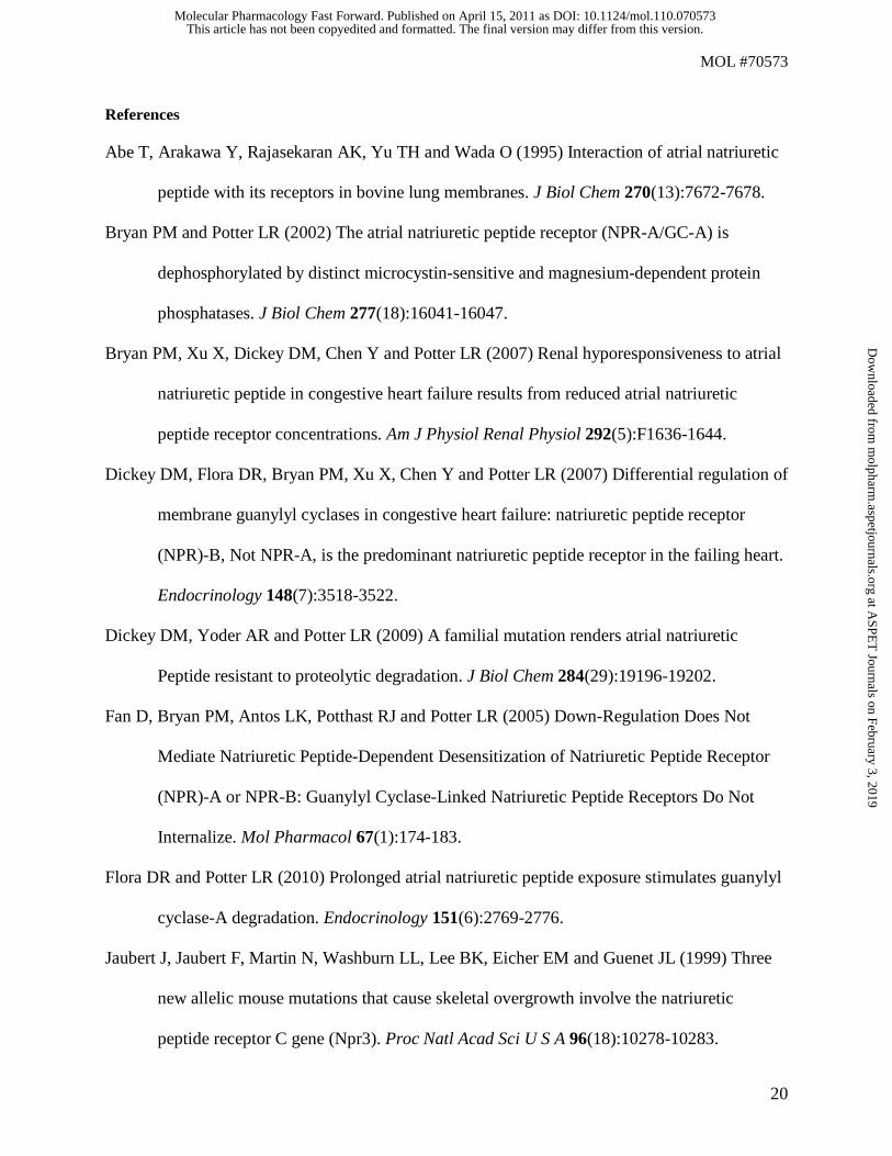
NPR-C, Not GC-A, Is the Major ANP Receptor in HeLa Cells. Because the 125I-ANP uptake curve in
the GFP transfected cells was similar to that reported for NPR-C (Nussenzveig et al., 1990) and different
from that observed in cells transfected with GC-A (Fig. 1A), we asked whether HeLa cells also express
NPR-C. 125I-ANP was chemically cross-linked to two independent HeLa cell lines. Reducing SDS-PAGE
fractionated membranes from cross-linked cells and labeled receptors were visualized by autoradiography
(Fig. 2). In both cell lines, the major 125I-ANP binding protein migrated at the molecular weight of NPR-C
(~60 kDa) in the absence of unlabeled natriuretic peptides. In the presence of saturating concentrations of
cold ANP, no specific binding was observed, consistent with ANP blocking binding to both NPR-C and
GC-A. Addition of excess unlabeled CNP reduced 125I-ANP binding to NPR-C and increased binding to
GC-A. These data indicate that HeLa cells express much higher concentrations of NPR-C than GC-A and
that 125I-ANP preferentially binds NPR-C, not GC-A. Thus, the vast majority of the internalization
observed in the GFP-transfected HeLa cells in Fig. 1A was due to internalization of NPR-C, not GC-A.
This article has not been copyedited and formatted. The final version may differ from this version.Molecular Pharmacology Fast Forward. Published on April 15, 2011 as DOI: 10.1124/mol.110.070573
at ASPE
T Journals on February 3, 2019
molpharm
.aspetjournals.orgD
ownloaded from
MOL #70573
11
Development of 125I-IgG-based Intracellular Receptor Accumulation Assay. Because most cells, like
HeLa cells, express higher concentrations of NPR-C than GC-A, acid-resistant 125I-ANP uptake primarily
measures NPR-C internalization. Therefore, we developed a new assay that measures the uptake of a
single class of receptors in the presence or absence of ligand. HeLa cells were transfected with plasmids
expressing extracellular, amino-terminal FLAG-tagged versions of GC-A or NPR-C. Transfected cells
were successively incubated with mouse anti-FLAG M2 and 125I-conjugated anti-mouse IgG antibodies at
4°C to radioactively label surface receptors. Elevating the temperature of the cells to 37°C initiated the
internalization assay and acid washing at 4oC separated internalized receptors from surface receptors.
With this technique, 125I-IgG binding was dependent on expression of FLAG-GC-A as total counts were
more than ten-fold higher in cells transfected with FLAG-GC-A compared to cells transfected with GFP
(Fig. 3A). Binding was specific to the extracellular FLAG epitope because acid stripping removed the
vast majority of bound counts and inclusion of the FLAG peptide in the medium reduced total counts
from 10,525 cpm + 1,576 to 715 cpm + 39 (data not shown). Various primary and secondary antibody
concentrations were tested to optimize the assay. The final conditions chosen (labeled as 1X in Fig. 3A)
gave the highest signal to noise ratio while using the least amount of 125I-IgG. These conditions do not
saturate all secondary-binding sites because this would have been cost prohibitive.
To rule out the possibility that receptor overexpression artificially reduced the rate or magnitude of
FLAG-GC-A internalization, 293 cells were transfected with increasing amounts of FLAG-GC-A plasmid
and receptor uptake rates were measured. Western blot analysis indicated that FLAG-GC-A expression
increased in proportion to the level of the transfected DNA. However, neither the rate nor magnitude of
FLAG-GC-A internalization was reduced in cells expressing higher levels of receptor (Fig. 3B). Thus, at
all conditions tested, the same ratio of internalized to surface receptors was observed, which indicates that
the 125I-IgG uptake assay accurately measured the fate of the average cell surface receptor.
This article has not been copyedited and formatted. The final version may differ from this version.Molecular Pharmacology Fast Forward. Published on April 15, 2011 as DOI: 10.1124/mol.110.070573
at ASPE
T Journals on February 3, 2019
molpharm
.aspetjournals.orgD
ownloaded from
MOL #70573
12
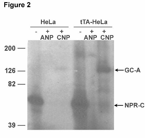
FLAG-GC-A Is Slowly Internalized in HeLa Cells. 125I-ANP uptake was much faster than 125I-IgG
uptake in FLAG-GC-A transfected tTA-HeLa cells, which is consistent with NPR-C, not GC-A,
mediating the majority of the 125I-ANP internalization in these cells (Fig. 4A). 125I-IgG uptake indicated
that basal FLAG-GC-A internalization was linear for 10-20 min with approximately 4% of the surface
receptors being internalized during this period of time (Fig. 4B). Multiple experiments determined that the
initial internalization rate was slow (0.2%/min ± 0.006, N = 4). Maximum receptor accumulation was
achieved between 10 and 20 min. Inclusion of 1 μM ANP in the assay had no effect on initial
internalization rates but increased accumulation after 1 h by 1.6-fold. To verify that the basal clathrin-
dependent internalization pathway in these cells was functional, 125I-transferrin uptake was measured and
found to be robust, rapid and saturable (Fig. 4C).
GC-A Internalization Is Rapid and ANP-Independent in 293 Cells. To measure internalization in cells
where GC-A is the only measurable 125I-ANP binding protein, FLAG-GC-A was stably expressed in 293
cells that do not endogenously express detectable levels of GC-A or NPR-C (Potter and Hunter, 1999). In
contrast to the HeLa cells, the uptake curves for 125I-ANP and 125IgG were virtually indistinguishable in
293 cells (Fig. 5A). Internalization was clearly mediated by GC-A because cells lacking FLAG-GC-A
failed to accumulate 125I-ANP or 125I-IgG (Fig. 5B and C, untransfected). Thus, 125I-IgG uptake faithfully
mirrors 125I-ANP internalization in these cells. Surprisingly, basal FLAG-GC-A internalization was eight-
fold higher and more robust in the 293 cells compared to the HeLa cells. Eighteen percent of the total cell
surface receptor population was internalized by 3 minutes at a rate of 4.9 %/min + 0.27. As in the HeLa
cells, ANP did not increase the initial rate of FLAG-GC-A internalization in the 293 cells, and unlike the
HeLa cells, had no effect on accumulation after 1 h (Fig. 5C).
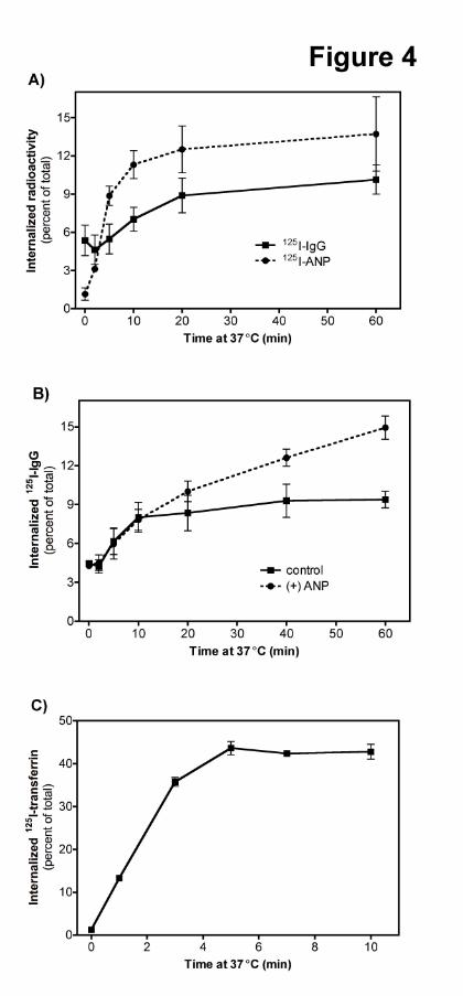
NPR-C Internalization Is Rapid and Ligand-Independent in HeLa and 293 Cells. NPR-C
internalization was also examined using the antibody-based assay. 125I-IgG was internalized at an initial
This article has not been copyedited and formatted. The final version may differ from this version.Molecular Pharmacology Fast Forward. Published on April 15, 2011 as DOI: 10.1124/mol.110.070573
at ASPE
T Journals on February 3, 2019
molpharm
.aspetjournals.orgD
ownloaded from
MOL #70573
13
rate of 2.4%/min + 0.01 in tTA-HeLa cells transfected with FLAG-NPR-C (Fig. 6A), which is similar to
the rate of internalization of 125I-ANP in GFP transfected cells (Fig. 1A). In 293 cells transiently
transfected with FLAG-NPR-C, receptor and initial ligand uptake were coincident with time. After 1 min,
the rate of 125I-ANP and 125I-IgG accumulation were 18.8%/min + 0.09 and 19.7%/min + 0.11,
respectively, and the percentage of surface receptor internalized as measured by 125I-IgG was increased to
25 to 40% depending on the assay (Fig. 6B). Neither ANP, BNP nor CNP increased NPR-C
internalization as measured by 125I-IgG uptake (Fig. 6C). Thus, the internalization of GC-A and NPR-C is
ligand-independent.
GC-A Downregulation Does Not Require Increased Internalization. We recently reported that GC-A
is downregulated in HeLa cells (Flora and Potter, 2010), but those experiments measured total receptor
levels in cell lysates. To specifically measure surface levels of receptor, HeLa cells were transiently
transfected with FLAG-GC-A and then incubated in the presence or absence of ANP for 8 h prior to
labeling with anti-FLAG/125I-IgG to measure surface receptor concentrations. Total counts (representing
surface labeled receptors) were reduced by 53% in cells exposed to ANP (data not shown), which is
consistent with previous studies showing that 8 h ANP exposure reduced total cellular GC-A
concentrations by nearly 60% in HeLa cells (Flora and Potter, 2010).
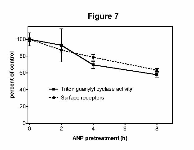
We also examined the downregulation of FLAG-GCA in stably expressing 293 cells. 293PMA-FLAG-
GCA cells were incubated at 37oC with ANP for 0, 2, 4 and 8 h to stimulate GC-A downregulation (Fig.
7). In one experiment, crude membranes were prepared and assayed for detergent-dependent guanylyl
cyclase activity to measure the effect of prior ANP exposure on total cellular receptor concentrations. In a
separate experiment, 125I-IgG binding was used to specifically measure receptors at the cell surface. Prior
ANP exposure significantly reduced both activities after 4 and 8 hours of exposure, and the reductions in
surface and total receptors were temporally correlated. These data indicate that GC-A downregulation
does not require increased receptor internalization, since ANP clearly stimulates downregulation but not
This article has not been copyedited and formatted. The final version may differ from this version.Molecular Pharmacology Fast Forward. Published on April 15, 2011 as DOI: 10.1124/mol.110.070573
at ASPE
T Journals on February 3, 2019
molpharm
.aspetjournals.orgD
ownloaded from

MOL #70573
14
internalization in 293 cells. Furthermore, it indicates that either the vast majority of receptors are at the
cell surface or that intracellular and extracellular pools of receptor rapidly exchange.
This article has not been copyedited and formatted. The final version may differ from this version.Molecular Pharmacology Fast Forward. Published on April 15, 2011 as DOI: 10.1124/mol.110.070573
at ASPE
T Journals on February 3, 2019
molpharm
.aspetjournals.orgD
ownloaded from

MOL #70573
15
Discussion
In this report, an antibody-based trafficking assay was developed that allowed the independent evaluation
of GC-A and NPR-C internalization for the first time. The validity of the 125I-IgG assay was established
by demonstrating identical uptake rates for 125I-IgG and 125I-ANP in cells expressing a single ANP-
binding receptor. All previous uptake studies followed receptors with 125I-ANP, which clearly measures
NRP-C, not GC-A, trafficking in HeLa cells. A major advantage of the 125I-IgG assay is that it measures a
single receptor class without complicating contributions from related receptors. Another advantage is that
it measures internalization rates of unbound receptors (basal rates), which was not possible using the 125I-
ANP binding approach. Although we demonstrated that 125I-ANP binding accurately measured GC-A and
NPR-C uptake in transfected 293 cells, 125I-ANP binding is unlikely to accurately measure GC-A uptake
in biologic systems because most, if not all, cells express higher levels of NPR-C than GC-A. Leitman
and colleagues studied eight cell lines and found that cells expressing GC-A also express higher
concentrations of NPR-C (Leitman et al., 1986). In contrast, Watt and Yip reported that HeLa cells only
express GC-A (Watt and Yip, 1989). However, our 125I-ANP chemical cross-linking studies indicated that
NPR-C, not GC-A, is the major ANP receptor in the two HeLa cell lines that we examined. We suggest
that past and future 125I-ANP uptake studies should be carefully interpreted so that NPR-C uptake is not
erroneously ascribed to GC-A.
Another clear conclusion from these studies is that cellular environment is a major determinant of
receptor uptake. In HeLa cells, GC-A internalization was very slow, whereas in 293 cells internalization
was eight times faster for both GC-A and NPR-C. The rapid internalization of 125I-ANP and 125I-
transferrin indicated that the meager FLAG-GC-A uptake in the HeLa cells was not due to a global
internalization defect caused by transfection but was due to a difference in the GC-A internalization
machinery. Likewise, the similar internalization rates of GC-A and NPR-C with either 125I-ANP or the
FLAG-based assay in the 293 cells suggest that the slow FLAG-GC-A internalization rate was not an
This article has not been copyedited and formatted. The final version may differ from this version.Molecular Pharmacology Fast Forward. Published on April 15, 2011 as DOI: 10.1124/mol.110.070573
at ASPE
T Journals on February 3, 2019
molpharm
.aspetjournals.orgD
ownloaded from

MOL #70573
16
artifact of the FLAG-tagged receptor and is representative of GC-A uptake in these cells. Additionally,
similar uptake rates in cells expressing various levels of receptors suggest that FLAG-GC-A uptake is
representative of the internalization of endogenous GC-A.
Another conclusion that can be drawn from these studies is that FLAG-NPR-C internalization was several
times faster than GC-A internalization regardless of cell line, which is consistent with unique trafficking
pathways mediating uptake of each receptor. Future work will focus on the identification of these
pathways. Surprisingly, ANP did not increase the internalization of either receptor in either cell line,
although it did increase total GC-A uptake at later times in the HeLa cells. The fact that ANP increased
uptake at later but not earlier time points is consistent with inhibition of recycling, not increased
internalization and is inconsistent with ANP binding increasing the ability of GC-A or NPR-C to attach to
cellular transport systems as occurs for other receptors.
As described in the introduction, many previous investigators have studied the trafficking of GC-A and/or
NPR-C. However, an important question pertaining to all previous studies is which receptor is being
measured. Our data suggest that previous internalization studies conducted on cells expressing both GC-A
and NPR-C, most likely measured NPR-C uptake. Nonetheless, regarding the debate over whether GC-A
internalizes 125I-ANP, it clearly does in the two cell lines that we tested. We do not know why our data
differ from a previous study showing that intracellular 125I-ANP radioactivity decreased rapidly in 293
cells transfected with GC-A (Pandey et al., 2002), but perhaps it is a function of the unique trafficking
properties of the individual cell lines.
Downregulation proceeds by three sequential processes: receptor internalization, endosomal sorting, and
receptor proteolysis (Katzmann et al., 2002). Downregulation of canonical G-protein coupled or tyrosine
kinase receptors is associated with receptor internalization rates that are increased several fold in response
to ligand binding (Vieira et al., 1996). However, since GC-A or NPR-C initial internalization rates were
This article has not been copyedited and formatted. The final version may differ from this version.Molecular Pharmacology Fast Forward. Published on April 15, 2011 as DOI: 10.1124/mol.110.070573
at ASPE
T Journals on February 3, 2019
molpharm
.aspetjournals.orgD
ownloaded from
MOL #70573
17
unchanged in the presence of ligand, we can conclude that increased uptake rates do not contribute to GC-
A downregulation in 293 cells. Furthermore, the similar loss of total and surface receptors indicates the
intracellular and extracellular pools of receptor are closely linked and rapidly exchange.
This article has not been copyedited and formatted. The final version may differ from this version.Molecular Pharmacology Fast Forward. Published on April 15, 2011 as DOI: 10.1124/mol.110.070573
at ASPE
T Journals on February 3, 2019
molpharm
.aspetjournals.orgD
ownloaded from
MOL #70573
18
Acknowledgments
We are grateful to Sean Conner for numerous reagents including the tTA-HeLa cells and advice regarding
these studies. We thank Christopher M. McGuirk for making the 293PMA-FLAG-GCA stable cell line
and Kathryn A. Barbieri for assistance in making the pCMV1-FLAG-NPR-C construct.
This article has not been copyedited and formatted. The final version may differ from this version.Molecular Pharmacology Fast Forward. Published on April 15, 2011 as DOI: 10.1124/mol.110.070573
at ASPE
T Journals on February 3, 2019
molpharm
.aspetjournals.orgD
ownloaded from
MOL #70573
19
Authorship Contribution
Participated in research design: Dickey, Flora, and Potter.
Conducted experiments: Dickey and Flora.
Performed data analysis: Dickey and Flora.
Wrote or contributed to the writing of the manuscript: Dickey, Flora, and Potter.
Other: Flora and Potter acquired funding for the research.
This article has not been copyedited and formatted. The final version may differ from this version.Molecular Pharmacology Fast Forward. Published on April 15, 2011 as DOI: 10.1124/mol.110.070573
at ASPE
T Journals on February 3, 2019
molpharm
.aspetjournals.orgD
ownloaded from
MOL #70573
20
References
Abe T, Arakawa Y, Rajasekaran AK, Yu TH and Wada O (1995) Interaction of atrial natriuretic
peptide with its receptors in bovine lung membranes. J Biol Chem 270(13):7672-7678.
Bryan PM and Potter LR (2002) The atrial natriuretic peptide receptor (NPR-A/GC-A) is
dephosphorylated by distinct microcystin-sensitive and magnesium-dependent protein
phosphatases. J Biol Chem 277(18):16041-16047.
Bryan PM, Xu X, Dickey DM, Chen Y and Potter LR (2007) Renal hyporesponsiveness to atrial
natriuretic peptide in congestive heart failure results from reduced atrial natriuretic
peptide receptor concentrations. Am J Physiol Renal Physiol 292(5):F1636-1644.
Dickey DM, Flora DR, Bryan PM, Xu X, Chen Y and Potter LR (2007) Differential regulation of
membrane guanylyl cyclases in congestive heart failure: natriuretic peptide receptor
(NPR)-B, Not NPR-A, is the predominant natriuretic peptide receptor in the failing heart.
Endocrinology 148(7):3518-3522.
Dickey DM, Yoder AR and Potter LR (2009) A familial mutation renders atrial natriuretic
Peptide resistant to proteolytic degradation. J Biol Chem 284(29):19196-19202.
Fan D, Bryan PM, Antos LK, Potthast RJ and Potter LR (2005) Down-Regulation Does Not
Mediate Natriuretic Peptide-Dependent Desensitization of Natriuretic Peptide Receptor
(NPR)-A or NPR-B: Guanylyl Cyclase-Linked Natriuretic Peptide Receptors Do Not
Internalize. Mol Pharmacol 67(1):174-183.
Flora DR and Potter LR (2010) Prolonged atrial natriuretic peptide exposure stimulates guanylyl
cyclase-A degradation. Endocrinology 151(6):2769-2776.
Jaubert J, Jaubert F, Martin N, Washburn LL, Lee BK, Eicher EM and Guenet JL (1999) Three
new allelic mouse mutations that cause skeletal overgrowth involve the natriuretic
peptide receptor C gene (Npr3). Proc Natl Acad Sci U S A 96(18):10278-10283.
This article has not been copyedited and formatted. The final version may differ from this version.Molecular Pharmacology Fast Forward. Published on April 15, 2011 as DOI: 10.1124/mol.110.070573
at ASPE
T Journals on February 3, 2019
molpharm
.aspetjournals.orgD
ownloaded from

MOL #70573
21
Katzmann DJ, Odorizzi G and Emr SD (2002) Receptor downregulation and multivesicular-body
sorting. Nat Rev Mol Cell Biol 3(12):893-905.
Koh GY, Nussenzveig DR, Okolicany J, Price DA and Maack T (1992) Dynamics of atrial
natriuretic factor-guanylate cyclase receptors and receptor-ligand complexes in cultured
glomerular mesangial and renomedullary interstitial cells. J Biol Chem 267(17):11987-
11994.
Leitman DC, Andresen JW, Kuno T, Kamisaki Y, Chang JK and Murad F (1986) Identification
of multiple binding sites for atrial natriuretic factor by affinity cross-linking in cultured
endothelial cells. J Biol Chem 261(25):11650-11655.
Lopez MJ, Wong SK, Kishimoto I, Dubois S, Mach V, Friesen J, Garbers DL and Beuve A
(1995) Salt-resistant hypertension in mice lacking the guanylyl cyclase-A receptor for
atrial natriuretic peptide. Nature 378(6552):65-68.
Matsukawa N, Grzesik WJ, Takahashi N, Pandey KN, Pang S, Yamauchi M and Smithies O
(1999) The natriuretic peptide clearance receptor locally modulates the physiological
effects of the natriuretic peptide system. Proc Natl Acad Sci U S A 96(13):7403-7408.
Nussenzveig DR, Lewicki JA and Maack T (1990) Cellular mechanisms of the clearance
function of type C receptors of atrial natriuretic factor. J Biol Chem 265(34):20952-
20958.
Oliver PM, Fox JE, Kim R, Rockman HA, Kim HS, Reddick RL, Pandey KN, Milgram SL,
Smithies O and Maeda N (1997) Hypertension, cardiac hypertrophy, and sudden death in
mice lacking natriuretic peptide receptor A. Proc Natl Acad Sci U S A 94(26):14730-
14735.
This article has not been copyedited and formatted. The final version may differ from this version.Molecular Pharmacology Fast Forward. Published on April 15, 2011 as DOI: 10.1124/mol.110.070573
at ASPE
T Journals on February 3, 2019
molpharm
.aspetjournals.orgD
ownloaded from

MOL #70573
22
Pandey KN (1992) Kinetic analysis of internalization, recycling and redistribution of atrial
natriuretic factor-receptor complex in cultured vascular smooth-muscle cells. Ligand-
dependent receptor down-regulation. Biochem J 288(Pt 1):55-61.
Pandey KN (2001) Dynamics of internalization and sequestration of guanylyl cyclase/atrial
natriuretic peptide receptor-A. Can J Physiol Pharmacol 79(8):631-639.
Pandey KN, Nguyen HT, Sharma GD, Shi SJ and Kriegel AM (2002) Ligand-regulated
internalization, trafficking, and down-regulation of guanylyl cyclase/atrial natriuretic
peptide receptor-A in human embryonic kidney 293 cells. J Biol Chem 277(7):4618-
4627.
Potter LR (2011) Regulation and therapeutic targeting of peptide-activated receptor guanylyl
cyclases. Pharmacology & therapeutics 130(1):71-82.
Potter LR and Hunter T (1999) A constitutively "Phosphorylated" guanylyl cyclase-linked atrial
natriuretic peptide receptor mutant is resistant to desensitization. Mol Biol Cell
10(6):1811-1820.
Potter LR, Yoder AR, Flora DR, Antos LK and Dickey DM (2009) Natriuretic peptides: their
structures, receptors, physiologic functions and therapeutic applications. Handb Exp
Pharmacol(191):341-366.
Potthast R, Abbey-Hosch SE, Antos LK, Marchant JS, Kuhn M and Potter LR (2004) Calcium-
dependent Dephosphorylation Mediates the Hyperosmotic and Lysophosphatidic Acid-
dependent Inhibition of Natriuretic Peptide Receptor-B/Guanylyl Cyclase-B. J Biol Chem
279(47):48513-48519.
Rathinavelu A and Isom GE (1991) Differential internalization and processing of atrial-
natriuretic-factor B and C receptor in PC12 cells. Biochem J 276(Pt 2):493-497.
This article has not been copyedited and formatted. The final version may differ from this version.Molecular Pharmacology Fast Forward. Published on April 15, 2011 as DOI: 10.1124/mol.110.070573
at ASPE
T Journals on February 3, 2019
molpharm
.aspetjournals.orgD
ownloaded from

MOL #70573
23
Sever S, Damke H and Schmid SL (2000) Dynamin:GTP controls the formation of constricted
coated pits, the rate limiting step in clathrin-mediated endocytosis. J Cell Biol
150(5):1137-1148.
Vieira AV, Lamaze C and Schmid SL (1996) Control of EGF receptor signaling by clathrin-
mediated endocytosis. Science 274(5295):2086-2089.
Vieira MA, Gao M, Nikonova LN and Maack T (2001) Molecular and cellular physiology of the
dissociation of atrial natriuretic peptide from guanylyl cyclase a receptors. J Biol Chem
276(39):36438-36445.
Watt VM and Yip CC (1989) HeLa cells contain the atrial natriuretic peptide receptor with
guanylate cyclase activity. Biochem Biophys Res Commun 164(2):671-677.
This article has not been copyedited and formatted. The final version may differ from this version.Molecular Pharmacology Fast Forward. Published on April 15, 2011 as DOI: 10.1124/mol.110.070573
at ASPE
T Journals on February 3, 2019
molpharm
.aspetjournals.orgD
ownloaded from

MOL #70573
24
Footnotes
This work was supported by the American Heart Association [Grants 0815607G, 0950053G].
Address reprint requests to:
Lincoln Potter
University of Minnesota - Twin Cities
6-155 Jackson Hall
321 Church St SE
Minneapolis, MN, USA 55455
Tel: 612-624-7251
Fax: 612-624-7282
Email: [email protected]
This article has not been copyedited and formatted. The final version may differ from this version.Molecular Pharmacology Fast Forward. Published on April 15, 2011 as DOI: 10.1124/mol.110.070573
at ASPE
T Journals on February 3, 2019
molpharm
.aspetjournals.orgD
ownloaded from

MOL #70573
25
Legends for figures
Fig 1. (A)125I-ANP uptake in tTA-HeLa cells transiently transfected with GFP or FLAG-GC-A. Cells
were labeled with subsaturating concentrations of 125I-ANP at 4°C. Aliquots of labeled cells were
incubated at 37°C for the times indicated before acid washing and counting. Values represent average ±
the range of the determinations where N = 2. The graph is representative of multiple experiments. (B)
FLAG-GC-A and wild type GC-A bind and are activated by ANP similarly. 293 cells were transiently
transfected with wild-type GC-A or FLAG-GC-A and incubated with increasing concentrations of ANP
for 1 min. Cellular cGMP concentrations were measured and plotted as a function of peptide
concentration. The data points represent the mean ± the SEM assayed in triplicate. (C) FLAG-GC-A has
similar affinity for ANP as wild-type GC-A. Transiently transfected 293 cells were incubated for 1 hr at
4°C with 125I-ANP in the presence or absence of increasing concentrations of unlabeled ligand.
Specifically bound 125I-ANP was plotted as a function of competing peptide concentration. The data
points represent the mean ± the SEM assayed in triplicate.
Fig 2. HeLa cells endogenously express high and low levels of NPR-C and GC-A, respectively. HeLa and
tTA-HeLa cells were incubated with 125I-ANP in the absence or presence of 1 μM ANP or CNP for 2 hr at
4°C before cross-linking 125I-ANP to the receptors with disuccinimidyl suberate. Membrane fractions
were separated by SDS-PAGE and 125I-ANP-receptor complexes were visualized by autoradiography.
Fig 3. The 125I-IgG uptake assay specifically measures FLAG-GC-A internalization. (A) tTA-Hela cells
were transiently transfected with GFP or FLAG-GCA. The cells were dispensed into tubes and incubated
with 0.05 μl (1X primary) or 0.1 μl (20X primary) anti-FLAG-M2 antibody. Excess antibody was
removed before addition of 5 (1X), 50 (10X secondary) or 100 μl 125I-IgG. Cellular radioactivity was
measured directly or after acid stripping to remove surface 125I-IgG. Values represent the range of
This article has not been copyedited and formatted. The final version may differ from this version.Molecular Pharmacology Fast Forward. Published on April 15, 2011 as DOI: 10.1124/mol.110.070573
at ASPE
T Journals on February 3, 2019
molpharm
.aspetjournals.orgD
ownloaded from

MOL #70573
26
determinations where N =2. The graph is representative of more than 3 experiments. (B) 293 cells were
transiently transfected with 10 μg (1x), 1 μg or 5 μg of FLAG-GC-A plasmid DNA. Internalization
assays were performed 48 h later. An equal number of cells from each transfection were separated by
SDS-PAGE, blotted to an Immobilon membrane and GC-A expression was detected by Western blot
using an anti-GC-A antibody (inset). Values represent average ± SEM where N = 6.
Fig 4. GC-A is slowly internalized in HeLa cells. (A) tTA-HeLa cells transiently transfected with FLAG-
GC-A were labeled with 125I-ANP or anti-FLAG-M2 antibody followed by 125I-anti-mouse-IgG at 4°C.
Values represent average ± SEM where N = 4 (B) ANP increases GC-A uptake at longer but not shorter
periods of time. Cells transfected with FLAG-GC-A were labeled with anti-FLAG-M2 antibody and 125I-
anti-mouse-IgG before incubation at 37°C in the absence or presence of 1 μM ANP for the periods of
time shown. Samples were then acid-washed and counted. Values represent average ± SEM where N = 6
(C) 125I-transferrin is rapidly internalized in tTA-HeLa cells. Aliquots of the tTA-HeLa cells transfected
with FLAG-GC-A were labeled with 125I-transferrin at 4°C. Internalized 125I-transferrin was determined
after the indicated periods of time at 37°C. Values represent the average ± SEM where N = 8.
Fig 5. GC-A is rapidly internalized in 293 PMA cells. (A) 293 cells stably expressing FLAG-GC-A were
incubated at 4°C with either 125I-ANP or anti-FLAG antibody followed by 125I-anti-mouse IgG. Cells
were incubated at 37°C for the indicated times before acid-washing and counting. Values represent
average ± SEM where N =14 (B) Untransfected 293 cells or 293 cells stably expressing FLAG-GC-A
were labeled at 4°C with 125I-ANP. Internalized radioactivity as a function of time at 37°C is shown.
Values represent average ± the range of two determinations (C) Untransfected or 293 cells stably
expressing FLAG-GC-A were labeled at 4°C with anti-FLAG antibody followed by 125I-IgG secondary
antibody. The cells were incubated at 37°C in the presence or absence of 1 μM ANP for the indicated
periods of time. Values represent the average ± the range determinations where N = 2.
This article has not been copyedited and formatted. The final version may differ from this version.Molecular Pharmacology Fast Forward. Published on April 15, 2011 as DOI: 10.1124/mol.110.070573
at ASPE
T Journals on February 3, 2019
molpharm
.aspetjournals.orgD
ownloaded from

MOL #70573
27
Fig 6. NPR-C is rapidly and constitutively internalized in HeLa and 293 cells. (A) tTA-HeLa cells were
transiently transfected with FLAG-NPR-C. Cells were then labeled with anti-FLAG antibody followed by
125I-anti-mouse-IgG at 4°C. Cells were incubated at 37°C for the indicated times before acid washing.
Values represent the average ± SEM where N = 14. (B) 293 cells were transfected with FLAG-NPR-C
and labeled with either 125I-ANP or 125I-IgG at 4°C. Aliquots were incubated at 37°C for the times shown
and before acid washing. Values represent the average ± SEM where N = 6 (C) 293 cells transiently
transfected with FLAG-NPR-C were labeled with anti-FLAG antibody and 125I-IgG secondary antibody at
4°C. Aliquots were incubated at 37°C in the presence or absence of 1 μM ANP, BNP or CNP for the
times indicated where N = 4.
Fig 7. Concomitant downregulation of extracellular and intracellular FLAG-GC-A in 293 cells. 293 cells
stably expressing FLAG-GC-A were incubated with 10 μg/ml cycloheximide in the absence or presence
of 200 nM ANP for the period of times indicated. In one experiment, crude membranes were prepared
and then assayed for guanylyl cyclase activity in the presence of 1 % Triton X-100 and Mn2+GTP. In a
second experiment, cells were incubated with ANP as described above and then labeled with anti-FLAG
antibody followed by 125I-IgG at 4ºC. Total 125I-IgG radioactivity and guanylyl cyclase activities were
normalized to activities obtained from cells not incubated with ANP (control) and plotted as a function of
time of ANP exposure.
This article has not been copyedited and formatted. The final version may differ from this version.Molecular Pharmacology Fast Forward. Published on April 15, 2011 as DOI: 10.1124/mol.110.070573
at ASPE
T Journals on February 3, 2019
molpharm
.aspetjournals.orgD
ownloaded from

This article has not been copyedited and formatted. The final version may differ from this version.Molecular Pharmacology Fast Forward. Published on April 15, 2011 as DOI: 10.1124/mol.110.070573
at ASPE
T Journals on February 3, 2019
molpharm
.aspetjournals.orgD
ownloaded from
This article has not been copyedited and formatted. The final version may differ from this version.Molecular Pharmacology Fast Forward. Published on April 15, 2011 as DOI: 10.1124/mol.110.070573
at ASPE
T Journals on February 3, 2019
molpharm
.aspetjournals.orgD
ownloaded from

This article has not been copyedited and formatted. The final version may differ from this version.Molecular Pharmacology Fast Forward. Published on April 15, 2011 as DOI: 10.1124/mol.110.070573
at ASPE
T Journals on February 3, 2019
molpharm
.aspetjournals.orgD
ownloaded from
This article has not been copyedited and formatted. The final version may differ from this version.Molecular Pharmacology Fast Forward. Published on April 15, 2011 as DOI: 10.1124/mol.110.070573
at ASPE
T Journals on February 3, 2019
molpharm
.aspetjournals.orgD
ownloaded from

This article has not been copyedited and formatted. The final version may differ from this version.Molecular Pharmacology Fast Forward. Published on April 15, 2011 as DOI: 10.1124/mol.110.070573
at ASPE
T Journals on February 3, 2019
molpharm
.aspetjournals.orgD
ownloaded from
This article has not been copyedited and formatted. The final version may differ from this version.Molecular Pharmacology Fast Forward. Published on April 15, 2011 as DOI: 10.1124/mol.110.070573
at ASPE
T Journals on February 3, 2019
molpharm
.aspetjournals.orgD
ownloaded from
This article has not been copyedited and formatted. The final version may differ from this version.Molecular Pharmacology Fast Forward. Published on April 15, 2011 as DOI: 10.1124/mol.110.070573
at ASPE
T Journals on February 3, 2019
molpharm
.aspetjournals.orgD
ownloaded from