antibody-mediated neutralization of human rhinovirus 14...
TRANSCRIPT
JOURNAL OF VIROLOGY,0022-538X/98/$04.0010
June 1998, p. 4610–4622 Vol. 72, No. 6
Copyright © 1998, American Society for Microbiology
Antibody-Mediated Neutralization of Human Rhinovirus 14Explored by Means of Cryoelectron Microscopy and
X-Ray Crystallography of Virus-Fab ComplexesZHIWEI CHE,1 NORMAN H. OLSON,1 DONNA LEIPPE,2 WAI-MING LEE,2 ANNE G. MOSSER,2
ROLAND R. RUECKERT,2 TIMOTHY S. BAKER,1 AND THOMAS J. SMITH1*
Department of Biological Sciences, Purdue University, West Lafayette, Indiana 47907,1 andInstitute of Molecular Virology, University of Wisconsin, Madison, Wisconsin 537062
Received 30 October 1997/Accepted 12 February 1998
The structures of three different human rhinovirus 14 (HRV14)-Fab complexes have been explored withX-ray crystallography and cryoelectron microscopy procedures. All three antibodies bind to the NIm-IA site ofHRV14, which is the b-B–b-C loop of the viral capsid protein VP1. Two antibodies, Fab17-IA (Fab17) andFab12-IA (Fab12), bind bivalently to the virion surface and strongly neutralize viral infectivity whereasFab1-IA (Fab1) strongly aggregates and weakly neutralizes virions. The structures of the two classes ofvirion-Fab complexes clearly differ and correlate with observed binding neutralization differences. Fab17 andFab12 bind in essentially identical, tangential orientations to the viral surface, which favors bidentate bindingover icosahedral twofold axes. Fab1 binds in a more radial orientation that makes bidentate binding unlikely.Although the binding orientations of these two antibody groups differ, nearly identical charge interactionsoccur at all paratope-epitope interfaces. Nucleotide sequence comparisons suggest that Fab17 and Fab12 arefrom the same progenitor cell and that some of the differing residues contact the south wall of the receptorbinding canyon that encircles each of the icosahedral fivefold vertices. All of the antibodies contact a significantproportion of the canyon region and directly overlap much of the receptor (intercellular adhesion molecule 1[ICAM-1]) binding site. Fab1, however, does not contact the same residues on the upper south wall (the sidefacing away from fivefold axes) at the receptor binding region as do Fab12 and Fab17. All three antibodiescause some stabilization of HRV14 against pH-induced inactivation; thus, stabilization may be mediated byinvariant contacts with the canyon.
Picornaviruses are among the largest of animal virus familiesand include the well-known poliovirus, rhinovirus, foot-and-mouth disease virus (FMDV), coxsackievirus, and hepatitis Avirus. The rhinoviruses, of which there are more than 100serotypes subdivided into two groups, are major causativeagents of the common cold in humans (42). The viruses arenonenveloped and have an ;300-Å-diameter protein shell thatencapsidates a single-stranded, plus-sense RNA genome ofabout 7,200 bases. The human rhinovirus 14 (HRV14) capsidexhibits a pseudo-T53 (P53) icosahedral symmetry and con-sists of 60 copies each of four viral proteins, VP1, VP2, VP3,and VP4, with VP4 at the RNA-capsid interface (40). An;20-Å deep canyon lies roughly at the junction of VP1 (form-ing the north rim) with VP2 and VP3 (forming the south rim)and surrounds each of the 12 icosahedral fivefold vertices. Thecanyon regions of HRV14 and HRV16, both major receptorgroup rhinoviruses, were shown to contain the binding site ofthe cellular receptor, intercellular adhesion molecule 1(ICAM-1) (8, 24a, 37). Four major neutralizing immunogenic(NIm) sites, NIm-IA, NIm-IB, NIm-II, and NIm-III, wereidentified by studies of neutralization escape mutants withmonoclonal antibodies (MAbs) (46, 47) and then mapped tofour protruding regions on the viral surface (40).
Several mechanisms of antibody-mediated neutralizationhave been proposed. Perhaps the simplest is based on aggre-gation of virions (5, 53, 54), which generally occurs over a
narrow range of antibody/virus ratios. This limited range hasraised questions about the role of aggregation in vivo. Alter-native suggestions are that antibodies may neutralize virions byinducing extensive conformational changes in the capsid (15,29), abrogate virus attachment to the host cell (8, 14), orprevent uncoating (57). There is no universal acceptance of asingle neutralization mechanism, and the various MAbs mayneutralize with different combinations of these mechanisms.
Neutralizing MAbs against HRV14 have been divided intothree groups: strong, intermediate, and weak neutralizers (26,34). All strongly neutralizing antibodies bind to the NIm-IAsite, which was defined by natural escape mutations at residuesD1091 and E1095 of VP1 on the loop between the b-B and b-Cstrands of the VP1 b-barrel (the letter designates the aminoacid, the first digit identifies the viral protein, and the remain-ing three digits specify the sequence number). Becausestrongly neutralizing antibodies form stable, monomeric virus-antibody complexes with a maximum stoichiometry of 30 an-tibodies per virion, it was concluded that they bind bivalently tothe virions (26, 34). Weakly neutralizing antibodies form un-stable, monomeric complexes with HRV14 and bind with astoichiometry of ;60 antibodies per virion (26, 52). The re-maining antibodies, all of which precipitate the virions, areclassified as intermediate neutralizers (26, 34).
The structures of two complexes, the strongly neutralizingantibody MAb17-IA and its Fab fragment, Fab17, bound toHRV14, were determined by means of cryo-transmission elec-tron microscopy (cryo-TEM) and three-dimensional image re-construction (51, 52) and interpreted on the basis of model-building studies that used the atomic structures of HRV14 (40)and Fab17 (28). These studies showed that no observable con-
* Corresponding author. Mailing address: Department of BiologicalSciences, Lilly Hall of Life Sciences, Purdue University, West Lafay-ette, IN 47907-1392. Phone: (765) 494-8038. Fax: (765) 496-1189. E-mail: [email protected].
4610

formational changes were induced in the viral capsid upon Fabor MAb binding. Modeling and site-directed mutagenesis stud-ies demonstrated that electrostatic interactions play a key rolein the binding of Fab17 to HRV14 (52). In the complex, theloop of the NIm-IA site on HRV14 sits clamped in the cleftbetween the heavy- and light-chain hypervariable regions andforms complementary electrostatic interactions with Lys58H
(on the heavy chain) and Arg91L (on the light chain) of Fab17.In addition, a cluster of lysines on HRV14 (K1236, K1097, andK1085) interact with two acidic residues, Asp45H and Asp54H,in the CDR2 (CDR stands for complementarity-determiningregion) of the Fab heavy chain (49). Earlier modeling studiesalso suggested that bidentate binding of MAb17-IA to HRV14is facilitated by rotation of the Fab constant domains about theelbow axes towards the viral twofold axes (51). This suggestedthat the flexibility of the elbow region (the junction betweenthe variable and constant domains) plays a role in the bivalentbinding process, which in turn increases antibody avidity. Fi-nally, the 4-Å-resolution crystal structure of the Fab17-HRV14complex clearly showed that the virion does not undergo con-formational changes upon Fab binding (49). This crystal struc-ture determination also revealed that the earlier docking of theHRV14 and Fab17 atomic structures into the 22-Å cryo-TEMdensity map (50) yielded a pseudo-atomic model that was veryclose to the real structure of the complex.
We have expanded our complementary X-ray crystallogra-phy and cryo-TEM microscopy studies to examine the struc-tures of two more Fab-virus complexes, using Fab fragmentsfrom two other NIm-IA antibodies, MAb1-IA (MAb1) andMAb12-IA (MAb12), bound to HRV14. MAb1 and MAb12are weak and strong neutralizing antibodies, respectively. Im-age reconstructions of these two complexes are interpreted onthe basis of pseudo-atomic models, which substantiate the pre-vious hypothesis that neutralizing efficacy and binding valencyare interrelated (34). Electrostatic interactions at the epitope-paratope interface are highly conserved and apparently impor-tant for the antibody binding to the virion surface. Like Fab17,Fab1 and Fab12 penetrate the canyon. There are, however,differences between the orientations of the strongly and weaklyneutralizing antibodies and in the contacts made with the re-ceptor binding region of the canyon. Finally, data suggestingthat antibody binding to HRV14 is alone sufficient for neutral-ization and that other possible mechanisms are not requiredare presented.
MATERIALS AND METHODS
Preparation of MAb1-IA and MAb12-IA. MAb1-IA and MAb12-IA were pro-duced as previously described (52) and were found to be immunoglobulin G1(IgG1) and IgG2a, respectively, by using Screentype (Boehringer MannheimBiochemicals, Indianapolis, Ind.). High-glucose Dulbecco’s modified Eagle me-dium (catalog no. 430-2100; GIBCO/Bethesda Research Laboratories, GrandIsland, N.Y.) containing 10% fetal bovine serum was used for the hybridoma cellcultures grown in the Cellmax Quad 4 cell culture system (Cellco Corp., Ger-mantown, Md.). Cells were removed from the culture aliquots with 10-mincentrifugations at 10,000 3 g. Antibodies were precipitated from the cellularsupernatant by using a 50% (final concentration) saturated solution of ammo-nium sulfate and collected with 10-min centrifugations at 10,000 3 g. The pre-cipitate was resuspended and dialyzed with 0.1 M sodium phosphate (pH 7.0)buffer. Aliquots were loaded onto a protein G affinity column (Pharmacia/LKBCorp., Piscataway, N.J.) and washed with 0.1 M sodium phosphate (pH 7.0), andantibodies were eluted with 50 mM sodium acetate buffer (pH 2.0). The pH ofthe eluant was immediately brought back up to neutrality by collecting fractionsin tubes containing 1 M sodium phosphate (pH 9.0). The antibody samples werepooled and dialyzed against 0.1 M sodium phosphate (pH 7.0).
Antibody neutralization plaque assays. Two different protocols were used toascertain the efficacy of the antibodies on virus neutralization prior to uncoating(method A) and the ability of the antibodies to prevent plaque formation (meth-od B).
(i) Method A. Samples of purified HRV14 (at ;5 3 106 PFU/ml) wereincubated with various concentrations of purified antibodies in phosphate-buff-
ered saline with bovine serum albumin (PBSA) for 1 h at room temperature andthen overnight at 4°C. These samples were then serially diluted in PBSA, and200-ml aliquots were added to monolayers of HeLa cells. After incubation for 1 hat room temperature to allow for viral attachment to the cells, each plate wasrinsed with 2.5 ml of PBSA. The monolayers were then covered with 2.5 ml of0.8% agar in medium P6. The hardened agar was then covered with 2.5 ml ofmedium P6 supplemented with 4 mM glutamine, 4 mM oxaloacetic acid, 2 mMpyruvate, and 0.2% glucose. Plates were incubated in 5% CO2 for 48 h at 35°C,and plaques were visualized by removing the overlays, staining the cell mono-layers with 0.5% crystal violet in 20% ethanol, and rinsing with water.
(ii) Method B. The ability of antibodies to inhibit plaque formation was testedby maintaining a high concentration of antibodies in the plaque assay. Seriallydiluted samples of HRV14 were allowed to attach to monolayers of HeLa cellsfor 1 h at room temperature. The monolayers were then covered with the agarand medium overlays as described above with the exception that antibodies wereadded to the overlays (;28-mg/ml final concentration) rather than being washedaway. Subsequent incubation and visualization were performed as in method A.
Generation and purification of Fab1 and Fab12. Fab fragments were gener-ated by papain cleavage at a 1:50 (wt/wt) enzyme-to-antibody ratio for 12 h at37°C in the presence of 30 mM b-mercaptoethanol. The reaction was quenchedby adding iodoacetamide to give a final concentration of 75 mM. After extensivedialysis (.3 changes of buffer every 8 h) against 20 mM Tris (pH 7.5), thedigested sample was purified by using a Mono-Q anion-exchange column on afast protein liquid chromatography system (Pharmacia/LKB). Pure Fab frag-ments eluted in the void volume, whereas Fc fragments and intact antibodieseluted at 0.1 to 0.2 M NaCl. The Fabs were pooled and concentrated withCentricon 10 microconcentrators (Amicon Corporation, Beverly, Mass.).
Preparation of HRV14-Fab1 and HRV14-Fab12 complexes. Fabs and HRV14(prepared as previously described [16]) were mixed at a ratio of four Fab mol-ecules per NIm-IA site (240 Fabs per virion). The mixture was incubated at 4°Covernight and then passed through a Superose 6 column (Pharmacia/LKB)equilibrated with 50 mM sodium phosphate buffer (pH 7.0) to separate thecomplex and the unbound Fab molecules. Pure complexes were concentrated to;10 mg/ml with Centricon 10 microconcentrators (Amicon Corp.).
Cryo-TEM and image reconstructions of HRV14-Fab1 and HRV14-Fab12complexes. Cryo-TEM of the HRV14-Fab complexes was performed essentiallyas previously described (3, 7, 36, 49). Micrographs were recorded at a magnifi-cation of 349,000, at ;1.2 mm for Fab1 and ;1.3 mm underfocus for Fab12, andunder minimal dose conditions (;20 e2/Å2), on an EM420 electron microscope(Philips Electronic Instruments, Mahwah, N.J.) equipped with a 626 cryotransferholder (Gatan, Warrendale, Pa.). Twenty-six images of the Fab1 complexes and41 images of the Fab12 complexes were used to calculate 3D reconstructionsaccording to established protocols (2, 17). Effective resolutions of 30 and 27 Åwere achieved for the Fab1 and Fab12 reconstructions, respectively. The calcu-lated eigenvalues of each data set exceeded 10.0, which indicated that randomand unique data were used for each reconstruction (11). The reconstructionswere then corrected for effects of the phase-contrast transfer function as de-scribed previously (52).
Sequence determinations of Fab1 and Fab12. The amino acid sequences of thevariable domains of Fab1 and Fab12 were derived from cDNAs by using theMouse Ig-Prime kit (Novagen, Madison, Wis.). Total RNAs from MAb1 andMAb12 hybridoma cells were isolated by the rapid GuSCN method, which issimilar to conventional phenol-chloroform extraction. cDNAs for both lightchains were synthesized and amplified by PCR with the primer pair MuIgkVL59-G–MuIgkVL39-1, whereas the primer pairs MuIgVH59-B–MuIgGVH39-2 andMuIgVH59-C–MuIgGVH39-2 were used in the synthesis and PCR amplificationof the heavy chains of Fab1 and Fab12, respectively. The PCR products werecloned into a T-A vector (pT7BlueT) and then transformed into competentEscherichia coli (NovaBlue). E. coli containing plasmids with cDNA insertsproduced white colonies after the transformed competent cells were plated ontoampicillin–X-Gal (5-bromo-4-chloro-3-indolyl-b-D-galactopyranoside) plates.These plasmids were extracted and purified from the minipreps grown out of thewhite colonies. Double-stranded DNA sequencing was then performed with aSequenase version 2.0 DNA sequencing kit (United States Biochemical, Cleve-land, Ohio). Each sequence was determined from at least three independentPCRs. The amino acid sequences were derived from the nucleotide sequencesand aligned to the Fab17 sequence according to previously published nomencla-ture (23).
Fab1 crystal structure determination. Details of the X-ray crystallographicstructure determination of Fab1 will be published elsewhere. Briefly, Fab1 crys-tals were obtained by the sitting-drop method with 18 to 22% polyethylene glycol8000, 0.1 M sodium phosphate buffer (pH 6.0), and 1% 2-methyl-2,4-pentanedioland an Fab1 concentration of 18 mg/ml. Crystals appeared within a day andcontinued growing for a week to maximum dimensions of 0.5 to 0.7 mm. Oscil-lation X-ray diffraction data were collected from two crystals, using an R-axisimaging detector and a Rigaku X-ray generator. The oscillation angle was 1°, andthe exposure time was 10 min for each image. The intensities were integrated andmerged by using the programs DENZO and SCALEPACK (38). The final dataset extended to a 2.7-Å resolution. The crystal belonged to space group P21212,with unit cell dimensions of a 5 92.17, b 5 135.95, and c 5 81.08 Å and two Fabmolecules in the asymmetric unit. The structure was determined by the molec-ular replacement method (41) with the Fab17 structure (28) as the initial phasing
VOL. 72, 1998 STRUCTURES OF HRV14-Fab COMPLEXES 4611

model. Rotation and translation functions were calculated with a combination ofX-PLOR (6) and AMORE (34a) in the CCP4 program suite (1). The model wasbuilt by using “O” (22), taking into account the differences in Fab17 and Fab1sequences, and was refined with X-PLOR with all reflections in the range of 6 to2.7 Å. The final R factor was 16.9% (Rfree 5 26.6%), with root mean squaredeviations in bond lengths and angles of 0.010 Å and 1.45°, respectively. FromRamachandran analysis, 85.2% of the residues were in the most favored regionsand none of the residues were in disallowed regions.
Modeling of the HRV14-Fab1 complex. Initial modeling studies of the HRV14-Fab1 complex were performed with the program “O” (22) by fitting the atomicstructures of HRV14 and Fab1 into the electron density map of the complexgenerated by cryo-TEM and image reconstruction. The SFALL program in theCCP4 program suite (1) was used to generate structure factors from the cryo-TEM maps. The rigid-body refinement algorithm within X-PLOR (6) was thenused to refine the initial models against the computed structure factors. TheHRV14 structure was constrained, and only the position and orientation of Fab1were refined.
Effect of antibodies on acid inactivation. Aliquots of 4 3 1010 virions wereincubated with 2 3 1012 antibodies in 200 ml of PBSA at room temperature for3 to 4 h, except for MAb3, -4, -6, and -7, whose concentrations are unknownbecause they could not be purified by protein-A affinity chromatography andprecipitated upon purification by ion-exchange chromatography. In these cases,enough antibody was added to neutralize .99.99% of infectivity. Ten microlitersof this mixture was diluted into 490 ml of 10 mM citrate buffer containing 0.4%BSA at various pH values. After incubation at room temperature for 10 min, 50ml of each treated sample was diluted 10-fold into 450 ml of PBSA to neutralize
the pH. Each sample was then serially diluted with PBSA and frozen at 270°Cto dissociate the antibodies from the virions. After 1 h or more, each dilutedsample was thawed, and the surviving infectivity was measured on HeLa cellmonolayers.
Calculation of Fab-virus surface contacts. The buried surfaces within theFab-virus contact interfaces were determined by using the programs MSPDB,MS, MSSEP, and MSAV (9) and ATMSRF (45) and a solvent probe radius of1.7 Å. The Fab17 portion of the complex crystal structure (49) and the Fab1structure from the cryo-TEM fit were used with the surface of HRV14 aroundthe epitope region that had been generated by using the native HRV14 structure(40) and icosahedral symmetry.
Atomic coordinates. The atomic coordinates for Fab1-IA have been depositedin the Brookhaven Protein Database (accession no. 1a6t).
RESULTS
Antibody neutralization properties. Experiments were per-formed to ascertain the relative neutralizations (neutralizationassay method A) and aggregation efficacies of the NIm-IAantibodies MAb17, MAb12, and MAb1. Both MAb17 andMAb12 were highly efficacious at inhibiting HRV14 infection,with MAb12 being slightly better than MAb17 (Fig. 1). Underoptimal neutralization conditions, both antibodies inhibitedplaque initiation by about 5 orders of magnitude. However,neither antibody formed appreciable amounts of precipitateover the same antibody/virus ratios. These results, and previ-ous results demonstrating a maximal antibody/virus stoichiom-etry of ;30, strongly suggest that both MAb12 and MAb17 (26,52) bind bivalently to the virion surface and that virion aggre-gation cannot account for neutralization.
In contrast, MAb1 only weakly inhibited plaque formation,with a maximum inhibition of ;2 orders of magnitude and witha slight enhancement in neutralization at intermediate anti-body/virus ratios (Fig. 2). Unlike the other two MAbs, MAb1strongly aggregated the virions over the same range of ratios atwhich neutralization enhancement was observed. At very highantibody/virus ratios, there was little precipitation but signifi-cant (;1.5-log-unit) neutralization. Therefore, MAb1 does notneutralize HRV14 solely by precipitating it, but neutralizationis enhanced by aggregation at intermediate antibody/virus ra-tios. Since MAb1 strongly aggregates HRV14, cannot formstable, monomeric virus-antibody species, and binds with amaximal stoichiometry of ;60 antibodies/virion, this antibodybinds monovalently to the virion surface. It is important tonote, however, that when antibodies are maintained in theplaque assay overlays (method B), all three antibodies neutral-
FIG. 1. Fab12 and Fab17 neutralization and precipitation properties. (A)The abilities of MAb12 and MAb17 to inhibit plaque formation were measured(method A) at various antibody concentrations. (B) The abilities of the antibod-ies MAb12 and MAb17 to precipitate the virions were measured over a compa-rable range of antibody concentrations.
FIG. 2. Neutralization (E) and precipitation (‚) properties of the NIm-IA antibodies MAb1 (strong precipitator, weak neutralizer), MAb23 (strong precipitator,strong neutralizer), and MAb14 (strong precipitator, weak neutralizer).
4612 CHE ET AL. J. VIROL.

ize HRV14 infectivity with comparable efficacies. This latterresult may more accurately represent in vivo conditions whereantibodies are not removed after the first round of viral at-tachment.
For comparison, results for MAb23-IA (MAb23) andMAb14-IA (MAb14) are also shown in Fig. 2. Both of theseantibodies precipitate HRV14 over a wider range of antibodyconcentrations than MAb1. Since MAb23 is also a strong neu-tralizer, bivalent binding is not a prerequisite for efficient neu-tralization. This can be explained if antibody avidity mainlydictates neutralization efficacy and the intrinsic affinity ofMAb23 for HRV14 is greater than that of either MAb1 orMAb14.
Image reconstructions of strongly and weakly neutralizingantibody-virus complexes. The cryo-TEM structures of threeFab-HRV14 complexes and one MAb-HRV14 complex weredetermined to explore possible correlations between the ori-entations and locations of antibody binding and neutralizationefficacy (Fig. 3). The orientation of Fab17 on the viral surfacesuggested that MAb17 would bind bivalently across icosahe-dral twofold axes (Fig. 3). Bivalent binding of MAb17 wasclearly demonstrated in the subsequent image reconstructionof the MAb17-HRV14 complex (Fig. 3) (51). To test whetherthis binding mode is common to other strongly neutralizingantibodies, the cryo-TEM image reconstruction of the Fab12-
HRV14 complex was determined (Fig. 3). Even though bothMAb12 and MAb17 are IgG2a antibodies from the samemouse, it was assumed that they were unique since MAb12 ismore soluble (data not shown) and a stronger neutralizer (Fig.1) than MAb17. Nonetheless, the image reconstructions of therespective virus-Fab complexes appeared to be virtually iden-tical. Any differences in these reconstructions were accountedfor by the higher resolution of the Fab12-HRV14 density map.As was demonstrated for the Fab17-HRV14 complex, thebinding orientation of Fab12 strongly supports the contentionthat MAb12 binds to virions in a bivalent manner.
The cryo-TEM image reconstruction of the Fab1-HRV14complex (Fig. 3) clearly differs from those of the other virus-Fab complexes. The Fab arms of the weakly neutralizing,strongly aggregating MAb1 bind in a more radially directedorientation on the capsid, and they are rotated ;25° abouttheir long axes compared to Fab12 and Fab17. In this orien-tation, the constant domains of the symmetry-related, boundFab1 fragments point away from each other. Unlike the casefor MAb17 (51), rotation of the constant domain about theelbow axis cannot generate a bivalently bound model of MAb1.The large separation of neighboring Fab1 molecules and themore radial orientation of the constant region in Fab1 presum-ably favor monovalent binding. Although the hinge region ofantibodies is known to be highly flexible (55, 58), the distance
FIG. 3. Shaded, surface representations of the cryo-TEM image reconstructions of HRV14-Fab17, HRV14-MAb17, HRV14-Fab1, and HRV14-Fab12. Fab17,MAb17, Fab1, and Fab12 are blue, brown, green, and mauve, respectively, with the surface of HRV14 shown in gray.
VOL. 72, 1998 STRUCTURES OF HRV14-Fab COMPLEXES 4613

4614 CHE ET AL. J. VIROL.
between icosahedral twofold-related NIm-IA sites is suffi-ciently large to cause this relatively small difference betweenthe Fab17 and Fab1 binding orientations to have a profoundeffect on binding valency. A similar distance constraint onbivalent binding has been demonstrated for the antibody-FMDV complex (20).
The HRV14 capsid surfaces in all three virus-Fab complexesappear to be similar to that in HRV14 alone. Hence, thebinding of Fab12 or Fab1 does not induce conformationalchanges that can be detected in the cryo-TEM reconstructions.This correlates with the results of the determination of thecrystal structure of the HRV14-Fab17 complex at a 4-Å reso-lution, in which antibody-induced conformational changes inthe viral capsid were not observed (49).
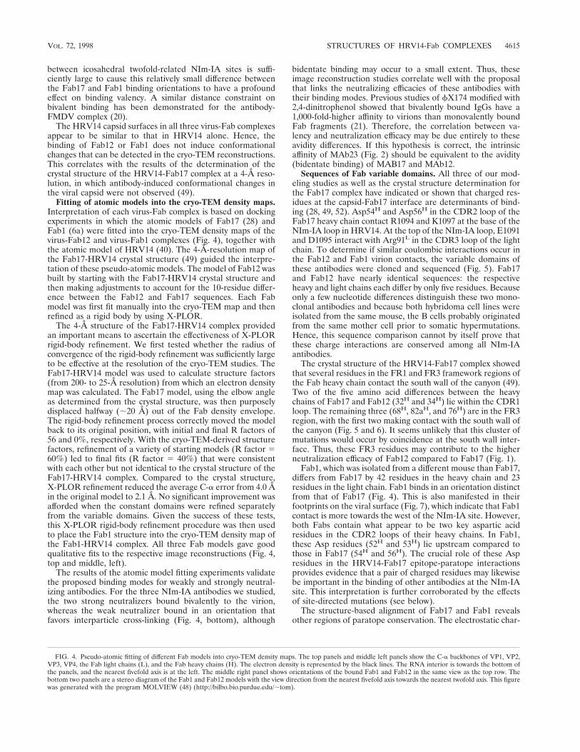
Fitting of atomic models into the cryo-TEM density maps.Interpretation of each virus-Fab complex is based on dockingexperiments in which the atomic models of Fab17 (28) andFab1 (6a) were fitted into the cryo-TEM density maps of thevirus-Fab12 and virus-Fab1 complexes (Fig. 4), together withthe atomic model of HRV14 (40). The 4-Å-resolution map ofthe Fab17-HRV14 crystal structure (49) guided the interpre-tation of these pseudo-atomic models. The model of Fab12 wasbuilt by starting with the Fab17-HRV14 crystal structure andthen making adjustments to account for the 10-residue differ-ence between the Fab12 and Fab17 sequences. Each Fabmodel was first fit manually into the cryo-TEM map and thenrefined as a rigid body by using X-PLOR.
The 4-Å structure of the Fab17-HRV14 complex providedan important means to ascertain the effectiveness of X-PLORrigid-body refinement. We first tested whether the radius ofconvergence of the rigid-body refinement was sufficiently largeto be effective at the resolution of the cryo-TEM studies. TheFab17-HRV14 model was used to calculate structure factors(from 200- to 25-Å resolution) from which an electron densitymap was calculated. The Fab17 model, using the elbow angleas determined from the crystal structure, was then purposelydisplaced halfway (;20 Å) out of the Fab density envelope.The rigid-body refinement process correctly moved the modelback to its original position, with initial and final R factors of56 and 0%, respectively. With the cryo-TEM-derived structurefactors, refinement of a variety of starting models (R factor 560%) led to final fits (R factor 5 40%) that were consistentwith each other but not identical to the crystal structure of theFab17-HRV14 complex. Compared to the crystal structure,X-PLOR refinement reduced the average C-a error from 4.0 Åin the original model to 2.1 Å. No significant improvement wasafforded when the constant domains were refined separatelyfrom the variable domains. Given the success of these tests,this X-PLOR rigid-body refinement procedure was then usedto place the Fab1 structure into the cryo-TEM density map ofthe Fab1-HRV14 complex. All three Fab models gave goodqualitative fits to the respective image reconstructions (Fig. 4,top and middle, left).
The results of the atomic model fitting experiments validatethe proposed binding modes for weakly and strongly neutral-izing antibodies. For the three NIm-IA antibodies we studied,the two strong neutralizers bound bivalently to the virion,whereas the weak neutralizer bound in an orientation thatfavors interparticle cross-linking (Fig. 4, bottom), although
bidentate binding may occur to a small extent. Thus, theseimage reconstruction studies correlate well with the proposalthat links the neutralizing efficacies of these antibodies withtheir binding modes. Previous studies of fX174 modified with2,4-dinitrophenol showed that bivalently bound IgGs have a1,000-fold-higher affinity to virions than monovalently boundFab fragments (21). Therefore, the correlation between va-lency and neutralization efficacy may be due entirely to theseavidity differences. If this hypothesis is correct, the intrinsicaffinity of MAb23 (Fig. 2) should be equivalent to the avidity(bidentate binding) of MAB17 and MAb12.
Sequences of Fab variable domains. All three of our mod-eling studies as well as the crystal structure determination forthe Fab17 complex have indicated or shown that charged res-idues at the capsid-Fab17 interface are determinants of bind-ing (28, 49, 52). Asp54H and Asp56H in the CDR2 loop of theFab17 heavy chain contact R1094 and K1097 at the base of theNIm-IA loop in HRV14. At the top of the NIm-IA loop, E1091and D1095 interact with Arg91L in the CDR3 loop of the lightchain. To determine if similar coulombic interactions occur inthe Fab12 and Fab1 virion contacts, the variable domains ofthese antibodies were cloned and sequenced (Fig. 5). Fab17and Fab12 have nearly identical sequences: the respectiveheavy and light chains each differ by only five residues. Becauseonly a few nucleotide differences distinguish these two mono-clonal antibodies and because both hybridoma cell lines wereisolated from the same mouse, the B cells probably originatedfrom the same mother cell prior to somatic hypermutations.Hence, this sequence comparison cannot by itself prove thatthese charge interactions are conserved among all NIm-IAantibodies.
The crystal structure of the HRV14-Fab17 complex showedthat several residues in the FR1 and FR3 framework regions ofthe Fab heavy chain contact the south wall of the canyon (49).Two of the five amino acid differences between the heavychains of Fab17 and Fab12 (32H and 34H) lie within the CDR1loop. The remaining three (68H, 82aH, and 76H) are in the FR3region, with the first two making contact with the south wall ofthe canyon (Fig. 5 and 6). It seems unlikely that this cluster ofmutations would occur by coincidence at the south wall inter-face. Thus, these FR3 residues may contribute to the higherneutralization efficacy of Fab12 compared to Fab17 (Fig. 1).
Fab1, which was isolated from a different mouse than Fab17,differs from Fab17 by 42 residues in the heavy chain and 23residues in the light chain. Fab1 binds in an orientation distinctfrom that of Fab17 (Fig. 4). This is also manifested in theirfootprints on the viral surface (Fig. 7), which indicate that Fab1contact is more towards the west of the NIm-IA site. However,both Fabs contain what appear to be two key aspartic acidresidues in the CDR2 loops of their heavy chains. In Fab1,these Asp residues (52H and 53H) lie upstream compared tothose in Fab17 (54H and 56H). The crucial role of these Aspresidues in the HRV14-Fab17 epitope-paratope interactionsprovides evidence that a pair of charged residues may likewisebe important in the binding of other antibodies at the NIm-IAsite. This interpretation is further corroborated by the effectsof site-directed mutations (see below).
The structure-based alignment of Fab17 and Fab1 revealsother regions of paratope conservation. The electrostatic char-
FIG. 4. Pseudo-atomic fitting of different Fab models into cryo-TEM density maps. The top panels and middle left panels show the C-a backbones of VP1, VP2,VP3, VP4, the Fab light chains (L), and the Fab heavy chains (H). The electron density is represented by the black lines. The RNA interior is towards the bottom ofthe panels, and the nearest fivefold axis is at the left. The middle right panel shows orientations of the bound Fab1 and Fab12 in the same view as the top row. Thebottom two panels are a stereo diagram of the Fab1 and Fab12 models with the view direction from the nearest fivefold axis towards the nearest twofold axis. This figurewas generated with the program MOLVIEW (48) (http://bilbo.bio.purdue.edu/;tom).
VOL. 72, 1998 STRUCTURES OF HRV14-Fab COMPLEXES 4615
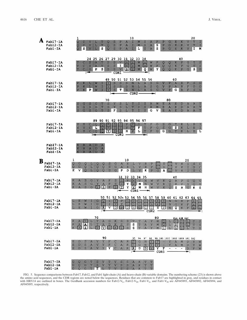
FIG. 5. Sequence comparisons between Fab17, Fab12, and Fab1 light-chain (A) and heavy-chain (B) variable domains. The numbering scheme (23) is shown abovethe amino acid sequences, and the CDR regions are noted below the sequences. Residues that are common to Fab17 are highlighted in gray, and residues in contactwith HRV14 are outlined in boxes. The GenBank accession numbers for Fab12-VL, Fab12-VH, Fab1-VL, and Fab1-VH are AF045893, AF045892, AF045894, andAF045895, respectively.
4616 CHE ET AL. J. VIROL.

acters of both paratope surfaces are quite similar (Fig. 8). InFab17, Arg91L (which interacts with D1091 and E1095) con-tributes to a positively charged patch located in the cleft be-tween the heavy and light chains. The two aspartic acid resi-dues (Asp54H and Asp56H) in the CDR2 loop of the Fab17heavy chain form a negatively charged patch juxtaposed withviral residues R1094 and K1097. Fab1 also has a positivelycharged patch, but it is comprised of heavy-chain residuesArg50H and Arg95H. The negatively charged patch is formedby the two conserved aspartic acid residues Asp52H andAsp53H. Interestingly, the two charged patches are aligned inFab1 in an orientation that is rotated counterclockwise by ;25°compared to that in Fab17, and this directly correlates with thedifferences in the Fab1 and Fab12 binding orientations. Thisagrees with the previous suggestions that electrostatic fieldinteractions may be important in NIm-IA antibody binding(52) and with recent calculations of electrostatic field comple-mentarity at protein-protein interfaces (32).
Interactions between HRV14 and NIm-IA antibodies. De-spite differences in the orientation of bound Fab1 compared tothose of Fab17 and Fab12, the light-chain CDR2 loops of allthree antibodies contact very little of the viral surface (Fig. 5).As the light chains of these antibodies contact a steep surfaceeast of the NIm-IA site, it is difficult for them to make exten-sive contact. In contrast, the heavy chain fits quite well into thecanyon, thereby allowing all three heavy-chain CDRs to con-tact the viral surface (Figs. 5 to 7). Such dominance of antigencontact via the antibody heavy-chain contact has been observedin other Fab-antigen complexes (see, e.g., reference 10) and isnot unexpected in view of the inherent genetic diversity ofheavy chains compared to light chains (e.g., the D geneticcassettes and the activation of terminal dideoxynucleotidetransferase during heavy-chain somatic recombination). In ad-dition, recent results with camelid antibodies have shown thatantibodies comprised of only heavy chains do occur in vivo andbind antigens (13).
Electrostatic interactions dominate the interface betweenthe NIm-IA loop region and Fab1 (Table 1; Fig. 5). E1095, oneof the two residues that define the NIm-IA site, is clamped bythe positively charged cleft between the heavy- and light-chainhypervariable regions (Fig. 8). The Fab1 heavy-chain arginines(Arg50H and Arg95H) have direct interactions with E1095. The
other NIm-IA residue, D1091, lies outside this region of pos-itive charge. The side chain of D1091 in the crystal structure ofFab17-HRV14 rotates to form interactions with the corre-sponding bases in Fab17. K1097 interacts with only one aspar-tic acid residue (Asp52H) in Fab1 but with two (Asp54H andAsp56H) in Fab17. The other aspartic acid of Fab1 (Asp53H)forms a salt bridge with K1240, which lies near the NIm-IAloop. R1094 is located close to the negatively charged region.It is quite possible that its side chain would move into thisregion, as was observed in the Fab17-HRV14 crystal structure.
Further study of HRV14 site-directed mutants, constructedpreviously for our Fab17-virus complex work (52), supportsour interpretations of the two new Fab-virus complexes anddemonstrates the importance of electrostatic interactions inFab binding to the NIm-IA site. Of the residues tested, muta-tion of K1097, which makes extensive interactions with Fab17(49), had the greatest effect on the neutralization of all NIm-IAantibodies (Table 2). The K1097E mutation reduced NIm-IAantibody neutralization by 102- to 104-fold. This mutation isjust as effective as a naturally occurring escape mutation inblocking neutralization. When an uncharged residue (Gln) wassubstituted for K1097, little to no effect was observed exceptfor MAb17. The K1085E mutation affected almost all NIm-IAantibodies by about 10-fold, whereas the K1236E mutation hadlittle to no effect on any of these antibodies. This result agreeswith the crystal structure study of the Fab17-HRV14 complex,in which Fab17 was observed to contact K1085 but made verylittle contact with K1236. These mutagenesis results demon-strate that coulombic interactions with these viral surface ly-sine residues (not identified in the initial selection of naturallyoccuring escape mutations) are very important to both weaklyand strongly neutralizing antibodies.
Antibody-mediated stabilization of HRV14 against acid in-activation. The crystal structure of the Fab17-HRV14 complexdemonstrated that large conformational changes in the virionare not required for antibodies to mediate neutralization.Thus, antibodies may achieve their effect by stabilizing virionswithout necessitating large conformational changes. To testthis hypothesis, HRV14 was complexed with several antibod-ies, incubated in buffers at various pHs, and examined forresidual infectivity (Table 3). Interestingly, all NIm-IA anti-bodies stabilized virions against acid inactivation, whereas an-
FIG. 6. The C-a backbone of the HRV14-Fab17 complex, with the residues that differ from Fab12 in the framework (FR) and hypervariable (CDR) regionshighlighted. The orientation and program used are the same as for Fig. 4.
VOL. 72, 1998 STRUCTURES OF HRV14-Fab COMPLEXES 4617
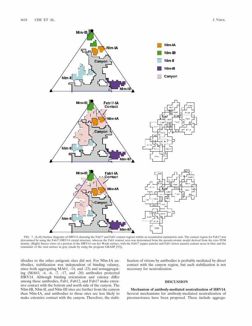
tibodies to the other antigenic sites did not. For NIm-IA an-tibodies, stabilization was independent of binding valancy,since both aggregating MAb1, -14, and -23) and nonaggregat-ing (MAb3, -4, -6, -7, -17, and -20) antibodies protectedHRV14. Although binding orientation and valency differamong these antibodies, Fab1, Fab12, and Fab17 make exten-sive contact with the bottom and north side of the canyon. TheNIm-IB, NIm-II, and NIm-III sites are further from the canyonthan NIm-IA, and antibodies to these sites are less likely tomake extensive contact with the canyon. Therefore, the stabi-
lization of virions by antibodies is probably mediated by directcontact with the canyon region, but such stabilization is notnecessary for neutralization.
DISCUSSION
Mechanism of antibody-mediated neutralization of HRV14.Several mechanisms for antibody-mediated neutralization ofpicornaviruses have been proposed. These include aggrega-
FIG. 7. (Left) Surface diagrams of HRV14 denoting the Fab17 and Fab1 contact regions within an icosahedral asymmetric unit. The contact region for Fab17 wasdetermined by using the Fab17-HRV14 crystal structure, whereas the Fab1 contact area was determined from the pseudo-atomic model derived from the cryo-TEMdensity. (Right) Stereo views of a portion of the HRV14 van der Waals surface, with the Fab17 (upper panels) and Fab1 (lower panels) contact areas in blue and theremainder of the viral surface in gray (made by using the program GRASP [35]).
4618 CHE ET AL. J. VIROL.

tion, induction of conformational changes, virion stabilization,and abrogation of cellular attachment.
(i) Aggregation. It has been suggested that aggregation oc-curs concomitantly with neutralization and that virus/antibodyratios in vivo are conducive to aggregation (4, 5, 54). However,our data strongly suggest that aggregation is not a major con-tributor to neutralization of HRV. First, antibodies that bindbivalently to virions do not aggregate them over a wide rangeof antibody/virus ratios, yet such antibodies are strong neutral-izers (Fig. 1). Second, even antibodies that are strong aggre-gators neutralize virus at antibody/virus concentration ratiosthat do not favor aggregation. The neutralization profile foraggregating antibodies sometimes displays a dip that is oftencoincident with aggregation. Hence, in such circumstances,neutralization may be enhanced in a narrow range of antibody/virus ratios that favors precipitation. This enhancement mayresult from a decrease of independent infectious particles orfrom avidity effects caused by antibodies bound bivalently, inan interparticle manner, to the large immunocomplexes. Al-though aggregation probably does not play a significant role invitro, it may facilitate innate immunological responses via op-sonization in vivo.
(ii) Stabilization. It has also been suggested that antibodiesmight neutralize virions by stabilizing the capsid (34), whichmight then prevent uncoating or receptor-induced conforma-tional changes. All antibodies that bind to NIm-IA (aggregat-ing and nonaggregating) stabilize virions against acidic pH tovarious extents. However, none of the non-NIm-IA antibodiesthat we tested cause such stabilization, although some areefficacious neutralizers. Therefore, these stabilization effectsdo not correlate well to neutralization efficacy or binding va-lency. In addition, antibodies to all four sites have been shownto block cellular attachment (8), and this would precede anystabilization effects. Notably, all known escape mutations maponly to residues around the epitope. An escape mutation whichdoes not affect antibody binding but prevents neutralizationhas not yet been observed. If capsid stabilization-destabiliza-tion was a major determinant of neutralization, at least someescape mutations that abrogated these effects might be ex-pected to arise. Analogous distal-site resistance mutationshave been found when poliovirus and rhinovirus are grown inthe presence of capsid-stabilizing antiviral agents (19).
(iii) Conformational changes. Antibodies and Fab frag-ments cause an apparent decrease in the pI of the viral capsid
FIG. 7—Continued.
VOL. 72, 1998 STRUCTURES OF HRV14-Fab COMPLEXES 4619

concomitant with neutralization (8, 29). This fact has beencited as evidence that antibodies neutralize by distorting thecapsid. The crystal structure of the Fab17-HRV14 complexclearly demonstrated that efficacious neutralization occurs inthe absence of large conformational changes. Instead, Fab17undergoes large conformational changes to better accommo-date the epitope without inducing structural changes in thevirion (49). Even though all antibodies to the four differentantigenic sites that were tested (MAb13, -17, -21, -28, -29, -33,-34, and -35) caused apparent changes in the pI of the capsid(8), it seems unlikely that dissimilar antibodies, which bind todistinct epitopes, would all cause the same effect on the capsid.Antibodies might cause conformational changes in proteinstructure upon binding, but such changes would be expected tooccur on flexible portions of the viral structure. Antibody-induced conformational changes on less flexible regions wouldcost significant Gibbs free energy and would greatly affectantibody affinity. Therefore, it seems unlikely that induction ofconformational changes contributes significantly to antibodyneutralization.
(iv) Abrogation of cellular attachment. Previous studieshave clearly demonstrated that antibodies to all four HRV14antigenic sites block cellular attachment (8). The three
NIm-IA antibodies that we have studied clearly bind in a man-ner that overlaps the ICAM footprint as determined by cryo-TEM (37). Steric hindrance effects can be used to explain thecompetition between receptor and antibody binding. NIm-II isimmediately adjacent to the ICAM binding region, and the;600- to 900-Å2 contact region of these antibodies possiblyoverlaps the ICAM site as well. However, NIm-III is quitedistal (;40 Å away) to the receptor binding region, yet NIm-III antibodies also compete with receptor binding (8). Perhaps,then, antibody competition with the receptor is merely a resultof the sheer bulk of an antibody molecule (an IgG is ;140 Ålong and approximately equal to the radius of HRV14) anddoes not require direct overlap with the ICAM binding region.This steric model would also explain why cellular attachment isinhibited at a nonsaturating stoichiometry of 7 to 10 antibodiesper virion (25, 52).
Summary. Consideration of all of these results suggests thatthe mechanism of HRV neutralization in vitro may be muchsimpler than previously envisioned: antibodies bound to thesurface of HRV14 are sufficient to block attachment of virusreceptors. For poliovirus and rhinovirus, interactions with theirreceptors appear to be essential for the proper release of RNAinto the cytoplasm of the host cell. Indeed, antibody-polioviruscomplexes were shown to enter cells, but this import mode led
FIG. 8. Electrostatic character of the Fab17 and Fab1 paratope surfaces. The positive charge (blue) and the negative charge (red) are mapped onto the van derWaals surface. The heavy-chain hypervariable region is towards the bottom of the image and the light chain is towards the top. This figure was made by using theprogram GRASP (35).
TABLE 1. Percentages of surface contact according to residue type
Residue type
Contact with:
Fab17 Fab1
Area(Å2)
% oftotal
Area(Å2)
% oftotal
Acidic (D, E) 96 11 95 14Basic (H, K, R) 142 17 84 12Polar (N, Q, S, T) 187 22 191 28Small (A, G) 61 7 76 11Hydrophobic (C, I, L, M, P, V) 23 3 84 12Aromatic Polar (W, Y) 336 38 158 23
Total 845 100 685 100
TABLE 2. Residual infectivity of HRV14 wild-type (WT) andmutant viruses after treatment with NIm-IA antibodies
Antibody% of residual plaques
LP1 (WT) K1085E K1236E K1097Q K1097E
MAb1 0.0021 0.032 0.0088 0.0054 0.27MAb3 0.0064 0.060 0.014 0.011 83MAb4 0.0031 0.045 0.0082 0.0059 0.44MAb6 0.0045 0.050 0.014 0.012 71MAb7 0.0064 0.063 0.014 0.015 106MAb14 0.0 0.0 0.0 0.0 0.19MAb17 0.0190 0.052 0.016 3.1 87MAb20 0.0000 0.003 0.0013 0.00066 0.018
4620 CHE ET AL. J. VIROL.

to digested viral RNA (29). In contrast, the picornavirusFMDV needs its receptor only to enter the cell and not forRNA release (31), yet this virus is also effectively neutralizedby antibodies. Therefore, a simple steric effect in which anti-body binds to the virion surface and blocks receptor attach-ment is sufficient to explain the neutralization behavior ofmany antibodies. Although some antibodies might induce sec-ondary effects (e.g., causing conformational changes in theviral capsid) upon binding (12, 27, 30, 60), such effects are notrequired for neutralization. In addition, antibodies that inducesuch secondary effects could not be exclusively selected forduring B-cell clonal expansion.
Our results clearly do not rule out the existence of antibod-ies that induce changes in virion structure upon binding, just asthey do not imply that all antibodies neutralize by abrogatingcellular attachment. For example, the details of antibody in-teractions with FMDV and poliovirus might be expected to bequite different. The receptor binding region of FMDV is lo-cated on the end of a highly mobile, immunodominant loop.Receptor or antibodies binding to this loop are unlikely totransmit conformational changes to the virion but will mostcertainly affect cell attachment. In contrast, the top of thecanyon region of poliovirus is involved in both receptor andantibody binding (18, 59). For poliovirus, therefore, some an-tibodies might bind to this region and either mimic receptorbinding and cause conformational changes or inhibit changesin this region. Indeed, FMDV-antibody complexes can infectcells that have Fc receptors, whereas poliovirus-antibody com-plexes cannot (31). Antibodies to human immunodeficiencyvirus apparently neutralize by blocking attachment or eventsafter uncoating, depending on which viral protein is targetedby the antibody (56). Antibodies to the hemagglutinin of in-fluenza virus can prevent attachment and replication, but an-tibodies to the neuraminidase only interfere with virus release(24). Therefore, the effects of antibodies on viruses can be asdiverse as the viruses.
Notably, in vivo studies have clearly shown that the types ofin vitro mechanisms that we have described may be of limitedconsequence in protecting animals from viral infections. Forexample, antibodies that are not efficacious in vitro against
Sindbis virus (43) and FMDV (33) are still capable of protect-ing animals from viral challenge. Although antibodies againstneuraminidase from influenza virus are not neutralizing, theydo affect disease progression in vivo (44). Therefore, the pri-mary role of antibodies in vivo may be to act synergisticallywith other components of the immune system. This furtherimplies that the design of vaccines should focus on the pro-duction of high-affinity antibodies rather than on a particular invitro neutralization property. This has been recently shown tobe true in the case of human immunodeficiency virus type 1,where the occupancy of binding sites on the virus is the majorfactor in neutralization efficacy irrespective of the epitopespecificity (39). This goal of eliciting high-affinity antibodies isclearly more straightforward than having to create vaccinesthat yield antibodies which neutralize by a particular mecha-nism.
ACKNOWLEDGMENTS
We thank E. S. Chase for growing the hybridoma cells for MAb12and MAb1, T. J. Schmidt for helpful suggestions in the processing ofthe Fab1 crystallographic data set, and Z. Zhu for help and advice inthe sequence determinations of Fab1 and Fab12.
This work was supported by grants from the National Institutes ofhealth (GM10704 to T.J.S. and GM33050 to T.S.B.).
REFERENCES1. Bailey, S. 1994. The CCP4 suite: programs for protein crystallography. Acta
Crystallogr. D. 50:760–763.2. Baker, T. S., and R. H. Cheng. 1996. A model-based approach for determin-
ing orientations of biological macromolecules imaged by cryoelectron mi-croscopy. J. Struct. Biol. 116:120–130.
3. Baker, T. S., W. W. Newcomb, N. H. Olson, L. M. Cowsert, C. Olson, andJ. C. Brown. 1991. Structures of bovine and human papilloma viruses: anal-ysis by cryoelectron microscopy and three-dimensional image reconstruction.Cell 60:1007–1015.
4. Brioen, P., D. Dekegel, and A. Boeye. 1983. Neutralization of poliovirus byantibody-mediated polymerization. Virology 127:463–468.
5. Brioen, P., A. A. M. Thomas, and A. Boeye. 1985. Lack of quantitativecorrelation between the neutralization of poliovirus and the antibody-medi-ated pI shift of the virions. J. Gen. Virol. 66:609–613.
6. Brunger, A. T. 1992. X-plor (version 3.1) user’s guide. Yale University, NewHaven, Conn.
6a. Che, Z., and T. J. Smith. Unpublished data.7. Cheng, R. H., N. H. Olson, and T. S. Baker. 1992. Cauliflower mosaic virus:
a 420 subunit (T57), multilayer structure. Virology 186:655–668.8. Colonno, R. J., P. L. Callahan, D. M. Leippe, and R. R. Rueckert. 1989.
Inhibition of rhinovirus attachment by neutralizing monoclonal antibodiesand their Fab fragments. J. Virol. 63:36–42.
9. Connolly, M. L. 1983. Analytical molecular surface calculation. J. Appl.Crystallogr. 16:548.
10. Corper, A. L., M. K. Sohi, V. R. Bonagura, M. Steinitz, R. Jefferis, A.Feinstein, D. Beale, M. J. Taussig, and B. J. Sutton. 1997. Structure ofhuman IgM rheumatoid factor Fab bound to its autoantigen IgG Fc revealsa novel topology of antibody-antigen interaction. Nat. Struct. Biol. 4:374–381.
11. Crowther, R. A. 1971. Procedure for the three-dimensional reconstruction ofspherical viruses by Fourier synthesis from electron micrographs. Philos.Trans. R. Soc. London B 261:221–230.
12. Delaet, I., and A. Boeye. 1993. Monoclonal antibodies that disrupt poliovirusonly at fever temperatures. J. Virol. 67:5299–5302.
13. Desmyter, A., T. R. Transue, M. A. Ghahroudi, M. H. Thi, F. Poortmans, R.Hamers, S. Muyldermans, and L. Wyns. 1996. Crystal structure of a camelsingle-domain VH antibody fragment in complex with lysozyme. Nat. Struct.Biol. 3:803–811.
14. Emini, E. A., S. Kao, A. J. Lewis, R. Crainic, and E. Wimmer. 1983. Func-tional basis of poliovirus neutralization determined with monospecific neu-tralizing antibodies. J. Virol. 46:466–474.
15. Emini, E. A., P. Ostapchuk, and E. Wimmer. 1983. Bivalent attachment ofantibody onto poliovirus leads to conformational alteration and neutraliza-tion. J. Virol. 48:547–550.
16. Erickson, J. W., E. A. Frankenberger, M. G. Rossmann, G. S. Fout, K. C.Medappa, and R. R. Rueckert. 1983. Crystallization of a common cold virus,human rhinovirus 14: “isomorphism” with poliovirus crystals. Proc. Natl.Acad. Sci. USA 80:931–934.
17. Fuller, S. D., S. J. Butcher, R. H. Cheng, and T. S. Baker. 1996. Three-dimensional reconstruction of icosahedral particles—the uncommon line. J.Struct. Biol. 116:48–55.
TABLE 3. Effects of antibodies on pH inactivation of HRV14with strong neutralizers (SN), intermediate neutralizers (IN),
weak neutralizers (WN), precipitating antibodies (P), andnonprecipitating antibodies (NP) to all four antigenic sites
MAb Nim site Type
% Residual infectivity afterincubation at pHa:
7.0 5.0 4.5
None 100 0.0 0.03 IA SN, NP 101 99 0.144 IA SN, NP 84 89 496 IA SN, NP 116 91 617 IA SN, NP 127 104 3514 IA SN, P 65 67 5.623 IA SN, P 82 87 0.017 IA IN, NP 92 91 0.020 IA IN, NP 80 82 0.291 IA WN, P 52 50 0.3026 IB WN, P 81 0.0012 0.018 II WN, P 44 0.0047 0.08 III IN, NP 104 0.0 0.09 III IN, P 106 0.0 0.0
a Residual infectivity after incubation at the indicated pH and removal ofantibody by freezing.
VOL. 72, 1998 STRUCTURES OF HRV14-Fab COMPLEXES 4621

18. Harber, J., G. Bernhardt, H. H. Lu, J. Y. Sgro, and E. Wimmer. 1995.Canyon rim residues, including antigenic determinants, modulate serotype-specific binding of polioviruses to mutants of the poliovirus receptor. Virol-ogy 214:559–570.
19. Heinz, B. A., R. R. Rueckert, D. A. Shepard, F. J. Dutko, M. A. McKinlay, M.Francher, M. G. Rossmann, J. Badger, and T. J. Smith. 1989. Genetic andmolecular analysis of spontaneous mutants of human rhinovirus 14 resistantto an antiviral compound. J. Virol. 63:2476–2485.
20. Hewat, E. A., N. Verdaguer, I. Fita, W. Blakemore, S. Brookes, A. King, J.Newman, E. Domingo, M. G. Mateau, and D. I. Stuart. 1997. Structure of thecomplex of an Fab fragment of a neutralizing antibody with foot-and-mouthdisease virus: positioning of a highly mobile antigenic loop. EMBO J. 16:1492–1500.
21. Hornick, C. L., and F. Karush. 1972. Antibody affinity III. The role ofmultivalence. Immunochemistry 9:325–340.
22. Jones, T. A., J.-Y. Zou, and S. W. Cowan. 1991. Improved methods forbuilding protein models in electron density maps and the location of errorsin these models. Acta Crystallogr. A 47:110–119.
23. Kabat, E. A., T. T. Wu, M. Reid-Miller, H. M. Perry, and K. S. Gottesman.1987. Sequences of proteins of immunological interests. National Institutesof Health, Bethesda, Md.
24. Kilbourne, E. D., W. G. Laver, J. L. Shulman, and R. G. Webster. 1968.Antiviral activity of antiserum specific for an influenza virus neuraminidase.J. Virol. 2:281–288.
24a.Kolatchar, P., et al. Unpublished data.25. Lee, W. M. 1992. Human rhinovirus 14: synthesis and characterization of a
molecular cDNA clone which makes highly infectious transcripts. Universityof Wisconsin, Madison.
26. Leippe, D. M. 1991. Stoichiometry of picornavirus neutralization by murinemonoclonal antibodies. University of Wisconsin, Madison.
27. Li, Q., A. G. Yafal, Y. M. H. Lee, J. Hogle, and M. Chow. 1994. Poliovirusneutralization by antibodies to internal epitopes of VP4 and VP1 resultsfrom reversible exposure of the sequences at physiological temperatures.J. Virol. 68:3965–3970.
28. Liu, H., T. J. Smith, W. M. Lee, A. Mosser, R. R. Rueckert, N. H. Olson,R. H. Cheng, and T. S. Baker. 1994. Structure determination of an fabfragment that neutralizes human rhinovirus 14 and analysis of the fab-viruscomplex. J. Mol. Biol. 240:127–137.
29. Mandel, B. 1967. The interaction of neutralized poliovirus with HeLa cells.II. Elution, penetration, uncoating. Virology 31:247–259.
30. Mandel, B. 1976. Neutralization of poliovirus: a hypothesis to explain themechanism and the one-hit character of the neutralization reaction. Virology69:500–510.
31. Mason, P. W., B. Baxt, F. Brown, J. Harber, A. Murdin, and E. Wimmer.1993. Antibody-complexed foot-and-mouth disease virus, but not poliovirus,can infect normally insusceptible cells via the Fc receptor. Virology 192:568–577.
32. McCoy, A. J., V. Chandana Epa, and P. M. Coleman. 1997. Electrostaticcomplementarity at protein/protein interfaces. J. Mol. Biol. 268:570–584.
33. McCullough, K. C., F. De Simone, E. Brocchi, L. Capucci, J. R. Crowther,and U. Kihm. 1992. Protective immune response against foot-and-mouthdisease. J. Virol. 66:1835–1840.
34. Mosser, A. G., D. M. Leippe, and R. R. Rueckert. 1989. Neutralization ofpicornaviruses: support for the pentamer bridging hypothesis, p. 155–167. InB. L. Semler and E. Ehrenfeld (ed.), Molecular aspects of picornavirusinfection and detection. American Society for Microbiology, Washington,D.C.
34a.Navaza, J. 1994. AMoRe: an automated package for molecular replacement.Acta Crystallogr. A 50:157–163.
35. Nicholls, A. 1993. GRASP: graphical representation and analysis of surfaceproperties. Columbia University, New York, N.Y.
36. Olson, N. H., T. S. Baker, P. Willingmann, and N. L. Incardona. 1992. Thethree-dimensional structure of frozen-hydrated bacteriophage fX174. J.Struct. Biol. 108:168–175.
37. Olson, N. H., P. R. Kolatkar, M. A. Oliveira, R. H. Cheng, J. M. Greve, A.McClelland, T. S. Baker, and M. G. Rossmann. 1993. Structure of a humanrhinovirus complexed with its receptor molecule. Proc. Natl. Acad. Sci. USA90:507–511.
38. Otwinoski, Z. 1993. DENZO, p. 56–62. In L. Sawyer, N. Isaacs, and S. Bailey(ed.), Data collection and processing. SERC Daresbury Laboratory, War-
rington, United Kingdom.39. Parren, P. W. H. I., I. Mondor, D. Naniche, H. J. Ditzel, P. J. Klasse, D. R.
Burton, and Q. J. Sattentau. 1998. Neutralization of human immunodefi-ciency virus type 1 by antibody to gp120 is determined primarily by occu-pancy of sites on the virion irrespective of epitope specificity. J. Virol.72:3512–3519.
40. Rossmann, M. G., E. Arnold, J. W. Erickson, E. A. Frankenberger, J. P.Griffith, H. J. Hecht, J. E. Johnson, G. Kamer, M. Luo, A. G. Mosser, R. R.Rueckert, B. Sherry, and G. Vriend. 1985. Structure of a human commoncold virus and functional relationship to other picornaviruses. Nature 317:145–153.
41. Rossmann, M. G., R. McKenna, L. Tong, D. Xia, H. Wu, and H. Choi. 1992.Molecular replacement real-space averaging. J. Appl. Cryst. 25:166–180.
42. Rueckert, R. R. 1996. Picornaviridae and their replication. Raven Press, NewYork, N.Y.
43. Schmaljohn, A. L., E. D. Johnson, J. M. Dalrymple, and G. A. Cole. 1982.Nonneutralizing monoclonal antibodies can prevent lethal alphavirus en-cephalitis. Nature 297:70–72.
44. Schulman, J. L. 1975. Immunology of influenza, p. 373–393. In E. D. Kil-bourne (ed.), The influenza viruses and influenza. Academic Press, NewYork, N.Y.
45. Sheriff, S., W. A. Hendrickson, R. E. Stenkamp, L. C. Sieker, and L. H.Jensen. 1985. Influence of solvent accessibility and intermolecular contactson atomic mobilities in hemerythrins. Proc. Natl. Acad. Sci. USA 82:1104–1107.
46. Sherry, B., A. G. Mosser, R. J. Colonno, and R. R. Rueckert. 1986. Use ofmonoclonal antibodies to identify four neutralization immunogens on acommon cold picornavirus, human rhinovirus 14. J. Virol. 57:246–257.
47. Sherry, B., and R. R. Rueckert. 1985. Evidence for at least two dominantneutralization antigens on human rhinovirus 14. J. Virol. 53:137–143.
48. Smith, T. J. 1995. MolView: a program to analyze and display atomic struc-tures on the Macintosh personal computer. J. Mol. Graphics. 13:122–125.
49. Smith, T. J., E. S. Chase, T. J. Schmidt, N. H. Olson, and T. S. Baker. 1996.Neutralizing antibody to human rhinovirus 14 penetrates the receptor-bind-ing canyon. Nature 383:350–354.
50. Smith, T. J., A. G. Mosser, and T. S. Baker. 1995. Structural studies on themechanisms of antibody-mediated neutralization of human rhinovirus. Se-min. Virol. 6:233–242.
51. Smith, T. J., N. H. Olson, R. H. Cheng, E. S. Chase, and T. S. Baker. 1993.Structure of a human rhinovirus-bivalently bound antibody complex: impli-cations for virus neutralization and antibody flexibility. Proc. Natl. Acad. Sci.USA 90:7015–7018.
52. Smith, T. J., N. H. Olson, R. H. Cheng, H. Liu, E. Chase, W. M. Lee, D. M.Leippe, A. G. Mosser, R. R. Ruekert, and T. S. Baker. 1993. Structure ofhuman rhinovirus complexed with Fab fragments from a neutralizing anti-body. J. Virol. 67:1148–1158.
53. Thomas, A. A., R. Vrijsen, and A. Boeye. 1986. Relationship between polio-virus neutralization and aggregation. J. Virol. 59:479–485.
54. Thomas, A. A. M., P. Brioen, and A. Boeye. 1985. A monoclonal antibodythat neutralizes poliovirus by cross-linking virions. J. Virol. 54:7–13.
55. Thouvenin, E., S. Laurent, M.-F. Madelaine, D. Rasschaert, J.-F. Vautherot,and E. A. Hewat. 1997. Bivalent binding of a neutralizing antibody to acalicivirus involves the torsional flexibility of the antibody hinge. J. Mol. Biol.270:238–246.
56. Ugolini, S., I. Mondor, P. W. Parren, D. R. Burton, S. A. Tilley, P. J. Klasse,and Q. J. Sattentau. 1997. Inhibition of virus attachment to CD41 targetcells is a major mechanism of T cell line-adapted HIV-1 neutralization. J.Exp. Med. 186:1287–1298.
57. Vrijsen, R., A. Mosser, and A. Boeye. 1993. Postabsorption neutralization ofpoliovirus. J. Virol. 67:3126–3133.
58. Wade, R. H., J. C. Taveau, and J. N. Lamy. 1989. Concerning the axialrotational flexibility of the Fab regions of immunoglobulin G. J. Mol. Biol.206:349–356.
59. Wien, M. W., S. Curry, D. J. Filman, and J. M. Hogle. 1997. Structuralstudies of poliovirus mutants that overcome receptor defects. Nat. Struct.Biol. 4:666–674.
60. Wien, M. W., D. J. Filman, E. A. Stura, S. Guillot, F. Delpeyroux, R. Crainic,and J. M. Hogle. 1995. Structure of the complex between the Fab fragmentof a neutralizing antibody for type 1 poliovirus and its viral epitope. Nat.Struct. Biol. 2:232–243.
4622 CHE ET AL. J. VIROL.