antibody-based immunotherapy of lymphoma department student lecture april 10, 2014 ... headache,...
TRANSCRIPT

Antibody-Based Immunotherapy of Lymphoma
Immunology Department Student Lecture
April 10, 2014
Myron S. Czuczman, MD Chief, Lymphoma/Myeloma Service Head, Lymphoma Translational Research Lab Roswell Park Cancer Institute

NHL: Incidence and Mortality
• United States: 54,370 new cases 20,730 deaths
– Sixth most common type of cancer – Increasing since early 1970s
Year 1973 1978 1983 1988 1993 1998 2001
0
2.5
20.0
17.5
15.0
12.5
10.0
7.5
5.0
11.1
5.6
11.9
5.9
14
6.7
17.2
7.5
18.9
8.2 8.7
8.4
19.3 19.4
Rate per 100,000
Incidence Mortality

NHL: Risk Factors
• Cause of NHL unknown • Inherited Familial: accounts for a small percentage
of cancers • Environmental
– Certain chemical suspected (eg, certain pesticides/ herbicides)
– High-dose radiation exposure suspected • Immunosuppression
– Immune deficiency (AIDS, post–organ transplant) • Viral and Bacterial
– Infections (HTLV-1 virus, EBV, H pylori bacteria)

Lymphocytes
• T cells – Release cytokines
• B cells – Produce antibodies
• Natural killer (NK) cells – Kill infected cells – Attack cancer cells
• Non-Hodgkin lymphoma – 85% B cells – 15% T cells

Treating Non-Hodgkin Lymphoma

Features of an Ideal Anticancer Target
• Crucial to the malignant phenotype • Not significantly expressed in vital organs / tissues • A biologically relevant molecular feature • Reproducibly measurable in readily obtainable
clinical samples • Correlated with clinical outcome • Clinical response in significant % of target-positive
patients when target is interrupted, interfered with, or inhibited
• Minimal effects in target –negative patients

Monoclonal Antibody Therapy
• Biotherapy targeted treatment • Effective, low toxicity • Targets tumor cells • Two types
– Unconjugated – Conjugated

Limitations of early mAbs
• Poor target selection • Limited biological activity of unlabeled mAbs • Poor tumor cell penetration of mAbs • Immunogenicity (i.e. high HAMA titers) • Infusional toxicity (i.e. purity) • “Biotechnology to the Rescue” – 1980’s / early
1990’s

9
B-Cells: Express Many Surface Antigens That May Serve as Targets for mAbs
Antigen expression variable1,2
Most involved in B-cell growth, differentiation, proliferation, and activation; other functions include1,2: – Immune regulation – Complement inhibition
Many are targets of therapeutic mAbs for current or potential use in B-cell malignancies1,2
B-Cell
CD19
CD20
CD21
CD22
CD23
CD38
B-cell receptor (BCR)
CD40
CD52
CD46, CD55, CD59
CD74
CD80
Marker
1Bello C, Sotomayor EM. Hematology Am Soc Hematol Educ Program. 2007;2007:233-242 2Hotta T. Acta Histochem Cytochem. 2002;35(4):275-279

Rationale for mAb / RIT of NHL • B-cell lymphomas
– Express tumor-associated antigens – Accessible to the vascular system – Rx of minimal residual disease may alter natural history
• mAbs – Greater tumor specificity and less non-specific toxicities – Unique MOA – Demonstrated activity alone and in combination therapy
• Radioimmunoconjugates (RIC) – B-cell NHL is radiosensitive – “Cross-fire” effect – Not dependent on host-immune function

Anti-CD20 MAbs: Mechanism-of-Action
Maloney DG. N Engl J Med 2012

Rituximab: First mAb approved by FDA for Cancer Therapy

ADCC
Fc region of CD20-bound MAb binds to Fc receptor (FcR) on effector cell (e.g. macrophage, NK cell, neutrophil, etc)
Effector cell releases mediators that damage and destroy CD20-positive cell
CD20-positive cell is phagocytosed
Anti-CD20 Monoclonal Antibodies Induce ADCC
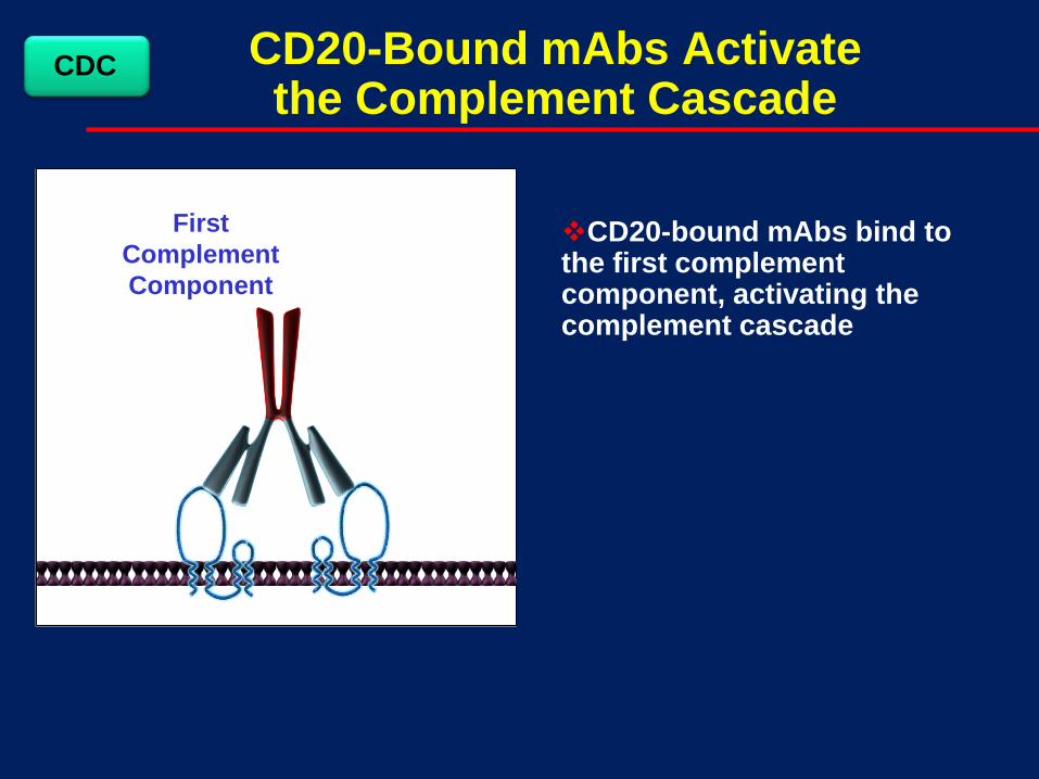
CD20-Bound mAbs Activate the Complement Cascade
First Complement Component
CD20-bound mAbs bind to the first complement component, activating the complement cascade
CDC

MAC
Inrushing fluids
MAC
Complement Activation Causes MAC Formation and B-Cell Lysis
Activation of complement components on the B-cell surface leads to their incorporation into the membrane attack complex (MAC)
MAC forms a pore through target cell membrane, causing osmotic cell lysis
CDC

mAb binding to CD20 may induce transmission of intracellular signals that trigger cell cycle arrest and programmed cell death
Anti-CD20 MAbs May Directly Induce B-Cell Apoptosis
Death Signal
Apoptosis
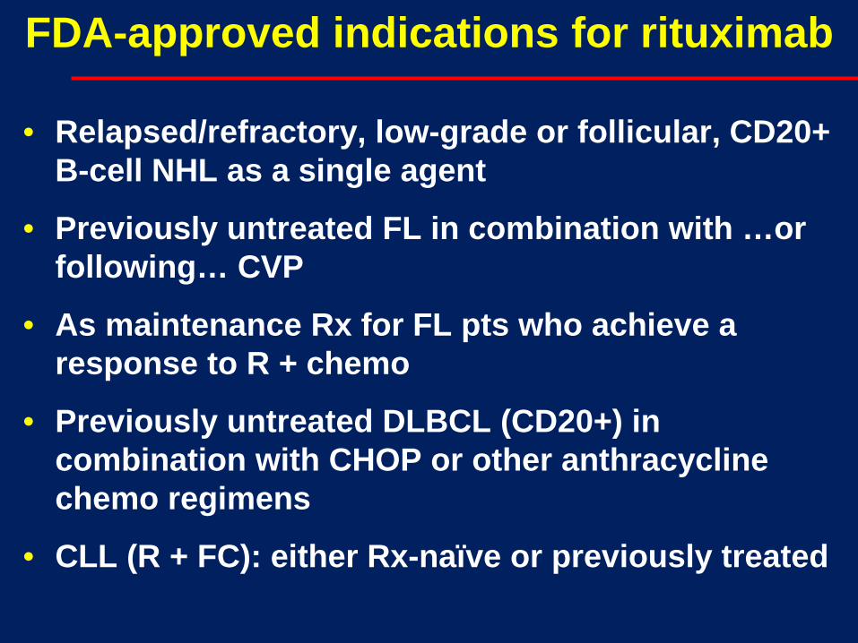
FDA-approved indications for rituximab
• Relapsed/refractory, low-grade or follicular, CD20+ B-cell NHL as a single agent
• Previously untreated FL in combination with …or following… CVP
• As maintenance Rx for FL pts who achieve a response to R + chemo
• Previously untreated DLBCL (CD20+) in combination with CHOP or other anthracycline chemo regimens
• CLL (R + FC): either Rx-naïve or previously treated

Next Generation anti-CD20 mAbs (+ more)
Name Comparison to Rituximab Status Ofatumumab1,2 •Human mAb
•Novel membrane proximal CD20 epitope
•Stronger CDC •Slower dissociation rate •Stronger binding to B-cells
•FDA-approved in r/r CLL •S/P Ph III in rituximab-refractory FL •Ph III: in CLL, FL, DLBCL •Several Ph II trials (also RA and MS)
GA1011 =
Obinutuzumab •Type II anti-CD20 (glycol-engineered Fc Region)
•Increased ADCC/Apoptosis •Stronger binding to effectors •Limited CDC
•S/P Ph I trials •Ph III Benda vs. Benda + GA101 in rituximab-refractory indolent NHL
•Several Ph II trials
Veltuzumab1 •Humanized IgG1 mAb •Single a.a. change in CDR3-VH (Asn to Asp)
•Epratuzumab framework •Slower dissociation rate •Stronger CDC •Enhances epratuzumab activity
•Low-dose subq formulation
•S/P Ph I/II studies (IV) •Phase I/II sub q in NHL/CLL •Phase I subq in ITP •Phase I combo with Milatuzumab (anti-CD74): Christian et al; ASH 2011, Abstr # 3707
1. Robak T & Robak E. Biodrugs 2011;25:13–25; 2. Lin TS. Pharmacogenomics and Personalized Medicine 2010;3:51–59

Type I mAbs Type II mAbs
Localize CD20 to lipid rafts Do not localize CD20 to lipid rafts
High CDC Minimal CDC ADCC activity ADCC activity Full number of binding sites /B-cell
Half number of binding sites / B-cell
Weak homotypic aggregation Strong homotypic aggregation
Limited direct apoptosis Strong direct apoptosis
Rituximab Ofatumumab Veltuzumab Ocrelizumab AME-133 PRO131921
Tositumumab (B1) GA101=Obinutuzumab
CD20: Type I and Type II mAbs

GA101: Type II Glycoengineered anti-CD20 mAb (Obinutuzumab)
ADCC, antibody-dependent cell-mediated cytotoxicity CDC, complement-dependent cytotoxicity Mössner et al. Blood 2010
Lower CDC Type II vs. Type I antibody
Effector cell
Increased Direct Cell Death Type II vs. Type I antibody
Enhanced ADCC Glycoengineering for
increased affinity to FcγRIIIa
CD20 FcγRIIIa
Complement GA101
B cell
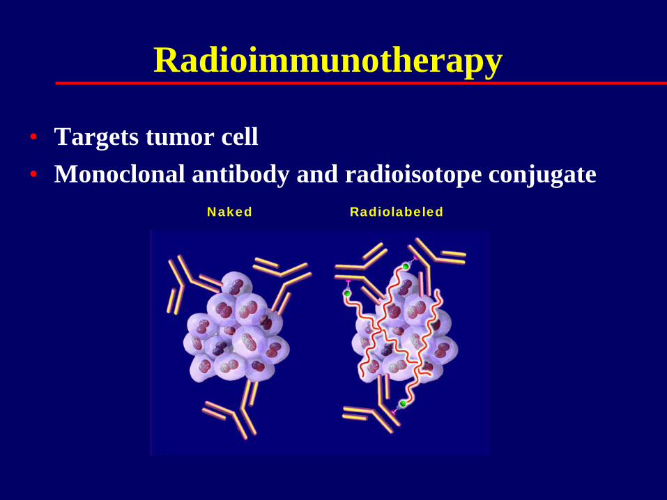
Radioimmunotherapy
• Targets tumor cell • Monoclonal antibody and radioisotope conjugate
Naked Radiolabeled

Efficacy of Radioimmunotherapy Enhanced Through the Crossfire Effect
Unlabeled “cold” Antibody Radiolabeled Antibody
Courtesy of Andrew Zelenetz, M.D.

Conjugated
• Yttrium-90 ibritumomab tiuxetan (Zevalin®) – β only – shorter t1/2 (64 hrs) – dosing based on wt +
PLTs
• Iodine-131 tositumomab (Bexxar®) – β and γ – longer t1/2 (192 hrs) – γ-emission allows dosing – need SSKI to protect
thyroid

BiTE® Technology: Blinatumomab
An investigational bispecific single-chain antibody construct with dual specificity for the CD19 and CD3 antigens on B cells
Nagorsen and Baeuerle. Exp Cell Res 2011
Apoptosis of tumor cells Membrane blebbing Activation of caspases Cleavage of PARP Fragmentation of DNA Morphological changes

BiTE® Technology: Blinatumomab Pharmacodynamic analysis in NHL patients showed complete
depletion of B lymphocytes from the circulation at blinatumomab doses ≥5 μg/m2/d, the depletion being faster at higher doses.
Encouraging single-agent activity in both adult and pediatric patients with ALL, as well as adult patients with NHL
Currently under investigation in 5 trials: Phase 1 trial for adult patients with relapsed/refractory NHL Two Phase 2 trials for adult patients with relapsed/refractory ALL Phase 1/2 trial for pediatric patients with relapsed/refractory ALL Phase 2 trial for adult ALL patients with minimal residual disease
(MRD)
Hijazi et al. ASCO 2013

Blinatumomab: Safety
CNS-related adverse events resulting in discontinuation
Most common clinical adverse events are flu-like and are of grade 1 or 2 (pyrexia, headache, chills, fatigue) Transient: Seen only during first days following start of
infusion Caused by onset of T cell activation (first dose reaction)
Most common laboratory abnormalities are lymphopenia and leukopenia Related to mode of action: Initial T cell redistribution and
sustained B target cell depletion

Antibody-Drug Conjugates (ADCs)
• Arose as an effort to combine cytotoxic chemotherapy and antibody specificity in order to obtain the benefit of their complementarity
• The antibody can be used to direct the cytotoxic agent to the tumor cell and thereby accomplish 2 objectives:
• Diminish the side effect profile of the cytotoxic agent
• Enable delivery of a more potent therapeutic because of the ability to control the target and the side effects

Components of an ADC
• The cancer, or target, antigen
• The antibody to that target
• The linker that connects the drug to the antibody
• The drug itself

The Target Antigen
• Should have high expression on a tumor
• Should have little or no expression in normal tissue
• Should be present on the cell surface
• Should be an internalizing antigen

Optimal Target Antigen for ADC

The Linker: Cleavable vs Noncleavable
• The linker of an ADC should be stable in the circulation so that the cytotoxic agent is not released systemically where it can be internalized into normal, nontarget cells
• The linker should also maintain attachment of the cytotoxic agent (the conjugate to the antibody) until the ADC reaches the tumor and is internalized1
• The early, cleavable linkers were too labile,2 which led to release of free drug in the circulation and consequent off-target toxicity
• Approximately 10 years ago, a non-cleavable linker was developed
• This type of linker is extremely stable in the circulation, and it prevents premature release of the cytotoxic agent into the circulation2
1. Teicher BA, Chari RV. Clin Cancer Res. 2011;17:6389-6397. 2. Ducry L, Stump B. Bioconjug Chem. 2010;21:5-13.
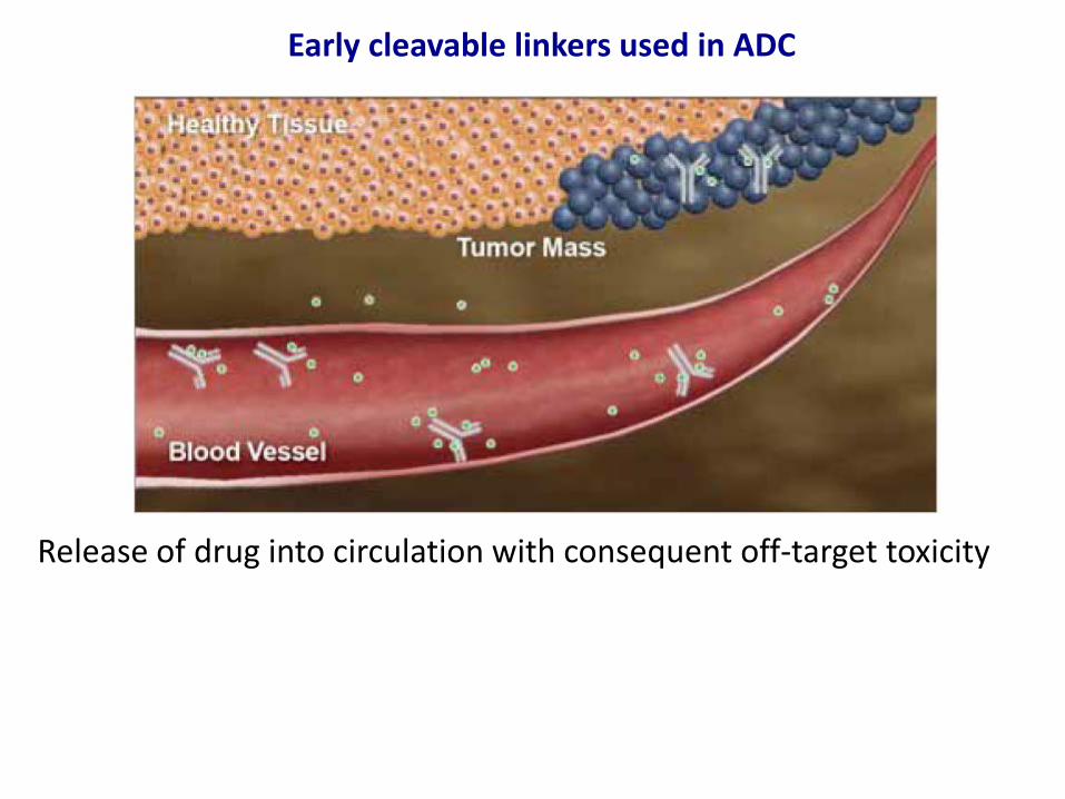
Early cleavable linkers used in ADC
Release of drug into circulation with consequent off-target toxicity

Development of a non-cleavable linker for ADC
“Stable” in circulation and prevents premature release of cytotoxic agent into circulation

Cytotoxic Agents
• The most common cytotoxic agents currently used in ADCs—maytansinoids and monomethyl auristatin E—have IC50s that are 100-1,000–fold more potent than those of conventional chemotherapeutic agents from the same or a similar class
• Most current ADCs use a ratio of cytotoxic drug to antibody in the range of 2:1 to 4:1

Brentuximab Vedotin (SGN35) Antibody-drug conjugate (ADC) directed to CD30
Expressed on virtually all Reed Sternberg and ALCL cells
Present in several T-cell lymphoproliferative diseases
In healthy tissue: limited to activated B and T lymphs and NK cells
Granted accelerated FDA approval in August 2011 for 2 indications:
Hodgkin lymphoma patients who relapse after autologous
transplant or fail at least two prior multi-agent chemotherapy
regimens if transplant ineligible
Systemic anaplastic large cell lymphoma (ALCL) patients who
fail at least one prior multi-chemo regimen

Mechanism of action of brentuximab vedotin
Deng C et al. Clin Cancer Res 2013;19:22-27
©2013 by American Association for Cancer Research

Brentuximab Vedotin Significant Adverse Events
Grade 3-4 (from Phase II studies) Peripheral neuropathy 8-10% Neutropenia 20% Febrile neutropenia 0% Thrombocytopenia 8-14%
Progressive Multifocal Leukoencephalopathy
Pulmonary Toxicity when given in combination
with Bleomycin

Preferred term
ABVD with brentuximab vedotin
N=25
AVD with brentuximab vedotin
N=26 Any event 11 (44) 0
Pulmonary toxicity 9 (36) 0
Interstitial lung disease 1 (4) 0
Pneumonitis 1 (4) 0
Pulmonary Toxicity
Events generally occurred during Cycles 3−4
Two patient deaths were associated with pulmonary toxicity
Events resolved in 9 of 11 patients (82%)
8 of 11 patients with events discontinued bleomycin and were able to complete treatment with AVD combined with brentuximab vedotin
Ansell S.M. et al. American Society of Hematology (ASH) Annual Meeting; December 8-11, 2012; Atlanta GA (Abstract 798).

Other ADCs in Clinical Trials for Lymphoid Malignancies
• Inotuzumab ozogamicin (CMC-544), a humanized anti-CD22 antibody conjugated to calicheamicin, a potent DNA-binding antibiotic
• SAR3419, a humanized IgG1 anti-CD19 monoclonal antibody conjugated to the maytansinoid derivative DM4
• Anti-CD22 or -CD79b conjugated to MMAE
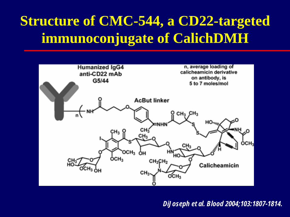
Structure of CMC-544, a CD22-targeted immunoconjugate of CalichDMH
DiJoseph et al. Blood 2004;103:1807-1814.

Anti-CD22 and -CD79b: MOA
Step 1 ADC specifically binds to
corresponding BCR
Step 2 Once bound, ADC
internalized into target cell
Step 3 Cytotoxic gent released
inside target cell, leading to microtubule disruption and
cell death
Beck et al. MAbs 2012; Polson et al. Blood 2007; http://www.biooncology.com/pipeline-molecules/anti-cd79b

Anti-CD79b (DCDS4501A) and -CD22 (DCDT2980S) in NHL: Phase 1 Safety
Common AEs (all grades)
Diarrhea Fatigue Nausea Neutropenia
(59% for anti-CD79b and 26% for CD22 )
Decreased appetite
Vomiting Peripheral
edema
Hyperglycemia Constipation Peripheral
neuropathy Pyrexia Leukopenia Chills Cough
Anti-CD79b Anti-CD22
≥3 AEs in ≥ 10% of patients Anti-CD22: Neutropenia (24%) Anti-CD79b: Neutropenia (39%) and leukopenia (12%)

Conclusion/Future • Exciting era of biotechnology: Continue to advance our
understanding of mAb structure vs function and lead to the production of even “more effective” mAbs and innovative immunoconjugates in the future
• Future: “Individualized Rx”: Choose which specific mAb(s) to use based on: target (e.g. epitope) and the predominant MOA (unlabeled biologically-augmented vs drug vs toxin vs radiolabel) we wish to achieve
• Major challenge: Determining these novel agents true value and how to optimize their use in today’s clinical arena

44