antibodies used in this study antibody supplier catalog ... · agilent 1290 uhplc and 6490 triple...
TRANSCRIPT

Antibodies used in this study Antibody Supplier Catalog
Number IHC Dilution
IF Dilution Western Blot Dilution
P53 Vector Labs VP-P956 1:500 P53 Novacastra P53-
CM5P-L 1:1000 1:5000
RFP Rockland 600-401-379
1:300 1:300
pERK1/2 Cell Signaling 4370L 1:100 1:1000 tERK1/2 Cell Signaling 4695S 1:1000 pAKT Cell Signaling 4060L 1:1000 tAKT Cell Signaling 9272S 1:1000 CD45 BD
Biosciences 553076 1:200
pSTAT3 Cell Signaling
9131
1:100
CK19 (TROMAIII)
Iowa Developmental Hybridoma Bank
- 1:100 1:100
Ki67 Vector Laboratories
VP-RM04 1:100
Cleaved Caspase 3
Cell Signaling 9961 1:300
β-Actin Santa Cruz SC-69879 1:2500 Primers Used in this study
Gene Forward Primer Reverse Primer Cyclophilin TCACAGAATTATTCCAGGATTCATG
TGCCGCCAGTGCCATT
Bax AAACTGGTGCTCAAGGCCCT
AGCAGCCGCTCACGGAG
Bcl-2 CCGGGAGAACAGGGTATGATAA
CCCACTCGTAGCCCCTCTG
Bcl-X GGTCGCATCGTGGCCTTT
TCCGACTCACCAATACCTGCAT
Bak TATTAACCGGCGCTACGACAC
CTTAAATAGGCTGGAGGCGATCTT
Cdkn1a CCTGGTGATGTCCGACCTG CCATGAGCGCATCGCAATC
(p21) Thbs1 GGGGAGATAACGGTGTGTTTG
CGGGGATCAGGTTGGCATT
Cdh1 (E-Cadherin)
CAGCCTTCTTTTCGGAAGACT
GGTAGACAGCTCCCTATGACTG
Hey1 GCGCGGACGAGAATGGAAA
TCAGGTGATCCACAGTCATCTG
Igf2 GTGCTGCATCGCTGCTTAC
ACGTCCCTCTCGGACTTGG
Vim CGTCCACACGCACCTACAG
GGGGGATGAGGAATAGAGGCT
Twist CGGGTCATGGCTAACGTG
CAGCTTGCCATCTTGGAGTC
Zeb1 CATTTGATTGAGCACATGCG
AGCGGTGATTCATGTGTTGAG
Metabolomics details Label-free targeted metabolomics
Agilent 1290 UHPLC and 6490 Triple Quadrupole (QqQ) Mass Spectrometer (LC-MS) was used in this study. Agilent MassHunter Optimizer and Workstation Software LC/MS Data Acquisition for 6400 Series Triple Quadrupole B.08.00 was used for standard optimization and data acquisition. Agilent MassHunter Workstation Software Quantitative Analysis Version B.0700 for QqQ was used for data analysis.
For reversed-phase chromatography (RPC), a Waters Acquity UPLC BEH TSS C18 column (2.1 x 100mm, 1.7µm) column was used with mobile phase (A) consisting of 0.5 mM NH4F and 0.1% formic acid in water; mobile phase (B) consisting of 0.1% formic acid in acetonitrile. Gradient program: mobile phase (B) was held at 1% for 1.5 min, increased to 80% in 15 min, then to 99% in 17 min and held for 2 min before going to initial condition and held for 10 min.
For hydrophilic interaction chromatography (HILIC), a Waters Acquity UPLC BEH amide column (2.1 x 100mm, 1.7µm) column was used with mobile phase (A) consisting of 20mM ammonium acetate, pH 9.6 in water; mobile phase (B) consisting of acetonitrile. Gradient program: mobile phase (B) was held at 85% for 1 min, increased to 65% in 12 min, then to 40% in 15 min and held for 5 min before going to initial condition and held for 10 min.
Both columns were at 40 ̊C and 3 µl of each sample was injected into the LC-MS with a flow rate of 0.2 ml/min. Calibration of TOF MS was achieved through Agilent ESI-Low Concentration Tuning Mix. Optimization was performed on the 6490 QqQ in the positive or negative mode for the RPC or HILIC respectively for each of 220 standard compounds to get the best fragment ion and other MS parameters for each standard. Retention time for each of 220 standards was measured from a pure standard solution or a mix standard solution. The LC-MS/MS method was created with dynamic dMRMs with RTs, RT windows and MRMs of all 220 standard compounds.
Key parameters of AJS ESI in both the positive and the negative acquisition modes are: Gas temp 275 ̊C, Gas Flow 14 l/min, Nebulizer at 20 psi, SheathGasHeater 250 ̊C, SheathGasFlow 11 l/min, and Capillary 3000 V. For MS: Delta EMV 200V or 350V for the positive or negative acquisition mode respectively and Cycle Time 500ms and Cell Acc 4V for both modes.

Metabolomics data post-processing and bioinformatic analysis
The QqQ data pre-processed with Agilent MassHunter Workstation Software Quantitative Analysis were post-processed for further quality control in the programming language R. We calculated coefficient of variation (CV) across replicate samples for each metabolite given a cut-off value of peak areas in both the positive and the negative modes. We then compared distributions of CVs for the whole dataset for a set of peak area cut-off values of 0, 1000, 5000, 10000, 15000, 20000, 25000 and 30000 in each mode. A noise cut-off value of peak areas in each mode was chosen by manual inspection of the CV distributions: 5000 for the positive mode and 0 for the negative mode. Each sample is then normalized by the total intensity of all metabolites to reflect the same protein content as a normalization factor. We then retained only those metabolites with at least 2 replicate measurements. The remaining missing value in each condition for each metabolite was filled with the mean value of the other replicate measurements. Finally, each metabolite abundance level in each sample was divided by the median of all abundance levels across all samples for proper comparisons, statistical analyses, and visualizations among metabolites. The statistical significance test was done by a two-tailed t-test with a significance threshold level of 0.05. The p-values were not adjusted in favor of more flexible biological interpretation. All other bioinformatics analyses including graphs and plots were also done using R/Bioconductor.

Anti-p53
Doxy + + - - +
Anti- β-actin
Supplemental Figure 1 – TREp53R270H Validation in vitro and in vivo (A) Western Blot for p53 to confirm p53R270H expression upon dox administration. (B) IHC for RFP (dsRed) and p53 in Control and Krt5-tTa; TREp53R270H animal epithelium to confirm p53R270H expression.
Ant
i-p53
A
nti-R
FP
Control Krt5-tTa; TREp53R270H
A
B
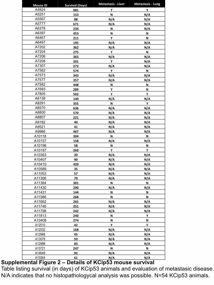
MouseID Survival(Days) Metastasis-Liver Metastasis-Lung
A4924 681 Y YA5251 153 N N/AA5567 88 N/A N/AA5777 671 N/A N/AA6378 234 N N/AA6397 453 N NA6467 211 Y NA6497 195 N/A N/AA7202 362 N/A N/AA7204 275 Y NA7206 363 N/A N/AA7208 201 Y N/AA7307 273 N/A N/AA7562 574 Y NA7573 343 N/A N/AA7577 357 N/A N/AA7582 448 N NA7693 289 Y NA7805 502 Y YA8138 140 N/A N/AA8291 355 N YA8570 636 N/A N/AA8800 570 N/A N/AA8807 221 N/A N/AA9182 40 N/A N/AA9521 41 N/A N/AA9966 467 N/A N/AA10118 304 N NA10157 158 N/A N/AA10196 58 N NA10197 260 Y YA10363 39 N/A N/AA10407 90 N/A N/AA10410 420 N/A N/AA10589 35 N/A N/AA11053 57 N/A N/AA11306 79 N/A N/AA11384 301 N NA11430 290 N/A N/AA11431 144 N NA11586 268 N NA11662 265 N/A N/AA11746 251 N/A N/AA11798 242 N/A N/AA11813 240 N YA10408 274 N NA12510 42 Y YA12532 168 N/A N/AA12968 45 N/A N/AA13076 59 N/A N/AA12988 83 N/A N/AA13721 247 N NA14649 92 N/A N/AA15564 61 N/A N/A
Supplemental Figure 2 – Details of KCip53 mouse survival Table listing survival (in days) of KCip53 animals and evaluation of metastasic disease. N/A indicates that no histopathologycal analysis was possible. N=54 KCip53 animals.

Supplemental Figure 3 – KPR270HC animals develop metastatic pancreatic tumors (A) Scheme of Pdx1-Cre; LSLKrasG12D; Tp53R270H/+ animals, termed KPR270HC here (B) Survival curve for KC and KPR270HC animals. (C) H&E from pancreas, liver and lung from two separate KPR270HC animals with tumors, shown at 20x magnification. N=43 KPR270HC and 39 KC. Survival significance analysis by Log Rank Test.

0 200 400 6000
50
100
Days
Perc
ent s
urvi
val
KCip53
KCip53; p53fl/+
n.s.
MouseID Age(Days) Tumor Mets?
A13458 275 Y N
A13720 220 Y N
A13984 106 Y N
A14561 171 NoHistology
A14857 106 NoHistology
A14915 139 Y N
A15822 60 NoHistology
A B
C
4x 20x
KCip53;p53fl/+Tumor
Supplemental Figure 4 – KCip53;p53fl/+ animals develop locally invasive pancreatic tumors (A) Survival of Ptf1aCre; LSL-KrasG12D; TREp53R270H; R26rtTa/rtTa; p53flox/+ animals, termed KCip53; p53fl/+ here. (B) Table detailing survival length of KCip53; p53fl/+
animals, with metastasis information for animals from which histology was available. (C) H&E from the tumor of a KCip53;p53fl/+ animal. Arrow indicates area of local invasion into the adjacent intestine. Histology shown at both low (4X) and high (20x) magnification, as noted on images. n= 18 KCip53; p53fl/+ animals in survival curve, 7 animals with tumors. Survival significance analysis by Log Rank Test.

KC10weeksONDox KCip5310weeksNODox
Supplemental Figure 5 – KCip53 histology at ten weeks of age without dox H&E analysis of KCip53 animals never on dox at 10 weeks of age compared to KC animals on dox at 10 weeks of age. Histology images shown at 20x magnification. n=3 KC on dox, 4 KCip53 never on dox.
H&E
20X

KC KCip5310weeks20X
Supplemental Figure 6 – KCip53 histology at ten weeks of age IHC for (A) CD45 (20x magnification) and (B) pSTAT3 (40x magnification) in 10-week-old KC or KCip53 animals on dox. N=3-6 animals per condition. For CD45 at least 5 high power fields were analyzed per animal.
A
B 40x
CD45
pSTA
T3

KC KCip532daysoffdox
Supplemental Figure 7 – KCip53 histology at two days and one week off dox (A) Scheme for dox treatment in KCip53 animals. (B) H&E analysis of KC and KCip53 animals taken off dox. Histology images shown at 20x magnification. n=3-6 animals per condition.
KCip531weekoffdox
1w 4w 10w
p53R270H ON p53R270H OFF
2d
A
B20X
E-CadherinKC
ip53
OnDo
xKC
ip533weeksOffDo
x
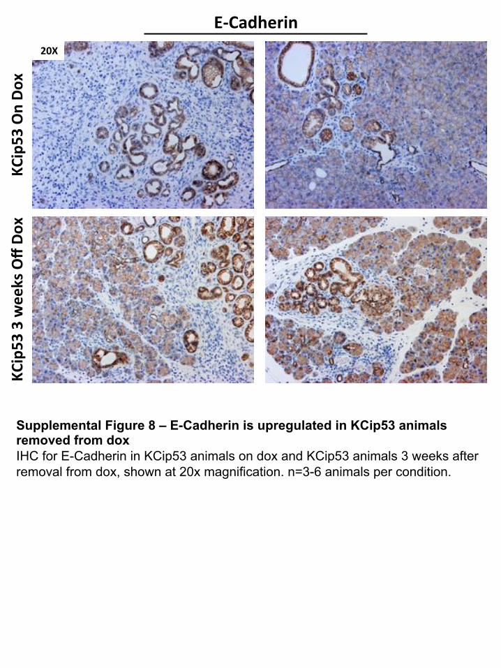
Supplemental Figure 8 – E-Cadherin is upregulated in KCip53 animals removed from dox IHC for E-Cadherin in KCip53 animals on dox and KCip53 animals 3 weeks after removal from dox, shown at 20x magnification. n=3-6 animals per condition.
20X

Supplemental Figure 9 – KCip53-1 Cell line validation by dsRed and p53 gene target expression (A) Bright field and dsRed fluorescence images of KCip53-1 cells before and after cell sorting for dsRed expression. (B) qRT-PCR analysis for p53 targets in KCip53-1 cells grown with or without dox. For all qPCR unpaired t-test with Welsh’s correction was used, and data is represented as mean with SD. N=3.
Unsorted dsRedPosi-ve dsRedNega-veBrigh4
ield
dsRe
dA
B
No Dox
Plus D
ox0.050
0.055
0.060
0.065
0.070
Thrombospondin1
Rel
ativ
e E
xpre
ssio
n
p=0.08
No Dox
Plus D
ox0.000
0.002
0.004
0.006
0.008
0.010
p21
Rel
ativ
e E
xpre
ssio
n
p<0.0001
No Dox
Plus D
ox0.8
1.0
1.2
1.4
1.6
1.8
2.0
Vimentin
Rel
ativ
e E
xpre
ssio
n
p=0.0007

269 270 271 269 270 271 269 270 271
WT KCip53-1 KCip53-2
WT KCip53-1 KCip53-2
>500(GenomicDNA)
250(TransgeneDNA)
A
B
Supplemental Figure 10 – KCip53 Cell line sequencing of Trp53 exon 7-8. (A) PCR amplification of p53 amino acid 270 region in mouse pancreatic cancer cells that are wild type at that region compared to KCip53 cell lines, and scheme of primer placement. (B) Sequencing results from same cell lines showing the wild type sequence at codon 270 (left) and a single sequence containing the point mutation at codon 270 in both KCip53 cell lines (middle and right).
Exon 7 Exon 8
Exon 7 Exon 8 *
*

Supplemental Figure 11 – Histology of subcutaneous tumors from KCip53-1 cells IHC analysis of subcutaneous tumors from KCip53-1 cells including (A) CK19, (B) Ki67, (C) Cleaved Caspase 3 and (D) dsRed. Quantification of number of dsRed expressing cells per high power field in final subcutaneous tumors from KCip53-1 cells, analyzed using multiple unpaired t-tests. Represented as mean with SD. All histology images shown at 20x magnification. n=6-8 tumors per condition, experiment repeated three times.
DsRed
CK
19
Ki67
NoDox PlusDox OffDoxCleavedCaspase3
B
A
C
D
No Dox
Plus Dox
Off Dox
0
20
40
60
80
100
Cel
ls P
er H
FP
p=0.001 p=0.01p=0.002
dsRed Quantification
20X

A
Supplemental Figure 12 –PDGFRb expression in KCip53 tumors (A) IHC analysis for PDGFRβ (20x magnification) of final subcutaneous tumors grown from KCip53-2 cells shows prevalent expression in the stromal compartment. N=6-8 tumors per group. (B) qRT-PCR analysis of PDGFRβ levels in KCip53 animals on dox compared to those removed from dox for 3 weeks. A trend towards higher expression upon dox removal might be explained by a change in the relative stroma composition. Unpaired t-test with Welsh’s correction was used, and data is represented as mean with SD. N=3-6 animals per condition.
NoDox PlusDox OffDoxPD
GFRβ
B
KCip53 O
n Dox
KCip53 3w
Off D
ox0.06
0.07
0.08
0.09
0.10
0.11PDGFRβ
Rel
ativ
e Ex
pres
sion
p=0.04
20X

No Dox
Plus Dox
Off Dox
0
1
2
3
4
Cm
3
Final Tumor Volumep=0.01
p=0.18p=0.25
Day 15
Day 16
Day 22
Day 23
Day 26
Day 30
0
500
1000
Perc
ent C
hang
e
Orthotopic Tumor Growth
No DoxPlus DoxOff Dox
Supplemental Figure 13 - Assessing the effect of mutant p53R270H expression on orthotopic tumor growth (A) Scheme for orthotopic tumor growth and MRI measurement (C) Final tumor volume. (D) H&E for final orthotopic tumors and representative liver metastases (20x magnification). (E) Quantification of average metastasis size. (F) Quantification of average number of metastases. N=3-4 animals per group. Analyzed by multiple unpaired t-tests, shown as mean with SD.
B
D
C
F
A
Harvest tumors
p53R270H ON p53R270H OFF
2w
MRI
NoDox PlusDox OffDox
Tumor
Liver
No Dox
Plus Dox
Off Dox
No Dox
Plus Dox
Off Dox
0.0
5.0×10⁶
1.0×10⁷
1.5×10⁷
Size
Liver Lung
p=0.04
Metastasis Size G
No Dox
Plus Dox
Off Dox
No Dox
Plus Dox
Off Dox
0
5
10
15
Num
ber P
er M
ouse
Liver Lung
Metastasis Number
20X

Supplemental Figure 14 – Further Differentially Regulated Pathways in Plus Dox Tumors Pathway analysis in “No dox” compared to “Plus dox” tumors. N=4 samples per group for RNA sequencing.

Supplemental Figure 15 – Metabolomics Analysis of KCip53 cells (A) Scheme for cell growth for metabolomics analysis (B) Heatmap of the significant metabolites changed in KCip53-1 cells with p53R270H (Plus dox) or without (No dox). (C) Summary of changed TCA intermediates and substrates. Unpaired t-test with Welsh’s correction was used, and data is represented as mean with SD. N=3 samples per group.
NoDox
PlusDox
KCip53-1
C
B
TCA Cycle
A NodoxorPlusdox3days Harvestformetabolicflux
assayandmetabolomics
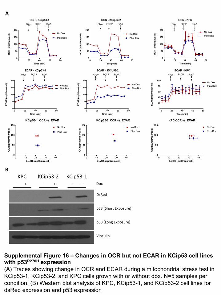
Supplemental Figure 16 – Changes in OCR but not ECAR in KCip53 cell lines with p53R270H expression (A) Traces showing change in OCR and ECAR during a mitochondrial stress test in KCip53-1, KCip53-2, and KPC cells grown with or without dox. N=5 samples per condition. (B) Western blot analysis of KPC, KCip53-1, and KCip53-2 cell lines for dsRed expression and p53 expression
KPC KCip53-2 KCip53-1Dox
DsRed
Vinculin
+- +- +-
p53(LongExposure)
p53(ShortExposure)
0 20 40 60 800
50
100
150
200
OCR - KCip53-1
Time (min)
OC
R (p
mol
/min
/cel
l)
Oligo FCCP R/AA
Plus Dox
No Dox
0 20 40 60 800
20
40
60
80
ECAR - KCip53-1
Time (min)
ECA
R (m
pH/m
in/c
ell)
Oligo FCCP R/AA
Plus DoxNo Dox
0 20 40 60 800
50
100
150
200
OCR - KCip53-2
Time (min)
OC
R (p
mol
/min
/cel
l)
Oligo FCCP R/AA
Plus Dox
No Dox
0 20 40 60 800
50
100
150
200
OCR - KPC
Time (min)
OC
R (p
mol
/min
/cel
l)
No DoxPlus Dox
Oligo FCCP R/AA
0 20 40 60 800
20
40
60
80
100
ECAR - KPC
Time (min)
ECA
R (m
pH/m
in/c
ell) No Dox
Plus Dox
Oligo FCCP R/AA
0 20 40 60 800
20
40
60
80
ECAR - KCip53-2
Time (min)
ECA
R (m
pH/m
in/c
ell)
Oligo FCCP R/AA
Plus DoxNo Dox
A
B

Supplemental Figure 17 – Histology of KCip53-1 subcutaneous tumors with MEK inhibition and p53R270H expression Resulting histology from final tumors from MEK inhibition and on and off dox subcutaneous tumor growth groups (20x). N=10 tumors per group. IHC for pERK1/2, Ki67 and Cleaved Caspase 3. IF for dsRed as a surrogate for p53R270H expression. Western Blot for pERK1/2 to confirm MEK inhibition and dsRed to confirm p53R270H expression. Each lane represents lysate from one subcutaneous tumor.
Vehicle On Dox
MEK Inhibited On Dox
Vehicle Off Dox
MEK Inhibited Off Dox
pERK
1/2
DsRe
dCK
19DAP
IKi67
CleavedCa
spase3
pERK1/2
tERK
Vehicle-DOX
MEKi-DOX
Vehicle+DOX
MEKi+DOX
β-acbn
Anb-RFP(DsRed)
Gomori
20X

Supplemental Figure 18 – Histology of KCip53-1 subcutaneous tumors with MEK and/or PI3K inhibition Resulting histology from final tumors from MEK and/or PI3K inhibition in subcutaneous tumor growth groups, shown at 20x magnification. N=8 tumors per group. IHC for pERK1/2, Ki67 and Cleaved Caspase 3. Western Blot for pAKT and total AKT to confirm PI3K inhibition and pERK1/2 to confirm MEK inhibition. Each lane represents lysate from one subcutaneous tumor.
Vehicle MEK Inhibited PI3K Inhibited MEK and PI3K
Inhibited pE
RK1/2
Ki67
CleavedCa
spase3
pAKT
tAKT
pERK1/2
tERK
Vehicle MEKi AKTiMEKiandAKTi
Gomori
20X