antibodies that inhibit fusionof human - pnas.org · antibodies that inhibit...
TRANSCRIPT

Medical Sciences: Correction
Correction. In the article "Antibodies that inhibit fusion ofhuman immunodeficiency virus-infected cells bind a 24-amino acid sequence of the viral envelope, gp120" by JamesR. Rusche, Kashi Javaherian, Charlene McDanal, JoanPetro, Debra L. Lynn, Raymond Grimaila, Alphonse Lang-
Proc. NatI. Acad. Sci. USA 86 (1989) 1667
lois, Robert C. Gallo, Larry 0. Arthur, Peter J. Fischinger,Dani P. Bolognesi, Scott D. Putney, and Thomas J. Matthews,which appeared in number 9, May 1988, ofProc. Natl. Acad.Sci. USA (85, 3198-3202), the authors request that Table 5 onp. 3202 be replaced with the table that appears below.
Table 5. HIVRP135 (BH10)IIIB (BH10)IIIB (BH8)RFMNSCWMJ-2LAV-MALSF-2NY5Z3
isolate variability of RP135N N T R K
C T R P N N N T R K
C - - - - Y -K--C T RC - - - Y --V - RC ---G - - - - RC---Y--__-_
C K-C - - - G SDKKI - Q
S I R I QS I R I QK - - - -
S - T- - HS - HS L SG - HS - yG - AS - R
R G P G R A F V T I G KR G P G R A F V T I G K
K ----V I Y A T - QI.-- - - - - Y - T K NI.-- - - - - Y A T - DI .-- - - -- R -R EF - - - Q- L Y -T -I .-- - - -- H- T - RI - - - - T L Y A R E -I - - -K V - Y A K - G
I GI G N
I - -DI -- TI -- DI --IDI - - II V- DI -- DI -- DI T-
M R Q A H C
I - K - -CI - - -- CI - - -- CI - - -- CI - R - YCI - K - -CI - - -- C
_ - C
The sequences listed (in single-letter amino acid code) are aligned at the cysteine residue at amino acid 302 by using thenumbering system of Ratner et al. (16). The listed sequences have all been published (15, 16, 22, 24, 25).

Proc. Nati. Acad. Sci. USAVol. 85, pp. 3198-3202, May 1988Medical Sciences
Antibodies that inhibit fusion of human immunodeficiencyvirus-infected cells bind a 24-amino acid sequence of theviral envelope, gpl20JAMES R. RUSCHE*, KASHI JAVAHERIAN*, CHARLENE MCDANALt, JOAN PETRO*, DEBRA L. LYNN*,RAYMOND GRIMAILA*, ALPHONSE LANGLOISt, ROBERT C. GALLO*, LARRY 0. ARTHUR§,PETER J. FISCHINGER¶, DANI P. BOLOGNESIt, SCOTr D. PUTNEY*, AND THOMAS J. MATTHEWSt*Repligen Corporation, One Kendall Square, Cambridge, MA 02139; tDepartment of Surgery, Duke University Medical School, Durham, NC 27710;*Laboratory of Tumor Cell Biology, National Cancer Institute, National Institutes of Health, Bethesda, MD 20892; tProgram Resources, Inc., National CancerInstitute-Frederick Cancer Research Facility, and 1Office of Director, National Cancer Institute-Frederick Cancer Research Facility, Frederick, MD 21701
Communicated by Maurice R. Hilleman, January 4, 1988
ABSTRACT Antisera to recombinant human immunode-ficiency virus (HIV) proteins containing the entire envelope,gpl60, or the central portion of the envelope, PB1, can inhibitfusion of virally infected cells in culture. This fusion inhibitionis HIV-variant specific-that is, anti-gpl60-IB inhibits fusionof isolate HTLV-IB-infected cells but not of isolate HTLV-IURF-infected cells. Both anti-gpl6O and anti-PB1 are com-pletely blocked in fusion inhibition activity by the addition ofPB1 protein. A 24-amino acid peptide (RP135, amino acids307-330) completely blocks fusion inhibition activity of bothantisera and also blocks the activity of serum from a chimpan-zee infected with HTLV-IB. Thus, the principal epitope thatelicits fusion-inhibiting antibodies is located in the centralportion of gp120.
Recent experiments to evaluate vaccine candidates able toprevent acquired immunodeficiency syndrome (AIDS) havefocused on the outer envelope of the human immunodefi-ciency virus (HIV). The envelope protein, gpl60, is glycosyl-ated and cleaved to an external glycoprotein, gpl20, and atransmembrane glycoprotein, gp4l (1, 2). gpl20 binds thecellular receptor CD4 (3, 4), and cells expressing the enve-lope gene product can fuse with CD4-positive cells in culture(5, 6). Fusion of infected and noninfected cells may be animportant component of the pathogenicity of HIV. Theassays used to evaluate humoral immune responses to viralsubunits include the ability of antiserum to block virusinfectivity (virus neutralization) or inhibit fusion of HIV-infected cells.
Purified gpl20 from virus-infected cells has been shown toelicit antibodies that neutralize the infectivity of HIV inculture (7). Recombinant envelope proteins expressed inmammalian cells (8), insect cells (9), or yeast (10) haveelicited neutralizing antibodies in test animals. A proteinmade in Escherichia coli, PB1, containing the central portionof the envelope gene (11), can also raise neutralizing anti-body. This shows that epitopes that elicit neutralizing anti-body can form in the absence of glycosylation and do notrequire the entire polypeptide. Native gpl20 (12) and therecombinant proteins listed above result in immune sera thatcan neutralize some but not all of the HIV isolates. Variant-specific neutralization with subunit immunogens suggeststhat the critical antigenic epitope(s) are located in hypervari-able regions of gpl20 (13, 14). A more complete understand-ing of the number and location of immunologically relevantprotein epitopes would facilitate a rational vaccine develop-ment.
We have analyzed recombinant proteins and peptides forthe ability to elicit HIV-neutralizing or fusion-inhibitingactivity. We have also used these proteins to block thefusion-inhibition activity of antisera in vitro. The results ofthese experiments highlight a single location in the viralenvelope as a target of fusion-inhibiting antiserum raised byeither subunit immunization in experimental animals or byvirus infection of chimpanzees. This antigenic epitope canexist on a synthetic peptide of 24 amino acids.
MATERIALS AND METHODSProteins. Recombinant gpl60-IIIB (rl6O-IIIB) was obtained
from insect cells infected with a recombinant baculovirus(9). A similar recombinant baculovirus expressing gp160 wasconstructed that contains sequences from the variant isolateHTLV-IIIRF (15); HTLV-III refers to earlier nomenclaturefor HIV-human T-cell leukemia virus type III and is usedhere to designate specific virus variants. Amino acids fromposition 132 through 731 are homologous to HTLV-IIIRFenvelope, and the remainder are homologous to the BH10clone from HTLV-IIIB (16). The recombinant envelopeprotein is glycosylated and inserted into the insect-cellmembrane but is not cleaved to gpl20 and gp4l. Purifiedrl60-IIIB and r160-RF were obtained by polyacrylamide gelelectrophoresis and used to immunize goats.Recombinant proteins PB1 (11) and its subfragments subi,
sub2, sub6, and sub8 are fusion proteins produced in E. coli.These proteins contain amino acid sequences homologous tothe BH10 clone of HTLV-IIIB (16): PB1, amino acids295-474; subl, 295-350; sub2, 295-404; sub6, 350-474; andsub8, 350-404. All five proteins were purified to at least 90%purity from cell extracts by ion-exchange chromatographyand gel filtration. Proteins PE3 and PENV9 (11) wereprovided by J. Ghrayeb (Centocor, Malvern, PA) and S.Petteway (duPont), respectively.
Protein Fragments and Peptides. Purified sub2 was cleavedat the single internal methionine by overnight reaction withCNBr at a ratio of 150 mol of CNBr per mol of sub2 protein.The fragments CNBr 1 (amino acids 295-333) and CNBr2(amino acids 334-404) were separated by reverse-phaseHPLC. The identity of each fragment was confirmed byN-terminal sequence analysis. Synthetic peptides RP134(amino acids 295-314), RP135 (amino acids 307-330), andRP136 (amino acids 295-332) contain sequences analogousto the BH10 clone of HTLV-IIIB. Synthetic peptides containa terminal cystinyl residue for crosslinking to carrier proteinprior to immunization. RP137 is analogous to RP135 except
Abbreviations: HIV, human immunodeficiency virus; HTLV-IIIisolates, early HIV isolates (human T-cell leukemia virus type III);r160, recombinant gp160.
3198
The publication costs of this article were defrayed in part by page chargepayment. This article must therefore be hereby marked "advertisement"in accordance with 18 U.S.C. §1734 solely to indicate this fact.

Proc. Natl. Acad. Sci. USA 85 (1988) 3199
for changes at HIV envelope position 316 (Gln-+Thr) and317 (Arg-)Lys). Peptides were prepared by solid-phasesynthesis, and subsequent purification was by reverse-phaseHPLC.
Sera. Antisera to PB1 (11) and rl6O-IIIB (9) were raised ingoats as described. Proteins subl, sub2, sub6, r160-RF, andPB1-RF were mixed with complete Freund's adjuvant andinjected into two goats for each immunogen. After 30 days,animals were reinjected with protein in incomplete Freund'sadjuvant, and blood samples were drawn 7 days after thesecond immunization. Synthetic peptides were crosslinkedthrough a sulfhydryl bond to keyhole limpet hemocyanin byusing succinimidyl 4-(N-maleimidomethyl) cyclohexane-1-carboxylate (Pierce), and 1 mg was used to immunize goatsas described above. Antibody titers of immune sera weredetermined by end-point dilution in solid-phase ELISA onmicrotiter plates containing PB1 protein.The chimpanzee was inoculated intravenously with 40
TCID50 (dose infecting 5Olo of cells in tissue culture) ofHTLV-IIIB, and blood was taken every 2 weeks afterinoculation. HIV was isolated from peripheral blood lym-phocytes obtained at the 4-wk bleeding. Antibodies to bothviral proteins gpl20 and p24 were found in blood extracted at6 weeks. Experiments described in Results were obtainedwith blood extracted at 12 weeks.
RESULTS
Sera to Recombinant Envelope Proteins Inhibit Fusion ofHIV-Infected Cells. Immune sera from goats immunized withrecombinant proteins r160 (gp160 made in insect cells; ref. 9)or PB1 (11) were examined for the ability to inhibit the fusionof uninfected CD4-positive lymphocytes with cells chroni-cally infected with HIV. Dilutions of immune sera weremixed with infected and uninfected cells and allowed toincubate 24 hr. A positive fusion inhibition is that dilution ofserum that shows at least 90%o decrease in the number ofsyncytia (giant cells). Fig. 1 shows that antisera to the entire
-._
cnC,)
0
I).0E
zU)0)cma
luv8080- --8
:::~8
60
40-
0 a
20
10 0 I0 I
10 20 40 80 160 320
1/ Serum Dilution
FIG. 1. Inhibition of cell fusion with goat antisera raised againstthe PB1 and r160 antigens. Serial 1:1 dilutions of each antiserumwere incubated with 5000 HTLV-IIIB chronically infected CEM (11)cells and 75,000 uninfected MOLT-4 cells in a 96-well half-area platefor 24 hr. Duplicate wells were tested for each dilution of serum, andthe number of giant cells (syncytia) were counted at a x 20 magni-fication. The serum levels shown indicate the final dilution in theassay for preimmune goat (o), goat anti-r160 (-); preimmune goat(o), and goat anti-PB1 (a).
envelope or to PB1 inhibited fusion of chronically infectedHTLV-IIIB lymphocytes. Concentrations of serum requiredto inhibit cell fusion were generally 5- to 10-fold greater thanconcentrations of serum required to inhibit infectivity of thevirus.The inhibition offusion observed with antisera to rl60-IIIB
or PB1-IIIB was variant specific. These sera did not preventthe fusion of chronically infected HTLV-IIIRF cells. Simi-larly, PB1-RF protein and r160-RF elicited immune seracapable of inhibiting fusion of HTLV-IIIRF-infected cells butnot of HTLV-IIIB-infected cells (Table 1). Thus, variantspecificity of immune sera with respect to fusion inhibitionparallels the results of virus-neutralization assays (9, 12).Antiserum capable of inhibiting fusion of infected cells can
be competitively blocked by proteins that contain the epi-tope target. For example, the activity of antiserum to PB1-IIIB was blocked when coincubated with PBl-IIIB protein(Table 1). The ability of PB1 protein to block fusion inhibi-tion was also variant specific. That is, PB1-III protein wascapable of blocking antiserum raised to PB1-IIIB, and PB1-RF protein was capable of blocking antisera raised to PB1-RF (Table 1).
Fusion Inhibition by Sera to the Entire Envelope Is Blockedby the Homologous PB1 Protein. To discern which regionscontain epitopes that elicit fusion-inhibiting antibodies, an-tisera to the entire envelope protein were tested for fusioninhibition in the presence of recombinant protein fragmentsof the HIV envelope. Immune serum from goats immunizedwith r160 isolated from insect cells expressing the HIVenvelope gene was tested in the presence of envelope proteinfragments listed in Fig. 2. Table 1 shows that although PB1protein contains less than one-third of the amino acidsencoded by the envelope gene, it removed all of the fusion-inhibition activity from serum raised to the entire envelope.This blockade of fusion inhibition was not observed withrecombinant proteins containing the amino-terminal portion(PE3) or the carboxyl-terminal portion (PENV9) of theenvelope (data not shown). The blockade of immune serumto r160 by PB1 protein was variant specific, as anti-rl60-RFwas blocked by PB1-RF but not PB1-IIIB protein (Table 1).These data confirm that the protein epitopes that elicit
fusion-inhibiting sera are variant specific and, in two distinctvariants, are located within the central portion of the enve-lope gene-that is, within PB1.PB1 Subfragments and Synthetic Peptides That Block Fu-
sion-Inhibiting Activity. A number of genetic constructionswere engineered to express various regions of the PB1-IIIBsequence, and the recombinant fusion proteins were pun-
Table 1. Variant specificity of fusion inhibition and blockage byPB1 proteins
ProteintVirus Serum* None PBl-IIIB PB1-RF
HTLV-IIIB Anti-PB1-IIIB 0 65 0Anti-PB1-RF 73 NDt NDAnti-r160-IIIB 0 71 0Anti-r160-RF 68 ND ND
HTLV-IIIRF Anti-PBl-IIIB 58 ND NDAnti-PB1-RF 0 0 75Anti-r160-IIIB 60 ND NDAnti-r160-RF 0 0 62
Cells chronically infected with the indicated viral isolate aremixed with uninfected cells, serum, and protein. The number ofsyncytia were determined in duplicate wells after 24 hr.*Serum was used at a 1:20 dilution. An average of 64 giant cells wasobserved in the presence of preimmune sera. A similar result wasobtained at serum dilutions of 1:10 and 1:40.tWhere indicated, PB1 protein was added to a concentration of 0.5,uM. ND, not done.
Medical Sciences: Rusche et al.
3200 Medical Sciences: Rusche et al.
295
gp 120
PE 3
Sub 6
Sub 2
Sub 8
I'm (I ~~~gp 160 la(IatI I WIr.
=i PB 1
759 863
l l
Sub 1
CnBr 2
CnBr 1
RP 136
RP 134 ,=4RP 135
NNTR KSIR I0RGPGRAFVCTIGKIGC
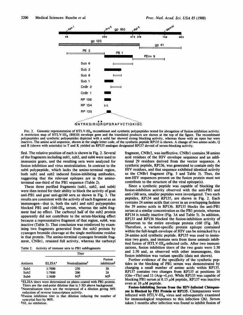
FIG. 2. Genomic representation of HTLV-IIIB recombinant and synthetic polypeptides tested for abrogation of fusion-inhibition activity.A restriction map of HTLV-IIIB (BH10) envelope gene and the translated products are shown at the top of the figure. The recombinantpolypeptides and synthetic polypeptides depicted with a solid bar showed strong blocking activity, whereas those with an open bar wereinactive. The amino acid sequence, shown in the single letter code, of the synthetic peptide RP135 is shown. A change of two amino acids, Qand R (shown with asterisks) to T and K yielded an RP135 analogue designated RP137 devoid of serum-blocking activity.
fied. The relative position of each is shown in Fig. 2. Severalof the fragments including subl, sub2, and sub6 were used toimmunize goats, and the resulting sera were analyzed forfusion inhibition and virus neutralization. In contrast to thesub6 polypeptide, which lacks the amino-terminal region,both subl and sub2 induced fusion-inhibiting antibodies,suggesting that the relevant epitopes are in the amino-terminal one-third of the PB1 sequence (Table 2).These three purified fragments (subl, sub2, and sub6)
were then tested for their ability to block the activity of goatanti-PBi and goat anti-gpl60 sera as shown in Fig. 3. Theresults are consistent with the activity of each fragment as animmunogen-that is, both the subl and sub2 polypeptidesblocked PB1 and rl60-IIIB serum, whereas the sub6 frag-ment had no effect. The carboxyl half of the sub2 proteinapparently did not contribute to the serum-blocking effectbecause a representative fragment of that region, sub8, wasinactive (Table 3). That conclusion was reinforced by exam-ining two fragments generated from the sub2 protein bycyanogen bromide cleavage at the single methionine residuein that protein. The amino-terminal cyanogen bromide frag-ment, CNBr1, retained full activity, whereas the carboxyl
Table 2. Activity of immune sera to PB1 subfragmentsTiter
FusionAntisera ELISA* Neutralizationt inhibition*Subl 1:7000 250 30Sub2 1:7800 200 20Sub6 1:3600 NIO NI§
*ELISA titers were determined on plates coated with PB1 protein.Titers are the end-point dilution that is 3 SD above background.
tNeutralization titers are the reciprocal of a dilution giving 50%0oreduction of reverse transcriptase activity.tFusion inhibition titer is that dilution reducing the number ofsyncytial foci by 90o.§NI, no inhibition.
fragment, CNBr2, was ineffective. CNBr1 contains 38 aminoacid residues of the HIV envelope sequence and an addi-tional 29 residues derived from the vector sequence. Asynthetic peptide, RP136, was generated to contain only theHIV residues, and that sequence exhibited identical activityas the CNBrl fragment (Fig. 3 and Table 3). Thus, thenon-HIV sequences present on the fusion protein must notcontribute to the structure of the viral epitope(s).
Since a synthetic peptide was capable of blocking thefusion-inhibition activity observed with the anti-PB1 andanti-r160 sera, smaller peptides were investigated. Two suchpeptides, RP134 and RP135, are shown in Fig. 2. Eachcontains 24 amino acids that cover in an overlapping fashionthe 39 amino acids in RP136. RP135 blocks the anti-PB1serum at a similar concentration as the PB1 protein, whereasRP134 is totally inactive (Fig. 3A and Table 3). In addition,RP135 and RP136 blocked the fusion-inhibition activity ofantiserum to the entire envelope protein r160 (Fig. 3B).Therefore, a variant-specific protein epitope containedwithin the full-length envelope of HIV can be mimicked by a24-amino acid synthetic peptide. RP135 was used to immu-nize two goats, and immune sera from these animals inhib-ited fusion of HTLV-IIIB-infected cells. After two immuni-zations, fusion inhibition titers of the two goats were 1:30and 1:50 and, as observed with other immunogens, thisfusion inhibition was variant specific (data not shown).
Further evidence of the specificity of the synthetic pep-tides in the blocking of PB1 serum was demonstrated bychanging a small number of amino acids within RP135.RP137 contains two changes from RP135 at positions 10(Gln-+Thr) and 11 (Arg--Lys). While RP135 was capable ofblocking PB1 serum at 0.15 AM peptide, RP137 was inactiveeven at 10 ,uM peptide.
Fusion-Inhibiting Serum from the flV-Infected Chimpan-zee Is Blocked by PB1 Protein or RP135. Chimpanzees wereinfected with HTLV-IIIB virus and subsequently examinedfor immunological responses to this infection (26). Serumtaken 3 months after infection was found to inhibit fusion of
48 474 518
gp 41
PEnv 9l
Proc. Natl. Acad. Sci. USA 85 (1988)

Proc. Natl. Acad. Sci. USA 85 (1988) 3201
*_
Co
.;0
c
CO
z
0I
80
60 .
40.
20.
,LM Protein
B. anti-r 160
04
0 00 10.01 0.1 1.0 10.00
ELM Protein
FIG. 3. Test of HTLV-IIIB polypeptide sequences to block thecell-fusion inhibition activity of the goat anti-PB1 (A) and the goatanti-r160 (B) sera. Antisera at a final dilution of 1:20, HTLV-IIIB-infected CEM cells, uninfected MOLT-4 cells, and polypeptides atindicated concentrations were incubated for 24 hr, at which timesyncytia in duplicate wells were counted. The number of syncytia inwells containing no sera is shown by the arrows. e, PB1; A, sub6; a,subl; *, RP134; n, RP135; o, RP136.
HTLV-IIIB-infected cells (Table 4). That activity made itpossible to repeat the subunit competition studies describedabove. Table 4 shows that PB1 protein (but not proteins fromother regions of the envelope) was capable of blockinginhibition activity present in this chimpanzee's serum. PB1
Table 3. Blockade of fusion inhibition by polypeptides
ICED (jM)*
Polypeptide Anti-PBlt Anti-r160t
PB1 0.16 0.06Subl 0.17 0.2Sub2 0.08 0.08Sub6 NI NISub8 NI NI
CNBr1 0.06 0.04CNBr2 NI NIRP134 NI NIRP135 0.3 0.15RP136 0.06 0.05RP137 NI NI
The cell-fusion assay utilized HTLV-IIIB chronically infectedCEM cells. Blockade of cell fusion by goat anti-PB1 and goatanti-r160 sera was tested in the presence of serial dilutions ofpolypeptides as shown in Fig. 2.*IC~o, protein concentration that yielded 50%o of the syncytiaobserved in uninhibited (no serum) cultures. NI, no inhibitoryeffect was observed at the highest concentrations tested (4 /AM forproteins and 30 IAM for peptides).tEach antiserum was used at a final dilution of 1:20.
Table 4. Peptide blockade of serum from infected chimpanzeeSerum* Competing peptidet Syncytia, mean no.
Preinfection None 69Postinfection None 5
PE3 (2.5) 7Sub6 (4.0) 3PB1-IIIB (0.8) 57RP135 (3.8) 55
Sera in the presence and absence of peptides were tested forblockade of cell fusion mediated by the HTLV-IIIB-infected CEMcells.*Sera were tested at a final dilution of 1:20. Serum from the infectedchimp was obtained 3 months after infection with 40 TCID50 (seeMaterials and Methods) of HTLV-IIIB.tThe final concentrations (JM) of the peptides in the assay areshown in parentheses. These protein concentrations are in excessand do not represent end-point dilutions of the polypeptide.
proteins from viral variants HTLV-IIIRF and HTLV-IIIMNwere incapable of blocking this activity (data not shown).Table 4 also shows that RP135 but not RP134 could blockactivity of the chimpanzee serum. This shows that the initialimmune response to infection parallels the immune responseobserved with the entire envelope protein as immunogen-that is, fusion-inhibiting antibodies bind to a 24-amino acidsequence within the central portion of the envelope protein.
DISCUSSIONThe humoral immune response can be an important compo-nent of protective immunity against infectious agents. Herewe show that antisera to recombinant subunits of the HIVenvelope (r160, PB1, and PB1 subfragments) block fusion ofHIV-infected cells. The inhibition of cell fusion by immuneserum raised to the entire HIV envelope provides a means foridentifying the locations of antigenic epitopes. Fusion-inhibition activity of anti-r160 was found to be totally blockedby the PB1 protein. The effect was traced to a 24-amino acidsequence capable of adopting a conformation able to com-pletely absorb the fusion-inhibition activity from sera raisedto the entire envelope. All results obtained to date usingRP135 either as an immunogen or as a block of sera to theentire envelope are consistent with this single site (aminoacids 307-330) as the principal protein epitope eliciting afusion-inhibition immune response. Immune sera from ma-caques infected with recombinant vaccinia virus expressinggpl60 also can be blocked with RP135 (P. Earl, B. Moss, andT.J.M., unpublished observation). In addition, the bindingsite of a monoclonal antibody that shows variant-specificneutralizing activity was also localized to RP135 (21). Theseresults do not exclude the possibility of obtaining serumshowing some neutralizing activity by immunization withselected subunits from other regions ofthe HIV envelope (18,19). It is interesting to note that adjacent to the RP135 portionof the HIV envelope is the envelope domain purported tointeract directly with the cellular receptor CD4 (17).
Subunit immunogens examined to date raise variant-specific neutralizing and fusion-inhibiting antibodies. The 24amino acids ofRP135 are located in a hypervariable region ofthe envelope protein (amino acids 307-330) as might beexpected considering the variant specificity of the immuneresponse (Table 5). Amino acid sequence divergence of thisregion approaches 50%. A hypervariable loop formed by adisulfide bridge of the conserved flanking cysteines is onepossible structural organization of the region. Another indi-cation of the specificity of fusion-inhibition activity is shownby changing the amino acids in RP135 at positions 10 and 11(RP137). This peptide is unable to block immune serum evenat peptide concentrations 50-fold more than that of RP13S.
Medical Sciences: Rusche et al.
sO

3202 Medical Sciences: Rusche et al.
Table 5. HIV isolate variability of RP135RP135(BH10) NNTRKSIRIQRGPGRAFVT IGKI G
"'IB(BH10) CTRP NNNTRKSIRIQRGPGRAFVT IGKI GNMRQAHC
IIIB (BH8) C--- K CRF C- TK VI-A T-Q-I-D-K-CMN C- -Y-K- -HI TKN-I-T CSC C- TR-H- A T-D-I-D- CWMJ-2 C- V-R-LS- R- RE-I-I CLAV-MA C-G - RG- HF-Q-L-T - -V-D-R-YCSF-2 C- Y- H- T-R--D-K-CNY5 C- K-G-A- TL-A RE-I-D CZ3 C-GSOKKI-Q -KV-A K-G-T- -C
The sequences listed (in single-letter amino acid code) are alignedat the cysteine residue at amino acid 302 by using the numberingsystem of Ratner et al. (16). The listed sequences have all beenpublished (15, 16, 22, 24, 25).
The fusion inhibition activity of serum from an infectedchimpanzee was examined in the presence of recombinantproteins and peptides. Both PB1 protein and RP135 wereable to block the activity of this serum. Similar results havebeen obtained with three other experimentally infectedchimpanzees. This suggests that immunization with pureprotein or infection with a viral isolate results in a neutral-izing humoral response in which a crucial subset of antibod-ies are directed at a region contained in a 24-amino acidportion of the HIV envelope. The ability to elicit fusion-inhibiting activity with synthetic peptides allows severalapproaches to overcome the problem of raising neutralizingantibodies against multiple HIV variants. For example, thecritical epitope of multiple variants can easily be included ina single vaccinepreparation. In addition, hybrid syntheticpeptide sequences might be identified that are capable ofeliciting humoral responses effective against more than asingle HIV variant. These possibilities must be tempered bythe fact that the role of humoral or cellular response indisease protection has not been determined.
In contrast to immunized or experimentally infected ani-mals, the majority of seropositive individuals possess anti-bodies that neutralize virus infection of multiple HIV vari-ants (20, 23) and block fusion of infected cells (5) from anumber of diverse viral isolates. This humoral activity mayrepresent a maturation of immune response after chronicexposure to infection or a fundamental difference in theimmune response of humans to HIV infection. Furtheranalysis of this portion of the HIV envelope and the rele-vance of humoral responses in disease prevention is neces-sary to answer these questions.Note Added in Proof. While this work was in progress, T. Palker etal. (27) and W. Kenealy and S. Petteway (personal communication),following independent approaches, also found that synthetic pep-tides that overlap the RP135 sequence induce high titers of isolate-restricted fusion-inhibiting antibody production.
We thank J. Farley, C. Jellis, M. L. Fleishell, T. O'Keeffe, C.Dennis, H. Carson, and S. Fields for excellent technical assistance;B. Bothwell for manuscript preparation; and Drs. S. Luria, J.Jackson, and W. Herlihy for helpful discussions. Portions of thiswork were funded by National Institute of Allergy and InfectiousDiseases Contract NO1-1-62558 to Repligen Corporation, NationalInstitutes of Health Contract FOD 0758 to Duke University, andNational Cancer Institute Contract NO1-CO-23910 to ProgramResources, Inc.
1. Robey, W. G., Safai, B., Oroszlan, S., Arthur, L. O., Gonda,M.A., Gall R. C. & Fischinger, P. J. (1985) Science 228,593-595.
2. Allan, J. S., Coligan, J. E., Barin, F., McLane, M. F., Sodroski,J. G., Rosen, C. A., Haseltine, W. A., Lee, T. H. & Essex, M.(1985) Science 228, 1091-1093.
3. Dalgleish, A. G., Beverly, P. C. L., Clapham, P. R., Crawford,D. H., Greaves, M. F. & Weiss, R. A. (1984) Nature (London) 312,763-767.
4. McDougal, J. S., Nicholson, J. K. A., Cross, G. D., Cort, S. P.,Kennedy, M. S. & Mawle, A. C. (1986) J. Immunol. 137,2937-2944.
5. Lifson, J. D., Feinberg, M. B., Reyes, G. R., Rabins, L., Bana-pour, B., Chakrabarti, S., Moss, B., Wong-Staal, F., Steimer,K. S. & Engleman, E. G. (1986) Nature (London) 323, 725-728.
6. Sodroski, J., Goh, W. C., Rosen, C., Campbell, K. & Haseltine,W. A. (1986) Nature (London) 322, 470-474.
7. Robey, W. G., Arthur, L. O., Matthews, T. J., Langlois, A.,Copeland, T. D., Lerche, N. W., Oroszlan, S., Bolognesi, D. P.,Gilden, R. V. & Fischinger, P. J. (1986) Proc. Nat!. Acad. Sci.USA 83, 7023-7027.
8. Lasky, L. A., Groopman, J. E.; Fennie, C. W., Benz, P. M.,Capon, D. J., Dowbenko, D. J., Nakamura, G. R., Nunes, W. M.,Renz, M. E. & Berman, P. W. (1986) Science 233, 209-233.
9. Rusche, J. R., Lynn, D. L., Robert-Guroff, M., Langlois, A. J.,Lyerly, H. K., Carson, H., Krohn, K., Ranki, A., Gallo, R. C.,Bolognesi, D. P., Putney, S. D. & Matthews, T. J. (1987) Proc.Nat!. Acad. Sci. USA 84, 1-5.
10. Steimer, K. S., Nest, G. V., Dina, D., Barr, P. J., Luciw, P. A. &Miller, E. T. (1987) Vaccine 87, 236-241.
11. Putney, S. D., Matthews, T. J., Robey, W. G., Lynn, D. L.,Robert-Guroff, M., Mueller, W. T., Langlois, A. J., Ghrayeb, J.,Petteway, S. R., Weinhold, K. J., Fischinger, P. J., Wong-Staal,F., Gallo, R. C. & Bolognesi, D. P. (1986) Science 234, 1392-1395.
12. Matthews, T. J., Langlois, A. J., Robey, W. J., Chang, N. T.,Gallo, R. C., Fischinger, P. J. & Bolognesi, D. P. (1986) Proc.Nat!. Acad. Sci. USA 83, 9709-9713.
13. Ratner, L., Gallo, C. & Wong-Staal, F. (1985) Nature (London)313, 636-637.
14. Modrow, S., Hahn, B. H., Shaw, G. M., Gallo, R. C., Wong-Staal,F. & Wolf, H. (1987) J. Virol. 61, 570-578.
15. Starcich, B. R., Hahn, B. H., Shaw, G. M., McNeely, P. D.,Modrow, S., Wolf, H., Parks, E. S., Parks, W. P., Josephs, S. F.,Gallo, R. C. & Wong-Staal, F. (1986) Ce!! 45, 637-648.
16. Ratner, L., Haseltine, W., Patarca, R., Livak, K. J., Starcich, B.,Josephs, S. F., Doran, E. R., Rafalski, J. A., Whitehorn, E. A.,Baumeister, K., Ivanoff, L., Petteway, S. R., Pearson, M. L.,Lautenberger, J. A., Papas, T. S., Ghrayeb, J., Chang, N. T.,Gallo, R. C. & Wong-Staal, F. (1985) Nature (London) 313,277-284.
17. Lasky, L. A., Nakamura, G., Smith, D. H., Fennie, C., Shima-saki, C., Patzer, E., Berman, P., Gregory, T. & Capon, D. J. (1987)Cell 50, 975-985.
18. Chanh, T. C., Dreesman, G. R., Kanda, P., Linette, G. P., Spar-row, J. T., Ho, D. D. & Kennedy, R. C. (1986) EMBO J. 5,3065-3071.
19. Ho, D. D., Sarngadharan, M. G., Hirsh, M. S., Schooley, R. T.,Rota, T. R., Kennedy, R. C., Chanh, T. C. & Sato, V. L. (1987) J.Virol. 61, 2024-2028.
20. Weiss, R. A., Clapham, P. R., Cheingsong-Popov, R., Dalgleish,A. G., Carne, C. A., Weller, I. V. D. & Tedder, R. S. (1985)Nature (London) 316, 69-71.
21. Matsushita, S., Robert-Guroff, M., Rusche, J., Koito, A., Hattori,T. R., Hoshino, H., Javaherian, K., Takatsuki, K. & Putney, S.(1987) J. Virol., in press.
22. Sanchez-Pescador, R., Power, M. D., Barr, P. J., Steimer, K. S.,Stempien, M. M., Brown-Shimer, S. L., Gee, W. W., Renard, A.,Randolf, A., Levy, J. A., Dina, D. & Luciw, P. A. (1985) Science227, 484-492.
23. Robert-Guroff, M., Brown, M. & Gallo, R. C. (1985) Nature(London) 316, 72-74.
24. Willey, R. W., Rutledge, R. A., Dias, S., Folks, T., Theodore, T.,Buckler, C. E. & Martin, M. A. (1986) Proc. Nat!. Acad. Sci. USA83, 5038-5042.
25. Alizon, M., Wain-Hobson, S., Montegnier, L. & Sonigo, P. (1986)Cell 46, 63-74.
26. Nara, P. L., Robey, W. G., Arthur, L. O., Asher, D. M., Wolff,A. V., Gibbs, C. J., Gajdusek, D. C. & Fischinger, P. J. (1987) J.Virol. 61, 3173-3180.
27. Palker, T. J., Clark, M. E., Langlois, A. J., Matthews, T. J.,Weinhold, K. J., Randall, R. R., Bolognesi, D. P. & Haynes, B. F.(1988) Proc, Nati. Acad. Sci. USA 85, 1932-1936.
Proc. Natl. Acad. Sci. USA 85 (1988)