antibiotic resistance: multi-drug profiles and genetic
TRANSCRIPT

East Tennessee State UniversityDigital Commons @ East
Tennessee State University
Electronic Theses and Dissertations Student Works
12-2001
Antibiotic Resistance: Multi-Drug Profiles andGenetic Determinants.LaShan Denise TaylorEast Tennessee State University
Follow this and additional works at: https://dc.etsu.edu/etd
Part of the Biology Commons
This Thesis - Open Access is brought to you for free and open access by the Student Works at Digital Commons @ East Tennessee State University. Ithas been accepted for inclusion in Electronic Theses and Dissertations by an authorized administrator of Digital Commons @ East Tennessee StateUniversity. For more information, please contact [email protected].
Recommended CitationTaylor, LaShan Denise, "Antibiotic Resistance: Multi-Drug Profiles and Genetic Determinants." (2001). Electronic Theses andDissertations. Paper 43. https://dc.etsu.edu/etd/43

Antibiotic Resistance: Multi-drug Profiles and Genetic Determinants
A thesis
presented to
the faculty of the Department of Biological Sciences
East Tennessee State University
In partial fulfillment
of the requirements for the degree
Masters of Science in Biology
by
LaShan D. Taylor
December 2001
Dr. Foster Levy, Co-Chair
Dr. Elaine Walker, Co-Chair
Dr. Laraine Powers
Keywords: Antibiotic Profiles, Antibiotic Resistance, β-lactamase, Moraxella catarrhalis
2
ABSTRACT
Antibiotic Resistance: Multi-drug Profiles and Genetic Determinants
by
LaShan D. Taylor
Antimicrobial susceptibility profiles were assembled for isolates of Moraxella catarrhalis collected from the Mountain Home Veteran's Affairs Medical Center (VAMC) clinical laboratory in Johnson City, Tennessee. The goal of the study was to identify isolates for genetic characterization using comparisons of susceptibility profiles. Isolates of Moraxella catarrhalis collected from July 1984 through 1994 were analyzed for β-lactamase production using a Cefinase disk assay. A multi-drug profile consisting of 11 β-lactam antibiotics was performed on the 41 M. catarrhalis isolates. Kirby Bauer disk assays were performed for 7 cephalosporin and 4 non-cephalosporin antibiotics. In summary, 2 observations implicate more complex resistance determinants than the 2 known forms of the BRO β-lactamase. First, there was overlap in the ranges of inhibition zones. Second, several isolates had antibiotic-specific deviations from typical profiles. These data suggest either more variation in the M. catarrhalis BRO β-lactamase than described or contributions to resistance from undescribed determinants.

3
CONTENTS
Page
ABSTRACT ....................................................................................................................... 2
LIST OF FIGURES .............................................................................................................. 5
Chapter
1. INTRODUCTION ........................................................................................................ 6
Moraxella catarrhalis Profile.................................................................................... 6
BRO-1 and BRO-2 Alleles........................................................................................ 7
β-lactam Antibiotics.................................................................................................. 8
Antibiotic Resistance................................................................................................. 9
Antibiotic Resistance Testing.................................................................................... 11
2. MATERIALS AND METHODS ................................................................................. 15
Bacterial Strains and Cultures ................................................................................... 15
Cefinase Disk Assay for β-lactamase Activity ......................................................... 15
Kirby Bauer Disk Assay for β-lactamase Sensitivity................................................ 16
Statistical Analysis .................................................................................................... 17
Haplotype Analysis ................................................................................................... 18
3. RESULTS ..................................................................................................................... 20
Multi-Antibiotic Profiles ........................................................................................... 20
Comparisons of β-lactamase Producers and Non-Producers .................................... 21
Outlier Test................................................................................................................ 21
Haplotype Comparisons ............................................................................................ 21

4
Chapter Page
4. DISCUSSION .............................................................................................................. 28
Purpose of Antibiotic Profiles .................................................................................. 28
Possible Modes of Antibiotic Resistance .................................................................. 29
Profiles in Genetically Identical Isolates................................................................... 30
Alternative Hypotheses and Expectations................................................................. 31
Suggestions for Future Projects ............................................................................... 32
BIBLIOGRAPHY ................................................................................................................. 36
VITA …………. ................................................................................................................... 40

5
LIST OF FIGURES
Figure Page
1. Proportion Of β-Lactamase Producers in the VAMC Population............................. 14
2. Nitrocefin Disk Assay for β-Lactamase Activity...................................................... 19
3. Kirby Bauer disk assay.............................................................................................. 19
4. Diagrammatic Depiction of Expected Patterns in Susceptibility Under Alternative
Hypothesis for the Role of β-Lactamase in Resistance............................................. 33
a. Hypothesis 1: β-Lactamase as Sole Determinant ........................................ 33
b. Hypothesis 2: β-Lactamase has No Effect ………………………………… 33
5. Cefamandole Antibiotic Susceptibility Profile ........................................................ 34
6. Amoxicillin/Clavulanic Acid Antibiotic Response................................................... . 35

6
CHAPTER 1
INTRODUCTION
Moraxella catarrhalis Profile
Moraxella (Branhamella) catarrhalis, a Gram-negative diplococcus previously thought
to be a commensal of the upper respiratory tract, has more recently gained recognition as an
emerging pathogen (Enright and McKenzie 1997). Moraxella (Branhamella) catarrhalis is the
3rd most common bacterium isolated from the middle-ear fluid of children with otitis media and
it is frequently found in the sputum of adults with acute exacerbations of chronic obstructive
pulmonary disease (Bootsma et al. 2000). A striking feature of M. catarrhalis is the rapid
worldwide and local increase in β-lactamase producing strains (Bootsma et al. 2000; Walker et
al. 2000) (Fig.1). This dramatic rise probably represents the fastest increase in prevalence of any
known β-lactamase within a bacterial species (Wallace et al. 1989).
Resistance to β-lactam antibiotics has emerged in a number of pathogens over the past
years, including M. catarrhalis (Jacoby 1994). The 1st reports of β-lactamase production in M.
catarrhalis appeared in 1977 (Malmvall et al.1977; Percival et al. 1977), and a rapid increase in
the frequency of β-lactamase producing strains was reported from different localities shortly
thereafter (Doern et al. 1980; Doern and Jones 1988; Wallace et al. 1989). Currently, greater than
90% of M. catarrhalis strains are clinically resistant to β-lactam antibiotics such as penicillin,
ampicillin, and amoxicillin (Doern et al. 1996; Walker et al. 2000). Several classification
schemes of β-lactamases have been proposed based on the enzyme hydrolytic spectrum,
susceptibility to inhibitors, genetic localization (plasmid or chromosome), DNA gene or amino
acid protein sequence (Thornsberry 1991).

7
β-lactam antibiotics belong to a family of antibiotics characterized by a β-lactam ring, the
presence of which aids the antibiotic in exerting its bactericidal activity. Penicillins,
cephalosporins, clavams (or oxapenams), cephamycins, and carbapenems are members of the β-
lactam family of antibiotics. The antibiotic activity results in the inactivation of a set of
transpeptidases that catalyze the final cross-linking reactions of peptidoglycan synthesis (Yao
and Moellering 1991).
The production of β-lactamases is the most common mechanism of β-lactam resistance
and, as these enzymes are frequently plasmid encoded (Jacoby 1994), resistance can be easily
transferred between bacteria. Probably the most clinically important characteristic of a β-
lactamase is its ability to hydrolyze β-lactam antibiotics (Bush and Sykes 1986). However, an
alternate mechanism of antibiotic resistance has emerged in several species (Spratt 1994). This
type of resistance is mediated by target alterations, or the development of altered penicillin-
binding proteins (PBPs) (Dowson et al. 1994; Maiden 1998). Uptake and recombination of DNA
by naturally competent bacteria may result in mosaic genes, the products of which have
decreased affinity for β-lactam antibiotics (Dowson et al. 1994; Maiden 1998). Although, the β-
lactamase encoding bla locus of M. catarrhalis does not appear to be a mosaic gene, its
dissemination mediated by transformation and recombination is reminiscent of this process
(Bootsma et al. 2000).
BRO-1 and BRO-2 Alleles
Moraxella catarrhalis strains may produce either BRO-1 or BRO-2 β-lactamase, which
can be distinguished on the basis of differences in their isoelectric focusing pattern (Wallace et
al. 1989). BRO-1 strains represent the majority of β-lactamase producing M. catarrhalis and
express higher levels of resistance to ampicillin (Bootsma et al. 2000). BRO-1 and BRO-2 were

8
shown to be alleles of the same chromosomal locus and they were also almost identical in DNA
sequence, differing in only 5 base pairs, of which 1 resulted in an amino acid substitution
(Bootsma et al. 1996). The β-lactamase gene of M. catarrhalis has been shown to be expressed
as a 33-kDa lipoprotein associated with the outer membrane (Bootsma et al. 1999). A 33-kDa
lipoprotein thus far has been described only for β-lactamases of Gram-positive species,
suggesting that the BRO β-lactamase was derived from a Gram-positive bacterium. A significant
difference was observed in the promoter region of the 2 BRO alleles, possibly explaining the
lower expression level of BRO-2 compared with BRO-1. Also, the distinct G+C content of the
bla locus compared to those of other M. catarrhalis genes is strong evidence for a relatively
recent acquisition. The present data suggest that BRO β-lactamase originated from a Gram-
positive bacterium and that its lipidation is a remnant of its origin (Bootsma et al. 1999).
β-lactam Antibiotics
Penicillins comprise a group of natural and semisynthetic antibiotics consisting of a β-
lactam ring fused to a thiazolidine ring (Yao and Moellering 1991). The antibiotic actions of
penicillins are based on their ability to inhibit a number of bacterial enzymes, known as
penicillin binding proteins (PBP), that are essential for peptidoglycan synthesis (Yao and
Moellering 1991). Cephalosporins comprise a group of antibiotics that are derivatives of the
fermentation products from the fungus Cephalosporium (Yao and Moellering 1991). The
structure is composed of a β-lactam ring fused to a dihydrothiazine ring (Yao and Moellering
1991). Cephalosporins bind to PBPs, thereby inhibiting synthesis of peptidoglycan for the
bacterial cell wall. Cephalosporins are often classified based on general features of their
antibacterial activity. First generation cephalosporins have strong Gram-positive activity and
modest Gram-negative activity (Yao and Moellering 1991). Second-generation cephalosporins

9
act against certain β-lactamases found in Gram-negative organisms (Yao and Moellering 1991).
Third generation cephalosporins are generally less effective against Gram-positive cocci, but
more effective against the Enterobacteriaceae (Yao and Moellering 1991). Aztreonam is a
monobactam antibiotic that binds to PBP-3 of Gram-negative aerobes. It is often given
intravenously and its activity is limited to Gram-negative bacilli (Yao and Moellering 1991).
Imipenem is a semisynthetic derivative of thienamycin, which is produced by Streptomyces spp.
(Yao and Moellering 1991). Imipenem binds to PBP-1 and PBP-2 of Gram-negative and Gram-
positive bacteria leading to cell elongation and lysis (Yao and Moellering 1991).
Antibiotic Resistance
Antibiotic resistance among many pathogenic microbes has been increasing during the
last decade. It is mostly associated with: a) overuse of antibiotics in outpatient settings; b)
unwarranted use of very broad spectrum antibiotics; c) poor standards for bacterial identification
and patient monitoring; d) ineffective hospital infection control over nosocomial transmission of
resistant strains.
Resistance to antibiotics can be intrinsic or acquired. Intrinsic resistance dictates the
spectrum of activity of the antibacterial and it is always present (Thornsberry 1991). For
example, Gram-negative bacteria are intrinsically resistant to cloxacillin and vancomycin due to
the Gram-negative cell wall being multi-layered with a lipoprotein-lipopolysaccharide-
phospholipid outer membrane external to the relatively thin peptidoglycan layer that protects the
cell wall from many antibiotics and enzymes (lysozyme) (Thornsberry 1991). Of increased
clinical significance is acquired resistance, in which bacteria that were previously sensitive to
antibiotics become resistant. Bacteria can acquire resistance through chromosomal mutations or
acquisition of genetic material (e.g., plasmids, transposons), which confers resistance to
10
antibiotics (Thornsberry 1991). Transfer of these plasmids from 1 organism to another can lead
to widespread resistance. Of great concern is the potential for 1 species of bacteria (e.g.,
Enterococcus) to transfer plasmids to a different species of bacteria (e.g., Staphylococcus)
(Thornsberry 1991). Changes in resistance patterns can occur after years of exposure to an
antibiotic (e.g., penicillin-resistant pneumococcus) or can develop during the course of therapy
for an infection (e.g., extended-spectrum β-lactamases that are seen in certain Gram-negative
bacilli).
The mechanism by which bacteria become resistant to antibiotics often reflects the
mechanisms by which antibiotics kill bacteria. After an antibiotic penetrates the cell wall or
membrane of the bacteria, it targets a specific bacterial enzyme (e.g., penicillin-binding protein,
DNA gyrase) or ribosome, thereby interfering with bacterial protein synthesis or replication. The
mechanisms of resistance to different antibiotics, therefore, include the following: decreased
penetration through the bacterial cell membrane, enzymatic degradation or inactivation of the
antibiotic, alteration of the target site and active efflux of the antibiotic out of the bacteria.
Resistance to a given class of antibiotics can occur by several mechanisms. Furthermore, as
drugs of a similar class have the same mechanism of action, cross-resistance between drugs
within the same class is often expected.
The most common mechanism of antimicrobial resistance is the production of enzymes
that inactivate or modify the antibiotic (Medeiros 1997). Examples include the production of β-
lactamases by many Gram-positive and Gram-negative organisms as well as aminoglycoside-
modifying enzymes in Gram-negative pathogens (Livermore et al. 2001). Within the Gram-
negative bacteria, many different β-lactamases have been identified. While some classes of β-
lactamases may cause degradation of an entire class of β-lactam antibiotic (e.g., penicillinase,

11
cephalosporinase, carbapenemase), others are more specific to a smaller group of antibiotics
(e.g., development of resistance to 3rd generation cephalosporins in certain Klebsiella species)
(Livermore et al. 2001). Apart from protecting the producing bacteria against β-lactam
antibiotics, the β-lactamase of M. catarrhalis can also have indirect pathogenic effects by
blocking antibiotic therapy of concomitant infections with more dangerous respiratory pathogens
such as pneumococci, as suggested by Wardle (1986) and as experimentally confirmed by Hol et
al. (1994).
An alteration in the target site is another common mechanism through which bacteria
become resistant to antibiotics. β-lactam antibiotics bind to PBPs, enzymes involved in cell wall
synthesis of bacteria. By binding to PBPs, the antibiotic interferes with cell wall synthesis,
resulting in inhibition of bacterial cell division. Changes in PBPs have resulted in the
development of penicillin-resistant Streptococcus pneumoniae and methicillin-resistant
Staphylococcus aureus. Similarly, an alteration in DNA gyrase, the target site of quinol1 activity,
is responsible for resistance in Gram-negative bacteria.
Antibiotic Resistance Testing
Of the various tests for the detection of β-lactamases, a direct test is feasible in species
where few enzyme types occur and where enzyme production has clear implications for therapy
(Livermore and Brown 2001). For example, the nitrocefin test is a chromogenic cephalosporin
that changes from yellow to red upon hydrolysis (Livermore and Brown 2001). It is the most
sensitive test for most β-lactamases.
The clinical goal of antimicrobial susceptibility testing is to predict the in vivo success or
failure of antibiotic therapy. Tests are designed to measure the growth response of an isolated
organism to a particular drug or drugs under standardized conditions. The results of antimicrobial

12
susceptibility testing should be combined with clinical information and experience when
selecting the most appropriate antibiotic (Thornsberry 1991). The disk-diffusion method (Kirby-
Bauer disk assay) is more suitable for routine testing in a clinical laboratory where a large
number of isolates are tested for susceptibility to numerous antibiotics (Thornsberry 1991). An
agar plate is uniformly inoculated with the test organism and a paper disk impregnated with a
fixed concentration of an antibiotic is placed on the agar surface (Thornsberry 1991). Growth of
the organism and diffusion of the antibiotic commence simultaneously resulting in a circular
zone of inhibition in which the amount of antibiotic exceeds inhibitory concentrations
(Thornsberry 1991). The diameter of the inhibition zone is a function of the amount of drug in
the disk and susceptibility of the microorganism (Thornsberry 1991). This test must be
rigorously standardized because zone size is also dependent on inoculum size, medium
composition, temperature of incubation, excess moisture and thickness of the agar (Thornsberry
1991). If these conditions are uniform, reproducible tests can be obtained and zone diameter is a
function of the susceptibility of the test organism. Zone diameter can be correlated with
susceptibility as measured by the dilution method. Further correlations using zone diameters
allow for the designation of an organism as clinically "susceptible", "intermediate", or "resistant"
to concentrations of an antibiotic which can be attained in the blood or other body fluids of
patients requiring chemotherapy (Livermore et al. 2001).
The susceptibility category implies that an infection may be appropriately treated with
the usual dosage of the antimicrobial agent recommended for the type of infection present
clinically. The resistant category predicts possible failure of the antimicrobial agent (Thornsberry
1991). Resistant strains are not inhibited by the usually achievable systemic concentrations of the
agent with normal dosage schedules and/or fall in the range where specific microbial resistance

13
mechanisms are likely and/or where clinical efficacy has not been reliable in treatment studies
(Thornsberry 1991). The intermediate category provides a buffer zone between the susceptible
and resistant categories. It is intended to avoid major discrepancies in interpretation due to small,
uncontrolled technical factors in testing (Thornsberry 1991). It should also be noted that
susceptibility and resistance is a continuous scale, and that some organisms fall in a "gray zone"
which is difficult to categorize at 1 end of the spectrum. Organisms in this category may or may
not respond to therapy with the tested agent, depending on many factors, which include the site
of the infection and the ability to increase the dose of the agent (Thornsberry 1991).
This study was conducted in order to assess the susceptibility of Moraxella catarrhalis
isolates from the Johnson City VAMC. The susceptibility information was used to create a
profile of the isolates for further study. Profiles were analyzed statistically to uncover isolates
that fall out of the normal range of susceptibility. Those isolates were classified as deviant and
require further analysis, emphasizing the purpose of the study and the question. The question I
sought to answer was, what is the magnitude of phenotypic variation in antibiotic profiles within
a bacteria population?

14
0.00
0.20
0.40
0.60
0.80
1.00
Pro
p. o
f B-la
ctam
ase
Prod
ucer
s
1 2 3 4 5 6 7 8 9 10Sample Year
Figure Legend: Sample years correspond to collection years. 1= 1984-1985, 2= 1985-1986, 3= 1986-1987, 4= 1987-1988, 5= 1988-1989, 6= 1989-1990, 7= 1990-1991, 8= 1991-1992, 9= 1992-1993, 10= 1993-1994.
Figure 1. Proportion of β-lactamase Producers Among the VAMC Population

15
CHAPTER 2
MATERIALS AND METHODS
Bacterial Strains and Cultures
The vast majority of M. catarrhalis strains in the James H. Quillen Veterans Affairs
Medical Center (VAMC) collection were isolated from sputum samples of patients (Walker et al.
2000). The collection includes over 1000 isolates that were obtained during a 10-year time
period (1984-1994). An additional 40 isolates from previous years (1983-1984) and 40 isolates
from subsequent years (1994-1998) were available for testing. Isolates from the 10-year period
have been intensely studied (Walker et al. 1998; Walker et al. 2000; Walker and Levy 2001),
while the pre and post-dated isolates were not subjected to Kirby Bauer disk assay antibiotic
testing.
Strains in the collection were frozen in skim milk to prevent desiccation and stored at
-70°C. Aliquots of cells from frozen culture were used to inoculate Todd Hewitt (TH) agar
plates and incubated overnight at 35°C.
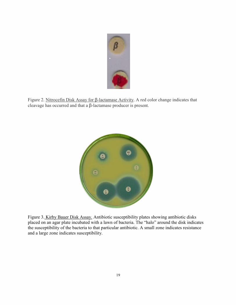
Cefinase Disk Assay for β-lactamase Activity
Nitrocefin disks, (“Cefinase”; Becton Dickson, Sparks, MD) were used to assay β-
lactamase activity. Cefinase disks were labeled with strain number corresponding to VAMC
collection number, placed on sterile aluminum foil, and moistened with 1 drop of sterile dH2O.
An inoculating loopful of cells was then spread directly from a plate onto a Cefinase disk and the
reaction was allowed to proceed for a maximum of 15 minutes. A positive reaction, observed as
a color change from yellow to red, was interpreted as indicating β-lactamase production (Fig. 2).
16
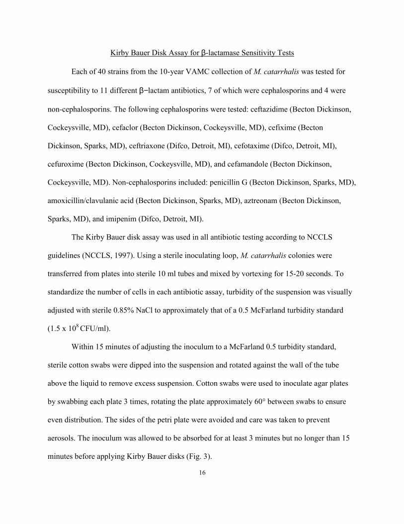
Kirby Bauer Disk Assay for β-lactamase Sensitivity Tests
Each of 40 strains from the 10-year VAMC collection of M. catarrhalis was tested for
susceptibility to 11 different β−lactam antibiotics, 7 of which were cephalosporins and 4 were
non-cephalosporins. The following cephalosporins were tested: ceftazidime (Becton Dickinson,
Cockeysville, MD), cefaclor (Becton Dickinson, Cockeysville, MD), cefixime (Becton
Dickinson, Sparks, MD), ceftriaxone (Difco, Detroit, MI), cefotaxime (Difco, Detroit, MI),
cefuroxime (Becton Dickinson, Cockeysville, MD), and cefamandole (Becton Dickinson,
Cockeysville, MD). Non-cephalosporins included: penicillin G (Becton Dickinson, Sparks, MD),
amoxicillin/clavulanic acid (Becton Dickinson, Sparks, MD), aztreonam (Becton Dickinson,
Sparks, MD), and imipenim (Difco, Detroit, MI).
The Kirby Bauer disk assay was used in all antibiotic testing according to NCCLS
guidelines (NCCLS, 1997). Using a sterile inoculating loop, M. catarrhalis colonies were
transferred from plates into sterile 10 ml tubes and mixed by vortexing for 15-20 seconds. To
standardize the number of cells in each antibiotic assay, turbidity of the suspension was visually
adjusted with sterile 0.85% NaCl to approximately that of a 0.5 McFarland turbidity standard
(1.5 x 10⁸ CFU/ml).
Within 15 minutes of adjusting the inoculum to a McFarland 0.5 turbidity standard,
sterile cotton swabs were dipped into the suspension and rotated against the wall of the tube
above the liquid to remove excess suspension. Cotton swabs were used to inoculate agar plates
by swabbing each plate 3 times, rotating the plate approximately 60° between swabs to ensure
even distribution. The sides of the petri plate were avoided and care was taken to prevent
aerosols. The inoculum was allowed to be absorbed for at least 3 minutes but no longer than 15
minutes before applying Kirby Bauer disks (Fig. 3).

17
Kirby Bauer disks were applied to the agar surface by using a dispenser and applying
gentle pressure with sterile forceps to ensure complete contact of disk with agar. Disks
impregnated with different antibiotics were separated by a minimum of 24 mm from center to
center and no more than 5 disks were placed on a 100 mm plate.
Plates were incubated for 16-18 hours at 35°C in an ambient-air incubator. Susceptibility
was measured only if a lawn of bacteria was present. To score susceptibility, plates were rested
lid down on a black non-reflecting surface and the diameter of the inhibition zone was measured
to the nearest whole millimeter by holding a caliper micrometer against the back of the plates.
Plates were examined visually for isolated colonies within the inhibition zone that may have
represented resistance. Because plates contained bacterial cells from a single strain of M.
catarrhalis, multi-drug profiles were easily assembled.
Statistical Analysis
The multi-drug profile was used as a method for inferring genetic variation and
highlighting isolates for further sequence analysis and determining possible variation among and
within antibiotics. A strain was considered significantly different from others in its susceptibility
if its inhibition zone was greater than 2 standard deviations from the mean. To assess
concordance of multi-drug profiles, strains showing significant deviation from means were
evaluated in a non-quantitative manner. For example, if 2 strains showed significant deviation
from means in regard to the same antibiotic, then the remainder of the profile was examined to
determine if the 2 strains showed similar susceptibilities to the remaining antibiotics. To compare
β-lactamase producers with β-lactamase non-producers, 2-sample t-tests, and confidence
intervals were computed using Minitab (Minitab, Inc. 1993). Grubbs outlier test was also

18
performed in order to assess if there existed any isolates detected as deviants other than those
identified by 2 standard deviations (Graphpad, Inc.).
Haplotype Analysis
Haplotype profiles were previously studied for the VAMC isolates tested. Of those some
of the isolates selected for analysis had identical multi-locus genotypes. Each of 8 genotypes was
represented by 2 or 3 isolates. Isolates representing 1 genotype were all β-lactamase non-
producers, isolates representing 6 genotypes were all β-lactamase producers (Table 1). Genotype
109CC was represented by 2 β-lactamase non-producers (isolates #604; #907) and 1 β-lactamase
producer (isolate #830) (Table 1).
19
Figure 2. Nitrocefin Disk Assay for β-lactamase Activity. A red color change indicates that cleavage has occurred and that a β-lactamase producer is present.
Figure 3. Kirby Bauer Disk Assay. Antibiotic susceptibility plates showing antibiotic disks placed on an agar plate incubated with a lawn of bacteria. The “halo” around the disk indicates the susceptibility of the bacteria to that particular antibiotic. A small zone indicates resistance and a large zone indicates susceptibility.

20
CHAPTER 3
RESULTS
Multi-Antibiotic Profiles
A total of 14 separate antibiotic profiles were found among the 41 isolates tested. Many
of the isolate profiles were typical which means that inhibition zones produced in response to
any of the antibiotics did not deviate from the mean by more than 2 standard deviations. Among
the β-lactamase producers, there were 8 different profiles consisting of 1 typical and 7 deviant
profiles (Table 1). The deviant profiles included antibiotic-specific reductions in susceptibility,
including: a cefuroxime specialist (#250); isolates with low susceptibility to amoxicillin/
clavulanic acid (#830 & 785); a cefixime specialist (#417); an isolate especially susceptible to
ceftazidime (#691); and an aztreonam and cefamandole specialist that was also sensitive to
cefotaxime (#566). The profile also included multi-specialists such as isolate #359 which was
highly sensitive to aztreonam, ceftazidime, ceftriaxone, and cefotaxime, and isolate #813 which
was highly sensitive to cefaclor, cefixime, ceftriaxone, cefotaxime, cefuroxime, cefamandole,
and penicillin G.
Among the β-lactamase non-producers there were 5 unique profiles and 1 typical profile
(Table 1). Isolate #604 had high resistance to cefuroxime, while isolate #123 showed
significantly lower susceptibility to ceftazidime, and isolate #444 was more sensitive to
aztreonam. Isolate # 347 appears to have been a multi-specialist with increased sensitivity to
both cefaclor and cefamandole. Isolate #474 appeared unique in showing resistance to
amoxicillin/clavulanic acid, with sensitivity to ceftriaxone and imipenem.

21
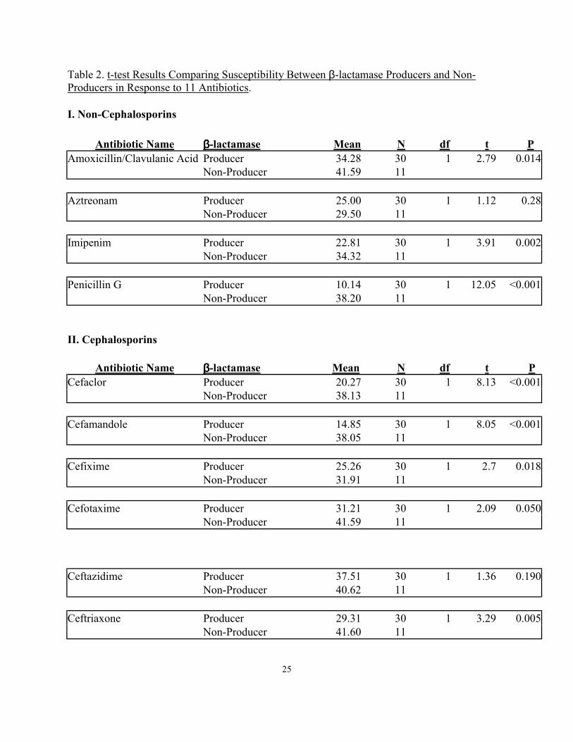
Comparisons of β-lactamase Producers and Non-Producers
β-lactamase producers were significantly less susceptible than non-producers to 9 of the
11 antibiotics (Table 2). Only ceftazidime and cefotaxime had similar sized inhibition zones in
producers and non-producers (Table 2). Among the non-cephalosporin antibiotics, only the
response to aztreonam was not significantly different between the β-lactamase producers and β-
lactamase non-producers (Table 2).
Outlier Test
Grubbs’ outlier test identified isolates that were outliers relative to the means. Isolates
with large z values are considered outliers. Among the non-producers, isolates #474, #123, and
#347 were identified as outliers (Table 3). Among the producers, isolates #691, #056, #359, and
#813 were identified as deviant isolates by the Grubbs’ test (Table 3). Isolates with large deviant
patterns include isolate #813, which was an outlier for 4 out of 11 antibiotics and was identified
as farthest from the mean, but not quite significantly different in susceptibility to 1 other
antibiotic (Table 3).
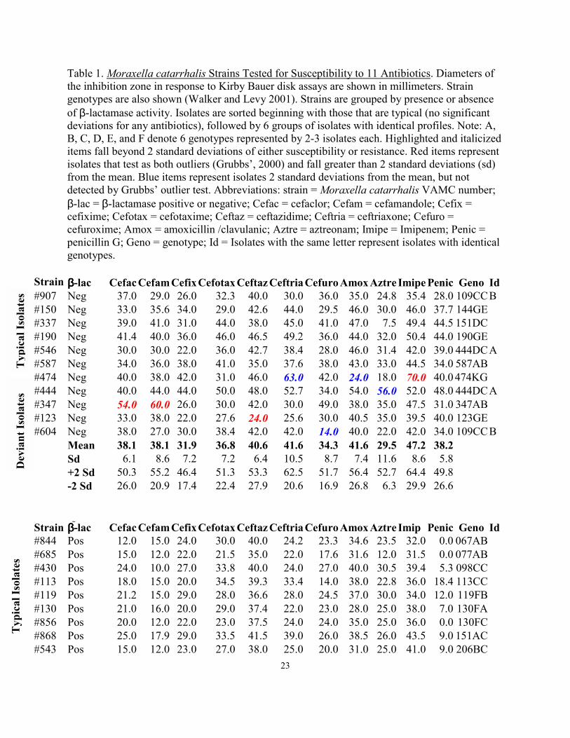
Haplotype Comparisons
Many isolates were grouped in pairs based on identical multi-locus genotypes (Walker
and Levy, 2001). Letter “A” denotes genotype #444DC, which contains 2 unique isolates, #444
and #546. Isolate #546 had a typical profile while isolate #444 showed increased susceptibility to
the antibiotic aztreonam. Letter “B” denotes genotype 109CC, which consisted of 3 separate
isolates, #604, #830, and #907. Isolates #907 and #604 were both β-lactamase non-producers and
isolate #830 was a β-lactamase producer. Isolate #907 displayed a typical profile, while isolate
#604 showed decreased susceptibility to cefuroxime. Letter “C” identified isolates #813 and
#417 that were both β-lactamase producers and showed deviant susceptibility profiles. Isolate
22
#813 displayed a deviant profile with increased susceptibility to 7 of 11 antibiotics (cefaclor,
cefamandole, cefixime, cefotaxime, ceftriaxone, cefuroxime, and penicillin G). Isolate #417
displayed increased susceptibility to only 1 antibiotic (cefixime). Letter “D” denoted haplotype
396AB, which contained isolates #566 and #785. Isolate #566 displayed decreased susceptibility
to both cefamandole and aztreonam and displayed increased susceptibility to cefotaxime. In
contrast, isolate #785 displayed increased resistance to amoxicillin. Letter “E”, haplotype
418CC, contained isolates #944, #770, and #980, which all displayed a typical antibiotic profile.
Letter “F”, haplotype 709CC, represented by isolates #712 and #709 also displayed a typical
antibiotic profile.
.
23
Table 1. Moraxella catarrhalis Strains Tested for Susceptibility to 11 Antibiotics. Diameters of the inhibition zone in response to Kirby Bauer disk assays are shown in millimeters. Strain genotypes are also shown (Walker and Levy 2001). Strains are grouped by presence or absence of β-lactamase activity. Isolates are sorted beginning with those that are typical (no significant deviations for any antibiotics), followed by 6 groups of isolates with identical profiles. Note: A, B, C, D, E, and F denote 6 genotypes represented by 2-3 isolates each. Highlighted and italicized items fall beyond 2 standard deviations of either susceptibility or resistance. Red items represent isolates that test as both outliers (Grubbs’, 2000) and fall greater than 2 standard deviations (sd) from the mean. Blue items represent isolates 2 standard deviations from the mean, but not detected by Grubbs’ outlier test. Abbreviations: strain = Moraxella catarrhalis VAMC number; β-lac = β-lactamase positive or negative; Cefac = cefaclor; Cefam = cefamandole; Cefix = cefixime; Cefotax = cefotaxime; Ceftaz = ceftazidime; Ceftria = ceftriaxone; Cefuro = cefuroxime; Amox = amoxicillin /clavulanic; Aztre = aztreonam; Imipe = Imipenem; Penic = penicillin G; Geno = genotype; Id = Isolates with the same letter represent isolates with identical genotypes.
Strain ββββ-lac Cefac Cefam Cefix Cefotax Ceftaz Ceftria Cefuro Amox Aztre Imipe Penic Geno Id #907 Neg 37.0 29.0 26.0 32.3 40.0 30.0 36.0 35.0 24.8 35.4 28.0 109CC B #150 Neg 33.0 35.6 34.0 29.0 42.6 44.0 29.5 46.0 30.0 46.0 37.7 144GE #337 Neg 39.0 41.0 31.0 44.0 38.0 45.0 41.0 47.0 7.5 49.4 44.5 151DC #190 Neg 41.4 40.0 36.0 46.0 46.5 49.2 36.0 44.0 32.0 50.4 44.0 190GE #546 Neg 30.0 30.0 22.0 36.0 42.7 38.4 28.0 46.0 31.4 42.0 39.0 444DC A #587 Neg 34.0 36.0 38.0 41.0 35.0 37.6 38.0 43.0 33.0 44.5 34.0 587AB #474 Neg 40.0 38.0 42.0 31.0 46.0 63.0 42.0 24.0 18.0 70.0 40.0 474KG #444 Neg 40.0 44.0 44.0 50.0 48.0 52.7 34.0 54.0 56.0 52.0 48.0 444DC A #347 Neg 54.0 60.0 26.0 30.0 42.0 30.0 49.0 38.0 35.0 47.5 31.0 347AB #123 Neg 33.0 38.0 22.0 27.6 24.0 25.6 30.0 40.5 35.0 39.5 40.0 123GE #604 Neg 38.0 27.0 30.0 38.4 42.0 42.0 14.0 40.0 22.0 42.0 34.0 109CC B Mean 38.1 38.1 31.9 36.8 40.6 41.6 34.3 41.6 29.5 47.2 38.2 Sd 6.1 8.6 7.2 7.2 6.4 10.5 8.7 7.4 11.6 8.6 5.8 +2 Sd 50.3 55.2 46.4 51.3 53.3 62.5 51.7 56.4 52.7 64.4 49.8 -2 Sd 26.0 20.9 17.4 22.4 27.9 20.6 16.9 26.8 6.3 29.9 26.6 Strain ββββ-lac Cefac Cefam Cefix Cefotax Ceftaz Ceftria Cefuro Amox Aztre Imip Penic Geno Id #844 Pos 12.0 15.0 24.0 30.0 40.0 24.2 23.3 34.6 23.5 32.0 0.0 067AB #685 Pos 15.0 12.0 22.0 21.5 35.0 22.0 17.6 31.6 12.0 31.5 0.0 077AB #430 Pos 24.0 10.0 27.0 33.8 40.0 24.0 27.0 40.0 30.5 39.4 5.3 098CC #113 Pos 18.0 15.0 20.0 34.5 39.3 33.4 14.0 38.0 22.8 36.0 18.4 113CC #119 Pos 21.2 15.0 29.0 28.0 36.6 28.0 24.5 37.0 30.0 34.0 12.0 119FB #130 Pos 21.0 16.0 20.0 29.0 37.4 22.0 23.0 28.0 25.0 38.0 7.0 130FA #856 Pos 20.0 12.0 22.0 23.0 37.5 24.0 24.0 35.0 25.0 36.0 0.0 130FC #868 Pos 25.0 17.9 29.0 33.5 41.5 39.0 26.0 38.5 26.0 43.5 9.0 151AC #543 Pos 15.0 12.0 23.0 27.0 38.0 25.0 20.0 31.0 25.0 41.0 9.0 206BC
Typ
ical
Isol
ates
D
evia
nt Is
olat
es
Typ
ical
Isol
ates

24
#339
Pos 25.0 10.0 23.0 30.0 37.0 28.5 21.7 33.0 26.0 38.3 0.0 327AB
E
#327 Pos 21.0 16.0 28.0 34.0 39.0 28.0 27.6 39.7 41.0 41.0 0.0 327AB E #980 Pos 20.0 15.0 24.0 21.0 36.0 23.0 12.0 33.0 20.0 37.0 0.0 418CC E #498 Pos 17.4 13.4 20.0 25.0 31.0 23.0 19.0 33.0 21.5 30.0 6.0 498AB #585 Pos 20.0 12.0 22.8 25.0 39.0 33.0 25.0 34.0 10.0 42.0 12.0 585CC #661 Pos 30.0 19.0 22.0 32.0 38.0 30.0 30.0 43.0 22.0 40.0 7.5 588DC #712 Pos 23.0 14.0 26.4 32.5 38.5 33.0 21.0 40.4 26.0 36.0 13.2 709CC F #709 Pos 18.5 15.0 22.0 28.8 38.0 26.8 25.0 36.0 23.6 33.0 12.0 709CC F #735 Pos 28.0 22.0 28.4 30.0 42.0 28.0 28.0 40.0 34.0 42.8 7.2 735DC #809 Pos 18.0 14.3 18.0 26.0 34.0 20.2 18.0 35.5 22.2 35.0 0.0 809EB #566 Pos 20.4 5.0 34.5 44.0 46.0 40.0 29.0 47.0 7.0 46.5 10.5 396AB D #359 Pos 22.0 20.0 22.0 57.0 52.0 60.0 18.0 38.0 48.6 62.0 16.0 359KG #691 Pos 14.0 12.4 30.0 34.0 18.0 33.0 25.0 38.0 24.0 42.0 10.0 206BC #813 Pos 40.3 38.0 37.4 49.0 38.1 52.0 39.0 24.0 32.0 50.6 45.0 151DC C #417 Pos 21.0 17.0 36.0 36.0 40.0 40.0 28.0 42.0 40.0 42.0 16.0 151DC C #830 Pos 18.7 15.7 28.0 40.0 42.6 34.0 25.0 19.0 27.0 39.0 17.0 109CC B #785 Pos 16.0 15.7 28.4 34.0 42.0 22.0 22.6 18.0 12.0 43.4 16.0 396AB D #250 Pos 10.0 12.0 23.0 26.0 24.0 22.2 11.0 30.0 16.0 26.0 0.0 077CC #056 Pos 20.0 9.0 26.0 24.0 38.3 22.0 18.0 30.0 32.6 10.0 0.0 056AB Mean 20.3 14.8 25.3 31.1 37.5 29.3 22.8 34.3 25.0 37.8 8.3 Sd 5.7 5.5 4.8 8.1 5.9 9.3 5.6 6.4 8.8 8.5 9.2 +2 Sd 31.6 25.8 34.9 47.2 49.4 47.9 34.0 47.2 42.7 54.7 26.7 -2 Sd 8.9 3.9 15.7 15.0 25.6 10.7 11.6 21.4 7.3 20.9 -10.1
Dev
iant
Isol
ates
Strain ββββ-lac Cefac Cefam Cefix Cefotax Ceftaz Ceftria Cefuro Amox Aztre Imip Penic Geno Id
Table 1. Continued
25
Table 2. t-test Results Comparing Susceptibility Between β-lactamase Producers and Non-Producers in Response to 11 Antibiotics. I. Non-Cephalosporins
Antibiotic Name ββββ-lactamase Mean N df t P Amoxicillin/Clavulanic Acid Producer 34.28 30 1 2.79 0.014 Non-Producer 41.59 11 Aztreonam Producer 25.00 30 1 1.12 0.28 Non-Producer 29.50 11 Imipenim Producer 22.81 30 1 3.91 0.002 Non-Producer 34.32 11 Penicillin G Producer 10.14 30 1 12.05 <0.001 Non-Producer 38.20 11 II. Cephalosporins
Antibiotic Name ββββ-lactamase Mean N df t P Cefaclor Producer 20.27 30 1 8.13 <0.001 Non-Producer 38.13 11 Cefamandole Producer 14.85 30 1 8.05 <0.001 Non-Producer 38.05 11 Cefixime Producer 25.26 30 1 2.7 0.018 Non-Producer 31.91 11 Cefotaxime Producer 31.21 30 1 2.09 0.050 Non-Producer 41.59 11
Ceftazidime Producer 37.51 30 1 1.36 0.190 Non-Producer 40.62 11 Ceftriaxone Producer 29.31 30 1 3.29 0.005 Non-Producer 41.60 11

26
Table 2. Continued Cefuroxime Producer 22.81 30 1 3.91 0.002 Non-Producer 34.32 11

27
Table 3. Isolates Showing Deviant Susceptibility Patterns Based on Grubbs’ Outlier Test. Isolates are Sorted Based on β-lactamase Activity. ββββ-lactamase – Genotype ID # Deviant Pattern Single Deviants 474KG 474 Imipenem susceptibility 123GE 123 Ceftazidime resistance Double Deviants 347AB 347 Cefaclor & Cefamandole susceptibility ββββ-lactamase + Genotype ID # Deviant Pattern Single Deviants 206BC 691 Ceftazidime resistance 056AB 056 Imipenem resistance Double Deviants 359KG 359 Cefotaxime & Ceftriaxone susceptibility Triple Deviants 151DC 813 Cefixime, Cefamandole, Cefaclor & Penicillin G susceptibility

28
CHAPTER 4
DISCUSSION
Purpose of Antibiotic Profiles
Antibiotic profiles were used to assess the susceptibility of M. catarrhalis isolates to a
series of β-lactam antibiotics. β-lactamase is the primary antibiotic resistance factor for β-lactam
antibiotics. Isolates that tested positive for β-lactamase production all displayed some degree of
resistance, but there was evidence of variation in the resistance profiles among those β-lactamase
positive isolates. Variation among β-lactamase producing isolates indicates that there must be a
difference in the activity of the β-lactamase or other factors must influence resistance. For
example, recent reports suggest that additional variation in BRO β-lactamase and/or in non-β-
lactamase factors may underlie novel susceptibility patterns (Baquero1996; Berk and Kalbfleisch
1996). Other factors that may underlie antibiotic resistance include alterations in the target
molecules that prevent interaction with the drug and/ or impermeability of the cell.
Livermore et al. (2001) has offered a protocol for using antibiotic susceptibilities to guide
strategies to manage resistance. Livermore suggested that first susceptibility testing be performed
in order to determine those isolates that are resistant. Resistant isolates should then be subjected
to an extensive battery of antibiotic tests (Livermore et al. 2001). The use of indicator drugs to
detect the presence of a mechanism that gives resistance not only to the indicator itself, but also
to related agents is another suggestion for detecting those isolates that are antibiotic resistant
mutants (Livermore et al. 2001). Livermore further suggested that the information gathered from
antibiotic profiles should be used to determine resistance patterns, especially indicating that β-
lactams are ideal drugs for discovering deviants.
29
This study used β-lactam antibiotics in a profile analysis of isolates from the VAMC. I
suggest that deviant isolates may also be tested as suggested by Livermore in an effort to
determine their resistance mechanism. Livermore suggested that ceftazidime can be used as an
antibiotic indicator for most of the TEM and SHV-derived extended-spectrum β-lactamases
(ESBL) types, while cefotaxime resistance is a better indicator for the CTX-M type enzymes in
other countries (Livermore et al. 2001). From the VAMC, isolates and #691 of the β-lactamase
producers showed increased resistance to ceftazidime hinting to the possibility of some variant
alteration of the BRO β-lactamase, while isolate #123 of the β-lactamase non-producers showed
decreased susceptibility to ceftazidime.
Possible Modes of Antibiotic Resistance The most frequent explanation for intrinsic antimicrobial resistance is explained by
decreased accumulation of the antibiotic or impermeability (Thornsberry 1991). Impermeability
to some β-lactam antibiotics may also be mediated by bacterial modifying enzymes that do not
inactivate the compounds but rather bind to them and alter their structures (Thornsberry 1991).
Alterations in antibacterial target molecules that prevent interaction with the antibiotic represents
1 of the most important mechanisms to clinically used antibiotics (Thornsberry 1991). The
cellular targets of β-lactam antibiotics are the penicillin binding proteins, and alterations in the
binding sites are known to affect resistance to β-lactam antibiotics. Each of these mechanisms
work together to confer a certain degree of antibiotic resistance and sometimes they work in
conjunction with the β-lactamase enzyme.
Isolates that have the β-lactamase enzyme show resistance, but variation was present
among the different β-lactamase positive isolates. In analyzing the susceptibility profiles to
30
penicillin, the only isolate that had a deviant profile was isolate #813. Isolate #813 also had a
deviant profile for cefaclor, cefamandole, cefixime cefotaxime, ceftriaxone, and cefuroxime with
statistically significant decreased resistance to those antibiotics. The profile of #813 suggests an
additional factor that confers resistance among the β-lactamase positive isolates tested. Isolates #
813 and #359 should be subjected to sequencing of the β-lactamase gene in order to determine if
alterations exist in their ß-lactamases.
Profiles in Genetically Identical Isolates Comparisons of genetically identical isolates offer information on potential differences in
β-lactamase activity. Isolates denoted by AA, BB, CC etc. in table 1 refer to groups of
genetically identical isolates. There are evident differences in the susceptibility profiles of some
genetically identical isolates. For example, among the β-lactamase positive isolates, there were 7
different antibiotic profiles. Within those profiles the most profound discovery involved 3
isolates that were genetically similar but differed in their β-lactamase activity. Genotype 109CC,
(Table 1) included isolates #604 and #907 that were β-lactamase negative and isolate #830 that
was β-lactamase positive. This genotype encompassed the entire spectrum of antibiotic
resistance by phenotypes. Within genotype 109CC, isolate #604, a β-lactamase non-producer,
showed relatively low susceptibility to cefuroxime but the remainder of its profile appeared
similar to the genetically identical isolate #907. Isolate #907 had a profile that was typical of β-
lactamase non-producers. Isolate #830 had a profile similar to other β-lactamase producers with
the exception of decreased susceptibility to amoxicillin/clavulanic acid. The antibiotic specific
increase in resistance suggests a mutated β-lactamase gene may be present in isolate #830.
31
Genotype 418CC, represented by isolates #944, #770, and #980 and genotype 709CC,
represented by isolates #709 and #712, were each β-lactamase producers with typical profiles
and no deviant isolates. In contrast, genotype 151DC was represented by isolates #813 and #417,
which were also β-lactamase producers, but they represent a different end of the spectrum.
Isolate #813 as previously mentioned, has an increased susceptibility to 7 out of the 11
antibiotics tested, while isolate #417 displays increased susceptibility only to cefixime. The
differences in profiles between isolates #813 and #417 continues to suggest that alterations in
target site and/or decreased permeability may also work with the β-lactamase enzyme to aid in
conferring antibiotic resistance.
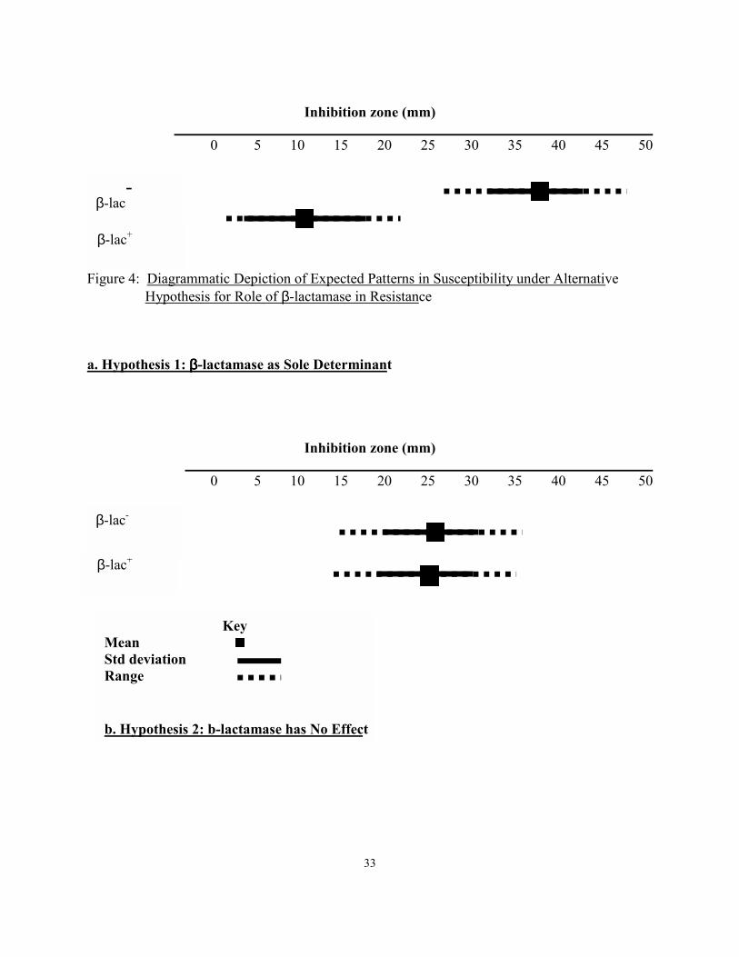
Alternative Hypotheses and Expectations
Alternative hypotheses for the role of β-lactamase susceptibility include the scenario of
β-lactamase as the sole determinant of β-lactamase resistance, in which case the susceptibilities
of β-lactamase positive and β-lactamase negative isolates are not expected to overlap (Figure
4a). The alternative hypothesis that β-lactamase has no influence on resistance predicts complete
overlap of the susceptibilities (Figure 4b). A 3rd hypothesis is that if β-lactamase has some
influence on the susceptibility, but it is not the sole determinant, then you would expect to see
partial overlap. The susceptibility tests showed overlap in susceptibility, especially when
comparing the cephalosporin antibiotics such as cefamandole with penicillin G (Fig. 5). The non-
cephalosporin, amoxicillin/clavulanic acid, used clinically as Augmentin also showed overlap of
its susceptibility profiles indicating a β-lactamase affect with additional determinants that aid in
explaining resistance to that antibiotic (Fig. 6). Also, in 9 of the 11 the antibiotic profiles
compiled from the VAMC isolates, there existed overlap between the ß-lactamase producers and

32
non-producers, suggesting that β-lactamase is not the only determinant of resistance in the
isolates tested. If other determinants were not essential to the resistance profiles we should
expect that there would not be any difference between the isolates and how they react with the
antibiotics.
Suggestions for Future Projects Those isolates determined to be different from the typical isolate (#831, #359, etc.) based
on their antibiotic profile should be further analyzed. I suggest that the isolates determined to be
ß-lactamase producers and that display unusual profiles should have their β-lactamase gene
sequenced to determine the type of β-lactamase gene present. Based on the fact that 2 alleles are
known that confer β-lactamase activity in M. catarrhalis, determining the type of allele present
in those deviant isolates might offer important information about resistance.
33
Inhibition zone (mm)
0 5 10 15 20 25 30 35 40 45 50
Figure 4: Diagrammatic Depiction of Expected Patterns in Susceptibility under Alternative
Hypothesis for Role of β-lactamase in Resistance
a. Hypothesis 1: ββββ-lactamase as Sole Determinant
Inhibition zone (mm)
0 5 10 15 20 25 30 35 40 45 50
β-lac+
β-lac-
β-lac+
β-lac-
Key Mean Std deviation Range b. Hypothesis 2: b-lactamase has No Effect

34
Inhibition zone (mm)
0 5 10 15 20 25 30 35 40 45 50
β-lac+
β-lac-
ANOVA P<0.001
Key Mean Std deviation Range Figure 5. Cefamandole Antibiotic Susceptibility Profile. Profile indicates a major β-lactamase effect between the producers and non-producers.

35
Inhibition zone (mm)
0 5 10 15 20 25 30 35 40 45 50
β-lac+ β-lac-
Key Mean Std deviation Range Figure 6. Amoxicillin/clavulanic Acid Antibiotic Response. Shows overlap in inhibition zones of β-lactamase producers and β-lactamase non-producers
ANOVA P = 0.014

36
BIBLIOGRAPHY
Baquero, F. 1996. Trends in antibiotic resistance of respiratory pathogens: an analysis and
commentary on a collaborative surveillance study. Journal of Antimicrobial
Chemotherapy 38: Suppl. A, 117–132.
Berk, S. L. Kalbfleisch, J. H. 1996. Antibiotic susceptibility patterns of community-acquired
respiratory isolates of Moraxella catarrhalis in Western Europe and in the USA. Journal
of Antimicrobial Chemotherapy 38: Suppl. A, 85–96.
Bootsma, H. J., van Dijk, H., Verhoef, J., Fleer, A., Mooi, F. R. 1996. Molecular
characterization of the BRO ß-lactamase of Moraxella (Branhamella) catarrhalis.
Antimicrobial Agents and Chemotherapy 40: 966–972.
Bootsma H. J., Aerts, P. C., Posthuma, G., Harmsen, T., Verhoef, J., van Dijk, H., Mooi, F. R.
1999. Moraxella (Branhamella) catarrhalis: BRO β-lactamase: A lipoprotein of Gram-
positive origin? Journal of Bacteriology 181(16): 5090-5093.
Bootsma, H. J., van Dijk, H., Vauterin, P., Verhoef, J., Mooi, F. R. 2000. Genesis of BRO β-
lactamase-producing Moraxella catarrhalis: Evidence for transformation-mediated
horizontal transfer. Molecular Microbiology 36(1): 93-104.
Bush, K., Sykes, R. B. 1986. Methodology for the study of β-lactamases. Antimicrobial Agents
and Chemotherapy 30: 6-10.
Doern, G. V., Brueggemann, A. B., Pierce, G., Hogan, T., Holley, H. P., Rauch, A. 1996.
Prevalence of antimicrobial resistance among 723 outpatient clinical isolates of
Moraxella catarrhalis in the United States in 1994 and 1995: Results of a 30-center
national surveillance study. American Society of Microbiology 40(12): 2884-2886.
Doern, G. V., Jones, R. N. 1988. Antimicrobial susceptibility testing of Haemophilus influenzae,
37
Branhamella catarrhalis, and Neisseria gonorrhoeae. Antimicrobial Agents and
Chemotherapy 32(12): 1747-1753.
Doern, G. V., Siebers, K. G., Hallick, L. M., Morse, S. A. 1980. Antibiotic susceptibility of
β-lactamase-producing strains of Branhamella (Neisseria) catarrhalis. Antimicrobial
Agents and Chemotherapy 1: 24-29.
Dowson, C. G., Coffey, T. J., Spratt, B. G. 1994. Origin and molecular epidemiology of
penicillin-binding-protein-mediated resistance to β-lactam antibiotics. Trends in
Microbiology 2: 361-366.
Enright, M. C., McKenzie, H. 1997. Moraxella (Branhamella) catarrhalis – clinical and
molecular aspects of a rediscovered pathogen. Journal of Medical Microbiology 46:
360-371.
Graphpad Software Inc. World Wide Web URL: http://216.46.227.18/welcome.htm.
Hol, C., Van Dijke, E. E. M., Verduin, C. M., Verhoef, J., Van Dijk, H. 1994. Experimental
evidence for Moraxella-induced penicillin neutralization in pneumococcal pneumonia.
Journal of Infectious Disease 170: 1613–1616.
Jacoby, G. A. 1994. Prevalence and resistance mechanism of common bacterial respiratory
Pathogens. Clinical Infection Disease 18: 951-957.
Livermore, D. M., Brown, D. F. J. 2001. Detection of β-lactamase-mediated resistance. Journal
of Antimicrobial Chemotherapy 48(SupplS1): 59-64.
Livermore, D. M., Winstanley, T. G., Shannon, K. P. 2001. Interpretative reading: Recognizing
the unusual and inferring resistance mechanisms from resistance phenotypes. Journal of
Antimicrobial Chemotherapy 48(SupplS1): 87-102.
Maiden, M. C. J. 1998. Horizontal genetic exchange, evolution, and spread of antibiotic

38
resistance in bacteria. Clinical Infection Disease 27(SupplS1): S12-S20.
Malmvall, B.E., Brorsson, J.E., Johnson, J. 1997. In vitro sensitivity to penicillin V and
β-lactamase production of Branhamella catarrhalis. Journal of Antimicrobial
chemotherapy 3: 374-375.
Medeiros A. A. 1997. Evolution and dissemination of β-lactamases accelerated by generations of
β-lactam antibiotics. Clinical Infection Disease 24(Suppl1): S19-45.
Minitab Inc. 1993. Minitab Reference Manual, Release 9 for Windows. Sowers Printing
Co. Lebanon, PA.
National Committee for Clinical Laboratory Standards. 1997. Methods for Dilution
Antimicrobial Susceptibility Tests for Bacteria that Grow Aerobically—Fourth Edition:
Approved Standard M7-A4. NCCLS, Villanova, PA.
Percival, A., Corkill, J. E., Rowlands, J., Sykes, R.B. 1977. Pathogenicity of and β-lactamase
production by Branhamella (Neisseria) catarrhalis. Lancet 2: 1175.
Spratt, B.G. 1994. Resistance to antibiotics mediated by target alterations. Science 264: 388-393.
Thornsberry, C. 1991. Antimicrobial susceptibility testing: general considerations. In:
Balows, A., Houston, W., Hermann, K. L., Isenberg, H., editors. Manual of Clinical
Microbiology. 5th ed. Washington, DC: American Society for Microbiology. p 1059-
1064.
Walker, E. S., Levy, F. 2001. Genetic trends in a population evolving antibiotic resistance.
Evolution 55(6): 1110-1122.
Walker, E. S., Neal, C. L., Laffan, E., Kalbfleisch, J. H., Berk, S. L., Levy F. 2000. Long-term
trends in susceptibility of Moraxella catarrhalis: a population analysis. Journal of
Antimicrobial Chemotherapy 45: 175-182.

39
Walker, E. S., Preston, R. A., Post, J. C., Ehrlich, G. D., Kalbfleisch, J. H., Klingman, K. L.
1998. Genetic diversity among strains of Moraxella catarrhalis: Analysis using multiple
DNA probes and a single-locus PCR-restriction fragment length polymorphism method.
Journal of Clinical Microbiology 36: 1977-1983.
Wallace, R. J., Jr, Steingrube, V. A., Nash, D. R., Hollis, D. G., Flanagan, C., Brown, B. A. et al.
1989. BRO ß-lactamases of Branhamella catarrhalis and Moraxella subgenus
Moraxella, including evidence for chromosomal ß-lactamase transfer by conjugation in
B. catarrhalis, M. nonliquifaciens, and M. lacunata. Antimicrobial Agents and
Chemotherapy 33: 1845–1854.
Wardle, J. R. 1986. Branhamella catarrhalis as an indirect pathogen. Drugs 31 (Suppl. 3):93–96.
Yao, J. D., Moellering, R. C. 1991. Antimicrobial agents. In: Balows, A., Houston, W.,
Hermann, K. L., Isenberg, H., editors. Manual of Clinical Microbiology. 5th ed.
Washington, DC: American Society for Microbiology. p 1065-1099.

40
VITA
LASHAN D. TAYLOR
Personal Data: Date of Birth: December 21, 1973 Place of Birth: Chattanooga, TN Marital Status: Single Education: Public Schools, Chattanooga, Tennessee University of Tennessee at Chattanooga, Chattanooga, Tennessee;
Biology, B.S., 1996 East Tennessee State University, Johnson City, Tennessee;
Biological Sciences, M.S., 2001 Professional Experience: Americorp VISTA Volunteer, The Urban League of Greater Chattanooga,
Chattanooga, TN 1997-1999 Graduate Assistant, East Tennessee State University, College of
Arts and Sciences, 1999-2002 .