antibiofilmactivityofpropolisextracton fusarium ... · antibiofilmactivityofpropolisextracton...
TRANSCRIPT
Research Article
For reprint orders, please contact: [email protected]
Antibiofilm activity of propolis extract onFusarium species from onychomycosisJuliana Galletti1, Flavia K Tobaldini-Valerio1,2, Sonia Silva2, Erika Seki Kioshima1, LarissaTrierveiler-Pereira1, Marcos Bruschi3, Melyssa Negri*,1 & Terezinha Inez Estivalet Svidzinski11Laboratory of Medical Mycology, Departamento de Analises Clınicas e Biomedicina, Universidade Estadual de Maringa (UEM),Avenida Colombo, 5790, Maringa, PR, CEP 87020-900, Brazil2CEB Centre of Biological Engineering, Universidade do Minho, Braga, Portugal3Laboratory of Research & Development of Drug Delivery Systems, Department of Pharmacy, State University of Maringa, CEP87020-900 Maringa, Parana, Brazil* Author for correspondence: Tel.: +55 44 3011 4809; Fax: +55 44 3011 4850; [email protected]
Aim: The present study evaluated the capacity of three species of Fusarium isolated from onychomycosisto form biofilms and the antibiofilm effect of propolis extract on these biofilms. Materials & methods: Thebiofilms and antibiofilm effects were evaluated by quantifying the colony-forming units, mitochondrialmetabolic activity assays, total biomass by crystal violet staining and scanning electron microscopy. Results:Propolis extract demonstrated significant antibiofilm efficiency on Fusarium spp. isolates and reduced F.solani, F. oxysporum and F. subglutinans mature biofilms. Conclusion: Propolis extract can be an alternativetopical treatment of onychomycosis caused by Fusarium spp.
First draft submitted: 20 March 2017; Accepted for publication: 5 July 2017; Published online: 4 October2017
Keywords: antifungal • fusariosis • paronychia • treatment • ungueal mycosis
Fusarium species are geophilic fungi, frequently isolated from opportunistic infections in immunocompromisedpatients, causing disseminated infections with high rates of mortality [1]. Onychomycosis, a fungal infection causedby Fusarium spp., can also affect immunocompetent patients [2]. It has previously been reported that paronychia,a characteristic of onychomycosis caused by this fungus, can be an entry portal to disseminated infections [3].Moreover, fusariosis has the highest incidence of infection among invasive fungal diseases in Brazil, with highmortality in hematological patients [4].
While the pathogenesis of fusariosis is not completely understood, the virulence factors of these fungi appearto be determinants for infection. Of these, biofilms are related to antifungal resistance and greater difficulty intreatment [5]. Additionally, some studies have described the capacity of F. oxysporum to form biofilm in vitro [6] oras an onychomycosis agent [7].
Studies focusing on finding new antifungal agents are urgently required, as there is a limited therapeutic arsenalavailable for Fusarium spp. infections. Alternatives include natural products, especially as they are inexpensive andhave low toxicity. Propolis is a natural product collected from the hives of Apis mellifera L. Several studies havedescribed the medicinal properties of propolis, such as its anti-inflammatory and antitumor effects and antifungaland antibacterial activity [8,9]. In the present study, biofilms formed by three species of Fusarium were characterizedand the activity of propolis extract (PE) against these biofilms was evaluated.
Materials & methodsFusarium strainsFungi were isolated from the nail samples of patients with onychomycosis. Nail scrapings were cultivated in threetubes containing Sabouraud dextrose agar (SDA; Himedia Laboratories, Mumbai, India) and three tubes thatcontained Mycosel medium (Himedia Laboratories, Mumbai, India). The identification of fungi was performedusing classic methods, including the examination of colonies and microscopic morphology [10,11]. The fungi isolatedand identified were maintained in a freeze-dried state in the Mycological Collection of the Laboratory of MedicalMycology of the State University of Maringa (UEM), Brazil. One clinical isolate was randomly chosen from each of
10.2217/fmb-2017-0052 C© 2017 Future Medicine Ltd Future Microbiol. (Epub ahead of print) ISSN 1746-0913
Research Article Galletti, Tobaldini-Valerio, Silva et al.
the three most frequent species of Fusarium in our region: F. oxysporum (FO42), F. solani (FS04) and F. subglutinans(FSub39). We also used a reference strain of F. solani (ATCC 36031), kindly provided by Fiocruz (Oswaldo CruzFoundation, Brazil).
Biofilm formationThis assay was based on the methodology described by Silva et al. [12] with modifications. The strains of Fusariumspp. were grown on potato dextrose agar (PDA; Himedia Laboratories, Mumbai, India) for 7 days at 25◦C. Thecolonies were gently scraped, harvested in 0.85% sterile saline and the conidia were counted in a Neubauer chamber.The inoculum was adjusted to a final concentration of 1 × 10⁶ conidia ml-1 [13,14] on Sabouraud dextrose brothmedium (SDB; Himedia Laboratories), and 200 μl of this suspension was placed into 96-well plates. The plateswere then incubated without agitation at 37◦C for 2 h in order to settle and to adhere the conidia to the bottom ofthe plate, and were then incubated at 37◦C in a shaker at 110 rev min-1. Every 24 h, 100 μl of SDB was removedand an equal volume of fresh SDB without Fusarium was added to each well for renewal of the culture medium.The plates were then incubated for 72 h. All experiments were repeated on three occasions with individual samplesevaluated in triplicate.
Biofilm biomass quantification by crystal violet stainingBiofilms were allowed to grow for 24, 48 and 72 h. They were then washed three-times with sterile saline and afterdrying, 200 μl of methanol was added to each well for 15 min to affix the biofilms. Later, 200 μl of crystal violet(1% v/v) was added for 5 min. The wells were washed with sterile distilled water and 200 μl of acetic acid (33%v/v) was then added to dissolve the stain. The obtained solution was read in a microtiter plate reader at 570 nmand the absorbance values were standardized per unit area of well (absorbance cm-2).
Biofilm metabolic activity assay by XTT reductionThe reduction assay of the tetrazolium salt 2,3-(2-methoxy-4-nitro-5-sulphophenyl)-5-([phenylamino]carbonyl)-2H tetrazolium hydroxide (XTT; Sigma-Aldrich, MO, USA) was used to determine in situ biofilm mitochondrialactivity. After biofilm formation, each well was washed three-times with sterile saline. A total of 100 μl of a solutioncontaining XTT, phenazine methosulphate (PMS) and 0.85% sterile saline was added to each well: 60 μl of sterilesaline, then 20 μl of XTT stock solution (500 μg ml-1) and 20 μl of PMS stock solution (50 μg ml-1) were added.The final concentrations of XTT and PMS in the wells were 100 and 10 μg ml-1, respectively. The plates werethen incubated at 35◦C for 3 h, protected from the light. After this period, the absorbance of the obtained solutionwas read in a microtiter plate reader at 492 nm and the absorbance values were standardized per unit area of well(absorbance cm-2).
Scanning electron microscopyThis assay was based on the methodology described by Silva et al. [12]. To examine the structure of biofilms byscanning electron microscopy (SEM), biofilms were allowed to form in 24-well polystyrene microtiter plates asdescribed above for 72 h. The wells were washed with sterile saline and the biofilms were dehydrated with alcohol(using 70% for 10 min, 80% for 10 min, 95% for 10 min and 100% ethanol for 20 min) and air dried. Prior toobservation, the bases of the wells were cut out and mounted onto aluminum stubs, sputter coated with gold andobserved with an S-360 scanning electron microscope (Leo, MA, USA).
Biofilm viability assayBiofilms were formed for 24, 48 and 72 h and were washed three-times with sterile saline. Then, 200 μl of salinewas added to each well and the biofilms were vigorously scraped with a pipette, transferred to a conical tube andvortexed for 1 min. Aliquots of 15 μl were placed in a plate containing Dichloran rose bengal chloramphenicolagar (Merck, Darmstadt, Germany) and were incubated at 35◦C. Dichloran rose bengal chloramphenicol agaris a selective medium for the isolation and enumeration of viable molds and restricts colony size, improving theenumeration and detection of the molds. The number of colony-forming units was determined after 5 days ofincubation.
10.2217/fmb-2017-0052 Future Microbiol. (Epub ahead of print) future science group
Antibiofilm activity of propolis on Fusarium spp. Research Article
PE & phenol contentThe propolis source was as described by Capoci et al. [15], with green propolis collected from hives of A. mellifera inCianorte (Parana, Brazil). The samples were triturated and stored at -20◦C until further analysis. PE was preparedwith a propolis/ethanol ratio of 30/70 (w/w) by turbo extraction, at 3500 rpm, three-times at 15 min with twointervals of 5 min. PE was filtered through filter paper and adjusted to the initial weight with ethanol. The totalphenol content (TPC) of PE was evaluated by the Folin–Ciocalteau method [16]. PE was added to 6 ml of theFolin–Ciocalteau and 6 ml of 20% Na2CO3 (Sigma-Aldrich). After 2 h, absorbance was determined by ShimadzuUV-1650PC spectrophotometer (Tokyo, Japan) at a wavelength of 760 nm. A calibration curve with solutionsof gallic acid was used as a reference. TPC was expressed as a percentage of total phenolic substances in PE andcorresponded to a mean of six replicates.
Antifungal assayIn vitro antifungal susceptibility testing of planktonic cells was performed using a microdilution method adapted
from Capoci et al. [15] and the Clinical and Laboratory Standards Institute M38-A2 protocol [17] with serial dilutionof PE 34.17, 68.35, 136.71, 273.43, 546.87, 1093.75, 2187.5, 4375, 8750 and 17,500μg ml-1 of TPC expressedin gallic acid in RPMI 1640 (Roswell Park Memorial Institute; Gibco, NY, USA) with l-glutamine (with sodiumbicarbonate) and 0.165 M 3-(N-morpholino) propanesulfonic acid (pH 7.2) as a buffer (Sigma-Aldrich). Thestrains of Fusarium spp. were grown on potato dextrose agar for 7 days at 25◦C. The colonies were gently scrapedand harvested in 0.85% sterile saline solution. The number of conidia were counted in a Neubauer chamber andthe inoculum was adjusted to a final concentration of 5 × 10⁴ conidia ml-1 in RPMI 1640. An aliquot of PE(100 μl) was dispensed into 96-well plates and further incubated with aliquots (100 μl) of the Fusarium inoculum.The 96-well plates were incubated at 35◦C for 72 h. The minimal inhibitory concentration (MIC) values weredetermined by visualization, corresponding to the lowest concentration of antifungal agent in which no visiblegrowth was observed, in comparison with the control (cells grown without PE) [15,17]. The minimal fungicidalconcentration (MFC) was determined after 72 h of incubation at 35◦C and 5 μl of each well was transferred to aplate containing sabouraud dextrose agar and incubated at 25◦C for 48 h.
Antibiofilm assayOnce stable and mature biofilms were formed (for 24 h as described above) they were washed three-times with sterilesaline. Biofilms were formed as described above for 24 h. Then, PE was added at the MFC concentration found inthe latter assay. The 96-well plates were incubated at 37◦C for a further 24 h. The activity of PE against biofilms wasdetermined by biomass quantification using crystal violet staining and by biofilm viability assay described above.
Epifluorescence microscopyAntibiofilm assay was also evaluated by epifluorescence microscopy. Biofilms were formed in 24-well polystyrenemicrotiter plates as described above for 24 h, then PE was added at the MFC concentration and the plates wereincubated at 37◦C for a further 24 h. The matrix of biofilms was then stained with calcofluor white (FlukaAnalytical, MO, USA) diluted in sterile saline (1:4) for 5 min and washed twice with sterile saline. Stained biofilmswere observed with a microscope (Evos FL, Live Technologies, CA, USA) with filters capable of detecting the fungicell wall (BP 365–370, FT 400, LP 421).
Statistical analysisData were analyzed using Prism 6.0 software (GraphPad, CA, USA). The t-test and one-way analysis of variancewith Bonferroni were used. All of the tests were performed with a confidence level of 95% and values of p ≤ 0.05were considered statistically significant.
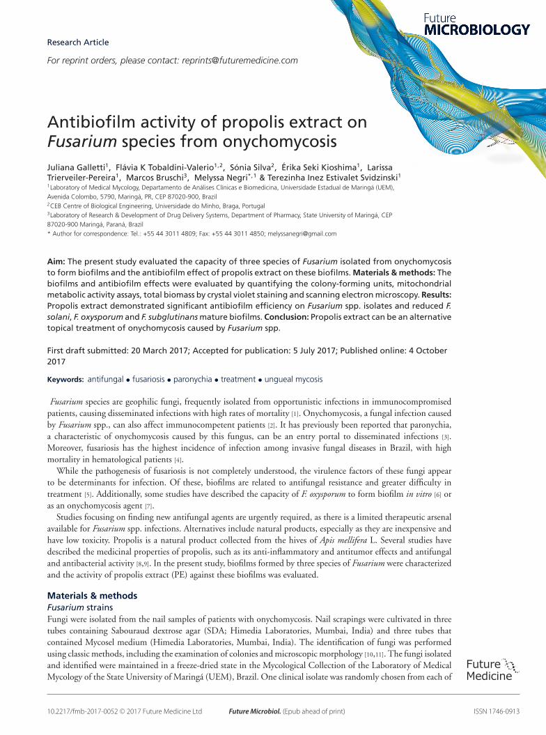
ResultsThe three species of Fusarium tested in this study formed biofilms. Nevertheless, the F. solani strains were moreefficient than the other species in relation to total biomass and the number of viable cells. Furthermore, the biomassproduced by both the clinical isolates and reference strain of this species was directly proportional to incubationtime (Figure 1). While the total biomass of biofilms formed by F. solani increased from 24 to 48 h and from 48to 72 h of incubation, the total biomass of F. oxysporum and F. subglutinans biofilms increased only until 48 h
future science group 10.2217/fmb-2017-0052
Research Article Galletti, Tobaldini-Valerio, Silva et al.
15
10
5
0ATCC 36031 FS04 FO42 FSub39
Ab
sorb
ance
cm
-2 (
570
nm
)*
*
*
*
*
24 h48 h72 h
Figure 1. Crystal violet staining of biofilm biomass of Fusarium solani (ATCC 36031 and FS04), Fusarium oxysporum(FO42) and Fusarium subglutinans (FSub39) at different times of incubation. ANOVA with Bonferroni, *p < 0.05.
and apparently became stable after this period (Figure 1). However, these increases in biomass production weresignificant only for strains of F. solani and F. subglutinans, and mainly from 24 to 72 h (Figure 1).
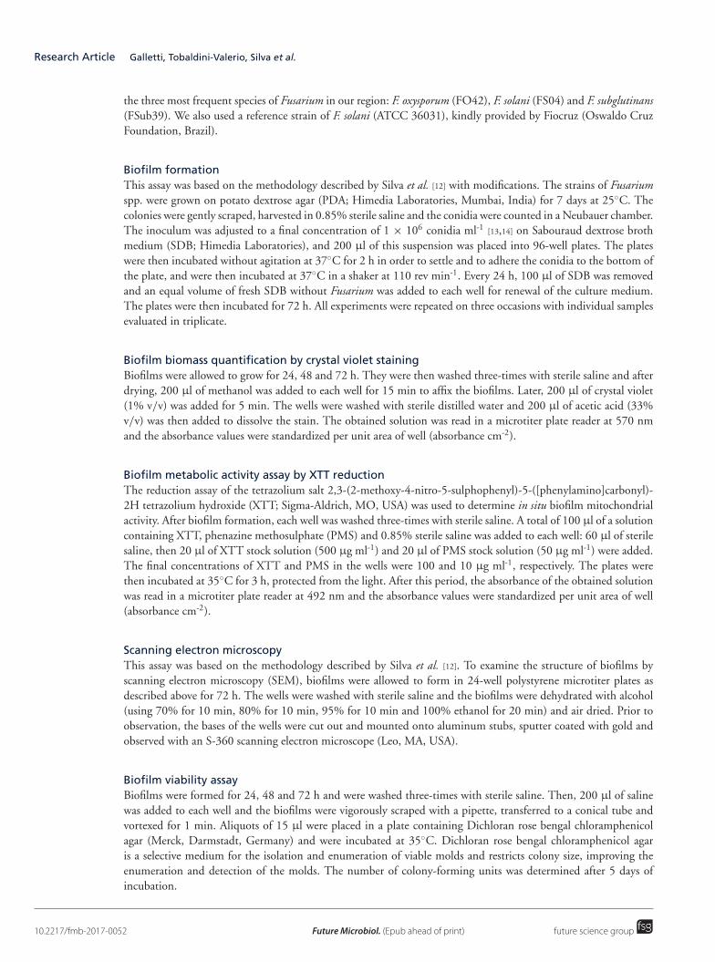
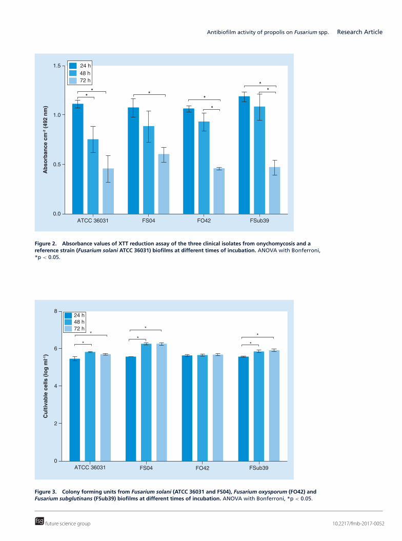
In contrast, the absorbance values from the XTT reduction assay, related to biofilm metabolic activity, decreasedwith incubation time. The decrease in absorbance values was significant from 24 to 72 h for all the isolates (Figure 2).
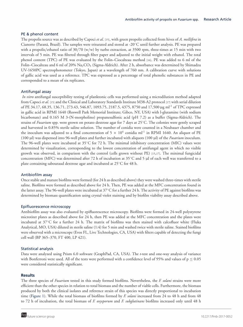
The clinical isolate of F. solani also presented significantly more viable cells on the biofilm than the other speciesat 48 and 72 h of incubation. Most isolates, except for F. oxysporum, exhibited a significant increase in viable cellsfrom 24 to 48 h and from 24 to 72 h (Figure 3). The number of viable cells and the quantity of biomass werequite similar among species with 24 h of incubation, but after this period, they began to show differences amongthemselves.
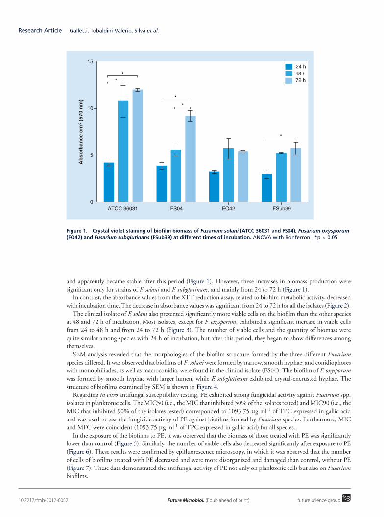
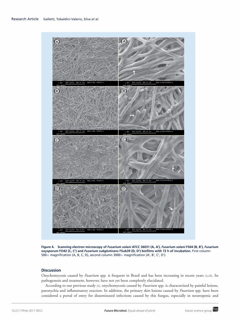
SEM analysis revealed that the morphologies of the biofilm structure formed by the three different Fusariumspecies differed. It was observed that biofilms of F. solani were formed by narrow, smooth hyphae; and conidiophoreswith monophiliades, as well as macroconidia, were found in the clinical isolate (FS04). The biofilm of F. oxysporumwas formed by smooth hyphae with larger lumen, while F. subglutinans exhibited crystal-encrusted hyphae. Thestructure of biofilms examined by SEM is shown in Figure 4.
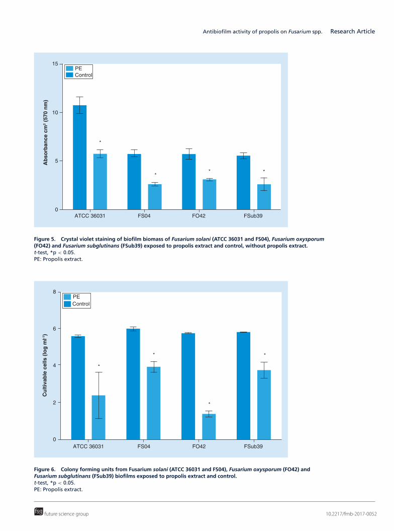
Regarding in vitro antifungal susceptibility testing, PE exhibited strong fungicidal activity against Fusarium spp.isolates in planktonic cells. The MIC50 (i.e., the MIC that inhibited 50% of the isolates tested) and MIC90 (i.e., theMIC that inhibited 90% of the isolates tested) corresponded to 1093.75 μg ml-1 of TPC expressed in gallic acidand was used to test the fungicide activity of PE against biofilms formed by Fusarium species. Furthermore, MICand MFC were coincident (1093.75 μg ml-1 of TPC expressed in gallic acid) for all species.
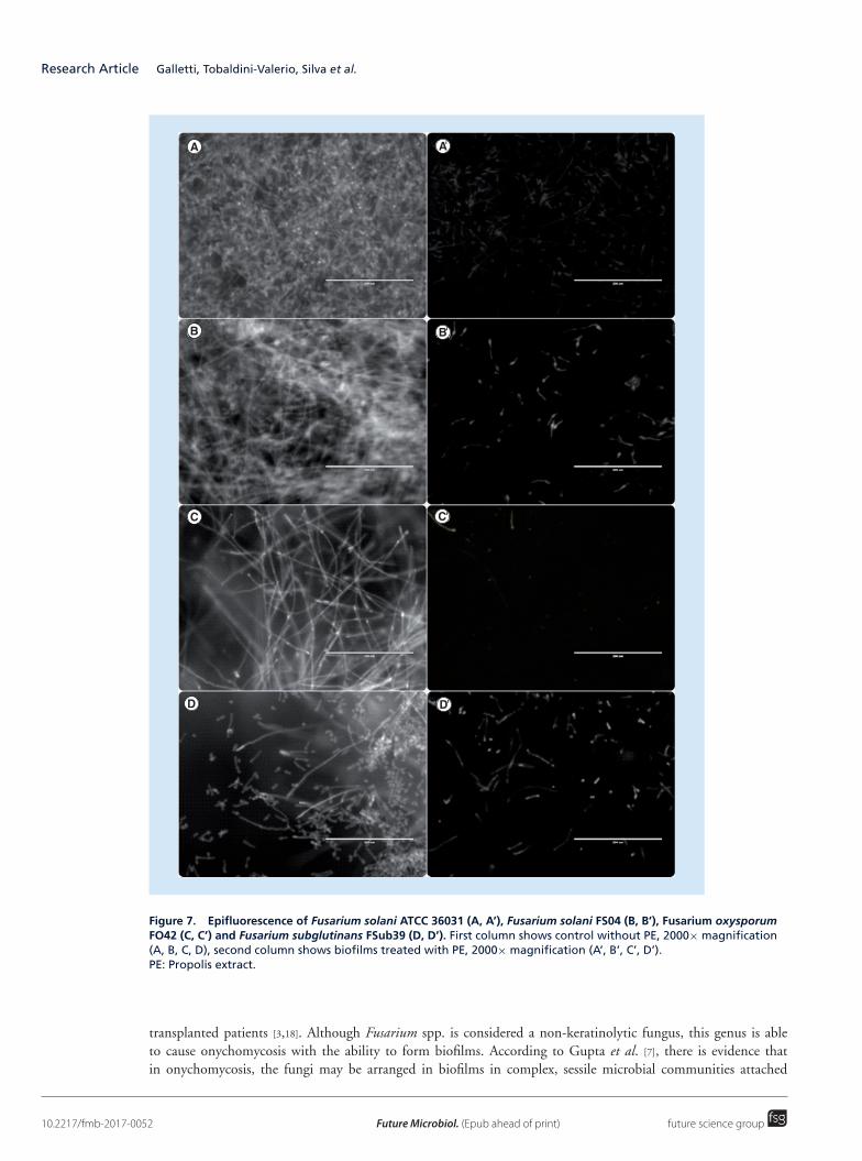
In the exposure of the biofilms to PE, it was observed that the biomass of those treated with PE was significantlylower than control (Figure 5). Similarly, the number of viable cells also decreased significantly after exposure to PE(Figure 6). These results were confirmed by epifluorescence microscopy, in which it was observed that the numberof cells of biofilms treated with PE decreased and were more disorganized and damaged than control, without PE(Figure 7). These data demonstrated the antifungal activity of PE not only on planktonic cells but also on Fusariumbiofilms.
10.2217/fmb-2017-0052 Future Microbiol. (Epub ahead of print) future science group
Antibiofilm activity of propolis on Fusarium spp. Research Article
ATCC 36031 FS04 FO42 FSub39
Ab
sorb
ance
cm
-2 (
492
nm
)
1.5
1.0
0.5
0.0
**
**
*
**
24 h48 h72 h
Figure 2. Absorbance values of XTT reduction assay of the three clinical isolates from onychomycosis and areference strain (Fusarium solani ATCC 36031) biofilms at different times of incubation. ANOVA with Bonferroni,*p < 0.05.
Cu
ltiv
able
cel
ls (
log
ml-1
)
8
6
4
2
0ATCC 36031 FS04 FO42 FSub39
*
*
*
* *
*
24 h48 h72 h
Figure 3. Colony forming units from Fusarium solani (ATCC 36031 and FS04), Fusarium oxysporum (FO42) andFusarium subglutinans (FSub39) biofilms at different times of incubation. ANOVA with Bonferroni, *p < 0.05.
future science group 10.2217/fmb-2017-0052
Research Article Galletti, Tobaldini-Valerio, Silva et al.
L- SE1 EHT- 10.0 kv50.0 µm
WD- 10 mm MAG- X 500. PHOTO- 0
UNIV. MINHO
L- SE1 EHT- 10.0 KV10.0 µm
WD- 10 mm MAG- X 3.00 K PHOTO= 0
UNIV. MINHO
L- SE1 EHT- 10.0 KV10.0 µm
WD- 10 mm MAG- X 3.00 K PHOTO= 0
UNIV. MINHO
L- SE1 EHT- 10.0 KV10.0 µm
WD- 10 mm MAG- X 3.00 K PHOTO= 0
UNIV. MINHO
L- SE1 EHT- 10.0 KV10.0 µm
WD- 10 mm MAG- X 3.00 K PHOTO= 0
UNIV. MINHO
L- SE1 EHT- 10.0 KV50.0 µm
WD- 10 mm MAG- X 500. PHOTO- 0
UNIV. MINHO
L- SE1 EHT- 10.0 KV50.0 µm
WD- 10 mm MAG- X 500. PHOTO- 0
UNIV. MINHO
L- SE1 EHT- 10.0 KV50.0 µm
WD- 10 mm MAG- X 500. PHOTO- 0
UNIV. MINHO
'
'
'
'
Figure 4. Scanning electron microscopy of Fusarium solani ATCC 36031 (A, A’), Fusarium solani FS04 (B, B’), Fusariumoxysporum FO42 (C, C’) and Fusarium subglutinans FSub39 (D, D’) biofilms with 72 h of incubation. First column500× magnification (A, B, C, D), second column 3000× magnification (A’, B’, C’, D’).
DiscussionOnychomycosis caused by Fusarium spp. is frequent in Brazil and has been increasing in recent years [2,10]. Itspathogenesis and treatment, however, have not yet been completely elucidated.
According to our previous study [2], onychomycosis caused by Fusarium spp. is characterized by painful lesions,paronychia and inflammatory reaction. In addition, the primary skin lesions caused by Fusarium spp. have beenconsidered a portal of entry for disseminated infections caused by this fungus, especially in neutropenic and
10.2217/fmb-2017-0052 Future Microbiol. (Epub ahead of print) future science group
Antibiofilm activity of propolis on Fusarium spp. Research Article
15
10
5
0ATCC 36031 FS04 FO42 FSub39
Ab
sorb
ance
cm
2 (5
70 n
m)
PEControl
*
** *
Figure 5. Crystal violet staining of biofilm biomass of Fusarium solani (ATCC 36031 and FS04), Fusarium oxysporum(FO42) and Fusarium subglutinans (FSub39) exposed to propolis extract and control, without propolis extract.t-test, *p < 0.05.PE: Propolis extract.
8
6
4
2
0ATCC 36031 FS04 FO42 FSub39
Cu
ltiv
able
cel
ls (
log
ml-1
)
PEControl
*
*
*
*
Figure 6. Colony forming units from Fusarium solani (ATCC 36031 and FS04), Fusarium oxysporum (FO42) andFusarium subglutinans (FSub39) biofilms exposed to propolis extract and control.t-test, *p < 0.05.PE: Propolis extract.
future science group 10.2217/fmb-2017-0052
Research Article Galletti, Tobaldini-Valerio, Silva et al.
'
'
'
'
Figure 7. Epifluorescence of Fusarium solani ATCC 36031 (A, A’), Fusarium solani FS04 (B, B’), Fusarium oxysporumFO42 (C, C’) and Fusarium subglutinans FSub39 (D, D’). First column shows control without PE, 2000× magnification(A, B, C, D), second column shows biofilms treated with PE, 2000× magnification (A’, B’, C’, D’).PE: Propolis extract.
transplanted patients [3,18]. Although Fusarium spp. is considered a non-keratinolytic fungus, this genus is ableto cause onychomycosis with the ability to form biofilms. According to Gupta et al. [7], there is evidence thatin onychomycosis, the fungi may be arranged in biofilms in complex, sessile microbial communities attached
10.2217/fmb-2017-0052 Future Microbiol. (Epub ahead of print) future science group
Antibiofilm activity of propolis on Fusarium spp. Research Article
to epithelial surfaces. The capacity of F. oxysporum to form biofilms on fragments of human nails has also beendocumented [6].
It has recently been shown for the first time that Fusarium spp. can grow in vitro using human nails as a singlesource of nutrient; and also that members from this genus have the ability to invade healthy human nails [10].In the current study, the capacity of clinical isolates of F. solani, F. oxysporum and F. subglutinans obtained fromonychomycosis to form biofilms was demonstrated, a finding similar to those described by Costa-Orlandi etal. (2014) [14] for the dermatophytes T. rubrum and T. mentagrophytes, which are the most common agents ofonychomycosis.
While all the species of Fusarium tested in this study formed biofilms, the F. solani strains were more efficient thanthe others for all the studied parameters. The results of total biomass quantification and viable cell characterizationrevealed the greater capacity of F. solani to form biofilms (Figures 1 & 2), suggesting that this species is morevirulent than the others. This finding is in accordance with the fact that this species is most frequently isolatedfrom invasive fusariosis [19]. Other studies have also described F. solani as the most virulent species, such as when F.solani was injected intravenously and caused the death of all the animals tested, which other Fusarium species didnot [20].
Curiously, the values of XTT reduction assay in the present study decreased over time (Figure 2), suggesting areduction in the metabolic activity of the biofilms. It is important to point out that according to biofilm structure,the fungal cells in sessile populations may display a poor physiological state, as confirmed by XTT-based assay [21].Furthermore, Fusarium biofilms exhibit opposing kinetics (Figures 1 & 2), as metabolic activity was lowest at themoment of peak biomass production and the inverse effect was also observed, corroborating the findings of otherstudies [22,23]. This low metabolic activity could be due to the increased adsorption of compounds on the biofilmmatrix, meaning there is less contact between the tetrazolium salt and the cells of the mature biofilm [21–23]. It isalso important to emphasize that the association between these parameters (low activity and high biomass) is wellestablished in literature and is considered responsible for biofilm antifungal resistance, as fungi arranged in biofilmsare considered to be up to 1000-fold more resistant to antifungal agents than planktonic cells [21].
Regarding fungal biofilm formation, although most attention has been paid to the yeast pathogen Candidaalbicans, it is known that many fungal species, including filamentous fungi, can form such structures [24,25]. Biofilmmorphology can vary according to the species involved and it has been found that even species from the samegenus can form biofilms with distinct arrangements [14]. The SEM results of the present study (Figure 4) haveshown that the Fusarium species used here can form biofilms with different morphologies and all the isolates wereable to organize dense growth, with a well-structured community constituted by large hyphae, sometimes withconidiophores and conidia, suggesting a mature ecosystem.
When considering its ability to form biofilms, Peiqian et al. [13] reported that F. oxysporum when found in biofilmswas less susceptible to cold, heat, UV light and some fungicides than those grown in planktonic (free-living) cells. Itis well known that the capacity to form biofilms is related to higher resistance and difficulty in clinical treatment [5].Considering that onychomycosis can be a portal of entry for invasive fusariosis, especially in immunocompromisedpatients, the search for effective new topical treatments for onychomycosis caused by Fusarium spp. is highlyrelevant. In addition, few antifungal agents are efficacious against fungal biofilms (echinocandins and liposomalamphotericin B) and these drugs are not used for onychomycoses [21].
In this way, it is important to evaluate if a new antifungal option for planktonic cells exists, as PE also actsagainst fungi organized in biofilms. Propolis is a resin that presents a number of medicinal properties [8], includinganti-inflammatory activity, which could potentiate a possible antifungal topical action. Furthermore, the capacityof propolis to inhibit the biofilm formation of C. albicans has already been reported, along with the low cytotoxicityin human cells with the same PE used in the present study [15,26]. However, the antifungal property of PE has notyet been proved in biofilms formed by Fusarium species.
While microbiologists have historically tested the effect of drugs on planktonic cells, the present study evaluatedthe action of PE on both planktonic cells and biofilm formation by Fusarium species. Initially, good results wereobtained for the MIC and MFC of PE against Fusariun spp. planktonic cells, which were 1093.75 μg ml-1, similarto those described by Dota et al. [27] for Candida spp. The majority of isolates tested had MIC values between550.3 and 1100.63 μg ml-1. Capoci et al. [15] also found similar values of MFC and MIC for Candida, provingthat propolis possesses a fungicide mechanism.
Next, the performance of this PE on biofilms formed by the Fusarium species was evaluated. The resultswere exciting, as PE appears to be efficient, significantly reducing both the biomass and the number of viable
future science group 10.2217/fmb-2017-0052
Research Article Galletti, Tobaldini-Valerio, Silva et al.
cell parameters (p < 0.05) of the biofilms produced by all the evaluated isolates (Figures 5 & 6). In addition,epifluorescence assay showed clearly that the number of cells of the biofilms formed by all the strains treated withPE were reduced and the structure of the biofilm was lost, leaving only some damaged and isolated cells (Figure 7).
A possible limitation of this study was the absence of a comparison between the antibiofilm effects of propolis andconventional antifungals. However, it should be pointed out that other works carried out by our group [15,26] haveshown propolis to be as or more efficient than other antifungal drugs. Moreover, Fusarium spp. are highly resistantto azole antifungals which are not usually active against this fungus and terbinafine response varies according tospecies [10]. We have not therefore included a conventional antifungal as a control.
In summary, it is known that biofilm-forming capacity is related to greater resistance and difficulty in treat-ment [21]. PE is therefore promising as propolis can be found in its extract form in pharmacies, ready for topical usein patients with onychomycosis, justifying the importance of the development of new treatment approaches withthe capacity to control fungal biofilms present in nails affected by Fusarium spp.
ConclusionThe results of the present study described the biofilm-forming capacity of three species of Fusarium isolated fromonychomycosis, with F. solani exhibiting the greatest such capacity, reinforcing that it is more virulent than theother Fusarium species tested. We also demonstrated the antifungal and antibiofilm capacity of PE against clinicalisolates of F. solani, F. oxysporum and F. subglutinans. This reinforces the previous results and suggests that PEcould be an alternative option for the topical treatment of onychomycosis and avoid the possible dissemination ofthis fungi of onychomycosis, since the most frequent species that cause human invasive fusariosis, F. solani and F.oxysporum, are also the most frequent species that cause onychomycosis.
Acknowledgements
The authors thank Fiocruz for kindly providing the strain of Fusarium solani.
Financial & competing interests disclosure
This study was supported by grants from Conselho Nacional de Desenvolvimento Cientıfico e Tecnologico (CNPq) and Coordenacao
de Aperfeicoamento de Pessoal de Nıvel Superior (CAPES). The authors have no other relevant affiliations or financial involvement
with any organization or entity with a financial interest in or financial conflict with the subject matter or materials discussed in the
manuscript apart from those disclosed.
No writing assistance was utilized in the production of this manuscript.
Ethical conduct of research
The authors state that they have obtained appropriate institutional review board approval or have followed the principles outlined
in the Declaration of Helsinki for all human or animal experimental investigations. In addition, for investigations involving human
subjects, informed consent has been obtained from the participants involved.
Summary points
� Fusarium species isolated from onychomycosis form biofilm efficiently.� Propolis presents potential antifungal activity.� Propolis is a strong inhibitor of Fusarium spp. from onychomycosis.� Propolis is able to reduce and destroy biofilms formed by Fusarium species.� Propolis is a promising alternative to traditional antifungal therapy.
ReferencesPapers of special note have been highlighted as: • of interest; •• of considerable interest
1. Nucci M, Anaissie E. Fusarium infections in immunocompromised patients. Clin. Microbiol. Rev. 20(4), 695–704 (2007).
2. Guilhermetti E, Takahachi G, Shinobu CS, Svidzinski TIE. Fusarium spp. as agents of onychomycosis in immunocompetent hosts. Int.J. Dermatol. 46(8), 822–826 (2007).
•• Excellent article for understanding Fusarium species infections.
3. Bourgeois GP, Cafardi JA, Sellheyer K, Andea AA. Disseminated Fusarium infection originating from paronychia in a neutropenicpatient: a case report and review of the literature. Cutis 85(4), 191–194 (2010).
10.2217/fmb-2017-0052 Future Microbiol. (Epub ahead of print) future science group
Antibiofilm activity of propolis on Fusarium spp. Research Article
4. Nucci M, Garnica M, Gloria AB et al. Invasive fungal diseases in haematopoietic cell transplant recipients and in patients with acutemyeloid leukaemia or myelodysplasia in Brazil. Clin. Microbiol. Infect. 19(8), 745–751 (2013).
5. Mukherjee PK, Chandra J, Yu C, Sun Y, Pearlman E, Ghannoum MA. Characterization of fusarium keratitis outbreak isolates:contribution of biofilms to antimicrobial resistance and pathogenesis. Investig. Opthalmol. Vis. Sci. 53(8), 4450–4457 (2012).
6. Vila TVM, Rozental S, de Sa Guimaraes CMD. A new model of in vitro fungal biofilms formed on human nail fragments allows reliabletesting of laser and light therapies against onychomycosis. Lasers Med. Sci. 30(3), 1031–1039 (2015).
7. Tamura NK, Kira G, Patussi EV, Donatti L, Inez T, Svidzinski E. Adherence and biofilm formation of Fusarium oxysporum isolated froma corneal ulcer. Glob. Adv. Res. J. Med. Med. Sci. 4(1), 28–34 (2015).
8. Burdock GA. Review of the biological properties and toxicity of bee propolis (propolis). Food Chem. Toxicol. 36(4), 347–363 (1998).
9. Negri M, Salci T, Shinobu-Mesquita C, Capoci I, Svidzinski T, Kioshima E. Early state research on antifungal natural products.Molecules 19(3), 2925–2956 (2014).
10. Galletti J, Negri M, Grassi FL, Kioshima-Cotica ES, Svidzinski TIE. Fusarium spp. is able to grow and invade healthy human nails as asingle source of nutrients. Eur. J. Clin. Microbiol. Infect. Dis. 34(9), 1767–1772 (2015).
• Excellent standard for understanding the pathogenicity of Fusarium.
11. De Hoog GS, Guarro J. Atlas of Clinical Fungi. Centraalbureau voor Schimmelcultures, Reus, Spain (1995).
12. Silva S, Henriques M, Martins A, Oliveira R, Williams D, Azeredo J. Biofilms of non-Candida albicans Candida species: quantification,structure and matrix composition. Med. Mycol. 47(7), 681–689 (2009).
13. Peiqian L, Xiaoming P, Huifang S, Jingxin Z, Ning H, Birun L. Biofilm formation by Fusarium oxysporum f. sp. cucumerinum andsusceptibility to environmental stress. FEMS Microbiol. Lett. 350(2), 138–145 (2014).
14. Costa-Orlandi CB, Sardi JCO, Santos CT, Fusco-Almeida AM, Mendes-Giannini MJS. In vitro characterization of Trichophyton rubrumand T. mentagrophytes biofilms. Biofouling 30(6), 719–727 (2014).
15. Capoci GRI, Bonfim-mendonca PDS, Arita GS et al. Propolis is an efficient fungicide and inhibitor of biofilm production by vaginalCandida albicans. Evid. Based Complement Alternat. Med. 2015, 28769 (2015).
• Excellent article for understanding the potential of propolis extract.
16. Pereira RRA, Bruschi ML. Evaluation of two spectrophotometric methods for analysis of green propolis extract. Lat. Am. J.Pharm. 32(5), 719–726 (2013).
17. Clinical and Laboratory Standards Institute Reference Method for Broth Dilution Antifungal Susceptibility Testing of Filamentous Fungi (2ndEdition). Clinical and Laboratory Standards Institute, PA, USA, 1–35 (2008).
18. Nucci M, Varon AG, Garnica M et al. Increased incidence of invasive fusariosis with cutaneous portal of entry, Brazil. Emerg. Infect.Dis. 19(10), 1567–1572 (2013).
19. Muhammed C, Anagnostou T, Desalermos A et al. Fusarium infection: report of 26 cases and review of 97 cases from the literature.Medicine 92(6), 305–316 (2013).
• Excellent article for understanding Fusarium infections.
20. Mayayo E, Pujol I, Guarro J. Experimental pathogenicity of four opportunist Fusarium species in a murine model. J. Med.Microbiol. 48(4), 363–366 (1999).
21. Ramage G, Rajendran R, Sherry L, Williams C. Fungal biofilm resistance. Int. J. Microbiol. 2012, 528521 (2012).
22. Marcos-Zambrano LJ, Escribano P, Bouza E, Guinea J. Production of biofilm by Candida and non-Candida spp. isolates causingfungemia: comparison of biomass production and metabolic activity and development of cut-off points. Int. J. Med. Microbiol. 304(8),1192–1198 (2014).
• Excellent standard for understanding the biofilm cut-off points.
23. Negri M, Silva S, Capoci IRG, Azeredo J, Henriques M. Candida tropicalis biofilms: biomass, metabolic activity and secreted aspartylproteinase production. Mycopathologia 181(3–4), 217–224 (2016).
24. Sardi JDCO, Pitangui NDS, Rodrıguez-Arellanes G, Taylor ML, Fusco-Almeida AM, Mendes-Giannini MJS. Highlights in pathogenicfungal biofilms. Rev. Iberoam. Micol. 31(1), 22–29 (2014).
25. Imamura Y, Chandra J, Mukherjee PK et al. Fusarium and Candida albicans biofilms on soft contact lenses: model development,influence of lens type, and susceptibility to lens care solutions. Antimicrob. Agents Chemother. 52(1), 171–182 (2008).
26. Tobaldini-Valerio FK, Bonfim-Mendonca PS, Rosseto HC et al. Propolis: a potential natural product to fight Candida species infections.Future Microbiol. 11, 1035–1046 (2016).
27. Dota KFD, Consolaro MEL, Svidzinski TIE, Bruschi ML. Antifungal activity of Brazilian propolis microparticles against yeasts isolatedfrom vulvovaginal candidiasis. Evid. Based Complement Alternat. Med. 2011, 201953 (2011).
future science group 10.2217/fmb-2017-0052
