antibacterial activity of phenoxypropan-2-ol against
TRANSCRIPT

fmicb-08-02585 December 22, 2017 Time: 13:34 # 1
ORIGINAL RESEARCHpublished: 22 December 2017
doi: 10.3389/fmicb.2017.02585
Edited by:Maria Olivia Pereira,
University of Minho, Portugal
Reviewed by:Rodolfo García-Contreras,
Universidad Nacional Autónomade México, MexicoVishvanath Tiwari,
Central University of Rajasthan, IndiaPaul Cos,
University of Antwerp, Belgium
*Correspondence:Jan Michiels
†Present address:Romu Corbau,
Freeline Therapeutics, UCL RoyalFree Medical School, London,
United Kingdom
‡These authors are joint seniorauthors.
Specialty section:This article was submitted to
Antimicrobials, Resistanceand Chemotherapy,
a section of the journalFrontiers in Microbiology
Received: 07 August 2017Accepted: 12 December 2017Published: 22 December 2017
Citation:Defraine V, Verstraete L,
Van Bambeke F, Anantharajah A,Townsend EM, Ramage G,
Corbau R, Marchand A, Chaltin P,Fauvart M and Michiels J (2017)
Antibacterial Activityof 1-[(2,4-Dichlorophenethyl)amino]-
3-Phenoxypropan-2-ol againstAntibiotic-Resistant Strains of Diverse
Bacterial Pathogens, Biofilmsand in Pre-clinical Infection Models.
Front. Microbiol. 8:2585.doi: 10.3389/fmicb.2017.02585
Antibacterial Activity of1-[(2,4-Dichlorophenethyl)amino]-3-Phenoxypropan-2-ol againstAntibiotic-Resistant Strains ofDiverse Bacterial Pathogens,Biofilms and in Pre-clinical InfectionModelsValerie Defraine1,2, Laure Verstraete1,2, Françoise Van Bambeke3,Ahalieyah Anantharajah3, Eleanor M. Townsend4,5, Gordon Ramage4, Romu Corbau6†,Arnaud Marchand6, Patrick Chaltin6,7, Maarten Fauvart1,8‡ and Jan Michiels1,2*‡
1 Centre of Microbial and Plant Genetics, University of Leuven, Leuven, Belgium, 2 Center for Microbiology, Vlaams Instituutvoor Biotechnologie, Leuven, Belgium, 3 Pharmacologie Cellulaire et Moléculaire, Louvain Drug Research Institute, Universitécatholique de Louvain, Brussels, Belgium, 4 Oral Science Research Group, Glasgow Dental School, University of Glasgow,Glasgow, United Kingdom, 5 Institute of Healthcare Policy and Practice, University of West of Scotland, Paisley,United Kingdom, 6 CISTIM Leuven vzw, Leuven, Belgium, 7 Centre for Drug Design and Discovery, Leuven, Belgium,8 Department of Life Sciences and Imaging, Smart Electronics Unit, imec, Leuven, Belgium
We recently described the novel anti-persister compound 1-[(2,4-dichlorophenethyl)amino]-3-phenoxypropan-2-ol (SPI009), capable of directly killingpersister cells of the Gram-negative pathogen Pseudomonas aeruginosa. Thiscompound also shows antibacterial effects against non-persister cells, suggestingthat SPI009 could be used as an adjuvant for antibacterial combination therapy.Here, we demonstrate the broad-spectrum activity of SPI009, combined withdifferent classes of antibiotics, against the clinically relevant ESKAPE pathogensEnterobacter aerogenes, Staphylococcus aureus, Klebsiella pneumoniae, Acinetobacterbaumannii, P. aeruginosa, Enterococcus faecium and Burkholderia cenocepacia andEscherichia coli. Importantly, SPI009 re-enabled killing of antibiotic-resistant strainsand effectively lowered the required antibiotic concentrations. The clinical potentialwas further confirmed in biofilm models of P. aeruginosa and S. aureus where SPI009exhibited effective biofilm inhibition and eradication. Caenorhabditis elegans infectedwith P. aeruginosa also showed a significant improvement in survival when SPI009 wasadded to conventional antibiotic treatment. Overall, we demonstrate that SPI009, initiallydiscovered as an anti-persister molecule in P. aeruginosa, possesses broad-spectrumactivity and is highly suitable for the development of antibacterial combination therapiesin the fight against chronic infections.
Keywords: antibacterials, P. aeruginosa, ESKAPE pathogens, anti-persister therapies, antibiotic resistance
Frontiers in Microbiology | www.frontiersin.org 1 December 2017 | Volume 8 | Article 2585

fmicb-08-02585 December 22, 2017 Time: 13:34 # 2
Defraine et al. Pre-clinical Assessment of a Novel Antibacterial
INTRODUCTION
Antibiotic resistance is rapidly increasing in the majority ofnosocomial pathogens, complicating the effective treatmentof bacterial infections and transforming once easily cureddiseases into serious human health threats (European Centre forDisease Prevention and Control, 2013; O’Neill, 2016). Althoughselection for resistance in microorganisms is inevitable, thewidespread and excessive use of antibiotics allowed pathogensto efficiently adapt to these stressful conditions, resulting inthe occurrence of extensively drug-resistant and pan-drugresistant strains (Livermore, 2004; Fischbach and Walsh, 2009).In an attempt to guide research and development towardthe most critical pathogens, the World Health Organization(WHO) recently published their ‘global priority list,’ containing12 bacterial pathogens that raise particular concern (WHO,2017). Among these are the so-called ESKAPE pathogens,Enterococcus faecium, Staphylococcus aureus, Klebsiellapneumoniae, Acinetobacter baumannii, P. aeruginosa, andEnterobacter spp., which efficiently evade antibiotic treatmentand represent new paradigms in pathogenesis, transmission,and resistance (Rice, 2008). Together, this select group ofbacteria is responsible for most of the hospital-acquiredinfections and, despite increasing research efforts, therapeuticoptions remain scarce (Bassetti et al., 2013; Pendleton et al.,2013). Greatly contributing to the difficult treatment ofthese bacterial infections is the presence of non-growingpersister cells. These phenotypic variants show a reducedmetabolic activity, are able to withstand intensive antibiotictreatment, and when antibiotic pressure drops, are capableof restoring the bacterial population, causing recurrence ofinfection (Fauvart et al., 2011; Van den Bergh et al., 2017).Persistence is widely acknowledged as a major culprit oftreatment failure in chronic and biofilm infections and recentresearch has identified the persister fraction as a possiblereservoir for the development of resistance (Lewis, 2007;Cohen et al., 2013). Effective elimination of persister cellscould significantly improve patient outcomes, but their smallnumbers and the apparent redundancy in persister mechanismsgreatly hampers the development of targeted anti-persistertherapies.
We recently reported the identification of a novel anti-persister molecule capable of directly killing persister cellsof P. aeruginosa (Liebens et al., 2017). SPI009 was identifiedin a screening of 23,909 small molecules for compoundsthat decrease the persister fraction of P. aeruginosa incombination with the conventional antibiotic ofloxacin.In the present study, we explore the activity of SPI009in several additional pathogens and demonstrate broadspectrum activity and the ability to sensitize resistant strains.Furthermore, SPI009 was shown to retain activity in differentbiofilm models and is capable of significantly improvingantibiotic efficacy both in in vitro and in vivo infectionmodels. Overall, these results further increase the clinicalpotential of SPI009 and offer compelling perspectives forthe use of SPI009 as an adjuvant in effective antimicrobialtherapies.
MATERIALS AND METHODS
Bacterial Strains, Human Cell Lines,C. elegans, and Culture ConditionsBacterial strains used in this study are listed in Table 1. Allstrains were cultured in 1:20 diluted Trypticase Soy Broth (1/20TSB) at 37◦C shaking at 200 rpm. For solid medium, TSBwas supplemented with 1.5% agar. Human THP-1 cell lineswere cultivated in RPMI-1640 medium containing 10% fetalcalf serum at 37◦C with 5% CO2. The C. elegans AU37 strain[glp-4(bn2); sek-1(km4)] was obtained from the CaenorhabditisGenetics Center (CGC) and maintained according to standards(Stiernagle, 2006). The following antibacterials were used:ofloxacin, ciprofloxacin, rifampicin, polymyxin B, vancomycin(Sigma–Aldrich), and 1-[(2,4-dichlorophenethyl)amino]-3-phenoxypropan-2-ol (SPI009; CD3) with concentrationsindicated throughout the text.
Antibacterial AssaysAntibacterial assays were performed on different clinicallyrelevant pathogens as previously described (Liebens et al.,2017). Briefly, stationary phase cultures were treated for 5 hwith 17 or 34 µg/mL of SPI009 alone or in combinationwith an appropriate antibiotic to assess anti-bacterial and anti-persister effects, respectively. To evaluate activity against resistantstrains, stationary phase cultures were treated for 5 h with 1x,4x, and 8x MIC concentrations of the respective antibiotic;17 or 34 µg/mL SPI009 or the combination of both. Aftertreatment, cells were washed and viability was assessed viaplating.
Quantification of Biofilm Formation andEradication after Treatment with SPI009Overnight cultures of P. aeruginosa PA14 WT or S. aureus ATCC33591 were diluted 1:100 in 1/20 TSB medium supplementedwith 2% DMSO (carrier control) or increasing concentrationsof SPI009 (4.25–68 µg/mL). Biofilms were grown for 24 h at37◦C on the bottom of a polystyrene 96-well plate, non-shaking.Medium and free-living cells were removed and the biofilmswere washed, scraped off and passed five times through a syringe(0.5 mm × 1.6 mm) to disrupt any cell clumps and obtain singlecells (Hermans et al., 2011). Appropriate dilutions made in 1xPBS were plated on solid TSB agar plates to assess biofilm growthunder different conditions.
To explore the biofilm eradicating effects of SPI009, overnightcultures of P. aeruginosa PA14 WT or S. aureus ATCC 33591were diluted 1:100 in 1/20 TSB medium and incubated for 24 hat 37◦C (non-shaking). Mature biofilms were treated for 5 hwith 2% DMSO and increasing concentrations of SPI009 (8.5–136 µg/mL) at 37◦C, non-shaking, after which the remainingbiofilms were processed as described above.
Chronic Wound ModelA three-dimensional wound biofilm model was used, aspreviously described (Townsend et al., 2016). P. aeruginosacoated cellulose matrices, obtained after 2 h of adhesion (1 × 10⁶
Frontiers in Microbiology | www.frontiersin.org 2 December 2017 | Volume 8 | Article 2585

fmicb-08-02585 December 22, 2017 Time: 13:34 # 3
Defraine et al. Pre-clinical Assessment of a Novel Antibacterial
TABLE 1 | Strains used in this study.
Strain Description Source or reference
P. aeruginosa PA14 Wild type; UBCPP-PA14 Pierre Cornelis; Lee et al., 2006
P. aeruginosa PAO1 Wild type Dieter Haas (ETH)
P. aeruginosa PA62 Broncho-pulmonary clinical isolate OFXR, CIPR, GENR, AMKR, ATMR, TICR,PIPR, TZPR, CAZR, FEPR
Françoise van Bambeke (UCL)
P. aeruginosa 9BR Clinical isolate, PBMR, MEMR, CIPR, and FEPR, CAZR, or TZPR Bob Hancock; Boyle et al., 2012
E. aerogenes ATCC 13048 (KCTC 2190) Shin et al., 2012
S. aureus Rosenbach 1844 Wild type, methicillin resistant, ATCC 33591 BCCM/LGM bacterial collection; Conlon et al., 2013
K. pneumoniae ATCC 13883 Arivett et al., 2015
A. baumannii RUH134 Jean-Paul Pirnay; Merabishvili et al., 2014
E. faecium LMG 8148 Descheemaeker et al., 1997
B. cenocepacia K56-2 LMG 18863 Van Acker et al., 2013
E. coli BW25113 F−, 1(araD-araB)567, 1lacZ4787(::rrnB-3), λ−, rph-1, 1(rhaD-rhaB)568,hsdR514
Baba et al., 2006
Resistance profiles determined according to EUCAST MIC breakpoints (European Committee on Antimicrobial Susceptibility Testing, 2017). OFX, ofloxacin; CIP,ciprofloxacin; GEN, gentamicin; AMK, amikacin; ATM, aztreonam; TIC, ticarcillin; PIP, piperacillin; TZP, piperacillin-tazobactam; CAZ, ceftazidime; FEP, cefepime; PBM,polymyxin B; MEM, meropenem.
cells/mL), were placed onto the hydrogels after which 3D biofilmdevelopment was allowed for 24 h at 37◦C. Mature biofilmswere treated for 24 h with DMSO (1%), 10 µg/mL ofloxacin, 34and 69 µg/mL of SPI009 or the combination of ofloxacin andSPI009. Any non-adherent cells were removed by rinsing afterwhich biomass was removed by sonication at 35 kHz for 10 minand DNA was extracted. Samples were prepared as previouslydescribed and viability-based qPCR using P. aeruginosaspecific primers F- GGGCGAAGAAGGAAATGGTC andR- CAGGTGGCGTAGGTGGAGAA was used to determine liveand total fractions of biofilm cells under different treatmentconditions (Smith et al., 2016). Standard curves were used toconvert the obtained qPCR values to colony forming estimates(CFEs), after which log10-transformed values were used forstatistical analysis, as described below. All experiments werecarried out in triplicate, each containing three technicalrepeats.
Intracellular Infection ModelInfection of human THP-1 cells was performed as describedpreviously, with minor modifications (Buyck et al., 2013).Since a newly synthesized batch of SPI009 was used for thisexperiment, cytotoxicity assessment via an LDH enzymeassay was repeated for the THP-1 cell line, as previouslydescribed (Liebens et al., 2017). After THP-1 infection withP. aeruginosa PAO1 and subsequent removal of any non-phagocytozed or adherent bacteria, ciprofloxacin and SPI009were added in final concentrations of, respectively, 0–20 µg/mLand 6.8 or 10.2 µg/mL. After 5 h of treatment, eukaryoticcells were collected in three consecutive centrifugationsteps and complete cell lysis was obtained by sonication(10 s). Lysates were used for bacterial CFU counting anddetermination of protein content by Lowry’s assay (Bio-RadDC protein assay kit; Bio-Rad laboratories, Hercules, CA,United States). For analysis of surviving bacterial cells, CFUdata were divided by corresponding protein content fornormalization.
C. elegans Toxicity Testing and SurvivalAssayAU37 nematodes were synchronized as previously described(Porta-de-la-Riva et al., 2012) to obtain L4 worms suitable fortoxicity and infection assays (Briers et al., 2014). Larvae obtainedafter bleaching were plated onto solid NGM-OP50 agar platesand incubated at 25◦C during 2 days to allow development of theworms to the L4 stage. Worms were transferred to fresh NGMagar plates containing OP50 (toxicity testing and uninfectedcontrol) or PA14 (infection) for an additional 24 h at 25◦C.
To evaluate toxicity of SPI009 L4 nematodes grown on OP50were transferred to 12-well plates (20–30 worms/well) containingdifferent concentrations of SPI009 (8.5–136 µg/mL) in 1.5 mLNGM:M9 (1:4). Controls consisted of untreated worms andDMSO (2% and 20%). For the infection assay, adult wormswere allowed to feed on NGM-PA14 plates for 24 h, after whichresidual bacteria were removed and nematodes were divided overa 12-well plate (20–30 worms/well). Different treatments wereprepared in 1.5 mL NGM:M9 (1:4) and consisted of an untreatedcontrol, 1.56 µg/mL ciprofloxacin (5x MIC), 8.5 µg/mL ofSPI009 and the combination of ciprofloxacin and SPI009. As anadditional control, uninfected worms were included. For bothassays, worms were incubated at 25◦C and survival was scoredvisually for 6 days.
Statistical AnalysisUnless mentioned otherwise, all statistical analyses wereperformed on log10-transformed data using GraphPad Prismsoftware (version 6.01). Bacterial survival after differenttreatments was compared to the untreated or antibiotic controlusing a one-way ANOVA (α = 0.05), with Dunnett’s correctionfor multiple comparisons. Statistical comparison of mono-and combination treatment in resistant strains was done usinga two-way ANOVA (α = 0.05) with Tukey correction formultiple comparisons. Statistical analysis of the in vivo C. elegansdata was done by means of a log-rank test using GraphPadPrism.
Frontiers in Microbiology | www.frontiersin.org 3 December 2017 | Volume 8 | Article 2585

fmicb-08-02585 December 22, 2017 Time: 13:34 # 4
Defraine et al. Pre-clinical Assessment of a Novel Antibacterial
RESULTS
SPI009 Shows Broad-Spectrum Activityagainst Different Clinically RelevantBacterial SpeciesThe activity of SPI009 was previously assessed in P. aeruginosaPA14 and several clinical isolates where combination withofloxacin significantly decreased the persister fraction in allstrains tested (Liebens et al., 2017). In the present study, wechallenged a panel of clinically relevant species, including theESKAPE pathogens (Figure 1A), B. cenocepacia and E. coli(Figure 1B). For each species appropriate concentrationsof a conventional antibiotic used in the clinic were selectedto allow only persister cells to survive (SupplementaryFigure S1). Combination of the antibiotic with 17 µg/mLSPI009 significantly decreased the number of surviving bacteriafor five of the eight species with reductions in CFU rangingbetween 1.5 ± 0.1 and 6.0 ± 0.2 log units and completeeradication of K. pneumoniae. Addition of 34 µg/mL completelyeradicated the bacterial cultures of five of the eight speciestested and resulted in significant 6.6 ± 0.5 log, 6.2 ± 1.3 log,and 5.4 ± 0.5 log reductions in bacterial survival for S. aureus,E. faecium, and B. cenocepacia, respectively. No reduction insurvival is observed after treatment with 17 µg/mL for eitherof the Gram-positive species, E. faecium and S. aureus. Theseresults suggest that the latter two species, and the Gram-negativeB. cenocepacia, are slightly less sensitive toward the combinationtherapy. K. pneumoniae proved the most susceptible speciestoward SPI009. Overall, the obtained results further supportthe antibacterial effect of SPI009 and reveal a broad-spectrumactivity.
SPI009 Sensitizes Antibiotic-ResistantStrainsTo investigate the possible use of SPI009 as an adjuvantin antibacterial combination therapies, several (multi)drug-resistant strains were treated with 1x, 4x, and 8x MICconcentrations of the antibiotic, alone and in combination withSPI009. While SPI009 alone did not cause a significant decreasein survival of the ofloxacin resistant P. aeruginosa PA62, additionof 17 or 34 µg/mL of SPI009 significantly reduced the number ofsurviving cells by 5.3 ± 0.9 and 7.8 ± 0.9 log units at 4x MIC ofofloxacin while combination with 8x MIC completely eradicatedthe bacterial culture (Figure 2A). In comparison, treatment withofloxacin alone caused 0.8 ± 0.9 log and 2.8 ± 0.9 log decreasesin surviving cells at concentrations of 4x MIC and 8x MIC,respectively.
A similar trend was observed in the polymyxin B resistantP. aeruginosa 9BR (Figure 2B). Here, addition of the antibioticalone had a slightly greater effect but combination with SPI009still significantly improved the treatment and 17 µg/mL of SPI009successfully eradicated the entire bacterial culture in combinationwith 4x MIC of polymyxin B. A somewhat smaller effect wasobserved in the polymyxin B resistant B. cenocepacia strainK56-2, for which addition of 17 µg/mL and 34 µg/mL SPI009to 4x MIC polymyxin B resulted in significant 4.9 ± 0.5 and
FIGURE 1 | SPI009 possesses broad-spectrum activity against differentclinically important pathogens. 200 µL volumes of stationary phase cultures of(A) ESKAPE pathogens E. aerogenes, S. aureus, K. pneumoniae, A.baumannii, P. aeruginosa, and E. faecium and (B) B. cenocepacia and E. coliwere treated for 5 h with the combination of a conventional antibiotic;ofloxacin (OFX), ciprofloxacin (CIP), or rifampicin (RIF) and 17 or 34 µg/mLSPI009. Black bars represent the antibiotic and white bars the combination ofantibiotic with SPI009. Results are the mean of at least three independentexperiments with error bars depicting SEM values. One-way ANOVA withDunnett’s correction for multiple comparisons was used to detect significantdifferences to the antibiotic control with ∗P ≤ 0.05, ∗∗P ≤ 0.01, ∗∗∗P ≤ 0.001,∗∗∗∗P ≤ 0.0001. ND, not detected.
Frontiers in Microbiology | www.frontiersin.org 4 December 2017 | Volume 8 | Article 2585

fmicb-08-02585 December 22, 2017 Time: 13:34 # 5
Defraine et al. Pre-clinical Assessment of a Novel Antibacterial
FIGURE 2 | SPI009 re-enables the treatment of (multi)drug-resistant strains. Stationary phase cultures of (A) P. aeruginosa PA62 (OFXR), (B) P. aeruginosa 9BR(PMBR) and (C) B. cenocepacia K56-2 (PMBR) were treated for 5 h with 1x MIC, 4x MIC, and 8x MIC concentrations of the respective antibiotic alone and incombination with 17 or 34 µg/mL of SPI009. Data points represent the average of at least three biological repeats. SEM values are shown as error bars. Statisticalanalysis was done by means of two-way ANOVA (α = 0.05) with Tukey correction for multiple comparisons and ∗P ≤ 0.05, ∗∗P ≤ 0.01, ∗∗∗P ≤ 0.001,∗∗∗∗P ≤ 0.0001; ND, not detected.
FIGURE 3 | Anti-biofilm effects of SPI009 in P. aeruginosa and S. aureus. Increasing concentrations of SPI009 (4.25–136 µg/mL) and a DMSO control were addedfor (A) 24 h to 1:100 diluted cultures or (B) 5 h to 24-h-old biofilms in 96-well microtiter plates to assess biofilm inhibition and eradication, respectively. Aftertreatment, biofilms were washed, disturbed and plated out to calculate the number of surviving cells. Data points represent the percentage of surviving cells relativeto the untreated control as an average of at least three biological repeats, each containing three technical repeats. Error bars depict SEM values. Statisticalsignificance was calculated on log10 transformed CFU counts using a one-way ANOVA with Dunnett’s correction for multiple comparisons. ∗P ≤ 0.05, ∗∗P ≤ 0.01,∗∗∗P ≤ 0.001, ∗∗∗∗P ≤ 0.0001. ND, not detectable.
5.2 ± 0.5 log decreases in survival. Combinations with higherconcentrations of polymyxin B (8x MIC) did not further decreasethe number of surviving cells (Figure 2C). The obtained results
clearly demonstrate the effective use of SPI009 as an adjuvantfor antibacterial therapy thereby facilitating the treatment ofdifferent antibiotic-resistant strains. Furthermore, SPI009 retains
Frontiers in Microbiology | www.frontiersin.org 5 December 2017 | Volume 8 | Article 2585

fmicb-08-02585 December 22, 2017 Time: 13:34 # 6
Defraine et al. Pre-clinical Assessment of a Novel Antibacterial
activity in multidrug-resistant strains, revealing the lack of cross-resistance. Importantly, resensitization of resistant strains couldrestore the effectiveness of established antibiotics.
Biofilm Inhibition and Eradication Effectsof SPI009To assess biofilm inhibiting properties of SPI009 in P. aeruginosaand S. aureus, biofilm growth was allowed in the presenceof increasing concentrations of SPI009 (Figure 3A). Analysisof the obtained results clearly show an effective inhibitionof biofilm growth in both P. aeruginosa and S. aureus. ForP. aeruginosa, a steep increase in inhibitory activity was observedat concentrations above 8.5 µg/mL, resulting in 1.8 ± 0.5 log and2.4 ± 0.4 log decreases at 17 or 34 µg/mL SPI009, respectively,and complete inhibition of biofilm growth at 68 µg/mL. S. aureusshowed a more gradual decrease in biofilm formation with34 µg/mL and 68 µg/mL resulting in significant 6.2 ± 0.6 logand 6.4 ± 0.6 log decreases in biofilm formation, respectively.These results clearly demonstrate the potent biofilm inhibitingactivity of SPI009 for both Gram-negative and Gram-positivemodel pathogens.
To explore biofilm eradication, SPI009 was added to maturebiofilms and survival was assessed after 5 h of treatment. ForP. aeruginosa the lower concentrations (8.5 and 17 µg/mL)caused a decrease in biofilm survival of about 0.8 log units(Figure 3B). Doses of 34 µg/mL or higher significantly decreasedthe number of surviving biofilms cells, resulting in 4.2 ± 0.6;6.2 ± 0.6; and 6.6 ± 0.6 log reductions. In comparison, 10 µg/mLof the conventional antibiotic ofloxacin caused a significant4.5 ± 1 log decrease in the number of surviving biofilm cells(Supplementary Figure S2A). For S. aureus, the treatment ofmature biofilms with lower concentrations of SPI009 provedslightly less effective than for P. aeruginosa. Treatment withhigher concentrations did cause extensive damage, resultingin significant decreases in biofilm survival ranging between2.5 ± 0.7 and 5.4 ± 0.6 log. For the 96-well biofilm models usedin this study, the combination of SPI009 with a conventionalantibiotic did not further decrease the number of surviving cellsas compared to mono-treatment with SPI009 (SupplementaryFigure S2). Overall, SPI009 shows potent activity in biofilms ofboth Gram-negative and Gram-positive species and is capable ofsignificantly inhibiting biofilm formation and decreasing survivalof mature biofilms.
SPI009 Reduces Bacterial Load in aChronic Wound ModelAfter confirming the biofilm eradication capacity of SPI009in a standard biofilm set-up, a more clinically relevant modelwas used to assess the clinical potential of SPI009 as a topicalantibacterial treatment. Using a porous cellulose matrix placedupon a moist hydrogel allowed the growth of a complex, three-dimensional hydrated structure, effectively mimicking biofilmsin a chronic wound environment (Townsend et al., 2016;Kean et al., 2017). Assessment of viability was performedby means of live/dead quantitative PCR (Figure 4). For theviable cells, treatment with increasing concentrations of SPI009
FIGURE 4 | Effect of SPI009 in a chronic wound model. Mature biofilms,grown on hydrogels supporting a cellulose matrix, were treated for 24 h with10 µg/mL ofloxacin, 34 or 68 µg/mL of SPI009 alone or in combination withofloxacin. Washed biofilms were removed by sonication and viability wasassessed by means of Live/Dead PCR. Values are shown as the log10
transformation of colony forming estimates (CFEs), as determined for threebiological repeats each containing three technical repeats. Error barsrepresent SEM values. Significant differences relative to untreated control aredepicted by ∗, # represent significant differences relative to ofloxacin treatmentwith ∗/# P ≤ 0.05, ∗∗/## P ≤ 0.01, as determined by means of one-wayANOVA with Dunnett’s correction.
alone resulted in significant 1.6 ± 0.5 log (34 µg/mL) and2.0 ± 0.5 log (68 µg/mL) decreases in the number of survivingcells. The obtained results confirm the biofilm eradicationcapacity of SPI009, both as an antimicrobial and as part ofa combination therapy, and this in a more complex, realisticbiofilm environment.
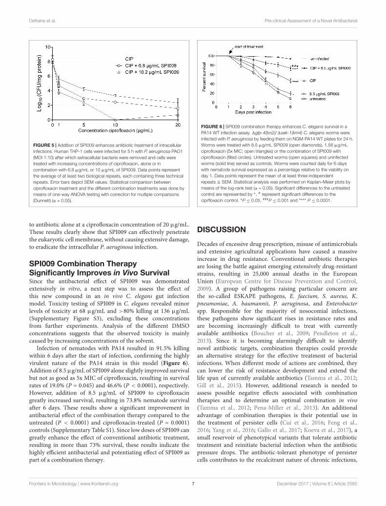
SPI009 Potentiates Antibiotic Activity inan Intracellular Infection ModelNext, the anti-persister and antibacterial activities of SPI009were verified in a recently developed P. aeruginosa intracellularinfection model (Buyck et al., 2013). Human THP-1 cellswere infected with PAO1 cells (MOI 10) and treated for5 h with different concentrations of ciprofloxacin, alone or incombination with 6.8 or 10.2 µg/mL of SPI009. Concentrationsof SPI009 were chosen to be well below the determinedIC50 value of 24.5 ± 1.36 µg/mL. After treatment, boththe number of surviving PAO1 cells and the amount ofeukaryotic proteins present was assessed, as this can provideinformation about the possible toxic effect of the differenttreatments and the infecting bacteria. While treatment withSPI009 alone caused non-significant decreases of 0.78 ± 0.7and 0.89 ± 0.7 log units in surviving bacteria, addition ofSPI009 to ciprofloxacin greatly improved the antibacterial effectfor all concentrations tested and this in a dose-dependentmanner (Figure 5). Maximal antibacterial activity for thecombination therapy with 10.2 µg/mL of SPI009 occurs atciprofloxacin concentrations of 10 µg/mL, resulting in completeeradication of the bacterial culture. Moreover, all combinationstested significantly reduced the bacterial load as compared tociprofloxacin alone. Combination treatment with 6.8 µg/mLSPI009 showed a maximal 0.78 ± 0.6 log decrease as compared
Frontiers in Microbiology | www.frontiersin.org 6 December 2017 | Volume 8 | Article 2585
fmicb-08-02585 December 22, 2017 Time: 13:34 # 7
Defraine et al. Pre-clinical Assessment of a Novel Antibacterial
FIGURE 5 | Addition of SPI009 enhances antibiotic treatment of intracellularinfections. Human THP-1 cells were infected for 5 h with P. aeruginosa PAO1(MOI 1:10) after which extracellular bacteria were removed and cells weretreated with increasing concentrations of ciprofloxacin, alone or incombination with 6.8 µg/mL or 10 µg/mL of SPI009. Data points representthe average of at least two biological repeats, each containing three technicalrepeats. Error bars depict SEM values. Statistical comparison betweenciprofloxacin treatment and the different combination treatments was done bymeans of one-way ANOVA testing with correction for multiple comparisons(Dunnett) (α = 0.05).
to antibiotic alone at a ciprofloxacin concentration of 20 µg/mL.These results clearly show that SPI009 can effectively penetratethe eukaryotic cell membrane, without causing extensive damage,to eradicate the intracellular P. aeruginosa infection.
SPI009 Combination TherapySignificantly Improves in Vivo SurvivalSince the antibacterial effect of SPI009 was demonstratedextensively in vitro, a next step was to assess the effect ofthis new compound in an in vivo C. elegans gut infectionmodel. Toxicity testing of SPI009 in C. elegans revealed minorlevels of toxicity at 68 µg/mL and >80% killing at 136 µg/mL(Supplementary Figure S3), excluding these concentrationsfrom further experiments. Analysis of the different DMSOconcentrations suggests that the observed toxicity is mainlycaused by increasing concentrations of the solvent.
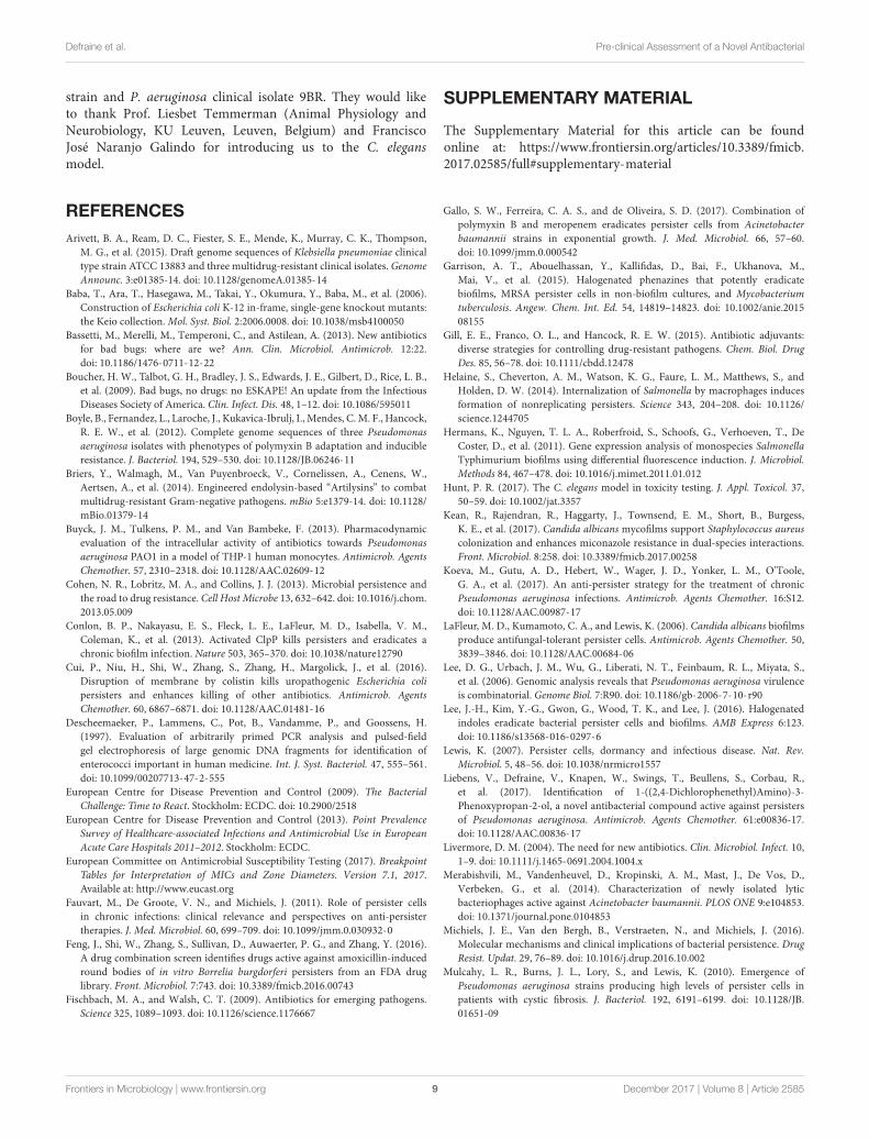
Infection of nematodes with PA14 resulted in 91.5% killingwithin 6 days after the start of infection, confirming the highlyvirulent nature of the PA14 strain in this model (Figure 6).Addition of 8.5 µg/mL of SPI009 alone slightly improved survivalbut not as good as 5x MIC of ciprofloxacin, resulting in survivalrates of 19.0% (P = 0.045) and 46.6% (P < 0.0001), respectively.However, addition of 8.5 µg/mL of SPI009 to ciprofloxacingreatly increased survival, resulting in 73.8% nematode survivalafter 6 days. These results show a significant improvement inantibacterial effect of the combination therapy compared to theuntreated (P < 0.0001) and ciprofloxacin-treated (P = 0.0001)controls (Supplementary Table S1). Since low doses of SPI009 cangreatly enhance the effect of conventional antibiotic treatment,resulting in more than 73% survival, these results indicate thehighly efficient antibacterial and potentiating effect of SPI009 aspart of a combination therapy.
FIGURE 6 | SPI009 combination therapy enhances C. elegans survival in aPA14 WT infection assay. 1glp-4(bn2)/1sek-1(km4) C. elegans worms wereinfected with P. aeruginosa by feeding them on NGM-PA14 WT plates for 24 h.Worms were treated with 8.5 µg/mL SPI009 (open diamonds), 1.56 µg/mLciprofloxacin (5x MIC; open triangles) or the combination of SPI009 withciprofloxacin (filled circles). Untreated worms (open squares) and uninfectedworms (solid line) served as controls. Worms were counted daily for 6 dayswith nematode survival expressed as a percentage relative to the viability onday 1. Data points represent the mean of at least three independentrepeats ± SEM. Statistical analysis was performed on Kaplan–Meier plots bymeans of the log-rank test (α = 0.05). Significant differences to the untreatedcontrol are represented by ∗, # represent significant differences to theciprfloxacin control. ∗P ≤ 0.05, ###P ≤ 0.001 and ∗∗∗∗ P ≤ 0.0001.
DISCUSSION
Decades of excessive drug prescription, misuse of antimicrobialsand extensive agricultural applications have caused a massiveincrease in drug resistance. Conventional antibiotic therapiesare losing the battle against emerging extensively drug-resistantstrains, resulting in 25,000 annual deaths in the EuropeanUnion (European Centre for Disease Prevention and Control,2009). A group of pathogens raising particular concern arethe so-called ESKAPE pathogens, E. faecium, S. aureus, K.pneumoniae, A. baumannii, P. aeruginosa, and Enterobacterspp. Responsible for the majority of nosocomial infections,these pathogens show significant rises in resistance rates andare becoming increasingly difficult to treat with currentlyavailable antibiotics (Boucher et al., 2009; Pendleton et al.,2013). Since it is becoming alarmingly difficult to identifynovel antibiotic targets, combination therapies could providean alternative strategy for the effective treatment of bacterialinfections. When different mode of actions are combined, theycan lower the risk of resistance development and extend thelife span of currently available antibiotics (Tamma et al., 2012;Gill et al., 2015). However, additional research is needed toassess possible negative effects associated with combinationtherapies and to determine an optimal combination in vivo(Tamma et al., 2012; Pena-Miller et al., 2013). An additionaladvantage of combination therapies is their potential use inthe treatment of persister cells (Cui et al., 2016; Feng et al.,2016; Yang et al., 2016; Gallo et al., 2017; Koeva et al., 2017), asmall reservoir of phenotypical variants that tolerate antibiotictreatment and reinitiate bacterial infection when the antibioticpressure drops. The antibiotic-tolerant phenotype of persistercells contributes to the recalcitrant nature of chronic infections,
Frontiers in Microbiology | www.frontiersin.org 7 December 2017 | Volume 8 | Article 2585

fmicb-08-02585 December 22, 2017 Time: 13:34 # 8
Defraine et al. Pre-clinical Assessment of a Novel Antibacterial
greatly complicates treatment and increases the chances ofresistance development (Lewis, 2007; Fauvart et al., 2011;Michiels et al., 2016).
We recently described the discovery of the propanol-aminederivative SPI009, a novel anti-persister molecule capable ofdirectly killing persister cells of P. aeruginosa (Liebens et al.,2017). Most anti-persister molecules described in literatureare only active against one or a very limited number ofbacterial species, which can be explained by a very specificmode of action or the sensitizing of persister cells to aspecific class of antibiotics (Wood, 2015; Van den Bergh et al.,2017). Other examples of small organic compounds capableof directly killing persister cells include the recently describedα-bromocinnamaldehyde (Shen et al., 2017), 5-iodoindole (Leeet al., 2016), halogenated phenazines (Garrison et al., 2015) andthe nitroimidazole prodrug PA-284 (Singh et al., 2008). In thisstudy, we showed that SPI009 possesses broad-spectrum activityand is capable of significantly decreasing or even eradicatingthe bacterial culture for all pathogens tested, including thenotorious ESKAPE pathogens. In addition, combination therapyof conventional antibiotics with SPI009 allowed the efficienttreatment of polymyxin B and ofloxacin resistant strains andcould lower the required concentration of antibiotics, therebyenabling their use in resistant strains.
The close relationship between persisters and chronicinfections (LaFleur et al., 2006; Mulcahy et al., 2010) is partlycaused by their presence in biofilms. The presence of the biofilmmatrix is capable of physically protecting the persister cellsagainst the human immune system, thereby enabling the persistercells to resume growth when antibiotic pressure drops and causerecurrence of infection. When compared to other anti-biofilmcompounds or conventional antibiotics, SPI009 monotherapyshows a promising anti-biofilm effect, both decreasing biofilmformation and causing a strong reduction in the number ofsurviving biofilm cells, for both Gram-negative and Gram-positive species. A more clinically relevant biofilm model wasobtained by P. aeruginosa growth on cellulose matrices andhydrogels, providing a three-dimensional structure and moistenvironment closely mimicking the environment of a chronicallyinfected wound. In this 3D model, clinical treatments havebeen shown to have less impact on the viability of biofilms incomparison to traditional 2D models, which are more susceptibleto eradication (Townsend et al., 2016; Kean et al., 2017).Therefore this further supports the ability of SPI009 mono-treatment to eradicate cells in a more complex biofilm modeland suggests the possible use of SPI009 in the topical treatmentof chronically infected wounds. For all biofilm experimentsexecuted, the addition of SPI009 to a conventional antibioticdid not further decrease the biofilm population as comparedto SPI009 alone. In comparison to planktonic cultures, wherecombination therapy with antibiotics strongly enhances theantibacterial effect, the specific lay-out and environment ofthe bacterial biofilm, including a possibly reduced penetrationof antibacterials, could impair the cooperation between bothantibacterials.
Besides the biofilm matrix, persister cells have also been shownto use eukaryotic cells to shield themselves from the human
immune system. The presence of intracellular persister reservoirshas been confirmed in vivo and can be associated with thechronic nature of infections (Buyck et al., 2013; Helaine et al.,2014). The ability of SPI009 to effectively reduce the intracellularbacteria further confirms the potential of SPI009 as an adjuvant incombination therapies. Capable of increasing nematode survivalto more than 70% when combined with ciprofloxacin, the in vivoC. elegans model further contributes to the clinical potentialof SPI009. The C. elegans model has been extensively used inthe identification and clinical assessment of novel antibacterialsand antifungals with ample studies confirming the consistentcorrelation between toxic effects in C. elegans and mammalianmodels (Hunt, 2017).
CONCLUSION
We demonstrated that the anti-persister molecule SPI009possesses a broad-spectrum antibacterial activity and, takeninto account that it can be combined with different classesof antibiotics, shows great potential for the development ofcase-specific antibacterial combination therapies. The clinicalpotential of SPI009 was further confirmed by the observationof an excellent anti-biofilm activity, successful eradicationof an intracellular infection in human eukaryotes and thesignificant increase in C. elegans survival after treatment withthe combination of SPI009 and ciprofloxacin. Additional in vivoexperiments will be required to assess the future applicabilityof SPI009 but its excellent activity in antibacterial combinationtherapies holds great promise.
AUTHOR CONTRIBUTIONS
Conceptualization, VD, RC, AM, PC, MF, and JM. Methodology,VD, FVB, GR, MF, and JM. Formal analysis, VD. Investigation,VD, LV, AA, and EMT. Wrote the original draft, VD. Contributedin writing review and editing, VD, FVB, GR, MF, and JM.Visualization, VD. Supervision, MF and JM.
FUNDING
This work was supported by Ph.D. grants of the Agencyfor Innovation through Science and Technology (IWT)to VD; the KU Leuven Excellence Center (grant numberPF/2010/07), the KU Leuven Research Council (grant numberPF/10/010, ‘NATAR’); the Belgian Science Policy Office(BELSPO) (IAP P7/28) and the Fund for Scientific Research,Flanders (FWO) (grant numbers G047112N; G0B2515N;G055517N).
ACKNOWLEDGMENTS
The authors thank Pierre Cornelis and Bob Hancockfor providing us with the P. aeruginosa PA14 wild type
Frontiers in Microbiology | www.frontiersin.org 8 December 2017 | Volume 8 | Article 2585
fmicb-08-02585 December 22, 2017 Time: 13:34 # 9
Defraine et al. Pre-clinical Assessment of a Novel Antibacterial
strain and P. aeruginosa clinical isolate 9BR. They would liketo thank Prof. Liesbet Temmerman (Animal Physiology andNeurobiology, KU Leuven, Leuven, Belgium) and FranciscoJosé Naranjo Galindo for introducing us to the C. elegansmodel.
SUPPLEMENTARY MATERIAL
The Supplementary Material for this article can be foundonline at: https://www.frontiersin.org/articles/10.3389/fmicb.2017.02585/full#supplementary-material
REFERENCESArivett, B. A., Ream, D. C., Fiester, S. E., Mende, K., Murray, C. K., Thompson,
M. G., et al. (2015). Draft genome sequences of Klebsiella pneumoniae clinicaltype strain ATCC 13883 and three multidrug-resistant clinical isolates. GenomeAnnounc. 3:e01385-14. doi: 10.1128/genomeA.01385-14
Baba, T., Ara, T., Hasegawa, M., Takai, Y., Okumura, Y., Baba, M., et al. (2006).Construction of Escherichia coli K-12 in-frame, single-gene knockout mutants:the Keio collection. Mol. Syst. Biol. 2:2006.0008. doi: 10.1038/msb4100050
Bassetti, M., Merelli, M., Temperoni, C., and Astilean, A. (2013). New antibioticsfor bad bugs: where are we? Ann. Clin. Microbiol. Antimicrob. 12:22.doi: 10.1186/1476-0711-12-22
Boucher, H. W., Talbot, G. H., Bradley, J. S., Edwards, J. E., Gilbert, D., Rice, L. B.,et al. (2009). Bad bugs, no drugs: no ESKAPE! An update from the InfectiousDiseases Society of America. Clin. Infect. Dis. 48, 1–12. doi: 10.1086/595011
Boyle, B., Fernandez, L., Laroche, J., Kukavica-Ibrulj, I., Mendes, C. M. F., Hancock,R. E. W., et al. (2012). Complete genome sequences of three Pseudomonasaeruginosa isolates with phenotypes of polymyxin B adaptation and inducibleresistance. J. Bacteriol. 194, 529–530. doi: 10.1128/JB.06246-11
Briers, Y., Walmagh, M., Van Puyenbroeck, V., Cornelissen, A., Cenens, W.,Aertsen, A., et al. (2014). Engineered endolysin-based “Artilysins” to combatmultidrug-resistant Gram-negative pathogens. mBio 5:e1379-14. doi: 10.1128/mBio.01379-14
Buyck, J. M., Tulkens, P. M., and Van Bambeke, F. (2013). Pharmacodynamicevaluation of the intracellular activity of antibiotics towards Pseudomonasaeruginosa PAO1 in a model of THP-1 human monocytes. Antimicrob. AgentsChemother. 57, 2310–2318. doi: 10.1128/AAC.02609-12
Cohen, N. R., Lobritz, M. A., and Collins, J. J. (2013). Microbial persistence andthe road to drug resistance. Cell Host Microbe 13, 632–642. doi: 10.1016/j.chom.2013.05.009
Conlon, B. P., Nakayasu, E. S., Fleck, L. E., LaFleur, M. D., Isabella, V. M.,Coleman, K., et al. (2013). Activated ClpP kills persisters and eradicates achronic biofilm infection. Nature 503, 365–370. doi: 10.1038/nature12790
Cui, P., Niu, H., Shi, W., Zhang, S., Zhang, H., Margolick, J., et al. (2016).Disruption of membrane by colistin kills uropathogenic Escherichia colipersisters and enhances killing of other antibiotics. Antimicrob. AgentsChemother. 60, 6867–6871. doi: 10.1128/AAC.01481-16
Descheemaeker, P., Lammens, C., Pot, B., Vandamme, P., and Goossens, H.(1997). Evaluation of arbitrarily primed PCR analysis and pulsed-fieldgel electrophoresis of large genomic DNA fragments for identification ofenterococci important in human medicine. Int. J. Syst. Bacteriol. 47, 555–561.doi: 10.1099/00207713-47-2-555
European Centre for Disease Prevention and Control (2009). The BacterialChallenge: Time to React. Stockholm: ECDC. doi: 10.2900/2518
European Centre for Disease Prevention and Control (2013). Point PrevalenceSurvey of Healthcare-associated Infections and Antimicrobial Use in EuropeanAcute Care Hospitals 2011–2012. Stockholm: ECDC.
European Committee on Antimicrobial Susceptibility Testing (2017). BreakpointTables for Interpretation of MICs and Zone Diameters. Version 7.1, 2017.Available at: http://www.eucast.org
Fauvart, M., De Groote, V. N., and Michiels, J. (2011). Role of persister cellsin chronic infections: clinical relevance and perspectives on anti-persistertherapies. J. Med. Microbiol. 60, 699–709. doi: 10.1099/jmm.0.030932-0
Feng, J., Shi, W., Zhang, S., Sullivan, D., Auwaerter, P. G., and Zhang, Y. (2016).A drug combination screen identifies drugs active against amoxicillin-inducedround bodies of in vitro Borrelia burgdorferi persisters from an FDA druglibrary. Front. Microbiol. 7:743. doi: 10.3389/fmicb.2016.00743
Fischbach, M. A., and Walsh, C. T. (2009). Antibiotics for emerging pathogens.Science 325, 1089–1093. doi: 10.1126/science.1176667
Gallo, S. W., Ferreira, C. A. S., and de Oliveira, S. D. (2017). Combination ofpolymyxin B and meropenem eradicates persister cells from Acinetobacterbaumannii strains in exponential growth. J. Med. Microbiol. 66, 57–60.doi: 10.1099/jmm.0.000542
Garrison, A. T., Abouelhassan, Y., Kallifidas, D., Bai, F., Ukhanova, M.,Mai, V., et al. (2015). Halogenated phenazines that potently eradicatebiofilms, MRSA persister cells in non-biofilm cultures, and Mycobacteriumtuberculosis. Angew. Chem. Int. Ed. 54, 14819–14823. doi: 10.1002/anie.201508155
Gill, E. E., Franco, O. L., and Hancock, R. E. W. (2015). Antibiotic adjuvants:diverse strategies for controlling drug-resistant pathogens. Chem. Biol. DrugDes. 85, 56–78. doi: 10.1111/cbdd.12478
Helaine, S., Cheverton, A. M., Watson, K. G., Faure, L. M., Matthews, S., andHolden, D. W. (2014). Internalization of Salmonella by macrophages inducesformation of nonreplicating persisters. Science 343, 204–208. doi: 10.1126/science.1244705
Hermans, K., Nguyen, T. L. A., Roberfroid, S., Schoofs, G., Verhoeven, T., DeCoster, D., et al. (2011). Gene expression analysis of monospecies SalmonellaTyphimurium biofilms using differential fluorescence induction. J. Microbiol.Methods 84, 467–478. doi: 10.1016/j.mimet.2011.01.012
Hunt, P. R. (2017). The C. elegans model in toxicity testing. J. Appl. Toxicol. 37,50–59. doi: 10.1002/jat.3357
Kean, R., Rajendran, R., Haggarty, J., Townsend, E. M., Short, B., Burgess,K. E., et al. (2017). Candida albicans mycofilms support Staphylococcus aureuscolonization and enhances miconazole resistance in dual-species interactions.Front. Microbiol. 8:258. doi: 10.3389/fmicb.2017.00258
Koeva, M., Gutu, A. D., Hebert, W., Wager, J. D., Yonker, L. M., O’Toole,G. A., et al. (2017). An anti-persister strategy for the treatment of chronicPseudomonas aeruginosa infections. Antimicrob. Agents Chemother. 16:S12.doi: 10.1128/AAC.00987-17
LaFleur, M. D., Kumamoto, C. A., and Lewis, K. (2006). Candida albicans biofilmsproduce antifungal-tolerant persister cells. Antimicrob. Agents Chemother. 50,3839–3846. doi: 10.1128/AAC.00684-06
Lee, D. G., Urbach, J. M., Wu, G., Liberati, N. T., Feinbaum, R. L., Miyata, S.,et al. (2006). Genomic analysis reveals that Pseudomonas aeruginosa virulenceis combinatorial. Genome Biol. 7:R90. doi: 10.1186/gb-2006-7-10-r90
Lee, J.-H., Kim, Y.-G., Gwon, G., Wood, T. K., and Lee, J. (2016). Halogenatedindoles eradicate bacterial persister cells and biofilms. AMB Express 6:123.doi: 10.1186/s13568-016-0297-6
Lewis, K. (2007). Persister cells, dormancy and infectious disease. Nat. Rev.Microbiol. 5, 48–56. doi: 10.1038/nrmicro1557
Liebens, V., Defraine, V., Knapen, W., Swings, T., Beullens, S., Corbau, R.,et al. (2017). Identification of 1-((2,4-Dichlorophenethyl)Amino)-3-Phenoxypropan-2-ol, a novel antibacterial compound active against persistersof Pseudomonas aeruginosa. Antimicrob. Agents Chemother. 61:e00836-17.doi: 10.1128/AAC.00836-17
Livermore, D. M. (2004). The need for new antibiotics. Clin. Microbiol. Infect. 10,1–9. doi: 10.1111/j.1465-0691.2004.1004.x
Merabishvili, M., Vandenheuvel, D., Kropinski, A. M., Mast, J., De Vos, D.,Verbeken, G., et al. (2014). Characterization of newly isolated lyticbacteriophages active against Acinetobacter baumannii. PLOS ONE 9:e104853.doi: 10.1371/journal.pone.0104853
Michiels, J. E., Van den Bergh, B., Verstraeten, N., and Michiels, J. (2016).Molecular mechanisms and clinical implications of bacterial persistence. DrugResist. Updat. 29, 76–89. doi: 10.1016/j.drup.2016.10.002
Mulcahy, L. R., Burns, J. L., Lory, S., and Lewis, K. (2010). Emergence ofPseudomonas aeruginosa strains producing high levels of persister cells inpatients with cystic fibrosis. J. Bacteriol. 192, 6191–6199. doi: 10.1128/JB.01651-09
Frontiers in Microbiology | www.frontiersin.org 9 December 2017 | Volume 8 | Article 2585

fmicb-08-02585 December 22, 2017 Time: 13:34 # 10
Defraine et al. Pre-clinical Assessment of a Novel Antibacterial
O’Neill, J. (2016). Tackling Drug-resistant Infections Globally: Final Report andRecommendations. The Review on Antimicrobial Resistance. London: HMGovernment.
Pena-Miller, R., Laehnemann, D., Jansen, G., Fuentes-Hernandez, A.,Rosenstiel, P., Schulenburg, H., et al. (2013). When the most potentcombination of antibiotics selects for the greatest bacterial load: the smile-frown transition. PLOS Biol. 11:e1001540. doi: 10.1371/journal.pbio.1001540
Pendleton, J. N., Gorman, S. P., and Gilmore, B. F. (2013). Clinical relevanceof the ESKAPE pathogens. Expert Rev. Anti Infect. Ther. 11, 297–308.doi: 10.1586/eri.13.12
Porta-de-la-Riva, M., Fontrodona, L., Villanueva, A., and Cerón, J. (2012). BasicCaenorhabditis elegans methods: synchronization and observation. J. Vis. Exp.64:e4019. doi: 10.3791/4019
Rice, L. B. (2008). Federal funding for the study of antimicrobial resistance innosocomial pathogens: no ESKAPE. J. Infect. Dis. 197, 1079–1081. doi: 10.1086/533452
Shen, Q., Zhou, W., Hu, L., Qi, Y., Ning, H., Chen, J., et al. (2017). Bactericidalactivity of alpha-bromocinnamaldehyde against persisters in Escherichia coli.PLOS ONE 12:e0182122. doi: 10.1371/journal.pone.0182122
Shin, S. H., Kim, S., Kim, J. Y., Lee, S., Um, Y., Oh, M.-K., et al. (2012). Completegenome sequence of Enterobacter aerogenes KCTC 2190. J. Bacteriol. 194,2373–2374. doi: 10.1128/JB.00028-12
Singh, R., Manjunatha, U., Boshoff, H. I. M., Ha, Y. H., Niyomrattanakit, P.,Ledwidge, R., et al. (2008). PA-824 kills nonreplicating Mycobacteriumtuberculosis by intracellular NO release. Science 322, 1392–1395. doi: 10.1126/science.1164571
Smith, K., Collier, A., Townsend, E. M., O’Donnell, L. E., Bal, A. M., Butcher, J.,et al. (2016). One step closer to understanding the role of bacteria in diabeticfoot ulcers: characterising the microbiome of ulcers. BMC Microbiol. 16:54.doi: 10.1186/s12866-016-0665-z
Stiernagle, T. (2006). Maintenance of C. elegans. Available at: http://www.wormbook.org
Tamma, P. D., Cosgrove, S. E., and Maragakis, L. L. (2012). Combination therapyfor treatment of infections with Gram-negative bacteria. Clin. Microbiol. Rev.25, 450–470. doi: 10.1128/CMR.05041-11
Townsend, E. M., Sherry, L., Rajendran, R., Hansom, D., Butcher, J., Mackay, W. G.,et al. (2016). Development and characterisation of a novel three-dimensionalinter-kingdom wound biofilm model. Biofouling 32, 1259–1270. doi: 10.1080/08927014.2016.1252337
Van Acker, H., Sass, A., Bazzini, S., De Roy, K., Udine, C., Messiaen, T., et al. (2013).Biofilm-grown Burkholderia cepacia complex cells survive antibiotic treatmentby avoiding production of reactive oxygen species. PLOS ONE 8:e58943.doi: 10.1371/journal.pone.0058943
Van den Bergh, B., Fauvart, M., and Michiels, J. (2017). Formation, physiology,ecology, evolution and clinical importance of bacterial persisters. FEMSMicrobiol. Rev. 41, 219–251. doi: 10.1093/femsre/fux001
WHO (2017). Global Priority List of Antibiotic-resistant Bacteria to Guide Research,Discovery, and Development of New Antibiotics. Geneva: WHO.
Wood, T. K. (2015). Combatting bacterial persister cells. Biotechnol. Bioeng. 113,476–483. doi: 10.1002/bit.25721
Yang, S., Hay, I. D., Cameron, D. R., Speir, M., Cui, B., Su, F., et al. (2016). Antibioticregimen based on population analysis of residing persister cells eradicatesStaphylococcus epidermidis biofilms. Sci. Rep. 5:18578. doi: 10.1038/srep18578
Conflict of Interest Statement: The authors declare that the research wasconducted in the absence of any commercial or financial relationships that couldbe construed as a potential conflict of interest.
Copyright © 2017 Defraine, Verstraete, Van Bambeke, Anantharajah, Townsend,Ramage, Corbau, Marchand, Chaltin, Fauvart and Michiels. This is an open-accessarticle distributed under the terms of the Creative Commons Attribution License(CC BY). The use, distribution or reproduction in other forums is permitted, providedthe original author(s) or licensor are credited and that the original publication in thisjournal is cited, in accordance with accepted academic practice. No use, distributionor reproduction is permitted which does not comply with these terms.
Frontiers in Microbiology | www.frontiersin.org 10 December 2017 | Volume 8 | Article 2585

Supplementary Material
Antibacterial activity of 1-((2,4-dichlorophenethyl)amino)-3-phenoxypropan-2-ol against antibiotic-resistant strains of diverse bacterial pathogens, biofilms and in pre-clinical infection models
Valerie Defraine, Laure Verstraete, Françoise Van Bambeke, Ahalieyah Anantharajah, Eleanor M. Townsend, Gordon Ramage, Romu Corbau, Arnaud Marchand, Patrick Chaltin, Maarten Fauvart, Jan Michiels
* Correspondence: corresponding author: [email protected]
1 Supplementary Data
1.1 Supplementary Figures
Figure S1: Determination of the persister plateau. Stationary phase cultures of (A) E. aerogenes (open circle), K. pneumoniae (open square), A. baumannii (open triangle), P. aeruginosa (filled triangle), E. coli (open diamond) and (B) S. aureus (open circle), E. faecium (open square) and B. cenocepacia (open triangle) were treated for 5 hours with increasing concentrations of (A) ofloxacin or (B) rifampicin (RIF) or ciprofloxacin (CIP). Data points represent the means of three independent experiments, with error bars depicting the SEM value.

Supplementary Material
2
Figure S2: Effect of conventional antibiotics in the treatment of bacterial biofilms. 24 hour, mature biofilms of (A) P. aeruginosa and (B) S. aureus were treated for 5 hours with a conventional antibiotic, ofloxacin (OFX) and vancomycin (VAN) respectively, or the combination of this antibiotic with increasing concentrations of SPI009. Assessment of biofilm survival was done by CFU counting and statistical differences to the control treatment were detected by means of a one-way ANOVA (α= 0.05) with Dunnett’s correction for multiple comparisons. Data points represent the average of three independent repeats, each containing three technical repeats, ± SEM with ** P < 0.01 and *** P < 0.001. No significant decreases in biofilm survival were observed for the combination therapy, as compared to treatment with equal concentrations of SPI009 alone.
T im e a fte r s ta r t o f tre a tm e n t (d a y s )
Ne
ma
tod
e s
urv
iva
l (%
)
1 2 3 4 5 60
2 0
4 0
6 0
8 0
1 0 0 u n tre a ted
8 .5 µ g /m L
1 7 µ g /m L
3 4 µ g /m L
6 8 µ g /m L
1 3 6 µ g /m L
2 % D M S O
2 0 % D M S O
********
********
Figure S3: Toxicity testing of SPI009 in C. elegans. OP50 fed nematodes were monitored for 5 days in the presence of increasing concentrations of SPI009 (8.5-134 µg/mL). Controls consisted of untreated worms (negative control, black line), 2% DMSO( carrier control, red solid line) and 20% DMSO (positive control, red dotted line). Data point represent the average of at least two independent experiments, as determined by means of Kaplan-Meier survival plots. Statistical analysis was done

3
using log-rank test which showed significant differences between the untreated control and 68 µg/mL, 136 µg/mL, 2% DMSO and 20% DMSO. No toxicity was observed for SPI009 concentrations of 8.5 µg/mL (P = 0.359), 17 µg/mL (P = 0,556) and 34 µg/mL (P = 0.094).
1.2 Supplementary tables
Table S1: Statistical analysis of C. elegans survival data. The Kaplan-Meier method was used to analyze the obtained survival data. N represents the total number of subjects, median lifespan is considered >6 if, at the end of the experiment > 50% of the population is still alive. Kaplan-Meier curves (Figure 6) were statistically analyzed using a log-rank test to compare between treatments and control or different treatments, as indicated in the table. Plog-rank indicates the P-value as a result of the log-rank analysis (α = 0.05), the last column indicates significance levels after the Bonferroni correction for multiple comparisons (α = 0.01).
Group N median lifespan (days)
Plog-rank Bonferroni correction
un-infected 351 >6
untreated 165 4 < 0.0001 vs un-infected
****
8.5 µg/mL SPI009 85 4 0.0452 vs untreated ns CIP (5 x MIC) 132 6 < 0.0001 vs untreated ****
CIP + 8.5 µg/mL SPI009
81 >6 < 0.0001 vs untreated ****
0.0001 vs CIP ****