anti-tumor effect of a novel soluble recombinant human endostatin
TRANSCRIPT

Anti-Tumor Effect of a Novel Soluble RecombinantHuman Endostatin: Administered as a Single Agent or inCombination with Chemotherapy Agents in MouseTumor ModelsZhihua Ren1,3., Yanan Wang2., Wenhong Jiang1, Wei Dai3,4*, Yongping Jiang1,3*
1 Biopharmagen Corp., Suzhou, China, 2 Department of Laboratory Diagnosis, Suzhou Municipal Hospital Affiliated Nanjing Medical University, Suzhou, China,
3 Biopharmaceutical R&D Center, Chinese Academy of Medical Sciences & Peking Union Medical College, Suzhou, China, 4 Department of Environmental Medicine, New
York University Langone Medical Center, Tuxedo, New York, United States of America
Abstract
Background: Angiogenesis has become an attractive target in cancer treatment. Endostatin is one of the potent anti-angiogenesis agents. Its recombinant form expressed in the yeast system is currently under clinical trials. Endostatinsuppresses tumor formation through the inhibition of blood vessel growth. It is anticipated that combined therapy usingendostatin and cytotoxic compounds may exert an additive effect. In the present study, we expressed and purifiedrecombinant human endostatin (rhEndostatin) that contained 3 additional amino acid residues (arginine, glycine, andserine) at the amino-terminus and 6 histidine residues in its carboxyl terminus. The recombinant protein was expressed in E.Coli and refolded into a soluble form in a large scale purification process. The protein exhibited a potent anti-tumor activityin bioassays. Furthermore, rhEndostatin showed an additive effect with chemotherapy agents including cyclophosphamide(CTX) and cisplatin (DDP).
Methods: rhEndostatin cDNA was cloned into PQE vector and expressed in E. Coli. The protein was refolded through dialysiswith an optimized protocol. To establish tumor models, nude mice were subcutaneously injected with human cancer cells(lung carcinoma A549, hepatocellular carcinoma QGY-7703, or breast cancer Bcap37). rhEndostatin and/or DDP wasadministered peritumorally to evaluate the rate of growth inhibition of A549 tumors. For the tumor metastasis model, micewere injected intravenously with mouse melanoma B16 cells. One day after tumor cell injection, a single dose ofrhEndostatin, or in combination with CTX, was administered intravenously or at a site close to the tumor.
Results: rhEndostatin reduced the growth of A549, QGY-7703, and Bcap37 xenograft tumors in a dose dependent manner.When it was administered peritumorally, rhEndostatin exhibited a more potent inhibitory activity. Furthermore,rhEndostatin displayed an additive effect with CTX or DDP on the inhibition of metastasis of B16 tumors or growth ofA549 tumors.
Conclusion: Soluble rhEndostatin exhibits a potent anti-tumor activity in mouse xenograft models and it also has anadditive effect with CTX and DDP, implying possible applications in clinical settings.
Citation: Ren Z, Wang Y, Jiang W, Dai W, Jiang Y (2014) Anti-Tumor Effect of a Novel Soluble Recombinant Human Endostatin: Administered as a Single Agent orin Combination with Chemotherapy Agents in Mouse Tumor Models. PLoS ONE 9(9): e107823. doi:10.1371/journal.pone.0107823
Editor: Irina V. Lebedeva, Columbia University, United States of America
Received March 21, 2014; Accepted August 15, 2014; Published September 17, 2014
Copyright: � 2014 Ren et al. This is an open-access article distributed under the terms of the Creative Commons Attribution License, which permits unrestricteduse, distribution, and reproduction in any medium, provided the original author and source are credited.
Data Availability: The authors confirm that all data underlying the findings are fully available without restriction. All relevant data are within the paper and itsSupporting Information files.
Funding: This study was supported in all by the Key State projects of China (2011ZX09401-027) and (2011ZX09102-010-04). The funders had no role in studydesign, data collection and analysis, decision to publish, or preparation of the manuscript.
Competing Interests: Zhihua Ren, Wenhong Jiang and Yongping Jiang are employed by Biopharmagen Corp. There are no patents, products in developmentor marketed products to declare. This does not alter the authors’ adherence to all the PLOS ONE policies on sharing data and materials, as detailed online in theguide for authors.
* Email: [email protected] (WD); [email protected] (YK)
. These authors contributed equally to this work.
Introduction
Angiogenesis plays a key role in promoting growth, invasion and
metastasis of tumors [1,2,3]. It has become an attractive target for
tumor therapy. Endostatin, a cleaved product of the carboxyl-
terminal domain of collagen XVIII, was originally found in
conditioned media from a murine endothelial tumor cell line
(hemangio-endothelioma). It was reported that endostatin inhibits
endothelial cell proliferation and migration both in vivo and in
vitro, thus inhibiting tumor growth [4,5]. Endostatin has been
evaluated in clinical trials. In a Phase I clinical study, Endostatin
was reported to be safe and well tolerated in advanced cancer
PLOS ONE | www.plosone.org 1 September 2014 | Volume 9 | Issue 9 | e107823
patients, demonstrating some antitumor activities [6,7]. Of the 74
patients’ cohort, minor responses have been found in 2 patients
(40% and 17% reduction in tumor volume for 22 and 11 months,
respectively), and mixed responses in 2 patients for 14 or 5 months;
In addition, 8 patients were reported to have stable disease for
more than 3 months [8]. Furthermore, independent studies have
shown that Endostatin decreases blood flow in tumors in a dose-
dependent manner as measured by positron emission tomography
scan [9,10] and that it induces apoptosis in tumor cells [11,12].
Endostatin that was used in previous clinical studies was
expressed in yeast [13,14], which makes the preparation costly and
laborious. Endostatin has no direct cytotoxicity against tumor cells.
The anti-tumor effect of endostatin stems from its ability to inhibit
neo-angiogenesis around tumors, thus preventing tumor growth
and progression. while it shows no direct cytotoxicity against
tumor cells [15]. It has been reported that endostatin-mediated
inhibition of angiogenesis was most effective when it is combined
with other therapies, especially chemotherapy [16,17,18].
The conventional chemotherapy agents, such as cyclophospha-
mide (CTX) and cisplatin (DDP), directly target tumor cells and
induce cytotoxicity. However, the efficacy of chemotherapy agents
is often compromised due to their adverse side effects such as
myelosuppression and development of drug resistance [19,20]. It is
believed that a combination of anti-angiogenesis agents and
chemotherapy agents will significantly enhance the therapeutic
efficacy and reduce side effects [17,21]. In this study, we expressed
rhEndostatin with additional arginine, glycine, and serine residues
at the N-terminus using the prokaryotic expression system. The
added amino acid residues were expected to enhance the binding
affinity of target molecule to the cellular receptor [22].
rhEndostatin also contained 6-histidine residues at the caboxyl
terminus for affinity purification. We also developed and
optimized a protocol for efficient refolding of bioactive rhEndos-
tatin in a large scale. We demonstrated that rhEndostatin
exhibited a potent bioactivity and was capable of synergizing with
CTX and DDP in suppression of tumor cell growth. Our
combined studies strongly suggest that rhEndostatin can be
developed as a therapeutical agent for the treatment of various
malignancies.
Materials and Methods
Ethics StatementAll research involving animals was conducted according to
relevant national and international guidelines. Male BALB/c nude
mice, weight 16.0,25.1 g were purchased from SLAC Company
(Shanghai, China). The experiment protocols were approved by
the Institutional Animal Care and Use Committees of Soochow
University (IACUC permit number: SYXK(Su) 2012-0045), and
were in accordance with the Guidelines for the Care and Use of
Laboratory Animals (National Research Council, People’s Re-
public of China, 2010). We further attest that all efforts were made
to ensure minimal suffering.
Cell lines and reagentsHuman A549 lung carcinoma cell line [23], human umbilical
vein endothelial cells (HUVEC) [4,24], and mouse B16 melanoma
cell line [25] were purchased from ATCC (Manassas, VA, USA).
Human hepatocellular carcinoma cell line QGY-7703 [26], and
human breast cancer cell line Bcap37 [27] were from Shanghai
Institute of Cell Biology (Shanghai, China). Restriction enzyme
BamHI and HindIII were purchased from New England Biolabs
(Ipswich, MA, USA). The PQE60 plasmid, E.Coli M15 and Ni-
NTA purification kit were obtained from QIAGEN (Valencia,
CA, USA). Isopropyl-1-thio-ß- D-galactopyranoside (IPTG) was
from Bio-Basic Inc (Amherst, NY, USA) to induce rhEndostatin
expression in bacteria. Guanidine Hydrochloride was from
Amresco Inc (Solon, OH, USA). Urea was from Shanghai
Experimental Reagent Inc (Shanghai, China). M199 medium
and RPMI 1640 medium were from Gibco (Grand Island, NY,
USA). Fetal bovine serum was from Hyclone (Logan, UT, USA).
Basic fibroblast growth factor (bFGF) and vascular endothelial
growth factor (VEGF) were from R&D systems (Minneapolis, MN,
USA). MTT was from Cell Biolabs (San Diego, CA, USA).
DMSO was from Sigma Aldrich (St. Louis, MO, USA). Cisplatin
(DDP) was purchased from Shandong Qilu Pharmaceutical Co.,
Ltd (Shandong, China), and Cyclophosphamide (CTX) was from
Shanghai Hualian Pharmaceutical Co, Ltd (Shanghai, China).
Sequence Modified rhEndostatin and Expression ofrhEndostatin in E.Coli
Human endostatin mRNA was extracted from Chinese embryo
hepatocytes and cDNA was obtained by reverse transcription-
polymerase chain reaction (RT-PCR). The amplified products
were fractionated on agarose gels, and then ligated to plasmid
PQE60, after been digested with restriction enzyme BamHI and
HindIII. Mutant rhEndostatin was made by adding CGAG-
GUUCC after the starting codon of AUG, and ligating
CACCACCACCACCACCAC before the stop codon. The
PQE60 plasmid expressing mutant rhEndostatin was transformed
into E.Coli M15, in a form as inclusion body.
rhEndostatin purification and refolding in large scaleThe inclusion body was collected from IPTG induced bacteria
lysate and dissolved with Guanidine Hydrochloride (6 Mol). Ni-
NTA Purification System has been employed to purify poly-
histidine-containing rhEndostatin as previously described [28].
The purified rhEndostatin protein were then diluted with a
phosphate buffer containing 8 Mol of Urea (PH 4.5) to 0.3 mg/ml,
and the refolding protein was obtained by a series of dialysis
procedure with optimized buffer conditions at 4uC.
rhEndostatin inhibits human umbilical vein endothelialcell proliferation in vitro
Primary human umbilical vein endothelial cells (HUVEC) at 2–
5 passages were suspended in M199 basal medium supplemented
with 10% heat-inactivated fetal bovine serum, and then plated in
96-well culture plates at 56103 cells per 100 ml/well. Cells were
incubated with rhEndostatin at various concentrations (0.1, 0.2,
0.5, 1, 2, 5, 8, 16 mg/ml) in bFGF(5 ng/ml) supplemented M199
medium or basal medium alone at 200 ml volume for 72 hours at
37uC. 20 ml of MTT(5%) was added to each well at 4 hours before
the end of treatment, and then aspirated carefully. Cells were
further incubated with 100 ml of DMSO for 15 minutes on the
shaker, and optical density (OD) value was read at 570 nm wave
length with the microplate reader (Bio-Rad, Hercules, CA. USA).
Inhibition of chick embryo chorioallantoic membrane(CAM) angiogenesis by rhEndostatin
Chick embryonic chorioallantoic membrane assay was em-
ployed for evaluating the anti-angiogenesis activity of rhEndosta-
tin. All procedures were carried out in a biosafety hood under
sterile conditions. Briefly, fertilized eggs were incubated for 7 days
at 37uC and 60% humidity. A window was made on the top of
each egg after 2 days’ incubation. Chick embryonic chorioallan-
toic membrane was exposed by tearing up the egg membrane with
a tip. A filter paper disc of 5 mm diameter containing 20 ml of
Anti-Tumor Effect of a Novel Human Endostatin with Chemotherapy Agents
PLOS ONE | www.plosone.org 2 September 2014 | Volume 9 | Issue 9 | e107823

rhEndostatin (0.3 mg/mL) or saline was gently implanted, and
changed every day for 10 days. The membrane was cut around the
air sac, which was turned upside down and observed and
photographed by a stereomicroscope. The number of blood vessel
branch points was counted, and the vessel density was calculated
according to the size of area where the blood vessel distributed to
evaluate the anti-angiogenesis activity.
In vitro anti-tumor study on rhEndostatinAll tumor cell lines (human A549 lung carcinoma cell line and
mouse B16 melanoma cell line, human hepatocellular carcinoma
cell line QGY-7703, and human breast cancer cell line Bcap37)
were maintained in RPMI 1640 supplemented with 10% FBS.
Human umbilical vein endothelial cells were cultured in RPMI
1640 supplemented with 2% FBS and 10 ng/mL each of VEGF
and b-FGF. In vitro activity assay on rhEndostatin was performed
as O’Reilly MS et al. described [4], with minor modifications.
AnimalsMale BALB/c nude mice, at 4–6 weeks of age, with an average
weight of 20.561.2 g (16.0,25.1 g) were employed in our studies.
A total of 50 mice were involved in each of the following anti-
tumor studies with single rhED treatment, including hepatocellu-
lar carcinoma QGY-7703, A549 lung carcinoma, or Bcap37
breast cancer, which were further divided into 5 groups, randomly.
To evaluate the combination effects with CTX or DDP, a total of
160 nude mice (N = 10 in each treatment group) were selected.
Another 60 nude mice (N = 10 each) were used to analyze if local
rhEndostatin delivery will improve the tumor inhibition rate, in
A549 lung carcinoma and Bcap37 breast cancer models. Animals
were housed in individual stainless steel cages in a SPF facility in
Soochow University, with a regulated temperature of 2462uC,
relative humidity of 50610%, and a 12-h light cycle. Animals
were sacrificed by Carbon dioxide (CO2) inhalation at 24 hours
post the final treatment. Tumor tissues and organs were collected
after euthanasia.
Tumor-bearing mouse studiesThe inhibition capabilities of rhEndostatin to tumor growth and
tumor metastasis were evaluated. The tumor-bearing mouse
model was generated by injecting BALB/c strain nude mice
subcutaneously with 66106/100 ml of each tumor cells, including
human A549 lung carcinoma, human hepatocellular carcinoma
QGY-7703 and human breast cancer Bcap37 cells. When the
tumors volume were about of 170 mm3, approximately 15 days
after implantation, either rhEndostatin alone, or in combination
with DDP were delivered to the animal model either intravenously
or subcutaniously at the site close to the inoculated tumor, to
evaluate the inhibition rate of tumor growth. Whereas rhEndos-
tatin alone or along with CTX was given intraperitoneally to
observe the metastatic rate of tumor. The mice from control group
were received injections of phosphate-buffered saline (PBS) at the
same volume intravenously. The mice were evaluated for tumor
growth at every other day. The formula, width26length60.52, was
applied to estimate the volume of a spheroid as described [5]. The
formula, [(tumor volume of control-tumor volume of treatment)/
tumor volume of control]6100%, was used to calculate the
inhibition rate of tumor growth [5]. For the estimation of the
metastasis inhibition rate, tumor-bearing mice were sacrificed by
carbon dioxide asphyxiation, and evaluation was calculated with
following formula: [(numbers of tumor colonies in lung of control
mice numbers of tumor colonies in lung of treatment mice)/
numbers of tumor colonies in lung of control mice]6100%. All
procedures involving animals were done in accordance with
national and international laws and policies.
Statistical analysisComparison of variables was performed using One-Way
ANOVA in SPSS program (SPSS). A statistically significant
difference was defined as P,0.05.
Results
Expression, refolding, and purification of rhEndostatin inE. coli
Modified rhEndostatin was expressed in E. coli using a pQE
vector expression system. Following the addition of IPTG,
rhEndostatin was highly induced (Figure 1A). Harvested from
40-L bio-reactor, about 1.5 g/L of rhEndostatin inclusion body
was collected. The rhEndostatin was purified by affinity purifica-
tion with nickel (Ni) resin followed by size-exclusion chromatog-
raphy, and then the denatured protein underwent an optimized
refolding procedure to gain a refolding rate of over 80%.
Eventually, about 200 mg/L of purified rhEndostatin with
bioactivity was generated. SDS-PAGE analysis revealed that
rhEndostatin was purified to homogeneity and remained intact
(Figure 1A). Immunoblotting showed that the commercial avail-
able rhEndostatin antibody detected IPTG-induced sequence-
modified- rhEndostatin in the total bacterial lysates as well as its
purified form (Figure 1B), suggesting that the amino acids addition
did not significantly change the overall conformation of the
protein. Protein sequencing analysis confirmed that the purified
protein was the modified form of rhEndostatin with the addition of
arginine, glycine, and serine residues at the amino-terminus (data
not shown).
Anti-angiogenesis assay on rhEndostatin in vitro and invivo
To determine whether rhEndostatin could inhibit the growth of
endothelial cells in vitro, HUVECs had been treated with
rhEndostatin at the dose range from 0.1 to 16 mg/ml. Cell
variability has been evaluated by MTT assay. rhEndostatin
Figure 1. SDS-PAGE and immunoblotting of expression andpurified rhEndostatin. A. SDS-PAGE of rhEndostatin. Lane 1: Pre-stained molecular weight markers; Lane 2: Purified rhEndostatin; Lane 3:Induced bacterial lysates; Lane 4: Non-induced bacterial lysates. (B).Immunoblotting of rhEndostatin. Lane 1: Purified rhEndostatin Lane 2:Whole bacterial cell lysates after IPTG induction; Lane 3: Non-inducedbacterial lysates.doi:10.1371/journal.pone.0107823.g001
Anti-Tumor Effect of a Novel Human Endostatin with Chemotherapy Agents
PLOS ONE | www.plosone.org 3 September 2014 | Volume 9 | Issue 9 | e107823

remarkably inhibited HUVEC proliferation in a dose depended
manner (figure 2). rhEndostatin significantly inhibited HUVECs
growth at the concentration above 0.2 mg/ml compared with the
saline vehicle control. In vivo anti-angiogenic effect was investi-
gated on a chicken embryonic chorioallantoic membrane assay
(CAMs). CAMs vascularization was inhibited by rhEndostatin at
6 mg/disc for 10 days incubation. Figure 3B showed a normal
looking of vascularization of untreated CAMs with primary,
secondary and tertiary vessels and dendritic branching. On the
contrary, CAMs treated with rhEndostatin demonstrated a
distorted vasculature or the absence of blood vessels (Figure 3A).
Antitumor efficacy of rhEndostatin administered as asingle agent
We first studied the anti-tumor bioactivity of rhEndostatin both
in vitro and in vivo. rhEndostatin had no effect on the growth of
any kind of tumor cells in vitro (data not shown). In the in vivostudy, tumor cells were implanted subcutaneously, and measurable
solid tumors were developed approximately 14 days post
implantation at the injection site in all mice. Administration of
rhEndostatin alone significantly delayed the growth of human
A549 lung carcinoma, human hepatocellular carcinoma QGY-
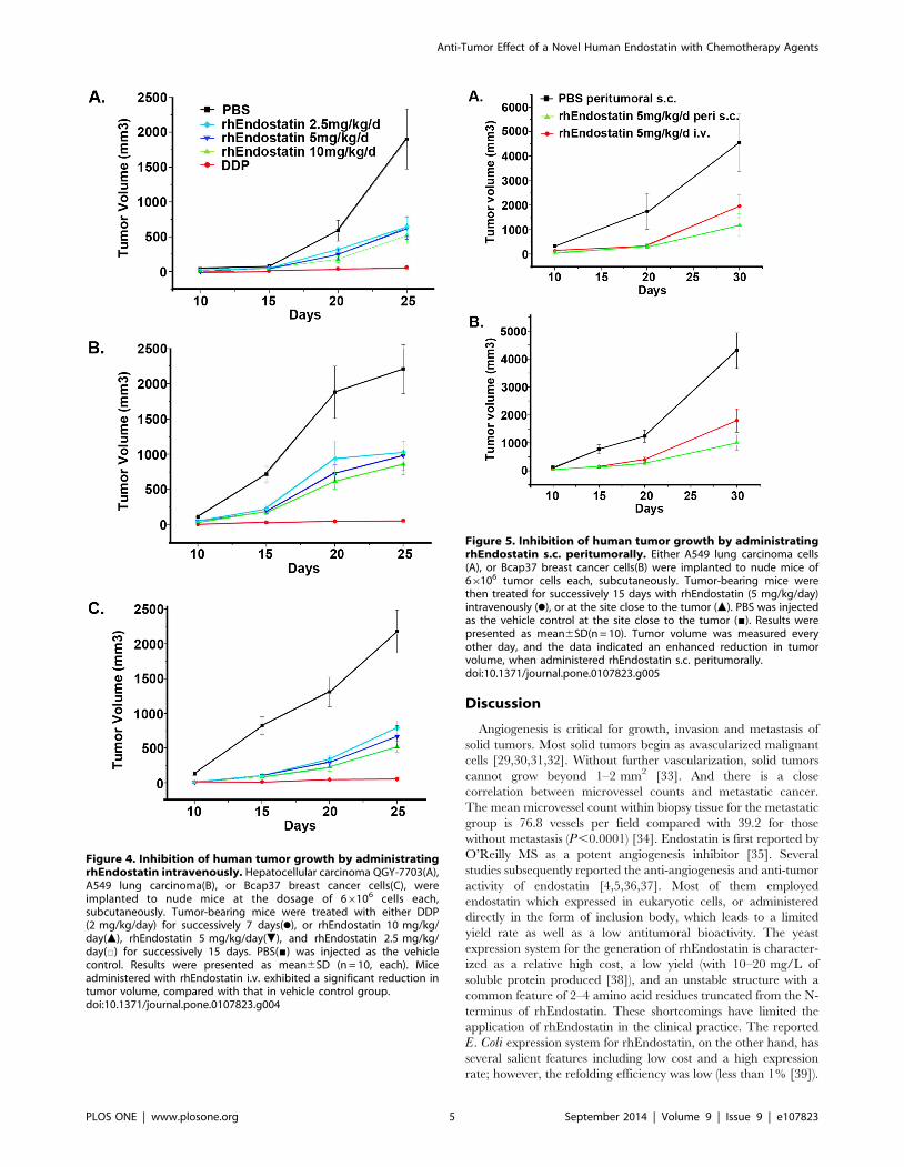
7703 and human breast cancer Bcap37 (Figure 4A, B, C), by 45%
-35% at a high to medium dosage, compared with PBS vehicle
control. Moreover, subcutaneous administration of rhEndostatin
peritumorally exhibited a higher inhibition rate on day 30 post
treatment than that by intravenous injection (Figure 5). Endostar,
a version of commercially available endostatin, also exhibited an
inhibitory activity toward A549 xenograft tumors. Although no
statistically significant difference was observed between Endostar
and rhEndostatin in blocking tumor growth, on day 9 rhEndos-
tatin appeared to be more efficacious in reducing the tumor size
(Figure S1).
rhEndostatin administered in combination with CTXB16 melanoma metastasis model was employed to assay the
additive effect of rhEndostatin with CTX. As shown in Figure 6,
intravenously administration of three dosages of rhEndostatin
(10 mg/kg/day, 5 mg/kg/day, and 2.5 mg/kg/day, respectively)for successive 15 days significantly inhibited the metastasis of B16
melanoma, with the inhibition rate of 34.87%, 29.66% and
29.26%, respectively. Furthermore, rhEndostatin and CTX
exhibited an additive effect. CTX (15 mg/kg/day) administered
alone in the first 7 days inhibited the metastasis by 59.32%,
whereas rhEndostatin, which was intravenously injected at the
dosage of 10 mg/kg/day, 5 mg/kg/day, and 2.5 mg/kg/day for
successive 15 days, increased the inhibition rate to 77.15%,
70.94%, and 65.93%, respectively.
rhEndostatin used in combination with DDPA nude mouse model with xenograft tumors (A549 cells) was
used to evaluate the additive effect of rhEndostatin with DDP. As
shown in Figure 7, three dosages of rhEndostatin, 5 mg/kg/day,
10 mg/kg/day, and 15 mg/kg/day, were administered peritumo-
rally, twice a day for successive 15 days. Significant inhibition of
tumor growth was observed with the inhibition rate of 28.79%,
28.88% and 32.43% (N = 10, each), respectively. Furthermore, an
additive effect was also exhibited when rhEndostatin administered
in combined with DDP. DDP (3 mg/kg/day) administered alone
by i.p. in the first 7 days suppressed the tumor volume by 45.19%.
After the first 7 days treatment, DDP (3 mg/kg/day), was
administered in combination with rhEndostatin at the dose of
5 mg/kg/day, 10 mg/kg/day, and 15 mg/kg/day, respectively,
for another 15 days successively. The inhibition rate was increased
to 54.24%, 47.26%, and 60.66%, respectively.
Figure 2. The inhibition effect of rhEndostatin on the growth ofHUVEC in vitro. HUVECs were planted in fibronectin coated 96-wellmicrotiterplates at 5610‘3cells per well with bFGF supplemented M199medium. rhEndostatin was applied at the dosage of 0.1, 0.2, 0.5, 1.0, 2.0,4.0, 8.0, 16 mg/ml, as described before. All the treatments weretriplicated. The OD value were shown as mean 6 SD. Treatment with2 mg/mL of rhEndostatin and above significantly inhibited HUVECproliferation compared to saline vehicle control.doi:10.1371/journal.pone.0107823.g002
Figure 3. In vivo inhibition of angiogenesis using chickenembryo CAMs. Treated with rhEndostatin at 6 mg/disc (A), or treatedwith saline vehicle control disc (B) the vessels numbers werecalculated(n = 8, each) in CAMs assay after 10 days treatment, and datawere presented as mean6SD. # indicated P,0.05. (C).doi:10.1371/journal.pone.0107823.g003
Anti-Tumor Effect of a Novel Human Endostatin with Chemotherapy Agents
PLOS ONE | www.plosone.org 4 September 2014 | Volume 9 | Issue 9 | e107823
Discussion
Angiogenesis is critical for growth, invasion and metastasis of
solid tumors. Most solid tumors begin as avascularized malignant
cells [29,30,31,32]. Without further vascularization, solid tumors
cannot grow beyond 1–2 mm2 [33]. And there is a close
correlation between microvessel counts and metastatic cancer.
The mean microvessel count within biopsy tissue for the metastatic
group is 76.8 vessels per field compared with 39.2 for those
without metastasis (P,0.0001) [34]. Endostatin is first reported by
O’Reilly MS as a potent angiogenesis inhibitor [35]. Several
studies subsequently reported the anti-angiogenesis and anti-tumor
activity of endostatin [4,5,36,37]. Most of them employed
endostatin which expressed in eukaryotic cells, or administered
directly in the form of inclusion body, which leads to a limited
yield rate as well as a low antitumoral bioactivity. The yeast
expression system for the generation of rhEndostatin is character-
ized as a relative high cost, a low yield (with 10–20 mg/L of
soluble protein produced [38]), and an unstable structure with a
common feature of 2–4 amino acid residues truncated from the N-
terminus of rhEndostatin. These shortcomings have limited the
application of rhEndostatin in the clinical practice. The reported
E. Coli expression system for rhEndostatin, on the other hand, has
several salient features including low cost and a high expression
rate; however, the refolding efficiency was low (less than 1% [39]).
Figure 4. Inhibition of human tumor growth by administratingrhEndostatin intravenously. Hepatocellular carcinoma QGY-7703(A),A549 lung carcinoma(B), or Bcap37 breast cancer cells(C), wereimplanted to nude mice at the dosage of 66106 cells each,subcutaneously. Tumor-bearing mice were treated with either DDP(2 mg/kg/day) for successively 7 days(N), or rhEndostatin 10 mg/kg/day(m), rhEndostatin 5 mg/kg/day(.), and rhEndostatin 2.5 mg/kg/day(%) for successively 15 days. PBS(&) was injected as the vehiclecontrol. Results were presented as mean6SD (n = 10, each). Miceadministered with rhEndostatin i.v. exhibited a significant reduction intumor volume, compared with that in vehicle control group.doi:10.1371/journal.pone.0107823.g004
Figure 5. Inhibition of human tumor growth by administratingrhEndostatin s.c. peritumorally. Either A549 lung carcinoma cells(A), or Bcap37 breast cancer cells(B) were implanted to nude mice of66106 tumor cells each, subcutaneously. Tumor-bearing mice werethen treated for successively 15 days with rhEndostatin (5 mg/kg/day)intravenously (N), or at the site close to the tumor (m). PBS was injectedas the vehicle control at the site close to the tumor (&). Results werepresented as mean6SD(n = 10). Tumor volume was measured everyother day, and the data indicated an enhanced reduction in tumorvolume, when administered rhEndostatin s.c. peritumorally.doi:10.1371/journal.pone.0107823.g005
Anti-Tumor Effect of a Novel Human Endostatin with Chemotherapy Agents
PLOS ONE | www.plosone.org 5 September 2014 | Volume 9 | Issue 9 | e107823

Therefore, our strategy focused on the optimization of refolding
and purification procedure, in order to provide a sufficient amount
of soluble form of rhEndostatin which can be potentially used for
clinical trials. Our current study is among the very few that use
rhEndostatin expressed in E. Coli and refolds into a soluble form
with an optimized dialysis procedure (patent pending), which
yields about 100 mg/L of soluble protein with a purity of 99%.
Structural integrity of rhEndostatin is evaluated by protein
sequencing, and anti-angiogenic and antitumor activities are also
determined, which yields a comparable result as compared with
the commercially available Endostar.
In order to achieve an optimal tumor inhibition and regression,
researchers have demonstrated that it is critical to continuously
elevate endostatin levels in blood circulation [40,41]. A short half-
life of 2 h of endostatin has been reported, which requires a more
frequent drug administration in addition to escalating drug doses
to achieve the effective plasma concentration. Our modified
rhEndostatin contains 3 additional amino acid residues (arginine,
glycine, and serine) at the amino-terminus, which appears to
increase the overall positive charge of protein, which may in turn
enhance its affinity to the cognate receptor molecule, as previously
reported [42,43,44]. Thus, it is not surprising that a prolonged
half-life and better antitumoral activity were observed. Future
studies will be focused on pharmacokinetics on non-human
primates. In addition, 6 histidine residues are included in the
carboxyl terminus of rhEndostatin, which are used for affinity
purification. At present, we are not sure whether the addition of
these residues would affect the activity of rhEndostatin.
Previous studies showed that endostatin has an additive effect
with chemotherapy agents towards various types of tumors
[45,46,47] with little toxicity [5,7,12]. Endostar has been approved
for clinical trials in China. Preclinical studies indicated that the
combination of Endostar with chemotherapy is more effective to
inhibit lung tumor growth and metastases [48,49]. Most recently,
clinical data from multiple institutes suggest that the strategy to
combine Endostar and chemotherapy agents can dramatically
improve outcomes of cancer patients [50,51,52]. Similar results
have been observed in our preclinical models that the rhEndos-
tatin exhibits a potent antitumor activity against homogeneic and
allogeneic tumors in mice alone or in combination with CTX, as
well as DDP. The commercially available Endostar is also
included in our study. The tumor inhibition rate by Endostar
and rhED are about 22% and 28%, respectively. On the other
hand, no significant difference has been found between these two
factors. Furthermore, our studies reveal that an enhanced tumor
inhibitory activity can be achieved when rhEndostatin is
administered peritumorally, implying that the route of drug
delivery may affect its bioactivity. It has been shown that
endostatin binds to the blood vessels when it is administered
systemically, resulting in the decreased angiogenic protein
concentration around the tumor microenvironment [53]. Angio-
genesis inhibitors are most effective when administered in a way
which maintains a stable concentration in the circulation [54].
Local administration of rhEndostatin may help to maintain an
effective concentration around tumors.
A combination of endostatin with chemotherapy agents is
currently employed to improve the therapeutic efficacy. However,
its application is often hampered by either non-overlapping or
overlapping toxicities [10,55]. Moreover, due to unstable genome,
tumor cells frequently develop multi-drug resistance to various
Figure 6. Additive effect of rhEndostatin with CTX on themetastasis of B16 melanoma. Nude mice (n = 10, each) wereinoculated with 2.56105 tumor cells intravenously to establish ametastasis model. Mice were either administered i.v. with rhEndostatinalone at 10 mg/kg/day, 5 mg/kg/day, or 2.5 mg/kg/day for 15 dayssuccessively, or combined treated with CTX at the dosage of 15 mg/kg/d for successively 7 days, then intravenously administered withrhEndostatin at 10 mg/kg/day, 5 mg/kg/day, or 2.5 mg/kg/day forsuccessively 15 days. The mice were sacrificed to count the metastasiscolony in the lung on day 25 post inoculation. The formula, [(meancolony of PBS control- mean colony of treated mice)/mean colony ofPBS control]6100, was used to calculate the inhibition rate ofmetastasis. #: P,0.05 vs mice administered with single rhEndostatintreatment at the same dose, respectively; *: P,0.05 vs mice treated withCTX alone, at the dosage of 15 mg/kg/day.doi:10.1371/journal.pone.0107823.g006
Figure 7. Additive inhibition effect of rhEndostatin with DDPon the growth of A549 lung cancer. Nude mice (n = 10, each) wereinoculated with 66106 tumor cells subcutanously to establish thetumor-bearing model. The animals were either administered peritumo-rally with rhEndostatin alone at 5 mg/kg/day, 10 mg/kg/day, and15 mg/kg/day for 15 days, successively, or combined with DDPtreatment at 3 mg/kg/d every other day for 15 days. The tumor volumewas measured every other day, and the formula, [(tumor volume ofcontrol-tumor volume of treatment)/tumor volume of control]6100%,was used to calculate the inhibition rate of tumor growth. #: P,0.05 vsmice administered with single rhEndostatin treatment at the samedose, respectively; *: P,0.05 vs mice treated with DDP alone at 3 mg/kg/day.doi:10.1371/journal.pone.0107823.g007
Anti-Tumor Effect of a Novel Human Endostatin with Chemotherapy Agents
PLOS ONE | www.plosone.org 6 September 2014 | Volume 9 | Issue 9 | e107823
chemotherapies [56]. While Endostatin exerts a cytostatic effect on
endothelium, which is less prone to the development of drug
resistance [55,57]. In the present study, we have demonstrated
that a combination of conventional chemotherapy agent, CTX,
with rhEndostatin exhibits an additive effect on the metastasis of
B16 melanoma. Our research data also confirm that a combina-
tion of cytotoxic antitumor drug, DDP, with rhEndostatin results
in an additive effect on growth inhibition of A549 xenograft
tumors. This combination strategy aims at multiple targets of
tumor development, thus enhancing the therapeutic efficacy and
reducing the toxicity of chemotherapy and occurrence of drug
resistance.
Our current study demonstrates that prokaryotically expressed,
soluble rhEndostatin exhibits a potent anti-tumor effect. rhEndos-
tatin that is administered peritumorally achieved a better anti-
tumor activity than that achieved by intravenous injection.
Moreover, rhEndostatin displays increased tumor inhibition in
A549 tumor cell xenografts in mice when it is combined with
DDP. Furthermore, rhEndostatin exhibits an additive effect with
CTX on inhibition of metastasis of B16 melanoma. Our
preclinical studies strongly suggest that rhEndostatin can be
further explored for clinical trials for eventual applications in the
clinic.
Supporting Information
Figure S1 Inhibition of human A549 lung carcinomagrowth by administrating rhEndostatin or Endostar.A549 lung carcinomas were implanted to nude mice at the dosage
of 66106 cells each, subcutaneously. Tumor-bearing mice were
treated peritumorally with either Endostar (m, 5 mg/kg/day) or
rhEndostatin (&, 5 mg/kg/day), for successively 15 days. PBS
(X)was injected at the same volume as the vehicle control. The
tumor volume was measured on day 0, 3, 6, 9, 12, 15 post
injections. Results were presented as mean6SD (n = 10, each).
(TIF)
Checklist S1 ARRIVE Guidelines.(DOC)
Acknowledgments
We thank co-workers in the laboratory for the valuable discussions and
suggestions. The authors read and approved the manuscript.
Author Contributions
Conceived and designed the experiments: YJ ZR WD. Performed the
experiments: YW WJ ZR YJ. Analyzed the data: YW ZR YJ WD.
Contributed reagents/materials/analysis tools: YW. Contributed to the
writing of the manuscript: YJ ZR WD.
References
1. Folkman J (2002) Role of angiogenesis in tumor growth and metastasis. Semin
Oncol 29: 15–18.
2. Zetter BR (1998) Angiogenesis and tumor metastasis. Annu Rev Med 49: 407–
424.
3. Rao JS, Gondi C, Chetty C, Chittivelu S, Joseph PA, et al. (2005) Inhibition of
invasion, angiogenesis, tumor growth, and metastasis by adenovirus-mediated
transfer of antisense uPAR and MMP-9 in non-small cell lung cancer cells. Mol
Cancer Ther 4: 1399–1408.
4. O’Reilly MS, Boehm T, Shing Y, Fukai N, Vasios G, et al. (1997) Endostatin: an
endogenous inhibitor of angiogenesis and tumor growth. Cell 88: 277–285.
5. Boehm T, Folkman J, Browder T, O’Reilly MS (1997) Antiangiogenic therapy
of experimental cancer does not induce acquired drug resistance. Nature 390:
404–407.
6. Mundhenke C, Thomas JP, Wilding G, Lee FT, Kelzc F, et al. (2001) Tissue
examination to monitor antiangiogenic therapy: a phase I clinical trial with
endostatin. Clin Cancer Res 7: 3366–3374.
7. Eder JP, Jr., Supko JG, Clark JW, Puchalski TA, Garcia-Carbonero R, et al.
(2002) Phase I clinical trial of recombinant human endostatin administered as a
short intravenous infusion repeated daily. J Clin Oncol 20: 3772–3784.
8. Kulke MH, Bergsland EK, Ryan DP, Enzinger PC, Lynch TJ, et al. (2006)
Phase II study of recombinant human endostatin in patients with advanced
neuroendocrine tumors. J Clin Oncol 24: 3555–3561.
9. Sun L, Ye HY, Zhang YH, Guan YS, Wu H (2007) Epidermal growth factor
receptor antibody plus recombinant human endostatin in treatment of hepatic
metastases after remnant gastric cancer resection. World J Gastroenterol 13:
6115–6118.
10. Plum SM, Hanson AD, Volker KM, Vu HA, Sim BK, et al. (2003) Synergistic
activity of recombinant human endostatin in combination with adriamycin:
analysis of in vitro activity on endothelial cells and in vivo tumor progression in
an orthotopic murine mammary carcinoma model. Clin Cancer Res 9: 4619–
4626.
11. Roy Choudhury S, Karmakar S, Banik NL, Ray SK (2012) Targeting
angiogenesis for controlling neuroblastoma. J Oncol: 782020.
12. Herbst RS, Mullani NA, Davis DW, Hess KR, McConkey DJ, et al. (2002)
Development of biologic markers of response and assessment of antiangiogenic
activity in a clinical trial of human recombinant endostatin. J Clin Oncol 20:
3804–3814.
13. Dhanabal M, Ramchandran R, Volk R, Stillman IE, Lombardo M, et al. (1999)
Endostatin: yeast production, mutants, and antitumor effect in renal cell
carcinoma. Cancer Res 59: 189–197.
14. Boehm T, Pirie-Shepherd S, Trinh LB, Shiloach J, Folkman J (1999) Disruption
of the KEX1 gene in Pichia pastoris allows expression of full-length murine and
human endostatin. Yeast 15: 563–572.
15. Dome B, Hendrix MJ, Paku S, Tovari J, Timar J (2007) Alternative
vascularization mechanisms in cancer: Pathology and therapeutic implications.
Am J Pathol 170: 1–15.
16. Morioka H, Weissbach L, Vogel T, Nielsen GP, Faircloth GT, et al. (2003)
Antiangiogenesis treatment combined with chemotherapy produces chondro-sarcoma necrosis. Clin Cancer Res 9: 1211–1217.
17. Ma J, Waxman DJ (2008) Combination of antiangiogenesis with chemotherapy
for more effective cancer treatment. Mol Cancer Ther 7: 3670–3684.
18. Kaya M, Wada T, Nagoya S, Yamashita T (2007) Prevention of postoperative
progression of pulmonary metastases in osteosarcoma by antiangiogenic therapyusing endostatin. J Orthop Sci 12: 562–567.
19. Kerbel RS, Viloria-Petit A, Klement G, Rak J (2000) ‘Accidental’ anti-
angiogenic drugs. anti-oncogene directed signal transduction inhibitors andconventional chemotherapeutic agents as examples. Eur J Cancer 36: 1248–
1257.
20. Klement G, Baruchel S, Rak J, Man S, Clark K, et al. (2000) Continuous low-dose therapy with vinblastine and VEGF receptor-2 antibody induces sustained
tumor regression without overt toxicity. J Clin Invest 105: R15–24.
21. Cabebe E, Wakelee H (2007) Role of anti-angiogenesis agents in treating
NSCLC: focus on bevacizumab and VEGFR tyrosine kinase inhibitors. Curr
Treat Options Oncol 8: 15–27.
22. Jiang Y, Jiang W, Qiu Y, Dai W Effect of a structurally modified human
granulocyte colony stimulating factor, G-CSFa, on leukopenia in mice andmonkeys. J Hematol Oncol 4: 28.
23. Giard DJ, Aaronson SA, Todaro GJ, Arnstein P, Kersey JH, et al. (1973) In vitro
cultivation of human tumors: establishment of cell lines derived from a series ofsolid tumors. J Natl Cancer Inst 51: 1417–1423.
24. Hoshi H, McKeehan WL (1984) Brain- and liver cell-derived factors are
required for growth of human endothelial cells in serum-free culture. Proc NatlAcad Sci U S A 81: 6413–6417.
25. Briles EB, Kornfeld S (1978) Isolation and metastatic properties of detachmentvariants of B16 melanoma cells. J Natl Cancer Inst 60: 1217–1222.
26. Luo D, Cheng SC, Xie H, Xie Y (1999) Chemosensitivity of human
hepatocellular carcinoma cell line QGY-7703 is related to bcl-2 protein levels.Tumour Biol 20: 331–340.
27. Xiong X, Sui M, Fan W, Kraft AS (2007) Cell cycle dependent antagonistic
interactions between paclitaxel and carboplatin in combination therapy. CancerBiol Ther 6: 1067–1073.
28. Song S JW, Jiang Y (2012) Large-scale production, refolding, and purification ofa novel recombinant human granulocyte colony stimulating factor. Chinese
Journal of Biomedical Engineering 31: 552–557.
29. Folkman J (1996) New perspectives in clinical oncology from angiogenesisresearch. Eur J Cancer 32A: 2534–2539.
30. Saphir A (1997) Angiogenesis: the unifying concept in cancer? J Natl Cancer
Inst 89: 1658–1659.
31. Hagedorn M, Bikfalvi A (2000) Target molecules for anti-angiogenic therapy:
from basic research to clinical trials. Crit Rev Oncol Hematol 34: 89–110.
32. Harris AL (1998) Anti-angiogenesis therapy and strategies for integrating it withadjuvant therapy. Recent Results Cancer Res 152: 341–352.
33. Brem H, Folkman J (1975) Inhibition of tumor angiogenesis mediated bycartilage. J Exp Med 141: 427–439.
Anti-Tumor Effect of a Novel Human Endostatin with Chemotherapy Agents
PLOS ONE | www.plosone.org 7 September 2014 | Volume 9 | Issue 9 | e107823
34. Weidner N, Carroll PR, Flax J, Blumenfeld W, Folkman J (1993) Tumor
angiogenesis correlates with metastasis in invasive prostate carcinoma.Am J Pathol 143: 401–409.
35. O’Reilly MS, Holmgren L, Shing Y, Chen C, Rosenthal RA, et al. (1994)
Angiostatin: a circulating endothelial cell inhibitor that suppresses angiogenesisand tumor growth. Cold Spring Harb Symp Quant Biol 59: 471–482.
36. O’Reilly MS, Holmgren L, Chen C, Folkman J (1996) Angiostatin induces andsustains dormancy of human primary tumors in mice. Nat Med 2: 689–692.
37. Boehm T, O’Reilly M S, Keough K, Shiloach J, Shapiro R, et al. (1998) Zinc-
binding of endostatin is essential for its antiangiogenic activity. Biochem BiophysRes Commun 252: 190–194.
38. Dhanabal M, Volk R, Ramchandran R, Simons M, Sukhatme VP (1999)Cloning, expression, and in vitro activity of human endostatin. Biochem Biophys
Res Commun 258: 345–352.39. Folkman J (1995) Seminars in Medicine of the Beth Israel Hospital, Boston.
Clinical applications of research on angiogenesis. N Engl J Med 333: 1757–
1763.40. Capillo M, Mancuso P, Gobbi A, Monestiroli S, Pruneri G, et al. (2003)
Continuous infusion of endostatin inhibits differentiation, mobilization, andclonogenic potential of endothelial cell progenitors. Clin Cancer Res 9: 377–382.
41. Kisker O, Becker CM, Prox D, Fannon M, D’Amato R, et al. (2001) Continuous
administration of endostatin by intraperitoneally implanted osmotic pumpimproves the efficacy and potency of therapy in a mouse xenograft tumor model.
Cancer Res 61: 7669–7674.42. Scheer A, Costa T, Fanelli F, De Benedetti PG, Mhaouty-Kodja S, et al. (2000)
Mutational analysis of the highly conserved arginine within the Glu/Asp-Arg-Tyr motif of the alpha(1b)-adrenergic receptor: effects on receptor isomerization
and activation. Mol Pharmacol 57: 219–231.
43. Hawtin SR, Simms J, Conner M, Lawson Z, Parslow RA, et al. (2006) Chargedextracellular residues, conserved throughout a G-protein-coupled receptor
family, are required for ligand binding, receptor activation, and cell-surfaceexpression. J Biol Chem 281: 38478–38488.
44. Joseph MK, Solomon LR, Petros AM, Cai J, Simmer RL, et al. (2004)
Divergence of Genbank and human tumor Bcl-2 sequences and implications forbinding affinity to key apoptotic proteins. Oncogene 23: 835–838.
45. Wu Y, Yang L, Hu B, Liu JY, Su JM, et al. (2005) Synergistic anti-tumor effect
of recombinant human endostatin adenovirus combined with gemcitabine.Anticancer Drugs 16: 551–557.
46. Jia Y, Liu M, Huang W, Wang Z, He Y, et al. Recombinant human endostatin
endostar inhibits tumor growth and metastasis in a mouse xenograft model ofcolon cancer. Pathol Oncol Res 18: 315–323.
47. Huang X, Wong MK, Zhao Q, Zhu Z, Wang KZ, et al. (2001) Solublerecombinant endostatin purified from Escherichia coli: antiangiogenic activity
and antitumor effect. Cancer Res 61: 478–481.
48. Dong XP, Xiao TH, Dong H, Jiang N, Zhao XG Endostar combined withcisplatin inhibits tumor growth and lymphatic metastasis of lewis lung carcinoma
xenografts in mice. Asian Pac J Cancer Prev 14: 3079–3083.49. Fu XH, Li J, Zou Y, Hong YR, Fu ZX, et al. Endostar enhances the
antineoplastic effects of combretastatin A4 phosphate in an osteosarcomaxenograft. Cancer Lett 312: 109–116.
50. Xu M, Xu CX, Bi WZ, Song ZG, Jia JP, et al. Effects of endostar combined
multidrug chemotherapy in osteosarcoma. Bone 57: 111–115.51. Cui C, Mao L, Chi Z, Si L, Sheng X, et al. A phase II, randomized, double-
blind, placebo-controlled multicenter trial of Endostar in patients with metastaticmelanoma. Mol Ther 21: 1456–1463.
52. Zhang LP, Liao XY, Xu YM, Yan LJ, Yan GF, et al. Efficacy and safety of
endostar(R) combined with chemotherapy in patients with advanced soft tissuesarcomas. Asian Pac J Cancer Prev 14: 4255–4259.
53. Calvo A, Feldman AL, Libutti SK, Green JE (2002) Adenovirus-mediatedendostatin delivery results in inhibition of mammary gland tumor growth in
C3(1)/SV40 T-antigen transgenic mice. Cancer Res 62: 3934–3938.54. Kerbel R, Folkman J (2002) Clinical translation of angiogenesis inhibitors. Nat
Rev Cancer 2: 727–739.
55. Brandwijk RJ, Dings RP, van der Linden E, Mayo KH, Thijssen VL, et al.(2006) Anti-angiogenesis and anti-tumor activity of recombinant anginex.
Biochem Biophys Res Commun 349: 1073–1078.56. Abdollahi A, Hlatky L, Huber PE (2005) Endostatin: the logic of antiangiogenic
therapy. Drug Resist Updat 8: 59–74.
57. Prokopiou EM, Ryder SA, Walsh JJ Tumour vasculature targeting agents inhybrid/conjugate drugs. Angiogenesis 16: 503–524.
Anti-Tumor Effect of a Novel Human Endostatin with Chemotherapy Agents
PLOS ONE | www.plosone.org 8 September 2014 | Volume 9 | Issue 9 | e107823