anti-lba1 flyer dec16 - alphalabs.co.uk · eear irogia arer not for diagnostic use ordering...
TRANSCRIPT

Anti-Iba1Microglia Marker
Rese
arch
Not for diagnostic use
Ordering InformationCat. No. Description Pack Size019-19741 Anti-Iba1 Rabbit IC 50µg
016-26461 Anti-Iba1 Biotin Conjugate 100µl
013-26471 Anti-Iba1 Red Fluorochrome 635 Conjugate 100µl
Microglial cells are the only brain cells to express Iba-1 (ionised calcium binding adapter molecule 1). Iba-1 is a 17kDa protein from the large EF hand family of proteins which contain the EF-hand motif. Iba-1 expression is up-regulated in activated microglia enabling differentiation between cells engaged in routine surveillance and those which are activated in response to injury. For this reason Iba-1, also known as Allograft Inflammatory factor 1 (AIF-1), is often used in immunohistochemistry as a marker for microglia. Enhanced Iba-1 expression has been observed in traumatic brain injury, ischemia and inflammation.
The Wako Anti-Iba-1 polyclonal antibodies (pAbs) for immunocytochemistry have been raised against a synthetic peptide corresponding to the carboxyl-terminus of Iba-1, which is conserved amongst human, rat and mouse Iba-1 protein sequences. These antibodies are specific to microglia and macrophages and do not cross react with neurons or astrocytes.
The original Iba-1 pAb (Cat. 019-19741) is widely referenced in scientific literature and well suited to double-immunostaining of brain tissue or cell cultures in combination with a monoclonal antibody specific to astrocytes, such as Glial Fibrillary Acid Protein (GFAP) (Figure 1).
Anti-Iba1 pAb is now available pre-conjugated, with biotin (016-26461) or red fluorochrome-635 (013-26471) (Figure 2). These convenient pre-conjugated antibodies eliminate the secondary antibody process, saving time but also reducing background staining.
Figure 2: Cerebral cortex 7-wk Wistar rat, frozen section, 50µm. Antibody concentration 1:200 (A) Anti-Iba1 Biotin Conjugate (016-26461) & (B) Anti-Iba1 red fluorochrome-635 conjugate.
(Data provided by Sanagi, T, Ichinohe, N, and Kohsaka, S. National Centre of Neurology and Psychiatry, Japan)
Figure 1: Immuno-double staining of rat primary mixed cell culture: Green: Iba1 (019-19741) Red: Astrocytes reacting with anti-GFAP mAb
(Data was provided by Department of Neurochemistry, National Institute of Neuroscience (Japan))
Cat. No. 019-19741 016-26461 013-26471
Antigen Synthetic peptide corresponding to C-terminus of Iba1
Presentation TBS no preservative PBS with 0.05% Sodium Azide
Conjugate None Biotin Red Fluorochrome
Species cross reactivity
Mouse, Rat, Human Mouse, Rat, Marmoset
Mouse, Rat
A
B
Rese
arch
Tel: +44 (0)23 8048 3000Email: [email protected]: www.alphalabs.co.uk
JBN149A 3.17
Ordering InformationCat. No. Description Pack Size016-26721 Anti-Iba1 mAb Clone NCNP24 50µl
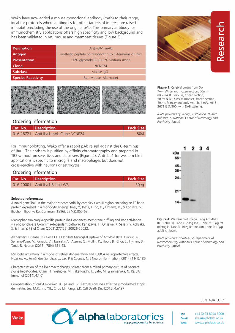
Wako have now added a mouse monoclonal antibody (mAb) to their range, ideal for protocols where antibodies for other targets of interest are raised in rabbit precluding the use of the original pAb. This primary antibody for immunochemistry applications offers high specificity and low background and has been validated in rat, mouse and marmoset tissues (Figure 3).
Figure 4: Western blot image using Anti-Iba1 (016-20001). Lane 1: 20ng Iba1. Lane 2: 10µg rat microglia, Lane 3: 10µg Rat neuron, Lane 4: 10µg adult rat brain.
(Data provided: Courtesy of Department of Neurochemistry, National Centre of Neurology and Psychiatry, Japan)
Figure 3: Cerebral cortex from (A) 7-wk Wistar rat, frozen section, 50µm (B) 7-wk ICR mouse, frozen section, 50µm & (C) 7-wk marmoset, frozen section, 40µm. Primary antibody Anti-Iba1 mAb (016-26721) (1/500) with DAB staining.
(Data provided by Sanagi, T, Ichinohe, N, and Kohsaka, S. National Centre of Neurology and Psychiatry, Japan)
Description Anti-iBA1 mAb
Antigen Synthetic peptide corresponding to C-terminus of Iba1
Presentation 50% glycerol/TBS 0.05% Sodium Azide
Clone NCNP24
Subclass Mouse IgG1
Species Reactivity Rat, Mouse, Marmoset
Ordering InformationCat. No. Description Pack Size016-20001 Anti-Iba1 Rabbit WB 50µg
For immunoblotting, Wako offer a rabbit pAb raised against the C-terminus of Iba1. The antisera is purified by affinity chromatography and prepared in TBS without preservatives and stabilisers (Figure 4). Anti-Iba1 for western blot applications is specific to microglia and macrophages but does not cross-reactive with neurons or astrocytes.
Selected references:A novel gene iba1 in the major histocompatibility complex class III region encoding an EF hand protein expressed in a monocytic lineage. Imai, Y., Ibata, I., Ito, D., Ohsawa, K., & Kohsaka, S. Biochem Biophys Res Commun (1996): 224(3):855-62.
Macrophage/microglia-specific protein Iba1 enhances membrane ruffling and Rac activiation via phospholipase C-gamma-dependant pathway. Kanazawa, H. Ohsawa, K. Sasaki, Y. Kohsaka, S. & Imai, Y. J Biol Chem (2002):277(22):20026-20032.
Alzheimer’s Disease Risk Gene CD33 Inhibits Microglial Uptake of Amyloid Beta. Griciuc, A., Serrano-Pozo, A., Parrado, A., Lesinski, A., Asselin, C., Mullin, K., Hooli, B., Choi, S., Hyman, B., Tanzi, R. Neuron (2013): 78(4):631-43.
Microglia activation in a model of retinal degeneration and TUDCA neuroprotective effects. Noailles, A., Fernández-Sánchez, L., Lax, P & Cuenca, N. J Neuroinflammation. (2014):11(1):186
Characterisation of the liver-macrophages isolated from a mixed primary culture of neonatal swine hepatocytes. Kitani, H., Yoshioka, M., Takenouchi, T., Sato, M. & Yamanaka, N. Results Immunol (2014):4:1-7
Compensation of cATSCs-derived TGFβ1 and IL-10 expressions was effectively modulated atopic dermatitis. Jee, M.K., Im, Y.B., Choi, J.I., Kang, S.K. Cell Death Dis. (2013):4:e497
A B C