anti-inflammatory, antiallergic and covid-19 protease
TRANSCRIPT

RSC Advances
PAPER
Ope
n A
cces
s A
rtic
le. P
ublis
hed
on 1
5 O
ctob
er 2
020.
Dow
nloa
ded
on 2
/4/2
022
12:2
5:21
AM
. T
his
artic
le is
lice
nsed
und
er a
Cre
ativ
e C
omm
ons
Attr
ibut
ion-
Non
Com
mer
cial
3.0
Unp
orte
d L
icen
ce.
View Article OnlineView Journal | View Issue
Anti-inflammato
aInstitute of Pharmaceutical Biology and Bio
D-40225 Dusseldorf, Germany. E-mail: ss_e
pharma.asu.edu.eg; [email protected];
81-14163bDepartment of Pharmaceutical Chemistry, F
61710 Al-Karak, JordancDepartment of Pharmacognosy, Faculty
Abbasia, 11566 Cairo, EgyptdDepartment of Pharmacognosy, Faculty o
Minia, EgypteInstitut fur Anorganische Chemie und Struk
D-40204 Dusseldorf, GermanyfDepartment of Pharmaceutical Chemistry
University in Egypt (BUE), Al-Sherouk City,gGraduate Institute of Natural Products, Co
University, Kaohsiung, TaiwanhDepartment of Biotechnology, College
University, Kaohsiung, Taiwan
Cite this: RSC Adv., 2020, 10, 38128
Received 2nd June 2020Accepted 5th October 2020
DOI: 10.1039/d0ra04876c
rsc.li/rsc-advances
38128 | RSC Adv., 2020, 10, 38128–3
ry, antiallergic and COVID-19protease inhibitory activities of phytochemicalsfrom the Jordanian hawksbeard: identification,structure–activity relationships, molecularmodeling and impact on its folk medicinal uses†
Sherif S. Ebada, *abc Nariman A. Al-Jawabri,b Fadia S. Youssef, ac Dina H. El-Kashef,ad Tim-Oliver Knedel,e Amgad Albohy,f Michal Korinek,ghi
Tsong-Long Hwang, hij Bing-Hung Chen,gkl Guan-Hua Lin,m Chia-Yi Lin,m
Sa'ed M. Aldalaien,b Ahmad M. Disi,n Christoph Janiak e and Peter Proksch*a
On Wednesday 11th March, 2020, the world health organization (WHO) announced novel coronavirus
(COVID-19, also called SARS-CoV-2) as a pandemic. Due to time shortage and lack of either a vaccine
and/or an effective treatment, many trials focused on testing natural products to find out potential
lead candidates. In this field, an edible and folk medicinal Jordanian plant Crepis sancta (Asteraceae)
was selected for this study. Phytochemical investigation of its enriched polyphenolic extract afforded
four eudesmane sesquiterpenes (1–4) together with (6S,9R)-roseoside (5) and five different
methylated flavonols (6–10). Structure elucidation of isolated compounds was unambiguously
determined based on HRESIMS, X-ray crystallography, and exhaustive 1D and 2D NMR experiments. All
isolated compounds were assessed for their in vitro anti-inflammatory, antiallergic and in silico
COVID-19 main protease (Mpro) inhibitory activities. Among the tested compounds, compounds 5–10
revealed potent anti-inflammatory, antiallergic and COVID-19 protease inhibitory activities.
Chrysosplenetin (10) is considered as a promising anti-inflammatory and antiallergic lead structure
adding to the phytotherapeutic pipeline. Moreover, its inhibitory activity against SARS-CoV-2 Mpro,
supported by docking and molecular dynamic studies, strengthens its potential as a lead structure
paving the way toward finding out a natural remedy to treat and/or to control the current COVID-19
pandemic.
technology, Heinrich-Heine University,
[email protected]; sherif_elsayed@
Fax: +49-211-81-11923; Tel: +49-211-
aculty of Pharmacy, Mu'tah University,
of Pharmacy, Ain-Shams University,
f Pharmacy, Minia University, 61519
turchemie, Heinrich-Heine-Universitat,
, Faculty of Pharmacy, The British
Cairo-Suez Desert Road, 11837, Egypt
llege of Pharmacy, Kaohsiung Medical
of Life Science, Kaohsiung Medical
iGraduate Institute of Natural Products, College of Medicine, Chang Gung
University, Taoyuan, TaiwanjResearch Center for Chinese Herbal Medicine, Research Center for Food and
Cosmetic Safety, Graduate Institute of Health Industry Technology, College of
Human Ecology, Chang Gung University of Science and Technology, Taoyuan,
TaiwankDepartment of Anesthesiology, Chang Gung Memorial Hospital, Taoyuan, TaiwanlDepartment of Medical Research, Kaohsiung Medical University Hospital,
Kaohsiung, TaiwanmThe Institute of Biomedical Sciences, National Sun Yat-sen University, Kaohsiung,
TaiwannDepartment of Basic Pharmaceutical Sciences, Faculty of Pharmacy, Al-Isra
University, 11622 Amman, Jordan
† Electronic supplementary information (ESI) available. CCDC 1994762. For ESIand crystallographic data in CIF or other electronic format see DOI:10.1039/d0ra04876c
8141 This journal is © The Royal Society of Chemistry 2020

Paper RSC Advances
Ope
n A
cces
s A
rtic
le. P
ublis
hed
on 1
5 O
ctob
er 2
020.
Dow
nloa
ded
on 2
/4/2
022
12:2
5:21
AM
. T
his
artic
le is
lice
nsed
und
er a
Cre
ativ
e C
omm
ons
Attr
ibut
ion-
Non
Com
mer
cial
3.0
Unp
orte
d L
icen
ce.
View Article Online
Introduction
Polymorphonuclear neutrophils (PMNs) are a major componentof the innate immunity defending the human body againstinadvertent entrance of xenobiotics and/or pathogens. PMNsperform their physiological function principally through theproduction of oxidative free radicals and non-oxidative prote-ases including neutrophil elastase (NE, a serine protease akaleukocyte elastase).1 The more PMNs inltrated, the higher NElevels secreted in response accomplishing their defensive roleby demolishing functional proteins of phagocytosed bacterial,fungal and/or viral pathogens. NE is an intracellular enzymeprincipally stored in azurophilic granules of PMNs. Beside itsdefensive role against pathogens, they were found to producedevastating effects on elastin-rich connective tissue particularlyin the lungs.2 NE can also split and sever collagen, proteogly-cans and other plasma proteins. Hence, its overexpression mayresult in deleterious effects on permeability barrier integritybetween alveolar epithelial and endothelial cells leading topathologic edematous symptoms in the inamed lungs under-going either acute lung injury (ALI), acute respiratory distresssyndrome (ARDS) or chronic respiratory disorders such asasthma and chronic obstructive pulmonary disease (COPD).3
However, ARDS, ALI and COPD are among the leading causes ofmorbidity and mortality in the intensive care units (ICU)worldwide,4 some recurrent comorbidities have been reportedfor COPD with chronic disorders such as hypertension, dia-betes, heart diseases, depression and pneumonia.5
During the last two decades, two epidemics of zoonoticorigins have been introduced into human community, namely,severe acute respiratory syndrome (SARS) commenced atGuangzhou province (China) in 2003 (ref. 6) and Middle Eastrespiratory syndrome (MERS) emerged in Saudi Arabia about 10years later in 2012.7 In December 2019 at Wuhan city (China),early reports indicated a group of ve patients primarily diag-nosed with pneumonia of an unknown etiology featuring ARDSand one of them died later. About six weeks later, the WorldHealth Organization (WHO) announced the onset of a potentialcoronavirus outbreak caused by SARS-CoV2 (COVID-19, namedby WHO on February 11, 2020) and one month later on March11, 2020, WHO raised alerts and announced this outbreak asa pandemic.8
Their causative pathogens belong to betacoronavirusesfamily and are of zoonotic origins. They all are believed toemerge from the horseshoe bats as a natural reservoir for theirimmense majority and recently moved through intermediateanimal hosts, dromedary camels for MERS and Malayanpangolins for COVID-19, to humans that then adopted human–human transmission leading to epidemics among humans.9
Till preparing this manuscript, COVID-19 spread worldwideaffecting about 37.1 million patients including more than onemillion deaths that obliged many governments to implementsocial distancing together with total lockdown strategies todefend against this pandemic.
Due to the current lack of an effective pharmaceutical and/oran approved vaccine, the major WHO recommendations focus
This journal is © The Royal Society of Chemistry 2020
on infection control measures, improving lifestyle and dietenrichment with more-healthy components. Based on theongoing research efforts directed toward nding a plausibletarget for developing a specic antiviral agent, a very recentpaper describing COVID-19 virus main protease (Mpro), a keyCoV enzyme mediating viral replication and transcription, thatwas recognized as a very promising target for drugdevelopment.10
As a part of our ongoing research and building on theprevious notions, we thought of isolating and probing plantnatural products as NE and/or COVID-19 Mpro inhibitors whichmay help to ameliorate the clinical manifestations related toARDS and to cease or even slow down viral replication andtranscription granting additional time for the immune systemto defend itself against COVID-19.
In this study, we explored an edible and a folk medicinalplant of the genus Crepis (aka hawksbeard including about 200species) belonging to the tribe Cichorieae and family Aster-aceae. It is widely eaten and/or used for herbal medicinepurposes in different countries and territories within thecircumference of the Mediterranean Basin including Italy,Greece and Jordan. The fresh leaves of many Crepis speciesincluding that discussed in this study, C. sancta, are eaten eitherfresh, boiled or browned in salads by the locals. Traditionally,its decoction has been used for treating different ailmentsrelated mainly to respiratory and gastrointestinal systems suchas common cold, cough, abdominal colic, constipation, hepa-titis along with hypertension and hyperglycemia.11
Based on searching the reported literature of the genusCrepis, its aerial parts were reported to be rich in both avonoidaglycones and glycosides12 in addition to eudesmanes andguaianolides sesquiterpenes present as both aglycone andglycosidic forms.13 Regarding assessed bioactivities, the extractsof genus Crepis have been reported to possess antimicrobial,antiviral and antiproliferative activities together with antioxi-dant, analgesic, antiulcer and anti-inammatory.12,13
In this study, we report phytochemical exploration ofpolyphenolic-rich extract of C. sancta aerial parts affording tendifferent compounds belonging to eudesmane sesquiterpenesand methoxylated avonoids. Structure elucidation of isolatedcompounds was unambiguously conrmed through HRESIMS,exhaustive 1D, 2D NMR spectroscopy and X-ray crystallography.We also report in this study results of in vitro anti-inammatoryand antiallergic activities of isolated compounds in addition toin silico COVID-19 main protease (Mpro) inhibitory activityassessment.
Materials and methodsGeneral experimental procedures
Perkin-Elmer-241 MC polarimeter was used for determiningoptical rotation. LRESIMS and HRESIMS were determined ona LC-MS HP1100 Agilent Finnigan LCQ Deca XP Thermoquestand FTHRMS-Orbitrap (ThermoFinnigan) mass spectrometer,respectively. Chromatographic workups were conducted viacolumn chromatography using different stationary phases suchas silica gel 60 M (0.04–0.063 mm) and Sephadex LH20. For
RSC Adv., 2020, 10, 38128–38141 | 38129
RSC Advances Paper
Ope
n A
cces
s A
rtic
le. P
ublis
hed
on 1
5 O
ctob
er 2
020.
Dow
nloa
ded
on 2
/4/2
022
12:2
5:21
AM
. T
his
artic
le is
lice
nsed
und
er a
Cre
ativ
e C
omm
ons
Attr
ibut
ion-
Non
Com
mer
cial
3.0
Unp
orte
d L
icen
ce.
View Article Online
screening purposes, ready-made silica gel 60 F254 TLC plates(Merck, Darmstadt, Germany) were used. For visualizing TLCplates, UV light at 254 and 365 nm wavelengths were applied asnon-destructive technique or aer spraying with anisaldehydereagent and heating. For analytical HPLC measurements,a Dionex Ultimate 3000 LC system equipped with a ready-madeseparation column (125 � 4 mm, L � ID), pre-packed withEurospher-10C18 (Knauer, Germany) and coupled to a photo-diode array (PDA) detector (UVD340S) was implemented, settingup the detection channels at 235, 254, 280 and 340 nm wave-lengths. A standard gradient elution was applied using (MeOH,0.01% formic acid in water): 0 min, 10% MeOH; 5 min, 10%MeOH; 35 min, 100% MeOH; 45 min, 100% MeOH, with a owrate of 1 mL min�1. Preparative HPLC separations wereaccomplished using a RP-HPLC system of LaChrom-MerckHitachi equipped with a pump L7100, UV detector L7400 anda column (300 � 8 mm, L� ID) prelled with Euroshper-100C18
(Knauer, Germany) at a ow rate of 5.0 mL min. 1D (1H and 13CNMR) and 2D NMR (chemical shis in ppm) spectra weredetermined on Bruker AVANCE DMX 600 (Switzerland) usingmethanol-d4 and DMSO-d6 solvents (Sigma-Aldrich, Germany).
Plant material
Crepis sancta aerial parts were collected in November, 2017 at Al-Basirah region (Al-Talah Governorate, southern Jordan). Theplant identity was authenticated by Prof. Dr Saleh A. Al-Qur'anat Department of Botany, Faculty of Science, Mu'tah Universityand a voucher specimen coded CSA-201711 was kept atDepartment of Pharmaceutical Chemistry, Faculty of Pharmacy,Mu'tah University.
Extraction, isolation and purication
The freshly collected aerial parts were kept in shade to dry,ground to afford a one-kg net weight that was then extracted bymethanol (3 � 1 L). Methanol extract was then concentratedunder vacuum till dryness affording a total residue (9.8 g). Thetotal extract was then defatted via liquid–liquid fractionationprocedure by being dispersed in 90% MeOH (500 mL) andshaken against n-hexane (1 L). The two immiscible liquid pha-ses were then separated and the aqueous methanol layer wasevaporated till dryness under reduced pressure resulting ina solid residue (5.9 g). Aerwards, the defatted residual solidwas chromatographed through vacuum liquid chromatography(VLC) using silica gel stationary phase and implementinga gradient elution system using n-hexane : EtOAc andDCM : MeOH with a 20% increment affording 12 fractions(CSV1-CSV12). All obtained fractions were subjected to TLC andanalytical HPLC procedures. Fractions CSV2, CSV5-CSV8, CSV11and CSV12 were chosen for further preparative HPLC purica-tion procedure. Fractions CSV2 (58 mg) and CSV11 (63 mg),eluted with n-hexane : EtOAc (8:2) and with DCM : MeOH (2:8),were subjected to preparative HPLC yielding 4 (2.7 mg) and 3(3.5 mg), respectively. Four fractions (CSV5-CSV8) were collectedtogether and applied on column chromatography usingSephadex LH-20 as stationary phase and methanol as a mobilephase followed by preparative HPLC for nal purication to
38130 | RSC Adv., 2020, 10, 38128–38141
yield 6 (4.5 mg), 7 (5.7 mg), 8 (6 mg), 9 (8 mg) and 10 (12 mg).Fraction CSV12 (82 mg) eluted by DCM : MeOH (1:4) was furtherpuried using preparative HPLC yielding 1 (1.3 mg), 2 (1.6 mg)and 5 (1.2 mg).
(6S,7S,10R)-3-Oxo-di-nor-eudesm-4-en-6a-hydroxy-11-oicacid (1). Amorphous yellow solid; ½a�20D +97.6� (c 0.02, MeOH);UV (MeOH) lmax 248 nm; 1H NMR and 13C NMR see ESI TableS1;† HRESIMS m/z 239.1282 [M+H]+ (calcd for C13H19O4; m/z239.1283) and m/z 237.1128 [M�H]� (calcd for C13H17O4; m/z237.1127).
(6S,7S,10R)-3-Oxo-6a-hydroxy-g-costic acid (2). Amorphousyellow solid; [a]20D +79.0� (c 0.02, MeOH); UV (MeOH) lmax
248 nm; 1H NMR and 13C NMR see ESI Table S1;†HRESIMS m/z263.1283 [M�H]� (calcd for C15H19O4; m/z 263.1282).
Cell viability assay
The mucosal mast-cell derived rat basophilic leukemia cells(RBL-2H3) were obtained from Bioresource Collection andResearch Center (Hsin-Chu, Taiwan). The cells were cultured inDMEM containing 10% FBS, 100 U mL�1 penicillin, and 100 mgmL�1 streptomycin in 10 cm cell culture dishes at 37 �C ina humidied chamber with 5% CO2 in air. The potential cyto-toxic effects of the samples (10 to 100 mg mL�1 for the crudeextract and 1 to 400 mM for the pure compounds) on RBL-2H3cells were determined by the methylthiazole tetrazolium(MTT) assay as described previously.14
Superoxide anion generation and elastase release assays byhuman neutrophils
The human neutrophils were obtained from venous blood ofhealthy adult volunteers (20–30 years old) following the re-ported procedure.15 Measurement of superoxide anion genera-tion by the activated neutrophils was based on the reduction offerricytochrome c as described before.15,16 Elastase release byactivated neutrophils was determined using N-methox-ysuccinyl-Ala-Ala-Pro-Val-p-nitroanilide as elastase substrateaccording to the reported method.16 The concentration was 1 to10 mg mL�1 for the crude samples and 0.3 to 10 mM for the purecompounds. LY294002 was used as the positive control.
Degranulation assay in mast cells
The level of degranulation in mast cells was evaluated based onA23187- and antigen-induced b-hexosaminidase release in RBL-2H3 cells according to a reported method with some modi-cations.14 Briey, the cells were seeded in a 96-well plate (2 �104 cells per well, for the A23187-induced assay) or a 48-wellplate (3 � 104 cells per well, for the antigen-induced assay)overnight. At the same time, the cells for the antigen-inducedassay were sensitized with anti-DNP IgE (0.5 mg mL�1; Sigma).RBL-2H3 cells were treated with the samples (0.5, 5 and 50 mM)for 30 min in Tyrode's buffer with maximal DMSO dose of 0.5%.For the A23187-induced assay, the cells were activated by addingA23187 (nal concentration 0.5 mM), while cells for antigen-induced assay were activated by the addition of DNP-BSA(nal concentration 100 ng mL�1) for 30 min. Azelastineserved as a positive control. The amount of b-hexosaminidase
This journal is © The Royal Society of Chemistry 2020
Paper RSC Advances
Ope
n A
cces
s A
rtic
le. P
ublis
hed
on 1
5 O
ctob
er 2
020.
Dow
nloa
ded
on 2
/4/2
022
12:2
5:21
AM
. T
his
artic
le is
lice
nsed
und
er a
Cre
ativ
e C
omm
ons
Attr
ibut
ion-
Non
Com
mer
cial
3.0
Unp
orte
d L
icen
ce.
View Article Online
was detected using the method utilizing p-NAG as a substrateaccording to the procedure described before.14
X-ray crystallographic analysis
Crystals of chrysosplenetin (10) have been obtained by crystal-lization from pure methanol.
Data collection: measurements were done with a BrukerKappa APEX2 CCD diffractometer with a microfocus tube andCu Ka radiation (l ¼ 1.54178 A). Data collection, structuresolving, renement and the hydrogen atom positioning were allperformed according to the recently reported procedure.17 Thestructural data have been deposited in the Cambridge Crystal-lographic Data Center (CCDC no 1994762†).
Molecular modeling studies
The proposed binding mode of isolated compounds withneutrophil elastase (NE) and SARS-CoV-2 main protease (Mpro)was studied using Autodock Vina and a method similar to whatwe reported earlier.12 Here, crystal structures of NE (PDB ID:1H1B) and SARS-CoV-2 Mpro (PDB ID: 6LU7) were used.Prepared and co-crystalized ligands were docked in a grid box inthe active site (25 � 25 � 25 A3, centered on co-crystalizedligand) using exhaustiveness of 16. For each ligand, the top 9binding poses were ranked according to their binding affinitiesand the predicted binding interactions were analyzed. The posewith the best binding affinity and binding mood similar to co-crystalized ligand was reported.
Molecular dynamic (MD) simulation
MD simulations were done using GROMACS 2018.1 moleculardynamics package.18 CHARMM36 all-atom force eld19 was usedfor protein simulations while ligands were parameterized usingSwissParam.20 All simulations were performed in TIP3P explicitwater21 in a dodecahedron box and neutralized by Na+ or Cl�
ions. Steepest descent algorithmwas used for minimization andmaximum force was set to less than 1000 kJ mol�1 nm�1.Systems were initially equilibrated using NVT and NPTensembles for 100 ps each. The temperature was maintained at300 K using the V-rescale algorithm22 and pressure wascontrolled by the Parrinello–Rahman barostat.23 The LINearConstraint Solver (LINCS) algorithm24 was used for bond'slength constraints and Particle mesh Ewald (PME) method25
was used for long-range electrostatics calculations. For allsimulations 2 fs timestep was used. van der Waals cut-offdistance (rvdw) was set to 1.2 nm. Initial coordinates wastaken from docking poses or from crystal structure if available.
Determination of elastase enzymatic activity
The compounds were further tested for direct inhibition ofelastase enzymatic activity.26 The neutrophil suspension (6 �105 cells per mL) was preheated for 5 min in the presence ofCaCl2 (1 mM) at 37 �C. Priming agent CB (1.5 mg mL�1) wasadded for 2 min, followed by fMLF (0.1 mM) for 20min to releaseelastase from the cells. Aer centrifugation at 1000g for 5 min at4 �C, the supernatant containing elastase was preheated at 37 �C
This journal is © The Royal Society of Chemistry 2020
for 5 min, and the test compounds were added. Then, 0.1 mM ofsubstrate methoxysuccinyl-Ala-Ala-Pro-Val-p-nitroanilide wasadded for 10 min. The effect of the compounds on elastaseenzymatic activity was quantied by measuring the absorbanceat 405 nm.
Coronavirus 229E assay
The protective effects of the samples against human coronavi-rus 229E (HCoV-229) was determined similarly to the previouslydescribed method.27 Huh7 cells (human liver carcinoma cellline) were infected with 9TCID50 (Median Tissue CultureInfectious Dose) of each coronavirus 229E in the presence orabsence of the compounds or vehicle. Aer incubation at 33 �Cfor 6 days, the surviving cells were then stained with MTT (3-[4.5-dimethylthiazol-2-yl]-2,5-diphenyl tetrazolium bromide).The percentage of surviving cells was then calculated.
Results and discussionIsolation and identication of major metabolites in the plantextract
A careful phytochemical investigation of the plant extract,implementing various chromatographic techniques, MS, 1D, 2DNMR spectroscopic analyses and by comparison with the re-ported literature, afforded four eudesmane sesquiterpenelactones (1–4). Two were unambiguously identied and re-ported previously as new natural products from the same extractnamely, (6S,7S,10R)-3-oxo-di-nor-eudesm-4-en-6a-hydroxy-11-oic acid (1)28 and (6S,7S,10R)-3-oxo-6a-hydroxy-g-costic acid(2)28 together with two known congeners, 3-oxo-g-costic acid(3)12,29 and its methyl ester (4).12,30 In addition, (6S,9R)-roseoside(5)28 and several methoxylated avonoids namely, jaceidin(6),12,31 kumatakenin (7),12,32 penduletin (8),12,33 pachypodol(9)12,34 and chrysosplenetin (10)12,35 were also isolated. Structureelucidation of the isolated compounds were unambiguouslydetermined based on extensive 1D, 2D NMR analyses togetherwith mass spectrometry (see ESI†).
Compounds (1 and 2) were individually puried as whiteamorphous solids whose molecular formulas were determinedto be C13H18O4 and C15H20O4 based on their HRESIMS spectrathat revealed pseudomolecular ion peaks at m/z 237.1128[M�H]� and m/z 263.1283 [M�H]� indicating the existence ofve and six degrees of unsaturation, respectively. The largermolecular weight of 2 by 26 atomic units and its additional onedegree of unsaturation compared to 1 suggested that the formermight have an additional olen moiety with one of its sp2
carbons turned into a quaternary one. The 1D NMR spectra of 1including 1H and 13C NMR (see ESI, Table S1†) unravelleda close resemblance to the known natural metabolite 3-oxo-di-nor-eudesma-4-en-11-oic acid with an exocyclic carboxylic acidgroup (dC 176.8, C-11).36,37 By careful investigation of extensive2D NMR spectra such as 1H–1H COSY, HMQC and HMBC,a clear difference was determined in 1 compared to the knownmetabolite that is the presence of an oxymethine group at dH5.25 (d, 3.0, 1H, dC 67.6) instead of a methylene group at dH 2.29/dH 2.99 (d, 15.0, 2H, dC 29.2) ascribed for C-6.37 This notion
RSC Adv., 2020, 10, 38128–38141 | 38131
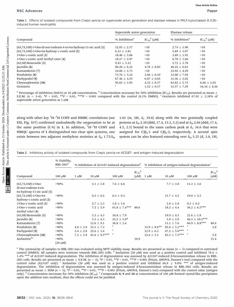
Table 1 Effects of isolated compounds from Crepis sancta on superoxide anion generation and elastase release in fMLF/cytochalasin B (CB)-induced human neutrophils
Compound
Superoxide anion generation Elastase release
% Inhibitiona IC50b (mM) % Inhibitiona IC50
b (mM)
(6S,7S,10R)-3-Oxo-di-nor-eudesm-4-en-6a-hydroxy-11-oic acid (1) 12.05 � 2.37 >10 2.74 � 1.90 >10(6S,7S,10R)-3-Oxo-6a-hydroxy-g-costic acid (2) 6.23 � 3.81 >10 5.89 � 1.07 >103-Oxo-g-costic acid (3) 18.46 � 5.06 >10 3.89 � 1.91 >103-Oxo-g-costic acid methyl ester (4) 18.27 � 4.97 >10 0.70 � 1.60 >10(6S,9R)-Roseoside (5) 9.43 � 5.12 >10 2.72 � 1.78 >10Jaceidin (6) 80.26 � 6.22 4.76 � 0.83 48.22 � 6.61 >10Kumatakenin (7) 24.06 � 2.75 >10 22.92 � 4.29 >10Penduletin (8) 73.76 � 5.24 5.66 � 0.19 33.00 � 7.94 >10Pachypodol (9) 67.46 � 4.55 6.07 � 0.65 31.56 � 5.02 >10Chrysosplenetin (10) 92.03 � 3.95 4.32 � 0.57 65.82 � 5.75 6.66 � 1.03Genistein c 1.52 � 0.17 31.57 � 7.29 16.34 � 4.26
a Percentage of inhibition (Inh%) at 10 mM concentration. b Concentration necessary for 50% inhibition (IC50). Results are presented as mean �S.E.M. (n ¼ 3–6). *P < 0.05, **P < 0.01, ***P < 0.001 compared with the control (0.1% DMSO). c Genistein inhibited 67.01 � 2.38% ofsuperoxide anion generation at 3 mM.
RSC Advances Paper
Ope
n A
cces
s A
rtic
le. P
ublis
hed
on 1
5 O
ctob
er 2
020.
Dow
nloa
ded
on 2
/4/2
022
12:2
5:21
AM
. T
his
artic
le is
lice
nsed
und
er a
Cre
ativ
e C
omm
ons
Attr
ibut
ion-
Non
Com
mer
cial
3.0
Unp
orte
d L
icen
ce.
View Article Online
along with other key 1H–1H COSY and HMBC correlations (seeESI, Fig. S3†) conrmed undoubtedly the oxygenation to be atthe same position (C-6) in 1. In addition, 1H–1H COSY andHMQC spectra of 1 distinguished two clear spin systems, oneexists between two adjacent methylene moieties at dH 1.72/dH
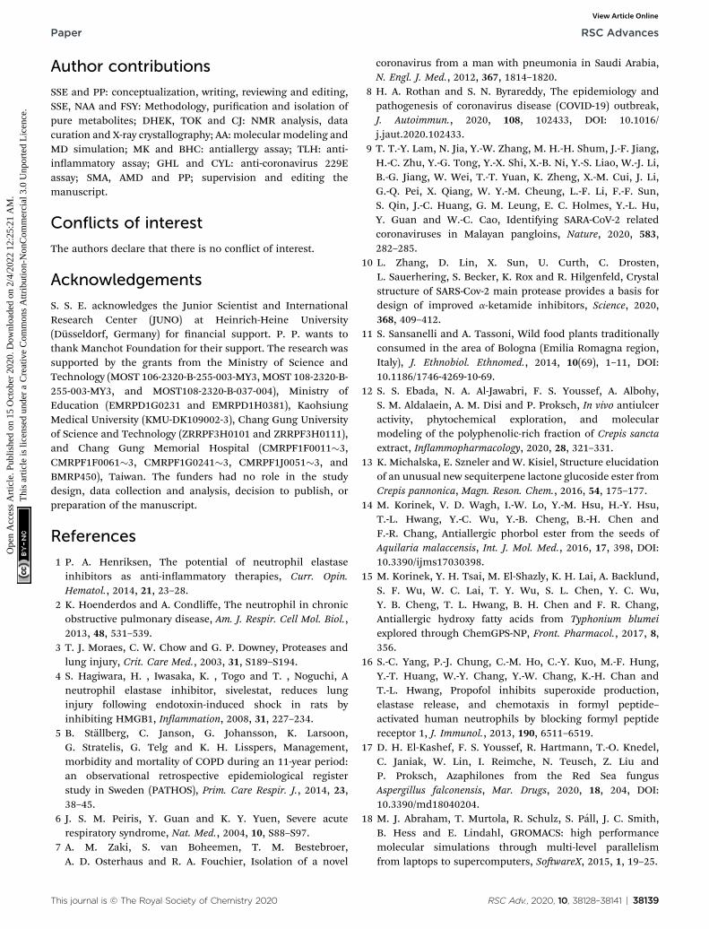
Table 2 Inhibitory activity of isolated compounds from Crepis sancta o
Compound
% Viability,RBL-2H3a % Inhibition of A23187-induced
100 mM 1 mM 10 mM 100
(6S,7S,10R)-3-Oxo-di-nor-eudesm-4-en-6a-hydroxy-11-oic acid (1)
>90% 2.1 � 1.8 7.6 �
(6S,7S,10R)-3-Oxo-6a-hydroxy-g-costic acid (2)
>90% 0.2 � 0.2 0.3 �
3-Oxo-g-costic acid (3) >90% 2.7 � 2.3 1.8 �3-Oxo-g-costic acidmethyl ester (4)
>90% 7.2 � 5.9 61.8
(6S,9R)-Roseoside (5) >90% 5.3 � 4.3 16.8Jaceidin (6) >90% 5.3 � 4.3 25.5Kumatakenin (7) >90% 12.6 � 9.1 36.8Penduletin (8) >90% 4.8 � 3.9 25.1 � 7.3 e
Pachypodol (9) >90% 3.4 � 2.8 22.6 � 3.4 e
Chrysosplenetin (10) >90% 7.3 � 5.9 41.6 � 9.5*** e
Azelastineb,c >90%(20 mM)
a The cytotoxicity of samples to RBL-2H3 was evaluated using MTT viabilcontrol (DMSO). All samples were nontoxic towards RBL-2H3 cells. b Az1.4% *** of A23187-induced degranulation. The inhibition of degranulat2H3 cells. Results are presented as mean � S.E.M. (n ¼ 3); *P < 0.05, **Pcontrol value (A23187 only). c Azelastine (20 mM) was used as a podegranulation. The inhibition of degranulation was assessed by antigpresented as mean � SEM (n ¼ 3); *P < 0.05, **P < 0.01, ***P < 0.001 (Ponly). d Concentration necessary for 50% inhibition (IC50).
e Compoundsupon the addition into medium, thus the effects could not be justied.
38132 | RSC Adv., 2020, 10, 38128–38141
1.83 (m, 2H, dC 39.8) along with the two geminally coupledprotons at dH 2.38 (ddd, 17.4, 15.2, 5.2) and at dH 2.66 (ddd, 17.4,4.5, 2.5) bound to the same carbon peak at dC 34.6 that wereassigned for CH2-1 and CH2-2, respectively. A second spinsystem can be also featured extending over dH 5.25 (d, 3.0, 1H,
n A23187- and antigen-induced degranulation
degranulationb % Inhibition of antigen-induced degranulationc
mMIC50
d
(mM) 1 mM 10 mM 100 mMIC50
d
(mM)
6.6 7.7 � 3.8 11.3 � 3.6
0.3 11.7 � 4.2 19.8 � 2.5
1.6 2.8 � 1.0 0.3 � 0.2� 7.4*** 80.6 18.2 � 4.4 36.2 � 6.5***
� 7.9 19.9 � 4.3 21.6 � 1.8� 5.0* 3.9 � 2.9 46.5 � 10.2***� 5.4 11.1 � 7.6 60.9 � 8.8*** 80.4
33.9 � 8.6** 85.8 � 5.1*** e 3.812.9 � 6.5 47.2 � 5.0*** e
12.4 � 5.1 83.3 � 7.5*** e 5.810.9 15.4
ity assay. Results are presented as mean (n ¼ 3) compared to untreatedelastine (20 mM) was used as a positive control and inhibited 78.4 �ion was assessed by A23187-induced b-hexosaminidase release in RBL-< 0.01, ***P < 0.001 (Prism, ANOVA, Dunnet's test) compared with thesitive control and inhibited 66.8 � 9.8% *** of antigen-induceden-induced b-hexosaminidase release in RBL-2H3 cells. Results arerism, ANOVA, Dunnet's test) compared with the control value (antigen8, 9 and 10 at concentration of 100 mM formed crystal-like precipitates
This journal is © The Royal Society of Chemistry 2020

Paper RSC Advances
Ope
n A
cces
s A
rtic
le. P
ublis
hed
on 1
5 O
ctob
er 2
020.
Dow
nloa
ded
on 2
/4/2
022
12:2
5:21
AM
. T
his
artic
le is
lice
nsed
und
er a
Cre
ativ
e C
omm
ons
Attr
ibut
ion-
Non
Com
mer
cial
3.0
Unp
orte
d L
icen
ce.
View Article Online
dC 67.6), dH 2.46 (dt, 12.9, 3.0, 1H, dC 49.0) and two methylenegroups at dH 2.21 (ddd, 13.4, 12.9, 3.2, 1H) and dH 1.74 (m, 1H)bound to a carbon peak at dC 18.5 in addition to dH 1.39 (dd,13.3, 3.2) and dH 1.70 (dd, 6.7, 3.0, 1H) bound to a carbon peakat dC 41.4 that were ascribed to CH-6, CH-7, CH2-8 and CH2-9,respectively.
A careful exploration of 1H and 13C NMR spectral data of 2(see ESI, Table S1†) and by comparison with the reportedliterature, it was interpreted to be structurally resembling 3-oxo-g-costic acid (3)29 with one additional oxygen atom in 2 asdeduced from their molecular formulas. The presence of anadditional oxygen atom was explained by comparing 1D and 2DNMR spectral data of 2 (see ESI, Table S1†) and 3 (ref. 19) thatconrmed the presence of an oxymethine proton, similar to 1,at dH 5.52 (d, 6.5, 1H, dC 77.3) in 2 replacing a methylene groupat dH 2.83 (br d, 13.5, 1H) and dH 2.02 (br t, 13.5, 1H) bound toa carbon peak at dC 33.3 assigned to C-6 in 3.29
To unambiguously determine the positions of the hydroxyland carboxylic acid moieties in both 1 and 2, gHMBC experi-ments were performed (see ESI, Fig. S3, S9 and S17†). The keyHMBC correlations of 1 and 2 (see ESI, Fig. S3†) undoubtedlyconrmed the existence of a hydroxyl group at C-6 in bothwhereas a terminal carboxylic acid group was ascertained to beat C-11 and C-12, respectively.
The relative conguration of 1 was dened by interpretingthe coupling constant (J) values and by key ROESY correlations(see ESI, Fig. S3†). The determined large J values for H-7 and H-8a (12.9 Hz) revealed a diaxial orientation, however, a small
Fig. 1 Chemical structures of isolated compounds 1–10.
This journal is © The Royal Society of Chemistry 2020
coupling constant value (3.0 Hz) for H-7 and H-6 suggested anaxial-equatorial orientation, respectively.36–38 In addition, theROESY spectrum of 1 disclosed clear correlations (see ESI,Fig. S3 and S11†) namely these from H-6 to H-7 and a singletmethyl group at dH 1.87 (br s, Me-13) together with other keycorrelations in particular those from a singlet methyl group atdH 1.42 (s, Me-12) to two proton peaks at dH 2.66 (H-2a) and at dH2.21 (H-8a) indicated that 6-OH and Me-12 are in an axialorientation while 7-COOH adopts an equatorial one. Supportedby the results of X-ray crystallography36 and modied Moshermethod37 for exploring the absolute conguration of biosyn-thetically related eudesmane-type sesquiterpenes such as 3-oxo-di-nor-eudesma-4-en-11-oic acid and (1R,2S,6R,7S,10S)-1,2,6-trihydroxyeudesma-4-en-3-one, compound 1 was deduced tohave similar absolute conguration to the related congeners36,37
apart from H-6 which was unambiguously distinguished to bein an equatorial position in 1 rather than an axial orientation inthe related congeners. Accordingly, compound 1 was deter-mined to be (6S,7S,10R)-3-oxo-di-nor-eudesm-4-en-6a-hydroxy-11-oic acid.
The relative and absolute congurations of 2 (see ESI,Fig. S3†) were dened as for 1 based on ROESY spectrum thatrevealed comparable key correlations to those found in 1 inaddition to the J values analysis that revealed similar orienta-tion pattern of H-6 and H-7 like that noted in 1. As a conclusion,compound 2 was unambiguously identied as (6S,7S,10R)-3-oxo-6a-hydroxy-g-costic acid.
RSC Adv., 2020, 10, 38128–38141 | 38133

Fig. 2 Docking results of isolated compounds with NE (1H1B). (A) Validation of docking through redocking of the co-crystalized ligandGW475151 (gray) overlaid with the docked structure (pink). (B, C and D) Docking poses of the best docked ligands 10, 9 and 7, respectively.
RSC Advances Paper
Ope
n A
cces
s A
rtic
le. P
ublis
hed
on 1
5 O
ctob
er 2
020.
Dow
nloa
ded
on 2
/4/2
022
12:2
5:21
AM
. T
his
artic
le is
lice
nsed
und
er a
Cre
ativ
e C
omm
ons
Attr
ibut
ion-
Non
Com
mer
cial
3.0
Unp
orte
d L
icen
ce.
View Article Online
Superoxide anion generation and elastase release assays byhuman neutrophils
Assessment of the anti-inammatory activity (Table 1) revealedthat the isolated avonols signicantly suppressed the super-oxide anion generation with IC50 values ranging between 4.32 �0.57 and 6.07� 0.65 mM. Flavonoids were previously reported tohave potent inhibitory activity against pro-inammatory medi-ators such as inducible nitric oxide synthase (iNOS), tumornecrosis factor a (TNF-a), interleukin-1b (IL-1b) and interleukin-6 (IL-6).39 Among the tested compounds in this study,compound 10 revealed potent inhibitory effect on NE releaseand superoxide anion generation with IC50 values of 6.66� 1.03and 4.32 � 0.57 mM, respectively. The results indicated inhibitoryeffect on human neutrophils activation. Human neutrophil elas-tase plays a major role in neutrophils-associated diseases.According to a previous study, structurally similar avonolsincluding 8 (IC50 ¼ 65.4 mM) showed inhibitory effect on humanneutrophil elastase which correlates with 33.0% inhibitory effectof 8 on NE release in human neutrophils at 10 mM.40 The obtainedresults in this study strengthened the previous reports about theanti-inammatory effects of avonols combating the respiratorydamage and NE release along with plausible antiviral activity aspreviously reported.41
38134 | RSC Adv., 2020, 10, 38128–38141
Degranulation assay in mast cells
All isolated compounds (1–10) were assessed for their toxic effectson normal RBL-2H3 cells up to 50 mM. The results (Table 2)revealed that all tested compounds were non-toxic (viability morethan 90%). Antiallergic activity of isolated compounds (1–10) wasthen tested via assessing their inhibitory activities against A23187-and antigen-induced b-hexosaminidase release in RBL-2H3 cells.Calcium ionophore A23187 represents an inducer facilitatingcalcium transport through mast cells membrane while antigen(IgE plus DNP-BSA) acts via Fc3RI receptor similar to physiologicalenvironment.15 The obtained results (Table 2) revealed that amongeudesmane sesquiterpene lactones (1–4), 3-oxo-g-costic acidmethyl ester (4) moderately suppressed A23187-induced degran-ulation (IC50 80.6 mM) indicating importance of carboxylic acidesterication for the activity. The obtained results (Table 2)revealed that compounds 8 and 10 displayed potent inhibitoryactivities against antigen-induced degranulation with IC50 valuesof 3.8 and 5.8 mM, respectively, exceeding that of azelastine asa standard antiallergic drug (IC50 ¼ 15.4 mM). Also, compounds 4and 7 exerted signicant effects on antigen-induced degranulationof mast cells. In a previous report, a structurally-related avonoidaglycone, kaempferol, was found to exert in vitro antiallergicactions in RBL-2H3 cells through inducing heme oxygenase-1 (ref.
This journal is © The Royal Society of Chemistry 2020

Table 3 Binding energy for compounds (1–10) in the active sites of neutrophil elastase (NE) and SARS-CoV-2 main protease (Mpro) showingresidues forming hydrogen bonds with docked ligands. Energy and interacting residues are for the first pose unless otherwise stated
Compound/ligand
1H1B (elastase) 6LU7 (Mpro)
Binding affinity(kcal mol�1)
Interactingresidues
Binding affinity (kcalmol�1) Interacting residues
(6S,7S,10R)-3-Oxo-di-nor-eudesm-4-en-6a-hydroxy-11-oic acid (1)
�5.5 Ser195, Val216 �5.9 Gly143
(6S,7S,10R)-3-Oxo-6a-hydroxy-g-costic acid (2)
�6.2 Ser195 �6.1 Gly143, Ser144, His163
3-Oxo-g-costic acid (3) �5.9 Ser195 �5.9 (4th pose) Gly143, His1633-Oxo-g-costic acid methyl ester(4)
�5.9 Ser195 �6.0 (3rd pose) Gly143, His163
(6S,9R)-Roseoside (5) �6.4 Phe41, Asn61, Ser195,Val216
�7.2 Thr26, Leu141, Gly143,Ser144, Cys145, His163,Glu166
Jaceidin (6) �6.2 Cys191, Gly193, Ser195 �7.3 Leu141, Gly143, Ser144,Cys145, Arg188
Kumatakenin (7) �6.7 Phe41, Cys191, Gly193,Ser195, Val216
�6.8 (3rd pose) Leu141, Gly143, Ser144
Penduletin (8) �6.1 Cys191, Gly193, Ser195 �6.7 (4th pose) Leu141, Gly143, Ser144,Cys145, His163
Pachypodol (9) �6.7 Phe41, Cys191, Gly193,Asp194, Ser195
�7.1 Gly143, Ser144, Cys145
Chrysosplenetin (10) �6.5 Phe41, His57, Gly193,Ser195, Val216
�7.1 Leu141, Gly143, Ser144,Cys145
1H1B-ligand �6.9 Ser195 — —6LU7-ligand (N3) — — �7.1 (3rd pose) Phe140, Gly143, His163,
His164, Glu166, Asp187,Thr190
Paper RSC Advances
Ope
n A
cces
s A
rtic
le. P
ublis
hed
on 1
5 O
ctob
er 2
020.
Dow
nloa
ded
on 2
/4/2
022
12:2
5:21
AM
. T
his
artic
le is
lice
nsed
und
er a
Cre
ativ
e C
omm
ons
Attr
ibut
ion-
Non
Com
mer
cial
3.0
Unp
orte
d L
icen
ce.
View Article Online
42) which might be a putative mechanism for the antiallergicactivity of 8 and 10 as well (Fig. 1).
X-ray crystallographic analysis
Being unprecedentedly reported and based on the ndingsobtained from anti-inammatory and antiallergic assays, wecrystallized chrysosplenetin (10) using its pure methanol solu-tion. Herein, we report for the rst time its crystal structure (seeESI, Fig. S2 and Table S2†). From the single-crystal structurerenement, the structure for chrysosplenetin was obtained,containing two independent molecules in the asymmetric unit,which are connected by a hydrogen bond. Both moleculescontain also intramolecular hydrogen bonds.
Molecular modeling studies
In order to predict the binding mode of the isolated compoundsin the active site of NE, compounds were docked using Auto-dock Vina. To validate docking method and parameters, co-crystalized ligand GW475151 was redocked in the active site.When the extracted crystal structure and the docked structurewere aligned using DockRMSD server, a root mean squaredeviation (RMSD) value of 1.317 was obtained which indicatesthe validity of the method used.12 Results obtained from dock-ing study of isolated compounds with NE showed that isolatedmethoxylated avonols (6–10) possess better binding in theactive site when compared to sesquiterpenes (1–4). Amongst
This journal is © The Royal Society of Chemistry 2020
avonols, those with a methoxy group at C-7, compounds 7, 9and 10, demonstrated better binding. This methoxy was foundto be in close proximity to His57, one of the catalytic triad of theserine proteases like NE. Another important residue which isalso one of the catalytic triad is Ser195 which was found to formhydrogen bond with the C-4 carbonyl group in all the threeavonols. The third residue among the catalytic triad, Asp102was found to lie at the bottom of the binding site and is posi-tioned beneath the docked avonols. It is generally acceptedthat catalytic activity of NE is enhanced by these three residues(catalytic triads; His57, Asp102 and Ser195).43 The former tworesidues act as a base to increase nucleophilicity of Ser195through accepting proton transferred from serine hydroxylgroup.44 In addition to the catalytic triad, docked structureswere found to form several hydrogen bonds, represented bydotted yellow line, with residues in the active site as illustratedin Fig. 2 and Table 3. These residues include Phe41, Cys191, Gly193 and Val216. In addition, hydrophobic interactions betweenring A and C from the avonols with Phe192 and Val216 areobserved, respectively. Hydrophobic interaction between ring Cand Val216 could explain the differences in dihedral anglesbetween rings B and C in the docked structure and the crystalstructure of free avonols. For example, when we crystalized 10alone, the dihedral angle between ring B and C was either 0.5�
(almost coplanar) or 40� with two independent molecules in theasymmetric unit. On the other hand, when 10 was docked in the
RSC Adv., 2020, 10, 38128–38141 | 38135

RSC Advances Paper
Ope
n A
cces
s A
rtic
le. P
ublis
hed
on 1
5 O
ctob
er 2
020.
Dow
nloa
ded
on 2
/4/2
022
12:2
5:21
AM
. T
his
artic
le is
lice
nsed
und
er a
Cre
ativ
e C
omm
ons
Attr
ibut
ion-
Non
Com
mer
cial
3.0
Unp
orte
d L
icen
ce.
View Article Online
active site of NE a dihedral angle of 85� (almost perpendicular)between rings B and C was obtained due interaction withVal216.
It is worth to mention that docking data has shown goodagreement with the elastase release assay in human neutrophils
Fig. 3 Docking results of isolated compounds with SARS-CoV-2 Mpro (6ligandN3 (gray) overlaid with the docked structure (pink). (B and C) Dockintheir overlapping in (D). (E) Docking poses of 5 (pink) and 6 (golden) ov(golden) and 10 (violet) comparing their binding modes.
38136 | RSC Adv., 2020, 10, 38128–38141
indicating a possible direct effect on elastase enzymatic activity.To justify this notion, Pearson's correlation coefficient wasdetermined whose value revealed a medium to strong negativeassociation between the inhibition of human elastase releaseand the docking scores (r¼�0.5). This indicates that the higher
LU7). (A) Validation of docking through redocking of the co-crystalizedg poses of the best compounds 6 (golden) and 5 (pink) respectively anderlapped with co-crystalized ligand N3 (blue). (F) Docking poses of 6
This journal is © The Royal Society of Chemistry 2020

Table 4 Coulombic and Lennard-Jones interactions as well as total interaction energy of the complexes studied by MD
Target Complex
Energy (kJ mol�1)
Coulombic interaction Lennard-Jones (L-J) energy Total interaction energy
Human elastase 1H1B-chrysosplenetin (10) �31.12 � 3.7 �84.81 � 10 �115.93 � 101H1B-control �20.36 � 3.8 �99.14 � 6.0 �119.50 � 7.1
SARS-CoV-2 main protease 6LU7-chrysosplenetin (10) �24.29 � 6.9 �81.58 � 17 �105.87 � 186LU7-control �173.04 � 5.2 �249.57 � 2.8 �422.61 � 5.96LU7-jaceidin (6) �43.72 � 2.0 �113.80 � 1.7 �157.52 � 2.6
Paper RSC Advances
Ope
n A
cces
s A
rtic
le. P
ublis
hed
on 1
5 O
ctob
er 2
020.
Dow
nloa
ded
on 2
/4/2
022
12:2
5:21
AM
. T
his
artic
le is
lice
nsed
und
er a
Cre
ativ
e C
omm
ons
Attr
ibut
ion-
Non
Com
mer
cial
3.0
Unp
orte
d L
icen
ce.
View Article Online
the inhibitory activity against human elastase release is, thelower the docking score (larger absolute value).
Considering NE as a protease, this encouraged us to assess thebinding modes and affinity of the isolated compounds to SARS-CoV-2 main protease (Mpro).10 This enzyme was proposed to bea good target for treatment of COVID-19. Chen and co-workersreported the virtual screening of Mpro against a medicinal plantlibrary containing 32 297 compounds including avonoids.45
To test that virtually, the isolated compounds' docking intothe active site of Mpro was studied. Before running the actualdocking, validation was done by redocking of the co-crystalizedligand N3. The docked structure had a RMSD of 1.855 comparedto the co-crystalized ligand structure (Fig. 3A).
Among the docked structures, tested avonols, jaceidin (6),pachypodol (9) and chrysosplenetin (10) along with (6S,9R)-roseoside (5) revealed binding energy values equal or evenhigher than the co-crystalized ligand (6LU7, Table 3).
Among isolated avonols, compounds 6, 9 and 10 featurea similar ring C structure with adjacent methoxy and hydroxylgroups that proved to form hydrogen bond network with resi-dues Cys145, Ser144, Gly143, and Leu141 (Fig. 3B). A very recentreport by Zhang and coworkers indicated the inevitable role ofCys145, Ser144 and Gly143 forming the canonical “oxyanionhole” of the cysteine protease, in the binding of SARS-CoV-2Mpro inhibitors.10 In addition, only compound 6 possessesa free hydroxyl group at position 7 of ring A which was found tobe involved in a hydrogen bond with Arg188 which may explainthe slightly higher binding energy value compared tocompounds 9 and 10 (Table 3).
For compound 5 (Fig. 3C), the binding pose is partiallyoverlapped with that of avonol (6) where the glycosidic sugar
Fig. 4 RMSD plot of ligands heavy atoms in the active sites of (A), humproduction MD run.
This journal is © The Royal Society of Chemistry 2020
moiety in 5 is overlapping with ring C of 6. The hydroxy groupson the sugar unit in 5 are involved in hydrogen bond networkwith the same residues similar to 6, 9 and 10 binding (Cys145,Ser144, Gly143 and Leu141). In addition, hydrogen bonds withHis163, Glu166 and Thr26 are also seen. Among those, His163and Glu166 were reported in Zhang and his fellows' workmentioned above.10 It will be interesting to synthesize and testa compound that has the aglycon of (6S,9R)-roseoside linked to30-oxygen of 6 (Fig. 3D and E) as a proposed structure for a leadSARS-CoV-2 Mpro inhibitor.
Molecular dynamic simulation
Beside being the best compound in the elastase release assaywith IC50 of 6.66 mM (Table 1), chrysosplenetin (10) was amongthe top compounds that showed the best docking in the activesite of human elastase (1H1B) and has shown comparablebinding affinity to the co-crystalized ligand in the active site ofSARS-CoV-2 main protease (6LU7) as demonstrated in Table 3.These ndings encouraged us to further investigate thebonding of 10 with both targets using molecular dynamicsimulation studies. Four complexes were studied including 10in both targets and the co-crystalized ligand corresponding toeach target. Using production run trajectories, the average totalinteraction energy composed of coulombic interaction andLennard-Jones (L-J) energy was calculated (Table 4).
For elastase complexes, both compound 10 and the co-crystalized ligand have shown comparable average total inter-action energy ranging from �115 to �120 kJ mol�1 supportingits potential activity as elastase inhibitor that was proved by thebiological testing. In addition, plotting of RMSD of ligand heavyatoms during the MD production run (Fig. 4A), shows RMSD < 1
an elastase 1H1B and (B), SARS-CoV-2 main proteae 6LU7 during the
RSC Adv., 2020, 10, 38128–38141 | 38137

Fig. 5 Elastase enzymatic activity of compounds 6 to 10. Humanneutrophils were incubated with fMLF/CB for 15 min. The elastasesupernatant was obtained and incubated with DMSO (as control), orwith compounds 6–10 (10 mM) for 2 min before the addition ofsubstrate (100 mM). Elastase activity was measured at 405 nm. Data arepresented asmeans� S.E.M. (n¼ 4). *p < 0.05, **p < 0.01, ***p < 0.001compared with the control group (t-test).
RSC Advances Paper
Ope
n A
cces
s A
rtic
le. P
ublis
hed
on 1
5 O
ctob
er 2
020.
Dow
nloa
ded
on 2
/4/2
022
12:2
5:21
AM
. T
his
artic
le is
lice
nsed
und
er a
Cre
ativ
e C
omm
ons
Attr
ibut
ion-
Non
Com
mer
cial
3.0
Unp
orte
d L
icen
ce.
View Article Online
A during more than 90% of production run time which suggeststhe stability of the proposed docking binding mode discussedearlier.
On the other hand, the average total interaction energybetween 10 and SARS-CoV-2 main protease was much less thanthe interaction energy seen in case of the co-crystalized ligand(Table 4). The RMSD plot (Fig. 4B) shows stability of the 6LU7–10 complex only during the initial MD production run. Thissuggested that compound 10 could be a lead for the design ofcompounds targeting main protease but need furtherimprovement. To investigate the last hypothesis, we decided torun MD for 6LU7 complex with 6 whose choice was based on itsclose structural similarity to 10 and also on its better dockingscore. In elastase release inhibitory activity, compound 6 wasalso the second-most potent compound. We found that 6 hasbetter total interaction energy (�157 kJ mol�1) which resultedfrom slight structural modication of compound 10. Moreover,jaceidin (6) has also shown less RMSD uctuation during theMD production run (Fig. 4B). This clearly supports that furtherimprovement can be done in this scaffold to design SARS-CoV-2main protease inhibitors against COVID-19. This could be donethrough different medicinal chemistry techniques but it isbeyond the scope of this work.
Elastase enzymatic activity
To address the results from molecular docking and moleculardynamic simulation studies, neutrophil elastase (NE) wasextracted from activated neutrophils and cell-free assay on theelastase enzymatic activity was performed (Fig. 5). The resultsrevealed, that compounds 6 to 10 signicantly inhibited theactivity of elastase enzyme by 20.3 to 27.1% at the concentrationof 10 mM (Fig. 5). Thus, the effects of 6 to 10 on elastase release(22.9 to 65.8% inhibition at 10 mM), could be, at least partly,attributed to the direct interaction with elastase enzyme which isin agreement with themolecular docking of compounds with NE.
38138 | RSC Adv., 2020, 10, 38128–38141
Protective effects against human coronavirus 229E infection
We further tested the compounds that docked well with Mpro
(compounds 5 to 10) in the human coronavirus 229E (HCoV-229E) assay in vitro (see ESI, Fig. S4†). The cells were infectedwith HCoV-229E strain (grey) and the protective effects ofcompounds (5–10) were evaluated. None of the testedcompounds protected the cells from HCoV-229E infection.However, having no in vitro protective activity against viral hostcell entry doesn't deny the Mpro inhibitory activity that convertsviral polyprotein into functional units within host cells to benally assembled into new viruses.
Recently, natural products have been focused on as a sourceof inhibitors affecting SARS-CoV-2 Mpro, a functional proteinconsidered as a potential target for developing new therapeuticsto help combatting the current COVID-19 pandemic. Manynatural compounds, belonging to various chemical classes suchas alkaloids, coumarins, avonoids, peptides, terpenoids andtannins, have been assessed for their SARS-CoV-2 Mpro inhibi-tory activity.46 Many potential Mpro inhibitors were chemicallybelonging to avonoids such as quercetin, apigenin and luteo-lin (IC50 of 20–200 mM) in addition to the biavonoid amento-avone that was the most potent with IC50 of 8.3 mM.47 Tofurther investigate and comprehend their Mpro inhibitoryactivity, in silico molecular docking and molecular dynamicsimulation studies were also conducted.48,49 In our study, theobtained results enriched potential SARS-CoV-2 Mpro inhibitorswith some more candidates supported by more details abouttheir binding modes in addition to being proved in vitro asinhibitors of human elastase release. These features altogethermay also escalate their efficiency in defeating the respiratorydistress symptoms triggered by inammatory responses toSARS-CoV-2 infection.
Conclusion
Phytochemical investigation of polyphenolic-rich extract of theJordanian hawksbeard, prescribed by local herbalists for treat-ment of cough and u, afforded several eudesmane sesquiter-penes and avonols. They were assessed for their in vitro anti-inammatory and antiallergic activities. Among the testedcompounds, avonols, in particularly chrysosplenetin (10),exhibited potent anti-inammatory activity that was proved viain vitro experimental results through assessing its inhibitoryactivity against neutrophil elastase release along with further insilicomolecular modeling that included docking and moleculardynamic simulation studies. By assessing the binding to SARS-CoV-2 (COVID-19) Mpro as an important target, chrysosplenetin(10) was also found to possess high binding energy comparableto that exhibited by its standard ligand.
These ndings are supporting the introduction of chrys-osplenetin (10) as a potential lead candidate into the phyto-therapeutic arena as an anti-inammatory and antiallergicdrug. Moreover, its inhibitory activity against SARS-CoV-2(COVID-19) Mpro will support considering it as a potential leadstructure paving the way toward nding out a natural remedyfor treatment of the current COVID-19 pandemic.
This journal is © The Royal Society of Chemistry 2020
Paper RSC Advances
Ope
n A
cces
s A
rtic
le. P
ublis
hed
on 1
5 O
ctob
er 2
020.
Dow
nloa
ded
on 2
/4/2
022
12:2
5:21
AM
. T
his
artic
le is
lice
nsed
und
er a
Cre
ativ
e C
omm
ons
Attr
ibut
ion-
Non
Com
mer
cial
3.0
Unp
orte
d L
icen
ce.
View Article Online
Author contributions
SSE and PP: conceptualization, writing, reviewing and editing,SSE, NAA and FSY: Methodology, purication and isolation ofpure metabolites; DHEK, TOK and CJ: NMR analysis, datacuration and X-ray crystallography; AA: molecular modeling andMD simulation; MK and BHC: antiallergy assay; TLH: anti-inammatory assay; GHL and CYL: anti-coronavirus 229Eassay; SMA, AMD and PP; supervision and editing themanuscript.
Conflicts of interest
The authors declare that there is no conict of interest.
Acknowledgements
S. S. E. acknowledges the Junior Scientist and InternationalResearch Center (JUNO) at Heinrich-Heine University(Dusseldorf, Germany) for nancial support. P. P. wants tothank Manchot Foundation for their support. The research wassupported by the grants from the Ministry of Science andTechnology (MOST 106-2320-B-255-003-MY3, MOST 108-2320-B-255-003-MY3, and MOST108-2320-B-037-004), Ministry ofEducation (EMRPD1G0231 and EMRPD1H0381), KaohsiungMedical University (KMU-DK109002-3), Chang Gung Universityof Science and Technology (ZRRPF3H0101 and ZRRPF3H0111),and Chang Gung Memorial Hospital (CMRPF1F0011�3,CMRPF1F0061�3, CMRPF1G0241�3, CMRPF1J0051�3, andBMRP450), Taiwan. The funders had no role in the studydesign, data collection and analysis, decision to publish, orpreparation of the manuscript.
References
1 P. A. Henriksen, The potential of neutrophil elastaseinhibitors as anti-inammatory therapies, Curr. Opin.Hematol., 2014, 21, 23–28.
2 K. Hoenderdos and A. Condliffe, The neutrophil in chronicobstructive pulmonary disease, Am. J. Respir. Cell Mol. Biol.,2013, 48, 531–539.
3 T. J. Moraes, C. W. Chow and G. P. Downey, Proteases andlung injury, Crit. Care Med., 2003, 31, S189–S194.
4 S. Hagiwara, H. , Iwasaka, K. , Togo and T. , Noguchi, Aneutrophil elastase inhibitor, sivelestat, reduces lunginjury following endotoxin-induced shock in rats byinhibiting HMGB1, Inammation, 2008, 31, 227–234.
5 B. Stallberg, C. Janson, G. Johansson, K. Larsoon,G. Stratelis, G. Telg and K. H. Lisspers, Management,morbidity and mortality of COPD during an 11-year period:an observational retrospective epidemiological registerstudy in Sweden (PATHOS), Prim. Care Respir. J., 2014, 23,38–45.
6 J. S. M. Peiris, Y. Guan and K. Y. Yuen, Severe acuterespiratory syndrome, Nat. Med., 2004, 10, S88–S97.
7 A. M. Zaki, S. van Boheemen, T. M. Bestebroer,A. D. Osterhaus and R. A. Fouchier, Isolation of a novel
This journal is © The Royal Society of Chemistry 2020
coronavirus from a man with pneumonia in Saudi Arabia,N. Engl. J. Med., 2012, 367, 1814–1820.
8 H. A. Rothan and S. N. Byrareddy, The epidemiology andpathogenesis of coronavirus disease (COVID-19) outbreak,J. Autoimmun., 2020, 108, 102433, DOI: 10.1016/j.jaut.2020.102433.
9 T. T.-Y. Lam, N. Jia, Y.-W. Zhang, M. H.-H. Shum, J.-F. Jiang,H.-C. Zhu, Y.-G. Tong, Y.-X. Shi, X.-B. Ni, Y.-S. Liao, W.-J. Li,B.-G. Jiang, W. Wei, T.-T. Yuan, K. Zheng, X.-M. Cui, J. Li,G.-Q. Pei, X. Qiang, W. Y.-M. Cheung, L.-F. Li, F.-F. Sun,S. Qin, J.-C. Huang, G. M. Leung, E. C. Holmes, Y.-L. Hu,Y. Guan and W.-C. Cao, Identifying SARA-CoV-2 relatedcoronaviruses in Malayan pangloins, Nature, 2020, 583,282–285.
10 L. Zhang, D. Lin, X. Sun, U. Curth, C. Drosten,L. Sauerhering, S. Becker, K. Rox and R. Hilgenfeld, Crystalstructure of SARS-Cov-2 main protease provides a basis fordesign of improved a-ketamide inhibitors, Science, 2020,368, 409–412.
11 S. Sansanelli and A. Tassoni, Wild food plants traditionallyconsumed in the area of Bologna (Emilia Romagna region,Italy), J. Ethnobiol. Ethnomed., 2014, 10(69), 1–11, DOI:10.1186/1746-4269-10-69.
12 S. S. Ebada, N. A. Al-Jawabri, F. S. Youssef, A. Albohy,S. M. Aldalaein, A. M. Disi and P. Proksch, In vivo antiulceractivity, phytochemical exploration, and molecularmodeling of the polyphenolic-rich fraction of Crepis sanctaextract, Inammopharmacology, 2020, 28, 321–331.
13 K. Michalska, E. Szneler and W. Kisiel, Structure elucidationof an unusual new sequiterpene lactone glucoside ester fromCrepis pannonica, Magn. Reson. Chem., 2016, 54, 175–177.
14 M. Korinek, V. D. Wagh, I.-W. Lo, Y.-M. Hsu, H.-Y. Hsu,T.-L. Hwang, Y.-C. Wu, Y.-B. Cheng, B.-H. Chen andF.-R. Chang, Antiallergic phorbol ester from the seeds ofAquilaria malaccensis, Int. J. Mol. Med., 2016, 17, 398, DOI:10.3390/ijms17030398.
15 M. Korinek, Y. H. Tsai, M. El-Shazly, K. H. Lai, A. Backlund,S. F. Wu, W. C. Lai, T. Y. Wu, S. L. Chen, Y. C. Wu,Y. B. Cheng, T. L. Hwang, B. H. Chen and F. R. Chang,Antiallergic hydroxy fatty acids from Typhonium blumeiexplored through ChemGPS-NP, Front. Pharmacol., 2017, 8,356.
16 S.-C. Yang, P.-J. Chung, C.-M. Ho, C.-Y. Kuo, M.-F. Hung,Y.-T. Huang, W.-Y. Chang, Y.-W. Chang, K.-H. Chan andT.-L. Hwang, Propofol inhibits superoxide production,elastase release, and chemotaxis in formyl peptide–activated human neutrophils by blocking formyl peptidereceptor 1, J. Immunol., 2013, 190, 6511–6519.
17 D. H. El-Kashef, F. S. Youssef, R. Hartmann, T.-O. Knedel,C. Janiak, W. Lin, I. Reimche, N. Teusch, Z. Liu andP. Proksch, Azaphilones from the Red Sea fungusAspergillus falconensis, Mar. Drugs, 2020, 18, 204, DOI:10.3390/md18040204.
18 M. J. Abraham, T. Murtola, R. Schulz, S. Pall, J. C. Smith,B. Hess and E. Lindahl, GROMACS: high performancemolecular simulations through multi-level parallelismfrom laptops to supercomputers, SowareX, 2015, 1, 19–25.
RSC Adv., 2020, 10, 38128–38141 | 38139

RSC Advances Paper
Ope
n A
cces
s A
rtic
le. P
ublis
hed
on 1
5 O
ctob
er 2
020.
Dow
nloa
ded
on 2
/4/2
022
12:2
5:21
AM
. T
his
artic
le is
lice
nsed
und
er a
Cre
ativ
e C
omm
ons
Attr
ibut
ion-
Non
Com
mer
cial
3.0
Unp
orte
d L
icen
ce.
View Article Online
19 J. Huang and A. D. MacKerell, CHARMM36 all-atom additiveprotein force eld: Validation based on comparison to NMRdata, J. Comput. Chem., 2013, 34, 2135–2145, DOI: 10.1002/jcc.23354.
20 V. Zoete, M. A. Cuendet, A. Grosdidier and O. Michielin,SwissParam, a Fast Force Field Generation Tool for SmallOrganic Molecules, J. Comput. Chem., 2011, 32, 2359–2368,DOI: 10.1002/jcc.21816PMID: 21541964.
21 P. Mark and L. Nilsson, Structure and dynamics of theTIP3P, SPC, and SPC/E water models at 298 K, J. Phys.Chem. A, 2001, 105, 9954–9960.
22 G. Bussi, D. Donadio and M. Parrinello, Canonical samplingthrough velocity rescaling, J. Chem. Phys., 2007, 126, 014101.
23 M. Parrinello and A. Rahman, Polymorphic transitions insingle crystals: a new molecular dynamics method, J. Appl.Phys., 1981, 52, 7182–7190.
24 B. Hess, H. Bekker, H. J. Berendsen and J. G. Fraaije, LINCS:a linear constraint solver for molecular simulations, J.Comput. Chem., 1997, 18, 1463–1472.
25 T. Darden, D. York and L. Pedersen, Particle mesh Ewald:an N log(N) method for Ewald sums in large systems, J.Chem. Phys., 1993, 98, 10089–10092.
26 Y. F. Tsai, H. P. Yu, W. Y. Chang, F. C. Liu, Z. C. Huang andT. L. Hwang, Sirtinol inhibits neutrophil elastase activity andattenuates lipopolysaccharide-mediated acute lung injury inmice, Sci. Rep., 2015, 5, 8347, DOI: 10.1038/srep08347.
27 C.-F. Hsieh, J.-R. Jheng, G.-H. Lin, Y.-L. Chen, J. Y. Ho,C.-J. Liu, K.-Y. Hsu, Y.-S. Chen, Y. F. Chan, H.-M. Yu,P.-W. Hsieh, J.-H. Chern and J.-T. Horng, Rosmarinic acidexhibits broad anti-enterovirus A71 activity by inhibitingthe interaction between the ve-fold axis of capsid VP1 andcognate sulfated receptors, Emerg. Microb. Infect., 2020, 9,1194–1205.
28 S. S. Ebada, D. H. El-Kashef, W. E. G. Muller and P. Proksch,Cytotoxic eudesmane sesquiterpenes from Crepis sancta,Phytochem. Lett., 2019, 33, 46–48.
29 F. R. Garcez, W. S. Garcez, L. Hamerski andA. C. de M. Miranda, Eudesmane and rearrangedeudesmane sesquiterpenes from Nectandra cissiora, Quım.Nova, 2010, 33, 1739–1742.
30 M.-E. F. Hegazy, H. Matsuda, S. Nakamura, T. A. Hussein,M. Yoshikawa and P. W. Pare, Chemical constituents andtheir antibacterial and antifungal activity from theEgyptian herbal medicine Chiliadenus montanus,Phytochemistry, 2014, 103, 154–161.
31 G. Flamini, E. Antognoli and I. Morelli, Two avonoids andother compounds from the aerial parts of Centaureabracteata from Italy, Phytochemistry, 2001, 57, 559–564.
32 J.-H. Woo, J.-H. Ahn, D.-S. Jang, K.-T. Lee and J.-H. Choi,Effect of kumatakenin isolated from cloves on theapoptosis of cancer cells and the alternative activation oftumor-associated macrophages, J. Agric. Food Chem., 2017,65, 7893–7899.
33 S. E. Sajjadi, S. M. Ghanadian, M. Rabbani and F. Tahmasbi,Isolation and identication of secondary metabolites fromthe aerial parts of Stachys lavandulifolia Vahl, Iran, J.Pharm. Res., 2017, 16, 58–63.
38140 | RSC Adv., 2020, 10, 38128–38141
34 H.-A. Ali, A. K. A. Chowdhury, A. K. M. Rahman,T. Borkowski, L. Nahar and S. D. Sarker, Pachypodol,a avonol from the leaves of Calycopteris oribunda,inhibits the growth of Caco 2 colon cancer cell line in vitro,Phytother Res., 2008, 22, 1684–1687.
35 X.-Y. Xie, R. Wang and Y.-P. Shi, Flavonoids from the owersof Matricaria chamomilla, Chem. Nat. Compd., 2014, 50, 910–911.
36 E. Fattorusso, F. U. Santelia, G. Appendino, M. Ballero andO. Taglialatela-Scafati, Polyoxygenated eudesmanes andtrans-chrysanthemanes from the aerial parts of Santolinainsularis, J. Nat. Prod., 2004, 67, 37–41.
37 G.-C. Wang, G.-Q. Li, H.-W. Geng, T. Li, J.-J. Xu, F. Ma, X. Wu,W.-C. Ye and Y.-L. Li, Eudesmane-type sesquiterpenederivatives from Laggera alata, Phytochemistry, 2013, 96,201–207.
38 M.-E. F. Hegazy, A. A. El-Beih, A. R. Hamed, A. A. Abd El Aty,N. S. Mohamed and P. W. Pare, 3-Oxo-g-costic acid fungal-transformation generates eudesmane sesquiterpenes within vitro tumor-inhibitory activity, Bioorg. Med. Chem. Lett.,2017, 27, 3825–3828.
39 R. Chen, Y. Yang, J. Xu, Y. Pan, W. Zhang, Y. Xing, H. Ni,Y. Sun, Y. Hou and N. Li, Tamarix hohenackeri Bung exertsanti-inammatory effects on lipopolysaccharide activatedmicroglia in vitro, Phytomedicine, 2018, 40, 10–19.
40 Z. Uddin, Z. Li, Y. H. Song, J. Y. Kim and K. H. Park,Visconata: a rare avonol having long chain fatty acid fromDodonaea viscosa which inhibits human neutrophilelastase (HNE), Tetrahedron Lett., 2017, 58, 2507–2511.
41 V. S. Somerville, A. J. Braakhuis and W. G. Hopkins, Effect ofavonoids on upper respiratory tract infections and immunefunction: A systematic review and meta-analysis, Adv. Nutr.,2016, 7, 488–497, DOI: 10.3945/an.115.010538.
42 E. Hirose, M. Matsushima, K. Takagi, Y. Ota, K. Ishigami,T. Hirayama, Y. Hayashi, T. Nakamura, N. Hashimoto,K. Imaizumi, K. Baba, Y. Hasegawa and T. Kawabe,Involvement of heme oxygenase-1 in kaempferol-inducedantiallergic actions in RBL-2H3 cells, Inammation, 2009,32, 99–108.
43 L. Crocetti, I. A. Schepetkin, A. Cilibrizzi, A. Graziano,C. Vergelli, D. Giomi, A. I. Khlebnikov, M. T. Quinn andM. P. Giovannoni, Optimization of N-benzoylindazolederivatives as inhibitors of human neutorphil elastase, J.Med. Chem., 2013, 56, 6259–6272.
44 I. M. Ayoub, M. Korinek, T.-L. Hwang, B.-H. Chen,F.-R. Chang, M. El-Shazly and A. N. B. Singab, Probing theantiallergic and anti-inammatory activity of biavonoidsand dihydroavonols from Dietes bicolour, J. Nat. Prod.,2018, 81, 243–253.
45 M. T. ul Qamar, S. M. Alqahtani, M. A. Alamri and L.-L. Chen,Structural basis of SARS-CoV-2 3CLpro and anti-COVID-19drug discovery from medicinal plants, J. Pharm. Sci., 2020,10, 313–319.
46 A. M. Sayed, A. R. Khattab, A. M. AboulMagd, H. M. Hassan,M. E. Rateb, H. Zaid and U. R. Abdelmohsen, Nature asa treasure trove of potential anti-SARS-CoV drug leads:
This journal is © The Royal Society of Chemistry 2020

Paper RSC Advances
Ope
n A
cces
s A
rtic
le. P
ublis
hed
on 1
5 O
ctob
er 2
020.
Dow
nloa
ded
on 2
/4/2
022
12:2
5:21
AM
. T
his
artic
le is
lice
nsed
und
er a
Cre
ativ
e C
omm
ons
Attr
ibut
ion-
Non
Com
mer
cial
3.0
Unp
orte
d L
icen
ce.
View Article Online
a structural/mechanistic rationale, RSC Adv., 2020, 10,19790–19802.
47 Y. B. Ryu, H. J. Jeong, J. H. Kim, Y. M. Kim, J. Y. Park, D. Kim,T. T. Nguyen, S. J. Park, J. S. Chang, K. H. Park, M. C. Rho andW. S. Lee, Biavonoids from Torreya nucifera displayingSARS-CoV 3CL(pro) inhibition, Bioorg. Med. Chem., 2010,18, 7940–7947.
48 R. S. Joshi, S. S. Jagdale, S. B. Bansode, S. S. Shankar,M. B. Tellis, V. K. Pandya, A. Chugh, A. P. Giri andM. J. Kulkarni, Discovery of potential multi-target-directed
This journal is © The Royal Society of Chemistry 2020
ligands by targeting host-specic SARS-CoV-2 structurallyconserved main protease, J. Biomol. Struct. Dyn., 2020, 38,DOI: 10.1080/07391102.2020.1760137.
49 D. S. N. B. K. Prasanth, M. Murahari, V. Chandramohan,S. P. Panda, L. R. Atmakuri and C. Guntupalli, In silicoidentication of potential inhibitors from Cinnamonagainst main protease and spike glycoprotein of SARS-CoV-2, J. Biomol. Struct. Dyn., 2020, 38, DOI: 10.1080/07391102.2020.1779129.
RSC Adv., 2020, 10, 38128–38141 | 38141