annexin a1 complex mediates oxytocin vesicle transport · annexin a1 complex mediates oxytocin...
TRANSCRIPT

Journal of Neuroendocrinology, 2013, 25, 1241–1254
ORIGINAL ARTICLE © 2013 British Society for Neuroendocrinology
Annexin A1 Complex Mediates Oxytocin Vesicle TransportV. Makani*, R. Sultana†, K. S. Sie*, D. Orjiako*, M. Tatangelo*, A. Dowling‡, J. Cai§, W. Pierce§, D. Allan Butterfield†, J. Hill‡ and
J. Park*
*Department of Neurosciences, College of Medicine, University of Toledo, Toledo, OH, USA.
†Department of Chemistry, University of Kentucky, Lexington, KY, USA.
‡Department of Physiology and Pharmacology, College of Medicine, University of Toledo, Toledo, OH, USA.
§Department of Pharmacology and Toxicology, University of Louisville, Louisville, KY, USA.
Journal ofNeuroendocrinology
Correspondence to:
Joshua Park, Department of
Neurosciences, College of Medicine,
University of Toledo, Toledo, OH
43614, USA (e-mail: joshua.
Oxytocin is a major neuropeptide that modulates the brain functions involved in social behav-
iour and interaction. Despite of the importance of oxytocin for the neural control of social
behaviour, little is known about the molecular mechanism(s) by which oxytocin secretion in the
brain is regulated. Pro-oxytocin is synthesised in the cell bodies of hypothalamic neurones in
the supraoptic and paraventricular nuclei and processed to a 9-amino-acid mature form during
post-Golgi transport to the secretion sites at the axon terminals and somatodendritic regions.
Oxytocin secreted from the somatodendritic regions diffuses throughout the hypothalamus and
its neighbouring brain regions. Some oxytocin-positive axons innervate and secrete oxytocin to
the brain regions distal to the hypothalamus. Brain oxytocin binds to its receptors in the brain
regions involved in social behaviour. Oxytocin is also secreted from the axon terminal at the
posterior pituitary gland into the blood circulation. We have discovered a new molecular com-
plex consisting of annexin A1 (ANXA1), A-kinase anchor protein 150 (AKAP150) and microtubule
motor that controls the distribution of oxytocin vesicles between the axon and the cell body in
a protein kinase A (PKA)- and protein kinase C (PKC)-sensitive manner. ANXA1 showed signifi-
cant co-localisation with oxytocin vesicles. Activation of PKA enhanced the association of kine-
sin-2 with ANXA1, thus increasing the axon-localisation of oxytocin vesicles. Conversely,
activation of PKC decreased the binding of kinesin-2 to ANXA1, thus attenuating the axon-local-
isation of oxytocin vesicles. The result of the present study suggest that ANXA1 complex coordi-
nates the actions of PKA and PKC to control the distribution of oxytocin vesicles between the
axon and the cell body.
Key words: oxytocin, annexin A1, kinesin-2, AKAP150, protein kinase A, protein kinase C
doi: 10.1111/jne.12112
Pro-oxytocin is synthesised in the cell bodies of hypothalamic neu-
rones in the supraoptic (SON) and paraventricular (PVN) nuclei of
the hypothalamus and is processed to a 9-amino-acid mature form
during post-Golgi transport to the secretion sites at the axon ter-
minals and somatodendritic regions (1,2). The axon terminals of
PVN neurones innervate the posterior pituitary gland (neurohypoph-
ysis) and secrete oxytocin directly into the bloodstream. Plasma
oxytocin facilitates lactation, parturition and natriuresis, and also
reduces blood pressure and heart rate (3–6). Oxytocin secreted from
the soma and dendrites diffuses throughout the hypothalamus and
the neighbouring brain regions of the hypothalamus. Oxytocin-posi-
tive axons also innervate and secrete oxytocin into the amygdala,
septum, substantia nigra, stria terminalis, olfactory bulb, brainstem
(nucleus tractus solitarius) and spinal cord (7–14). Oxytocin secreted
inside the brain affects brain functions involved in behaviour, anxi-
ety, cognition and autonomic nerve activity (15).
Acute oxytocin secretion at the axon terminal differs from that
at the somatodendritic region with respect to its sensitivity to
membrane depolarisation and calcium. Plasma membrane depolar-
isation by high K+ induces axonal (but not somatodendritic) oxyto-
cin secretion (16). Conversely, somatodendritic (but not axonal)
oxytocin secretion is induced by increased cytoplasmic calcium
([Ca2+]i) (16,17). In addition to the differential sensitivity to mem-
brane depolarisation and Ca2+ influx, axonal and somatodendritic
oxytocin secretion appears to be affected differently by protein
kinase A (PKA) and protein kinase C (PKC). Activation of the seroto-

ninergic receptors increases cAMP, a PKA activator, and enhances
axonal oxytocin secretion to the neurohypophysis (18,19). Gluca-
gon-like peptide-1 is released from the neurones of the nucleus of
the solitary tract that innervate into the PVN and causes accumula-
tion of oxytocin vesicles at the axon terminal in the pituitary gland
in a PKA-dependent manner (20). Norepinephrine and prolactin
releasing peptide released from neurones of the medulla oblongata
innervating into the PVN also increase axonal oxytocin secretion
from the neurohypophysis (21).
Conversely, signalling pathways that increase [Ca2+]i and activate
PKC facilitate somatodendritic oxytocin secretion. a-Melanocyte
stimulating hormone (MSH) that increases [Ca2+]i and activates PKC
via the Gq receptor enhances somatodendritic oxytocin secretion
(16). Simultaneously, a-MSH decreases axonal oxytocin secretion by
reducing cAMP via the Gi/o receptor and attenuating the membrane
potential of oxytocin neurones (16). Endocannabinoids have a simi-
lar effect on the secretion of oxytocin (22). Galanin, an activator of
the Gi/o receptor, appears to decrease axonal oxytocin secretion by
inhibiting PKA (23).
All of these previous studies demonstrate the effects of extracel-
lular stimuli on the exocytosis of oxytocin vesicles from secretion
sites at the axon terminals and somatodendritic regions. However,
little is known about the molecular mechanism by which the distri-
bution of oxytocin vesicles between the axon and the somatoden-
dritic region is regulated by extracellular stimuli. We have
discovered a molecular complex consisting of annexin A1 (ANXA1),
A-kinase-associated protein 150 (AKAP150) and kinesin-2 that
coordinates the actions of PKA and PKC to enhance and attenuate,
respectively, the targeting of oxytocin vesicles to the axon
terminals.
A-kinase anchor protein 5 (AKAP5 = rat AKAP150 = human
AKAP79) binds to PKA and PKC and coordinates their phosphoryla-
tion actions (24–28). AKAP5 targets the phosphorylating action of
PKA to AMPA receptor (29,30). AKAP5 also coordinates PKC-medi-
ated phosphorylation of voltage-gated M-type K+ channels (31,32).
By contrast, little is known about the function of AKAP5 in hor-
mone secretion. AKAP5 coordinates glucagon-like peptide-1-induced
enhancement of insulin secretion (33,34). In melanocytes, AKAP5
appears to form a complex with PKA, kinesin-2, cytoplasmic dynein
and dynactin on melanosomes, thus affecting the direction of
melanosome transport (35).
ANXA1, a phospholipid/Ca2+-binding protein (36), is abundant in
the pituitary gland and, to lesser extent, in the hypothalamus (37).
ANXA1 mediates some of the steroid-mediated stress responses
(38,39). Glucocorticoids increase the de novo synthesis of ANXA1
(40) and cause the extracellular secretion of ANXA1 (41). In pan-
creatic b cells, ANXA1 was almost exclusively observed on most of
the insulin-containing vesicles (approximately 90%) (42), suggest-
ing that ANXA1 may mediate the interaction of vesicles with cyto-
plasmic machinery. According to a recent finding in which
knockout of ANXA1 blocked the anterograde transport of Shiga
toxin (43), one of the AXNA1-interacting cytoplasmic machineries
appears to be microtubule-based transport system Hence, ANXA1
may be involved in intracellular trafficking of hormone-containing
vesicles.
In the present study, we demonstrate how ANXA1 mediates the
anterograde transport of oxytocin vesicles via its interaction with
kinesin-2 and AKAP150 in PKA- and PKC-sensitive manners.
Materials and methods
Antibodies and materials
The 4659 and 4660 antibodies were gifts from Dr Y. Peng Loh (NIH, Bethesda,
MD, USA). The 4659 and 4660 antibodies were generated against the recombi-
nant protein of full-length snapin in chicken by AVES Labs, Inc. (Tigard, OR,
USA). In accordance with the manufacturer’s protocol, antigen was injected
four times into the breast muscle of chicken: the first in complete Freunds’
adjuvant and the remaining injections at a 1 : 1 dilution of complete and
incomplete Freunds’ Adjuvants. One week after the fourth injection, the
immunoglobulin (Ig)Y batches (4659 and 4660) were purified from pre-
immune and immune eggs. Anti-snapin antibody (clone L8/15) was purchased
from NeuroMab Inc. (Davis, CA, USA). Antibodies to ANXA1 (sc-12740),
AKAP150 (sc-10765), dynein intermediate chain (DIC, sc-13524), kinesin-1 (ki-
nesin heavy chain, sc-28538), kinesin-2 (KIF3A, sc-135960), carboxypeptidase
E (CPE) (sc-136252), dynactin (p150, sc-11363) and syntaxin-1 (sc-12736)
were purchased from Santa Cruz Biotechnology (Santa Cruz, CA, USA). Anti-
bodies to microtubule-associated protein 2 (MAP2) (610460), kinesin-2 (KIF3A,
611508), kinesin-3 (KIF1A, 612094), p115 (612260), CPE (610758) and calreti-
culin (612137) were obtained from BD Biosciences (San Jose, CA, USA). Anti-
synaptophysin antibody (S5768) was obtained from Sigma (St Louis, MO,
USA). Anti-oxytocin antibody (ab2078) was obtained from Abcam (Cambridge,
MA, USA). Forskolin, PKI-ester and phorbol ester [phorbol 12-myristate 13-
acetate (PMA)] were purchased from Sigma and dissolved in dimethyl sulph-
oxide (DMSO) (Sigma). Control and annexin-1 short hairpin (sh)RNA lentiviral
particles and Polybrene� were purchased from Santa Cruz Biotechnology.
GST-ANXA1 expression and purification
Glutathione-S-transferase (GST) tagged ANXA1 was generated by inserting
the BamHI-XhoI digest of the polymerase chain reaction product of annex-
in-1 cDNA in GFP-ANXA1 that was purchased from Origene Co. (Rockville,
MD, USA) into pGEX4T-2. GST tag alone or GST-tagged ANXA1 was purified
on 40 ll of glutathione (GSH) beads (Novagen Co., Madison, WI, USA) from
bacterial cell cytosol in 5 ml of Bugbuster solution (EMD Chemicals Inc., San
Diego, CA, USA) containing protein inhibitor cocktails. After extensive wash-
ing in phophate-buffered saline (PBS), GST proteins were eluated from GSH
beads in 100 ll of 10 mM GSH + protein inhibitor cocktails. The eluate from
the GSH beads was mixed with 40 ll sodium dodecylsulphate (SDS) loading
buffer and loaded onto NuPAGE SDS gel (Invitrogen, Carlsbad, CA, USA) for
immunoblotting.
Cell culture and drug treatment
AtT20 cells that were used for our study are F2 subclone [AtT-20/D16V-F2,
ATCC� CRL-1795TM, ATCC (Manassas, VA, USA)]. The F2 subclone of AtT20
cells is different from the D1 and D16 subclones of AtT20 cells that do not
express endogenous ANXA1 (44). N11 cells were purchased from Cedarlane
Co. (Burlington, NC, USA). N11 cells are cloned from mouse embryonic
hypothalamic primary cultures immortalised by retroviral transfer of SV40 T-
Ag. N11 cells express oxytocin, steroidogenic factor 1 (SF-1) and other hypo-
thalamic neurone markers. Both AtT20 (passages 14–16) and N11 cells (pas-
sages 2–3) were grown in Dulbecco’s modified Eagle’s medium [DMEM (Life
Technologies, Grand Island, NY, USA)] containing 10% heat-inactivated fetal
bovine serum, 1 g/l D-glucose, 110 mg/l sodium pyruvate, and 19 Pen Strap
(Life Technologies). To obtain primary hypothalamic neurones, female preg-
© 2013 British Society for Neuroendocrinology Journal of Neuroendocrinology, 2013, 25, 1241–1254
1242 V. Makani et al.

nant mice (BALB/C; Charles River Laboratories International, Inc., Wilmington,
MA, USA) at gestation day 17 (E17) were anaesthetised and dissected to
obtain eight or nine embryos per mouse. Cortical neurones were isolated
from embryonic brains and differentiated on poly-L-lysine-coated coverslips
in B27/neurobasal medium for 14 days (DIV14). All the cell cultures were
incubated at 37 °C in a humidified 5% CO2 incubator. For drug treatment,
N11 cells or primary hypothalamic neurones were treated with DMSO
(0.1%), 10 lM forskolin or 10 nM PMA before immunocytochemistry and
immunoprecipitation.
Transduction
Transduction of shRNA lentiviral particles (sc-29198-V; Santa Cruz Biotechnol-
ogy) was conducted in accordance with the manufacturer’s instructions. The
shRNA lentiviral particles contain four expression constructs each encoding
target-specific 19–25 nucleotides (plus hairpin) shRNA designed to knock-
down ANXA1. Primary hypothalamic neurones (E17, DIV14) differentiated on
coverslips in 24-well plates were incubated in 0.5 ml of B27/neurobasal med-
ium containing 5 lg/ml Polybrene� and 3 ll of shRNA particles (15 000
infectious units of virus) for 24 h. On the second day of transduction, the
medium containing shRNA particles was replaced with fresh medium minus
shRNA particles and incubated for 5 days before immunocytochemistry.
Immunocytochemistry
For microscopic purposes, neurones or N11 cells cultured on poly-L-lysine-
coated coverslips were fixed at 37 °C in DMEM medium containing 3.7%
paraformaldehyde for 30 min, and permeabilised with 0.1% Triton X-100 in
PBS for 30 min. After blocking with 2% bovine serum albumin (BSA) in T-
TBS (Tris-buffered saline+ 0.1% Tween 20), neurones or cells were immuno-
stained with primary antibodies. Bound primary antibodies were detected
using their corresponding secondary antibodies conjugated with Alexa488 nm
or Alexa594 nm (Life Technologies). Coverslips were then mounted on glass
slides using Fluoromount G (Fisher Scientific Co., Pittsburgh, PA, USA).
Confocal microscopy and image analysis
The images of immunostained neurones and N11 cells were taken using a TCS
SP5 multi-photon laser scanning confocal microscope (Leica Microsystems,
Bannockburn, IL, USA). The confocal microscope is equipped with conventional
solid state, a ti-sapphire tunable multi-photon laser (Coherent, Santa Clara,
CA, USA) and an acousto optical beam splitter. Images were taken with a
9 63 Zeiss alpha plan fluor oil objective (1.4 NA). METAMORPH software (Life
Technologies) was used to quantify the intensity of oxytocin and ANXA1 in
the axons, axon terminals and the cell body. The correlation co-efficient of co-
localisation (CCC) between ANXA1 and oxytocin vesicles was obtained using
METAMORPH as described by Park et al. (45). The region of interest was limited to
either the whole cell or a small cell region per image. According to the devel-
oper of METAMORPH, CCC ¼ Pi½Sc1i � Sc2i�=SQRTð
Pi½S1i�2 �
Pi½S2i�2Þ, where
i is the ith pixel in the region of interest, Sc1 is the signal intensity of pixel
involved in co-localisation in the first channel, Sc2 is the signal intensity of
pixel involved in co-localisation in the second channel, S1 is the signal inten-
sity of pixel, regardless of co-localisation, in the first channel, S1 is the signal
intensity of pixel, regardless of co-localisation, in the second channel and
SQRT is the square root. To obtain data objectively, all of the image were
acquired and analysed blindly by different persons.
In-gel trypsin digestion and mass spectrometry
A protein band of 38 kDa was excised from coomassie-stained NuPAGE gel
with a clean, sterilised blade and transferred to Eppendorf microcentrifuge
tubes. In-gel trypsin digestion and mass spectrometry were conducted as
described previously (46).
Co-immunoprecipitation
For coimmunoprecipitation studies, the cytosols of N11 cells or mouse
hypothalamus tissues were prepared. N11 cells from eight 100-mm dishes
at approximately 90% confluency or three hypothalami (BALB/C, female, 8-
weeks) were harvested and resuspended in 1.2 ml of PMEE buffer (pH 7.0;
35 mM KOH, 35 mM PIPES, 5 mM MgSO4, 1 mM ethylene glycol tetraacetic
acid, 1% BSA and 0.5 mM ethylenediaminetetraacetic acid) containing 1 mM
4-(2-aminoethyl) benzenesulfonyl fluoride hydrochloride and 19 HaltTM pro-
tease inhibitor cocktail (Thermo Scientific, Rockford, IL, USA). Cells were
then ruptured by passing them through a 27-gauge needle (Becton Dickin-
son, Franklin Lakes, NJ, USA) 20 times to break down any vesicular struc-
tures. Cell debris and nuclear membranes were removed by centrifugation
for 20 min at 14 500 g to obtain post-nuclear supernatant. Then, 1 ml of
the post-nuclear supernatant (5 mg/ml protein) was split into two sets of
500 ll. Each 500 ll of the cytosol was mixed with 100 ll of protein A-
agarose beads (Sigma) and incubated for 1 h at 4 °C on a rotating plat-
form to preclear the cytosol. After removal of the agarose beads, the cyto-
sol was mixed with either 5 lg of control IgGs or IgGs against ANXA1 or
p150 (dynactin) and incubated for 16 h at 4 °C. Then, 60 ll of protein A-
agarose beads was added to the tubes and incubated for 8 h. After incuba-
tion, the beads were washed four times with PMEE buffer and boiled in
60 ll of SDS loading buffer. Co-immunoprecipitation was performed twice
per condition.
Differential centrifugation
N11 cells were harvested from three 100-mm culture dishes of approxi-
mately 90% confluency. Hypothalamic tissues were dissected from two mice
(BALB/C, female, 8- weeks) and used for differential centrifugation. After
washing with PBS, cell pellet or hypothalamic tissue was resuspended in
0.3 ml of PMEE buffer + proteases/phosphatases inhibitors + 0.25 M
sucrose (PMEE-IS) and passed through a 27-gauge needle six times. A post-
nuclear supernatant (0.5–1 mg/ml) was generated from the lysate after cen-
trifugation at 600 g for 10 min at 4 °C. Post-nucleus supernatant (PNS)
was then spun sequentially at 3000, 4800, 15 000, 100 000 and 161 000 g.
After each spin, supernatant was used for next centrifugation and pellet
was dissolved in 15 ll of PMEE-IS buffer + 0.5% Igepal CA-630 (Rhodia, La
D�efense, France). The wall of centrifuge tube was wiped with cotton bud
after each spin to minimise contamination of the pellet with the superna-
tant. The markers of the plasma membrane, endoplasmic reticulum, Golgi
complex, and heavy and light-density vesicles were detected by immuno-
blotting. Differential centrifugation was performed twice per condition.
Immunoblotting
Proteins in the cytosols extracted from N11 cells and mouse hypothalamus
and in the samples of immunoprecipitation and differential centrifugation
were boiled with SDS loading buffer, separated in NuPAGE 4–11% Bis-Tris
protein gels (Life Technologies), transferred to nitrocellulose membrane (Pro-
tran� BA-85, 0.45 lm; Whatman-GE Healthcare Life Science, Piscataway,
NJ, USA) and detected by immunoblotting using primary antibodies.
Statistical analysis
All experiments were replicated multiple times with different batches of cell
cultures. Microscopic analysis was performed by students blinded to the
experimental set-up. Statistical significance between two groups was calcu-
© 2013 British Society for NeuroendocrinologyJournal of Neuroendocrinology, 2013, 25, 1241–1254
Annexin A1 mediates axon-localisation of oxytocin 1243
lated using unpaired Student’s t-test. P < 0.05 was considered statistically
significant. Multiple comparisons were performed using one-way ANOVA fol-
lowed by Dunnett’s multiple comparisons test (GraphPad Prism software, La
Jolla, CA, USA).
Results
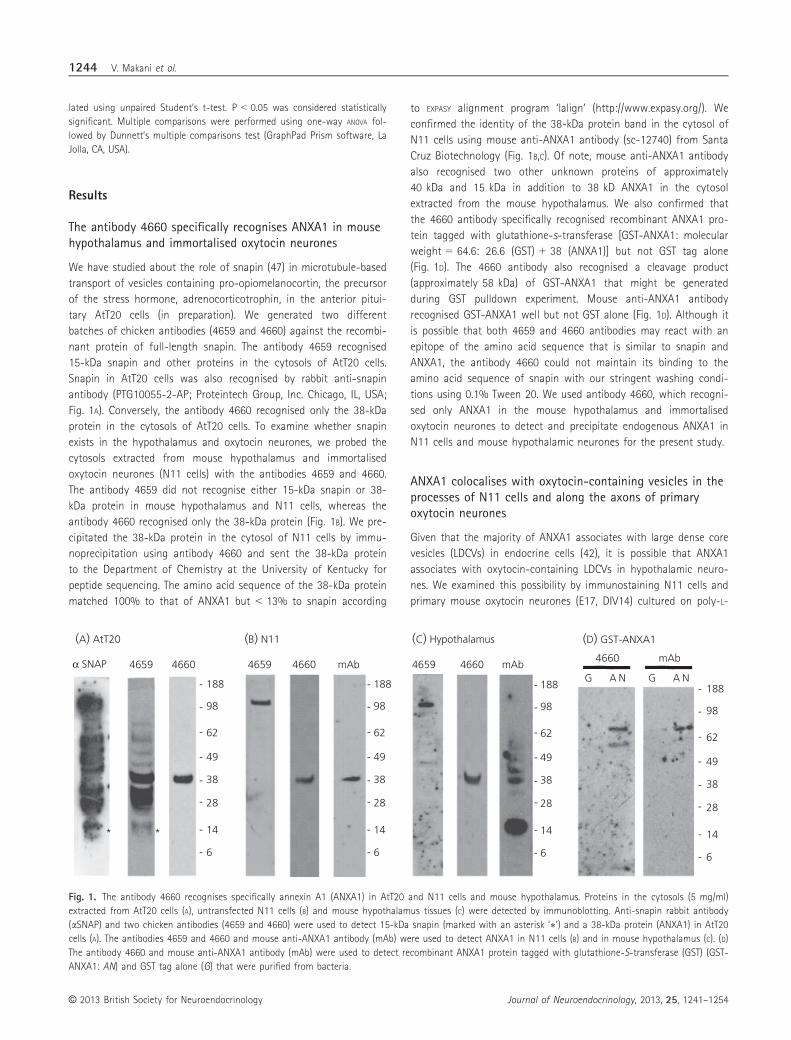
The antibody 4660 specifically recognises ANXA1 in mousehypothalamus and immortalised oxytocin neurones
We have studied about the role of snapin (47) in microtubule-based
transport of vesicles containing pro-opiomelanocortin, the precursor
of the stress hormone, adrenocorticotrophin, in the anterior pitui-
tary AtT20 cells (in preparation). We generated two different
batches of chicken antibodies (4659 and 4660) against the recombi-
nant protein of full-length snapin. The antibody 4659 recognised
15-kDa snapin and other proteins in the cytosols of AtT20 cells.
Snapin in AtT20 cells was also recognised by rabbit anti-snapin
antibody (PTG10055-2-AP; Proteintech Group, Inc. Chicago, IL, USA;
Fig. 1A). Conversely, the antibody 4660 recognised only the 38-kDa
protein in the cytosols of AtT20 cells. To examine whether snapin
exists in the hypothalamus and oxytocin neurones, we probed the
cytosols extracted from mouse hypothalamus and immortalised
oxytocin neurones (N11 cells) with the antibodies 4659 and 4660.
The antibody 4659 did not recognise either 15-kDa snapin or 38-
kDa protein in mouse hypothalamus and N11 cells, whereas the
antibody 4660 recognised only the 38-kDa protein (Fig. 1B). We pre-
cipitated the 38-kDa protein in the cytosol of N11 cells by immu-
noprecipitation using antibody 4660 and sent the 38-kDa protein
to the Department of Chemistry at the University of Kentucky for
peptide sequencing. The amino acid sequence of the 38-kDa protein
matched 100% to that of ANXA1 but < 13% to snapin according
to EXPASY alignment program ‘lalign’ (http://www.expasy.org/). We
confirmed the identity of the 38-kDa protein band in the cytosol of
N11 cells using mouse anti-ANXA1 antibody (sc-12740) from Santa
Cruz Biotechnology (Fig. 1B,C). Of note, mouse anti-ANXA1 antibody
also recognised two other unknown proteins of approximately
40 kDa and 15 kDa in addition to 38 kD ANXA1 in the cytosol
extracted from the mouse hypothalamus. We also confirmed that
the 4660 antibody specifically recognised recombinant ANXA1 pro-
tein tagged with glutathione-s-transferase [GST-ANXA1: molecular
weight = 64.6: 26.6 (GST) + 38 (ANXA1)] but not GST tag alone
(Fig. 1D). The 4660 antibody also recognised a cleavage product
(approximately 58 kDa) of GST-ANXA1 that might be generated
during GST pulldown experiment. Mouse anti-ANXA1 antibody
recognised GST-ANXA1 well but not GST alone (Fig. 1D). Although it
is possible that both 4659 and 4660 antibodies may react with an
epitope of the amino acid sequence that is similar to snapin and
ANXA1, the antibody 4660 could not maintain its binding to the
amino acid sequence of snapin with our stringent washing condi-
tions using 0.1% Tween 20. We used antibody 4660, which recogni-
sed only ANXA1 in the mouse hypothalamus and immortalised
oxytocin neurones to detect and precipitate endogenous ANXA1 in
N11 cells and mouse hypothalamic neurones for the present study.
ANXA1 colocalises with oxytocin-containing vesicles in theprocesses of N11 cells and along the axons of primaryoxytocin neurones
Given that the majority of ANXA1 associates with large dense core
vesicles (LDCVs) in endocrine cells (42), it is possible that ANXA1
associates with oxytocin-containing LDCVs in hypothalamic neuro-
nes. We examined this possibility by immunostaining N11 cells and
primary mouse oxytocin neurones (E17, DIV14) cultured on poly-L-
(A) AtT20
α SNAP
(B) N11 (C) Hypothalamus
4659 4660 4659 4660 mAb 4659 4660 mAb
(D) GST-ANXA1
G A N G A N
4660 mAb
-
-
-
-
-
-
-
98
62
49
38
28
14
6
- 188
-
-
-
-
-
-
-
98
62
49
38
28
14
6
- 188
-
-
-
-
-
-
-
98
62
49
38
28
14
6
- 188
-
-
-
-
-
-
-
98
62
49
38
28
14
6
- 188
* *
Fig. 1. The antibody 4660 recognises specifically annexin A1 (ANXA1) in AtT20 and N11 cells and mouse hypothalamus. Proteins in the cytosols (5 mg/ml)
extracted from AtT20 cells (A), untransfected N11 cells (B) and mouse hypothalamus tissues (C) were detected by immunoblotting. Anti-snapin rabbit antibody
(aSNAP) and two chicken antibodies (4659 and 4660) were used to detect 15-kDa snapin (marked with an asterisk ‘∗’) and a 38-kDa protein (ANXA1) in AtT20
cells (A). The antibodies 4659 and 4660 and mouse anti-ANXA1 antibody (mAb) were used to detect ANXA1 in N11 cells (B) and in mouse hypothalamus (C). (D)
The antibody 4660 and mouse anti-ANXA1 antibody (mAb) were used to detect recombinant ANXA1 protein tagged with glutathione-S-transferase (GST) (GST-
ANXA1: AN) and GST tag alone (G) that were purified from bacteria.
© 2013 British Society for Neuroendocrinology Journal of Neuroendocrinology, 2013, 25, 1241–1254
1244 V. Makani et al.

lysine-coated coverslips with primary antibodies to ANXA1 and oxy-
tocin. In N11 cells, ANXA1 co-localised with oxytocin-containing
vesicles accumulated at the process tips (Fig. 2A). We quantified the
correlation co-efficient of co-localisation (CCC) (45) between ANXA1
and oxytocin vesicles using METAMORPH. The CCC between ANXA1 and
oxytocin vesicles in the processes reached 0.80 � 0.01
(mean � SEM, n = 40 cells), indicating significant co-localisation.
Primary oxytocin neurones formed long thin axons negative for
MAP2 and thick dendrites positive for MAP2 (Fig. 2B). To examine
whether LDCVs containing CPE, a LDCV marker, co-localise with
oxytocin in the axons, we stained primary hypothalamic neurones
with anti-CPE and anti-oxytocin antibodies. CPE was very sparse
along the axons of oxytocin neurones but rich in the cell bodies
and dendrites of the neurones. At the axon terminals of untreated
(A) N11
iii i
iiOXT
OXT
OXT
ANXA1
ANXA1
MAP2
ANXA1
OXT
Axon
Axon
Merge
Merge
Merge
(B) Primary oxytocin neurones
(D) Primary oxytocin neurones
(E) Axon terminals
(C) Axon terminal
(F) Magnified vesicles
OXT CPE
Merge
0 25µm
0 50µm
0 50µm 0 50µm 0 50µm
0 50µm
0 25µm
0 25µm 0 25µm
0 25µm0 25µm
Fig. 2. Annexin A1 (ANXA1) colocalises with oxytocin vesicles (OXT) at the process tips of N11 cells and in the axons of primary hypothalamic neurones. (A)
N11 cells grown on poly-L-lysine-coated coverslips were processed for immunocytochemistry with antibodies to oxytocin (green) and ANXA1 (red). Scale
bars = 10 lm. (B–E) Primary mouse hypothalamic neurones at gestation day 17 (E17) were differentiated on poly-L-lysine-coated coverslips in neurobasal/B27
medium for 14 days (DIV14) and processed for immunocytochemistry with antibody to microtubule-associated protein 2 (MAP2) (B), carboxypeptidase E (CPE)
(C) or ANXA1 (D–F) along with anti-oxytocin antibody. (B) Primary oxytocin neurones formed a long axon that was not stained by anti-MAP2 antibody. (C) The
image of oxytocin vesicles and CPE at the axon terminals of the neurones was taken by confocal microscope. Scale bars = 5 lm. The inset of panel (D) shows
the co-localisation of oxytocin vesicles with ANXA1 in the axon. (D–F) ANXA1 showed significant co-localisation with vesicles containing oxytocin at the axon
terminals. Scale bars = 10 lm. The insets of panel (F) show the overlapping between the green pixels of oxytocin and the red pixels of ANXA1. Scale bars of
the insets = 0.13 lm (equivalent to the diameter of one pixel).
© 2013 British Society for NeuroendocrinologyJournal of Neuroendocrinology, 2013, 25, 1241–1254
Annexin A1 mediates axon-localisation of oxytocin 1245

oxytocin neurones, a small amount of CPE partially co-localised
with oxytocin vesicles (Fig. 2C). It suggests that only a subpopula-
tion of oxytocin-containing vesicles in the axon may have CPE.
There were distinguishable individual LDCVs containing oxytocin
along thin axons (Fig. 2D–F). Meanwhile, we could not find distin-
guishable individual oxytocin vesicles in the cell body and dendrites
(Fig. 2D). The oxytocin-containing LDCVs along the axons showed
significant co-localisation with ANXA1 (Fig. 2E, mean � SEM
CCC = 0.71 � 0.03; n = 40 neurones). To obtain more detailed
information about the co-localisation between ANXA1 and oxytocin
vesicles, we attempted to perform voxel co-localisation analysis, a
method to quantify the extent of the overlapping of volumic pixels
(voxels) between two different fluorescence colours. However, we
could not measure voxels because it was not possible to take multi-
ple images along the z-axis through the thin axons of oxytocin
neurones. Instead, we calculated the percent of the overlapping of
the green fluorescent pixels of oxytocin vesicle with the red fluo-
rescent pixels of ANXA1 on the vesicle using METAMORPH (Fig. 2F). The
mean � SEM percentage of ANXA1-colocalising oxytocin pixels per
vesicle was 70.4 � 1.2% (n = 47 vesicles). Thus, it is clear that
ANXA1 associates with oxytocin-containing LDCVs in the axons of
primary hypothalamic oxytocin neurones.
ANXA1 mediates the anterograde transport of oxytocinvesicles along the axon via its interaction with kinesin-2
A previous study showed that knockout of ANXA1 blocked the
anterograde transport of Shiga toxin (43), suggesting that ANXA1
may be involved in intracellular anterograde transport. We exam-
ined whether ANXA1 is required for the anterograde transport of
oxytocin-containing LDCVs by knocking down ANXA1 in primary
hypothalamic neurones using lentiviral shRNA against ANXA1. Pri-
mary hypothalamic neurones (E17, DIV14) were transduced with
lentiviral particles containing either control or anti-ANXA1 shRNA
for 5 days and processed for immunocytochemistry with antibodies
to oxytocin and ANXA1. The mean � SEM intensity of immuno-
stained ANXA1 in neurones transduced with anti-ANXA1 lentiviral
shRNA was decreased to approximately 26% (5.35 � 0.37) of that
(20.78 � 1.33) in neurones transduced with control lentiviral
shRNA (n > 50 neurones, P < 0.0001; Fig. 3A). Similarly, the mean
intensity of immunostained oxytocin vesicles along the axons of
neurones transduced with anti-ANXA1 lentiviral shRNA was signifi-
cantly lower than that of neurones transduced with control lentivi-
ral shRNA (n = 40 neurones; Fig. 3B,C). These results suggest that
ANXA1 is required for the anterograde transport of oxytocin vesi-
cles along the axons.
Given that ANXA1 is required for the anterograde transport of
oxytocin vesicles along the axons, it is possible that ANXA1 inter-
acts with kinesins, microtubule-based anterograde transporters. We
performed co-immunoprecipitation using the antibody 4660 and
the cytosols of mouse hypothalamus and N11 cells. ANXA1 inter-
acted with kinesin-2 (KIF3A) but not with kinesin-1 (kinesin heavy
chain) and kinesin-3 (KIF1A) in N11 cells and mouse hypothalamus
(Fig. 4A,B). Thus, kinesin-2 appears to mediate the anterograde
transport of oxytocin vesicles along the axons via its interaction
with ANXA1. In addition to kinesin-2, ANXA1 interacted with the
retrograde transporter, cytoplasmic dynein, and its activator, dynac-
tin (Fig. 4A,B).
Activation of PKA enhances the anterograde transport ofoxytocin vesicles to the axon terminals
Given that pharmacological treatments that activate PKA enhance
oxytocin secretion at the axon terminals (18–21), activation of
PKA may increase the anterograde transport of oxytocin vesicles
to the axon terminals for secretion. To examine this possibility, we
treated primary hypothalamic neurones with forskolin, which
increases the intracellular level of cyclic AMP, an activator of PKA,
and then quantified the intensity of immunostained oxytocin vesi-
cles along the axons and at the axon terminals. Mouse hypotha-
lamic neurones (E17, DIV14) were treated with either DMSO or
10 lM forskolin for 10, 20 and 30 min and immunostained using
antibodies to oxytocin and ANXA1 (n = 40 neurones per each
condition, two independent experiments). The mean intensity of
oxytocin vesicles in the axons was gradually increased by longer
period of incubation with forskolin (Fig. 5). The mean intensities of
oxytocin vesicles in neurones treated with DMSO were approxi-
mately 20 (in arbitrary units) in the cell bodies, 10–15 in the ax-
ons between 0 and 100 lm from the cell body, and seven in
those between 100 and 200 lm. After 10- and 20-min treatments
with forskolin, the mean intensity of oxytocin vesicles in the cell
body was decreased from approximately 20 (0 min) to 8 (10 min)
and 8 (20 min; Fig. 5). Meanwhile, the mean intensities of oxyto-
cin vesicles were increased to 20–22 in the axon between 0 and
100 lm and to 13–16 between 100 and 200 lm (Fig. 5B,C,E). At
30 min after forskolin treatment, the mean intensity of oxytocin
vesicles in the axon was significantly increased to approximately
30, 26, 21 and 20 in axons between 0–50, 50–100, 100–150 and
150–200 lm, respectively. We specified the involvement of PKA in
the enhancement of the axon-localisation of oxytocin vesicles by
pre-treating neurones with 10 lM PKI-ester, a PKA inhibitor, for
1 h before 30 min of forskolin treatment. Pre-treatment with PKI-
ester blocked forskolin from enhancing the axon-localisation of
oxytocin vesicles (Fig. 5D). The mean intensities of oxytocin vesicles
in the cell bodies and the axons of neurones pre-treated with PKI
and forskolin were similar to those of neurones treated with
DMSO (data not shown). In addition, we assessed the extents of
co-localisation between oxytocin vesicles and ANXA1 along the
axon during 30 min of stimulation with forskolin. ANXA1 main-
tained high levels of co-localisation with oxytocin vesicles
(mean � SEM CCC = 0.73 � 0.01; n = 60) in the axons during
30 min of stimulation by forskolin.
We also quantified the mean intensities of oxytocin vesicles at
the axon terminals of neurones treated with either DMSO or 10 lMforskolin for 30 min. The mean intensity of oxytocin vesicles at the
axon terminals of forskolin-treated neurones was approximately
three-fold higher than that of DMSO-treated neurones (n > 70
axon terminals; Fig. 6A,B,D).
All of these results suggest that activation of PKA enhances
the anterograde transport of oxytocin vesicles along the
© 2013 British Society for Neuroendocrinology Journal of Neuroendocrinology, 2013, 25, 1241–1254
1246 V. Makani et al.

axons, thus accumulating more oxytocin vesicles at the axon
terminals.
Activation of PKA increases the association of kinesin-2and AKAP150 with ANXA1
Because kinesins are the anterograde transporters that carry pro-
teins and vesicles toward the axon terminals (48), it is possible
that activation of PKA recruits more kinesins to oxytocin vesicles,
thus enhancing the anterograde transport of the vesicles. Possi-
bly, activation of PKA increases the binding of kinesins to ANXA1,
a protein associated with oxytocin vesicles. We therefore per-
formed co-immunoprecipitation using the antibody 4660 and the
cytosols extracted from N11 cells treated with either DMSO or
10 lM forskolin for 30 min. Activation of PKA did, indeed,
increase the association of kinesin-2 with ANXA1 compared to
control (Fig. 4C). Conversely, activation of PKA had no effect on
the binding of cytoplasmic dynein, dynactin, kinesin-1 and kine-
sin-3 to ANXA1. We also examined the effect of forskolin treat-
ment on the association of AKAP150, a scaffold protein that
targets the action of PKA to its associated proteins, with ANXA1.
In untreated N11 cells and mouse hypothalamus, there was a
basal level of interaction between AKAP150 and ANXA1 (Fig. 4A,B).
The basal interaction between AKAP150 and ANXA1 was also
observed in the counter pulldown by anti-dynactin antibody
(Fig. 4E). Upon forskolin treatment, more AKAP150 was associated
with ANXA1 (Fig. 4C). Thus, activation of PKA increases the bind-
ing of kinesin-2 to ANXA1, which should increase the antero-
grade transport of oxytocin vesicles. Increased binding of
AKAP150 to ANXA1 may concentrate the action of PKA on oxy-
tocin vesicles, thus enhancing the anterograde transport of the
vesicles.
(A) Control shRNA
(B) Anti-ANXA1shRNA
(C)
0
2
4
6
8
10
12
14
16
Cell body 0–50 50–100 100–150 um
150–200 um
200–250 um
Mea
n in
tens
ity o
f ox
ytoc
inst
aini
ng in
axo
n
Axon (at the distance from cell body)
*
* **
Control
ANXA1 KD
ANXA1 OXT
ANXA1 OXT
um um
Fig. 3. Annexin A1 (ANXA1) is required for the axon-localisation of oxytocin vesicles (OXT) in primary hypothalamic neurones. Primary mouse hypothalamic
neurones at gestation day 17 (E17) differentiated on poly-L-lysine-coated coverslips were transduced with control or anti-ANXA1 short hairpin (sh)RNA lentivi-
ral particles for 5 days. Transduced neurones were then processed for immunocytochemistry using antibodies to ANXA1 (red) and oxytocin (green). The insets
showed that neurones transduced with anti-ANXA1 shRNA (B) had low levels of ANXA1 and oxytocin vesicles in the axons compared to those transduced with
control shRNA (A). Scale bars = 10 lm. (C) Bar graphs show the mean � SEM intensities of oxytocin vesicles in the cell bodies and the axons between 0–50,
50–100, 100–150, 150–200 and 200–250 lm from the cell body of neurones transduced with either control or anti-ANXA1 shRNA (ANXA1 KD) (*P < 0.001).
© 2013 British Society for NeuroendocrinologyJournal of Neuroendocrinology, 2013, 25, 1241–1254
Annexin A1 mediates axon-localisation of oxytocin 1247

Activation of PKC decreases the binding of kinesin-2 toANXA1, which may attenuate the anterograde transport ofoxytocin vesicles
Previous studies suggest that activation of PKC may enhance so-
matodendritic oxytocin secretion but attenuate axonal secretion
(16,23). Hence, we examined the effect of PKC on the axon-locali-
sation of oxytocin vesicles and the interaction of ANXA1 with ki-
nesin-2 and other proteins. Primary hypothalamic neurones (E17,
DIV14) were treated with either DMSO or 10 nM phorbol ester
(PMA) and immunostained with antibodies to oxytocin and ANXA1.
The mean � SEM intensities of oxytocin (4.71 � 0.46) and ANXA1
(6.75 � 0.58) in the axons of neurones treated with PMA (Fig. 6F,G)
were similar to those of neurones treated with DMSO (oxyto-
cin = 7.1 � 0.7 and ANXA1 = 6.9 � 0.6; n = 60 neurones;
Fig. 6E,G). This finding suggests that PKC activation does not
enhance the axon-localisation of oxytocin vesicles. We also quanti-
fied the mean intensity of oxytocin vesicles at the axon terminals
of neurones treated with either DMSO or 10 nM PMA for 30 min.
The mean intensity of oxytocin vesicles at the axon terminals of
PMA-treated neurones was not different from that of DMSO-trea-
ted neurones (n > 70 axon terminals, P > 0.05; Fig. 6A,C,D). Next,
we performed co-immunoprecipitation using the antibody 4660
and the cytosols extracted from N11 cells treated with either
DMSO or 10 nM PMA. Activation of PKC did not affect the associ-
ation of cytoplasmic dynein, dynactin, and AKAP150 with ANXA1
but decreased the binding of kinesin-2 to ANXA1 (Fig. 4D). The
binding of kinesin-1 and kinesin-3 to ANXA1 was not affected by
activation of PKC. These results suggest that activation of PKC
may attenuate the anterograde transport of oxytocin vesicles
along the axon by decreasing the association of kinesin-2 with
oxytocin vesicles.
Activation of PKA increases the association of kinesin-2and AKAP150 with light-density vesicles
We performed differential centrifugation to examine the effect of
PKA and PKC on the association of ANXA1, AKAP150 and micro-
tubule motors with LDCVs in N11 cells. N11 cells treated with
DMSO, 10 lM forskolin or 10 nM PMA were processed to obtain
cytosols (1 mg/ml) in 0.3 ml of PMEE buffer (49) + protease/phos-
phatase inhibitors + 0.25 M sucrose. PNS was obtained by spin-
ning cell lysates at 600 g. PNS was spun sequentially at 3000,
4800, 15 000, 100 000 and 161 000 g to separate different mem-
branous compartments. The plasma membrane (syntaxin-1), Golgi
complex (p115) and endoplasmic reticulum (ER: calreticulin) were
pelleted at 3000, 4800 and 15 000 (Fig. 7A). CPE, a heavy-density
LDCV protein that travels from the ER through the Golgi complex
to the plasma membrane (50), was found in the ER, Golgi com-
plex, plasma membrane and heavy-density vesicle pool, and to a
lesser extent in the light-density vesicle pool. Synaptophysin
(SYN), a light-density vesicle marker, was pelleted at 161 000 and
to some extent at 3000. Oxytocin-containing intermediates (15,
20–28, 34 kDa) were pelleted at 100 000 and 161 000, suggesting
that there are two different-density LDCVs containing oxytocin.
Two different-density oxytocin vesicles were also found in the
mouse hypothalamus (Fig. 7D). We could not detect the mature
form (nine amino acids, < 1 kD) of oxytocin because the nitrocel-
lulose membrane (pore size = 0.45 lm) that we used could not
hold peptides smaller than 2 kDa. Given that oxytocin vesicles in
the axon contain a little CPE (Fig. 2C), it appears that light-density
vesicles containing oxytocin and a little CPE (Fig. 7A) are targeted
to the axon.
In untreated N11 cells, most ANXA1 was found in the heavy
membrane fractions pelleted at 3000 and 4800 and the cytosol,
IgG αp150
Dynactin
AKAP150
ANXA1
Dynein
Kinesin-2
Hypothalamus N11 (DMSO)
SN IgG αAN SN IgG αAN
Dynein
AXNA1
AKAP150
Kinesin-2
Dynactin
Kinesin-3
Kinesin-1
(A) (B) N11 (Forsk)
SN IgG αAN
(C) N11 (PMA)
SN IgG αAN
(D) (E) N11 (DMSO)
Fig. 4. Annexin A1 (ANXA1), A-kinase anchor protein 150 (AKAP150) and microtubule motors form a complex in mouse hypothalamus and N11 cells. The cy-
tosols (5 mg/ml) of mouse hypothalamus tissues (A) or N11 cells (B–D) that were treated with dimethyl sulphoxide (B), 10 lM forskolin (C, Forsk) or 10 nM phor-
bol 12-myristate 13-acetate (PMA) (D) were mixed with the antibody 4660 along with protein A beads (60 ll) at 4 °C for 24 h. Proteins pulled down by anti-
ANXA1 antibody (aAN) were probed by immunoblotting. (E) The cytosols [5 mg/ml, post-nuclear supernatant (SN)] of N11 cells were mixed with anti-p150
(dynactin) antibody for co-immunoprecipitation. Proteins bound to dynactin were detected by immunoblotting.
© 2013 British Society for Neuroendocrinology Journal of Neuroendocrinology, 2013, 25, 1241–1254
1248 V. Makani et al.

whereas a small amount of ANXA1 was pelleted at 15 000,
100 000 and 161 000 (Fig. 7A). Conversely, in forskolin-treated N11
cells, the amount of ANXA1 that was associated with light-density
vesicles was increased (Fig. 7B). In PMA-treated N11 cells, ANXA1
was found in all the fractions (Fig. 7C). Kinesin-2 was found mostly
in the vesicle pools and cytosol of untreated N11 cells. Treatment
(A)
(B)
ANXA1OXT Merge
F10’ 0 50µm
Axon
Axon
F20’
F30’
Axon
(C)
iv
iv
PKI, F30’
Axon
i
i
iiii
iii
iii
(D)
0
5
10
15
20
25
30
35
Cell body 0–50 um 50–100 um 100–150 um 150–200 um
Mea
n in
tens
ity o
f ox
ytoc
inst
aini
ng in
axo
n
Axon (at the distance from cell body) –10 uM forskolin
0 min
10 min
20 min
30 min
(E)
*
#
##
# #
* *
0 50µm
0 50µm
0 50µm 0 50µm
0 50µm
0 50µm
0 50µm 0 50µm
0 50µm
0 50µm
0 50µm
Fig. 5. Vesicles containing oxytocin (OXT) and annexin A1 (ANXA1) co-migrate toward the axon terminal in response to protein kinase A (PKA) activation.
Mouse hypothalamic neurones (E17, DIV14) were treated with dimethyl sulphoxide or 10 lM forskolin for 10 min (A, F10′), 20 min (B, F20′), and 30 min (C,
F30′) and stained using antibodies to oxytocin and ANXA1. The axons are indicated by arrows. The magnified images in the insets show the co-localisation
between oxytocin and ANXA1 at the distal axons of neurones treated with forskolin for 10 min (inset i), 20 min (inset ii) and 30 min (inset iii). Scale
bar = 50 lm. (D) To specify the involvement of PKA in forskolin-induced axon-localisation of oxytocin vesicles, neurones were pretreated with 10 lM PKI-ester
for 1 h before 30 min of forskolin treatment (inset iv). (E) The mean intensities of oxytocin vesicles in cell bodies and along axons (0–50, 50–100, 100–150
and 150–200 lm) were quantified using METAMORPH (n = 30 per condition). Bar graphs show the mean � SEM intensities of oxytocin vesicles along the axons
(0–50, 50–100, 100–150 and 150–200 lm) of neurones treated with forskolin (0, 10, 20 and 30 min) (#P < 0.0001, *P < 0.01 compared to control).
© 2013 British Society for NeuroendocrinologyJournal of Neuroendocrinology, 2013, 25, 1241–1254
Annexin A1 mediates axon-localisation of oxytocin 1249

with forskolin increased the association of kinesin-2 with light-den-
sity vesicles (Fig. 7B), whereas that with PMA decreased the associ-
ation of kinesin-2 even with heavy-density vesicles (Fig. 7C).
AKAP150 was found mostly in the light-density vesicles and cyto-
sols of cells when the cells were treated with forskolin (Fig. 7B),
whereas it was in all the fractions of cells treated with either
DMSO (Fig. 7A) or PMA (Fig. 7C). The association of cytoplasmic
dynein, dynactin and kinesin-1 with heavy- and light-density vesi-
cles was not changed by treatment with either forskolin (Fig. 7B) or
PMA (Fig. 7C). These results suggest that activation of PKA increases
the association of kinesin-2 with light-density oxytocin vesicles that
contain oxytocin and a little CPE, thus enhancing the anterograde
transport of the vesicles to the axon. Conversely, activation of PKC
that decreases the association of kinesin-2 with ANXA1 on oxytocin
vesicles may block the anterograde transport of oxytocin vesicles to
the axon.
Discussion
Oxytocin is a major socialising neuropeptide in the brain (51). Oxy-
tocin secreted from the somatodendritic regions of the hypotha-
lamic neurones activates its receptors in the hypothalamus and the
brain regions neighbouring the hypothalamus (1,2). Some oxytocin
travels along the long axon to the distal brain regions such as sep-
tum, substantia nigra, stria terminalis, olfactory bulb, brainstem and
spinal cord (7–14). Brain oxytocin modulates brain functions
involved in behaviour, anxiety, cognition and autonomic nerve
activity (15). Oxytocin is also secreted from the axons that inner-
vate the posterior pituitary gland into the blood circulation, thus
increasing natriuresis and reducing blood pressure and heart rate
(3–6). There appears to be a ‘yin and yang’ mechanism for control-
ling the distribution of oxytocin secretion between the axon and
the somatodendritic region. Extracellular stimuli that activate PKA
(A) (B)
(C)DMSO Forsk
PMA02468
10121416
Mock Forsk. PMA
Mea
n ox
ytoc
in
inte
nsity
at a
xon
term
inal
s (D)*
(E)
(G)
Axon05
10152025
Moc
kPM
AFo
rsk.
P < 0.005
P < 0.0001
Mea
n in
tens
ity o
f O
XT
in a
xonOXT ANXA1
ANXA1OXT
(F)
Axon
0 50µm 0 50µm
Fig. 6. Activation of protein kinase A (PKA) increases the localisation of oxytocin-containing vesicles (OXT) to axon terminals, whereas that of protein kinase
C (PKC) decreases it. Mouse hypothalamic neurones (E17, DIV14) were treated with dimethyl sulphoxide (DMSO) (A), 10 lM forskolin (B, Forsk) or 10 nM phorbol
12-myristate 13-acetate (PMA) (C) for 30 min and stained using anti-oxytocin antibody. The images of oxytocin vesicles at the axon terminals of the neurones
were taken by confocal microscope. Scale bars = 10 lm. (D) Bar graphs show the mean � SEM intensities of oxytocin vesicles at the axon terminals of neuro-
nes treated with DMSO, forskolin or PMA (*P < 0.01). (E,F) Mouse hypothalamic neurones treated with DMSO (E) or PMA (F) were stained using antibodies to
oxytocin and ANXA1. The axons were indicated by arrows. Scale bars = 10 lm. (G) Bar graphs show the mean � SEM intensities of oxytocin vesicles in the
axons of neurones treated with DMSO, PMA and forskolin (Forsk.). (n = 60 neurones per condition; *P < 0.001: DMSO versus forskolin).
© 2013 British Society for Neuroendocrinology Journal of Neuroendocrinology, 2013, 25, 1241–1254
1250 V. Makani et al.

enhance the secretion of oxytocin at the axon terminals (18–21),
whereas those that activate PKC enhance somatodendritic oxytocin
secretion and attenuate oxytocin secretion at the axon terminal
(16,22,23). Thus far, the molecular mechanism underlying the regu-
lation of the distribution of oxytocin vesicles for secretion between
the axon and the somatodendritic region remains largely unknown.
We have discovered that ANXA1 forms a complex with AKAP150,
a protein that interacts with PKA and PKC (24–28), and microtubule
motors in the hypothalamus and immortalised oxytocin neurones.
Pixel analysis of the co-localisation between ANXA1 and oxytocin
vesicles clearly shows that ANXA1 associates with oxytocin-contain-
ing LDCVs in the axons of primary oxytocin neurones. The interac-
tion of ANXA1 complex with kinesin-2, an anterograde transporter,
is modified differently by PKA and PKC. Activation of PKA increases
the binding of kinesin-2 to ANXA1, which appears to enhance the
anterograde transport of oxytocin vesicles toward the axon termi-
nals. Conversely, activation of PKC decreases the binding of kine-
sin-2 to ANXA1, which may decrease the axon-localisation of
oxytocin vesicles. Thus, our results indicate that PKA and PKC deter-
mine the directionality of oxytocin vesicle movements in the axon
by controlling the binding of kinesin-2 to ANXA1 on oxytocin vesi-
cles (Fig. 8).
Our finding of a new molecular mechanism by which the direc-
tionality of vesicle movements is controlled should provide new
insight into peptidergic vesicle trafficking in endocrine and neuro-
endocrine cells. As reviewed recently (48), even the molecular
mechanism by which peptidergic vesicles are transported along
microtubules in peptidergic neurones and endocrine cells is not
fully understood. KIF1A mediates the anterograde transport of pep-
tidergic vesicles in Caenorhabditis elegan (52). A microtubule motor
complex consisting of kinesin-2, cytoplasmic dynein and dynactin
mediates the bi-directional movement of stress hormone vesicles in
AtT20 cells (49). A similar complex mediates the transport of vesi-
cles containing brain-derived neurotrophic factor in hippocampal
neurones (53) with the assistance of Htt-associated protein-1 (54,
55). In premature neurones, bicaudal-D-related protein 1 mediates
peptidergic vesicle transport required for neurite outgrowth by
interacting simultaneously with Rab6 on LDCVs and with a motor
complex consisting of dynactin, cytoplasmic dynein and kinesin-3
(56). We speculate that ANXA1 complex may coordinate the actions
of PKA and/or PKC in those transport systems, thus determining
the direction of hormone vesicle movements to different secretion
sites (e.g. the axon versus the cell body).
Is the binding of more kinesin-2 to vesicles sufficiently strong to
push the vesicles toward the anterograde direction? Under resting
conditions, bi-directional microtubule-based transport is nearly at
force balance between a small group of strong torque kinesins and
a large team of weak torque cytoplasmic dynein (57–59). A single
kinesin competes with at least five cytoplasmic dyneins for chang-
ing the direction of microtubule-based transport (59). Thus, the
directionality of microtubule-based transport is determined by
either the association/dissociation of a single strong torque kinesin
or the number of multiple weak torque cytoplasmic dyneins. There-
fore, the increase in the association of kinesin-2 with ANXA1 on
oxytocin vesicles without affecting that of cytoplasmic dynein
should push oxytocin vesicles toward the axon terminals of neuro-
Syntaxin-1
p115
SYN
ANXA1
CPE
OXT
Kinesin-1
Dynein
Dynactin
Kinesin-2
Calreticulin
AKAP150
(A)
(B) (C) (D)hypothalamus
N11 (DMSO)
N11 (Forsk) N11 (PMA)
3 4.8 15 100 161 cyt.
3 4.8 15 100 161 cyt. 3 4.8 15 100 161 cyt. 3 4.8 15 100 161 cyt.
Fig. 7. Activation of protein kinase A (PKA) increases the association of kinesin-2 with light-density vesicles. (A–D) Post-nucleus supernatant was obtained
from either N11 cells [treated with dimethyl sulphoxide (A), 10 lM forskolin (B) or 10 nM phorbol 12-myristate 13-acetate (C)] or mouse hypothalamus tissues
(D) by spinning cell or tissue extracts at 600 g to remove nuclei and cell debris. Post-nucleus supernatant was spun sequentially at 3000, 4800, 15 000,
100 000 and 161 000 g. The pellet after each spin was re-suspended in 0.5% Igepal CA-630-containing PMEE to extract proteins from lipid membrane. Pro-
teins in the pellets and cytosol (cyt.) were detected by immunoblotting. Markers for plasma membrane (syntaxin-1), p115 (Golgi complex), calreticulin (ER), CPE
(heavy density vesicles), synaptophysin (SYN: light density vesicles) and oxytocin (OXT) in the pellets and cytosol were detected by immunoblotting.
© 2013 British Society for NeuroendocrinologyJournal of Neuroendocrinology, 2013, 25, 1241–1254
Annexin A1 mediates axon-localisation of oxytocin 1251
nes treated with PKA activator. Conversely, the dissociation of kine-
sin-2 from ANXA1 on oxytocin vesicles in neurones treated with
PKC activator should shift the direction of vesicle movement
toward the cell body. This mechanism may be how extracellular
stimuli adjust the amount of oxytocin for secretion at the axon ter-
minal versus the somatodendritic region. It is possible that most
peptidergic neurones with long axons such as hippocampal neuro-
nes use ANXA1-AKAP150-kinesin-2 complex or a similar complex to
refill depleted secretion sites at the axon terminal quickly in a PKA-
dependent manner for release of a proper amount of hormones in
response to repetitive stimulation.
To clarify the difference of our finding of the association of
ANXA1 with peptidergic vesicles with respect to the findings of a
subset of previous studies (60, 61), we attempted to perform
immuno-electron microscopy using primary hypothalamic neurones
cultured in vitro, although it was found that this was not possible
as a result of technical difficulties with respect to obtaining sec-
tions of the intact thin axons of hypothalamic primary neurones.
Nonetheless, our high magnification images clearly show significant
co-localisation of ANXA1 with oxytocin vesicles in the axon. Given
that ANXA1 is expressed as an intracellular (not extracellular) pro-
tein (36) and found mostly on LDCVs in endocrine cells (42), it may
reside on the extravesicular surface of oxytocin vesicles.
We observed the presence of two different-density oxytocin vesi-
cles in immortalised N11 cells and mouse hypothalamus. Light-den-
sity vesicles appear to contain some CPE, an LDCV enzyme that
removes the C-terminal bi-basic residues of pro-hormones (50).
Most oxytocin vesicles in the axons may be light-density vesicles
containing a little CPE. Additional studies are needed to further
characterise these light-density oxytocin vesicles.
Cell body Microtubules Axon terminal
Anterograde+
+
OXT
Inhibited by PKC
OXT
Cell body Microtubules Axon terminal
Golgi
Cell
Micr
otubules
Axon
body
Dendrite
Light OXT vesicles Heavy OXT vesiclesPKC
PKA
(–)
(+)
DyneinKinesin-2
ANXA1
AKAP150 PKARR
CC
PKC
(A)
(B) (C)
Enhanced by PKARR
CC
RR
CC
Dynactin
Fig. 8. Schematic illustration of how protein kinase A (PKA) and protein kinase C (PKC) affect the interaction of annexin A1 (ANXA1) with A-kinase anchor
protein 150 (AKAP150) and kinesin-2, thus changing the direction of movement of oxytocin-containing vesicles. (A) Upon activation of PKA that consists of
two catalytic [C] and two regulatory [R] subunits, the regulated subunits withdraw their inhibitory action from the catalytic subunits. Then, the catalytic su-
bunits increase the binding of kinesin-2 and AKAP150 to ANXA1 on light-density oxytocin (OXT) vesicle without affecting the interaction of cytoplasmic
dynein-dynactin with ANXA1, resulting in enhancement of anterograde transport of the vesicles to the axon terminal. (B) Activated PKC inhibits the interaction
of kinesin-2 with AKAP150-ANXA1 complex without affecting the interaction of cytoplasmic dynein-dynactin with ANXA1 complex, resulting in inhibition of
anterograde transport of oxytocin vesicles to the axon terminal. (C) Activation of PKA enhances the anterograde transport of light-density oxytocin vesicles to
the axon terminals, whereas activation of PKC inhibits the anterograde transport. Activation of PKC increases somatic exocytosis of oxytocin vesicles. Some
heavy density oxytocin vesicles are transported to the axon and dendrites.
© 2013 British Society for Neuroendocrinology Journal of Neuroendocrinology, 2013, 25, 1241–1254
1252 V. Makani et al.

In conclusion, the findings of the present study demonstrate that
ANXA1 forms a complex with AKAP150 and microtubule motors in
the hypothalamus, specifically in oxytocin neurones. This complex
coordinates the actions of PKA and PKC to control the direction of
oxytocin vesicle movement by changing the association of the
anterograde strong torque motor, kinesin-2, with oxytocin vesicles
(Fig. 8). Our study reveals, for the first time, a novel molecular
mechanism that peptidergic neurones use to control the microtu-
bule-based transport of neuropeptide-containing vesicles to secre-
tion sites. Further studies should help identify molecular targets for
improving the treatments of endocrine and psychiatric disorders
that are caused by abnormal hormone secretion.
Acknowledgements
We thank Dr Andrea Kalinoski (University of Toledo Microscopy Imaging
Core) for technical support. We also thank Dr Y. Peng Loh for the 4659 and
4660 antibodies. This research was supported by NICHD K22
(1K22HD056137-01A1) and ARRA funding.
Received 4 March 2013,
revised 24 September 2013,
accepted 29 September 2013
References
1 Gimpl G, Fahrenholz F. The oxytocin receptor system: structure, func-
tion, and regulation. Physiol Rev 2001; 81: 629–683.
2 Hirasawa M, Kombian SB, Pittman QJ. Oxytocin retrogradely inhibits
evoked, but not miniature, EPSCs in the rat supraoptic nucleus: role of
N- and P/Q-type calcium channels. J Physiol 2001; 532: 595–607.
3 Lee HJ, Macbeth AH, Pagani JH, Young WS 3rd. Oxytocin: the great
facilitator of life. Prog Neurobiol 2009; 88: 127–151.
4 Gutkowska J, Jankowski M. Oxytocin revisited: its role in cardiovascular
regulation. J Neuroendocrinol 2011; 24: 599–608.
5 Petersson M. Cardiovascular effects of oxytocin. Prog Brain Res 2002;
139: 281–288.
6 Soares TJ, Coimbra TM, Martins AR, Pereira AG, Carnio EC, Branco LG,
Albuquerque-Araujo WI, de Nucci G, Favaretto AL, Gutkowska J, McCann
SM, Antunes-Rodrigues J. Atrial natriuretic peptide and oxytocin induce
natriuresis by release of cGMP. Proc Natl Acad Sci USA 1999; 96: 278–
283.
7 Buijs RM, deVries GJ, VanLeeeuwen FW. Distribution and synaptic
release of oxytocin in the central nervous system. In: Amico JA, Robin-
son AG, eds. Oxytocin: Clinical and Laboratory Aspects. Oxford: Elsevier
Science Publishers, 1985; 666: 77–86.
8 Blevins JE, Eakin TJ, Murphy JA, Schwartz MW, Baskin DG. Oxytocin
innervation of caudal brainstem nuclei activated by cholecystokinin.
Brain Res 2003; 993: 30–41.
9 Buijs RM, Van der Beek EM, Renaud LP, Day TA, Jhamandas JH. Oxy-
tocin localization and function in the A1 noradrenergic cell group: ul-
trastructural and electrophysiological studies. Neuroscience 1990; 39:
717–725.
10 Peters JH, McDougall SJ, Kellett DO, Jordan D, Llewellyn-Smith IJ, Andre-
sen MC. Oxytocin enhances cranial visceral afferent synaptic transmis-
sion to the solitary tract nucleus. J Neurosci 2008; 28: 11731–11740.
11 Llewellyn-Smith IJ, Kellett DO, Jordan D, Browning KN, Travagli RA. Oxy-
tocin-immunoreactive innervation of identified neurons in the rat dorsal
vagal complex. Neurogastroenterol Motil 2011; 24: e136–e146.
12 Ingram CD, Moos F. Oxytocin-containing pathway to the bed nuclei of
the stria terminalis of the lactating rat brain: immunocytochemical and
in vitro electrophysiological evidence. Neuroscience 1992; 47: 439–452.
13 Buijs RM, De Vries GJ, Van Leeuwen FW, Swaab DF. Vasopressin and
oxytocin: distribution and putative functions in the brain. Prog Brain
Res 1983; 60: 115–122.
14 Nicholas AP, Hancock MB. Evidence for substance P, serotonin and oxy-
tocin input to medullary catecholamine neurons with diencephalic pro-
jections. Brain Res Bull 1989; 22: 213–223.
15 Barberis C, Tribollet E. Vasopressin and oxytocin receptors in the central
nervous system. Crit Rev Neurobiol 1996; 10: 119–154.
16 Sabatier N. Alpha-melanocyte-stimulating hormone and oxytocin: a pep-
tide signalling cascade in the hypothalamus. J Neuroendocrinol 2006;
18: 703–710.
17 Ludwig M, Sabatier N, Bull PM, Landgraf R, Dayanithi G, Leng G. Intra-
cellular calcium stores regulate activity-dependent neuropeptide release
from dendrites. Nature 2002; 418: 85–89.
18 Galfi M, Radacs M, Juhasz A, Laszlo F, Molnar A, Laszlo FA. Serotonin-
induced enhancement of vasopressin and oxytocin secretion in rat neu-
rohypophyseal tissue culture. Regul Pept 2005; 127: 225–231.
19 Osei-Owusu P, James A, Crane J, Scrogin KE. 5-Hydroxytryptamine 1A
receptors in the paraventricular nucleus of the hypothalamus mediate
oxytocin and adrenocorticotropin hormone release and some behavioral
components of the serotonin syndrome. J Pharmacol Exp Ther 2005;
313: 1324–1330.
20 Bojanowska E, Stempniak B. tGLP-1 and release of vasopressin and oxy-
tocin from the isolated rat hypothalamo-neurohypophysial system:
effects of a tGLP-1 receptor agonist and antagonist. J Physiol Pharma-
col 2001; 52: 781–793.
21 Onaka T. Neural pathways controlling central and peripheral oxytocin
release during stress. J Neuroendocrinol 2004; 16: 308–312.
22 Sabatier N, Leng G. Presynaptic actions of endocannabinoids mediate
alpha-MSH-induced inhibition of oxytocin cells. Am J Physiol Regul In-
tegr Comp Physiol 2006; 290: R577–R584.
23 Ciosek J, Cisowska A. Centrally administered galanin modifies vasopres-
sin and oxytocin release from the hypothalamo-neurohypophysial sys-
tem of euhydrated and dehydrated rats. J Physiol Pharmacol 2003; 54:
625–641.
24 Beene DL, Scott JD. A-kinase anchoring proteins take shape. Curr Opin
Cell Biol 2007; 19: 192–198.
25 Coghlan VM, Perrino BA, Howard M, Langeberg LK, Hicks JB, Gallatin
WM, Scott JD. Association of protein kinase A and protein phosphatase
2B with a common anchoring protein. Science 1995; 267: 108–111.
26 McConnachie G, Langeberg LK, Scott JD. AKAP signaling complexes: get-
ting to the heart of the matter. Trends Mol Med 2006; 12: 317–323.
27 Pawson T, Nash P. Assembly of cell regulatory systems through protein
interaction domains. Science 2003; 300: 445–452.
28 Pawson T, Scott JD. Signaling through scaffold, anchoring, and adaptor
proteins. Science 1997; 278: 2075–2080.
29 Colledge M, Dean RA, Scott GK, Langeberg LK, Huganir RL, Scott JD. Tar-
geting of PKA to glutamate receptors through a MAGUK-AKAP complex.
Neuron 2000; 27: 107–119.
30 Tavalin SJ, Colledge M, Hell JW, Langeberg LK, Huganir RL, Scott JD.
Regulation of GluR1 by the A-kinase anchoring protein 79 (AKAP79)
signaling complex shares properties with long-term depression. J Neuro-
sci 2002; 22: 3044–3051.
31 Bal M, Zhang J, Hernandez CC, Zaika O, Shapiro MS. Ca2+/calmodulin
disrupts AKAP79/150 interactions with KCNQ (M-Type) K+ channels. J
Neurosci 2010; 30: 2311–2323.
32 Zhang J, Bal M, Bierbower S, Zaika O, Shapiro MS. AKAP79/150 signal
complexes in G-protein modulation of neuronal ion channels. J Neurosci
2011; 31: 7199–7211.
© 2013 British Society for NeuroendocrinologyJournal of Neuroendocrinology, 2013, 25, 1241–1254
Annexin A1 mediates axon-localisation of oxytocin 1253

33 Carnegie GK, Means CK, Scott JD. A-kinase anchoring proteins: from
protein complexes to physiology and disease. IUBMB Life 2009; 61:
394–406.
34 Lester LB, Langeberg LK, Scott JD. Anchoring of protein kinase A facili-
tates hormone-mediated insulin secretion. Proc Natl Acad Sci USA
1997; 94: 14942–14947.
35 Kashina AS, Semenova IV, Ivanov PA, Potekhina ES, Zaliapin I, Rodionov
VI. Protein kinase A, which regulates intracellular transport, forms com-
plexes with molecular motors on organelles. Curr Biol 2004; 14: 1877–
1881.
36 Bizzarro V, Petrella A, Parente L. Annexin A1: novel roles in skeletal
muscle biology. J Cell Physiol 2012; 227: 3007–3015.
37 Buckingham JC, Solito E, John C, Tierney T, Taylor A, Flower R, Christian
H, Morris J. Annexin 1: a paracrine/juxtacrine mediator of glucorticoid
action in the neuroendocrine system. Cell Biochem Funct 2003; 21:
217–221.
38 John CD, Christian HC, Morris JF, Flower RJ, Solito E, Buckingham JC.
Kinase-dependent regulation of the secretion of thyrotrophin and
luteinizing hormone by glucocorticoids and annexin 1 peptides. J Neuro-
endocrinol 2003; 15: 946–957.
39 Solito E, Mulla A, Morris JF, Christian HC, Flower RJ, Buckingham JC.
Dexamethasone induces rapid serine-phosphorylation and membrane
translocation of annexin 1 in a human folliculostellate cell line via a
novel nongenomic mechanism involving the glucocorticoid receptor,
protein kinase C, phosphatidylinositol 3-kinase, and mitogen-activated
protein kinase. Endocrinology 2003; 144: 1164–1174.
40 Taylor AD, Cowell AM, Flower J, Buckingham JC. Lipocortin 1 mediates
an early inhibitory action of glucocorticoids on the secretion of ACTH
by the rat anterior pituitary gland in vitro. Neuroendocrinology 1993;
58: 430–439.
41 Chapman LP, Epton MJ, Buckingham JC, Morris JF, Christian HC. Evi-
dence for a role of the adenosine 5′-triphosphate-binding cassette
transporter A1 in the externalization of annexin I from pituitary follicu-
lo-stellate cells. Endocrinology 2003; 144: 1062–1073.
42 Ohnishi M, Tokuda M, Masaki T, Fujimura T, Tai Y, Itano T, Matsui H, Ish-
ida T, Konishi R, Takahara J. Involvement of annexin-I in glucose-
induced insulin secretion in rat pancreatic islets. Endocrinology 1995;
136: 2421–2426.
43 Tcatchoff L, Andersson S, Utskarpen A, Klokk TI, Skanland SS, Pust S,
Gerke V, Sandvig K. Annexin A1 and A2: roles in retrograde trafficking
of Shiga toxin. PLoS ONE 2012; 7: e40429.
44 McArthur S, Yazid S, Christian H, Sirha R, Flower R, Buckingham J, Solito
E. Annexin A1 regulates hormone exocytosis through a mechanism
involving actin reorganization. FASEB J 2009; 23: 4000–4010.
45 Park JJ, Gondre-Lewis MC, Eiden LE, Loh YP. A distinct trans-Golgi
network subcompartment for sorting of synaptic and granule
proteins in neurons and neuroendocrine cells. J Cell Sci 2011; 124:
735–744.
46 Fiorini A, Sultana R, Barone E, Cenini G, Perluigi M, Mancuso C, Cai J,
Klein JB, St Clair D, Butterfield DA. Lack of p53 affects the expression of
several brain mitochondrial proteins: insights from proteomics into
important pathways regulated by p53. PLoS ONE 2012; 7: e49846.
47 Cai Q, Lu L, Tian JH, Zhu YB, Qiao H, Sheng ZH. Snapin-regulated late
endosomal transport is critical for efficient autophagy-lysosomal func-
tion in neurons. Neuron 2010; 68: 73–86.
48 Gondre-Lewis MC, Park JJ, Loh YP. Cellular mechanisms for the biogene-
sis and transport of synaptic and dense-core vesicles. Int Rev Cell Mol
Biol 2012; 299: 27–115.
49 Park JJ, Cawley NX, Loh YP. Carboxypeptidase E cytoplasmic tail-driven
vesicle transport is key for activity-dependent secretion of peptide hor-
mones. Mol Endocrinol 2008; 22: 989–1005.
50 Cawley NX, Wetsel WC, Murthy SR, Park JJ, Pacak K, Loh YP. New roles
of carboxypeptidase E in endocrine and neural function and cancer. En-
docr Rev 2012; 33: 216–253.
51 Moldin SO, Rubenstein JL. Understanding Autism: From Basic Neurosci-
ence to Treatment. Boca Raton, FL, CRC Press, 2006.
52 Jacob TC, Kaplan JM. The EGL-21 carboxypeptidase E facilitates acetyl-
choline release at Caenorhabditis elegans neuromuscular junctions. J
Neurosci 2003; 23: 2122–2130.
53 Park JJ, Cawley NX, Loh YP. A bi-directional carboxypeptidase E-driven
transport mechanism controls BDNF vesicle homeostasis in hippocampal
neurons. Mol Cell Neurosci 2008; 39: 63–73.
54 Gauthier LR, Charrin BC, Borrell-Pages M, Dompierre JP, Rangone H,
Cordelieres FP, De Mey J, MacDonald ME, Lessmann V, Humbert S, Sau-
dou F. Huntingtin controls neurotrophic support and survival of neurons
by enhancing BDNF vesicular transport along microtubules. Cell 2004;
118: 127–138.
55 Kwinter DM, Lo K, Mafi P, Silverman MA. Dynactin regulates bidirec-
tional transport of dense-core vesicles in the axon and dendrites of cul-
tured hippocampal neurons. Neuroscience 2009; 162: 1001–1010.
56 Schlager MA, Kapitein LC, Grigoriev I, Burzynski GM, Wulf PS, Keijzer N,
de Graaff E, Fukuda M, Shepherd IT, Akhmanova A, Hoogenraad CC.
Pericentrosomal targeting of Rab6 secretory vesicles by bicaudal-D-
related protein 1 (BICDR-1) regulates neuritogenesis. EMBO J 2010; 29:
1637–1651.
57 Hendricks AG, Holzbaur EL, Goldman YE. Force measurements on car-
goes in living cells reveal collective dynamics of microtubule motors.
Proc Natl Acad Sci USA 2012; 109: 18447–18452.
58 Hendricks AG, Perlson E, Ross JL, Schroeder HW 3rd, Tokito M, Holzbaur
EL. Motor coordination via a tug-of-war mechanism drives bidirectional
vesicle transport. Curr Biol 2010; 20: 697–702.
59 Soppina V, Rai AK, Ramaiya AJ, Barak P, Mallik R. Tug-of-war between
dissimilar teams of microtubule motors regulates transport and fission
of endosomes. Proc Natl Acad Sci USA 2009; 106: 19381–19386.
60 Traverso V, Morris JF, Flower RJ, Buckingham J. Lipocortin 1 (annexin 1)
in patches associated with the membrane of a lung adenocarcinoma cell
line and in the cell cytoplasm. J Cell Sci 1998; 111: 1405–1418.
61 Solito E, McArthur S, Christian H, Gavins F, Buckingham JC, Gillies GE.
Annexin A1 in the brain–undiscovered roles? Trends Pharmacol Sci
2008; 29: 135–142.
© 2013 British Society for Neuroendocrinology Journal of Neuroendocrinology, 2013, 25, 1241–1254
1254 V. Makani et al.