annex to guideline on format and content of applications ... · annex to guideline on format and...
TRANSCRIPT

Annex to Guideline on format and content of applications for designation as orphan medicinal products and on the transfer of designations
from one sponsor to another (ENTR/6283/00) October 2006

2/10
APPLICATION
for ORPHAN MEDICINAL PRODUCT
DESIGNATION
DECLARATION and SIGNATURE
Name of the active substance(s): Sponsor:
It is hereby confirmed that all data required for the designation of this medicinal product as an orphan medicinal product have been included in the dossier. It is hereby confirmed that the summaries provided in the application are an accurate account of the data obtained by the sponsor. ____________________(Dr. Wassil Nowicky, Chairman of the Board of Directors)_ (Signature(s) and function of sponsor) _Luxembourg, 05.02.2007___ (Place and date)
NOW PHARM AG
Chelidonii radix special liquid extract

3/10
APPLICATION FORM This application form is to be used to apply for the designation of a medicinal product for human use as an orphan medicinal product, according to Regulation (EC) No 141/2000 of 16 December 1999 and Commission Regulation (EC) No 847/2000. The application should be submitted to the European Agency for the Evaluation of Medicinal Products (EMEA). NOTE: PLEASE CONSULT THE ‘GUIDELINE FOR THE FORMAT AND CONTENT OF
APPLICATIONS FOR DESIGNATION AS ORPHAN MEDICINAL PRODUCTS (ENTR/6283/00)’ WHEN COMPLETING THIS FORM.
I. CRITERIA FOR DESIGNATION Note: The following sections should be ticked (√) and completed as appropriate. I.1. THIS APPLICATION CONCERNS: Note: A sponsor requesting designation of a medicinal product as an orphan medicinal product must request designation
before an application for marketing authorisation is made. A request for designation may, however, be made for a new indication for an already authorised medicinal product
I.1.1. AN ACTIVE SUBSTANCE NOT CURRENTLY AUTHORISED IN THE COMMUNITY
I.1.2. AN ACTIVE SUBSTANCE CURRENTLY AUTHORISED IN THE COMMUNITY
Note: The indication for which orphan designation is sought in this application must be different to that currently
authorised If you are the holder of an existing marketing authorisation in the Community for this product, please provide details of the
currently authorised indication and the type of marketing authorisation below:
I.1.2.1 Authorised indication(s)
I.1.2.2 Type of marketing authorisation (tick and complete as appropriate)
CENTRALISED (according to Regulation (EC) No 726/2004) Tradename: ................................................................................................................... Date of authorisation: ⎣⎦⎣⎦ ⎣⎦⎣⎦ ⎣⎦⎣⎦ Marketing authorisation number(s): .................................................................................. Marketing authorisation holder: ..................................................................................

4/10
MUTUAL RECOGNITION (according to Article 28 of Directive 2001/83/EC) Reference Member State: .............................................................................................. Date of authorisation: ⎣⎦⎣⎦ ⎣⎦⎣⎦ ⎣⎦⎣⎦ Marketing authorisation holder: .................................................................................. Concerned Member State(s) (specify):
AT BE BG CY CZ DE DK EE EL ES FI FR HU IS IE
IT LI LT LU LV MT NL NO PL PT RO SE SI SK UK Please attach details of tradename(s) and marketing authorisation number(s)
NATIONAL PROCEDURE Member State(s) where authorised (specify):
AT BE BG CY CZ DE DK EE EL ES FI FR HU IS IE
IT LI LT LU LV MT NL NO PL PT RO SE SI SK UK Marketing authorisation holder: .................................................................................. Please attach details of tradename(s) and marketing authorisation number(s)
I.2. THIS APPLICATION IS IN ACCORDANCE WITH THE FOLLOWING PARAGRAPHS IN ARTICLE
3, REGULATION (EC) 141/2000 Note: Both sections I.2.1 and I.2.2 should be completed for all designation applications, by ticking (√) as appropriate.
I.2.1. ARTICLE 3(1)(a), PARAGRAPHS 1 OR 2 (PLEASE TICK EITHER PARAGRAPH 1 OR 2)
PARAGRAPH 1 - PREVALENCE OF A CONDITION IN THE COMMUNITY
Note: For the documentation submitted in support of this application (see Table of Contents p.9). Sections A(1-4); B(1), B(3) should be completed.
PARAGRAPH 2 - POTENTIAL FOR RETURN ON INVESTMENT
Note: For the documentation submitted in support of this application (see Table of Contents p.10). Sections A(1-4); B(2-3); C(1-5) should be completed. I.2.2. ARTICLE 3(1)(b), EXISTENCE OF OTHER METHODS OF DIAGNOSIS, PREVENTION OR TREATMENT
(PLEASE CHOSE ONE OPTION)
NO OTHER METHODS EXIST IN THE COMMUNITY
Note: For the documentation submitted in support of this application (see Table of Contents p.10). Section D(1) should contain a statement that no other methods currently exist.
OTHER METHODS EXIST BUT ARE NOT CONSIDERED SATISFACTORY
Note: For the documentation submitted in support of this application (see Table of Contents p.10). Sections D(1) and D (2) should be completed.

5/10
OTHER SATISFACTORY METHODS EXIST BUT THIS MEDICINAL PRODUCT WILL BE OF SIGNIFICANT BENEFIT TO THOSE AFFECTED BY THE CONDITION
Note: For the documentation submitted in support of this application (see Table of Contents p.10). Section D(1) and D (3) should be completed
II. DESIGNATION APPLICATION PARTICULARS II.1. Name II.1.1 Name of the active substance(s): Chelidonii radix special liquid extract (PhEur)
Note: Only one name should be given in the following order of priority: INN1, Ph.Eur., National
Pharmacopoeia, common name, scientific name Please indicate in brackets after the name whether the name given is the recommended INN,
the PhEur name, or the common name etc. II.2. Proposed indication and ATC code II.2.1 Proposed indication:
Treatment of pancreatic cancer
Note: If more than one indication is applied for, separate applications should be submitted for each indication. The dossier should contain a more detailed description of the condition in Section A and a summary of the development of the product in Section E (see Table of Contents for Remainder of Dossier p.9)
II.2.2 Pharmacotherapeutic group (Please use current ATC code if known): ATC Code: Group:
L01C
Plant alkaloids and other natural products
Please indicate when the ATC Code is pending II.3. Tradename, Strength, pharmaceutical form and route of administration Note: For products that are in the early stages of development it may not be possible to complete this section. II.3.1 Proposed Tradename of the medicinal product in the Community/Member States(s):
Ukrain
1 The INN should be accompanied by its salt or hydrate form if relevant

6/10
II.3.2 Strength(s) and Pharmaceutical form(s) (use current list of standard terms - European
Pharmacopoeia) Strength(s) Ph. Form(s) II.3.3 Proposed route(s) of administration (use current list of standard terms - European
Pharmacopoeia) II.4. Sponsor / Contact person II.4.1 Sponsor: Name or corporate name of sponsor: NOW PHARM AG Address: 241, route d’Arlon, L-1150 Country: Luxembourg Telephone: +352 44 44 69 Telefax: +352 44 65 87 E-Mail: [email protected] Contact person at sponsor’s premises: Dr. Wassil Nowicky Attach proof of establishment of the sponsor in the EEA II.4.2 For sponsors whose main business is operated from outside the Community, address of
those premises and a contact name Name or corporate name of sponsor: n/a Contact name: Address: Country: Telephone: Telefax: E-Mail: II.4.3 Person/company responsible for research and development of the medicinal product, if
different from II.4.1: Name or corporate name: n/a Address: Country: Telephone: Telefax: E-Mail:
Intravenous, intramuscular
Solution for injections1 mg/ml

7/10
II.4.4 Person/company authorised for communication on behalf of the sponsor during the procedure:
Name of contact: DDr. G. Nahler If different to II.4.1 above, Address: Kaiserstr. 43, A-1070 Vienna Append a letter of authorisation Country: Austria Telephone: +43 1 523 4015 Telefax: +43 1 523 4015 99 E-Mail: [email protected] II.4.5 Person/company for communication between the sponsor and the Agency after
designation if different from II.4.1: Name: n/a If different to II.4.1 above, Address: Append a letter of authorisation Country: Telephone: Telefax: E-Mail: II.5 Manufacturers Note: For products that are in the early stages of development it may not be possible to complete section II.5.2. II.5.1 Name of Manufacturer(s) and site(s) of manufacture of the active substance(s): Name: LAT Dr. Tittel GmbH
Address: Am Haag 4, D-82166 Country: Germany Telephone: +49 89 854 3093 Telefax: +49 89 854 2120 E-Mail: [email protected] II.5.2 Name of Manufacturer(s) and site(s) of manufacture of the finished medicinal product: Name: Solvay Pharmaceuticals Address: C.J. van Houtenlaan 36, NL-1381 CP Weesp Country: The Netherlands Telephone: +31 294 477 526 Telefax: +31 294 417 772 E-Mail [email protected]

8/10
III OTHER INFORMATION III.1 Scientific Advice: III.1.1 Has scientific advice been given by the CPMP for this medicinal product? yes no
If yes, Date: Reference of the scientific advice letter: Append a copy of the scientific advice letter
III.2 Protocol assistance: III.2.1 Do you intend to seek protocol assistance for this medicinal product? yes no
If yes, when? 2007
III.3 Application for Marketing Authorisation: III.3.1 Details of planned submission of application for marketing authorisation (if known)?
Planned submission date: Do you intend to request a fee reduction? yes no
2007

9/10
TABLE OF CONTENTS
FOR REMAINDER OF APPLICATION
This table of contents/checklist is to be used as a guide to complete the documentation to be submitted in an application for designation of a medicinal product for human use as an orphan medicinal product, according to Regulation (EC) No 141/2000 of 16 December 1999 and Commission Regulation (EC) No 847/2000. NOTE: PLEASE CONSULT THE ‘GUIDELINE FOR THE FORMAT AND CONTENT OF
APPLICATIONS FOR DESIGNATION AS ORPHAN MEDICINAL PRODUCTS (ENTR/6283/00)’ WHEN PREPARING THE APPLICATION.
SECTION
A) DESCRIPTION OF THE CONDITION
CHECKLIST (tick , as
appropriate)
INDEX
1. Details of the condition.
Included
Page_1_ to_3_
2. Proposed therapeutic indication.
Included
Page_4_ to_4_
3. Medical plausibility.
Included
Page_5_ to_10_
4. Justification of the life-threatening or debilitating nature of the condition.
Included
Page_11_ to_11_
Note: - Section A(1-4) should be completed for all applications.
SECTION
B) PREVALENCE OF THE CONDITION
CHECKLIST (tick , as
appropriate)
INDEX
1. Prevalence of the orphan disease or condition in the Community.
Included
Not Applicable
Page_12_ to_14_
2. Prevalence and incidence of the condition in the Community.
Included
Not Applicable
Page_15_ to_15_
3. Information on participation in other Community projects.
Included
Page_16_ to_17_
Note: - Section B (1) should be completed for applications submitted in accordance with Article 3(1)(a) paragraph 1 - Section B (2) should be completed for applications submitted in accordance with Article 3(1)(a) paragraph 2 - Section B (3) should be completed for all applications

10/10
SECTION
C) POTENTIAL FOR RETURN ON INVESTMENT
CHECKLIST (tick , as
appropriate)
INDEX
1. Grants and tax incentives. Included
Not Applicable
Page_18_to__18_
2. Past and future development costs.
Included
Not Applicable
Page_18_to_18_
3. Production and marketing costs.
Included
Not Applicable
Page_18_ to_18_
4. Expected revenues
Included
Not Applicable
Page_18_to_18_
5. Certification by registered accountant.
Included
Not Applicable
Page_18_to_18_
Note: - This section should only be completed for applications submitted in accordance with Article 3(1)(a) para 2
SECTION
D) OTHER METHODS FOR DIAGNOSIS, PREVENTION OR TREATMENT OF THE CONDITION
CHECKLIST (tick , as
appropriate)
INDEX
1. Details of any existing diagnosis, prevention or treatment methods.
Included
Page_19_ to_23_
2. Justification as to why the methods are not considered satisfactory.
Included
Not Applicable
Page_24_ to_24_
3. Justification of significant benefit.
Included
Not Applicable
Page_25_ to_32_
Note: - Section D (1) should be completed for all applications - Section D (2) or D (3) should be completed as appropriate.
SECTION
E) DESCRIPTION OF THE STAGE OF DEVELOPMENT
CHECKLIST (tick , as
appropriate)
INDEX
1. Summary of the development of the product.
Included
Page_33_ to_42_
2. Details of regulatory status and marketing history in non EU countries.
Included
Page_43_ to_43_
Note: - This section should be completed for all applications.
SECTION
F) BIBLIOGRAPHY
CHECKLIST (tick , as
appropriate)
INDEX
This section should contain all published references referred to in the sections A to D above.
Included
Page_44_ to_52_

A) DESCRIPTION OF THE CONDITION
1. Details of the condition. Clinical presentation and symptoms The clinical presentation of pancreatic carcinoma varies depending on the localisation in head, body or tail of pancreas. About ninety percent of exocrine tumours of the pancreas are ductal adenocarcinomas (Marantz et al, 2001), and approximately three-quarters of these arise in the head of the pancreas. Small tumours of the pancreatic head may obstruct the intrapancreatic portion of the common bile duct and cause the patient to seek medical attention when the tumour is nonmetastatic and potentially resectable (Evans et al, 1999). Weight loss, abdominal pain, jaundice and anorexia are the most frequent presenting symptoms. The onset of symptoms for all localisations is usually gradual and progressive (Carter, 1990; Gudjonsson, 1987). Table 1 summarises the most frequent presenting symptoms of cancer of the pancreas by localisation in head, body or tail.
Head Body and Tail Symptoms % Patients Symptoms % Patients Weight loss 92 Weight loss 100 Jaundice 82 Pain 87 Pain 72 Weakness 43 Anorexia 64 Nausea 43 Dark urine 63 Vomiting 37 Light stools 62 Anorexia 33 Nausea 54 Constipation 27 Vomiting 37 Food intolerance 7 Weakness 35 Jaundice 7 Pruritus 24
Table 1. Symptoms and signs of pancreatic cancer (from Adler et al, 1996). Weight loss Weight loss is one of the most frequent symptoms of cancer of the pancreas and usually precedes other symptoms. Weight loss afflicts up to 90% patients at the time of diagnosis and may be rapid, despite normal appetite, due to steatorrhoea. Poor intake is usually present as well and the weight loss can usually be explained by this and malabsorption (Dowsett and Russell, 1995). Pancreatic exocrine insufficiency due to obstruction of the pancreatic duct system may result in malabsorption and steatorrhea. Although malabsorption and mild changes in stool frequency are common, diarrhoea occurs infrequently (Evans et al, 1999). Jaundice Jaundice is one of the most frequent presenting symptoms for carcinoma of the head of the pancreas, whereas it is a rare symptom in carcinoma of the body and tail. In the latter cases it occurs as a consequence of hepatic or extrahepatic biliary obstruction due to metastases of the pancreatic carcinoma. Pruritus, dark urine and pale stools are further signs of the tumourous obstruction of the bile ducts. Obstructive jaundice caused by cancer of the pancreas is frequently associated with pain. Painless jaundice is a more frequent finding in papillary, ampullary carcinomas as well as in primary bile duct carcinomas. Biliary obstruction, in the absence of other symptoms, is associated with a better prognosis (Todd and Reber, 2002).
1
Pain Abdominal pain is one of the first symptoms in 70-90% of all patients. It has been alleged to be more frequent in carcinoma of the body and tail than in carcinoma of the head of the pancreas. The pain is usually most intense in the abdomen and radiates to the back. The innervation of the pancreas is both sympathetic and parasympathetically derived. The sympathetic innervation arises from the preganglionic fibres from the upper splanchnic nerve as well as the lesser splanchnic nerve. Efferent cell bodies are located in the celiac ganglion; afferent pancreatic sympathetic nerves are located in dorsal root ganglia. Parasympathetic innervation of the pancreas is derived from the vagal nerve. The presence of multiple important nerve fibres and several ganglion system which affect other organs and structures also explains the multiple and far-reaching pain that patients describe (Alter, 1996). Other symptoms In addition to symptoms listed in Table 1 several other symptoms and syndromes are associated with pancreatic carcinoma. The occurrence of acute and chronic pancreatitis as well as pancreatic insufficiency will be described later. Glucose tolerance is pathological in most patients with cancer of the pancreas at the time of diagnosis. 20% of all patients display symptoms of diabetes mellitus during the course of the disease. Every patient with a tumourous disease may experience symptoms such as depression, low spirits, adynamia and anxiety. Incidence of these symptoms appears to be extremely high in patients with cancer of the pancreas. Up to 76% of all patients will report psychological symptoms during the course of their disease, which at an early stage may lead to a misjudgement of the situation). Pancreatic cancer patients reported significantly higher rates of anxiety and depression as compared to patients with other neoplasms (Holland et al, 1986; Alter, 1996). Multiple, migratory thrombosis and thrombophlebitis (Trousseau’s syndrome) have frequently been described as characteristic of cancer of the pancreas. Trousseau’s syndrome however may occur in other tumour diseases and only 1-10% of patients with cancer of the pancreas have been described as experiencing recurrent thrombosis and thrombophlebitis. In a number of patients with cancer of the pancreas, arthralgias and arthritis will occur in one or more joints. As early as 1908 Berner described a syndrome characterised by fever, polyarthritis and subcutaneous nodular fat necrosis most frequently occurring in men over 50 (Carter, 1990; van Klaveren et al, 1990). Eosinophilia and increased serum lipase activity are frequent laboratory findings. Acinar cell carcinoma but as well as pancreatic cancer associated with this syndrome are usually resistant to therapy and lead to death within weeks or months. Local invasion of stomach and bowel by the tumour may lead to hidden or manifest gastrointestinal bleeding. In rare cases, in particular in cancers of the body and tail of the pancreas, bleeding may occur from gastric varices caused by splenic vein occlusion (Smith and Brand, 2001; Chang, 2000). Laboratory tests Routine laboratory tests lack specificity in excluding diseases of the liver or pancreas from pancreatic carcinoma. Values from most laboratory tests are non-specific. Serum levels on amylase and lipase are increased in 20-50% of all patients with pancreatic carcinoma. The percentage is higher (up to 60%) in tumours of the head of the pancreas. However, this increase is mainly observed in the early stages of a tumour obstruction of the pancreatic duct. Alkaline phosphatase is increased in about 80% of patients. A decrease in serum albumin is observed in about 60%. When biliary obstruction is present in carcinoma of the head of the pancreas, increases in bilirubin, alkaline phosphatase, LDH and SGOT are observed in 70-80% of patients. In the majority of cases hyperglycemia is observed either after a fasting period or following a glucose load.
2

Some studies show that ductal pancreatic adenocarcinomas are associated with a high rate of genetic alterations. These include the presence of k-ras oncogenes in as many as 85% of cases, and p53 mutations in at least half (Dowsett and Russell, 1995; Tada et al, 1991). Diagnosis and staging For most patients diagnosed as having cancer of the exocrine pancreas, life expectancy is measured in months. Three factors underlie this poor outlook. First, pancreatic cancer disseminates to distant sites early in its history. Second, as the disease progresses it is associated with substantial morbidity, characterised by cachexia and asthaenia. Third, pancreatic cancer is resistant to most forms of treatment studied to date (Li et al, 2004). For patients who present with painless jaundice, the diagnostic work-up is generally straightforward. CT (computed tomography) of the abdomen is recommended as the first diagnostic procedure rather than endoscopic retrograde cholangiopancreatography because the appearance of the biliary tree and the pancreas are better defined before endoscopic retrograde cholangiopancreatography and stent placement. Once the biliary tree has been manipulated, visualisation of small tumours might be obscured on CT because of the presence of the stent or inflammatory changes caused by instrumentation of the bile duct. After the pancreatic and peripancreatic anatomy has been defined on CT, endoscopic retrograde cholangiopancreatography with stent placement is appropriate to manage obstructive jaundice (Pisters et al, 2001). Once a pancreatic mass has been identified, it is normally preferred to make a tissue diagnosis. Tissue can be obtained by CT-guided fine-needle aspiration, transabdominal ultrasound-guided fine-needle aspiration, or fine-needle aspiration under endoscopic ultrasound guidance (Di Stasi et al, 1998). Thin-cut dynamic multiphase helical CT scan of the abdomen and pelvis is the most important staging study. Such imaging can generally show the tumour and its relation to the surrounding structures, including the superior mesenteric artery and vein, the portal vein, and the coeliac axis (Li et al, 2004). Abdominal CT scan is also useful in revealing hepatic metastases, peritoneal implants, regional adenopathy, and ascites. Laparoscopy is frequently recommended to rule out the presence of small liver or peritoneal metastases for patients who seem to have resectable disease on the basis of preoperative imaging studies (Li et al, 2004).
3

2. Proposed therapeutic indication
Treatment of pancreatic carcinoma.
4
3. Medical plausibility.
Chelidonii radix special liquid extract (Ukrain): general information, physical, chemical and biological properties Since the first therapeutic use in 1978, Ukrain administered either as neoadjuvant treatment before surgery or as combination therapy or alone has been the subject of numerous experimental and clinical tests. Ukrain is reproducible and always produced as the same compound. It is confirmed chromatographically and in biological tests. Ukrain is a product that results from a treatment of alkaloids from greater celandine with Thio-TEPA in the presence of hydrochloric acid. Chelidonium majus (greater celandine) was first described in the so-called Ebers` papyrus in around 1550 BC and greater celandine extracts have been well known in herbal medicine for more than 3,000 years, in particular for the treatment of skin and gastrointestinal diseases. Greater celandine (Chelidonii herba) is listed in the European Pharmacopoea 5.0 (Eur Phar 5.0). Greater celandine is used for the production of extracts which are ingredients of many drugs from the groups cholagoga and bile duct therapeutics, for example Aristochol®, Chelidophyt®, Cholagogum N Nattermann®, Cholarist®, Esberigal®, Gallopas® 100, Horvilan®, Panchelidon®, Zettagall® V etc. The active substance of the herbal remedies and extracts are alkaloids. DAB demands for the herbal remedy a minimum content of 0.6% alkaloids estimated as chelidonine (Fulde et al, 1994). Chelidonine is the main alkaloid of greater celandine. Chelidonine, like some other celandine alkaloids, is hardly soluble in water. This makes intravenous injections impossible. For this reason drugs derived from celandine alkaloids are always administered only orally. In addition, these drugs cannot accumulate in cancer tissue. The special extract is manufactured in a multi-step procedure, starting with the ethanolic extraction of greater celandine roots. The crude extract is purified and then processed with hydrochloric acid and thiotepa BP. The result of this process is a precipitate, which might be unstable and hygroscopic. Therefore it is immediately purified and then dissolved in water for injection (33 mg/ml). This solution is the drug substance “Chelidonii radix special liquid extract”. The manufacturing process leads to the special extract in reproducible quality. The herbal medicinal product UKRAIN AMPOULES contains a sterile aqueous dilution of CHELIDONII RADIX SPECIAL LIQUID EXTRACT in a concentration of 0.0303 ml per ml finished product, according to 1 mg solid substance per ml. Sodium hydroxide solution and hydrochloric acid are used to adjust the pH value of the solution (3.0 – 5.5). The solution is filled in 5 ml amber glass ampoules glass type I under nitrogen as protective gas. Even though the definition of the active substance has sometimes been changed over a long period, the results of all clinical trials and clinical reports about the efficacy of “Ukrain” can be directly compared because the ampoules contained always the same pharmaceutical active principle. The manufacturing process of Ukrain ampoules has not been significantly changed since its invention in the seventies. The reproducibility has been proved by tests of retained samples over many years. The formulation was specifically developed for the administration of the active ingredient Chelidonii radix special liquid extract in the pharmaceutical form of ampoules. 0.1515 ml of Chelidonii radix special liquid extract are diluted with water for injection to 5 ml per ampoule as finished product. The solution is filled in 5 ml ampoules with a concentration of 5 mg solid substance per 5 ml solution (= 1 mg/1 ml).
5

The manufacturing process is extensively and sufficiently described: It starts with the solution of Chelidonii radix special liquid extract to the Ukrain® bulk solution. This solution is filled into ampoules and sterilised. The substance is patented: European Patent No. 0083600, US Patent No. 2,670,347. The active substance is a bright yellow crystalline hygroscopic powder which is readily water soluble. The injection solution is a transparent, bright yellow-brown liquid with the aroma of freshly cut grass and a bitter taste. The preparation comes as a sterile 0.1% (1 mg/ml) aqueous injection solution (pH: 3.5 to 5.5) in amber-coloured ampoules of 5 ml. Ukrain is readily soluble in water. Therefore it is possible to inject the drug intravenously. It has a very strong affinity to and accumulates in cancer cells. This has been proved by autofluorescence, radiography, and HPLC (Nowicky et al, 1988; Hohenwarter et al, 1992; Thakur et al, 1992). The National Cancer Institute (Bethesda, Maryland, USA) has proved that NSC 631570 (this abbreviation was given to Ukrain by the National Cancer Institute) has a completely different effect on malignant cells compared to Thio-TEPA (NSC 6396) and chelidonine hydrochloride (NSC 406034). For example: • NSC 631570 is least effective [log(TGI) = -3.4] against leukaemia-HL-60(TB) in
contrast to chelidonine hydrochloride, which is very effective [log(TGI) = -5.4] and to Thiotepa, which is only moderately effective [log(TGI) = -4.4].
• NSC 631570 is extremely effective [log(TGI) = -5.6] with Non-SmallLung-NCI-H460, chelidonine hydrochloride less effective [log(TGI) = -4.0] and Thiotepa shows very little effect [log(TGI) = -4.5].
• With Colon-SW-620, NSC 631570 is very effective [log(TGI) = -5.2], chelidonine hydrochloride is not effective [log(TGI) = -4.0], and Thiotepa is also not effective [log(TGI) = -4.2].
The profiles of these three different substances show very clearly that their effects on the same cell lines are very different. Results of the National Cancer Institute, Bethesda, USA, Human Cell Line Screen can be seen on the website of the Developmental Therapeutics Program NCI/NIH (National Cancer Institute, National Institute of Health) http://www.dtp.nci.nih.gov/. Until now NSC 631570 has been tested on more than 100 cancer cell lines and revealed malignotoxic action against all of them, including pancreas cancer cell lines, cis-platin resistant cell lines and human tumour xenografts. At the doses at which NSC 631570 kills cancer cells it does not affect healthy cell lines. The concentration of NSC 631570 which is toxic for healthy cells is more than 100 times higher than the concentration lethal for all cancer cell lines. Its therapeutic index is 1250 (Nowicky et al, 1996; Nowicky et al, 1996; Panzer et al. 1998; Roublevskaia et al, 2000; Cordes et al, 2002). Mechanism of action of NSC 631570 (Ukrain). In a recent in-vitro study where various pancreatic cancer cell lines were incubated with different concentrations of NSC 631570 and Thio-TEPA and chelidonine, it was found that NSC 631570 and chelidonine lead to a significant accumulation of cells in G2/M phase in all investigated cell lines in concentrations of 0.6 µg/ml chelidonine or above and 5 µg/ml NSC 631570 or above. At the same concentrations a significant reduction of proliferation rates after 48 hours was also observed (all cell lines: p < 0.05). In Giemsa stains, a significant accumulation of cells in the prophase was found; fluorescent immuno-histochemistry with antibodies against alpha-tubulin revealed that NSC 631570 and chelidonine lead to a disruption of the microtubule network in the investigated cell lines. Furthermore, it was
6

shown that in in-vitro polymerisation assays NSC 631570 and chelidonine stabilise monomeric tubulin (Ramadani et al, 2001). In another experiment with pancreas cancer cell lines, NSC 631570 (10 μg/ml) showed a high accumulation of treated cells in the G2/M phase, whereas the apoptosis rate of peripheral mononuclear cells did not show any differences between treated and untreated cells; mitogen-stimulated lymphocytes even showed an increased blastogenic response (Ramadani et al, 2000). In another experiment using cancer cell lines A431 and ME180 as well as normal human keratinocytes as control, it was demonstrated that, at concentrations of 7µM NSC 631570, cancer cells but not human keratinocytes accumulate in G2/M phase over a 24 h period. In addition, apoptosis was detected following 48 h treatment (Roublevskaia et al, 2000). Other investigations on the possible mechanism of action of NSC 631570 on malignant cells (K562 leukaemia cells) showed similar results, suggesting that NSC 631570 induces bimodal cell death programmes: First, apoptosis, mediated by quinidine sensitive Ca2+-dependent K+-channels and second, blister cell death, by preventing microtubule formation, thus inducing polyploidy (Liepins et al, 1996). From these experiments it can be concluded that NSC 631570 inhibits the cell cycle progression of pancreatic and other cancer cells in M-phase by stabilising monomeric tubulin, thus being an anti-tubulin drug agent. NSC 631570 also seems to inhibit (reversibly) angiogenesis at relatively low concentrations of 10-50 μmol, approx. 15-75 μg/ml (Koshelnick et al, 1998). In vitro tests by the National Cancer Institute (NCI), Bethesda, USA, demonstrated an inhibitory effect of NSC 631570 against all of the 8 colon cancer cell lines tested, at molar concentrations between 10-4.5 and 10-5.5 (corresponding to concentrations between ≈7.6 μg/ml and 76.0 μg/ml). In contrast, 5-FU barely showed any inhibition of the same cell lines at 100 to 1,000-fold higher concentrations, not achieving lethal effects even at the highest concentration (10-2.5) in contrast to NSC 631570 which is lethal at a concentration of 10-3.5, i.e. ≈760 μg/ml (Nowicky et al, 1992). Dose dependence of in vitro cytotoxic effects against tumour cell lines has also been confirmed independently by other research groups: The European Organisation for Research and Treatment of Cancer (EORTC) found that NSC 631570 was cytotoxic against 5 of 6 colorectal cell lines (human xenografts) at concentrations of 100 μg/mL (communication from the EORTC, New Drug Development Office, 9. June 1991). Fluoroscopic examinations of malignant cells show that NSC 631570 has a strong affinity to elements of the nuclei of cancer cells but not to normal cells. In a series of experiments with 14 different cell lines of human and animal origin, including normal and cancer cell lines, effects of 4 different doses of NSC 631570 (0.1, 1.0, 10, 100 mcg/ml) on DNA, RNA and protein synthesis was investigated by measuring the incorporation of 3H-labelled thymidine, uridine and leucine (Nowicky et al, 1996). Usually, a dose-dependent inhibition of all anabolic processes, DNA, RNA and protein synthesis was found that was more pronounced in malignant cells than in normal cells, even in those normal cell lines known for fast replication rates. According to the authors, no toxic effects were seen in normal cells treated in doses that are 100% growth inhibitory to cancer cell lines. Tumour tissues from human breast cancer, treated before surgery with NSC 631570 (5 mg i.v. every 2nd day for 20 days, followed by surgery 7-10 days later) show a number of striking changes compared to the untreated tumour of control patients (Brzosko et al, 1996): Histopathological examinations demonstrate that the tumour is surrounded by connective tissue (encapsulated) with massive infiltration by mononuclear cells (mostly lymphocytes and
7

plasma cells). Many neoplastic cells surrounded by inflammatory infiltrates are degenerated, enlarged with vacuolated cytoplasm, undergoing necrosis or already necrotic. Immunfluorescence examinations show connective tissues within the tumour heavily embedded in IgG and the predominance of IgM-positive cells. Mononuclears surrounding and infiltrating the tumour are B-lymphocytes and T-lymphocytes that are almost exclusively CD8-positive. IgM can be found in the cytoplasm and in the nucleus of tumour cells, but also on the surface of the cell membrane; it is particular abundant in necrotic foci, covering all disrupted cell fragments. Due to its autofluorescence, NSC 631570 can also be detected within neoplastic cells. When tissues of ten patients treated with NSC 631570 were examined under the electron microscope, massive changes were again found in comparison to an untreated control group (Uglyanica et al, 1996). Under the influence of NSC 631570 the endoplasmatic reticulum underwent fragmentation, and mitochondria became swollen with the cristae damaged. In addition, the cytoplasm was also swollen with an increased number of lysosomes, phagolysosomes and myelin bodies indicating destruction of cancer cells. Ultrastructures of other cells, however, were not affected. Treatment with NSC 631570 also resulted in a markedly higher number of fibroblasts and extracellular connective fibres as compared to controls. Histochemical examinations demonstrated quantitative changes in the enzyme content, in particular in those enzymes which are key factors in the citrate cycle and therefore in the flow of cell respiration; these enzymes and coenzymes are responsible for the generation and transfer of energy in the form of ATP, e.g., SDH, LDH, NADH. On the other hand, the activity of glucose-6-phosphate-dehydrogenase and acid phosphatase was increased, indicating an enhanced process of destruction of cancer cells. From these observations it may be concluded that NSC 631570 has direct effects on cancer cells in humans as it can be found within the cytoplasma; but also indirect cytotoxic activity via immunological processes, possibly changing the antigenic expression of tumour cells. NSC 631570 has low toxicity. The LD50 in rats after i.v. application is 43 and 76 mg/kg b.w. (males and females respectively), in mice 80 and 68 mg/kg b.w. (Hruby, 2000). NSC 631570 has no cumulative toxicity and is - in cases where no tumour is present – rapidly excreted (Doroshenko et al, 2000). In a 6-month i.v. toxicity study with rabbits (0 - negative control, 0 - negative control recovery, 0.07 - low dose, 0.30 - mid dose, 0.70 - high dose and 0.70 mg NSC 631570 /kg b.w. - high dose recovery, groups of 6 animals each), statistically significant differences between dosed groups and the control group were observed with regard to bone marrow (sternum) with hypocellularity (mid dosed males and females, high dosed males), karyorrhexis (mid dosed males and females), inactive megakaryocytes (high dosed males), pyknosis (mid dosed females), cytolysis (mid dosed males) and with regard to the kidneys with proximal tubuli epithelium degeneration (high dosed males and females). Differences also occurred in white blood cells, with a slight increase of leukocytes, lymphocytes and bands in the high dose group (both sexes) after 4 months. Haematocrit and reticulocytes were also slightly increased in the high dose group. Occasionally, other differences between the groups were observed but can be considered as not medically relevant (ARCS, 2001). Reproduction studies have given no indications of teratogenic, mutagenic or cancerogenic properties of the preparation, even in doses, which were 100 times larger than the therapeutic dose. NSC 631570 does not induce sensitisation and is also not genotoxic (Chlopkiewicz et al, 1992; Wyczolkowska et al, 1992; ARCS, 1999; ARCS, 2000).
8

Pancreas cancer A total of 2x21 patients with pancreas cancer received, after palliative surgery, either NSC 631570 (10 mg i.v. every 2nd day for 20 days) combined with vitamin C (3 g i.v. + 0.8 g p.o. every 8 h, every 2nd day for 20 days) or vitamin C alone. Median survival was significantly prolonged (p <0.001) and more than twice as long in the group treated with NSC 631570 than in the control group (574 versus 197 days, corresponding to 18.8 vs. 6.4 months); 5 of 21 patients survived 3 years, 1 patient is still alive after 5 years (Zemskov, 2000, and internal study report). The Karnofsky index was also significantly better after therapy with NSC 631570 (unblinded assessment). Patients of the NSC 631570-group also had significantly higher phagocytic activity. Adverse events occurred in a total of 17 of 21 patients of the group receiving NSC 631570 and 11 of 21 receiving the control treatment, in particular increase of body temperature (9 of 21 patients treated with NSC 631570, 1/21 control) and thirst. Nausea and vomiting were more frequently reported in the control group (11/21 versus 2/21). As the treating physicians were not “blinded” it cannot be excluded that this has resulted in bias, e.g., underreporting of adverse events. Serious adverse events observed were two cases of cholangitis in the group treated with NSC 631570 and seem to be related to the disease. Liver enzymes did not show clinically significant changes as a result of NSC 631570 therapy and no WHO grade III toxicity reaction was observed.
Survival NSC 631570 + Vitamin C Vitamin C N of 21 Patients (%) N of 21 Patients (%)
1 year – 365d 16 (76%) 2 (10%) 2 years – 730d 8 (38%) 0 3 years – 1,095d 5 (24%) - 4 years – 1,460d 1 (5%) 5 years – 1,825d 1 (5%)
Table 2. Survival of pancreas cancer patients in the study Zemskov et al, 2002. In another controlled clinical trial, a total of 3x30 patients with histologically proven unresectable adenocarcinoma of the pancreas were treated either with gemcitabine (group A, 1000 mg gemcitabine/sqm weekly, 7 weeks therapy, one week rest), NSC 631570 (group B, 20 mg/week, 7 weeks therapy, one week rest), or a combination of NSC 631570 + gemcitabine (group C, 1000 mg gemcitabine/sqm followed by 20 mg NSC 631570 weekly). Median survival was significantly longer after NSC 631570 alone or NSC 631570 combined with gemcitabine (A, B, C: 5.2 months, 7.9 months, and 10.4 months respectively; p <0.01); the 12-month survival rate in group A (gemcitabine), B (NSC 631570) and C (NSC 631570 + gemcitabine) was 13%, 29%, and 32% respectively (Gansauge et al, 2002). In all three groups therapy was well tolerated and no severe side effects occurred. In no cases was it necessary to stop therapy due to side effects. In arm A, nausea seemed to be more frequent than in arm B and arm C (total of 53% versus 22% versus 27% p < 0.05), whereas in arm B and arm C fever was observed more frequently (22% versus 42% versus 24%, p < 0.05). In arm C (gemcitabine plus NSC-631570) haematological toxicities WHO II occurred significantly more frequently than in arm A and arm B (85% versus 71% and 43%). Increases in liver enzymes occurred in all three arms in the same frequency and were related to stent occlusion or disease progression of hepatic metastases. In four patients tumour bleeding occurred (2 patients arm B, 2 patients arm C), which were treated angiographically. Other adverse events such as obstipation or diarrhoea were approximately equally distributed. WHO grade III reactions were rare (3 patients in each group with haematological reactions).
9
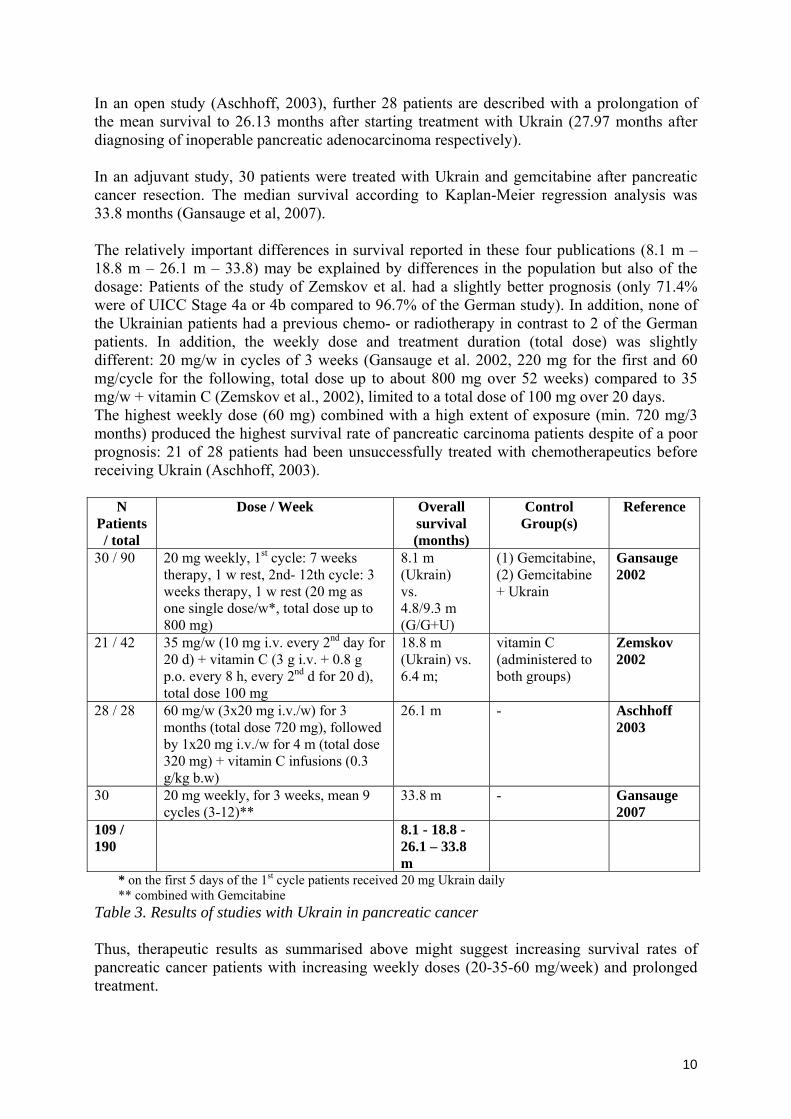
In an open study (Aschhoff, 2003), further 28 patients are described with a prolongation of the mean survival to 26.13 months after starting treatment with Ukrain (27.97 months after diagnosing of inoperable pancreatic adenocarcinoma respectively). In an adjuvant study, 30 patients were treated with Ukrain and gemcitabine after pancreatic cancer resection. The median survival according to Kaplan-Meier regression analysis was 33.8 months (Gansauge et al, 2007). The relatively important differences in survival reported in these four publications (8.1 m – 18.8 m – 26.1 m – 33.8) may be explained by differences in the population but also of the dosage: Patients of the study of Zemskov et al. had a slightly better prognosis (only 71.4% were of UICC Stage 4a or 4b compared to 96.7% of the German study). In addition, none of the Ukrainian patients had a previous chemo- or radiotherapy in contrast to 2 of the German patients. In addition, the weekly dose and treatment duration (total dose) was slightly different: 20 mg/w in cycles of 3 weeks (Gansauge et al. 2002, 220 mg for the first and 60 mg/cycle for the following, total dose up to about 800 mg over 52 weeks) compared to 35 mg/w + vitamin C (Zemskov et al., 2002), limited to a total dose of 100 mg over 20 days. The highest weekly dose (60 mg) combined with a high extent of exposure (min. 720 mg/3 months) produced the highest survival rate of pancreatic carcinoma patients despite of a poor prognosis: 21 of 28 patients had been unsuccessfully treated with chemotherapeutics before receiving Ukrain (Aschhoff, 2003).
N Patients / total
Dose / Week
Overall survival (months)
Control Group(s)
Reference
30 / 90 20 mg weekly, 1st cycle: 7 weeks therapy, 1 w rest, 2nd- 12th cycle: 3 weeks therapy, 1 w rest (20 mg as one single dose/w*, total dose up to 800 mg)
8.1 m (Ukrain) vs. 4.8/9.3 m (G/G+U)
(1) Gemcitabine, (2) Gemcitabine + Ukrain
Gansauge 2002
21 / 42 35 mg/w (10 mg i.v. every 2nd day for 20 d) + vitamin C (3 g i.v. + 0.8 g p.o. every 8 h, every 2nd d for 20 d), total dose 100 mg
18.8 m (Ukrain) vs. 6.4 m;
vitamin C (administered to both groups)
Zemskov 2002
28 / 28 60 mg/w (3x20 mg i.v./w) for 3 months (total dose 720 mg), followed by 1x20 mg i.v./w for 4 m (total dose 320 mg) + vitamin C infusions (0.3 g/kg b.w)
26.1 m - Aschhoff 2003
30 20 mg weekly, for 3 weeks, mean 9 cycles (3-12)**
33.8 m - Gansauge 2007
109 / 190
8.1 - 18.8 - 26.1 – 33.8 m
* on the first 5 days of the 1st cycle patients received 20 mg Ukrain daily ** combined with Gemcitabine
Table 3. Results of studies with Ukrain in pancreatic cancer Thus, therapeutic results as summarised above might suggest increasing survival rates of pancreatic cancer patients with increasing weekly doses (20-35-60 mg/week) and prolonged treatment.
10

4. Justification of the life threatening or debilitating nature of the condition.
In western countries, the treatment of pancreatic cancer is one of the greatest challenges today. The incidence of pancreatic carcinoma has increased during the past five decades and about 10/100 000 people/year die of the pancreatic cancer, making it the fourth commonest cause of cancer related mortality after lung, colorectal, and breast cancer (Eskelinen and Haglund, 1999). Pancreatic cancer is an aggressive lesion. It is a malignancy that causes late symptoms, and diagnosis is therefore late and cure rare. At the time of diagnosis most patients show progression of the disease beyond the pancreas, either through the direct invasion of neighbouring structures or metastases in regional lymph nodes, liver, peritoneum, lungs, bones, or brain. Therefore, up to 90% of patients present with incurable, advanced disease (Dowsett and Russell, 1995). Median survival time is approximately 4-6 months after diagnosis. Fewer than 10% of patients survive 1 year after diagnosis, and many suffer from increasingly severe pain, nausea and vomiting, anorexia, weight loss, and weakness as the disease progresses. The overall European mean 1 year relative survival rate is 15% for pancreatic cancer. (Faivre et al, 1998). The 5-year survival for pancreatic cancer is usually less than 5% and has not changed during the past 30 years (Crino et al, 2001; Philip et al, 2001; Faivre et al, 1998). For most patients diagnosed as having cancer of the exocrine pancreas, life expectancy is measured in months. Three factors underlie this poor outlook. First, pancreatic cancer disseminates to distant sites early in its history. Second, as the disease progresses it is associated with substantial morbidity, characterised by cachexia and asthaenia. Third, pancreatic cancer is resistant to most forms of treatment studied to date (Li et al, 2004). Causes of death in pancreatic cancer patients In about 30% of patients sepsis is the direct cause of death. This was the major cause of death in a retrospective study in 108 patients. Pneumonia, cholangitis and peritonitis, each leading to the formation of local abscesses were the source of the sepsis. Pulmonary embolism was found in 1/3 of the patients, 14% of patients with cancer of the pancreas died of pulmonary embolism. Cachexia and inanition were the cause of death is only 5-6% of patients. Liver failure (75%) due to metastatic destruction of the liver was the most common cause of death due to tumour growth or metastases. A considerable number of patients died as a consequence of metastases in other organs such as e.g. lung, pleura, pericard, perineum, brain and other localisations. Only 5% of all patients died of secondary non-tumour-related diseases (mostly cardiovascular disease) (Adler and Gress, 1996).
11
B) PREVALENCE OF THE CONDITION. 1. Prevalence of the orphan disease or condition in the European Union.
The incidence of pancreatic carcinoma has increased in Northern Europe and North America during recent decades and contrary to for example, lung, gastric and oesophageal carcinoma, its incidence is still increasing. Annual incidence is about 8-10/100,000 of the population. (Eskelinen et al, 1999) Pancreatic ductal adenocarcinoma (PDAC) currently has an incidence of approximately 8 to 10 cases per 100,000 citizens in European countries, and incidence has been increasing throughout the last decades. Approximately 30,000 patients die every year from PDAC in Western Europe and most of the newly diagnosed patients are at an already unresectable tumour stage (Kleeff et al, 2000). There are different trends in the epidemiology of pancreatic cancer within the EU. The incidence of pancreatic cancer has fallen during the last ten years in Sweden (Ihse et al, 2002) but increased in Spain (Fernandez et al, 2000; Ruiz Liso et al, 1993) where pancreas cancer trends increased for both sexes. In a region of France, incidence of the carcinoma remained stable during the observation period and no change was noticed with regard to housing conditions. (Pienkowski et al, 1992). This corresponds to other data. A study analysed data from a large health screening survey in Norway. The study included 31,000 men and 32,374 women initially free from any diagnosed cancer, and during 12 years of follow-up, 166 cases of pancreatic cancer were diagnosed at the Cancer Registry (Nilsen and Vatten, 2000). Pancreatic cancer mortality has appreciably increased for both sexes in Italy over the last few decades, although Italian rates are still relatively low on a European scale (7.0/100,000 men, 4.1/100,000 women, world standard). These rises are probably due, at least in part, to improved diagnosis and certification of the disease, and are related to increased exposure to tobacco smoking - the best recognised risk factor for the disease in subsequent generations of Italian men and women (La Vechia, 1996). A study was designed to assess time trends of the incidence of pancreatic cancer 1961-90 in Malmo, Sweden. The city of Malmo (population 230,000), situated in the south of Sweden, is in an area which has the highest incidence of pancreatic cancer in the country. 1,314 cases, 651 men and 663 women, were found in the Regional Tumour Register and the National Cause-of-Death Register. In 75% of cases diagnosis was based on autopsy. Twenty per cent of these cases were first found at autopsy, being undiagnosed. The average age-standardised incidence was 20.4 per 10 person-years for men and 13.7 for women. The incidence was higher for men than for women in all age groups above 44 years. No change in incidence over time was observed for men. In older and middle-aged women there was however a statistically significant increase. The average relative change in women above age 64 was 1.7% per year after age adjustment and in women aged 55-64 years 2.6% per year. No results have been found indicating that this increasing incidence could be caused by detection bias as a result of changing autopsy rates during the study period and hence conclude that the observed increase is explained by a growing number of women being exposed to factors with a potential tumour-promoting or initiating effect (Hedberg et al, 1996).
12

During the period between 1973 and 1992, 1,032 patients were diagnosed with pancreatic carcinoma at different medical institutions in the Asturias region of Spain. The incidence increased from 1.28 to 6.42 cases/100,000. The proportion male/female was 1.5/1. Mean age of the patients was 67.5 +/- 11.35 and the median age was 65 years. The age of women was higher than that of men: 70.2 +/- 11.81 (p < 0.01). This data is in agreement with general pancreatic carcinoma incidence in Spain (Gonzalez Martinez et al, 1995). Country Population* Incident cases 1-year
prevalence cases 5-year
prevalence caces Incidence
Austria 8,206,500 1,239 236 678 1.51 Belgium 10,445,900 965 257 666 0.92 Bulgaria 7,761,000 821 160 370 1.06 Cyprus 749,200 70 19** 44** 0.93 Czech Republic 10,220,600 1,534 303 728 1.50 Denmark 5,411,400 722 117 221 1.33 Estonia 1,347,500 184 37 97 1.37 Finland 5,236,600 691 163 292 1.32 France 62,518,600 5,321 1,761 3,605 0.85 Germany 82,500,800 10,334 2,738 5,933 1.25 Greece 11,082,800 1,211 309 657 1.09 Hungary 10,097,500 1,597 308 707 1.58 Iceland 293,600 19 4 9 0.65 Ireland 4,109,200 332 69 138 0.81 Italy 58,462,400 8,602 2,341 4,814 1.47 Latvia 2,306,400 347 74 152 1.50 Liechtenstein 34,600 4 1** 2** 1.16 Lithuania 3,425,300 391 82 168 1.14 Luxembourg 455,000 45 12 26 0.99 Malta 402,700 44 13 33 1.09 Netherlands 16,305,500 1,491 387 759 0.91 Norway 4,606,400 557 123 222 1.21 Poland 38,173,800 4,357 826 1,730 1.14 Portugal 10,529,300 874 221 471 0.83 Romania 21,658,500 2,049 398 921 0.95 Slovenia 1,997,600 246 49 90 1.23 Slovakia 5,384,800 615 117 288 1.14 Spain 43,038,000 3,879 843 2,016 0.90 Sweden 9,011,400 910 199 367 1.01 United Kingdom 60,059,900 7,225 1,400 3,072 1.20 Total, EU27+3*** 495,832,800 56,676 13,567 29,276 1.14 *As of January 1, 2005. Source: Lanzieri, 2006. **Own estimation. *** 27 member countries of European Union + Iceland, Liechtenstein, and Norway Table 4: Population, incidence and prevalence of pancreatic carcinoma in the European Union, Iceland, Liechtenstein, and Norway. Sources: Eurostat, EUCAN version 5.0, created 17-03-2003, and Globocan 2002, IARCPress, Lyon, 2004. The trends in treatment and outcome of 13,560 patients with pancreatic cancer, and in incidence of the disease, in the West Midlands health region (Great Britain) between 1957 and 1986 were determined using data from the West Midlands Region Cancer Registry. Patients were divided into those diagnosed in the first 20 years (1957-1976, n = 7,888) and the most recent 10 years (1977-1986, n = 5,672). The disease was more common in men and incidence increased up to 1970 after which it levelled off. (Bramhall et al, 1995).
13

Brown et al (1998) did not find any association in pancreatic cancer incidence with socio-economic status. According to EUCAN database published by European Network of Cancer Registries (ENCR), and ‘Globocan 2002: Cancer Incidence, Mortality and Prevalence Worldwide’, the incidence of pancreatic carcinoma in the European Union, Iceland, Liechtenstein and Norway (EU25+3) was estimated as 56,676 cases, 1-year prevalence as 13,567 cases, and 5-year prevalence as 29,276 cases. With an estimated population in the EU27+3 of about 495.8 million (as of 1 January 2005, Lanzieri, Eurostat, 2006), the total prevalence of pancreas cancer is estimated to be about 1.1 in 10,000. Summarising presented statistical data we estimate that
• the prevalence of pancreatic carcinoma in the European Community is about 1.1 in 10,000 and
• there are about 55,000-60,000 pancreas cancer patients in EU27+3. We conclude that pancreatic carcinoma corresponds in all criteria to the definition of an orphan disease according to Article 3(1)(a) paragraph 1 of the Regulation (EC) No141/2000 of 16 December 1999.
14

2. Prevalence and incidence of the condition in the Community.
See B.1.
15

3. Information on participation in other EU projects. European Community funded projects regarding pancreatic cancer (all data from www.cordis.lu/en/home.html): 1. A CORDIS RTD-PROJECT Record Control Number : 19844 Database on transcribed sequences in tumour cells and identification on transcription pattern changes related to transformation and other tumour cell properties for global fingerprinting analysis of human pancreatic cancer. Programme Type: 3rd FWP (Third Framework Programme) Programme Acronym: BIOMED 1 Project reference: BMH10401 2. A CORDIS RTD-PROJECT Record Control Number : 41431 Synthesis of the manumycin family of antibiotics and novel ras farnesyl transferase inhibitors for cancer chemotherapy. Programme Type: 4th FWP (Fourth Framework Programme) Programme Acronym: TMR Project reference: FMBI961177 Completed on 1998-09-30 3. A CORDIS RTD-PROJECT Record Control Number : 42489 99mTc labelling and preliminary evaluation of rc160. Programme Type: 4th FWP (Fourth Framework Programme) Programme Acronym: TMR Project reference: FMBI971966 Completed on 1998-11-02 3. A CORDIS RTD-PROJECT Record Control Number : 46509 Identification, structural and functional characterisation of disease genes in pancreatic cancer. Programme Type: 4th FWP (Fourth Framework Programme) Programme Acronym: INCO Project reference: IC20980202 Completed on 2001-04-30 3. A CORDIS RTD-PROJECT Record Control Number : 42627 Identification, structural and functional characterisation of disease genes in pancreatic cancer. Programme Type: 4th FWP (Fourth Framework Programme) Programme Acronym: BIOMED 2 Project reference: BMH4983085 Completed on 2001-11-30 4. A CORDIS RTD-PROJECT Record Control Number : 47635
16

Development of novel peptide based radiopharmaceuticals for in vivo receptor associated tumour diagnosis and therapy. Programme Type: 4th FWP (Fourth Framework Programme) Programme Acronym: BIOMED 2 Project reference: BMH4983198 Completed on 2001-03-31 5. A CORDIS RTD-PROJECT Record Control Number : 63720 Pancreatic cancer network: from candidate genes to medical applications. Programme Type: 5th FWP (Fifth Framework Programme) Programme Acronym: BIOMED 2 Project reference: LIFE QUALITY Start date: 2001-11-30 End date: 2005-08-31 Following European Community projects on pancreatic cancer are developing(all data from www.cordis.lu/en/home.html): 1. Record Control Number : 13324 Farnesylecysteine mimetics in cancer treatment. Stage of development: tested, available for demonstration. Update date: 1997-04-23 2. Record Control Number : 22832 Early detection of pancreatitis and pancreatic cancer by fluorescence. Stage of development: not specified. Update date: 1997-07-15 3. Record Control Number : 27615 Inhibitors of the tissue-type plasminogen activator (t-PA) with anti-tumour activity. Stage of development: prototype/demonstrator available for testing. Update date: 2002-03-15
17
C) POTENTIAL FOR RETURN ON INVESTMENT
1. Grants and tax incentives. Not applicable
2. Past and future development costs. Not applicable
3. Expected revenues. Not applicable
4. Certification by registered accountant. Not applicable
18
D) OTHER METHODS FOR DIAGNOSIS, PREVENTION OR TREATMENT OF THE CONDITION
1. Details of any existing diagnosis, prevention or treatment methods.
Pancreatic cancer remains one of the most difficult cancers to treat at the present time. In the few cases in which early diagnosis is made, surgical pancreatico-duodenectomy may be attempted by those with skill and experience in performing this challenging operation. Currently resection rates of up to 14% (Wade et al, 1996) and operative mortality rates of less than 5% to 10% are being achieved. Some studies showed better results in patients treated with postoperative radiotherapy (Dobelbower et al, 1997), but the presence of critical radiosensitive organs such as the liver, kidneys and small intestine limits the dose that can be delivered to this site (Morganti et al, 2002). The methods which enable the intensification of radiation treatment such as intraoperative radiation therapy and concomitant chemoradiation (Yeo et al, 1997) can improve treatment results of resectable carcinomas. The standard systemic treatment for advanced pancreatic cancer was 5-fluorouracil (Morrell et al, 1991). The drug acts as a pyrimidine antimetabolite (Peters et al, 1996). The addition of modulators to 5-FU such as folinic acid, hydroxyurea, or interferon-alpha did not produce substantial improvements in response rates and led to significant toxicity even in highly selected patients with an ambulatory performance status (Wadler et al, 1999; David et al, 2000). One of the better alternatives to 5-FU is gemcitabine, a deoxycytidine analog that became the standard first-line therapy for patients with advanced pancreatic carcinoma (Burris et al, 1997). The drug acts by intracellular activation into phosphorylated metabolites: one of them competes with endogens deoxycitidine triphosphate for incorporation into DNA, the other one inhibits ribonucleotide reductase. Three biochemical mechanisms underlie the so-called self potentiation process of gemcitabine activity: inhibition of ribonucleotide reductase, stimulation of deoxycitidine kinase and inhibition of deoxycitidine monophosphate deaminase (Peters et al, 1996). Gemcitabine monotherapy resulted in a median survival of 5.6 (Berlin et al, 2002), 7.3 (Crino et al, 2001) and 8.8 (Ulrich-Pur et al, 2000) months. Gemcitabine represents an attractive candidate for combination chemotherapy because of its excellent side-effect profile and the absence of overlapping toxicities with other chemotherapeutic agents; due to its chain termination masking activity the drug directly inhibits DNA repair which could represent a molecular basis for synergistic activity with other DNA-damaging chemotherapeutic agents (Neri et al, 2001). There are multiple clinical trials in patients with advanced pancreatic carcinoma that describe the administration of gemcitabine with other cytotoxic agents. Combined with 5-fluorouracil, median survival of 6.7 (Berlin et al, 2002), 4.4 (Berlin et al, 2000), 7.0 (Cascinu et al, 1999), and 10.3 months (Hidalgo et al, 1999) has been achieved. Combination of gemcitabine and epirubicin (inhibitor of topoisomerase II-mediated DNA releasing) led to 7.8 months median survival in the study by Scheithauer et al (1999) and to 10.9 median survival in the trial by Neri et al (2001). Gemcitabine plus cisplatin (the heavy metal alkylating-like agent) achieved 8.3 months median survival (Heinemann et al, 2000), 6.7 months (30 weeks) median survival in the study by Colucci et al (2002), 7.4 months – by Philip et al (1999). Gemcitabine plus docetaxel (enhances microtubule assembly and inhibits the depolymerisation of tubulin) led to 5.8 months (26 weeks) median survival (Stathopoulos et al, 2001) and to 8.9 months in the study by David (David et al, 2001).
19
Source (study) Total Therapy Response, % Median survival, months
1-year survival, %
Rothenberg et al, 2002 116 5-fluorouracil, eniluracil 8 and 21 3.6 and 3.42 Oman et al, 2001 30 5-fluorouracil, intraperitoneal,
leucovorin 7 (0-21)
Rougier et al, 2000 40 Docetaxel 15 - - Androulakis et al, 1999 33 Docetaxel 6 8 (36 weeks) 36.4 Okada et al, 1999 21 Docetaxel 0 4.0 - Ryan et al, 2002 333 Docetaxel, gemcitabine 6 8.9 29 Halford et al, 2001 164 Doxorubicin (Doxil, liposomal
doxorubicin) 0 3.2 10
Falconi et al, 2001 135 FLEC5 6.3 9.6 Crino et al, 2001 33 Gemcitabine 12 7.3 Ulrich-Pur et al, 2000 43 Gemcitabine 21 8.8 26.3 Philip et al, 2001 42 Gemcitabine , cisplatin 26 7.1 19 Berlin et al, 2000 36 Gemcitabine, 5-fluorouracil 14 4.4 8.6 Cascinu et al, 1999 54 Gemcitabine, 5-fluorouracil 4 7.0 - Hidalgo et al, 1999 26 Gemcitabine, 5-fluorouracil 19 10.3 39.5 Philip et al, 1999 26 Gemcitabine, cisplatin 36 7.4 - Heinemann et al, 2000 41 Gemcitabine, cisplatin 11.5 8.3 28.0 Stathopoulos et al, 2001 54 Gemcitabine, docetaxel 13 5.8 (26 weeks) 22.0 Scheithauer et al, 1999 66 Gemcitabine, epirubicin 21 7.8 21.2 Whitehead et al, 1997 39 Paclitaxel 8 5.0 - Okusaka et al, 2001 416 Radiation, cisplatin, 5-fluorouracil 7.7 36 Yavuz et al, 2001 10 Radiation, gemcitabine amifostine 40 Tsuruta et al, 2001 35 Radiotherapy 29 Boz et al, 2001 42 Radiotherapy, 5-fluorouracil 23 9.1 Tsuruta et al, 2001 10 Radiotherapy, 5-fluorouracil 50 Brunner et al, 2000 297 Radiotherapy, 5-fluorouracil,
mitomycin 9
Rocha Lima et al, 2003 360 Gemcitabine, irinotecan 6.6 22
Louvet et al, 2004 313 Gemcitabine, oxiplatin 9.0 14.9 Lee J et al, 2004 22 Gemcitabine, uracil-tegafur 5.8 Wilkowski et al, 2004 30 Surgery plus radiotherapy,
gemcitabine, cisplatin 10.6
Sangro et al, 2004 7 Adenovirus encoding interleukin-12 Not indicated Not indicated Rich et al, 2004 109 Radiotherapy, paclitaxel 11.2 43 Shepard et al, 2004 33 Gemcitabine, docetaxel 4.7 Van Cutsem et al, 2004 688 Gemcitabine, tipifarnib 193 days 27 Milella et al, 2004 17 Celecoxib, 5-fluoruracil 15 weeks
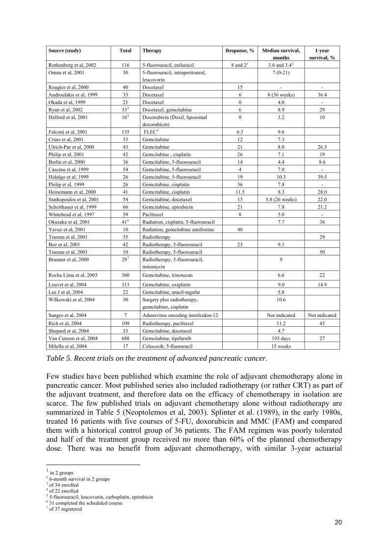
Table 5. Recent trials on the treatment of advanced pancreatic cancer. Few studies have been published which examine the role of adjuvant chemotherapy alone in pancreatic cancer. Most published series also included radiotherapy (or rather CRT) as part of the adjuvant treatment, and therefore data on the efficacy of chemotherapy in isolation are scarce. The few published trials on adjuvant chemotherapy alone without radiotherapy are summarized in Table 5 (Neoptolemos et al, 2003). Splinter et al. (1989), in the early 1980s, treated 16 patients with five courses of 5-FU, doxorubicin and MMC (FAM) and compared them with a historical control group of 36 patients. The FAM regimen was poorly tolerated and half of the treatment group received no more than 60% of the planned chemotherapy dose. There was no benefit from adjuvant chemotherapy, with similar 3-year actuarial
1 in 2 groups 2 6-month survival in 2 groups 3 of 34 enrolled 4 of 22 enrolled 5 5-fluorouracil, leucovorin, carboplatin, epirubicin 6 31 completed the scheduled course 7 of 37 registered
20

survival rates of 24% and 28% for the treatment and control groups, respectively. The first prospective, randomised controlled trial was by Bakkevold et al. from Norway (1993). Forty-seven patients with resected PDAC (plus 14 with ampullary tumours) were randomly assigned to receive either chemotherapy with moderate-dose FAM, or observation alone. No long-term survival benefit with chemotherapy was shown, with similar 5-year survival rates of 4% and 8% in the FAM and control groups, but there was an improvement in median survival (23 months for chemotherapy versus 11 months for controls), with a delay in time to disease recurrence. The multi-agent chemotherapy regimen was rather toxic, with one reported death directly attributed to chemotherapy, four cases of septicaemia and 16 patients hospitalised after the first course of chemotherapy. The inclusion of ampullary carcinomas in the study makes it difficult to draw firm conclusions regarding the benefits of chemotherapy for PDAC, as the two types of cancer were not differentiated in the survival analysis.
Actuarial survival (%) Series Period Number of cases Regimen Median survival, months
1 year 3 years 5 years
1972-1984 36 - 28 Splinter et al. 1980-1984 16 FAM 24
31 (24 PDAC)2 - 11 45 30 8 Bakkevold et al.1
1984-1987 30 (23 PDAC)2 FAM 23 70 27 4 527 - 12.4 Baumel et al. 1982-1988 43 Unspecified 11.5 235 - 14 Neoptolemos
et al.11994-2000
238 5-FU/FA 19.7 1 – randomized controlled trial; 2 – the remainder had other pancreatic cancers; FAM, 5-fluoruracil (5-FU), doxorubicin and mitomycin C; FA – folinic acid; - no treatment; empty cells – data not available. Table 6. Adjuvant systemic chemotherapy for pancreatic ductal adenocarcinoma (adapted from Neoptolemos et al, 2003). Recently, the European Study Group for Pancreatic Cancer randomly assigned 289 patients who had undergone complete macroscopic resection of histologically proven pancreatic ductal adenocarcinoma to receive postoperative chemoradiotherapy alone, chemotherapy alone, combination chemoradiotherapy and chemotherapy, or neither treatment (observation). This study was the largest randomized trial of adjuvant therapy for pancreatic cancer reported to date. Clinical features and characteristics of the tumors were similar among groups. After a median follow-up of 47 months, 237 patients (82%) had died. Median survival times were 17 months in the observation group, 14 months in the chemoradiotherapy alone group, 22 months in the chemotherapy alone group, and 20 months in the combination group. Median survival among all patients who received chemoradiotherapy was 16 months, compared with 18 months among all patients who did not receive chemoradiotherapy (p=0.05). Estimated 2- and 5-year survival rates were 29% and 10%, respectively, among patients who received chemoradiotherapy versus 41% and 20%, respectively, among those who did not receive chemoradiotherapy. Median survival was 20 months for all patients who received chemotherapy versus 15.5 months for those who did not receive chemotherapy (p=0.009). Estimated 2- and 5-year survival rates were 40% and 21%, respectively, among patients who received chemotherapy, and 30% and 8%, respectively, among those who did not receive chemotherapy. The adjusted hazard ratio for death was 1.47 with the use of chemoradiotherapy and 0.77 with the use of chemotherapy. The median time to tumor recurrence was 10.7 months among patients who received chemoradiotherapy and 15.2 months for those who did not receive chemoradiotherapy (p=0.04). Time to recurrence in patients who received chemotherapy was significantly longer than that in patients who did not receive chemotherapy (15.3 months vs.
21

9.4 months). Observed quality of life did not differ significantly between patients who received chemotherapy and those who did not, or between patients who received chemoradiotherapy and those who did not. Combinations of the other drugs protocols and chemotherapy with radiotherapy were also explored. Öman et al (2001) used intraperitoneal 5-fluorouracil and leucovorin, achieving 7 months median survival. Preoperative radiotherapy plus 5-fluorouracil plus mitomycin C (Brunner et al, 2000) resulted in 9 months medial survival. Radiation plus cisplatin plus 5-fluoruracil (1 week later) in a study (Okusaka et al, 2001) led to 7.7 months median survival. Radiotherapy with 5-fluorouracil (Boz et al, 2001) achieved 9.1 months median survival. Monotherapy with docetaxel led to 4.0 months median survival (Okada et al, 1999) and to 5 months in the study by Whitehead et al (1997). Doxorubicin (doxil, liposomal doxorubicin) led to 3.2 months median survival (Halford et al, 2001). In all these clinical studies numerous side effects (blood toxicity, diarrhoea, phlebitis, neurological disorders etc.) were observed in 70-90% of treated patients. Therefore, currently the optimal treatment of patients with pancreatic carcinoma can be considered as one of the most topical unresolved issues in oncology. Poor results have been achieved in numerous studies with monochemothepary (docetaxel, gemcitabine, paclitaxel) and polychemotherapy (using of 2 or 3 anticancer drugs: gemcitabine with docetaxel, 5-fluorouracil, epirubicin, cisplatin), combined radiochemotherapy (radiotherapy plus gemcitabine and amifostine, or radiotherapy with cisplatin and 5-fluorouracil) or radiotherapy alone. For this reason, the search for new drugs and drug combinations is of great importance in the treatment of pancreatic cancer. Currently, the administration of two and more anticancer drugs with different mechanisms of action is the most promising approach that can decrease the number of side effects and improve the clinical outcome. Followed medications are already designated for the treatment of pancreatic cancer in the EU (http://www.emea.europa.eu/htms/human/comp/a-zcompsumop.htm , last accessed on 26.01.2007):
• 4-imino-1, 3-diazobicyclo-[3.1.0]-hexan-2-one (EU designation: EU/3/05/299, designated orphan indication: treatment of pancreatic cancer, designation date 27/07/2005);
• 5,10-methylene-tetrahydrofolic acid (EU designation: EU/3/04/221/, designated orphan indication: treatment of pancreatic cancer in combination with 5-fluorouracil, designation date 2/09/2004);
• 5-10-Methylene-tetrahydrofolate (EU designation: EU/3/03/143/, designated orphan indication: treatment of pancreatic cancer in combination with 5-fluorouracil, designation date 11/06/2003);
• 26 base single stranded phosphodiester DNA oligonucleotide (EU designation: EU/3/06/352, designated orphan indication: treatment of pancreatic cancer, designation date 16/02/2006)
• bovine bile extract (EU designation: EU/3/05/287, designated orphan indication: treatment of pancreatic cancer, designation date 20/06/2005)
• cytochrome P450 isoform 2B1 gene transfected human embryonic kidney 293 cells encapsulated in polymeric cellulose sulphate (EU designation: EU/3/03/149/, designated orphan indication: treatment of pancreatic cancer in combination with ifosfamide, designation date 30/06/2003);
• deuterium oxide (EU designation: EU/3/04/239/, designated orphan indication: treatment of pancreatic cancer, designation date 20/10/2004);
• G17(9) gastrin-diphtheria toxoid conjugate (EU designation: EU/3/02/129/, designated orphan indication: treatment of pancreatic cancer, designation date 24/01/2003);
22

• iodine (131I) anti-CEA sheep-human chimeric monoclonal antibody (EU designation: EU/3/03/142/, designated orphan indication: treatment of pancreatic cancer, designation date 7/05/2003);
• rubitecan (EU designation: EU/3/03/145/, designated orphan indication: treatment of pancreatic cancer, designation date 10/06/2003).
Low toxicity (or its absence) and a wide range of influences on the organism of the patient, high affinity of the drug to cancer cells and immune modulating effects are very important properties that forecast the possibility of good clinical results.
23
2. Justification as to why the methods are not considered satisfactory.
Not applicable. According to the Annex to Guideline on format and content of applications the explanation of the section D(2) is made in section D(3).
24

3. Justification of significant benefit
Although operative mortality rates have much improved, surgery has only a slight effect on survival time. Median survival times following surgery are poor: 10 to 18 months with long-term survival rates of 10% to 24% (Edge et al, 1993; Yeo et al, 1995; Conlon et al, 1996). Most patients are never cured despite optimal surgical intervention (Ghaneh et al, 1999). Regional or extended radical surgery has been advocated as a means of increasing the rate of disease-free margins and hence patient survival. Long-term survival has not proved to be statistically different from that of conventional resection in retrospective series (Gudjonsson, 1987; Sperti et al, 1996). The most significant factors in predicting patient outcome are tumour grade, stage, and resection margin status. The survival of patients with negative resection margins is not as high as might be expected. A major reason for this is the pattern of recurrence in pancreatic cancer following potentially curative resection and the activity of the tumour. Most tumour recurrences are local, in the peritoneum and liver; local relapses are the most frequent cause of death. Adjuvant chemoradiation therapy has shown prolonged survival time in some trials but not in others. Gemcitabine was found to have a positive influence on the quality of life in pancreatic patients; however, median survival times were only marginally prolonged by several weeks (Burris et al, 1997). In another randomised trial, median survival for gemcitabine patients was only 5.7 months compared with 4.4 months for 5-FU patients (Moore et al, 1997). There are several new descriptions of the successful use of Ukrain in patients with pancreatic cancer. Aschhoff (2003) reported on palliative therapy with Ukrain (total first three-month dose 720 mg, and then next four-month dose 320 mg) of 28 patients with unresectable pancreatic adenocarcinoma (17 male, 11 female, aged 48-74 years, mean age 49.6 years). All the patients were presented with advanced and/or metastatic disease that made curable surgery impossible. Twenty-one patients had previously been treated with conventional chemotherapy modalities, however, this therapy had failed and disease progressed. Of the 28 patients treated with Ukrain, partial remission was achieved in 24 cases (85.7%) while four patients did not respond to treatment. The mean survival of the patients treated with Ukrain was 26.1 months after the start of Ukrain administration and 28.0 months after the diagnosis of inoperable pancreatic adenocarcinoma. In the report on the treatment with Ukrain and gemcitabine of four advanced pancreatic cancer cases (Gansauge, 2003) the author noted partial remission and the reduced toxicity profile of the drug. The findings were especially surprising because all the patients had exhausted traditional methods of therapy before Ukrain therapy, and surgical treatment was also impossible. Kleef published a case report on a 60 year-old male patient, who was diagnosed with pancreatic head cancer with liver metastases. After the surgical correction of biliary obstruction conventional chemotherapy was performed (five cycles of gemcitabine at a dose of 1000 mg/m2 and one cycle 1800 mg/m2; Camptothecin (Irinotecan) 160 mg/m2 and Tomudex, 3 mg/m2, 3 cycles every three weeks were administrated; Xeloda 1.5 g in the morning, 2 g in the evening for two weeks, one course). Due to massive toxicity and disease progression chemotherapy was discontinued. Therapy with Ukrain (20 mg intravenous in 500 ml normal saline solution), vitamin C (10 g) and local hyperthermy (radiofrequency 13.56 MHz, 100 W) was begun. CT performed after two months showed complete response of liver metastasis and stable status of local recurrence. Complete regression of ascites was found,
25
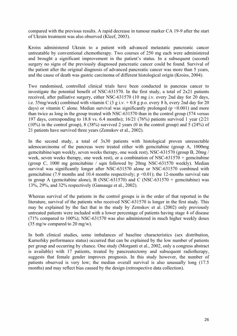
compared with the previous results. A rapid decrease in tumour marker CA 19-9 after the start of Ukrain treatment was also observed (Kleef, 2003). Kroiss administered Ukrain to a patient with advanced metastatic pancreatic cancer untreatable by conventional chemotherapy. Two courses of 250 mg each were administered and brought a significant improvement in the patient’s status. In a subsequent (second) surgery no signs of the previously diagnosed pancreatic cancer could be found. Survival of the patient after the original diagnosis of advanced pancreatic cancer was more than 5 years, and the cause of death was gastric carcinoma of different histological origin (Kroiss, 2004). Two randomised, controlled clinical trials have been conducted in pancreas cancer to investigate the potential benefit of NSC-631570. In the first study, a total of 2x21 patients received, after palliative surgery, either NSC-631570 (10 mg i.v. every 2nd day for 20 days, i.e. 35mg/week) combined with vitamin C (3 g i.v. + 0.8 g p.o. every 8 h, every 2nd day for 20 days) or vitamin C alone. Median survival was significantly prolonged (p <0.001) and more than twice as long in the group treated with NSC-631570 than in the control group (574 versus 197 days, corresponding to 18.8 vs. 6.4 months); 16/21 (76%) patients survived 1 year (2/21 (10%) in the control group), 8 (38%) survived 2 years (0 in the control group) and 5 (24%) of 21 patients have survived three years (Zemskov et al., 2002). In the second study, a total of 3x30 patients with histological proven unresectable adenocarcinoma of the pancreas were treated either with gemcitabine (group A, 1000mg gemcitabine/sqm weekly, seven weeks therapy, one week rest), NSC-631570 (group B, 20mg / week, seven weeks therapy, one week rest), or a combination of NSC-631570 + gemcitabine (group C, 1000 mg gemcitabine / sqm followed by 20mg NSC-631570 weekly). Median survival was significantly longer after NSC-631570 alone or NSC-631570 combined with gemcitabine (7.9 months and 10.4 months respectively; p <0.01); the 12-months survival rate in group A (gemcitabine alone), B (NSC-631570) and C (NSC-631570 + gemcitabine) was 13%, 29%, and 32% respectively (Gansauge et al., 2002). Whereas survival of the patients in the control groups is in the order of that reported in the literature, survival of the patients who received NSC-631570 is longer in the first study. This may be explained by the fact that in the study by Zemskov et al. (2002) only previously untreated patients were included with a lower percentage of patients having stage 4 of disease (71% compared to 100%); NSC-631570 was also administered in much higher weekly doses (35 mg/w compared to 20 mg/w). In both clinical studies, some imbalances of baseline characteristics (sex distribution, Karnofsky performance status) occurred that can be explained by the low number of patients per group and occurring by chance. One study (Morganti et al., 2002, only a congress abstract is available) with 17 patients, treated by pancreatectomy and subsequent radiotherapy, suggests that female gender improves prognosis. In this study however, the number of patients observed is very low; the median overall survival is also unusually long (17.5 months) and may reflect bias caused by the design (retrospective data collection).
26

NSC-631570
(35 mg/w) + Vitamin C
Vitamin C Gemcitabine NSC-631570 (20 mg/w)
NSC-631570 (20 mg/w) + Gemcitabine
21 Patients 21 Patients 30 Patients 30 Patients 30 Patients Survival 1 year, N patients (%)
16 (76%) 2 (10%) 4 (13%) 9 (29%) 10 (32%)
2 years 8 (38%) 0 Median Survival (months) 18.8 6.4 5.2 7.9 10.4Tumour response complete 0/21 (-%) 0/21 (-%) 1/15 (7%)
0/15 (-%) 1/20 (5%)
partial 3/21 (14%) 0/21 (-%) 5/15 (33%) 4/15 (27%) 7/20 (35%)stable 9/21 (43%) 3/21 (14%) 5/15 (33%) 5/15 (33%) 9/20 (45%)
UICC Stage 2 1 (5%) 2 (10%) -
- -
Stage 3 5 (24%) 5 (24%) 1 0 1Stage 4a 8 (38%) 9 (43%) 12 (40%) 13 (43%) 7 (23%)Stage 4b 7 (33%) 5 (24%) 17 (57%) 17 (57%) 22 (73%)
Karnofsky status 80-90 2 (10%) 7 (33%) Not reported Not reported Not reported50-70 17 (82%) 14 (67%) Not reported Not reported Not reported
Sex Male 17 10 22
16 19
Female 4 11 8 14 11Mean age (years) 60.7 65.4 63.8 60.6 58.2Therapies prior randomisation
none none 2 5 5
Chemotherapy - - 1 1 3Radiochemotherapy - - 1 4 2
Table 7. Baseline characteristics and efficacy (controlled clinical trials with NSC-631570)
NSC-631570 (35 mg/w)
+ Vitamin C
Vitamin C Gemcitabine NSC-631570 (20 mg/w)
NSC-631570 (20 mg/w) + Gemcitabine
21 Patients 21 Patients 30 Patients 30 Patients 30 Patients Toxicity Neutropenia <500/mcl, WHO 4 (6.9%) (- %) ⎫ (all grade 3,
no grade 4 tox.) 12%
⎫ (all grade 3, no grade 4 tox.) 11%
⎫ (all grade 3, no grade 4 tox.) 10%
Neutropenia, WHO 3 (19%) (- %) I I I Thrombocytopenia, WHO 4 (-%) (- %) I I I Thrombocytopenia, WHO 3 (9.7%) (- %) I I I
Hemoglobin, WHO 4 (3.2%) (- %) I I I Hemoglobin, WHO 3 (6.5%) (- %) ⎭ ⎭ ⎭
AST WHO 4 (1.6%) (- %) Not reported Not reported Not reportedAST WHO 3 (9.8%) (- %) Not reported Not reported Not reportedALT WHO 4 (1.6%) (- %) Not reported Not reported Not reportedALT WHO 3 (8.2%) (- %) Not reported Not reported Not reported
Nausea/Vomiting, WHO 4 (3.2%) (- %) (- %) (- %) (- %)Nausea/Vomiting, WHO 3 (9.5%) (- %) (11%) (3%) (3%)
Diarrhoea, WHO 4 (- %) (- %) (- %) (- %) (- %)Diarrhoea, WHO 3 (1.6%) (- %) (- %) (1%) (- %)
Fever, WHO 4 (- %) (- %) (- %) (- %) (- %)Fever, WHO 3 (- %) (- %) (- %) (- %) (- %)
Table 8. Toxicity (controlled clinical trials with NSC-631570)
27

Gemcitabine
1000 mg/m2/w Gemcitabine
1000 mg/m2/w Gemcitabine
1000 mg/m2/w+ 5-FU 600 mg/m2/w
+ Leucovorin 20 mg/m2/w
63 Patients 30 Patients 29 Patients Burris et al., 1997 Gansauge et al., 2002 Marantz et al., 2001
No control groupSurvival 1 year, N patients (%) 4 (18%) 4 (13%) 4 (36%) 2 years Median survival (months) 5,.65 5.2 8.4Tumour response
complete - 1/15 (7%) 1/29 (3%)partial 3/56 (5.4%) 5/15 (33%) 5/29 (17%)stable 22/56 (39%) 5/15 (33%) 16/29 (55%)
UICC Stage 2
9 (14%) - -
Stage 3 9 (14%) 1 15 Stage 4a ⎫ 45 (72%) 12 (40%) ⎫ 14 (48%) Stage 4b ⎭ 17 (57%) ⎭ Karnofsky status Not reported
80-90 44 (70%)50-70 19 (30%)
Sex Male 34 (54%) 22 Not reported
Female 29 (46%) 8 Not reportedMean age (years) 62 63.8 Not reportedTherapies prior randomisation 2
Chemotherapy 1 noneRadiochemotherapy 1 Not reported
Toxicity Neutropenia <500/mcl, WHO 4 (6.9%) 0Neutropenia <500/mcl, WHO 3 (19%) 3.4%
Thrombocytopenia , WHO 4 (-%) 0Thrombocytopenia, WHO 3 (9.7%) 0
Hemoglobin, WHO 4 (3.2%) 0Hemoglobin, WHO 3 (6.5%) 6.9%
Aspartate transaminase WHO 4 (1.6%) Not reportedAspartate transaminase WHO 3 (9.8%)
Alanine transaminase WHO 4 (1.6%) Not reportedAlanine transaminase WHO 3 (8.2%)
Nausea/Vomiting, WHO 4 (3.2%) 0Nausea/Vomiting, WHO 3 (9.5%) 0
Diarrhoea, WHO 4 (- %) 0Diarrhoea, WHO 3 (1.6%) 3.4%
Fever, WHO 4 (- %) Not reportedFever, WHO 3 (- %)
Table 9. Treatment results of other chemotherapeutic schemes: As sex has not previously been reported to be a prognostic factor for pancreatic cancer outcome, this observation remains to be confirmed in a prospective randomised, controlled study with a larger patient population. In these studies with NSC-631570, the influence of characteristics such as sex, tumour stage, Karnofsky status and prior therapy would - if anything – more likely favour the controls than NSC-631570 patients. As can be seen in the tabulated summary below, median overall survival of patients receiving gemcitabine alone in this study is well in line with the results of previous publications (Gansauge et al., 2002).
28
The final report of the study was presented 18 months after the study was closed (Gansauge, 2003). In each study arm two dropouts were noted and these patients were not taken into the final results. Median survival and survival rates are presented in the Table 12.
Survival rates, % Study arm Median survival 6 months 9 months 12 months 24 months
Arm A 4.8 32 11 11 0 Arm B 8.1* 61* 43** 32 18 Arm C 9.3** 64* 54** 29 4
* - p<0.02 versus arm A, ** - p<0.01 - versus arm A.
Table 10. Median survival time and survival rates in the study Gansauge et al, 2002. In arm A (gemcitabine only) all patients have died, in arm B (Ukrain only) two patients are still alive (7.1%) 26 and 28 months after the start of the therapy, in arm C (gemcitabine and Ukrain) all patients have died. In this final analysis the preliminary results were confirmed. The median survival time in arm C was reduced as compared to the study results 18 months before and remained unchanged in arm A and arm B. According to the authors’ conclusion, Ukrain proved to be well tolerated and can be easily used on an out-patient basis. In the study arms treated with Ukrain the median survival times were significantly prolonged as compared to the gemcitabine monotherapy arm. The combination of gemcitabine with Ukrain showed no significant advantage as compared to the Ukrain monotherapy. The same German group performed a clinical study on the Ukrain adjuvant therapy in pancreas cancer. From November 1999 to May 2002 30 patients (14 female, 16 male) were included in this study. All patients underwent pancreatic cancer resection with curative intent for locally advanced pancreatic cancer. All patients gave informed consent. Eight patients were classified UICC stage II, 22 patients were classified UICC stage III. The mean age was 62.3 years ranging from 31 to 78 years. In one patient a resection of the pancreatic tail was performed, 29 patients underwent pancreatic head resection (23 pylorus preserving partial duodenopancreatectomies, 6 partial duodenopancreatectomies). In all patients a R0 resection was performed. In addition an extensive lymph node resection was performed. Following resection 24 patients became tumor marker negative, in 6 patients tumor marker CA19-9 did not return to normal values following resection. Adjuvant chemotherapy consisting of Gemcitabine and NSC-631570 was performed according to previously published protocol (Gansauge et al, 2002) with a mean of 9.8 cycles (range 3-12 cycles). One cycle consisted of weekly infusions of Gemcitabine (1000 mg/sqm) and 20 mg of NSC-631570 for three weeks followed by one week without therapy. Toxicity was evaluated at every treatment, tumor marker CA19-9 was evaluated at every cycle. Every three months patients were reevaluated according to WHO-criteria, including chest X-ray, ultrasound of the abdomen and CT-scan of the upper abdomen during the first two years, followed by the same examinations every six months. A mean number of 9.0 cycles (range 3–12 cycles) were applied. There were no drop outs due to serious side effects or interruption of the therapy by the patient. Actually six patients are alive more than 5 years following operation for pancreatic cancer without recurrence of the disease. WHO Grade II toxicities were observed in 53% (table 13). These toxicities were mainly due to haematological reasons. Grade III and grade IV complications were not observed. No skin rash, hair loss, severe fever or stomatitis occurred during the treatment period. Although the treatment of several patients was a little delayed at some time during this study period, chemotherapy was well tolerated and there were no life-threatening complications. Gastrointestinal bleeding as observed in the previously published study in palliative treatment of pancreatic cancer (Gansauge et al, 2002) did not occur.
29

Toxicity (n=30) WHO I WHO II WHO III Hematological 42% 29% 0% Obstipation 3% 0% 0% Nausea 15% 8% 0% Diarrhea 17% 4% 0% Fever 22% 12% 0%
Table 11. Toxicity in the study Gansauge et al, 2007.
In 24 out of the 30 patients local recurrence or metastasing was observed. The sites of recurrences are shown in table 2. Local recurrence was found in 8 out of these 24 patients. Peritoneal recurrence or recurrence in retroperitoneal lymph nodes were observed in 7 out of these 24 patients. Hepatic metastases were found in 7 patients. Interestingly 2 patients developed bone metastases which is rather rare in pancreatic cancer. Especially bone metastases occurred late following operation and adjuvant chemotherapy (38 and 30.4 month following resection).
Site of relapse Number of patients Per cent Time after resection
(months) Local 8 / 24 33% 23.3 Liver 7 / 24 29% 16.7 Peritoneum 7 / 24 29% 23.7 Lymph nodes 7 / 24 29% 10.2 Lung 3 / 24 12.5% 34.2 Bone 2 / 24 16.7% 20.3
Table 12. Pattern of relapse and metastazing in the study Gansauge et al, 2007.
In Kaplan-Meier analysis the median relapse-free survival time was 21.7 months. The relapse free survival rates were 76.6% after one year, 50% after two years, 30% after three years and 20% after five years.
Relapse-free survival
0 10 20 30 40 50 60
months
1,0
0,9
0,8
0,7
0,6
0,5
0,4
0,3
0,2
0,1
0,0
Sur
viva
l pro
babi
lity
30

The actuarial survival rates were 86,7% after one year, 76,6% after two years, 46,7% after three years and 23,3% after five years. One patient developed recurrence of the disease 50 months following operation and died 62 months after operation. The median survival time according to Kaplan-Meier regression analysis was 33,8 months (figure 2). Six patients (20%) are still alive without recurrence of the disease, more than 5 years after operation. The therapeutic effect of NSC-631570 cannot be explained by a possible splitting of NSC-631570 under in vivo conditions and an effect of thiotepa: Even if NSC-631570 was splitted 100% the resulting weekly dose of thiotepa would not exceed 4.2 mg (assuming the typical dose of 10 mg NSC-631570 every second day) thus far below the recommended dose (15-60 mg/week). Gemcitabine and 5-FU have similar modes of action and both are pirimidine analogues. Gemcitabine (2’,2’-difluorodeoxycytidine) is a fluorinated analogue of deoxycytidine that can inhibit ribonucleotide reductase and be incorporated into DNA. Its maximum tolerated dose was 790 mg/sqm. The dose-limiting toxicity is myelosuppression, with thrombocytopenia and anaemia quantitatively more important than granulocytopenia. The active derivate of 5-FU fluorodeoxyuridine monophosphate (F-dUMP) inhibits thymidinnucleotide synthesis and DNA synthesis. Combination of these two antimetabolites is expected to cause more toxicity than each drug separately. Indeed, Eastern Cooperative Oncology Group Trial E2297 revealed that 5-FU, administered in conjunction with gemcitabine, did not improve median survival of patients with advanced pancreatic carcinoma compared with single-agent gemcitabine. Median survival was 5.4 months for gemcitabine alone and 6.7 months for gemcitabine plus 5-FU. Objective responses were rare and were observed in only 5.6% of patients treated with gemcitabine and 6.9% of patients treated with gemcitabine plus 5-FU. (Berlin et al, 2002). Epirubicine as the agent that directly inhibits topoisomerase II-mediated DNA release seemed to be especially effective in combination with gemcitabine (that acts by incorporation into DNA). In the study (Neri et al, 2002) a total of 44 patients were treated with gemcitabine and epirubicine. All WHO toxicity grades 1 and 2, and anaemia, leukopenia, thrombocytopenia, alopecia, diarrhoea and phlebitis of grade 3 were observed. Median survival was 10.9 months. Unsatisfactory survival results were explained by the advanced stage of disease (45.5% were patients with locally advanced and metastatic cancer) - this allowed the authors to forecast that epirubicine and gemcitabine combination would achieve greater success in earlier stages of the disease. Cisplatin is a heavy metal alkylating agent that exerts its effects by forming DNA-DNA cross links (both intrastrand and interstrand) and DNA-protein cross links. Preclinical in vitro and in vivo combination studies showed schedule-dependent and model-dependent synergistic effects between cisplatin and gemcitabine (acts as an antimetabolite) (Peters et al, 1996). Early clinical studies have demonstrated significant activity of gemcitabine and cisplatin in several malignancies (Crino et al, 2001). Some clinical trials (Philip et al, 2001; Heinemann et al, 1999; Philip et al, 1999) achieved median survival of 7.1-8.3 months with a wide range of toxicities included grade 3-4 neutropenia, thrombocytopenia, fatigue and weakness, nausea and emesis, nephrotoxicity, neuropathy, mucositis. In one study (Philip et al, 2001) 93% of patients developed grade 3 or 4 toxicity. The main toxicity found in recent studies is a high grade myelosuppression. The combination of gemcitabine (the competitive antagonist-antimetabolite) with drugs that enhance microtubule assembly, or monotherapy with these drugs (docetaxel, paclitaxel, taxol) did not achieve promising results in the treatment of pancreas cancer. These drugs act as enhancers of microtubule assembly and inhibit the depolymerization of tubulin in M phase of
31

mitosis. Microtubules composed of polymerized tubulin dimers play an important role in various cell functions: they maintain cell shape, form mitotic spindles in M phase of cell cycle and carry an axonal transport in nerve cells. This anti-tubulin action is non-specific to cancer cells, and all normal body cells with a high rate of proliferation can be blocked by antimitotic action of antitubulin drugs: germ cells, bone marrow cells, the epithelium of small and large intestine etc. Due of the low selectivity of these drugs, blockage of the mitotic spindle leads to numerous side effects and toxicities connected with damage to hemopoiesis, renewal processes in intestinal and renal epithelium and immunopoietic function. Docetaxel in combination with gemcitabine had a significant anti-tumour effect despite the relatively low doses, and median survival was 8.9 months (Ryan et al, 2002). Numerous toxicities were observed: Grade 3 and 4 neutropenia (48% of the patients), fatigue, venous tromboembolism, anorexia, nausea/emesis, diarrhoea, depression, motor neuropathy. The mechanism of action of NSC 631570 differs from that of gemcitabine, 5-FU, epirubicin, cisplatin and taxanes – it is an inhibitor of tubulin polymerization in G2/M phase. In contrast to taxol and other taxanes (which also act as tubulin inhibitors, but in phase M) NSC 631570 prevents the formation of mitotic spindles in the G2/M phase of the cell cycle, whereas taxol acts as a inhibitor of existing mitotic spindles in the M phase. Taking into account the difference in the NSC 631570 action on malignant and normal cells, it is of a great interest to compare the results action of NSC 631570 in G2/M phase that are present in cancer but not in normal cells. The reason for this effect could be important for our understanding of the origin of cancer, and an exact definition of the locus of this action could define the Achille`s heel of cancer initiation and progression and new possibilities for cancer treatment. The combination of NSC 631570 (that is inhibitor of tubulin-polymerization in G2/M phase and prevents mitotic spindle formation) and gemcitabine (that acts as a pyrimidine antagonist-antimetabolite) should be more effective against cancer cells because of it uses two different mechanisms of action against malignant cells and one of the drugs (NSC 631570) has no toxic effects and seems to have immune modulating action (Zemskov et al, 2002; Gansauge et al, 2002). In a study by Gansauge et al (2002) NSC 631570 reduced the rate of diarrhoea in comparison with gemcitabine. That is why cancerostatic action of Ukrain is not accompanied by immune suppressive action (as in the case of other cytostatics) and this allows for a wide range of dosage. This is the possible reason for the best survival in the gemcitabine + NSC 631570 group compared to NSC 631570 alone and gemcitabine alone groups in this study. Combination of these two drugs can extend therapeutic possibilities in the treatment of pancreatic carcinoma. NSC 631570 accumulates selectively in cancer cells (Nowicky et al, 1996). Due to an increase in oxygen consumption in cancer cells but not in normal cells (Brüller, 1992), this drug is less toxic for patients. This selective increase of oxygen consumption is the possible reason for the local feeling of warmth in the tumour area after the intravenous administration of NSC 631570. In contrast to most cytostatics which have a therapeutic index of 1.4-1.9, the therapeutic index of NSC 631570 is 1250. This increases therapeutic possibilities and allows for the treatment of patients whose general state of health is too bad to be treated with the usual cytostatics such as Gemcitabine or 5-FU. Recently, radiotherapy has again been the focus of possible therapy modalities for pancreatic carcinoma. Adjuvant radiotherapy after surgical resection was used (Morganti et al, 2002), however, combined chemoradiotherapy seems to have more perspectives due to better control of distant metastases. NSC 631570 could be used combined with radiotherapy because it protects human non-malignant but not human tumour cells in vitro against ionising radiation (Cordes et al, 2002).
32

E) Description of the stage of development.
1. Summary of the development of the product.
Since its first therapeutic use in 1978, NSC 631570 (administered either as neoadjuvant treatment before surgery or as combination therapy or alone) has been the subject of numerous experimental and clinical tests. NSC 631570 is a Chelidonium majus L. - thiophosphoric acid derivative, a complex of Chelidonium majus L.-alkaloids with triethylene-thiophosphoric acid triamide (Thio-TEPA). The injection solution contains NSC 631570 in a concentration of 1 mg/ml (at least 90% Chelidonium majus alkaloid-thiophosphoric acid derivative and a maximum of 10% of free Chelidonium majus alkaloids). NSC 631570 (active substance: Chelidonium majus L. alkaloid-thiophosphoric acid derivate) is readily soluble in water. Therefore it is possible to inject the drug intravenously. It has a very strong affinity to cancer tissue and it accumulates only in cancer cells. This has been proved by autofluorescence as well as by an American research team using a radioisotope method (Nowicky et al, 1988; Hohenwarter et al, 1992; Thakur et al, 1992). The substance is a bright yellow-brown crystalline powder. The injection solution is a transparent, bright yellow-brown liquid with the aroma of freshly cut grass and a bitter taste. The preparation comes as a sterile 0.1% (1 mg/ml) aqueous injection solution (pH: 3.5 to 6.5) in amber-coloured ampoules of 5 ml, with no excipients. Under UV light NSC 631570 shows a yellow-orange autofluorescence. Due to this autofluorescence NSC 631570 can also be easily detected in tissues. Celandine alkaloids and Thio-TEPA are the starting materials used for the production of NSC 631570. The alkaloids are hardly soluble in water. This means that it is not possible to give intravenous injections. For this reason these drugs are always only administered orally. However, by this means of administration the drugs cannot accumulate in cancer tissue. Thio-TEPA is listed in many pharmacopoeia (e.g. UK, Japan, France, USA) and is approved as a cytostatic in Austria. No free Thio-TEPA or aziridine ring compounds can be detected in NSC 631570. Ukrain is therefore definitively different from the starting materials. In vitro activity against cancer cell lines: In vitro tests by the National Cancer Institute (NCI), Bethesda, USA, demonstrated cytotoxic activity of NSC-631570 against all of the 8 colon cancer cell lines tested (pancreatic cell lines were not part of this test program) at molar concentrations between 10-4.5 and 10-5.5 (corresponding to concentrations between ≈7.6 μg/mL and 76.0 μg/mL). In contrast, 5-FU barely showed any inhibition of the same cell lines at 100 to 1,000-fold higher concentrations, not achieving lethal effects even at the highest concentration (10-2.5), in contrast to NSC-631570 which is lethal at a concentration of 10-3.5, i.e. ≈760 μg/mL. The experiments also show that the activity profile of NSC-631570 is clearly different from the profile of its basic components thiotepa and chelidonine hydrochloride, both less active in the majority of the 53 cell lines tested. NSC 631570 is produced from alkaloids from greater celandine and Thio-TEPA. These two compounds are approved and clinically widely used. The National Cancer Institute (Bethesda, Maryland, USA) has proved that NSC 631570 has a completely different effect on malignant cells to Thio-TEPA (NSC 6396) and chelidonine hydrochloride (NSC 406034; chelidonine is the main alkaloid of greater celandine and is an ingredient of many oral drugs). For example:
33

• NSC 631570 is least effective [log(TGI) = -3.4] against leukaemia-HL-60(TB) in contrast to chelidonine hydrochloride, which is very effective [log(TGI) = -5.4] and to Thiotepa, which is only moderately effective [log(TGI) = -4.4].
• NSC 631570 is extremely effective [log(TGI) = -5.6] with Non-SmallLung-NCI-H460, chelidonine hydrochloride less effective [log(TGI) = -4.0] and Thiotepa shows very little effect [log(TGI) = -4.5].
• With Colon-SW-620 NSC 631570 is very effective [log(TGI) = -5.2], chelidonine hydrochloride is not effective [log(TGI) = -4.0], and Thiotepa is also not effective [log(TGI) = -4.2].
The enclosed profiles of these three different substances show very clearly that their effects on the same cell lines are very different. Results of the National Cancer Institute, Bethesda, USA, Human Cell Line Screen can be seen on the website of the Developmental Therapeutics Program NCI/NIH (National Cancer Institute, National Institute of Health) http://www.dtp.nci.nih.gov/. In a series of experiments with 14 different cell lines of human and animal origin, including normal and cancer cell lines, the effects of 4 different doses of NSC-631570 (0.1, 1.0, 10, and 100 mcg/ml) on DNA, RNA and protein synthesis were investigated by measuring the incorporation of 3-H labelled thymidine, uridine and leucine (Nowicky et al, 1996). Usually, a dose-dependent inhibition of all anabolic processes, DNA, RNA and protein synthesis was found that was more pronounced in malignant cells than in normal cells, even in those normal cell lines known for fast replication rates. According to the authors, no toxic effects were seen in normal cells treated in doses that are 100% growth inhibitory to cancer cell lines. Until now NSC 631570 has been tested on more than 100 cancer cell lines and revealed malignotoxic action against all of them, including pancreas cancer cell lines, cis-platin resistant cell lines and human tumour xenografts. At the doses at which NSC 631570 kills cancer cells it does not affect healthy cell lines. The concentration of NSC 631570 which is toxic for healthy cells is more than 100 times higher than the concentration lethal for all cancer cell lines. Its therapeutic index is 1250 (Nowicky et al, 1996; Nowicky et al, 1996; Panzer et al, 1998; Roublevskaia et al, 2000; Cordes et al, 2002). Mechanism of action NSC-631570, in concentrations between 3.5 and 5 µM (≈2.5 to 5 µg/ml), causes a dose-dependent, reversible arrest of dividing cancer cells (human epidermoid cell lines, American Type Culture Collection A431 and ME180) in the G2/M phase and to apoptosis; normal human keratinocytes serving as controls in this experiment remained largely unaffected (Roublevskaia et al, 2000). These observations could be confirmed in a recent independent experiment with pancreas cancer cell lines (Ramadani et al, publication in preparation). It could be demonstrated that NSC-631570 is a potent mitotic inhibitor, acting through stabilisation of monomeric tubulin. When four different pancreatic cell lines (American Type Culture Collection cell lines AsPC1, BxPC3, MiaPaCa2, Panc1) were incubated with NSC-631570, chelidonine or thiotepa at different concentrations, only NSC-631570 and chelidonine led to a significant dose and time-dependent accumulation of cells in the G2/M phase and to a reduction of the proliferation rates in all cell lines in concentrations of ≥5µg/ml NSC-631570, and ≥0.6µg/ml chelidonine. It is worth mentioning that NSC-631570 has clearly different physical properties from chelidonine: NSC-631570 is freely soluble in water, whereas chelidonine has very low solubility.
34
NSC 631570 arrests pancreatic cancer cells in prophase via inhibition of tubulin polymerisation. NSC 631570 reduces the proliferation rate and induces apoptosis in pancreatic cancer cells (Gansauge et al, 2001). The Committee for Orphan Medicinal Products (COMP) of the European Agency for the Evaluation of Medicinal Products (EMEA) suggested that Now Pharm AG could establish a biological test procedure for the determination of the biological activity of its product Ukrain
(NSC-631570), as confirmation of the stability of Ukrain in addition to the data on its primary structure obtained by chemical, biochemical and physical methods. With this aim a biological test procedure was developed. The experiments and procedures were performed to 1) characterise the effects of Ukrain on pancreatic cancer cells and 2) develop a routine test procedure to ensure comparable biological activity in each new batch of Ukrain. Test criteria were: influence on the cell cycle of pancreatic cancer cells, effect on the proliferation rate of pancreatic cancer cells, inhibition of cell division by mitotic arrest in prophase, disruption of tubulin filaments and inhibition of tubulin polymerization in vitro. All cell lines investigated showed reduced proliferation rates following incubation with NSC-631570 in a time- and dose-dependent manner indicating that NSC-631570 affects replication of these cells. In cell cycle analysis, a G2/M arrest was observed in all cell lines tested, whereas the number of cells in G1 or S phase remained nearly stable. Since the fraction of cells in sub-G1 increased continuously under the influence of NSC-631570, with a time delay to the increase of the G2/M fraction, it was supposed that a significant number of cells undergo apoptosis following G2/M phase without re-entering the G1 phase. In order to further establish this hypothesis, Giemsa stains of the nuclei were performed and indeed a highly significant arrest of cells in the mitotic prophase was observed, indicating that NSC-631570 does not act on DNA- or RNA-levels but is involved in the cell division process. Since many mitotic inhibitors act through interaction with tubulin as the major component of the mitotic spindle apparatus, fluorescence immunostaining with antibodies against tubulin was performed. Disruption of the microtubule network in cells incubated with NSC-631570 was observed. To further clarify how NSC-631570 acts on the formation of microtubules, in vitro polymerization experiments with monomeric tubulin were performed. It was shown that NSC-631570 inhibits GTP-dependent and paclitaxel-mediated tubulin polymerization by stabilising monomeric tubulin. The stability and similarity of biological activity of all tested Ukrain batches was absolutely confirmed. The effect of Ukrain on the expression of genes and proteins involved in the cultured glioblastoma cells was investigated. Three human glioblastoma cell lines T60, T63 and GBM were treated with three concentrations of Ukrain (0.1, 1 and 10 µmol/l). Untreated cultures served as controls. Controls and treated cells were incubated for 24, 48 and 72 h. RT-PCR, SDS-zymography and Western blot were used. The high dose of Ukrain (10 µmol/l) significantly reduced cell proliferation after 48 h and 72 h. There was also a tendency to downregulation of MMP-2 and SPARC 48 and 72 h after incubation with 10 µmol/l Ukrain. Densiometric analysis of the pro-MMP-2 indicated a 26% decrease of pro-MMP-2 levels 72 h after 10 µmol/l Ukrain and a 17% decrease for pro-MMP-9 at the same time, compared with controls. SPARC levels tended to decrease in glioblastoma cells 48 h after 10 µmol/l Ukrain (20% less than controls). At 72 h, there were dose-dependent drops in protein levels. Authors concluded that Ukrain influences some major aspects of progression in human glioblastoma cells, such as cell proliferation and the expression of a pivotal protein in the mechanisms, leading to tumor cell invasion and survival. Thus, Ukrain may have some potential for the therapy of brain tumors, and could well also help extend our understanding of the mechanisms of this anti-tumor and chemopreventive potential (Gagliano et al, 2006).
35
In vitro effects of Ukrain on four human Ewing sarcoma (EWS) cell lines were studied and compared with the cytotoxicity of thiotepa, Chelidonium majus alkaloids, doxorubicin, cyclophosphamide and etoposide. EWS cell lines VH-64, STA-ET-1, STA-ET-2.1 and CADO-ES-1 were grown in RPMI 1640 medium supplemented with glutamine, penicillin G, streptomycin, amphotericin B and calf fetal serum on collagen coated tissue culture. A modified 3-(4,5-dimethylthiazol-2-yl)-2,5-diphenyl-2H-tetrayolium bromide (MTT) proliferation assay was used to determine cell viability. Ukrain inhibited the growth of all four EWS cell lines in a time and dose dependent manner. After 72 h and 96 h Ukrain significantly inhibited the growth of all cell lines treated with concentrations between 0.05-50 µmol. The effects of Ukrain were superior to that of thiotepa and comparable to that of etoposide, which has been proven effective in the treatment of EWS. Ukrain was inferior to doxorubicin and the activated form of cyclophosphamide, which belong to the most active drugs in the treatment of EWS. The resistance profile of Ukrain on the four EWS cell lines was comparable to that of Chelidonium majus alkaloids and to thiotepa (Lanvers-Kaminsky, 2006). Aim of another study was to elucidate the importance of apoptosis induction for the antineoplastic activity of Uktrain, to define the molecular mechanism of its cytotoxic effects and to identify its active constituents by mass spectrometry. Apoptosis induction was analysed in a Jurkat T-lymphoma cell model by fluorescence microscopy (chromatin condensation and nuclear fragmentation), flow cytometry (cellular shrinkage, depolarisation of the mitochondrial membrane potential, caspase activation) and Western blot analysis (caspase activation). Composition of Ukrain was analysed by mass spectrometry and LC-MS coupling. Ukrain turned out to be a potent inducer of apoptosis. Mechanistic analyses revealed that Ukrain induced depolarisation of the mitochondrial potential and activation of caspases. Lack of caspase-8, expression of cFLIP-L and resistance to death receptor ligand-induced apoptosis failed to inhibit Ukrain-induced apoptosis while lack of FADD caused a delay but not abrogation of Ukrain-induced apoptosis pointing to a death receptor independent signalling pathway. In contrast, the broad spectrum caspase inhibitor zVAD-fmk blocked Ukrain-induced cell death. Moreover, overexpression of Bcl-2 or Bcl-xL and expression of dominant negative caspase-9 partially reduced Ukrain-induced apoptosis pointing to Bcl-2 controlled mitochondrial signalling events. Chelidonium majus alkaloids chelidonine, sanguinarine, chelerythrine, protopine and allocryptopine were identified as major components of Ukrain. Apart from sanguinarine and chelerythrine, chelidonine turned out to be a potent inducer of apoptosis triggering cell death at concentrations of 0.001 mmol, while protopine and allocryptopine were less effective. Similar to Ukrain, apoptosis signalling of chelidonine involved Bcl-2 controlled mitochondrial alterations and caspase activation (Habermehl et al, 2006). In vivo activity In the study by Sotomayor et al. (University of Miami School of Medicine, Miami, Florida, USA, 1992) various doses of NSC 631570 and various routes of administration (intravenous, intraperitoneal, subcutaneous) were tested. The optimal administration route was judged to be intravenous and the optimal dose inducing the best remission was estimated to be 4 μg per mouse. This dose corresponds to a human single dose of about 7-10 mg for 70 kg body weight (Sotomayor et al, 1992).
36

Fluorescence microscopic examinations of malignant cells (NSC-631570 has a marked autofluorescence) show that NSC-631570 has a strong affinity to elements of the nuclei of cancer cells but not to those of normal cells. In breast cancer patients treated with a total of 10 injections of 5mg NSC-631570 every second day before mastectomy, NSC-631570 could still be detected in tissues removed 1 week after the last dose. When tissues of breast cancer patients treated with NSC-631570 were examined under the electron microscope, massive changes were found in comparison to an untreated control group (Uglyanica et al., 1996): Under the influence of NSC-631570 the endoplasmatic reticulum underwent fragmentation, and mitochondria became swollen with the cristae damaged. In addition, the cytoplasm was also swollen with an increased number of lysosomes, phagolysosomes and myelin bodies indicating destruction of the cancer cells. Ultrastructures of other cells however were not affected. Treatment with NSC-631570 also resulted in a markedly higher number of fibroblasts and extracellular connective fibres as enzyme content, in particular in those enzymes that are key factors in the Krebs-cycle (tricarboxylic acid cycle) and therefore in the flow of cell-respiration; these enzymes are responsible for the generation and transfer of energy in the form of ATP, e.g., NADH, SDH, LDH. On the other hand, the activity of glucose-6-phosphate-dehydrogenase and acid phosphatase was increased, indicating an enhanced destruction process of cancer cells. Susak (2003) performed a study to define histological features of pancreatic ductal adenocarcinoma after Ukrain administration. Six non-smoking, male, 57±5 years old, patients with histological verified pancreatic ductal adenocarcinoma with localisation in the mid part of the gland were operated on duodenal impassability. All the patients had previously received palliative surgical treatment with subsequent chemotherapy with gemcitabine or 5-fluoruracil. Due to extremely strong adverse events chemotherapy was discontinued. All the patients then received 2±1 courses Ukrain (30 mg weekly, 120 mg per course). The last injection of Ukrain was performed 10-12 hours before the operation. Necrosis areas squares were increased by 50-70% compared with existing previous (before Ukrain administration) stains. Tissue sclerosis was present as well as perivascular that is not common in spontaneous processes in the pancreas. In the sites where parenchymatic elements predominated on stromal necrosis, the areas were especially bright. Microcirculation disorders in the form of perivascular and perineural haemorrhages, tissue infiltration with blood, connective tissue disaggregation, marginal erythrocytes standing and pathological changes of vascular walls were present. The signs of fibrinoid infiltration and percolation of malignant tissues with fibrin is a universal phenomenon in therapeutic pathomorphosis of pancreatic cancer under Ukrain influence that leads to “immuring” of cancer cells and preventing metastasis. Neocollagenogenesis that follows fibrinoid infiltration separate single malignant cells or even little groups of malignant cells leading to their dystrophic changes, for example, impossibility of mucus secretion. The other important feature was tissue prosoplasia – increase of differentiation grade in previously less differentiated malignant cells. This event occurred predominantly in tumours rich in vascularisation and parenchyma elements. From these observations it may be concluded that NSC-631570 has direct effects on cancer cells in humans as it can be found in the cytoplasma but also that it is indirectly cytotoxic via immunological processes, possibly changing the antigenic expression of tumour cells. Increased cell-respiration may be the underlying mechanism of the sensation of heat in the area of tumours reported by many patients after treatment.
37

Toxicity NSC-631570 has a low acute toxicity. The LD50 in rats after i.v. application is 43 and 76mg/kg b.w. (males and females respectively), in mice 80 and 68 mg/kg b.w. (unpublished report of the Austrian Research Centre, Seibersdorf, Internal study code A-4483, Oct. 1998 and L-0400, May 2000). This is at least 300 times above the usual therapeutic dose in man. NSC-631570 has no cumulative toxicity and is - in cases where no tumour is present – rapidly excreted. In a 6-month i.v. toxicity study with rabbits (0-negative control, 0 -negative control recovery, 0.07 -low dose, 0.30 -mid dose, 0.70 -high dose and 0.70 mg NSC 631570 /kg -high dose recovery, groups of 6 animals each), statistically significant differences between dosed groups and the control group were observed with regard to bone marrow (sternum) with hypocellularity (mid dosed males and females, high dosed males), karyorrhexis (mid dosed males and females), inactive megakaryocytes (high dosed males), pyknosis (mid dosed females), cytolysis (mid dosed males) and with regard to the kidneys with proximal tubuli epithelium degeneration (high dosed males and females). Differences also occurred in white blood cells, with a slight increase of leukocytes, lymphocytes and bands in the high dose group (both sexes) after 4 months. Haematocrit and reticulocytes were also slightly increased in the high dose group. Occasionally, other differences between the groups were observed but can be considered as not medically relevant (Austrian Research Centre Seibersdorf, 2001). As was previously reported by Benninger et al, 1999, greater celandine drugs can lead to toxic liver damage when given per os. Studies were recently performed to detect the possible hepatotoxic activity of Ukrain. In several recent studies Ukrain was demonstrated to be free of hepatotoxicity. In a recent study Ukrain administered at a daily dose of 2 mg/kg to male Wistar rats had a slight activating effect on the drug metabolising enzymes of the liver (Zverinsky et al, 2003). It should also be mentioned that in more than 20 clinical studies performed with Ukrain no signs of toxic effects on the liver were found. Quite the contrary, the compound can be successfully used to protect the liver from toxic damages in the acetaminophen-induced hepatitis model in rats (Levina et al, 2004). The aim of the study performed by Muller (2004) was to examine to potential for Ukrain Ampoule 5 mg/5 ml to induce hepatotoxicity in the rat. The test substance was administered by intraperitoneal injection to two groups of 5 male and 5 female Sprague-Dawley rats for 5 consecutive days. Injections were made at the same time each day. The doses selected were equivalent to the maximum (0.3 mg/kg/day) and 5 times the maximum (1.5 mg/kg/day) human daily dose. Followed investigations were performed: body weights and body weight gain, observations in life, gross liver pathology, liver histopathology and plasma hepatic enzymes levels. All animals survived until the scheduled termination of the study. Body weights and body weight gain were normal. All animals were normal throughout the study. There were no increases in plasma levels of alanine aminotransferase (ALT), aspartate aminotransferase (AST) or alkaline phosphatase (AP) that were considered treatment-related. Gross liver pathology was normal. Histopathological examination of the liver revealed no lesions considered to be treatment-related. There were no increases in plasma levels hepatic enzymes that were considered treatment-related. There were no treatment-related sex differences. It was concluded that intraperitoneal injection of Ukrain at 0.3 mg/kg/day or 1.5 mg/kg/day for 5 consecutive days did not induce hepatotoxicity in the rat. The aim of the study by Zverinsky (2003) was to compare the effects of thiotepa, greater celandine alkaloids and Ukrain on the morphology of the liver and activity of liver enzymes in rats. Total 88 Wistar rats, weighed 180-220 g were used in the study. Rats were kept on the
38
standard laboratory feed, at natural light and room temperature 23-250С. Animals were divided into 11 groups, 8 animals each, and were treated as follows: I and II – control; III – thiotepa, 3 mg/kg/day, intraperitoneal, for 10 days; IV – alkaloids, 20 mg/kg/day, intraperitoneal, for 10 days; V - alkaloids, 10 mg/kg/day, intraperitoneal, for 10 days; VI – alkaloids, 5 mg/kg/day, intraperitoneal, for 10 days; VII – Ukrain (concentrated) 20 mg/kg/day, intraperitoneal, for 10 days; VIII – Ukrain (ampoules) 10 mg/kg/day, intraperitoneal, for 10 days; IX – Ukrain (ampoules) 5 mg/kg/day, intraperitoneal, for 10 days; X – thiotepa, 11 mg/kg/day, intraperitoneal, for 3 days; XI – thiotepa, 6 mg/kg/day, intraperitoneal, for 3 days. Thiotepa solution as well as celandine alkaloids solution were prepared immediately before administration. The injections was performed daily at noon. The animals were sacrificed 24 hours after the last administration. Blood was sampled. A part of the liver was removed for morphology and biochemistry examination, the remainder was washed out with 1.15% potassium chloride solution. Microsomal and cytosolic fractions were then separated by means of differentiated centrifugation. Activity of acid phosphatase and alkaline phosphatase, triglycerides and total cholesterol, choline esterase activity, thymol test and endogenic intoxication were measured. Administration of thiotepa at a dose of 11 mg/kg body weight for 3 days caused the death of all treated animals on the day 4. Administration of thiotepa at the dose of 6 mg/kg caused the death of all animals on the day 6 or 10. The treatment with thiotepa at all doses caused significant decrease of the body weight. In the group X (thiotepa, 11 mg/kg i.p. for 3 days) total cholesterol, triglycerides and acid phosphatase activity increased by 88, 133 and 196%, respectively (all values p<0.0001). Low molecular tyrosine- and tryptophan-containing peptides increased by 168 and 65%, respectively. Significant decrease in the concentration of reduced glutathione (GSH) by 34% was also revealed. In the groups III and X, superoxide dismutase activity increased significantly compared to the control group. A minor decrease of this enzyme activity was revealed in the group XI. Other parameters did not statistically differ compared to the control group. In the groups treated with celandine alkaloids (5, 10 or 20 mg/kg/day), the most pronounced changes of biochemistry parameters were revealed in the group IV (20 mg/kg/day). The body weight increase was by 36% lesser compared with the control group. Serum choline esterase activity decreased by 38%, indicating the damage to the liver cells. Low molecular tryptophan peptides increased by 43% and GSH decrease was revealed. Serum triglycerides were significantly increased. Superoxide dismutase activity was increased by 36% in the cytosolic liver fraction. All alkaloids treated groups revealed decrease of GSH in the liver and positive thymol test. Thymol test is considered as one of the most sensitive among liver function tests. Such results of the thymol test indicate the damage to the liver cells in the groups treated with alkaloids. Other parameters did not statistically differ from these in the control group. After administration of Ukrain at the doses of 5, 10 or 20 mg for 10 days, superoxide dismutase activity was increased in blood as well as in the liver; GSH was decreased in the liver. the These effects were non-dose-dependent. Other parameters did not statistically differ from the control group. It was concluded, that administration of Ukrain oppositely to thiotepa and alkaloids in similar doses has no hepatotoxic activity. It confirms the previously findings that the drug possesses other pharmacological properties comparing with the start components for its synthesis (see Results of the Ukrain, thiotepa and alkaloids testing at the NCI, Maryland, Bethesda, USA). Reproduction studies have given no indications of teratogenic, mutagenic or cancerogenic properties of the preparation, even in doses, which were 100 times larger than the therapeutic dose. NSC 631570 does not induce sensitisation and is also not genotoxic (Chlopkiewicz et al, 1992; Wyczolkowska et al, 1992; ARCS, 1999; ARCS, 2000).
39
Pharmacokinetics In a pilot study, NSC-631570 was administered to 6 healthy men at a dose of 20 mg / 20 ml, undiluted, as a slow intravenous injection; plasma concentration was determined 5, 15, 30, 45, 60, 90, 120, 150, and 180 min after administration, urine was collected over 24 hours. In this study the half life of NSC-631570, t1/2 β was 27.55±2.45 minutes and the apparent volume of distribution (V) was 27.93±1.38 l. Around 47% of NSC-631570 was found in the urine, more than half of the amount being eliminated during the first 6 hours (Uglianica, 1999). No significant changes with regard to results of physical examination, laboratory parameters and ECG were reported. Binding to human plasma proteins seems to be insignificant at around 2% (Doroshenko et al., 2000). In another study (Danysz et al, 1992) NSC 631570 was administered to 19 healthy volunteers intramuscularly or intravenously at doses from 5-50 mg every one, two or three days for up to 40 days. In all cases NSC 631570 was generally well tolerated. Some volunteers reported localised pain with a burning sensation during intramuscular injection. The pain disappeared spontaneously after about two minutes. Drowsiness during the day was also reported by some volunteers. There were no notable changes in clinical conditions. All haematological, chemical and urine parameters studied revealed only minimal fluctuations within normal range. Spasmotic and cholagogic actions of the preparation were reported by two volunteers who had mild dyspepsia. During the study period these symptoms disappeared. It is worth emphasising that during the period of NSC 631570 administration, numerous catarrhal and influenza infections were prevalent in the study area. However, no such infections were observed in any of the volunteers taking NSC 631570. A tendency to an increase in the CD4/CD8 cell ratio was noted. After drug administration all volunteers were in good or even better general states of health than before therapy. At the beginning of NSC 631570 administration some volunteers felt slight fatigue, a slight increase in body temperature and increased thirst and enhanced urination. The results of this study showed no evidence that NSC 631570 had any harmful side effects. From animal experiments it may be concluded that NSC-631570 concentrations are highest in tumour tissues (2.84-fold higher than in plasma) followed by normal liver and kidney tissues; the lowest concentration was found in muscles and the brain. NSC-631570 does not significantly cross the blood-brain barrier (Doroshenko et al, 2000). NSC-631570 can be detected in tumour tissues within minutes after i.v. injection and concentrates in the nucleoli of tumour cells; healthy cells remain unaffected. The presence of NSC-631570 in tumour tissues can be demonstrated up to 19 days after injection by means of its autofluorescence under UV light. However, NSC-631570 is rapidly excreted from healthy tissues. No dose-limiting signs of accumulation were observed during repeated injections of NSC-631570. Clinical studies with NSC-631570 Apart from studies devoted to the treatment of patients with pancreatic cancer, several other clinical studies with NSC-631570 have been carried out. One of them was carried out to estimate the optimal clinical dosage of NSC 631570 and included 70 advanced stage cancer patients. These patients had been treated with all conventional methods and because of recurrence and/or disease progression no further therapy
40

modality was available to them. These patients had exhausted all therapy options. NSC 631570 was given intramuscularly or intravenously every one, two, three, four or five days, in the dose range of 2.5, 5, 10, 15, 20 or 25 mg increasing (2.5 to 25 mg per injection), decreasing (25 to 2.5 mg per injection) and stable (5, 10, 15, 20 or 25 mg per injection). Duration of a course of therapy was between 10 and 90 days. Intervals between courses ranged from 7 days to 3 months. NSC 631570 was well tolerated in all cases. Some patients experienced analgesic effects, and morphine dosage could be reduced or discontinued. Some patients experienced subjective and objective phenomena such as headache, vertigo, thirst, sweating, increased urine production, fever (about 1 to 2 °C above normal temperature) and pain in tumour and metastases sites, but these phenomena were observed in patients treated with various doses of NSC 631570. Some patients complained of a feeling of warmth and heat, especially in malignant tumour areas, with flu-like symptoms; some showed increased temperature at the site of the tumour. Short lasting tumour swelling, increased pulse rate and slight decrease in blood pressure were also noted. In some cases rapid sequestration of large tumours was seen. The patients’ general condition improved in most cases, with normalisation of appetite and improvement of quality of life. Tumour regression was seen in some cases as encapsulation which made surgery possible. Positive results were clearly observed in patients whose tumours were not too extensive. The study failed to estimate a single optimal dose of NSC 631570, but the most beneficial were doses of 5, 10, 15 or 20 mg per injection every or every second or every third day depending on the general condition of the patient and the extent of the tumour. Products of degradation of larger tumours can cause intoxication to the whole body and worsen the general condition (Musianowycz et al, 1992; Lohninger et al, 1993). In small dosages NSC 631570 has immunomodulating effects (Liepins et al, 1992). The strong anticancer action of NSC 631570 have been confirmed in numerous out-patient observations, whereby some NSC 631570-induced complete remissions have lasted 19 years after surgery. Randomised clinical trials showed that these therapeutic successes were not accidental but due of the intrinsic properties of NSC 631570 Ninety-six colorectal cancer patients were included in a randomised study by Susak et al. 48 patients were treated with NSC 631570 monotherapy (15 with metastatic and 33 with non-metastatic colorectal carcinomas), and 48 patients were treated with 5-FU and x-ray therapy. The twenty-one month survival rate was 78% in the NSC 631570-treated group vs. 33% in the 5-FU+radiotherapy group. Operability was greatly facilitated by pre-treatment with NSC 631570. Quality of life was significantly improved in the NSC 631570-treated group (Susak et al, 1996). In a randomised study by Bondar et al. 48 rectal cancer patients received high fractional radiotherapy and a course of 5-FU before surgery (24 patients) or two courses of NSC 631570 treatment: one course before surgery, 10 mg every second day up to 60 mg, and a course after surgery, up to 40 mg. During a 14 month follow-up 6 patients (25%) in the 5-FU+radiotherapy group experienced relapses, and only 2 patients (8.3%) experienced relapses in the NSC 631570 group. Over a two-year follow-up, eight patients (33.3%) in the 5-FU+radiotherapy group and four patients (16.6%) in the NSC 631570 group had rectal cancer relapses (Bondar et al, 1998). 90 patients with histologically proven unresectable pancreatic cancer were included in a monocentric, controlled, randomised study at the University of Ulm, Germany. Patients in arm A received 1000 mg gemcitabine/m2, those in arm B received 20 mg NSC 631570, and those in arm C received 1000 mg gemcitabine/m2 followed by 20 mg NSC 631570 weekly.
41

Actuarial survival rates after 6 months were 26%, 65% and 74% in arms A, B and C, respectively. The authors concluded that in unresectable advanced pancreatic cancer, NSC 631570 alone and in combination with gemcitabine nearly doubled median survival times (Gansauge et al, 2002). 42 patients with pathologically diagnosed pancreatic cancer were included in a study by Zemskov et al. Patients were randomly assigned to treatment with vitamin C plus NSC 631570 or vitamin C plus normal saline. The NSC 631570 therapy cycle was 10 mg intravenously, every other day, up to 100 mg. One-year survival was 76% in the NSC 631570 group and 9.5% in the control group; 2-year survival was 48% in the NSC 631570 group and 5% in the control group (Zemskov et al, 2002). A systematic review on randomized clinical trials has been published recently. The authors conclude that “data from randomized clinical trials suggest Ukrain to have potential as an anticancer drug. However, numerous caveats prevent a positive conclusion, and independent rigorous studies are urgently needed” (Ernst, Schmidt, 2005).
42
2. Details of regulatory status and marketing history in non EU countries. NSC 631570 is Chelidonium majus special liquid extract. European Patent No. 0083600, US Patent No. 2,670,347. NSC 631570 has anticancer action and is about 300 times less toxic than Thio-TEPA. Therefore an application for registration of NSC 631570 in Austria was first made in 1976 as a second-line therapy after all conventional treatment modalities had failed. Further in vitro, in vivo, and clinical studies have given a lot of evidence for the anticancer effect of NSC 631570. This was the reason to make a second application in 1986 for registration in Austria for treatment of adenocarcinomas, especially in the colorectal area, mammary, bladder, prostate, ovary, cervix, endometrium, plate epithelial carcinomas, small cell and non-small cell lung cancer, tumours in the head-neck area, testicle carcinomas, sarcomas, malignant melanomas and lymphomas. Similar applications were made in other countries where later NSC 631570 was approved: Belarus (White Russia, 8.1.1995, reg. #1330/95), Ukraine (15.10.1998 and 3.9.2003, reg. #3641), Georgia (5.8.1999, reg. #002861), Turkmenistan (13.4.2000, reg. #0001707), Azerbaijan Republic (5.9.2000, reg. #00267), and Tajikistan (7.9.2000, reg. #000568) (Information for physicians in Ukrainian and English). 19 years after the application for NSC 631570 was made in Austria, the former Ministry of Health and Customer Protection with the settlement on 2.6.1995 refused my registration application at first time. I have taken the only legal remedy available for me and made a complaint against this settlement to the Supreme Administrative Court. The Supreme Administrative Court granted my appeal with the judgement on 26.2.1996, cancelled the contested settlement because of wrongfulness due to the procedure regulation violation, and ordered the procedure completion. Approximately six years after the cancelling judgement of the Supreme Administrative Court, the next negative settlement was issued on 25.4.2002, which I countered with a complaint on 7.6.2002. Ukrain has got the orphan drug status for the treatment of pancreatic cancer in Australia (Decision of Therapeutic Goods Administration, Department of Health and Ageing, Australian Government from 8.6.2004) and in the United States of America (Decision of the Office of Orphan Products Development (HF-35), Food and Drug Administration from 20.08.2003; for both documents see Bibliography).
43

F) Bibliography.
1. Adler G, Gress T. Clinical presentation of pancreatic cancer. In: Cancer of the pancreas. Molecular biology, recent progress in diagnostics and therapy. Eds.: Beger HG, Buechler MW, Schoenberg MH. Universitaetsverlag Ulm, 1996:223-229.
2. Alter CL. Palliative and supportive care of patients with pancreatic cancer. Seminars in oncology 1996;23(2):229-240.
3. Androulakis N, Kourousis C, Dimopoulos MA, et al. Treatment of pancreatic cancer with docetaxel and granulocyte colony-stimulating factor: a multicentral Phase II study. J Clin Oncol 1999;17(6):1779-1785.
4. ARCS (Austrian Research Center Seibersdorf). “UKRAIN – solution for injection 5 mg/5 mL”: 6-month intravenous toxicity study with rabbits. Summary data and statistical calculations. December 2001, 1-7.
5. ARCS (Austrian Research Center Seibersdorf). “UKRAIN 5 mg Ampullen”: Salmonella typhimurium reverse mutation test. January 1999, 1-5.
6. ARCS (Austrian Research Center Seibersdorf). “UKRAIN Ampullen”: micronucleus test with mice. November 1999, 5-10.
7. ARCS (Austrian Research Center Seibersdorf). “UKRAIN Konzentrat”: micronucleus test with mice. November 1999, 5-10.
8. ARCS (Austrian Research Center Seibersdorf). “UKRAIN Konzentrat”: acute intravenous toxicity study with mice. May 2000, 3-9.
9. ARCS (Austrian Research Center Seibersdorf). Intravenous, intraarterial, paravenous and intramuscular irritation study with “UKRAIN – Injektionslösung 5mg/5 ml” in rabbits. October 1997, 1-5.
10. Aschhoff B. Retrospective study of Ukrain treatment in 28 patients with unresectable pancreatic cancer. Int J Immunother 2003;XIX(2-4):81-85.
11. Bakkevold KE, Arnesjo B, Dahl O, Kambestad B. Adjuvant combination chemotherapy (AMF) following radical resection of carcinoma of the pancreas and papilla of Vater – results of a controlled, prospective, randomised multicenter study. Eur J Cancer 1993; 5: 698–703.
12. Berlin JD, Adak S, Vaughn DJ, Flinker D, Blaszkowsky L, Harris JE, et al. A phase II study of gemcitabine and 5-fluorouracil in metastatic pancreatic cancer: an Eastern Cooperative Oncology Group Study (E3296). Oncology 2000;58(3):215-218.
13. Berlin JD, Catalano P, Thomas JP, Kugler JW, Haller DG Benson AB. Phase III study of gemcitabine in combination with fluorouracil versus gemcitabine alone in patients with advanced pancreatic carcinoma: Eastern Cooperative Oncology Group Trial E2297. J Clin Oncology 2002;20(15):3270-3275.
14. Bondar GV, Borota AV, Yakovets YI, Zolotukhin SE. Comparative evaluation of the complex treatment of rectal cancer patients (chemotherapy and x-ray therapy, Ukrain monotherapy). Drugs Exptl Clin Res 1998; XXIV(5/6):221-226.
15. Boz G, De Paoli A, Innocente R, Rossi C, Tosolini G, Pederzoli P, Talamini R, Trovo MG. Radiotherapy and continuous infusion 5-fluorouracil in patients with nonresectable pancreatic carcinoma. Int J Radiat Oncol Biol Phys; 2001 Nov 1; 51(3):736-740.
16. Bramhall SR, Allum WH, Jones AG, Allwood A, Cummins C, Neoptolemos JP. Treatment and survival in 13,560 patients with pancreatic cancer and incidence of the disease in the West Midlands: an epidemiological study. Br J Surg 1995;82(1):111-115.
44

17. Brüller W. Studies concerning the effect of Ukrain in vivo and in vitro. Drugs Exptl Clin Res 1992; XVIII(Suppl.):13-16.
18. Brunner TB, Grabenbauer GG, Kastl S, Herrmann O, Baum U, Fietkau R, Klein P, Bautz W, Schneider T, Hohenberger W, Sauer R. Preoperative chemoradiation in locally advanced pancreatic carcinoma: a phase II study. Onkologie 2000 Oct;23(5).
19. Brzosko WJ, Uglanica KN, Fomin KA, Nowicky JW Influence of Ukrain on Breast Cancer. Drugs Exptl Clin Res 1996, XXII(Suppl):55-62.
20. Burris HA III, Moore MJ, Andersen J, Greene MR, Rothenberg MI, Modiano MR, et al. Improvements in survival and clinical benefit with gemcitabine as first-line therapy for patients with advanced pancreas cancer: a randomized trial. J Clin Oncol 1997; 15(6):2403-2413.
21. Carter DC. Cancer of the pancreas. Gut 1990; 31:494-496. 22. Cascinu S, Silva RR, Barni S, Labianca R, Frontini L, Piazza E, et al. A combination of
gemcitabine and 5-fkuorouracil in advanced pancreatic cancer, a report from the Italian Group for the study of Digestive Tract Cancer (GISCAD). Br J Cancer 1999;80(10):1595-1598.
23. Chang CY. Pancreatic adenocarcinoma presentino as sinistral portal hypertension: an unusual presentation of pancreatic cancer. Yale J Biol Med 1999;72:295-300.
24. Chlopkiewicz B, Marczewska J, Ejchart A, Anuszewska E, Koziorowska J. Evaluation of mutagenic, genotoxic and transforming properties of Ukrain. Drugs Exptl Clin Res 1992; XVIII(Suppl.):31-34.
25. Colucci G, Giuliani F, Gebbia V, Biglietto M, Rabitti P, Uomo G, Cigolari S, Testa A, Maiello E, Lopez M. Gemcitabine alone or with cisplatin for the treatment of patients with locally advanced and/or metastatic pancreatic carcinoma. Cancer 2002; 94(4):902-910.
26. Cordes N, Plasswilm L, Bamberg M, Rodemann HP. Ukrain®, an alkaloid thiophosphoric acid derivative of Chelidonium majus L. protects human fibroblasts but not human tumour cells in vitro against ionizing radiation. Int J Radiat Biol 2002; 78(1):17-27.
27. Crino L, Mosconi AM, Calandri C, Corgna E, Darwish S, Porrozzi S, Chiara S, Nobili MT, Tonato M. Gemcitabine in advanced pancreatic cancer: a phase II trial. Am J Clin Oncol 2001; 24(3):296-298.
28. DAB 10, 2. Nachtrag 1993 (Deutsches Arzneimittelbuch). Schöllkraut, 1-3. 29. Danysz A, Kokoschinegg M, Hamler F. Clinical studies of Ukrain in healthy volunteers
(phase I). Drugs Exptl Clin Res 1992; XVIII(Suppl.):39-44. 30. David AK, Vaughn DJ, Holroyde CP, Armstead B, Haller DG. A phase II trial of 5-
fluorouracil, leucovorin, and interferon alpha 2a (IFN-α 2a) in metastatic pancreatic carcinoma. A Penn Cancer Trials Group (PCCTG) trial. J Clin Oncol, 2000;23(1):37-39.
31. Di Stasi M, Lencioni R, Solmi L, Magnolfi F, Caturelli E, De Sio I, Salmi A, Buscarini L. Ultrasound-guided fine needle biopsy of pancreatic masses: results of a multicenter study. Am J Gastroenterol. 1998 Aug;93(8):1329-33.
32. Dobelbower RR, Merrcik HW, Khuder S, Battle JA, Herron LM, Pawlicki T. Adjuvant radiation therapy for pancreatic cancer: a 15-year experience. Int J Radiation Oncol Biol Phys 1997; 39:31-37.
45

33. Doroshenko YM, Karavay AV, Hodysh YY, Uglyanitsa KN, Nowicky WM, Nefyodov LI. The dynamics of concentration of the main fluorescent component of Ukrain in the tissues and blood plasma of rats with W-256 tumor after a single intravenous injection. Drugs Exptl Clin Res, 2000, XXVI (5/6):171-177.
34. Dowsett JF, Russell RCG. Tumours of the pancreas. In: Oxford textbook of oncology. Vol. 1. Eds.: Peckham M, Pinedo H, Veronesi U. 1995, Oxford University Press, 1177-1200.
35. EORTC, New Drug Development Office, 1991:1-2. 36. Ernst E, Schmidt K: Ukrain – a new cancer cure? A systematic review of randomised
clinical trials. BMC Cancer. 2005; 5: 69. Published online 2005 July 1. doi: 10.1186/1471-2407-5-69.
37. Eskelinen MJ, Haglund UH. Prognosis of human pancreatic adenocarcinoma: review of clinical and histopathological variables and possible uses of new molecular methods. Eur J Surg 1999 Apr;165(4):292-306.
38. Evans DB, Wolff RA, Abbrezzese JL. Cancer of the pancreas. In: Manual of clinical oncology. UICC, 7th ed. 1999, Wiley-Liss Inc., 453-475.
39. Evans TR, Colston KW, Lofts FJ, Cunningham D, Anthoney DA, Gogas H, de Bono JS, Hamberg KJ, Skov T, Mansi JL. A phase II trial of the vitamin D analogue Seocalcitol (EB1089) in patients with inoperable pancreatic cancer. Br J Cancer. 2002 Mar 4;86(5):680-5.
40. Faivre J, Forman D, Esteve J, Obradovic M, Sant M. Survival of patients with primary liver cancer, pancreatic cancer and biliary tract cancer in Europe. EUROCARE Working Group. Eur J Cancer 1998 Dec;34(14 Spec No):2184-2190.
41. Ferlay J, Bray F, Pisani P, Parkin DM. Globocan 2002: Cancer Incidence and Mortality Worldwide. IACR CancerBase No.5, version 2.0. IARCPress, Lyon, 2004.
42. Ferlay J, Bray F, Sankila R, Parkin DM. EUCAN: Cancer Incidence, Mortality and Prevalence in the European Union 1998, version 5.0, created 17-03-2003. International Agency for Research on Cancer, IARCPress, Lyon, 1999.
43. Fulde G, Wichtl M. Analytik von Schöllkraut. Deutsche Apotheker Zeitung 1994; 134(12):1031-1035.
44. Gagliano N, Moscheni C, Torri C, Magnani I, Bertelli AAE, Nowicky W and Gioia M (2006): Effect of Ukrain on matrix metalloproteinase-2 and Secreted Protein Acidic and Rich in Cysteine (SPARC) expression in human glioblastoma cells. Anti-Cancer Drugs 17:189-194.
45. Gansauge F, Ramadani M, Pressmar J, Gansauge S, Muehling B, Stecker B, Cammerer G, Leder G, Beger HG. NSC-631570 (Ukrain) in the palliative treatment of pancreatic cancer. Results of a phase II trial. Langenbeck’s Archives of Surgery (2002) 386:570-574.
46. Gansauge F. Treatment of pancreatic cancer patients with Ukrain: four case reports. Int J Immunother 2003;XIX(2-4):67-71.
47. Gansauge F. Final Report Ukrain Study. 13.03.2003. 48. Gansauge F, Ramadani M, Schwarz M, Beger HG, Lotspeich E and Poch B. The
Clinical Efficacy of Adjuvant Systemic Chemotherapy with Gemcitabine and NSC-631570 in Advanced Pancreatic Cancer. Hepatogastroenterology, 2007, submitted.
49. Ghaneh P, Kawesha A, Howes N, Jones L, Neoptolemos JP. Adjuvant therapy for pancreatic cancer. World J. Surg 1999; 23:937-945.
46

50. Gonzalez Martinez J, Truan Alonso N, Turienzo E, Martinez D, Florez L. [Epidemiology of exocrine pancreatic cancer in the Principality of Asturias, 1973-1992] (article in Spanish). Rev Esp Enferm Dig 1995 Sep;87(9):653-657.
51. Gudjonsson B. Cancer of the Pancreas. 50 years of surgery. Cancer 1987; 60:2284-2303.
52. Habermehl D, Kammerer B, Handrick R, Eldh T, Gruber C, Cordes N, Daniel PT, Plasswilm L, Bamberg M, Belka C, Jandrossek V (2006): Proapoptotic activity of Ukrain is based on Chelidonium majus L. alkaloids and mediated via a mitochondrial death pathway. BMC Cancer 6:14 doi:10.1186/1471-2407-6-14.
53. Halford S, Yip D, Karapetis CS, Strickland AH, Steger A, Khawaja HT, Harper PG. A phase II study evaluating the tolerability and efficacy of CAELYX (liposomal doxorubicin, Doxil) in the treatment of unresectable pancreatic carcinoma. Ann Oncol Oct;12(10):1399-1402.
54. Hedberg M, Anderson H, Borgstrom A, Janzon L, Larsson SA. Rising incidence of pancreatic carcinoma in middle-aged and older women - time trends 1961-90 in the city of Malmo, Sweden. Br J Cancer 1996 Mar;73(6):843-846.
55. Heinemann V, Wilke H, Possinger K, Mergenthaler K, Clemens M, Konig H, Illiger HJ, Arning M, Schalhorn A, Possinger K, Fink U. Gemcitabine and cisplatin in the treatment of advanced or metastatic pancreatic cancer. Ann Oncol 2000 Nov; 11(11): 1399-1403.
56. Hidalgo M, Castellano D, Paz-Arez L, Gravalos C, Diaz-Puente M, Hitt R, Alonso S, Cortes-Funes H. Phase I-II study of gemtabicine and fluorouracil as a continuous infusion in patients with pancreatic cancer. J Clin Oncol 1999;17(2):585-592.
57. Hohenwarter O, Strutzenberger K, Katinger H, Liepins A, Nowicky JW. Selective inhibition of in vitro cell growth by the anti-tumour drug Ukrain. Drugs Exptl Clin Res 1992, XVIII(1):1-4.
58. Holland JC, Hughes Korzun A, Tross S, Silberfarb P, Perry M, Cornis R, Oster M. Comparative psychological disturbance in patients with pancreatic and gastric cancer. Am J Psychiatry 1986; 143(8):982-986.
59. Hruby R. Ukrain: Acute toxicity after intravenous, intramuscular and oral administration in rats. Drugs Exptl Clin Res 2000, XXVI(5/6):157-161.
60. Ihse I, Permert J, Andersson R, Borgstrom A, Dawiskiba S, Enander LK, Glimelius B, Hafstrom L, Haglund U, Larsson J, Lindell G, Olmarker A, von Rosen A, Svanvik J, Svensson JO, Thune A, Tranberg KG. [Guidelines for management of patients with pancreatic cancer] (Article in Swedish). Lakartidningen 2002 Apr 11;99(15):1676-1680, 1683-1685.
61. Kleef R. Ukrain in combination with locoregional hyperthermia in the treatment of pancreatic cancer with liver metastases: case report (in print). Int J Immunother 2003;XIX(2-4):87-90.
62. Kleeff J, Friess H, Berberat PO, Martignoni ME, Z'graggen K, Buchler MW. Pancreatic cancer – new aspects of molecular biology research. Swiss Surg 2000;6(5):231-234.
63. Koshelnick Y, Moskvina E, Binder BR, Nowicky JW. Ukrain (NSC-631570) inhibits angiogenic differentiation of human endothelial cells in vitro. 17th International Cancer Congress, Rio de Janeiro, 1998 August 24-28, Monduzzi Editore, 91-95.
64. Kroiss T. Ukrain in the therapy of advanced metastatic pancreatic cancer: a case report. Int J Immunother 2003;XIX(2-4):91-94.
47

65. Lanvers-Kaminsky C, Nolting DM, Koster J, Schroder A, Sandkotter J, Boos J (2006): In-vitro toxicity of Ukrain against human Ewing tumor cell lines. Anticancer Drugs 17(9): 1025-1030.
66. Lanzieri G. Population in Europe 2005: first results. Eurostat, European Communities 2006.
67. Lee J, Park JO, Kim WS, Lee SII, Song SY, Lim DH, Choi SH, Heo JS, Lee KT, Lee JK, Heo JS, Kim K, Jung CW, Im YH, Lee MH, Kang WK, Park K. Phase II study of gemcitabine combined with uracil-tegafur in metastatic pancreatic cancer. Oncology 2000;66:32-37.
68. Levina OA, Goncharova IA, Filatova TG, Nadeev AP, Nowicky W, Sukhenko TG, Kolesnikova OP, Korolenko TA. Protective effect of Ukrain against acute acetaminophen-induced hepatitis in rats. Int J Immunother 2003;XIX(2-4):129-134.
69. Li D, Xie K, Wolff R, Abbruzzese JL. Pancreatic cancer. Lancet. 2004 Mar 27;363(9414):1049-57.
70. Liepins A, Nowicky JW, Bustamante JO, Lam E. Induction of bimodal programmed cell death in malignant cells by the derivative Ukrain (NSC-631570). Drugs Exptl Clin Res 1996, XXII (Suppl.):1-8.
71. Liepins A, Nowicky JW. Activation of spleen cell lytic activity by the alkaloid thiophosphoric acid derivative: Ukrain. Int J Immunopharmacology 1992;14(8):1437-1442.
72. Ling YH, Liebes L, Ng B, Buckley M, Elliott PJ, Adams J, Jiang JD, Muggia FM, Perez-Soler R. PS-341, a novel proteasome inhibitor, induces Bcl-2 phosphorylation and cleavage in association with G2-M phase arrest and apoptosis. Mol Cancer Ther. 2002 Aug;1(10):841-9.
73. Lohninger A, Hamler F. Chelidonium majus L. (Ukrain) in the treatment of cancer patients. Drugs Exptl Clin Res 1992; XXIII(Suppl.):73-78.
74. Louvet C., Labianca R., Hammel P., Lledo G., De Braud F., Andre T., Cantore M., Ducreux M., Zaniboni A., De Gramont A.. Gemcitabine versus GEMOX (gemcitabine + oxaliplatin) in non resectable pancreatic adenocarcinoma: Interim results of the GERCOR/GISCAD Intergroup Phase III. ASCO 2003. Abstract 1004.
75. Marantz A, Jovtis S, Almira E, Balbiani L, Castilla JL, Fein L, Lewi D, Pasccon G, Pinckevicius R, Franga G, Abal M, Muino M, Reale M, Agusto S, Lastiri F. Phase II study of gemcitabine, 5-fluorouracil, and leucovorin in patients with pancreatic cancer. Seminars in Oncology 2001;28(3), Suppl 10:44-49.
76. Milella M, Gelibter A, Di Cosimo S, Bria E, Ruggeri EM, Carlini P, Malaguti P, Pellicciotta M, Terzoli E, Cognetti F. Pilot study of celecoxib and infusional 5-fluorouracil as second-line treatment for advanced pancreatic carcinoma. Cancer. 2004 Jul 1;101(1):133-8.
77. Morganti A, Valentini V, Macchia G, Alfieri S, Trodella L, Brizi M, Bossola M, Ziccarelli L, Doglietto G, Cellini N. Adjuvant radiotherapy in resectable pancreatic carcinoma. Eur J Surg Oncol 2002 Aug;28(5):523-530.
78. Muller M. Report: hepatotoxicity study of Ukrain® (Ampoule 5 mg/5 ml) in the rat. March 2004.
79. Musianowycz J, Judmajer F, Manfreda D, Spängler P, Albrecht H, Hoffmann J, Meijer D. Clinical studies of Ukrain in terminal cancer patients (phase II). Drugs Exptl Clin Res 1992; XVIII(Suppl.):45-50.
48

80. Nawrocki ST, Bruns CJ, Harbison MT, Bold RJ, Gotsch BS, Abbruzzese JL, Elliott P, Adams J, David J. McConkey DJ. Effects of the Proteasome Inhibitor PS-341 on Apoptosis and Angiogenesis in Orthotopic Human Pancreatic Tumor Xenografts. Mol Cancer Therapeutics 2002;Dec 1:1243-1253.
81. Neoptolemos JP, Cunningham D, Friess H, Bassi C, Stocken DD, Tait DM, Dunn JA, Dervenis C, Lacaine F, Hickey H, Raraty MG, Ghaneh P, Buchler MW. Adjuvant therapy in pancreatic cancer: historical and current perspectives. Ann Oncol. 2003 May;14(5):675-92.
82. Neoptolemos JP, Stocken DD, Friess H, Bassi C, Dunn JA, Hickey H, Beger H, Fernandez-Cruz L, Dervenis C, Lacaine F, Falconi M, Pederzoli P, Pap A, Spooner D, Kerr DJ, Buechler MW. A randomized trial of chemoradiotherapy and chemotherapy after resection of pancreatic cancer. N Engl J Med 2004, Mar 18; 350:1200-10.
83. Neri B, Cini G, Doni L, Fulignati C, Turrini M, Pantalone D, Mini E, De Luca Cadrillo C, Fioretto LM, Ribecco AS, Moretti R, Scitizzi M, Zocchi G and Quattrone. Weekly gemcitabine plus epirubicine as effective chemotherapy for advanced pancreatic cancer: a multicentral phase II study. Br J Cancer 2002; 87:497-501.
84. Nilsen TI, Vatten LJ. A prospective study of lifestyle factors and the risk of pancreatic cancer in Nord-Trondelag, Norway. Cancer Causes Control 2000 Aug;11(7):645-652.
85. Nowicky JW, Greif M, Hamler F, Hiesmayr W, Staub W. Macroscopic UV-Marking through Affinity. J Tumor Marker Oncology 1988, 33, 4:463-465.
86. Nowicky JW, Hiesmayr W, Nowicky W, Liepins A. Influence of Ukrain on DNA, RNA and protein synthesis in malignant cells. Drugs Exptl Clin Res 1996, XXII (Suppl):9-20.
87. Nowicky JW, Hiesmayr W. Influence of Ukrain on human xenografts in vitro. Drugs Exptl Clin Res 1996; XXII (Suppl.):21-25.
88. Nowicky JW, Liepins A, Staniszewski A, Slezak B, Nowicky W, Hiesmayr W. The malignotoxic and immune modulating property of the alkaloid derivative Ukrain. J Cancer Res Clin Oncology, Suppl 1992, 118, V1. 09. 05.
89. Okada S, Sakata Y, Matsuno S, Kuruhara M, Sasaki Y, Ohasi Y, et al. Phase II study of docetaxel in patients with metastatic pancreatic cancer: a Japanese cooperative study. Cooperative Group of Docetaxel for Pancreatic Cancer in Japan. Br J Cancer 1999;80(3-4):438-443.
90. Okusaka T, Okada S, Tokuuye K, Wakasugi H, Saisho H, Ishikawa O, Matsuno S, Sato T, Sato K. Lack of effectiveness of radiotherapy combined with cisplatin in patients with locally advanced pancreas carcinoma. Cancer 2001;91(7):1384-1389.
91. Oman M, Blind PJ, Naredi P, B. Gustavsson and L.O. Hafstrom. Treatment of non-resectable pancreatic cancer with intraperitoneal 5-FU and leucovorin IV. EJSO 2001; 27:477-481.
92. Panzer A, Seegers JC. Ukrain, a semisynthetic alkaloid of Chelidonium majus, is selectively toxic to malignant cells by causing a metaphase block which results in apoptosis. Proc Amer Assoc Cancer Res 1998; 39:319-320.
93. Peters GJ, Ruiz van Haperen VW, Bergman AM, Veerman G, Smitskampf-Wilms E, van Moorsel CL, et al. Preclinical combination therapy with gemcitabine and mechanisms of resistance. Semin Oncol 1996;23(5 Suppl 10):16-24.
94. Philip PA, Zalupski MM, Vaitkevicius VK, Arlauskas P, Chaplen R, Heibrun LK, Adasy V, Weaver D, Shields AF. Phase II study of gemcitabine and cisplatin in the treatment of patients with advanced pancreatic carcinoma. Cancer 2001;92:569-577.
49

95. Pienkowski P, Fregevu J, Moreau J, Payen C, Vaysse N, Rumeau JL, Escourrou J, Frexinos J, Ribet A. [Incidence, treatment and prognosis of cancer of the pancreas in Haute-Garonne (1982-1986)] (Article in French). Gastroenterol Clin Biol 1992;16(3):220-226.
96. Pisters PW, Hudec WA, Hess KR, Lee JE, Vauthey JN, Lahoti S, Raijman I, Evans DB. Effect of preoperative biliary decompression on pancreaticoduodenectomy-associated morbidity in 300 consecutive patients. Ann Surg. 2001 Jul;234(1):47-55.
97. Ramadani M, Gansauge S, Braumüller H, Schlosser S, Beger HG, Gansauge F. Selective induction of apoptosis in pancreatic cancer cell lines by NSC-631570. In: Chirurgisches Forum 2000 für experimentelle und klinische Forschung. 117. Kongress der Deutschen Gesellschaft für Chirurgie, Berlin, 02.05.-06.05.2000, 79-83 (in German).
98. Re: Designation Request # 03-1693. Office of Orphan Products Development (HF-35), Food and Drug Administration. August 20, 2003.
99. Re: NSC-631570 (Ukrain) – Orphan Drug Application. Therapeutic Goods Administration, Department of Health and Ageing, Australian Government. 8th June 2004.
100. Register of designated Orphan Drugs (alphabetical), http://www.emea.europa.eu/htms/human/comp/a-zcompsumop.htm, last accessed on 26.01.2007.
101. Rich T, Harris J, Abrams R, Erickson B, Doherty M, Paradelo J, Small W Jr, Safran H, Wanebo HJ. Phase II study of external irradiation and weekly paclitaxel for nonmetastatic, unresectable pancreatic cancer: RTOG-98-12. Am J Clin Oncol. 2004 Feb;27(1):51-6.
102. Rocha Lima C. M. S., Rotche R., Jeffery M., Trudeau M., Cisar L. A., Morganti A., Gruia G., Miller L., Green M. R.. A randomised phase 3 study comparing efficacy and safety of gemcitabine (GEM) and irinotecan (I), to GEM alone in patients (pts) with locally advanced or metastatic pancreatic cancer who had not yet received prior systemic therapy. ASCO 2003. Abstract 1005.
103. Rothenberg ML, Moore MJ, Andersen J, Greene MR, Portenoy RK, Burris HA III, et al. A Phase II trial of gemcitabine in patients with 5-FU-refractory pancreas cancer. Ann Oncol 1996;7(4):347-53.
104. Roublevskaia IN, Polevoda BV, Ludlow JW, Haake AR. Induced G2/M arrest and apoptosis in human epidermoid carcinoma cell lines by semisynthetic drug Ukrain. Anticancer Res 2000 Sep-Oct; 20(5A):3163-3167.
105. Rougier P, Adenis A, Ducreux M, de Forni M, Bonneterre J, Dembak M, et al. A Phase II study : docetaxel as first-line chemotherapy for advanced pancreatic adenocarcinoma. Eur J Cancer 2000;36(8):1016-1025.
106. Ruiz Liso JM, Sanz Anquela J, Alfaro Torres J, Horndler Argarate C, del Villar Sordo V, Garcia Perez M, Rodriguez Manzanilla L. [Incidence of digestive system neoplasms. Trends. Soria 1981-1990] (Article in Spanish). Rev Esp Enferm Dig 1993 Jul;84(1):8-16.
107. Ryan DP, Kulke MH, Fuchs CS, Grossbard ML, Grossman SR, Morgan JA, Earle CC, Shivdasani R, Kim H, Mayer RJ, Clark JW. A phase II study of gemcitabine and docetaxel in patients with metastatic pancreatic carcinoma. Cancer 2002;94(1):97-103.
50

108. Safran H, Ramanathan RK, Schwartz J, King TC, Steinhoff MM, Nauman C, Hesketh P, Ianitti D, Quirk D, Akerman P, Cioffi W, Rathore R, Moore T, Ramdin N, Mass R, Wolff R. Herceptin and gemcitabine for metastatic pancreatic cancers that overexpress HER-2/neu. ASCO 2001. Abstract 517.
109. Sangro B, Mazzolini G, Ruiz J, Herraiz M, Quiroga J, Herrero I, Benito A, Larrache J, Pueyo J, Subtil JC, Olague C, Sola J, Sadaba B, Lacasa C, Melero I, Qian C, Prieto J. Phase I trial of intratumoral injection of an adenovirus encoding interleukin-12 for advanced digestive tumors. J Clin Oncol. 2004, Apr 15; 22(8):1389-97.
110. Scheithauer W, Kornek GV, Raderer M, Hejna M, Valencak J, Miholic J, et al. Phase II trial of gemcitabine, epirubicin and granulocyte colony-stimulating factor in patients with advanced pancreatic adenocarcinoma. Br J Cancer 1999;80(11):1797-1802.
111. Shepard RC, Levy DE, Berlin JD, Stuart K, Harris JE, Aviles V, Thomas JP, Benson AB 3rd. Phase II study of gemcitabine in combination with docetaxel in patients with advanced pancreatic carcinoma (E1298). A trial of the eastern cooperative oncology group. Oncology. 2004;66(4):303-9.
112. Smith TA, Brand EJ. Pancreatic cancer presenting as bleeding gastric varices. J Clin Gastroenterol 2001;32(5):444-447.
113. Stathopoulos GP, Mavroudis D, Tsavaris N, Kourussis C, Aravantinos G, Agelaki S, et al. Treatment of pancreatic cancer with a combination of docetaxel, gemcitabine and granulocyte colony-stimulating factor: a phase II study of the Greek Cooperative Group for Pancreatic Cancer. Ann Oncol 2001;12(1):101-103.
114. Susak YM, Zemskov VS, Yaremchuk OY, Kravchenko OB, Yatsyk IM, Korsh OB. Comparison of chemotherapy and x-ray therapy with Ukrain monotherapy for colorectal cancer. Drugs Exptl Clin Res 1996; XXII(Suppl.):43-50.
115. Susak Y. Ukrain stimulates fibrotic and sclerotic transformations of pancreatic cancer tissues: comparative description of six cases. Int J Immunother 2003;XIX(2-4):81-85.
116. Tada M, Omata M, Ohto M. Clinical application of gene mutations for diagnosis of pancreatic adenocarcinoma. Gastroenterology 1991; 100:233-238.
117. Thakur ML, De Fulvio J, Tong J, John E, McDevitt MR, Damjanov I. Evaluation of biological response modifiers in the enhancement of tumor uptake of technetium-99m labeled macromolecules. J Immunol Methods 1992; 152:209–216.
118. Todd KE, Reber HA. Surgical management of cancer of the pancreas. In: Surgical Oncology. Multidisciplinary approach to difficult problems. Eds.: Silberman H and Silberman AW. Arnold, 2002: 556-565.
119. Tsuruta K, Okamoto A, Egawa N, Kamisawa T, Karasawa K, Takahashi T. Survival benefits of adjuvant chemotherapy with oral doxifluridine following radiotherapy in patients with unresectable pancreatic cancer. Journal of Surgical Oncology 2001;78:202-207.
120. Uglanica KN, Nefyodov LI, Doroshenko YM, Karavay AV, Zverinskiy IV, Smirnov VY. UKRAIN ampoules 5 mg/5 ml: pilot study of human pharmacokinetics. Internal report, 2000: 5-10.
121. Uglanica KN, Maciuk JR, Fomin KA, Nefyodov LI, Kravchuk RI, Vinogradova LM, Nowicky JW, Brzosko WJ, Jankowski A. Influence of Ukrain on patients with surgically treated breast cancer. Part IV. Electromicroscopic and cytochemical evaluation. Drugs Exptl Clin Res 1996, XXII (Suppl):75-81.
51
122. Ulrich-Pur H, Kornek GV, Raderer M, Haider K, Kwasny, Depisch D, et al. A phase II trial of biweekly high dose gemcitabine for patients with metastatic pancreatic adenocarcinoma. Cancer 2000;88(11):2505-2511.
123. USP XXII. Thiotepa. Official monographs, 1367. 124. Van Cutsem E, van de Velde H, Karasek P, Oettle H, Vervenne WL, Szawlowski A,
Schoffski P, Post S, Verslype C, Neumann H, Safran H, Humblet Y, Perez Ruixo J, Ma Y, Von Hoff D. Phase III trial of gemcitabine plus tipifarnib compared with gemcitabine plus placebo in advanced pancreatic cancer. J Clin Oncol. 2004 Apr 15;22(8):1430-8.
125. Van Klaveren RJ, de Molder PHM, Boerbooms AMT, Kaa ven de CA, Haelst van UJGM, Wagner DJT, Hafkenscheid JCM. Pancreatic carcinoma with polyarthritis, fat necrosis, and high serum lipase and trypsin activity. Gut 1990; 31:953-955.
126. Wade TP, Halaby IA, Stapleton DR, Virgo KS, Johnson FE. Population-based analysis of treatment of pancreatic cancer and Whipple resection: Department of Defense hospitals, 1989-1994. Surgery 1996 Oct;120(4):680-685.
127. Wadler S, Damle S, Haynes H, Kaleya R, Schechner R, Berkenblit R, Ladner RD, Murgo A. Phase II/pharmacodynamic trial of dose-intensive, weekly parenteral hydroxyurea and fluorouracil administered with interferon alfa-2a in patients with refractory malignancies of the gastrointestinal tract. J Clin Oncology, 1999;17(6): 1771-1778.
128. Whitehead RP, Jacobson J, Brown TD, Taylor SA, Weiss GR, Macdonald JS. Phase II trial of paclitaxel and granulocyte colony-stimulating factor in patients with pancreatic carcinoma: a Southwest Oncology Group Study. J Clin Oncol 1997; 15(6):2414-2419.
129. Wilkowski R, Thoma M, Duhmke E, Rau HG, Heinemann V. Concurrent chemoradiotherapy with gemcitabine and cisplatin after incomplete (R1) resection of locally advanced pancreatic carcinoma. Int J Radiat Oncol Biol Phys. 2004 Mar 1;58(3):768-72.
130. Wyczolkowska J, Michon T., Nowicky JW. Inhibotory effect of thiophosphoric acid alkaloid derivatives from Chelidonium majus L. (Ukrain) on ovalbumin antigenicity and antiovalbumin IgE antibody response in mice. Drugs Exptl Clin Res 1996; XXII(Suppl.):123-128.
131. Yavuz AA, Aydin F, Yavuz MN, Ilis E, Ozdemir. Radiation therapy and concurrent fixed dose amifostine with escalating doses of twice-weekly gemcitabine in advanced pancreatic cancer. Int J Radiat Biol Phys 2001 Nov 15;51(4):974-981.
132. Yeo CJ, Abrams RA, Grochow LB, Sohn TA, Ord SE, Hruban RH, Zahurak ML, Dooley WC, Coleman J, Sauter PK et al. Pancreaticoduodenectomy for pancreatic adenocarcinoma: postoperative adjuvant chemoradiation improves survival. A prospective, single-institution experience. Ann Surg 1997; 225: 621-636.
133. Zemskov V, Prokopchuk O, Susak Y, Zemskov S, Tkachenko O, Hodysh Y, Nowicky W. Efficacy of Ukrain in the treatment of pancreatic cancer. Langenbeck’s Archives of Surgery (2002) 387:84-89.
134. Zverinsky I, Maximchuk Y, Melnichenko N, Zaloga I. Effects of Ukrain on hepatic drug-metabolizing enzymes in rats: pilot studies. Int J Immunother 2003;XIX(2-4):81-85.
52