anisotropy scanning: novel imaging analysis for beef
TRANSCRIPT
49
ANISOTROPY SCANNING:Novel Imaging Analysis for Beef Tenderness
Student Author
Mentors
Derico Setyabrata is a 2016 graduate of the Purdue University College of Agriculture with a major in food science. He joined Dr. Yuan H. Brad Kim’s Meat Science and Muscle Biology Lab as a research assistant during his junior year. His research interests
are food quality improvement and food sustainability, with a focus on meat products. Setyabrata will be continuing his studies at Purdue in the Meat Science and Food Safety PhD program.
Yuan H. Brad Kim is an assistant professor in the Department of Animal Sciences at Purdue University. Kim leads the Meat Science and Muscle Biology research program, focusing on identifying key regulatory molecular and biochemical mechanisms
governing meat quality attributes and developing systematic strategies and technologies from the live animal pre-harvest to the post-harvest chains to improve meat quality through both fundamental and applied
approaches. Kim currently teaches a graduate-level course, Advanced Meat Science, and works closely with industry and various stakeholders at the regional, national, and global levels.
Young L. Kim is currently an associate professor in the Weldon School of Biomedical Engineering and a codirector of the Biomedical Sciences Interdisciplinary Graduate Program at Purdue University. He received his PhD degree in biomedical engineering and his
MS degree in clinical investigation from Northwestern University in 2005 and 2007, respectively. His research interests include light-matter (e.g., natural materials and biological tissue) interactions; biophotonics technology development for sensing, imaging, and treatment; and biostatistical analyses.
http://dx.doi.org/10.5703/1288284316154

Journal of Purdue Undergraduate Research: Volume 6, Fall 201650
INTRODUCTION
Red meat plays a crucial role in people’s daily diet due to its high nutrient quality, providing good protein, vitamins, and minerals (McAfee et al., 2010). There are numerous factors that infl uence the development of meat quality, such as the individual animal (breed, sex, age), environmental conditions (feeding, transporting, and slaughtering condition), and processing (storing time/temperature condition) (Liu et al., 2003). Changes in these factors will undoubtedly affect the quality attributes of fresh meat products (Quevedo et al., 2013). Maintaining these factors to be constant at any given time is practically impossible, and thus the attempt to control these factors has proven to be a diffi cult challenge and important concern for the meat industry (Xiong, Sun, Zeng, & Xie, 2014). As a result, these changes are refl ected as a variation in meat quality attributes, such as tenderness, juiciness, fl avor, and overall quality of the meat, which in turn adversely affects consumers’ eating satisfaction. As tenderness is the single most important eating characteristic that affects consumers’ repeated purchasing decisions and eating satisfaction (Grobbel, Dikeman, Hunt, & Milliken, 2008), failure to meet consumers’ eating satisfaction can result in reduced profi ts for the beef industry over time.
Until now, Warner-Bratzler shear force (WBSF) has remained the most widely and extensively used instrumental measure of meat tenderness (Wheeler, Shackelford, & Koohmaraie, 1997). Even though WBSF is reliable and accurate, it is a destructive method that requires considerable time and labor during the process (Xiong et al., 2014). Therefore, a development of precise and consistent methods for predicting meat tenderness will greatly benefi t the meat industry by supplying quality-ensured/guaranteed meat products to consumers.
Imaging systems have shown their capability to consistently and quantitatively characterize complex systems such as fresh muscle (Sun, Chen, Berg, & Magolski, 2011). With this qualifi cation, the use of imaging systems has been suggested to quantify both muscle color and marbling (Wyle et al., 2003) as a rapid quality measurement method. Hyperspectral imaging systems and near-infrared imaging systems are some of the most common systems applied in the industry. The imaging system projected a light with different wavelength onto the sample and captured the refl ectance of the light to create an assessable image. In a study reported by Gerrard, Gao, and Tan (1996), the captured images showed that both marbling score (r2 = 0.84) and color (r2 = 0.86) could
Abstract
Providing consistently high-eating-quality meat products to consumers is crucial for the meat industry. Inconsistent meat tenderness has been identifi ed as one of the major quality-related challenges for the meat industry. Thus, development of a precise and consistent method to predict meat tenderness will greatly benefi t the meat industry by enabling it to ensure a constant high-quality meat supply to consumers. A novel noninvasive anisotropic image scanning technology has been recently developed to evaluate and predict microstructural and physicochemical changes in muscle. Thus, the objective of this study was to evaluate the effi cacy of the novel anisotropic imaging system in assessing meat tenderness during postmortem aging periods. In this study, loins (Musculus longissimus lumborum) from one side of three beef carcasses were removed at 1-day postmortem. Beef samples were divided into four equal sections, and four aging times (7, 14, 21, and 28 days) were randomly assigned to each section. Two steaks were collected from each section; one steak was used for meat tenderness measurement (using the Warner-Bratzler shear force—WBSF) after each assigned aging time. The other steak was scanned using the anisotropic imaging system to analyze the muscle structural change. The results showed that as the aging period increased, both the WBSF and refl ectance results decreased (p < 0.05), indicating an improvement in meat tenderness with aging. Furthermore, the WBSF and refl ectance results showed a strong correlation with an r2 value of 0.995 (p = 0.003). This result suggests that the scattering anisotropy imaging method can be used as a viable noninvasive prediction tool for determining the extent of meat tenderization.
Setyabrata, D. (2016). Anisotropy scanning: Novel imaging analysis for beef tenderness. Journal of Purdue Undergraduate Research, 6, 49–55. http://dx.doi.org/10.5703/1288284316154
Keywords
beef, quality, tenderness, anisotropy imaging analysis, loins, rapid analyzer, hyperspectral imaging, meat, telecentric lens, imaging system
51
be accurately measured using the imaging system. Thus, imaging could be considered a nondestructive way toward rapid quality determination methods for meat application. Though the system has shown its ability to measure color and marbling, the accuracy and repeatability of the system have not been fully developed. This fact underscores the conclusion that there remain signifi cant opportunities to exploit an imaging method that is highly sensitive to the scattering properties, potentially providing images with direct tenderness information.
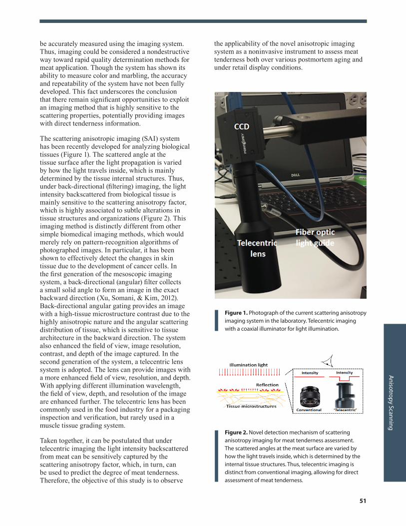
The scattering anisotropic imaging (SAI) system has been recently developed for analyzing biological tissues (Figure 1). The scattered angle at the tissue surface after the light propagation is varied by how the light travels inside, which is mainly determined by the tissue internal structures. Thus, under back-directional (fi ltering) imaging, the light intensity backscattered from biological tissue is mainly sensitive to the scattering anisotropy factor, which is highly associated to subtle alterations in tissue structures and organizations (Figure 2). This imaging method is distinctly different from other simple biomedical imaging methods, which would merely rely on pattern-recognition algorithms of photographed images. In particular, it has been shown to effectively detect the changes in skin tissue due to the development of cancer cells. In the fi rst generation of the mesoscopic imaging system, a back-directional (angular) fi lter collects a small solid angle to form an image in the exact backward direction (Xu, Somani, & Kim, 2012). Back-directional angular gating provides an image with a high-tissue microstructure contrast due to the highly anisotropic nature and the angular scattering distribution of tissue, which is sensitive to tissue architecture in the backward direction. The system also enhanced the fi eld of view, image resolution, contrast, and depth of the image captured. In the second generation of the system, a telecentric lens system is adopted. The lens can provide images with a more enhanced fi eld of view, resolution, and depth. With applying different illumination wavelength, the fi eld of view, depth, and resolution of the image are enhanced further. The telecentric lens has been commonly used in the food industry for a packaging inspection and verifi cation, but rarely used in a muscle tissue grading system.
Taken together, it can be postulated that under telecentric imaging the light intensity backscattered from meat can be sensitively captured by the scattering anisotropy factor, which, in turn, can be used to predict the degree of meat tenderness. Therefore, the objective of this study is to observe
the applicability of the novel anisotropic imaging system as a noninvasive instrument to assess meat tenderness both over various postmortem aging and under retail display conditions.
Figure 1. Photograph of the current scattering anisotropy imaging system in the laboratory. Telecentric imaging with a coaxial illuminator for light illumination.
Figure 2. Novel detection mechanism of scattering anisotropy imaging for meat tenderness assessment. The scattered angles at the meat surface are varied by how the light travels inside, which is determined by the internal tissue structures. Thus, telecentric imaging is distinct from conventional imaging, allowing for direct assessment of meat tenderness.
Anisotropy Scanning

Journal of Purdue Undergraduate Research: Volume 6, Fall 201652
METHOD AND MATERIALS
Sample Preparation
A total of three steers were slaughtered (±16 months old). At 7-day postmortem, loins (Musculus longissimus lumborum) from one side of each carcass were removed (Figure 3). Each muscle was then divided into four equal sections and randomly allocated to four different aging times (7, 14, 21, and 28 days). Samples excluding 7-day were individually vacuum-packaged and aged at 2° C. Two steaks (2.54 cm thick) were cut from each section: one steak for WBSF measurement and the other steak for the SAI analysis.
Cooking Protocol
For the WBSF analysis for the cooked steak samples, the cooking griddle was first set to a heating temperature of 290° F (143° C). After it reached 290° F (143° C), the griddle temperature was then adjusted to a cooking temperature of 275° F (135° C). Prior to placing the steak on the grill, a thermocouple (TruTemp 3519N) was inserted into the center of the steak to monitor the internal temperature of the steaks. When the internal temperature of the steak reached 106° F (41° C), the steak was turned over to cook the other side. The cooking process continued until the internal temperature reached 160° F (71° C). The steak was then wrapped using aluminum foil and cooled at 2° C for 24 hours.
Warner-Bratzler Shear Force Measurement
After 24 hours of cooling at 2° C, six cores parallel to the fiber direction were collected from each sample. The samples were sheared perpendicular to fiber direction using the calibrated TA-XT Exponent Stable Micro System adjusted for the WBSF measurement. The average peak shear force (kg) of the six cores was calculated and used for the instrumental tenderness analysis.
Scattering Anisotropic Image Scanning
Steak samples were scanned using the SAI system in Purdue University’s Weldon School of Biomedical Engineering Research Laboratory (Figure 4). The sample was illuminated using a white-light illuminator with a liquid crystal tunable filter via rig illumination. Reflected light was collected by a telecentric lens and recorded using a CCD camera. The wavelength was varied from 400 nm to 720 nm with a spectral resolution of 10 nm. A total of three scanned images were collected from each sample. To avoid a confounding effect from the strong absorption from myoglobin, the reflectance intensity images were analyzed at around 700 nm. The averaged intensity data from three animals were correlated with the WBSF data.
Figure 3. A schematic figure illustrating the experiment/sampling process. Beef loins were separated from carcasses and each loin was divided into four equal lengths for four aging treatment allocations. Once aging was done, steak cuts from each section were analyzed for tenderness by using either conventional WBSF analysis (using cooked steak samples) or the novel SAI analysis (using uncooked fresh steak samples).
Warner-Bratzler shear force (WBSF) analysis for cooked
steaks
Scattering anisotropic image analysis for uncooked
(fresh) steak samples

53
Figure 4. Representatively SAI results, compared with conventional digital photography. The reflectance intensity, which is normalized by a reflectance reference standard, is inversely correlated with the aging period.
Figure 5. WBSF (top) and SAI reflectance (bottom) values of loins over different postmortem aging periods. The image of the beef samples was captured using the SAI analysis system and averaged cosine values of the scattering angles calculated.
Figure 6. Comparison of averaged WBSF and SAI reflectance values over different aging periods. The comparison showed a very strong correlation between the two methods with r2 = 0.995 (p = 0.003). Both methods showed a significant decrease of tenderness with an increase in aging time.
Anisotropy Scanning
RESULTS
A significant decrease in WBSF measurement was observed as expected, indicating an increase in meat tenderness with aging (p < 0.05; Figure 5). The reflectance from the SAI image taken also significantly decreased as aging time increased (p < 0.05; Figure 5). The decreasing trends in both WBSF and SAI values were very similar, and thus we conducted a correlation analysis to determine the predictability. We found an excellent linear correlation of both averaged SAI reflectance and WBSF values with an r2 of 0.995 (p = 0.003; Figure 6). This observation suggests that meat tenderness can be quantified in a nondestructive, consistent, and highly accurate manner by the novel SAI system.
DISCUSSION
A noninvasive and rapid detection method for analyzing meat quality has been one of the major interests of the meat industry over the past decades, since the traditional methods (e.g., WBSF) for assessing meat quality are time-consuming, destructive, and inconsistent (ElMasry, Sun, & Allen, 2012). Various detection methods, including several imaging systems, have been tested for their efficacy and accuracy. The most extensively studied imaging methods are the hyperspectral imaging system and the near-infrared imaging system. Both of these systems share a common similarity with the SAI method used in the current study. All these imaging systems have a similar methodology, in which lights are used in order to obtain the information from the sample. The system is also commonly coupled with a computer vision technology, making automated

Journal of Purdue Undergraduate Research: Volume 6, Fall 201654
measurement and analysis possible, which greatly improves the consistency and accuracy of the prediction. The main differentiating factors of these imaging systems are the light source type and the method used in capturing the reflectance values.
In the hyperspectral imaging system, a light source with a visible wavelength is commonly used. This system is commonly used to highlight the surface of an object. It uses the principal of a spectrophotometer, in which only a single wavelength of the visible light spectrum is used in a given period of time of the scanning. This method provides both spatial and spectral images of the sample, increasing the amount and depth of the information that can be gathered from it (Barbin, Elmasry, Sun, & Allen, 2012). The wavelengths of the light transmitted are also able to penetrate the sample and cause scattering in the reflectance, which contains substantial information on the internal structure of the product (Jackman, Sun, & Allen, 2011). The near-infrared imaging system also applied the same measuring principle as the hyperspectral imaging system. This system is normally coupled with the hyperspectral imaging system; however, instead of visible light spectrum wavelength, the system utilizes 900–1700 nm near-infrared wavelength during the scanning process. The SAI system also applies the hyperspectral imaging scanning method. A single wavelength of the visible light spectrum is transmitted to the sample in a given time of the scanning process. For this study, the wavelength used during the scan ranged from 400 to 720 nm.
The main difference between the systems appears after the light being transmitted onto the sample. In both hyperspectral imaging and near-infrared imaging systems, the diffused reflectance commonly collected a backscattering angle of around 30–35°, providing images with <1 mm depth. In the SAI system, a back-directional gating collection system is applied in the anisotropic imaging system. The back-gating capturing system is able to reduce the collections angle to 2°, and hence effectively removes the diffused light reflected. The back-directional gating system also captured the reflectance in a backward direction (reflection), since meat structure is more sensitive in the backward direction due to its anisotropic properties. “Anisotropic” means that it is directionally dependent (Xu et al., 2012). For the meat application, it means that the reflected light comes out in a different direction depending on the microstructure of the meat, and hence the reduced amount of available reflectance will be captured. The SAI system is able to minimize this random direction reflectance factor, and thus able to create
an image with a unique image contrast. The system also utilized a telecentric lens, which increases the sensitivity, field of view, resolution, and imaging depth of the imaging system, thus providing an image with a unique contrast of the tissue microstructure and a depth of 1.2 mm compared to the previous imaging system.
Overall, all the imaging systems discussed above utilize mostly similar approaches in extracting information from the sample, but the slight modification in the systems leads to the generation of different results or extents of accuracy. In the near-infrared imaging system, 900–1700 nm near-infrared wavelength is used during the scanning process. Due to the usage and nature of infrared light, other than the physical properties of the sample, complete information on the chemical constituent in a sample can also be gathered (ElMasry et al., 2012). According to the study conducted by Rust and colleagues (2008), a low correlation coefficient between the observed shear force and predicted shear force value was found, indicating that the system is not accurate in predicting tenderness. From the same study, the near-infrared imaging system was only able to go up to a 70% tenderness certification level, which means only 70% of the scanned product actually meets the precise tenderness level. When the hyperspectral imaging system and near-infrared system were coupled, the prediction level increased. In the previous study, it was observed that tenderness measured using this system was viable, but still not highly accurate. From the study conducted by ElMasry and colleagues (2012), a strong correlation with an r2 value of 0.83 was found between the predicted shear force obtained using the imaging system and the actual shear force values. However, although having a strong correlation, the accuracy of the system was not increased much, indicating that the system was only able to go as high as a 75% tenderness certification level (Cluff, Naganathan, Subbiah, Samal, & Calkins, 2013). Taken together, large latent factors as well as high values of error in calibration and cross-validation would be the likely factors to overcome and thus to improve the accuracy and robustness of the system (ElMasry et al., 2012).
The novel scattering anisotropic imaging system showed a stronger correlation and accuracy in predicting meat tenderness in the current pilot study. The measured WBSF and reflectance from the SAI imaging system was highly correlated (r2 of 0.995; p = 0.003). This observation suggests that under telecentric imaging, the light intensity backscattered from meat can sensitively capture the scattering anisotropy factor, which, in turn, can be used to

55
predict the degree of meat tenderness. Further studies would be beneficial, as the current results were only based on a given limited condition (e.g., small sample replicates, loin muscle only, and so forth). Therefore, numerous other background factors that are known to be related to meat tenderness should be taken into consideration.
CONCLUSION
The result of the current pilot study indicates a potential use of the novel anisotropy imaging method as a viable noninvasive prediction tool for meat quality attributes, especially the extent of tenderization. Further studies determining the efficacy and viability of the system in predicting meat tenderness under various baseline factors, such as different marbling contents (USDA-quality grade), connective tissue amounts (different muscle cuts), and postmortem aging times should be warranted. The successful completion of the proposed research can be expected to make a positive impact by advancing knowledge and providing new strategies for use by meat producers and meat processors to consistently provide tenderness ensured/guaranteed beef products to consumers.
ACKNOWLEDGMENTS
This work was supported by the USDA National Institute of Food and Agriculture, Hacth-Multistate project 1006773. The authors appreciate Purdue DURI and SURF undergraduate research programs for the support. The authors also would like to thank the Purdue Meat Laboratory staff and Meat Science and Muscle Biology Lab members for the successful completion of sample and data collection for this project.
REFERENCESBarbin, D., ElMasry, G., Sun, D. W., & Allen, P. (2012).
Near-infrared hyperspectral imaging for grading and classification of pork. Meat Science, 90, 259–268. http://dx.doi.org/10.1016/j.meatsci.2011.07.011
Cluff, K., Naganathan, G. K., Subbiah, J., Samal, A., & Calkins, C. R. (2013). Optical scattering with hyperspectral imaging to classify longissimus dorsi muscle based on beef tenderness using multivariate modeling. Meat Science, 95, 42–50. http://dx.doi.org/10.1016/j.meatsci.2013.04.014
ElMasry, G., Sun, D. W., & Allen, P. (2012). Near-infrared hyperspectral imaging for predicting colour, pH and tenderness of fresh beef. Journal of Food Engineering, 110, 127–140. http://dx.doi.org/10.1016/j.jfoodeng.2011.11.028
Gerrard, D. E., Gao, X., & Tan, J. (1996). Beef marbling and color score determination by image processing. Journal of Food Science, 61(1), 145–148. http://dx.doi.org/10.1111/j.1365-2621.1996.tb14745.x
Grobbel, J. P., Dikeman, M. E., Hunt, M. C., & Milliken, G. A. (2008). Effects of packaging atmosphere on beef instrumental tenderness, fresh color stability and internal cooked color. Journal of Animal Science, 86(5), 1191–1199. http://dx.doi.org/10.2527/jas.2007-0479
Jackman, P., Sun, D. W., & Allen, P. (2011). Recent advances in the use of computer vision technology in the quality assessment of fresh meats. Trends in Food Science & Technology, 22, 185–197. http://dx.doi.org/10.1016/j.tifs.2011.01.008
Liu, Y., Lyon, B. G., Windham, W. R., Realini, C. E., Pringle, T. D. D., & Duckett, S. (2003). Prediction of color, texture, and sensory characteristics of beef steaks by visible and near infrared reflectance spectroscopy. A feasibility study. Meat Science, 65(3), 1107–1115. http://doi.org/10/1016/S0309-1740(02)00328-5
McAfee, A. J., McSorley, E. M., Cuskelly, G. J., Moss, B. W., Wallace, J. M. W., Bonham, M. P., & Fearon, A. M. (2010). Red meat consumption: An overview of the risks and benefits. Meat Science, 84(1), 1–13. http://dx.doi.org/10.1016/j.meatsci.2009.08.029
Quevedo, R., Valencia, E., Cuevas, G., Ronceros, B., Pedreschi, F., & Bastias, J. M. (2013). Color changes in the surface of fresh cut meat: A fractal kinetic application. Food Research International, 54(2), 1430–1436. http://dx.doi.org/10.1016/j.foodres.2013.10.006
Rust, S. R., Price, D. M., Subbiah, J., Kranzler, G., Hilton, G. G., Vanoverbeke, D. L., & Morgan, J. B. (2008). Predicting beef tenderness using near-infrared spectroscopy. Journal of Animal Science, 86(1), 211–218. http://dx.doi.org/10.2527/jas.2007-0084
Sun, X., Chen, K., Berg, E. P., & Magolski, J. D. (2011). Predicting fresh beef color grade using machine vision imaging and support vector machine (SVM) analysis. Journal of Animal and Veterinary Advances. http://dx.doi.org/10.3923/javaa.2011.1504.1511
Wheeler, T. L., Shackelford, S. D., & Koohmaraie, M. (1997). Standardizing collection and interpretation of Warner-Bratzler shear force and sensor tenderness data. Proceedings of the Reciprocal Meat Conference, 50, 68–77.
Wheeler, T. L., Shackelford, S. D., & Koohmaraie, M. (1999). Tenderness classification of beef: IV. Effect of USDA quality grade on the palatability of “tender” beef longissimus when cooked well done. Journal of Animal Science, 77(4), 882–888.
Wyle, A. M., Vote, D. J., Roeber, D. L., Cannell, R. C., Belk, K. E., Scanga, J. A., . . . & Smith, G. C. (2003). Effectiveness of the SmartMV prototype BeefCam system to sort beef carcasses into expected palatability groups. Journal of Animal Science, 81(2), 441–448.
Xiong, Z., Sun, D. W., Zeng, X. A., & Xie, A. (2014). Recent development of hyperspectral imaging systems and their applications in detecting quality attributes of red meats: A review. Journal of Food Engineering, 132, 1–13. http://dx.doi.org/10.1016/j.jfoodeng.2014.02.004
Xu, Z., Somani, A. K., & Kim, Y. L. (2012). Scattering anisotropy-weighted mesoscopic imaging. Journal of Biomedical Optics, 19(9), 090501. http://dx.doi.org/10.1117/1.jbo.17.9.090501
Anisotropy Scanning