angptl4 participates in gestational diabetes mellitus via ... · pact on gdm mothers and their...
TRANSCRIPT

5056
Abstract. – OBJECTIVE: To explore ANGPTL4 expressions in patients with gestational diabetes mellitus (GDM) and its underlying mechanism.
PATIENTS AND METHODS: We first detected serum expressions of ANGPTL4 in GDM patients and healthy pregnancies. Subsequently, effects of ANGPTL4 knockdown on apoptosis, proliferation, and cell cycle in 3T3-L1 cells were determined, re-spectively. Effects of ANGPTL4 on glucose uptake and adipocyte differentiation were also evaluat-ed, respectively. The cytokine secretion in adipo-cytes transfected with sh-ANGPTL4 was detected by quantitative Reverse Transcriptase-Polymerase Chain Reaction (qRT-PCR) and enzyme-linked im-munosorbent assay (ELISA). Furthermore, effects of ANGPTL4 knockdown on NF-kB and Akt path-way were detected by Western blot.
RESULTS: ANGPTL4 was down-regulated in se-rum of GDM patients. In vitro experiments suggest-ed that down-regulated ANGPTL4 inhibited apop-tosis and promoted proliferation of 3T3-L1 cells. Meanwhile, down-regulated ANGPTL4 significantly inhibited glucose uptake and Akt pathway. However, ANGPTL4 expression did not affect cell cycle and adipocyte differentiation. Detection of inflammatory cytokines suggested that down-regulated ANGPTL4 resulted in increased expressions of inflammatory cytokines and activation of NF-kB pathway.
CONCLUSIONS: ANGPTL4 is down-regulated in GDM and may participate in the GDM devel-opment by promoting insulin resistance and se-cretion of inflammatory cytokines.
Key WordsANGPTL4, Inflammatory factors, Insulin resistance,
Gestational diabetes mellitus.
Introduction
Gestational diabetes mellitus (GDM) is a con-dition in which a woman develops high blood sugar levels during pregnancy. The incidence of
GDM reported in different countries is 2%-12%, which has been risen annually1. More than 80% of pregnant women with diabetes mellitus are diagnosed as GDM. GDM significantly increas-es morbidity and mortality of pregnancies and fetuses, which seriously leads to a long-term im-pact on GDM mothers and their offspring2,3. The incidence of type 2 diabetes mellitus in GDM mothers within 20 years after pregnancy is up to 40%. More importantly, the risks of obesity and diabetes are greatly increased in their offspring4. Therefore, it is of great importance to explore the underlying mechanism of GDM.
Recently, great progress has been made in the investigation of the pathogenesis of GDM. So far, adipocyte dysfunction, changes in placenta and hormones, inflammation and oxidative stress are considered to be the possible pathogenesis of GDM5. Adipokines, including leptin, adiponec-tin, resistin, visfatin, etc., are closely related to in-sulin resistance (IR)6. Recent studies have found that many pregnancies experience abnormal se-cretion of adipokines, which exerts a crucial role in the occurrence, development, and prognosis of IR and GDM. Previous studies7,8 have specifically described changes and effects of leptin and ad-iponectin on GDM. Effects of other adipokines such as resistin, visfatin, RBP4, and vaspin on pregnancy complications remain controversial9,10. Further studies are urgently needed to explore the possible interactions between adipokines and in-flammatory factors.
The human ANGPTL4 gene is located on 19p131 with 7 exons and 6 introns. The full-length complementary Deoxyribose Nucleic Acid (cDNA) of human ANGPTL4 is 1943 bp. Addi-tionally, the open reading frame of ANGPTL4
European Review for Medical and Pharmacological Sciences 2018; 22: 5056-5062
M. LI1, X.-J. YANG2, G.-Y. ZHANG3, D.-X. SU4, L. LEI2, R. LI5
1Department of Obstetrics, Jining No. 1 People’s Hospital, Jining, China2Department of Gynaecology and Obstetrics, Shanghai East Hospital Affiliated to Tongji University, Shanghai, China3Department of Laboratory Medicine, The People’s Hospital of Zhangqiu Area, Jinan, China4Department of Cardiology, The People’s Hospital of Zhangqiu Area, Jinan, China5Department of Obstetrics, The Affiliated Qingdao Hiser Hospital of Qingdao University, Qingdao, China
Min Li and Xiaoju Yang contributed equally to this work
Corresponding Author: Rui Li, BM; e-mail: [email protected]
ANGPTL4 participates in gestational diabetes mellitus via regulating Akt pathway

ANGPTL4 participates in gestational diabetes mellitus via regulating Akt pathway
5057
contains 1218 bp and encodes 406 amino acids. ANGPTL4 protein is mainly expressed in ad-ipose tissue and embryo with organ specifici-ty11. ANGPTL4 is also found to be expressed in lung, kidney, liver, pituitary, skeletal muscle, and heart12,13. Currently, there is a controversy about the effect of ANGPTL4 on diabetes. Some schol-ars believed that ANGPTL4 can improve IR by in-creasing insulin sensitivity14. However, Mandard et al15 found that overexpression of ANGPTL4 decreased glucose tolerance. Therefore, the re-lationship between ANGPTL4 and diabetes still requires to be further confirmed. In the present study, we explored ANGPTL4 expressions in GDM patients and its underlying mechanism.
Patients and Methods
PatientsPatients diagnosed as GDM in Obstetrics De-
partment, Jining No. 1 People’s Hospital from June 2015 to May 2017 were selected as GDM group. Healthy pregnancies in the same period were se-lected as control group. The study was approved by the Ethics Committee of Jining No. 1 People’s Hos-pital and all subjects signed the informed consent. Fasting venous samples before natural delivery or caesarean surgery were collected and centrifuged, followed by preservation in liquid nitrogen. For di-agnosing GDM, all pregnancies at 24-30 weeks of gestation were screened for glucose test. If plasma glucose was over 7.8 mmol/L 1 h after breakfast, a 75 g oral glucose tolerance test (OGTT) was re-quired after one-week normal diet1.
Cell CultureMouse 3T3-L1 cells were purchased from Cell
Bank of Chinese Academy of Sciences (Shang-hai, China). 3T3-L1 cells were cultured in Dul-becco’s Modified Eagle Medium (DMEM) medi-um supplemented with 10% fetal bovine serum (FBS, Gibco, Rockville, MD, USA), 100 U/mL penicillin and 100 μg/mL streptomycin, and maintained in a 5% CO2 incubator at 37°C. Fresh medium was replaced every 2 to 3 days. The ad-ipocyte differentiation procedure was as follows: cells were cultured in DMEM medium containing 10% FBS, 1 μM dexamethasone, 0.5 mM IBMX, and 10 μg/ml insulin for 4 days. Normal medium containing 10 μg/mL insulin was then changed for 3-day incubation. Cytokine stimulation pro-cedure was as follows: at 10 days differentiation, cells were starved in serum-free medium for 16
h. Cells were then cultured for 24 h after adding 10 ng/mL IL-1β in the medium. Expressions of inflammatory cytokines were finally detected.
Cell TransfectionCells in LV-Vector group and LV-shANGPTL4
group were transfected with empty vector and lentivirus containing ANGPTL4 shRNA, respec-tively. Cell transfection was performed by Lipo-fectamine 2000 (Thermo Fisher Scientific, Inc. Waltham, MA, USA) based on the manufactur-er’s recommendations. Briefly, cells were seeded into a 6-well plate and then transfected with the above-mentioned plasmids when cell confluence was up to 60%. Lentivirus used in the study was purchased from GenePharm (Shanghai, China).
Cell Counting Kit-8 (CCK-8) AssayTransfected cells were collected and seeded
into a 96-well plate at a dose of 5×10³/mL. Af-ter 24 h-inoculation, 10 μL of CCK-8 (Dojindo, Kumamoto, Japan) solution was added into each well at 0 h, 24 h, 48 h, and 72 h, respectively. The absorbance (OD) values at the wavelength of 450 nm were accessed with a microplate reader.
Cell Apoptosis Transfected cells were digested with ED-
TA-free trypsin and washed with phosphate-buff-ered saline (PBS). 100 µL of 1×binding buffer was added. Subsequently, 5 μL of Annexin V-FITC was added and maintained at room temperature without light for 10 min. 400 µL of 1×binding buf-fer was added for gentle mixture. The apoptosis rate was analyzed by flow cytometry.
Cell CycleTransfected cells were harvested and prepared
for cell suspension. Cells were fixed with ice-cold 70% ethanol overnight. For cell cycle assay, cells were centrifuged, washed twice with PBS and in-cubated with 150 µL of propidium iodide (PI) in the dark for 30 min. Finally, the specific distri-bution of cell cycle was determined by analyzing 10000 events by flow cytometry.
Oil Red O StainingCells were seeded into a 6-well plate with pre-
placed slices, followed by cell differentiation for 6 days. For oil red O staining, cells were fixed with 4% formaldehyde and washed twice with 60% iso-propanol. Fixed cells were incubated with oil red O solution at room temperature for 30 min. Images of cells were captured with an inverted microscope.
M. Li, X.-J. Yang, G.-Y. Zhang, D.-X. Su, L. Lei, R. Li
5058
Enzyme-Linked Immunosorbent Assay (ELISA)
Samples were added in the coated well for 30-min incubation. Thereafter, enzyme-labeled anti-body was added for culturing at 37°C. Cells were washed and stained for 15 min. Subsequently, 0.05 mL of sulfuric acid was added to terminate the reaction. OD value at the wavelength of 450 nm was measured on the ELISA detector.
Glucose UptakeAt 10 days differentiation, cells were washed
with PBS and maintained at 37°C for 20 min af-ter 0.5 mM 2-deoxy-D-[2,6–3H] glucose (1.5 mCi/well, Moravek Biochemicals, CA, USA) was added into each well. Pre-cooled PBS was used to wash cells for terminating the reaction. Thereafter, cells were lysed in 200 µL of lysis buffer for scintillation counting with a liquid scintillation counter.
RNA Extraction and Quantitative Reverse Transcriptase-Polymerase Chain Reaction (qRT-PCR)
The mRNAs of cells were extracted by TRIzol reagent (Invitrogen, Carlsbad, CA, USA) and then reversely transcribed to cDNAs. The reac-tion conditions were as follows: denaturation at 95°C for 1 min, followed by annealing at 95°C for 30 s, and extension at 60°C for 40 s, for a total of 40 cycles. Each sample was repeated-ly performed for 3 times. Primers used in this study were as follows: ANGPTL4: F: GTCCAC-CGACCTCCCGTTA; R: CCTCATGGTCTAG-GTGCTTGT; IRS1: F: ACAAACGCTTCTTCG-TACTGC; R: AGTCAGCCCGCTTGTTGATG; IRS2: F: CGGTGAGTTCTACGGGTACAT; R: TCAGGGTGTATTCATCCAGCG; Akt1: F: AG-CGACGTGGCTATTGTGAAG; R: TCAGGGT-GTATTCATCCAGCG; FoxO1: F: CCAAGAG-GTGAGTGCTTCCC; R: CTGTTGTTCAGACTCTCTCCCT; IL-6: F: ACTCACCTCTTCAGAACGAATTG; R: CCATCTTTGGAAGGTTCAGGTTG; MCP1: F: CTGCAAGAGACTTCCATCCAG; R: AGTG-GTATAGACAGGTCTGTTGG.
Western BlotThe total protein of the transfected cells was
extracted. The concentration of each protein sam-ple was determined by a bicinchoninic acid (BCA) kit (Pierce, Rockford, IL, USA). Briefly, 50 μg of total protein was separated by sodium dodecyl sulphate-polyacrylamide gel electrophoresis (SDS-PAGE) under denaturing conditions and transferred to polyvinylidene difluoride (PVDF) membranes
(Millipore, Billerica, MA, USA). Membranes were blocked with 5% skimmed milk, followed by the incubation of specific primary antibodies over-night. Membranes were then incubated with the secondary antibody at room temperature for 1 h. Immunoreactive bands were exposed by enhanced chemiluminescence (ECL) method.
Statistical AnalysisImageJ and Statistical Product and Service
Solutions (SPSS19.0, IBM, Armonk, NY, USA) statistical software were used for data analysis. Measurement data were expressed as mean ± standard deviation (x–±s). Comparison of mea-surement data was conducted using t-test. p<0.05 was considered statistically significant.
Results
ANGPTL4 Expressions Were Decreased in Serum of GDM Patients
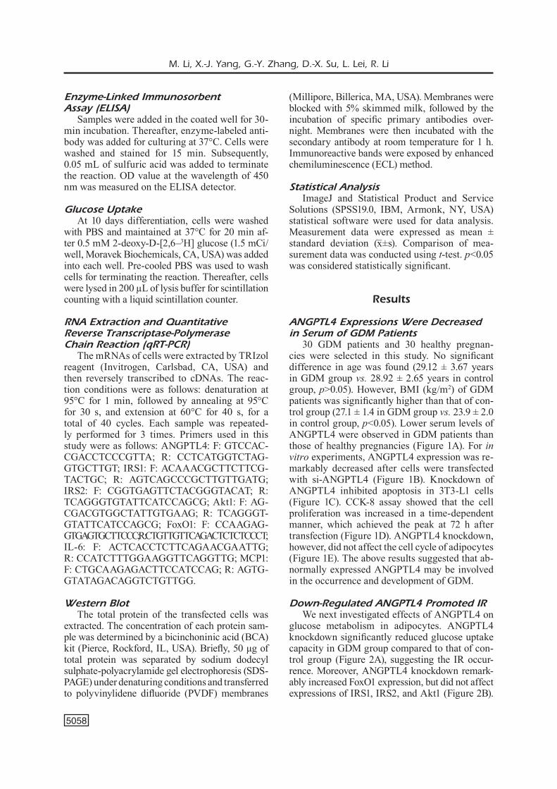
30 GDM patients and 30 healthy pregnan-cies were selected in this study. No significant difference in age was found (29.12 ± 3.67 years in GDM group vs. 28.92 ± 2.65 years in control group, p>0.05). However, BMI (kg/m2) of GDM patients was significantly higher than that of con-trol group (27.1 ± 1.4 in GDM group vs. 23.9 ± 2.0 in control group, p<0.05). Lower serum levels of ANGPTL4 were observed in GDM patients than those of healthy pregnancies (Figure 1A). For in vitro experiments, ANGPTL4 expression was re-markably decreased after cells were transfected with si-ANGPTL4 (Figure 1B). Knockdown of ANGPTL4 inhibited apoptosis in 3T3-L1 cells (Figure 1C). CCK-8 assay showed that the cell proliferation was increased in a time-dependent manner, which achieved the peak at 72 h after transfection (Figure 1D). ANGPTL4 knockdown, however, did not affect the cell cycle of adipocytes (Figure 1E). The above results suggested that ab-normally expressed ANGPTL4 may be involved in the occurrence and development of GDM.
Down-Regulated ANGPTL4 Promoted IRWe next investigated effects of ANGPTL4 on
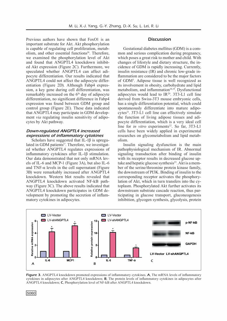
glucose metabolism in adipocytes. ANGPTL4 knockdown significantly reduced glucose uptake capacity in GDM group compared to that of con-trol group (Figure 2A), suggesting the IR occur-rence. Moreover, ANGPTL4 knockdown remark-ably increased FoxO1 expression, but did not affect expressions of IRS1, IRS2, and Akt1 (Figure 2B).

ANGPTL4 participates in gestational diabetes mellitus via regulating Akt pathway
5059
Figure 1. ANGPTL4 expressions were decreased in serum of GDM patients. A, Serum expressions of ANGPTL4 in GDM patients and healthy pregnancies; B, ANGPTL4 expression after lentivirus transfection; C, Cell apoptosis after ANGPTL4 knockdown; D, Cell proliferation after ANGPTL4 knockdown; E, Cell cycle after ANGPTL4 knockdown.
A B C
D E
Figure 2. Down-regulated ANGPTL4 decreased the glucose uptake. A, Glucose uptake detection after ANGPTL4 knock-down; B, The mRNA levels of key genes in insulin signaling pathway after ANGPTL4 knockdown; C, Phosphorylation level of Akt detected by Western blot; D-E, Cell differentiation and mRNA level of Fabp4 after ANGPTL4 knockdown.
A B C
D E F
M. Li, X.-J. Yang, G.-Y. Zhang, D.-X. Su, L. Lei, R. Li
5060
Previous authors have shown that FoxO1 is an important substrate for Akt. Akt phosphorylation is capable of regulating cell proliferation, metab-olism, and other essential functions16. Therefore, we examined the phosphorylation level of Akt and found that ANGPTL4 knockdown inhibit-ed Akt expression (Figure 2C). Furthermore, we speculated whether ANGPTL4 can affect adi-pocyte differentiation. Our results indicated that ANGPTL4 could not affect the adipocyte differ-entiation (Figure 2D). Although Fabp4 expres-sion, a key gene during cell differentiation, was remarkably increased on the 6th day of adipocyte differentiation, no significant difference in Fabp4 expression was found between GDM group and control group (Figure 2E). These data indicated that ANGPTL4 may participate in GDM develop-ment via regulating insulin sensitivity of adipo-cytes by Akt pathway.
Down-regulated ANGPTL4 increased expressions of inflammatory cytokines
Scholars have suggested that IL-1β is upregu-lated in GDM patients17. Therefore, we investigat-ed whether ANGPTL4 regulates expressions of inflammatory cytokines after IL-1β stimulation. Our data demonstrated that not only mRNA lev-els of IL-6 and MCP-1 (Figure 3A), but also IL-6 and TNF-α levels in the cell supernatant (Figure 3B) were remarkably increased after ANGPTL4 knockdown. Western blot results revealed that ANGPTL4 knockdown activated NF-kB path-way (Figure 3C). The above results indicated that ANGPTL4 knockdown participates in GDM de-velopment by promoting the secretion of inflam-matory cytokines in adipocytes.
Discussion
Gestational diabetes mellitus (GDM) is a com-mon and serious complication during pregnancy, which poses a great risk to mother and child. With changes of lifestyle and dietary structure, the in-cidence of GDM is rapidly increasing. Currently, insulin resistance (IR) and chronic low-grade in-flammation are considered to be the major factors of GDM3. Adipose tissue is well recognized as its involvement in obesity, carbohydrate and lipid metabolism, and inflammation18,19. Dysfunctional adipocytes would lead to IR20. 3T3-L1 cell line derived from Swiss-3T3 mouse embryonic cells, has a single differentiation potential, which could spontaneously differentiate into mature adipo-cytes21. 3T3-L1 cell line can effectively simulate the function of living adipose tissues and adi-pocyte differentiation, which is a very ideal cell line for in vitro experiments22. So far, 3T3-L1 cells have been widely applied in experimental researches on glycometabolism and lipid metab-olism.
Insulin signaling dysfunction is the main pathophysiological mechanism of IR. Abnormal signaling transduction after binding of insulin with its receptor results in decreased glucose up-take and hepatic glucose synthesis23. Akt is a mem-ber of the serine/threonine protein kinase family, the downstream of PI3K. Binding of insulin to the corresponding receptor activates the phosphory-lation of Akt, which in turn transfers into the cy-toplasm. Phosphorylated Akt further activates its downstream substrate cascade reaction, thus par-ticipating in glucose transport, gluconeogenesis inhibition, glycogen synthesis, glycolysis, protein
Figure 3. ANGPTL4 knockdown promoted expressions of inflammatory cytokines. A, The mRNA levels of inflammatory cytokines in adipocytes after ANGPTL4 knockdown; B, The protein levels of inflammatory cytokines in adipocytes after ANGPTL4 knockdown; C, Phosphorylation level of NF-kB after ANGPTL4 knockdown.
A BC

ANGPTL4 participates in gestational diabetes mellitus via regulating Akt pathway
5061
synthesis, and anti-apoptotic process24. FoxO1 is an important substrate of PI3K/Akt pathway, the activity of which is regulated by phosphorylation, ubiquitination, acetylation, and other post-trans-lational modifications. Transcriptional activity of phosphorylated FoxO1 is inhibited in the nucle-us, which negatively regulates insulin signaling pathway16. In this study, we found that ANGPTL4 knockdown significantly inhibits glucose uptake in adipocytes, which may be explained by the IR promotion via decreased Akt phosphorylation and increased FoxO1 expression.
NF-kB is a widespread transcription factor in almost all types of tissues and cells, which is in-volved in regulating inflammatory responses. In-flammatory cytokines are produced in adipocytes by oxidative stress, thereby activating the inflam-matory signaling pathway. Previous researches suggested that activated NF-κB increases expres-sions of TNF-α and IL-6 in 3T3-L1 cells, thus ex-acerbating IR and chronic inflammation25,26. Our study also found that ANGPTL4 knockdown can stimulate the secretion of inflammatory cytokines via NF-kB pathway, including IL-6 and TNF-α.
Conclusions
We found that ANGPTL4 is down-regulated in GDM and may participate in the GDM devel-opment by promoting insulin resistance and se-cretion of inflammatory cytokines.
Funding AcknowledgementsThis work was supported by Jiangxi Provincial Health and Family Planning Commission Technology Plan (20187312) and Shanghai Municipal Health and Family Planning Com-mission Research Project Plan (201640246).
Conflict of InterestsThe authors declared no conflict of interest.
References
1) Buchanan Ta, Xiang ah, Page Ka. Gestational diabetes mellitus: risks and management during and after pregnancy. Nat Rev Endocrinol 2012; 8: 639-649.
2) crowTher ca, hiller Je, Moss Jr, McPhee aJ, Jeffries ws, roBinson Js. Effect of treatment of gestational diabetes mellitus on pregnancy outcomes. N Engl J Med 2005; 352: 2477-2486.
3) li JY, wu gM, hou Z, cao YM. Expression of C1q/TNF-related protein-3 (CTRP3) in serum of patients with gestational diabetes mellitus and its relationship with insulin resistance. Eur Rev Med Pharmacol Sci 2017; 21: 5702-5710.
4) BoneY cM, VerMa a, TucKer r, Vohr Br. Metabolic syndrome in childhood: Association with birth weight, maternal obesity, and gestational diabe-tes mellitus. Pediatrics 2005; 115: e290-e296.
5) PanThaM P, aYe il, Powell Tl. Inflammation in ma-ternal obesity and gestational diabetes mellitus. Placenta 2015; 36: 709-715.
6) MaMaBolo rl, alBerTs M, leViTT ns, DeleMarre-Van Dwh, sTeYn nP. Prevalence of gestational dia-betes mellitus and the effect of weight on mea-sures of insulin secretion and insulin resistance in third-trimester pregnant rural women residing in the Central Region of Limpopo Province, South Africa. Diabet Med 2007; 24: 233-239.
7) winZer c, wagner o, fesTa a, schneiDer B, roDen M, Bancher-ToDesca D, Pacini g, funahashi T, KauTZKY-willer a. Plasma adiponectin, insulin sensitivity, and sub-clinical inflammation in women with prior gestational diabetes mellitus. Diabetes Care 2004; 27: 1721-1727.
8) wolf M, sauK J, shah a, Vossen sK, JiMeneZ-KiMBle r, ecKer Jl, ThaDhani r. Inflammation and glucose intolerance: a prospective study of gestational diabetes mellitus. Diabetes Care 2004; 27: 21-27.
9) Miehle K, sTePan h, fasshauer M. Leptin, adiponec-tin and other adipokines in gestational diabetes mellitus and pre-eclampsia. Clin Endocrinol (Oxf) 2012; 76: 2-11.
10) Vrachnis n, BeliTsos P, sifaKis s, DafoPoulos K, sirisTaTiDis c, PaPPa Ki, ilioDroMiTi Z. Role of adipokines and oth-er inflammatory mediators in gestational diabetes mellitus and previous gestational diabetes mellitus. Int J Endocrinol 2012; 2012: 549748.
11) genTil c, le Jan s, PhiliPPe J, leiBowiTch J, sonigo P, gerMain s, PieTri-rouXel f. Is oxygen a key factor in the lipodystrophy phenotype? Lipids Health Dis 2006; 5: 27.
12) KloPPer JP, BerenZ a, haYs wr, sharMa V, PugaZhenThi u, Janssen J, singh M, BissonneTTe rP, haugen Br. In vivo and microarray analysis of rexinoid-respon-sive anaplastic thyroid carcinoma. Clin Cancer Res 2008; 14: 589-596.
13) ge h, cha JY, goPal h, harP c, Yu X, rePa JJ, li c. Differential regulation and properties of angiopoi-etin-like proteins 3 and 4. J Lipid Res 2005; 46: 1484-1490.
14) MerKel M, ecKel rh, golDBerg iJ. Lipoprotein li-pase: genetics, lipid uptake, and regulation. J Lipid Res 2002; 43: 1997-2006.
15) ManDarD s, ZanDBergen f, Van sTraTen e, wahli w, KuiPers f, Muller M, KersTen s. The fasting-induced adipose factor/angiopoietin-like protein 4 is phys-ically associated with lipoproteins and governs plasma lipid levels and adiposity. J Biol Chem 2006; 281: 934-944.
16) carlsson P, MahlaPuu M. Forkhead transcription factors: key players in development and metabo-lism. Dev Biol 2002; 250: 1-23.
17) laPPas M. Activation of inflammasomes in adipose tissue of women with gestational diabetes. Mol Cell Endocrinol 2014; 382: 74-83.

M. Li, X.-J. Yang, G.-Y. Zhang, D.-X. Su, L. Lei, R. Li
5062
18) PiTTas ag, JosePh na, greenBerg as. Adipocytokines and insulin resistance. J Clin Endocrinol Metab 2004; 89: 447-452.
19) MaTsuZawa Y. The metabolic syndrome and adipo-cytokines. FEBS Lett 2006; 580: 2917-2921.
20) aPPel sJ, Jones eD, KenneDY-Malone l. Central obe-sity and the metabolic syndrome: Implications for primary care providers. J Am Acad Nurse Pract 2004; 16: 335-342.
21) green h, KehinDe o. An established preadipose cell line and its differentiation in culture. II. Factors af-fecting the adipose conversion. Cell 1975; 5: 19-27.
22) JoosT hg, schurMann a. Subcellular fractionation of adipocytes and 3T3-L1 cells. Methods Mol Biol 2001; 155: 77-82.
23) Kasuga M. Insulin resistance and pancreatic beta cell failure. J Clin Invest 2006; 116: 1756-1760.
24) TaZZari Pl, caPPellini a, ricci f, eVangelisTi c, PaPa V, grafone T, MarTinelli g, conTe r, cocco l, MccuBreY Ja, MarTelli aM. Multidrug resistance-associated protein 1 expression is under the control of the phosphoinositide 3 kinase/Akt signal transduc-tion network in human acute myelogenous leu-kemia blasts. Leukemia 2007; 21: 427-438.
25) aJuwon KM, sPurlocK Me. Palmitate activates the NF-kappaB transcription factor and induces IL-6 and TNFalpha expression in 3T3-L1 adipocytes. J Nutr 2005; 135: 1841-1846.
26) laPPas M, Yee K, PerMeZel M, rice ge. Sulfasalazine and BAY 11-7082 interfere with the nuclear fac-tor-kappa B and I kappa B kinase pathway to regulate the release of proinflammatory cytokines from human adipose tissue and skeletal muscle in vitro. Endocrinology 2005; 146: 1491-1497.