anesthetic managemen otf a child with stevens-johnson syndrome filecash report anesthetic managemen...
TRANSCRIPT
CASH R E P O R T
Anesthetic management of a child with Stevens-Johnson syndrome
S A N T O S H B. KALHAN, M B A N D S T E V E N R. DITTO, M D
• The authors describe the anesthetic management of a patient with Stevens-Johnson syndrome. Intravenous infusion of ketamine was optimal for multiple minor surgical procedures without the need for airway interference. • INDEX TERM: STEVENS-JOHNSON SYNDROME • CLEVE CLIN J MED 1988; 55:467^(69
STEVENS-JOHNSON SYNDROME (SJS), or erythema multiforme major, is an uncommon acute exfoliative disease involving the skin and mucous membranes. It is associated with severe
systemic manifestations and may be life threatening in the acute phase. For the anesthesiologist, it presents problems pertaining to airway management, venous ac-
• See also the editorial by Rasmussen (pp 412-414)
cess, fluid management, and application of monitors. However, despite descriptions of several surgical proce-dures involving patients with SJS,1 reports of anesthetic management are scant.2-4 This paper describes the suc-cessful anesthetic management of a child with SJS.
CASE REPORT
A two-year-old white girl was admitted to another hospital following a cluster of three seizures. An electro-encephalogram and computed tomogram of the head
From the Division of Anesthesiology, The Cleveland Clinic Foun-dation. Submitted for publication Sept 1987; accepted Feb 1988.
SEPTEMBER • OCTOBER 1988
were normal. A diagnosis of bilateral otitis media and febrile seizures (temperature, 38.5° C) was reached. She was treated with amoxicillin and phenobarbital (30 mg, twice daily). Twelve days later, she become febrile, and her eyes appeared to be "bloodshot" and "sunburnt." By the same evening, her eyes were swollen and some vesicular lesions had developed on the lids. The next day, vesiculobullous lesions were noted on the arms and legs. Over the next 12 hours, the lesions spread to her palms, soles, and mouth.
When she was referred to the Cleveland Clinic, she was febrile (temperature, 40° C). Skin and mucosal lesions were apparent. No lesions were in the pharynx, nor was there evidence of tracheal or pulmonary involve-ment. The chest radiograph, serum electrolytes, and a complete blood count were normal. A diagnosis of Ste-vens-Johnson syndrome secondary to phenobarbital was made. Phenobarbital was discontinued and diazepam (5 mg) was given every eight hours through an intravenous line into the saphenous vein. Amoxicillin was also dis-continued, and the patient was given cefuroxime intra-venously for otitis media and as a prophylaxis against Staphylococcus aureus infection of the skin. Examination of the eyes showed bullous epitheliopathy with mild in-jection of palpebral conjunctiva. Both corneas were normal.
Over the next several days, weeping and crusting
CLEVELAND CLINIC JOURNAL OF MEDICINE 467

STEVENS-JOHNSON SYNDROME • KALHAN AND DITTO
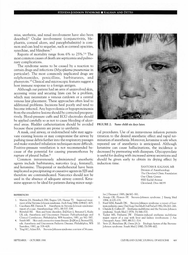
F I G U R E 1. Facial lesions with fibrin membrane on both eyelids and corners of the mouth.
lesions developed. The trunk lesions soon started to stabilize, but lesions of the face and eyelids continued to weep and crust. Fibrin adhesions formed bilaterally be-tween her upper and lower eyelids, and she was unable to open her eyes. She was scheduled for an examination under anesthesia and lysis of lid adhesions.
The preoperative examination showed the child to be irritable but well nourished (weight, 12.9 kg). Perioral fissuring and bleeding were noted in the extensively weeping and crusted facial lesions. Fibrin membranes were present in the corners of her mouth and a few anterior intraoral lesions were also apparent (Figure 1). She was having difficulty handling secretions and was drooling.
A decision was made to use ketamine anesthesia. She fasted after midnight and was given the scheduled dose of diazepam on the morning of surgery. She also received glycopyrrolate (0.1 mg, intravenously) one hour before administration of anesthesia. In the operating room, electrocardiogram (ECG) leads, a blood-pressure cuff, and precordial stethoscope were applied to the least involved areas of the skin. A pulse oximeter was applied to her toe (oxygen saturation, 98%) and axillary tem-perature was monitored with a nonsticky all-purpose
temperature probe. The patient was given diazepam intravenously (2 mg), followed by ketamine in incre-mental doses of 10 mg. Surgery was started after the patient had received 20 mg of ketamine. A total of 100 mg of ketamine was administered over 25 minutes. This was followed by a ketamine infusion (1 -2 mg/min as a 1 mg/mL solution) and the infusion rate was titrated to prevent movement of the extremities. A total of 60 mg of ketamine was infused over the next 40 minutes. Vital signs remained stable throughout the procedure. Oxygen saturation, blood pressure, and heart rate ranged from 97%—100%, 110/70 to 100/55 mmHg, and 120-140 beats per minute, respectively.
The extensive adhesions between the upper and lower eyelids were lysed bilaterally. Ultrasound examination of the left eye confirmed integrity of the eyeball. A thick fibrin membrane covering the cornea and bulbar con-junctivae was removed from both eyes.
In the recovery room, the patient started to move and complain of discomfort within 10 minutes. She main-tained good oxygen saturation and was discharged to the floor one hour later.
Over the next two days, the surgical procedure was repeated using the same anesthetic technique, except that the infusion was started after a 25-mg induction dose of ketamine administered intravenously. By the third procedure, the facial lesions were much improved. By the sixth day after the first procedure, the patient could open both eyes easily and her face was almost clear (Figure 2). Eight days after the first procedure, pressure-equalization tubes were inserted bilaterally for serous otitis media while the patient was given halothane anesthesia by mask. Diazepam was discontinued. She was discharged.
DISCUSSION
Stevens-Johnson syndrome is an acute disease charac-terized by target lesions of the skin that rapidly progress to macules, papules, vesicles, and bullae. These rupture, leaving extensive weeping and crusting areas. There is often a prodromal period of one to three days with fever, cough, sore throat, myalgias, and generalized malaise. In addition, all the mucous membranes may be severely involved.5,6 Although the presence of respiratory lesions is variable, stomatitis is present in almost all cases,7 and patients may have problems handling secretions. Pneu-monitis is not uncommon and is seen on the chest radiograph as bilateral pulmonary infiltrates.8 Pneumo-thorax may occur from rupture of surface bullae on vis-ceral pleura. Acute myocarditis with transitory arrhyth-
468 CLEVELAND CLINIC JOURNAL OF MEDICINE VOLUME 55 NUMBER 5
S T E V E N S - J O H N S O N S Y N D R O M E • K A L H A N A N D D I T T O
mias, urethritis, and renal involvement have also been described.9 Ocular involvement (conjunctivitis, ble-pharitis, corneal ulcers, and panophthalmitis) is com-mon and can lead to sequelae, such as corneal opacities, synechiae, and blindness.5
Reports of mortality range from 6 % to 25%.5 ,6 The most common causes of death are septicemia and pulmo-nary complications.
The syndrome seems to be caused by a reaction to certain drugs and infections (Mycoplasma pneumoniae in particular). The most commonly implicated drugs are sulphonamides, penic i l l ins , barbiturates , and phenytoin.10 Clinical and microscopic features suggest a host immune response to a foreign antigen.
Although our patient had an area of uninvolved skin, accessing veins and securing lines can be a problem, which may necessitate a venous cutdown or a central venous line placement. These approaches often lead to additional problems. Incisions heal poorly and tend to become infected. Any hypovolemia or hypoproteincmia from the exudative lesions should be corrected preopera-tively. Blood-pressure cuffs and ECG electrodes should be applied carefully so as not to cause bleeding of ulcer-ated areas. Bladder catheterization should be avoided because these patients are prone to urethritis.
A mask, oral airway, or endotracheal tube may aggra-vate existing lesions or may compromise the airway by pushing tissue debris further into the respiratory passages and make standard inhalation techniques more difficult. Positive-pressure ventilation is not recommended be-cause of the potential for causing pneumothorax by rupture of pleural bullae.4
Common intravenously administered anesthetic agents include barbiturates, narcotics (e.g., fentanyl), and ketamine. Thiopental or methohexital have been implicated as precipitating or causative agents in SJS and therefore are contraindicated. Narcotics should not be used in the absence of adequate airway control. Keta-mine appears to be ideal for patients during minor surgi-
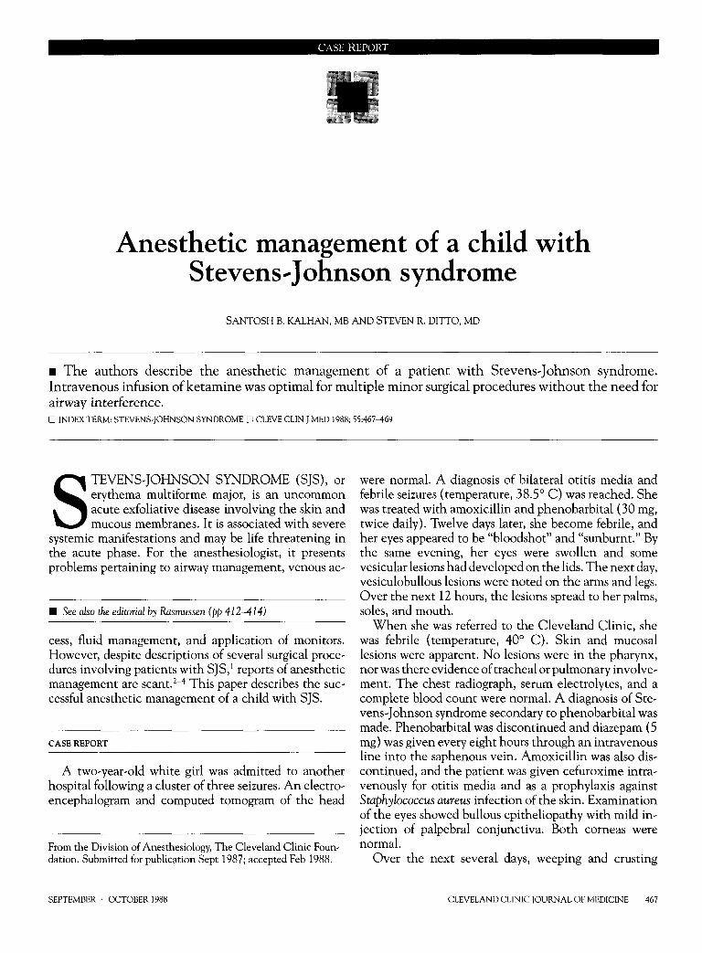
FIGURE 2. Same child six days later.
cal procedures. Use of an intravenous infusion permits titration to the desired anesthetic effect and rapid ter-mination of anesthesia. Moreover, ketamine is safe when repeated use of anesthetics is anticipated. Although ketamine can cause hallucinations, the incidence is decreased by pretreating with diazepam. Glycopyrrolate is useful for dealing with increased airway secretions and should be given early to obtain its drying effect by induction time.
S A N T O S H B. KALHAN, MB Division of Anesthesiology The Cleveland Clinic Foundation One Clinic Center 9500 Euclid Avenue Cleveland, Ohio 44195
REFERENCES
1. Marvin JA, Heimbach DM, Engrav LH, Harnar TJ. Improved treat-ment of the Stevens-Johnson syndrome. Arch Surg 1984; 119:601-605.
2. Cucchiara RF, Dawson B. Anesthesia in Stevens-Johnson syndrome: report of a case. Anesthesiology 1971; 35 :537-539.
3. Berryhill RE. Skin and bone disorders. [In] Katz J, Benumof J, Kadis LB, eds. Anesthesia and Uncommon Diseases: Pathophysiologic and Clinical Correlations. Philadelphia, W B Saunders, 1981, pp 562-587.
4. SmithMF. Skin and connective tissue diseases. [In] Katz J, Steward DJ, eds. Anesthesia and Uncommon Pediatric Diseases. Philadelphia, W B Saunders, 1987, pp 378-428.
5. Ting HC, Adam BA. Stevens-Johnson syndrome: a review of 34 cases.
Int J Dermatol 1985; 24:587-591. 6. Araujo OE, Flowers FP. Stevens-Johnson syndrome. J Emerg Med
1984; 2 :129-135. 7. Nazif MM, Ranalli DN. Stevens-Johnson syndrome: a report of four-
teen pediatric cases. Oral Surg Oral Med Oral Pathol 1982; 53 :263-266. 8. Chanda JJ, Callen JP. Erythema multiforme and the Stevens-Johnson
syndrome. South Med J 1978; 71:566-570. 9. Tucker MS, Fitzharris JW. Dilantin-induced erythema multiforme
major: report of a case with liver and kidney involvement. J Am Osteopath Assoc 1985; 85 :511-514.
10. Yetiv JZ, Bianchine JR, Owen JA Jr. Etiologic factors of the Stevens-Johnson syndrome. South Med J 1980; 73 :599-602.
SEPTEMBER • OCTOBER 1988 CLEVELAND CLINIC JOURNAL OF MEDICINE 469