anatomy & physiology laboratory manualanatomy and physiology laboratory 1 chapter 1 the...
TRANSCRIPT

Anatomy & PhysiologyLaboratory Manual
Second Edition
Main Version
Christine M. EckelCarroll College
Kyla Turpin RossGeorgia State University
Theresa Stouter BidleHagerstown Community College
9781259139437_FM_MAIN_i-xxiv_Print.indd 1 4/16/15 9:49 AM

ANATOMY & PHYSIOLOGY LABORATORY MANUAL: MAIN VERSION, SECOND EDITION
Published by McGraw-Hill Education, 2 Penn Plaza, New York, NY 10121. Copyright © 2016 by McGraw-Hill Education. All rights reserved. Printed in the United States of America. Previous edition © 2014. No part of this publication may be reproduced or distributed in any form or by any means, or stored in a database or retrieval system, without the prior written consent of McGraw-Hill Education, including, but not limited to, in any network or other electronic storage or transmission, or broadcast for distance learning.
Some ancillaries, including electronic and print components, may not be available to customers outside the United States.
This book is printed on acid-free paper.
1 2 3 4 5 6 7 8 9 0 RMN/RMN 10 9 8 7 6 5
ISBN 978-1-259-13943-7MHID 1-259-13943-3
Senior Vice President, Products & Markets: Kurt L. StrandVice President, General Manager, Products & Markets: Marty LangeVice President, Content Design & Delivery: Kimberly Meriwether DavidManaging Director: Michael HackettDirector of Digital Content: Michael G Koot, PhDBrand Manager: Amy ReedDirector, Product Development: Rose KoosProduct Developer: Donna NemmersMarketing Manager: Jessica CannavoDigital Product Analyst: Jake TheobaldDirector, Content Design & Delivery: Linda AvenariusProgram Manager: Angela R. FitzPatrickContent Project Managers: April R. Southwood/Christina NelsonBuyer: Sandy LudovissyDesign: David HashContent Licensing Specialist: Carrie BurgerCover Image: wheelchair athlete © David MoyerCompositor: MPS North America LLCPrinter: R. R. Donnelley
All credits appearing on page or at the end of the book are considered to be an extension of the copyright page.
The Internet addresses listed in the text were accurate at the time of publication. The inclusion of a website does not indicate an endorsement by the authors or McGraw-Hill Education, and McGraw-Hill Education does not guarantee the accuracy of the information presented at these sites.
www.mhhe.com
9781259139437_FM_MAIN_i-xxiv_Print.indd 2 4/16/15 9:49 AM

brief contentsPART I IntroductIon to
thE AnAtoMy And PhySIoLogy L AborAtory
Chapter 1The Laboratory Environment 1
Chapter 2Orientation to the Human Body 27
Chapter 3The Microscope 43
PART I I orgAnIzAtIon of thE huMAn body
Chapter 4Cell Structure and Membrane Transport 57
Chapter 5Histology 91
PART I I I SuPPort And body MovEMEnt
Chapter 6Integument 123
Chapter 7The Skeletal System: Bone Structure and Function 143
Chapter 8The Skeletal System: Axial Skeleton 161
Chapter 9The Skeletal System: Appendicular Skeleton 195
Chapter 10Articulations 229
Chapter 11The Muscular System: Muscle Structure and Function 247
Chapter 12The Muscular System: Axial Muscles 283
Chapter 13The Muscular System: Appendicular Muscles 311
PART IV coMMunIcAtIon And controL
Chapter 14Nervous Tissues 345
Chapter 15The Brain and Cranial Nerves 371
Chapter 16The Spinal Cord, Spinal Nerves, and Reflexes 419
Chapter 17The Autonomic Nervous System 443
Chapter 18General and Special Senses 459
Chapter 19The Endocrine System 501
PART V MAIntEnAncE And rEguL AtIon
Chapter 20The Cardiovascular System: Blood 523
Chapter 21The Cardiovascular System: The Heart 545
Chapter 22The Cardiovascular System: Vessels and Circulation 579
Chapter 23The Lymphatic System and Immunity 627
Chapter 24The Respiratory System 651
Chapter 25The Urinary System 685
Chapter 26The Digestive System 715
PART VI rEProductIon
Chapter 27The Reproductive System and Early Development 751
iii
9781259139437_FM_MAIN_i-xxiv_Print.indd 3 4/16/15 9:49 AM

CHRISTINE MARIE ECkEL received her B.A. in integrative biology and M.A. in human biodynamics from the University of California, Berkeley, and her Ph.D. in neurobiology and anatomy at the University of Utah School of Medicine. Christine is associate professor of biology at Carroll College in her hometown of Helena, Montana, where she teaches the two-semester anatomy and physiology course for pre-nursing and pre-health science majors, and an advanced dissection course for premedical students. She also serves as the faculty advisor for pre-physical therapy and pre-physician assistant students. Prior to her position at Carroll College, Christine was associate professor and course director for medical gross anatomy and medical microanatomy at West Virginia School of Osteopathic Medicine (WVSOM). In the 14 years prior to her position at WVSOM, Christine taught undergraduate human anatomy and human physiology courses at Salt Lake Community College and the University of California, Berkeley. She earned outstanding teaching awards at all three of these institutions.
Christine is the author of Human Anatomy Laboratory Manual, second edition (McGraw-Hill Education). In addition, her cadaver dissections and photographs are featured in several textbooks, including this laboratory manual.
Christine served as the Western Regional Director for the Human Anatomy & Physiology Society (HAPS) for two terms. She has also served on several committees for both HAPS and the American Association of Anatomists (AAA). Her research is in the field of educational outcomes, and she serves as a peer reviewer for the journals Anatomical Sciences Education and Medical Education.
With over 25 years of experience engaging with students at all levels, including community college students, medical students, and surgical residents, Christine has a unique appreciation for the learning challenges experienced by students at each level. Christine's passions for anatomy and physiology, teaching, dissection, and photography are evident throughout the pages of this laboratory manual. In her spare time, Christine loves to mountain bike, skate ski, and explore the great Montana outdoorsÐa lways with her camera in hand.
kyLA TURPIN ROSS received her undergraduate degree from Louisiana State University in biological and agricultural engineering and her Ph.D. in biomedical engineering from Georgia Institute of Technology and Emory University. Kyla then served as a postdoctoral fellow in the Fellowships in Research and Science Teaching (FIRST) program at Emory University, an NIH-funded program that provides training in both research and teaching. Kyla is now senior academic professional at Georgia State University (GSU), where she teaches and manages the introductory and graduate human anatomy and physiology courses. Kyla has extensive experience developing lecture and laboratory curricula, and incorporates active learning in the classroom as a method to reinforce difficult physiological concepts. In addition, Kyla plays an active role in mentoring GSU faculty and teaching assistants and planning and hosting an annual teaching assistant workshop. She is involved in STEM initiatives at GSU, and serves as the faculty advisor for the Department of Biology Tutorial Center. She has served as a reviewer for numerous publications, and has authored a custom laboratory manual for GSU' s human anatomy and physiology course. She is active in several committees within the Human Anatomy & Physiology Society (HAPS). In addition to academic endeavors, Kyla serves on the Decatur Family YMCA board of directors.
TERRI STOUTER BIDLE received her undergraduate degree from Rutgers University and her M.S. degree in biomedical science from Hood College in Maryland, and has completed additional graduate coursework in genetics at the National Institutes of Health. She is a professor at Hagerstown Community College where she teaches anatomy and physiology and genetics to pre-allied health students. Before joining the Hagerstown faculty in 1990, Terri was coordinator of the Science Learning Center, where she developed study materials and a tutoring program for students enrolled in science classes. She has been a developmental reviewer, and has written supplemental materials for both textbooks and laboratory manuals. Terri is a coauthor of Anatomy & Physiology: An Integrative Approach, second edition.
With love and thanks to my entire family, including the dogs.
With love and thanks
about the author
To my husband Jim, daughter Ella, and son
Cameron: I treasure your constant love and
unending support.
To my husband Jim,
With love and thanks to my husband Jay
and my daughter Stephanie for their continued support.
With love and thanks
iv
9781259139437_FM_MAIN_i-xxiv_Print.indd 4 4/16/15 9:49 AM

contentsPreface xiv
PArt I IntroductIon to thE AnAtoMy And PhySIoLogy L AborAtory 1
Chapter 1The Laboratory Environment 1GROSS ANATOMy 4
The Scientific Process of Discovery 4ExErCiSE 1.1 The ScienTific MeThod 6
ExErCiSE 1.2 PreSenTing daTa 7
Measurement in Science 8ExErCiSE 1.3 UniTS of MeaSUreMenT 10
Laboratory Equipment 10ExErCiSE 1.4 idenTificaTion of coMMon diSSecTion inSTrUMenTS 11
ExErCiSE 1.5 ProPer diSPoSal of laboraTory WaSTe 15
Dissection Techniques 15ExErCiSE 1.6 Placing a ScalPel blade on a ScalPel blade handle 16
ExErCiSE 1.7 diSSecTing WiTh a ScalPel 19
ExErCiSE 1.8 diSSecTing WiTh SciSSorS 20
ExErCiSE 1.9 blUnT diSSecTion TechniqUeS 22
Chapter 2Orientation to the Human Body 27GROSS ANATOMy 30
Anatomic Terminology and the Anatomic Position 30
Anatomic Planes and Sections 30ExErCiSE 2.1 anaToMic PlaneS and SecTionS 31
Directional Terms 33ExErCiSE 2.2 direcTional TerMS 33
regional Terms 34ExErCiSE 2.3 regional TerMS 35
Body Cavities and Membranes 36ExErCiSE 2.4 body caviTieS 36
Abdominopelvic regions and Quadrants 38ExErCiSE 2.5 locaTing Major body organS USing abdoMinoPelvic region and qUadranT TerMinology 38
Chapter 3The Microscope 43HISTOLOGy 46
The Compound Microscope 46
Caring for the Compound Microscope 46ExErCiSE 3.1 ParTS of a coMPoUnd MicroScoPe 47
Focus and Working Distance 49ExErCiSE 3.2 vieWing a Slide of The leTTer e 49
Diameter of the Field of View 50ExErCiSE 3.3 MeaSUring The diaMeTer of The field of vieW 51
ExErCiSE 3.4 eSTiMaTing The Size of a SPeciMen 51
Depth of Field 52ExErCiSE 3.5 deTerMining dePTh of field 52
Finishing Up 52
GROSS ANATOMy 53
The Dissecting Microscope 53ExErCiSE 3.6 ParTS of a diSSecTing MicroScoPe 54
PArt II orgAnIzAtIon of thE huMAn body 57
Chapter 4Cell Structure and Membrane Transport 57HISTOLOGy 61
Structure and Function of a Generalized Animal Cell 61ExErCiSE 4.1 obServing cellUlar anaToMy WiTh a coMPoUnd MicroScoPe 63
v
9781259139437_FM_MAIN_i-xxiv_Print.indd 5 4/16/15 9:49 AM

PArt II I SuPPort And body MovEMEnt 123
Chapter 6Integument 123HISTOLOGy 126
The Epidermis 126ExErCiSE 6.1 layerS of The ePiderMiS 127
ExErCiSE 6.2 fingerPrinTing 128
ExErCiSE 6.3 PigMenTed Skin 129
The Dermis 130ExErCiSE 6.4 layerS of The derMiS 130
ExErCiSE 6.5 Merocrine (eccrine) SWeaT glandS and SenSory recePTorS 131
ExErCiSE 6.6 The ScalP—hair follicleS and SebaceoUS glandS 133
ExErCiSE 6.7 axillary Skin—aPocrine SWeaT glandS 135
ExErCiSE 6.8 STrUcTUre of a nail 136
GROSS ANATOMy 137
integument Model 137ExErCiSE 6.9 obServing claSSrooM ModelS of inTegUMenT 137
Chapter 7The Skeletal System: Bone Structure and Function 143HISTOLOGy 146
Bone Tissue 146ExErCiSE 7.1 coMPacT bone 147
ExErCiSE 7.2 SPongy bone 148
ExErCiSE 7.3 endochondral bone develoPMenT 149
GROSS ANATOMy 151
Classification of Bones 151ExErCiSE 7.4 idenTifying claSSeS of boneS baSed on ShaPe 152
Structure of a Typical Long Bone 152ExErCiSE 7.5 coMPonenTS of a long bone 153
ExErCiSE 7.6 coW bone diSSecTion 154
Survey of the Human Skeleton 156ExErCiSE 7.7 The hUMan SkeleTon 156
Mitosis 65ExErCiSE 4.2 obServing MiToSiS in a WhiTefiSh eMbryo 66
GROSS ANATOMy 67
Models of a Generalized Animal Cell 67ExErCiSE 4.3 obServing claSSrooM ModelS of cellUlar anaToMy 67
PHySIOLOGy 68
Mechanisms of Passive Membrane Transport 68ExErCiSE 4.4 diffUSion (WeT lab) 68
ExErCiSE 4.5 oSMoSiS (WeT lab) 77
ExErCiSE 4.6 Ph.i.l.S. leSSon 1: oSMoSiS and diffUSion: varying exTracellUlar concenTraTion 79
Filtration 82ExErCiSE 4.7 filTraTion (WeT lab) 82
Chapter 5Histology 91Histology Slides 94
HISTOLOGy 94
Epithelial Tissue 94ExErCiSE 5.1 idenTificaTion and claSSificaTion of ePiThelial TiSSUe 97
Connective Tissue 103ExErCiSE 5.2 idenTificaTion of eMbryonic connecTive TiSSUe 104
ExErCiSE 5.3 idenTificaTion and claSSificaTion of connecTive TiSSUe ProPer 105
ExErCiSE 5.4 idenTificaTion and claSSificaTion of SUPPorTing connecTive TiSSUe 110
ExErCiSE 5.5 idenTificaTion and claSSificaTion of flUid connecTive TiSSUe 113
Muscle Tissue 113ExErCiSE 5.6 idenTificaTion and claSSificaTion of MUScle TiSSUe 115
Nervous Tissue 116ExErCiSE 5.7 idenTificaTion and claSSificaTion of nervoUS TiSSUe 117
vi Contents
9781259139437_FM_MAIN_i-xxiv_Print.indd 6 4/16/15 9:49 AM

Chapter 8The Skeletal System: Axial Skeleton 161GROSS ANATOMy 164
Bone Markings 164
The Skull 165ExErCiSE 8.1 anTerior vieW of The SkUll 168
ExErCiSE 8.2 addiTional vieWS of The SkUll 173
ExErCiSE 8.3 SUPerior vieW of The cranial floor 176
ExErCiSE 8.4 boneS aSSociaTed WiTh The SkUll 178
The Fetal Skull 178ExErCiSE 8.5 The feTal SkUll 179
The Vertebral Column 180ExErCiSE 8.6 verTebral colUMn regionS and cUrvaTUreS 182
ExErCiSE 8.7 STrUcTUre of a TyPical verTebra 183
ExErCiSE 8.8 characTeriSTicS of individUal verTebrae 184
The Thoracic Cage 188ExErCiSE 8.9 The STernUM 189
ExErCiSE 8.10 The ribS 190
Chapter 9The Skeletal System: Appendicular Skeleton 195GROSS ANATOMy 198
The Pectoral Girdle 198ExErCiSE 9.1 boneS of The PecToral girdle 199
The Upper Limb 201ExErCiSE 9.2 boneS of The UPPer liMb 203
ExErCiSE 9.3 SUrface anaToMy revieW—PecToral girdle and UPPer liMb 209
The Pelvic Girdle 210ExErCiSE 9.4 boneS of The Pelvic girdle 211
The Lower Limb 214ExErCiSE 9.5 boneS of The loWer liMb 216
ExErCiSE 9.6 SUrface anaToMy revieW—Pelvic girdle and loWer liMb 223
Chapter 10Articulations 229GROSS ANATOMy 232
Fibrous Joints 232ExErCiSE 10.1 fibroUS joinTS 232
Cartilaginous Joints 233ExErCiSE 10.2 carTilaginoUS joinTS 234
Synovial Joints 235ExErCiSE 10.3 general STrUcTUre of a Synovial joinT 235
ExErCiSE 10.4 claSSificaTion of Synovial joinTS 236
ExErCiSE 10.5 PracTicing Synovial joinT MoveMenTS 238
ExErCiSE 10.6 The knee joinT 239
Chapter 11The Muscular System: Muscle Structure and Function 247HISTOLOGy 250
Skeletal Muscle Tissue 250ExErCiSE 11.1 hiSTology of SkeleTal MUScle fiberS 252
ExErCiSE 11.2 connecTive TiSSUe coveringS of SkeleTal MUScle 253
The Neuromuscular Junction 253ExErCiSE 11.3 The neUroMUScUlar jUncTion 255
Cardiac Muscle Tissue 255ExErCiSE 11.4 cardiac MUScle TiSSUe 255
Smooth Muscle Tissue 256ExErCiSE 11.5 SMooTh MUScle TiSSUe 256
GROSS ANATOMy 257
Gross Anatomy of Skeletal Muscles 257ExErCiSE 11.6 naMing SkeleTal MUScleS 257
ExErCiSE 11.7 archiTecTUre of SkeleTal MUScleS 260
Organization of the Human Musculoskeletal System 261ExErCiSE 11.8 Major MUScle groUPS and faScial coMParTMenTS of The liMbS 262
viiContents
9781259139437_FM_MAIN_i-xxiv_Print.indd 7 4/16/15 9:49 AM

PHySIOLOGy 264
Force Generation of Skeletal Muscle 264ExErCiSE 11.9 MoTor UniTS and MUScle faTigUe (hUMan SUbjecT) 265
ExErCiSE 11.10 conTracTion of SkeleTal MUScle (WeT lab) 266
ExErCiSE 11.11 Ph.i.l.S. leSSon 4: STiMUlUS-dePendenT force generaTion 270
ExErCiSE 11.12 Ph.i.l.S. leSSon 7: The lengTh-TenSion relaTionShiP 271
ExErCiSE 11.13 Ph.i.l.S. leSSon 8: PrinciPleS of SUMMaTion and TeTanUS 274
ExErCiSE 11.14 Ph.i.l.S. leSSon 9: eMg and TWiTch aMPliTUde 275
ExErCiSE 11.15 bioPac elecTroMyograPhy (eMg) 276
Chapter 12The Muscular System: Axial Muscles 283GROSS ANATOMy 286
Muscles of the Head and Neck 286ExErCiSE 12.1 MUScleS of facial exPreSSion 286
ExErCiSE 12.2 MUScleS of MaSTicaTion 289
ExErCiSE 12.3 MUScleS ThaT Move The TongUe 290
ExErCiSE 12.4 MUScleS of The Pharynx 291
ExErCiSE 12.5 MUScleS of The neck 292
Muscles of the Vertebral Column 296ExErCiSE 12.6 MUScleS of The verTebral colUMn 296
Muscles of respiration 300ExErCiSE 12.7 MUScleS of reSPiraTion 300
Muscles of the Abdominal Wall 303ExErCiSE 12.8 MUScleS of The abdoMinal Wall 303
The rectus Sheath, inguinal Ligament, and inguinal Canal 305ExErCiSE 12.9 The recTUS SheaTh, ingUinal ligaMenT, and ingUinal canal 305
Chapter 13The Muscular System: Appendicular Muscles 311GROSS ANATOMy 314
Muscles That Act About the Pectoral Girdle/ Glenohumeral Joint 314ExErCiSE 13.1 MUScleS ThaT acT aboUT The PecToral girdle/glenohUMeral joinT 314
Upper Limb Musculature 317ExErCiSE 13.2 coMParTMenTS of The arM 317
ExErCiSE 13.3 coMParTMenTS of The forearM 320
ExErCiSE 13.4 inTrinSic MUScleS of The hand 325
Muscles That Act About the Hip Joint/Thigh 327ExErCiSE 13.5 MUScleS ThaT acT aboUT The hiP joinT/Thigh 329
Lower Limb Musculature 331ExErCiSE 13.6 coMParTMenTS of The Thigh 332
ExErCiSE 13.7 coMParTMenTS of The leg 335
ExErCiSE 13.8 inTrinSic MUScleS of The fooT 338
PArt Iv coMMunIcAtIon And controL 345
Chapter 14Nervous Tissues 345HISTOLOGy 348
ExErCiSE 14.1 gray and WhiTe MaTTer 348
Neurons 349ExErCiSE 14.2 general MUlTiPolar neUronS—anTerior horn cellS 350
ExErCiSE 14.3 cerebrUM—PyraMidal cellS 350
ExErCiSE 14.4 cerebellUM—PUrkinje cellS 351
Glial Cells 352ExErCiSE 14.5 aSTrocyTeS 352
ExErCiSE 14.6 ePendyMal cellS 353
ExErCiSE 14.7 neUroleMMocyTeS (SchWann cellS) 354
ExErCiSE 14.8 SaTelliTe cellS 354
Peripheral Nerves 355ExErCiSE 14.9 coveringS of a PeriPheral nerve 356
PHySIOLOGy 356
resting Membrane Potential 356ExErCiSE 14.10 Ph.i.l.S. leSSon 10: reSTing PoTenTial and exTernal [k+] 357
ExErCiSE 14.11 Ph.i.l.S. leSSon 11: reSTing PoTenTial and exTernal [na+] 359
Action Potential Propagation 360ExErCiSE 14.12 Ph.i.l.S. leSSon 12: The coMPoUnd acTion PoTenTial 362
ExErCiSE 14.13 Ph.i.l.S. leSSon 13: condUcTion velociTy and TeMPeraTUre 364
ExErCiSE 14.14 Ph.i.l.S. leSSon 14: refracTory PeriodS 365
viii Contents
9781259139437_FM_MAIN_i-xxiv_Print.indd 8 4/16/15 9:49 AM
Chapter 15The Brain and Cranial Nerves 371GROSS ANATOMy 374
The Meninges 374ExErCiSE 15.1 cranial MeningeS 374
Ventricles of the Brain 378ExErCiSE 15.2 brain venTricleS 379
ExErCiSE 15.3 circUlaTion of cerebroSPinal flUid (cSf) 380
The Human Brain 381ExErCiSE 15.4 SUPerior vieW of The hUMan brain 384
ExErCiSE 15.5 laTeral vieW of The hUMan brain 385
ExErCiSE 15.6 inferior vieW of The hUMan brain 386
ExErCiSE 15.7 MidSagiTTal vieW of The hUMan brain 387
Cranial Nerves 388ExErCiSE 15.8 idenTificaTion of cranial nerveS on a brain or brainSTeM Model 388
The Sheep Brain 390ExErCiSE 15.9 SheeP brain diSSecTion 390
PHySIOLOGy 398
Testing Cranial Nerve Functions 398ExErCiSE 15.10 TeSTing SPecific fUncTionS of The cranial nerveS 400
Testing Brain Function 407ExErCiSE 15.11 bioPac elecTroencePhalograPhy (eeg) 408
Chapter 16The Spinal Cord, Spinal Nerves, and Reflexes 419HISTOLOGy 422
Spinal Cord Organization 422ExErCiSE 16.1 hiSTological croSS SecTionS of The SPinal cord 422
GROSS ANATOMy 425
The Spinal Cord 425ExErCiSE 16.2 groSS anaToMy of The SPinal cord 426
Peripheral Nerves 427ExErCiSE 16.3 The cervical PlexUS 428
ExErCiSE 16.4 The brachial PlexUS 429
ExErCiSE 16.5 The lUMbar and Sacral PlexUSeS 432
Somatic reflexes 435ExErCiSE 16.6 idenTifying coMPonenTS of a reflex on a claSSrooM Model 436
PHySIOLOGy 437
reflex Physiology 437ExErCiSE 16.7 PaTellar reflex 437
ExErCiSE 16.8 WiThdraWal and croSS-exTenSor reflex 438
ExErCiSE 16.9 PlanTar reflex 438
Chapter 17The Autonomic Nervous System 443GROSS ANATOMy 446
Autonomic Nervous System 446ExErCiSE 17.1 ParaSyMPaTheTic diviSion 447
ExErCiSE 17.2 SyMPaTheTic diviSion 449
PHySIOLOGy 451
Autonomic reflexes 451ExErCiSE 17.3 PUPillary reflexeS 451
ExErCiSE 17.4 bioPac galvanic Skin reSPonSe 452
Chapter 18General and Special Senses 459HISTOLOGy 462
General Senses 462ExErCiSE 18.1 TacTile (MeiSSner) corPUScleS 462
ExErCiSE 18.2 laMellaTed (Pacinian) corPUScleS 463
Special Senses 464ExErCiSE 18.3 gUSTaTion (TaSTe) 464
ExErCiSE 18.4 olfacTion (SMell) 466
ExErCiSE 18.5 viSion (The reTina) 467
ExErCiSE 18.6 hearing 470
GROSS ANATOMy 472
General Senses 472ExErCiSE 18.7 SenSory recePTorS in The Skin 473
Special Senses 474ExErCiSE 18.8 groSS anaToMy of The eye 474
ExErCiSE 18.9 exTrinSic MUScleS of The eye 478
ExErCiSE 18.10 coW eye diSSecTion 479
ExErCiSE 18.11 groSS anaToMy of The ear 481
ixContents
9781259139437_FM_MAIN_i-xxiv_Print.indd 9 4/16/15 9:49 AM

PHySIOLOGy 485
General Senses 485ExErCiSE 18.12 TWo-PoinT diScriMinaTion 485
ExErCiSE 18.13 TacTile localizaTion 486
ExErCiSE 18.14 general SenSory recePTor TeSTS: adaPTaTion 487
Special Senses 488ExErCiSE 18.15 gUSTaTory TeSTS 488
ExErCiSE 18.16 olfacTory TeSTS 489
ExErCiSE 18.17 viSion TeSTS 491
ExErCiSE 18.18 hearing TeSTS 493
ExErCiSE 18.19 eqUilibriUM TeSTS 494
Chapter 19The Endocrine System 501HISTOLOGy 504
Endocrine Glands 504ExErCiSE 19.1 The hyPoThalaMUS and PiTUiTary gland 504
ExErCiSE 19.2 The Pineal gland 507
ExErCiSE 19.3 The Thyroid and ParaThyroid glandS 508
ExErCiSE 19.4 The adrenal glandS 509
ExErCiSE 19.5 The endocrine PancreaS—PancreaTic iSleTS (of langerhanS) 511
GROSS ANATOMy 513
Endocrine Organs 513ExErCiSE 19.6 groSS anaToMy of endocrine organS 513
PHySIOLOGy 516
Metabolism 516ExErCiSE 19.7 Ph.i.l.S. leSSon 19: Thyroid gland and MeTabolic raTe 516
ExErCiSE 19.8 a clinical caSe in endocrine PhySiology 518
PArt v MAIntEnAncE And rEguL AtIon 523
Chapter 20The Cardiovascular System: Blood 523
HISTOLOGy 526
ExErCiSE 20.1 idenTificaTion of forMed eleMenTS on a PrePared blood SMear 526
ExErCiSE 20.2 idenTificaTion of MegakaryocyTeS on a bone MarroW Slide 530
GROSS ANATOMy 531
ExErCiSE 20.3 idenTificaTion of forMed eleMenTS of The blood on claSSrooM ModelS or charTS 531
PHySIOLOGy 531
Blood Diagnostic Tests 531ExErCiSE 20.4 deTerMinaTion of leUkocyTe coUnTS 533
ExErCiSE 20.5 deTerMinaTion of heMaTocriT 534
ExErCiSE 20.6 deTerMinaTion of heMoglobin conTenT 535
ExErCiSE 20.7 deTerMinaTion of coagUlaTion TiMe 536
ExErCiSE 20.8 deTerMinaTion of blood TyPe 537
ExErCiSE 20.9 deTerMinaTion of blood choleSTerol 539
ExErCiSE 20.10 deTerMinaTion of blood glUcoSe 540
Chapter 21The Cardiovascular System: The Heart 545HISTOLOGy 548
ExErCiSE 21.1 cardiac MUScle 548
ExErCiSE 21.2 layerS of The hearT Wall 549
GROSS ANATOMy 550
ExErCiSE 21.3 locaTion of The hearT and The PericardiUM 550
ExErCiSE 21.4 groSS anaToMy of The hUMan hearT 551
ExErCiSE 21.5 The coronary circUlaTion 556
ExErCiSE 21.6 SUPerficial STrUcTUreS of The SheeP hearT 558
ExErCiSE 21.7 coronal SecTion of The SheeP hearT 561
ExErCiSE 21.8 TranSverSe SecTion of The SheeP hearT 562
PHySIOLOGy 563
Electrical Conduction Within the Heart 563ExErCiSE 21.9 elecTrocardiograPhy USing STandard ecg aPParaTUS 565
ExErCiSE 21.10 bioPac leSSon 5: elecTrocardio-graPhy i 566
x Contents
9781259139437_FM_MAIN_i-xxiv_Print.indd 10 4/16/15 9:49 AM

ExErCiSE 21.11 Ph.i.l.S. leSSon 22: refracTory Period of The hearT 568
ExErCiSE 21.12 Ph.i.l.S. leSSon 29: ecg and hearT block 570
ExErCiSE 21.13 Ph.i.l.S. leSSon 30: abnorMal ecgs 571
Cardiac Cycle and Heart Sounds 572ExErCiSE 21.14 aUScUlTaTion of hearT SoUndS 572
ExErCiSE 21.15 Ph.i.l.S. leSSon 26: The Meaning of hearT SoUndS 573
Chapter 22The Cardiovascular System: Vessels and Circulation 579HISTOLOGy 582
Blood Vessel Wall Structure 582ExErCiSE 22.1 blood veSSel Wall STrUcTUre 583
Elastic Arteries 584ExErCiSE 22.2 elaSTic arTery—The aorTa 584
Muscular Arteries 584ExErCiSE 22.3 MUScUlar arTery 585
Arterioles 585ExErCiSE 22.4 arTeriole 585
Veins 586ExErCiSE 22.5 vein 586
Capillaries 587ExErCiSE 22.6 obServing elecTron MicrograPhS of caPillarieS 587
GROSS ANATOMy 590
Pulmonary Circuit 590ExErCiSE 22.7 PUlMonary circUiT 590
Systemic Circuit 591ExErCiSE 22.8 circUlaTion To The head and neck 591
ExErCiSE 22.9 circUlaTion To The brain 593
ExErCiSE 22.10 circUlaTion To The Thoracic and abdoMinal WallS 596
ExErCiSE 22.11 circUlaTion To The abdoMinal caviTy 599
ExErCiSE 22.12 circUlaTion To The UPPer liMb 604
ExErCiSE 22.13 circUlaTion To The loWer liMb 608
Fetal Circulation 613ExErCiSE 22.14 feTal circUlaTion 613
PHySIOLOGy 615
Blood Pressure and Pulse 615ExErCiSE 22.15 Ph.i.l.S. leSSon 27: ecg and finger PUlSe 616
ExErCiSE 22.16 blood PreSSUre and PUlSe USing a STandard blood PreSSUre cUff 617
ExErCiSE 22.17 bioPac leSSon 16: blood PreSSUre 618
Chapter 23The Lymphatic System and Immunity 627HISTOLOGy 630
Lymphatic Vessels 630ExErCiSE 23.1 lyMPhaTic veSSelS 630
Mucosa-Associated Lymphatic Tissue (MALT) 631ExErCiSE 23.2 TonSilS 632
ExErCiSE 23.3 Peyer PaTcheS 633
ExErCiSE 23.4 The verMiforM aPPendix 634
Lymphatic Organs 634ExErCiSE 23.5 The ThyMUS 634
ExErCiSE 23.6 lyMPh nodeS 636
ExErCiSE 23.7 The SPleen 639
GROSS ANATOMy 642
ExErCiSE 23.8 groSS anaToMy of lyMPhaTic STrUcTUreS 642
PHySIOLOGy 644
ExErCiSE 23.9 a clinical caSe STUdy in iMMUnology 645
Chapter 24The Respiratory System 651HISTOLOGy 654
Upper respiratory Tract 654ExErCiSE 24.1 olfacTory MUcoSa 655
Lower respiratory Tract 656ExErCiSE 24.2 The Trachea 656
ExErCiSE 24.3 The bronchi and bronchioleS 658
Lungs 659ExErCiSE 24.4 The lUngS 660
GROSS ANATOMy 661
Upper respiratory Tract 661ExErCiSE 24.5 SagiTTal SecTion of The head and neck 661
Lower respiratory Tract 663ExErCiSE 24.6 The larynx 664
xiContents
9781259139437_FM_MAIN_i-xxiv_Print.indd 11 4/16/15 9:49 AM

The Pleural Cavities and the Lungs 665ExErCiSE 24.7 The PleUral caviTieS 665
ExErCiSE 24.8 The lUngS 665
ExErCiSE 24.9 The bronchial Tree 668
PHySIOLOGy 669
respiratory Physiology 669ExErCiSE 24.10 MechanicS of venTilaTion 671
ExErCiSE 24.11 aUScUlTaTion of reSPiraTory SoUndS 671
ExErCiSE 24.12 PUlMonary fUncTion TeSTS 672
ExErCiSE 24.13 Ph.i.l.S. leSSon 34: ph and hb-oxygen binding 676
ExErCiSE 24.14 Ph.i.l.S. leSSon 39: exerciSe-indUced changeS 678
Chapter 25The Urinary System 685HISTOLOGy 688
The Kidney 688ExErCiSE 25.1 hiSTology of The renal corTex 690
ExErCiSE 25.2 hiSTology of The renal MedUlla 691
The Urinary Tract 692ExErCiSE 25.3 hiSTology of The UreTerS 693
ExErCiSE 25.4 hiSTology of The Urinary bladder 694
GROSS ANATOMy 694
The Kidney 694ExErCiSE 25.5 groSS anaToMy of The kidney 694
ExErCiSE 25.6 blood SUPPly To The kidney 695
ExErCiSE 25.7 Urine-draining STrUcTUreS WiThin The kidney 698
The Urinary Tract 698ExErCiSE 25.8 groSS anaToMy of The UreTerS 698
ExErCiSE 25.9 groSS anaToMy of The Urinary bladder and UreThra 699
PHySIOLOGy 702
Urine Formation 702ExErCiSE 25.10 UrinalySiS 704
Acid-Base Balance 706ExErCiSE 25.11 a clinical caSe STUdy in acid-baSe balance 707
Chapter 26The Digestive System 715HISTOLOGy 718
Salivary Glands 718ExErCiSE 26.1 hiSTology of The Salivary glandS 719
The Stomach 720ExErCiSE 26.2 Wall layerS of The SToMach 720
ExErCiSE 26.3 hiSTology of The SToMach 722
The Small intestine 724ExErCiSE 26.4 hiSTology of The SMall inTeSTine 725
The Large intestine 726ExErCiSE 26.5 hiSTology of The large inTeSTine 726
The Liver 727ExErCiSE 26.6 hiSTology of The liver 727
The Pancreas 728ExErCiSE 26.7 hiSTology of The PancreaS 729
GROSS ANATOMy 729
The Gastrointestinal (Gi) Tract 729ExErCiSE 26.8 overvieW of The gi TracT 729
The Oral Cavity, Pharynx, and Esophagus 730ExErCiSE 26.9 groSS anaToMy of The oral caviTy, Pharynx, and eSoPhagUS 730
The Stomach 732ExErCiSE 26.10 groSS anaToMy of The SToMach 732
The Duodenum, Liver, Gallbladder, and Pancreas 734ExErCiSE 26.11 groSS anaToMy of The dUodenUM, liver, gallbladder, and PancreaS 735
The Jejunum and ileum of the Small intestine 736ExErCiSE 26.12 groSS anaToMy of The jejUnUM and ileUM of The SMall inTeSTine 736
The Large intestine 738ExErCiSE 26.13 groSS anaToMy of The large inTeSTine 739
PHySIOLOGy 741
Digestive Physiology 741ExErCiSE 26.14 digeSTive enzyMeS 743
ExErCiSE 26.15 a clinical caSe STUdy in digeSTive PhySiology 745
xii Contents
9781259139437_FM_MAIN_i-xxiv_Print.indd 12 4/16/15 9:49 AM

PArt vI | rEProductIon 751
Chapter 27The Reproductive System and Early Development 751HISTOLOGy 754
Female reproductive System 754ExErCiSE 27.1 hiSTology of The ovary 754
ExErCiSE 27.2 hiSTology of The UTerine TUbeS 758
ExErCiSE 27.3 hiSTology of The UTerine Wall 760
ExErCiSE 27.4 hiSTology of The vaginal Wall 761
Male reproductive System 761ExErCiSE 27.5 hiSTology of The SeMiniferoUS TUbUleS 761
ExErCiSE 27.6 hiSTology of The ePididyMiS 763
ExErCiSE 27.7 hiSTology of The dUcTUS deferenS 765
ExErCiSE 27.8 hiSTology of The SeMinal veSicleS 766
ExErCiSE 27.9 hiSTology of The ProSTaTe gland 767
ExErCiSE 27.10 hiSTology of The PeniS 768
GROSS ANATOMy 769
Female reproductive System 769ExErCiSE 27.11 groSS anaToMy of The ovary, UTerine TUbeS, UTerUS, and SUPPorTing ligaMenTS 769
ExErCiSE 27.12 groSS anaToMy of The feMale breaST 772
Male reproductive System 774ExErCiSE 27.13 groSS anaToMy of The ScroTUM, TeSTiS, SPerMaTic cord, and PeniS 775
PHySIOLOGy 777
reproductive Physiology 777ExErCiSE 27.14 a clinical caSe in reProdUcTive PhySiology 781
Fertilization and Development 783ExErCiSE 27.15 early develoPMenT: ferTilizaTion and zygoTe forMaTion 783
ExErCiSE 27.16 early develoPMenT: eMbryonic develoPMenT 784
appendix a-1
credits c-1
index i-1
xiii Contents
9781259139437_FM_MAIN_i-xxiv_Print.indd 13 4/16/15 9:49 AM

prefaceHuman anatomy and physiology is a complex yet fascinating subject,
and is perhaps one of the most personal subjects a student will encounter during his or her education. It is also a subject that can create concern for students because of the sheer volume of material, and the misconception that ª it is all about memorization.º The study of human anatomy and physiology really comes to life in the anatomy and physiology laboratory, where students get hands-on experience with human cadavers and bones, classroom models, preserved and fresh animal organs, histology slides of human tissues, and explore the process of scientific discovery through physiology experimentation. Yet, most students are at a loss regarding how to approach the anatomy and physiology laboratory. For example, students are often given numer-ous lists of structures to identify, histology slides to view, and ª wet labsº to conduct, but are given comparatively little direction regarding how to recognize structures, or how to relate what they encounter in the laboratory to the material presented in the lecture. In addition, most laboratory manu-als on the market contain little more than material repeated from anatomy and physiology textbooks, which provides no real benefit to a student. This laboratory manual takes a very focused approach to the labo-ratory experience, and provides students with tools to make the subject matter more relevant to their own bodies and to the world around them. Rather than providing a recap of material from classroom lectures and the main textbook for the course, this laboratory manual is much more of an interactive workbook for students: a ª how-toº guide to learning human anatomy and physiology through touch, dissection, observation, experimentation, and critical thinking exercises. Students are guided to formulate a hypothesis about each experiment before beginning physiol-ogy exercises. Diagrams direct students in how to perform experiments, and don' t just show the end results. The text is written in a friendly, conversational tone to put students at ease as they discover, organize, and understand the material presented in each chapter.
Organization
Because observation of histology slides, human cadavers or classroom models, and ª wet labº experiments are usually performed in separate physical spaces or at specific times within each laboratory classroom, chapters in this laboratory manual are similarly separated into three sections: Histology, Gross Anatomy, and Physiology. Each exercise within these chapter sections has been designed with the student's actual experience in the anatomy and physiology laboratory in mind. Thus, each exercise covers only a single histology slide, classroom model, region of the human body, or wet lab experiment. At the same time, within-chapter ª Concept Connectionº and ª Clinical Viewº boxes provide an opportunity to integrate the material from all three sections of each chapter. ª Learning Strategiesº boxes provide mnemonics, study tips, and other helpful hints to assist students in recall of pertinent information. In addition, ª Can You Apply What You've Learned?º and ª Can You Synthesize What You've Learned?º questions in Post-Laboratory Worksheets provide further opportunities for students to integrate the information and apply it to clinically relevant and practical situations. Organization of each chapter into a series of discreet exercises makes the laboratory manual easily customizable to any anatomy and physiology classroom, allowing an instructor to assign certain exercises, while telling students to ignore other exercises. Post-Laboratory Worksheets are also organized by exercise and are coded to Learning Objectives within the chapter, which makes it easy for an
instructor to assign questions that relate only to the exercises and/or Learning Objectives covered in their classroom.
Changes to the Second Edition
Anatomy & Physiology: An Integrative Approach Laboratory Manual, second edition, continues to serve as a resource for students both in and out of the lab, providing a ª how toº guide for learning anatomy and physiology. The interactive pages within serve as a stand-alone manual, while also complementing the textbook, McKinley/O' Loughlin/Bidle: Anatomy & Physiology: An Integrative Approach, second edition. Certain changes to the second edition of this lab manual have been applied throughout all chapters.
■ Word origins have been added to tables, where relevant.
■ Chapter opening pages now include a list of reference tables.
■ Ph.I.L.S. exercises throughout the manual have been updated to correlate with Ph.I.L.S. Version 4.0, including new screen captures to illustrate the software.
■ Pre-Laboratory Worksheets and Post-Laboratory Worksheets include a broader variety of question types.
■ Drawing circles have been enlarged throughout to allow more space for student drawings.
■ Tables have been reorganized to include headings and subheadings for ease of learning.
■ Chapters 25 and 26 have been reordered so that the urinary system is presented prior to the digestive system, in alignment with the McKinley/O'Loughlin /Bidle textbook.
■ Safety icons have been added throughout the manual to alert students to potential hazards in the lab.
■ New content has been added in numerous places throughout the manual, including:
■ three new clinical case studies
■ four new BIOPAC exercises
■ seven additional new exercises
■ thirteen new Concept Connection boxes
■ thirty new Clinical View boxes
■ thirty new Learning Strategy boxes
Changes by Chapter
The following is a list of the most significant changes by chapter in the second edition of this lab manual.
Chapter 1■ New Learning Strategy on studying anatomy and physiology
■ Safety icons emphasizing safe dissection techniques
Chapter 2■ New Figure 2.1 The Anatomic Position, Body Planes, and Directional
Terms
■ New Exercise 2.1B Sectioning a Specimen
■ New Figure 2.3 Sections Through a Sheep Heart
xiv
9781259139437_FM_MAIN_i-xxiv_Print.indd 14 4/16/15 9:49 AM

Chapter 3■ New Figure 3.3 Loading a Microscope Slide
■ Revised Figure 3.5 Estimating Specimen Size
■ Revised Concept Connection on the structure and function of cells
■ New Clinical View: Nail Fungus
Chapter 4■ Revised Exercise 4.2 Observing Mitosis in a Whitefish Embryo
to include space for students to sketch the phases of mitosis
■ Revised Figure 4.3 Classroom Model of a Generalized Animal Cell
■ Revised Exercise 4.3 Observing Classroom Models of Cellular Anatomy to include space for students to sketch a generalized cell with organelles
■ Moved Exercise 4.6 Ph.I.L.S. Lesson 1: Osmosis and Diffusion: Varying Extracellular Concentration to immediately follow laboratory exercises on osmosis and diffusion
■ New Learning Strategy on active and passive transport mechanisms
Chapter 5■ New Clinical View: Histopathology
■ New Table 5.1 Classification of Epithelial Tissue by Number of Cell Layers
■ New Learning Strategy on identifying a histological slide of pseudostratified ciliated columnar epithelial tissue
Chapter 6■ New Exercise 6.2 Fingerprinting
■ New Clinical View: Fingerprinting
■ Revised Figure 6.11 Classroom Model of the Integument
■ New Concept Connection on apocrine sweat glands
Chapter 7■ Revised Concept Connection on bone density to include the
influence of pregnancy on calcium deposition
■ New Learning Strategy on the zones of the epiphyseal plate and bone growth
■ New Concept Connection on the influence of hormones on bone formation
■ Revised Clinical View: Bones and Mechanical Stress to include quadriplegics and their struggle with bone density loss due to a lack of stress and loading
Chapter 8■ New introductory text on bone markings
■ New Table 8.1 Bone Markings
■ New Learning Strategy on relating skeletal structure to function
■ New Learning Strategy on the word origins of bones and bone markings
■ Revised Table 8.2 The Axial Skeleton: Skull Bones and Important Bony Landmarks to include word origins
■ Revised Table 8.3 The Axial Skeleton: Anterior View of the Skull to include word origins
■ New Learning Strategy on visualizing structures as they travel through the foramina of the skull
■ Revised Figure 8.12 The Hyoid Bone
■ Revised Table 8.4 The Axial Skeleton: Vertebral Column to include word origins
■ New Learning Strategy on learning the number of vertebrae in each region of the vertebral column
■ Replaced Clinical View: Spina Bifida with new Clinical View: Spondylolisthesis
■ New Concept Connection on the atlas and axis
■ New Learning Strategy on identifying vertebrae from each region of the vertebral column
■ Revised Table 8.5 The Axial Skeleton: Sternum and Ribs to include word origins
■ Revised Figure 8.23 A Typical Rib
Chapter 9■ New Concept Connection on learning the bony features of the
appendicular skeleton
■ Revised Exercise 9.1 Bones of the Pectoral Girdle
■ New Clinical View: Clavicular Fracture
■ Revised Exercise 9.2 Bones of the Upper Limb
■ Revised Figure 9.8 Surface Anatomy of the Upper Limb
■ Revised Exercise 9.4 Bones of the Pelvic Girdle
■ Revised Learning Strategy on distinguishing a male versus a female pelvis
■ New Learning Strategy on determining distinctive features for each bone
■ Revised Exercise 9.5 Bones of the Lower Limb
■ New Learning Strategy on remembering the names of the tarsal bones
Chapter 10■ Revised introduction to more clearly explain joint classification
■ Reorganized Exercise 10.1 Fibrous Joints to be consistent with Table 10.2 Classification of Fibrous Joints
■ Revised Table 10.4 Components of Synovial Joints to include most relevant terms
■ Revised Exercise 10.4 Classification of Synovial Joints to include more detailed description of each type of synovial joint
■ New Learning Strategy on distinguishing synchondroses and synovial joints
■ New Learning Strategy on the structural classification of synovial joints
■ New Clinical View: Bursitis
■ Revised Concept Connection on movement of synovial joints to include the relationship between mobility and stability in a synovial joint
■ New Clinical View: Low Back Pain
Chapter 11■ Revised introduction to more concisely summarize the
muscular system and chapter organization
■ New Concept Connection on comparing the three types of muscle tissue
■ New Clinical View: Muscular Dystrophies
xvPreface
9781259139437_FM_MAIN_i-xxiv_Print.indd 15 4/16/15 9:49 AM

■ Reorganized the order of chapter topics and exercises: skeletal, cardiac, and smooth muscle
■ New Learning Strategy on the movements of synovial joints
■ New Learning Strategy on common architecture of skeletal muscles
■ Revised introductory text for the physiology of muscle tissue
■ Revised Concept Connection on cardiac versus skeletal muscle
■ New Exercise 11.15 BIOPAC Electromyography (EMG)
Chapter 12■ Reorganized Exercise 12.1 Muscles of Facial Expression
■ New Concept Connection on the facial nerves
■ Moved Exercise 12.3 Extrinsic Eye Muscles to Chapter 18
■ New Clinical View: Dysphagia
■ New Concept Connection on pulmonary ventilation
■ New Learning Strategy on learning the external and internal oblique muscles
■ New Clinical View: Athletic Pubalgia
Chapter 13■ Revised Gross Anatomy introductory text: Muscles That Act
About the Pectoral Girdle/Glenohumeral Joint
■ Revised Table 13.1: Muscles That Act About the Pectoral Girdle
■ Revised Exercise 13.1: Muscles That Act About the Pectoral Girdle/Glenohumeral Joint to include Exercise 13.1A: Muscles That Act About the Pectoral Girdle and Exercise 13.1B: Muscles That Act About the Glenohumeral Joint
■ New Clinical View: Winged Scapula
■ Revised Table 13.2: Muscles That Act About the Glenohumeral Joint
■ New Learning Strategy for remembering muscles in the forearm
■ Reorganized Table 13.6: Posterior (Extensor) Compartment of the Forearm
■ Revised Gross Anatomy introductory text: Muscles That Act About the Hip Joint/Thigh
■ New Learning Strategy to remember muscles in medial compartment of the thigh
■ Revised Table 13.8: Muscles That Act About the Hip Joint/Thigh
■ Revised Exercise 13.5: Muscles That Act About the Hip Joint/Thigh
■ Revised Exercise 13.6: Compartments of the Thigh
■ New Table 13.9: Anterior Compartment of the Thigh
■ New Table 13.10: Posterior Compartment of the Thigh
■ New Clinical View: Graciloplasty
Chapter 14■ New Concept Connection on somatic motor neurons
■ New Concept Connection on the excitability and conductivity of nervous tissue
■ Revised Table 14.3 Glial Cells to include headings for the central nervous system and peripheral nervous system
■ New Clinical View: Peripheral Nerve Injury
■ New Learning Strategy on membrane potential
Chapter 15■ New Clinical View: Meningiomas
■ New Exercise 15.3 Circulation of Cerebrospinal Fluid (CSF)
■ New Figure 15.5 Cerebrospinal Fluid (CSF) Production and Circulation
■ Reorganized Table 15.3 Brain Structures Visible in Superficial Views of Whole or Sagittally Sectioned Brains
■ New Clinical View: Vasovagal Syncope
■ New Clinical View: Cranial Nerve Assessment
■ New Exercise 15.11 BIOPAC Electroencephalography (EEG), and corresponding figures for experiment setup and data collection
Chapter 16■ Chapter renamed The Spinal Cord, Spinal Nerves, and Reflexes
■ Revised Table 16.1 Regional Characteristics of the Spinal Cord to include word origins
■ Reorganized Table 16.2 Histology of the Spinal Cord in Cross Section
■ Reorganized Table 16.5 Major Nerves of the Brachial Plexus
■ Revised Exercise 16.5 The Lumbar and Sacral Plexuses
■ Reorganized original Table 16.6 into Table 16.6 Major Nerves of the Lumbar Plexus and Table 16.7 Major Nerves of the Sacral Plexus
■ New Gross Anatomy section on somatic reflexes
■ New Exercise 16.6 Identifying Components of a Reflex on a Classroom Model (moved from chapter 17)
■ New section on Reflex Physiology
■ New Exercise 16.7 Patellar Reflex (moved from chapter 17)
■ New Clinical View: Babinski Reflex (moved from chapter 17)
■ New Exercise 16.8 Withdrawal and Crossed-Extensor Reflex
■ New Exercise 16.9 Plantar Reflex (moved from chapter 17)
Chapter 17■ Chapter renamed The Autonomic Nervous System
■ New Learning Strategy on the two divisions of the autonomic nervous system
■ Revised introductory text for the Gross Anatomy of the Autonomic Nervous System
■ Revised Figure 17.2 Overview of the Parasympathetic Division of the ANS
■ New Clinical View: Pheochromocytoma
■ New Exercise 17.4 BIOPAC Galvanic Skin Response, and corresponding figures for experiment setup and data collection
Chapter 18■ Revised Table 18.2 Cells Associated with Taste Buds to include
word origins
■ New Concept Connection on olfaction and the ethmoid bone
■ Revised Figure 18.10 Skin
■ Revised Exercise 18.8 Gross Anatomy of the Eye to include Exercise 18.8A Accessory Structures of the Eye and Exercise 18.8B Internal Structures of the Eye
■ Revised Figure 18.11 Accessory Structures of the Eye (a) Classroom model
■ Revised Figure 18.12 Classroom Model of the Internal Eye
■ New Exercise 18.9 Extrinsic Eye Muscles (moved from Chapter 12)
xvi Preface
9781259139437_FM_MAIN_i-xxiv_Print.indd 16 4/16/15 9:49 AM

■ New Learning Strategy on remembering extrinsic eye muscle innervation
■ Revised Figure 18.13 Extrinsic Eye Muscles
■ New Clinical View: Pressure Changes in the Middle Ear
■ New Exercise 18.17D Color Blindness
■ Reorganized Exercise 18.18 Hearing Tests and Exercise 18.19 Equilibrium Tests
Chapter 19■ New Learning Strategy on hormones secreted by the anterior
pituitary gland
■ New Concept Connection on hormones secreted by the pituitary gland
■ Revised Figure 19.6 Adrenal Glands
■ New Clinical View: Anabolic Steroids
■ New Exercise 19.8 A Clinical Case in Endocrine Physiology
Chapter 20■ Reoriented Table 20.3 Leukocyte Characteristics for better readability
■ Revised Blood Diagnostic Tests to provide introductory text with each physiology exercise
■ Revised Figure 20.6 Separation of a Whole Blood Sample by Centrifugation
■ New Table 20.4 Normal Ranges for Laboratory Blood Tests
■ New Learning Strategy for learning the relative abundance of leukocytes in the blood
■ New Clinical View: Blood Type Abundance
■ New Exercise 20.10 Determination of Blood Glucose
■ New Figure 20.12 Blood Glucose Testing
■ New Clinical View: Hemoglobin A1c (Glycated Hemoglobin)
Chapter 21■ Revised Exercise 21.3 Location of the Heart and the Pericardium
■ New Learning Strategy on remembering the atrioventricular valves on the right versus the left side of the heart
■ Reorganized Table 21.3 Arterial Supply to the Heart
■ New Clinical View: Myocardial Infarction
■ New Exercise 21.9 Electrocardiography Using Standard ECG Apparatus
■ New Figure 21.19 Interpreting an ECG Tracing
■ New Exercise 21.10 BIOPAC Lesson 5: Electrocardiography I, and corresponding figures for experiment setup and data collection
Chapter 22■ New Concept Connection on endothelium
■ New Clinical View: Great Saphenous Vein and Varicose Veins
■ New Clinical View: Atherosclerosis in the Internal Carotid Artery
■ Revised Figure 22.11 Circulation to the Thoracic and Abdominal Walls
■ New Clinical View: Cardiac Catheterization via the Femoral Artery
Chapter 23■ Reorganized the order of chapter topics and exercises: thymus,
lymph nodes, and the spleen
■ New Clinical View: Appendicitis
■ New Table 23.5 Major Lymphatic Vessels of the Body
■ Revised Figure 23.13 Lymph Node and Its Components
■ New Clinical View: Mononucleosis
■ New Physiology section on the immune system
■ New Table 23.6 Cells of the Immune System
■ New Exercise 23.9 A Clinical Case Study in Immunology
Chapter 24■ New Learning Strategy on structure and function of the trachea
■ New Clinical View: Tuberculosis
■ New Learning Strategy to remember the lobes of the right versus the left lung
■ Revised Exercise 24.12 Pulmonary Function Tests to include Exercise 24.12A Wet Spirometry and Exercise 24.12B BIOPAC Lesson 12: Pulmonary Function Tests
Chapter 25■ Reorganized Table 25.1 Histological Features of the Kidney to
include headings and subheadings
■ New Clinical View: Glomerulonephritis
Chapter 26■ New Learning Strategy on distinguishing between gastric pits
and gastric glands
■ New Learning Strategy on distinguishing the three parts of the small intestine
■ Revised Figure 26.5 The Small Intestine
■ New Learning Strategy on histology of the pancreas
■ New Exercise 26.8 Overview of the GI Tract
■ New Figure 26.10 Overview of the Digestive System
■ Reorganized Table 26.6 Gross Anatomic Regions and Features Associated with the Stomach
■ Revised Figure 26.12 Classroom Model of the Stomach
■ Reorganized Table 26.7 Gross Anatomic Features of the Liver, Gallbladder, Pancreas, and Their Associated Ducts
■ Reorganized Table 26.9 The Cecum, Large Intestine, Rectum, and Anus
■ New Figure 26.17 The Cecum, Large Intestine, and Rectum
■ Revised Learning Strategy to a Concept Connection on motility in the GI tract
■ New Exercise 26.15 A Clinical Case Study in Digestive Physiology
Chapter 27■ New Learning Strategy on recognizing follicles in various
developmental stages
■ Reorganized Table 27.4 Components of the Uterine Tube
■ Reorganized Table 27.5 Phases of the Menstrual Cycle
■ New Clinical View: Erectile Dysfunction
■ New Concept Connection on lactation
■ Revised Table 27.14 Pre-Embryonic Period
■ Revised Table 27.15 Stages of Embryonic Development
xviiPreface
9781259139437_FM_MAIN_i-xxiv_Print.indd 17 4/16/15 9:49 AM
The Muscular System: Muscle Structure and Function
11CHAPTER
OUTLINE AND LEARNING OBJECTIVES
HISTOLOGY 250
Skeletal Muscle Tissue 250EXERCISE 11.1: HISTOLOGY OF SKELETAL MUSCLE FIBERS 252
1 Identify skeletal muscle tissue through the microscope, and describe the features unique to skeletal muscle tissue
2 Name the visible bands that form the striations in skeletalmuscle tissue
EXERCISE 11.2: CONNECTIVE TISSUE COVERINGS OF SKELETAL MUSCLE 253
3 Describe the layers of connective tissue that surround skeletalmuscle tissue
The Neuromuscular Junction 253EXERCISE 11.3: THE NEUROMUSCULAR JUNCTION 255
4 De�ne motor unit and describe how the concept of a motor unit applies to neuromuscular junctions
Cardiac Muscle Tissue 255EXERCISE 11.4: CARDIAC MUSCLE TISSUE 255
5 Identify cardiac muscle tissue through the microscope, and describe the features unique to cardiac muscle tissue
6 Compare and contrast the structure of skeletal, smooth, and cardiac muscle tissues
Smooth Muscle Tissue 256EXERCISE 11.5: SMOOTH MUSCLE TISSUE 256
7 Identify smooth muscle tissue through the microscope, and describe the features unique to smooth muscle tissue
8 Describe how two layers of smooth muscle tissue act as antagonists
GROSS ANATOMY 257
Gross Anatomy of Skeletal Muscles 257EXERCISE 11.6: NAMING SKELETAL MUSCLES 257
9 Explain some of the logic behind the naming of skeletal muscles
EXERCISE 11.7: ARCHITECTURE OF SKELETAL MUSCLES 26010 Use anatomic terminology to describe the architecture
of skeletal muscles, and describe how the architecture of a skeletal muscle is related to its action
Organization of the Human Musculoskeletal System 261EXERCISE 11.8: MAJOR MUSCLE GROUPS AND FASCIAL COMPARTMENTS OF THE LIMBS 26211 Describe the location and major actions of the major muscle
groups of the body
12 Describe the fascial compartments of the limbs, and explain the major actions associated with each fascial compartment
PHYSIOLOGY 264
Force Generation of Skeletal Muscle 264EXERCISE 11.9: MOTOR UNITS AND MUSCLE FATIGUE (HUMAN SUBJECT) 26513 Compare and contrast the characteristics of Type I, Type IIa,
and Type IIb muscle �bers14 Describe the sequence of motor unit recruitment15 Explain the relationship between motor unit recruitment,
muscle force, and fatigueEXERCISE 11.10: CONTRACTION OF SKELETAL MUSCLE(WET LAB) 26616 Describe the effect of adding ATP alone, ATP + salts, or salts
only on contraction of glycerinated muscle17 Explain the relationship between ATP supply to skeletal
muscle and rigor mortisEXERCISE 11.11: Ph.I.L.S. LESSON 5: STIMULUS-DEPENDENT FORCE GENERATION 27018 Describe the events of a muscle twitch19 Describe the relationship between stimulus intensity and
maximum isometric twitch force20 Describe the threshold voltage, and explain what happens
when suprathreshold stimuli are applied to skeletal muscleEXERCISE 11.12: Ph.I.L.S. LESSON 7: THE LENGTH-TENSION RELATIONSHIP 27121 Describe the relationship between muscle length and tension
for a maximal isometric contraction22 Describe what is happening at the sarcomere level when a
muscle contracts isometrically at its optimal lengthEXERCISE 11.13: Ph.I.L.S. LESSON 8: PRINCIPLES OFSUMMATION AND TETANUS 27423 Describe the relationship between stimulus frequency and
muscle tension24 Demonstrate the concepts of summation, incomplete tetanus,
and complete tetanusEXERCISE 11.14: Ph.I.L.S. LESSON 9: EMG AND TWITCHAMPLITUDE 27525 Describe the relationship between muscle tension and EMG
amplitude
EXERCISE 11tt.15: BIOPAC ELECTROMYOGRAPHY (EMG) 27626 Describe the relationship between motor unit recruitment and
EMG amplitude
MODULE 6: MUSCULAR SYSTEM
247247
LM 1
00x
LM 4
00x
LM 4
5x
Root oftongue
Body oftongue
Apex of tongue
(c) Fungiform papilla (a) Dorsal surface of tongue
(b) Filiform papilla
(e) Foliate papilla
(d) Vallate papillaEpithelium Papilla
Dermis
Epidermis
Epithelium Taste budFiliform papilla
Epithelium Taste bud
LM 1
40x
Epithelium
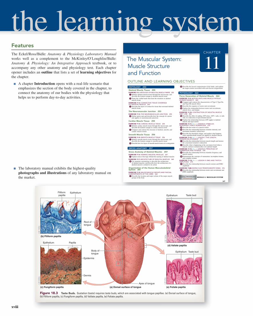
Figure 18.3 Taste Buds. Gustation (taste) requires taste buds, which are associated with tongue papillae. (a) Dorsal surface of tongue, (b) Filiform papilla, (c) Fungiform papilla, (d) Vallate papilla, (e) Foliate papilla.
Features
The Eckel/Ross/Bidle: Anatomy & Physiology Laboratory Manual works well as a complement to the McKinley/O' Loughlin/Bidle: Anatomy & Physiology: An Integrative Approach textbook, or to accompany any other anatomy and physiology text. Each chapter opener includes an outline that lists a set of learning objectives for the chapter.
■ A chapter Introduction opens with a real-life scenario that emphasizes the section of the body covered in the chapter, to connect the anatomy of our bodies with the physiology that helps us to perform day-to-day activities.
■ The laboratory manual exhibits the highest-quality photographs and illustrations of any laboratory manual on the market.
the learning system
xviii
9781259139437_FM_MAIN_i-xxiv_Print.indd 18 4/16/15 9:49 AM

CONCEPT CONNECTIONRecall that the gray matter of the spinal cord contains predominately neuron cell bodies, dendrites, and unmyelinated axons. In addition, gray matter also contains various types of neurons (e.g., motor neurons, somatic sensory neurons, etc.), which are typically contained within a speci�ed area of the gray matter. For example, the posterior horn of the spinal cord contains predominately unmyelinated axons of somatic sensory neurons. The cell bodies for those sensory neurous are located in the posterior root ganglion, and their axons enter the spinal cord via the posterior root. The anterior horn of the spinal cord contains cell bodies for somatic motor neturons. Somatic motor neurons are the neurons that stimulate skeletal muscles to contract. Recall from chapter 11 that a motor unit is a motor neuron and
all of the �bers that it innervates. That somatic motor neuron becomes “excited” within the anterior horn of gray matter within the spinal cord, and its axon exits the central nervous system through the anterior root. It then joins a spinal nerve and ultimately travels to individual muscle �bers within a skeletal muscle. An action potential traveling along a somatic motor neuron will excite the muscle �bers, causing an increase in force production by the muscle. The lateral horns of gray matter in the thoracic and lumber regions of the spinal cord contain cell bodies of autonomic motor neurons. Autonomic motor neurons innervate cardiac muscle, smooth muscle, and glands (see chapter 17). As with somatic motor neurons, the axons of autonomic motor neurons exit the central nervous system by traveling through the anterior root to a spinal nerve.
INTEGRATE■ The content of the laboratory manual is informed by the textbook, and both the textbook and the laboratory manual share similar pedagogic elements: Concept Connection, Learning Strategy, and Clinical View features from the text are also employed in the laboratory manual.
■ Integrate: Concept Connection boxes draw concepts from the classroom into the laboratory for a real-time review of how previously covered concepts relate to body systems.
■ Integrate: Learning Strategy boxes offer tried-and-tested learning strategies that consist of everyday analogies, mnemonics, and useful tips to aid understanding and memory.
■ Integrate: Clinical View sidebars reinforce facts through a clinical discussion of what happens when the body doesn' t perform normally.
■ Pre-Laboratory Worksheets at the start of each chapter consist of important refresher points to provide students with a ª warm-upº before entering the laboratory classroom. Some questions pertain to previous activities that are relevant to upcoming exercises, while others are basic questions that students should be able to answer if they have read the chapter from their lecture text before coming into the laboratory classroom. The goal of completing these worksheets is to have students arrive at the laboratory prepared to deal with the material they will be covering, so valuable laboratory time isn' t lost in reviewing necessary information. All Pre-Laboratory Worksheet questions are assignable within Connect.
LEARNING STRATEGYTo identify super�cial muscles and tendons, place your left palm on the medial epicondyle of your right humerus. In this position, the order of the muscles on your right forearm, from lateral to medial, is:
Index �nger—pronator teres (PT)
Middle �nger—�exor carpi radialis (FCR)
Ring �nger—palmaris longus (PL)
Pinky �nger—�exor carpi ulnaris (FCU)
While performing this exercise, �ex your wrist and digits to identify the tendons, from lateral to medial, of the �exor carpi radialis, palmaris longus, and �exor carpi ulnaris.
INTEGRATE
Pronator teresFlexor carpi radialisPalmaris longusFlexor carpi ulnaris
(Left hand coversmedial epicondyle)CLINICAL VIEW
Piriformis SyndromeThe piriformis muscle is a “pear-shaped” muscle that lies in close proxim-ity to important structures within the gluteal region, such as the sciatic nerve, and the gluteal arteries and nerves. Piriformis syndrome is a pain-ful condition that results from in�ammation or overuse of the piriformis muscle. The incidence of piriformis syndrome is relatively common in athletes such as runners and cyclists, who may develop an imbalance in the strength of the piriformis muscle as compared to the gluteal muscles. Speci�cally, the syndrome occurs when the piriformis muscle (which
laterally rotates the thigh) is stronger than the gluteus medius and gluteus minimus muscles (which are responsible for medial rotation of the thigh). As the piriformis muscle becomes in�amed or experiences spasms, it may also compress the underlying sciatic nerve, resulting in sciatica. Sciatica is a tingling, painful, or even numbing sensation that travels down the path of the sciatic nerve. Patients complain of shooting pain that runs from the gluteal region down the lateral aspect of the thigh, and toward the leg. Often the pain may be exacerbated when the body is held in certain positions, such as prolonged sitting or standing. The symptoms of piriformis syndrome can be reduced with the administration of anti-in�ammatory drugs and through stretching exercises.
INTEGRATE
These Pre-Laboratory Worksheet questions may be assigned by instructors through their course.
Chapter 14: Nervous Tissues Name: _____________________________
Date: ____________ Section: ___________
PRE-LABORATORY WORKSHEET
C
F
G
A
B
E
DH
I
J
1. For each structure listed, write the corresponding letter as labeled in the diagram.
axon
axon hillock
cell body (soma)
chromatophilic substance
dendrite
myelin sheath/neurolemmocyte (Schwann cell)
neuro�bril node (node of Ranvier)
nucleolus
nucleus
synaptic knobs
2. Match the description listed in column A with the corresponding cell type listed in column B.
Column A
1. cells that myelinate axons in the central nervous system
2. cells that help reinforce the blood-brain barrier
3. glial cells found within peripheral nerve ganglia
4. cells that myelinate axons in the peripheral nervous system
5. cells that engage in phagocytosis in response to tissue injury
6. cells that line the ventricles of the brain
3. Match the description listed in column A with the corresponding connective tissue structure listed in column B.
Column A
1. surrounds a fascicle of axons
2. surrounds an individual axon
3. surrounds the entire nerve
Column B
a. endoneurium
b. epineurium
c. perineurium
Column B
a. astrocytes
b. ependymal cells
c. microglia
d. neurolemmocytes (Schwann cells)
e. oligodendrocytes
f. satellite cells
347Chapter Fourteen Nervous Tissues
xixPreface
9781259139437_FM_MAIN_i-xxiv_Print.indd 19 4/16/15 9:49 AM

■ In-chapter activities offer a mixture of labeling exercises, sketching activities, table completion exercises, data recording and analysis, palpation of surface anatomy, and other sources of learning. In the gross anatomy exercises of this manual, structures such as cranial bones and muscles of the body are not always presented as labeled photos, since students already have labeled photos provided in their anatomy and physiology textbook. Instead, images are presented as labeling activities with a checklist of structures. The checklists serve two purposes: (1) they guide students to items they should be able to identify on classroom models, fresh specimens, or cadavers (if the laboratory uses human cadavers), and (2) they double as a list of terms students can use to complete the labeling activities. Answers to the labeling activities are provided in the Appendix. Thus, if a student does not know what a leader line is pointing to, or cannot remember the correct term, the Appendix serves as a resource for locating the correct answer.
■ Anatomy & Physiology Revealed® (APR) correlations, indicated by the APR logo, direct students to related content in this cutting-edge software.
■ Each chapter contains numerous tables, which concisely summarize critical information and key structures and serve as important points of reference while in the laboratory classroom. Most tables contain a column that provides word origins for each structure listed within the table. These word origins are intended to give students continual exposure to the origins of the language of anatomy and physiology, which is critical for learning and retention.
■ Numerous Physiology Interactive Lab Simulations© (Ph.I.L.S.) 4.0 exercises throughout the laboratory manual make otherwise difficult and expensive experiments a breeze, and offer additional opportunities to aid student understanding of physiology.
■ BIOPAC© exercises are included in chapters 11, 15, 17, 21, 22 and 24.
■ Post-Laboratory Worksheets at the end of each chapter serve as a review of the materials just covered, and challenge students to apply knowledge gained in the laboratory. The Post-Laboratory Worksheets contain in-depth critical thinking question types, and are perforated so they can be torn out and handed in to the instructor, if so desired. Assessment questions are organized by exercise, and are keyed to the Learning Objectives from the chapter opener outline.
■ ªD o You Know the Basics?º questions quiz students on the material they have just learned in the chapter, using a variety of question formats including labeling, table completion, matching exercises, and fill-in-the-blank.
■ ªC an You Apply What You've Learned?º questions are often clinically oriented and expose health-sciences students to problem solving in clinical contexts.
■ ªC an You Synthesize What You've Learned?º questions combine concepts learned in the chapter to ensure student understanding of each chapter's objectives.
7
8
1
5
6
3
2
4
Synovial JointsSynovial joints have a complex structure that includes a joint cavity filled with uid. The term synovial literally means “together with egg” (syn, together, + ovum, egg). This term refers to the uid inside the joint (the synovial uid), which has the consistency and appear-ance of egg white. The joint cavity filled with synovial uid allows
the articulating bones to move easily past one another with very little friction between the bones. The features of synovial joints are cov-ered in detail in the next several exercises. These include exercises covering the general structure of a synovial joint, categories of syno-vial joints, and the types of movements allowed by synovial joints.
EXERCISE 10.3
GENERAL STRUCTURE OF A SYNOVIAL JOINT
1. Observe a model of a synovial joint, preferably a model of the knee joint. Identify the features of a typical synovial joint listed in table 10.4.
2. Label the components of a synovial joint in figure 10.3, using table 10.4 and the textbook as guides.
3. Optional Activity: 5: Skeletal System—Watch the “Synovial Joint” animation for a summary of synovial joint structure and types.
Figure 10.3 Diagram of a Representative Synovial Joint. Use the terms listed to �ll in the numbered labels in the �gure.
articular capsule
articular cartilage
�brous layer of articular capsule
ligament
periosteum
synovial (joint) cavity
synovial membrane
yellow bone marrow
Table 10.4 Components of Synovial Joints
Structure Description Word Origin
Articular Capsule
Consists of two layers: an outer fi brous capsule and an inner synovial membrane
arthron, a joint, + capsa, a box
Articular Cartilage
Hyaline cartilage found on the epiphyses of the articulating bones
arthron, a joint
Fibrous Layer of Articular Capsule
Dense irregular connective tissue that anchors the periosteum of the two articulating bones to each other; thickenings of the fi brous capsule form several joint ligaments
fi bra, fi ber
Synovial Cavity
A cavity within the joint that is lined by a synovial membrane and fi lled with synovial fl uid
syn, together, + ovum, egg, + cavus, hollow
Synovial Fluid
A viscous, oily fl uid located within the synovial joint; functions as a lubricant, to nourish the articular cartilage, and as a shock absorber
syn, together, + ovum, egg, + fl uidus, to fl ow
Synovial Membrane
Composed primarily of areolar connective tissue that forms the inner lining of the articular capsule and covers internal joint surfaces not covered by cartilage; responsible for the formation of synovial fl uid
syn, together, + ovum, egg, + membrana, a skin
235Chapter Ten Articulations
l l ligament y y yellow bone mallow bone mallow rrow
Do You Know the Basics?
Exercise 9.1: Bones of the Pectoral Girdle
1. The head of the humerus articulates with which bony landmark of the scapula? (Circle one.) 1
a. acromion
b. coracoid process
c. glenoid fossa
d. spine
e. suprascapular notch
2. The only point of articulation between the pectoral girdle and the axial skeleton is the sternoclavicular joint. _____________________ (True/False) 2
3. Match the bones listed in column A with the corresponding joint listed in column B. 1 2 3
Column A Column B
____ 1. clavicle and scapula a. acromioclavicular joint
____ 2. humerus and ulna b. elbow joint
____ 3. scapula and humerus c. glenohumeral joint
____ 4. ulna, radius, and carpal bones d. wrist joint
4. Label the following diagram of an articulated shoulder girdle using the terms listed: 1 2
Right scapula and clavicle articulation, anterior view
Clavicle
Scapula
13
14
12
11
10
9
8
1
2
3
4
5
6
7
Scapula Clavicle
acromion acromial end
coracoid process sternal end
glenoid cavity
inferior angle
infraglenoid tubercle
lateral border
medial border
spine
subscapular fossa
superior angle
supraglenoid tubercle
suprascapular notch
Chapter 9: The Skeletal System: Appendicular Skeleton
POST-LABORATORY WORKSHEET
Name: ______________________________
Date: ____________ Section: ___________
The 1 corresponds to the Learning Objective(s) listed in the chapter opener outline.
225Chapter Nine The Skeletal System: Appendicular Skeleton
xx Preface
9781259139437_FM_MAIN_i-xxiv_Print.indd 20 4/16/15 9:49 AM

Teaching Supplements
Answers to the Pre-Laboratory and Post-Laboratory Worksheets can be found within the Instructor's Manual for this Laboratory Manual within Connect, by accessing the McKinley/O' Loughlin/Bidle: Anatomy & Physiology, 2nd edition Instructor Resources. Image files for use in presentations and teaching materials are also provided for instructor use at this location.
Anatomy & Physiology Revealed®: An Interactive Cadaver Dissection ExperienceAvailable online at www.aprevealed.com, and as an APR application on Apple® and Android™ tablets, this amazing multimedia tool is designed to help students learn and review human anatomy using cadaver specimens. Detailed cadaver photographs blended with a state-of-the-art layering technique provide a uniquely interactive dissection experience. This easy-to-use program features the following sections:
■ Dissection: Peel away layers of the human body to reveal the structures beneath the surface. Structures can be pinned and labeled, just as in a real dissection lab. Each labeled structure is accompanied by detailed information and an audio pronunciation. Dissection images can be captured and saved.
■ Animation: Compelling animations demonstrate muscle action, clarify anatomical relationships, and explain difficult concepts.
■ Histology: Labeled light micrographs presented with each body system allow students to study the cellular detail of tissues at their own pace.
■ Imaging: Labeled X-ray, MRI, and CT images familiarize students with the appearance of key anatomical structures as seen through different medical imaging techniques.
■ Self-test: Challenging exercises let students test their ability to identify anatomical structures in a timed practical exam format or with traditional multiple choice questions. A results page provides an analysis of test scores plus links back to all incorrectly identified structures for review.
■ Anatomy Terms: This visual glossary of general terms includes directional and regional terms, as well as planes and terms of movement.
Instructors may customize APR 3.0 to their course by selecting the specific structures they require in their course, and APR 3.0 does the rest. Once the structure list is generated, APR highlights these selected structures for students. APR contains all the material covered in an A&P course, including these three new modules:
■ Body Orientation
■ Cells and Chemistry
■ Tissues
APR is now available in two new versions!
Anatomy & Physiology Revealed | Cat® and Anatomy & Physiology Revealed | Fetal Pig® are online interactive cat dissection and fetal pig dissection experiences that use cat photos or fetal pig photos, combined with a layering technique that allows you to peel away layers to reveal structures beneath the surface. Both Anatomy & Physiology Revealed | Cat and Anatomy & Physiology Revealed | Fetal Pig offer animations, histologic and radiologic imaging, audio pronunciations, and comprehensive quizzing.
xxiPreface
9781259139437_FM_MAIN_i-xxiv_Print.indd 21 4/16/15 9:49 AM

Physiology Interactive Lab Simulations© (Ph.I.L.S.) offers 42 lab simulations that may be used to supplement or substitute for wet labs. Users may adjust variables, view outcomes, make predictions, draw conclusions, and print lab reports.
McGraw-Hill LearnSmart Labs™THE Virtual Lab Experience
Based on the same world-class super-adaptive techn ology as LearnSmart, McGraw-Hill's LearnSmart Labs™
are must-see, outcomes-based lab simulations. LearnSmart Labs assess a student's knowledge and adaptively correct deficiencies, allowing the student to learn faster and retain more knowledge with greater success.
First, a student's knowledge is adaptively leveled on core learn-ing outcomes: questioning reveals knowledge deficiencies that are corrected by the delivery of content that is conditional on a student's response. Then, a simulated lab experience requires the student to think and act like a scientist: recording, interpreting, and analyzing data using simulated equipment found in labs and clinics. The student is allowed to make mistakesÐa powerful part of the learning experi-ence! A virtual coach provides subtle hints when needed, asks ques-tions about the student's choices, and allows the student to reflect upon and correct those mistakes. Whether your need is to overcome the logistical challenges of a traditional lab, provide better lab prep, improve student performance, or make your online experience one that rivals the real world, LearnSmart Labs accomplishes it all.
A c k n o w L E d g M E n t S
This laboratory manual is the product of the excellent work and dedication of a consummate group of talented professionals who have helped lead us through this publishing process. We are forever indebted to all of you for embarking on this journey with us.
We wish to thank McGraw-Hill for providing us the unique opportunity to share our enthusiasm for teaching anatomy and physiology through the pages of this laboratory manual.
From start to finish, this book has been carried through the able hands of product developers Donna Nemmers and Kristine Queck. April Southwood, Content Project Manager, kept everyone on track and managed countless details in the production process.
The beautiful design and line art that make this laboratory manual shine are products of a team of hugely talented designers and artists including David Hash at McGraw-Hill, and the fantastic EPS illustration team. Carrie Burger's direction and guidance were instrumental in bringing out the best of our photography program. Danny Meldung, of Photo Affairs, Inc. researched wonderful photos for this edition.
We thank all of the reviewers of the manual (listed below) for taking the time to review this manual and provide us with their insight and perspective, gained from years of experience in the classroom. We hope we have honored your suggestions for improvement, and we welcome continued feedback. We also thank the many students we have had the pleasure of interacting with over the years, for teaching us what works or does not work in the classroom.
The extent of our gratitude is limitless when it comes to the love, understanding, and support that our families, friends, and colleagues gave to us throughout this process. We are truly honored to live our lives in the presence of such wonderful people.
To the users of this laboratory manual: We sincerely hope we have created a learning resource that not only will excite you about the study of anatomy and physiology, but also will actively engage you in the laboratory as you learn about the wonders of the human body. We welcome your thoughts and suggestions for improvements.
Christine M. EckelDepartment of Life and Environmental SciencesCarroll [email protected]
Kyla Turpin RossDepartment of BiologyGeorgia State [email protected]
Terri Stouter BidleScience DivisionHagerstown Community [email protected]
xxii Preface
9781259139437_FM_MAIN_i-xxiv_Print.indd 22 4/16/15 9:49 AM

ReviewersAndrew E. Accardi
Central Carolina Technical College
Kyle BartowIndian River State College
Gladys BoldingGeorgia Perimeter College
Pamela K. ElfUniversity of Minnesota—Crookston
Theresa FeltenPolk State College—Lakeland
Joseph D. GarWest Kentucky Community and Technical College
Sylvester HackworthBishop State Community College
Steven B. HammerIndian River State College
Lesleigh HastingsWake Tech Community College
Karen Ramey HlinkaWest Kentucky Community and Technical College
Kathy Jo Ann JacksonMcNeese State University
Scott JohnsonWake Tech Community College
Karen Dunbar KareivaIvy Tech Community College—Northwest Region
Jessica Burr LeaCentral Carolina Technical College
Tiffany B. McFalls-SmithElizabethtown Community and Technical College
Jill Y. O’MalleyErie Community College
Justicia OpokuUniversity of Maryland—College Park
Jo RogersUniversity of Cincinnati
Connie E. RyeEast Mississippi Community College
Dee Ann SatoCypress College
Jackie ThomasWake Tech Community College
Joyce E.M. WallHousatonic Community College
Charles M. WatsonMidwestern State University
Martha T. WolfeElizabethtown Community and Technical College
Jennie L. YatesSt. Petersburg College—Seminole Campus
xxiiiPreface
9781259139437_FM_MAIN_i-xxiv_Print.indd 23 4/16/15 9:49 AM

9781259139437_FM_MAIN_i-xxiv_Print.indd 24 4/16/15 9:49 AM