anatomy of female reproductive system teng yincheng m.d., ph.d., professor teng yincheng m.d.,...
Post on 19-Dec-2015
219 views
TRANSCRIPT

Anatomy of Female Anatomy of Female reproductive systemreproductive system
Teng YinchengTeng Yincheng M.D., Ph.D., Professor M.D., Ph.D., Professor
Department Of Obstetrics & GynecologyDepartment Of Obstetrics & Gynecology
Renji Hospital Affiliated to SJTU School of Medicine Renji Hospital Affiliated to SJTU School of Medicine

• bony pelvis
• female genital organs
• blood supply of female genital organs

Bony pelvisBony pelvis1. The bony pelvis is made up of 4
bones:
ilium
ischium
pubis
sacrum

1) 2 os coxae or innominate bones the iliumcoxae the ischium the pubis

2) 1 sacrum: Formed from 5 sacral vertebrae which fuse.
3)1 coccyx: Formed from 4 fused vertebrae.

2.Joint and ligament2.Joint and ligament
Sacro-iliac joint
Sacro-coccygeal
joint
腹股沟韧带骶棘韧带骶结节韧带

3.True pelvis and false pelvis3.True pelvis and false pelvis
The pelvis is divided into
the pelvis major (false pelvis) is bounded by the lumbar vertebrae
posteriorly,an iliac fossa bilaterally,and the abdominal wall anteriorly
the pelvis minor (true pelvis)
is formed by sacrum and coccyx posteriorly and by the ischium and pubis laterally and anteriorly

4.Pelvic planes4.Pelvic planes
• The pelvic inlet( pelvic brim): the promontory, and alae of the
sacrum ,linea terminalis the superior border of the
symphysis pubis • The plane of greatest diameter
is bounded by the junction of the second and third sacral vertebrae posteriorly,the upper part of the obturator foramina laterally,and the midpoint of the pubis anteriorly

• The plane of least diameter: extends from the lower border of the pubis anteriorly to lower sacrum at the level of the ischial spines
• The plane of the outlet is irregular
consisting of two intersecting triangles

5.The types of female pelvis5.The types of female pelvis
Gynecoid pelvis Occurs 40%--50% the inlet is rounded the side walls are
straight the sacrum is well
curved, and the sacrosciatic notch is adequate
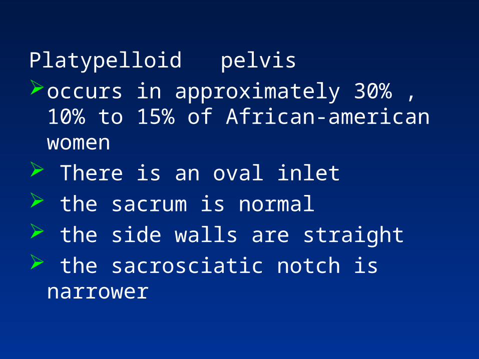
Platypelloid pelvisoccurs in approximately 30% , 10% to
15% of African-american women There is an oval inlet the sacrum is normal the side walls are straight the sacrosciatic notch is narrower

Android pelvis occurs in only 2% to 5% of women wedge-shaped inlet the side walls converge the sacrum is inclined forward the sacrosciatic notch is narrow

Anthropoid pelvisoccurs in approximately
20% ;approximately 40% of black womenthe inlet is oval, long, and narrow the side walls are straight(do not
converge)the sacrum is long and narrowthe sacrosciatic notch is wide.Both the interspinous and intertuberous
diameter are somewhat smaller

6.Length of pelvic plane diameters6.Length of pelvic plane diametersPelvic plane diameter Average length(cm)
Inlet
Greatest diameter
Midplane
outlet
True conjugate
Obstetric conjugate
Diagonal conjugate
Transverse diameter
Oblique diameter
Anterior-posterior
Transverse
Anterior-posterior
Bispinous(interspinous)
Anterior-posterior
bituberous
10.5-11.5
10.0-11.0
12.5
13.5
12.5
12.75
12.5
11.5-12.0
10.0
11.5
8.0—11.0

7.Pelvic floor7.Pelvic floor


female genital organsfemale genital organs

mons pubis: fatty tissue
labia majora labia minora which lie
inside the labia majora clitoris: is located
anterior to the labia minora
1.External genitalia1.External genitalia

vestibuleLies between the labia minora Is
bounded Anteriorly by clitoris Posteriorly by perineum
Urethra and vaginal orifice open to the midline of the vestibule
Bartholin’s glands also empty into the vestibule

2.Internal genitalia2.Internal genitalia 1)VaginaMuscular tube from vestibule extends
to uterusThe anterior vaginal wall is 2cm
shorter than the posterior wallThe area around the cervix---the
fornix the posterior fornix is important
pouch which allows access to the peritoneal cavity from vagina
The vaginal wall consists of a mucous membrane , Beneath this is a submucosal layer of connective tissue which contains a rich of veins and lymphatics
The muscular wall has three layers of smooth muscle.

2)uterus2)uterusCervix
cylindrical shape, 2-3cm long vaginal portion external os
cervix tube
stratified squamous epithelium
simple columnar epithelium

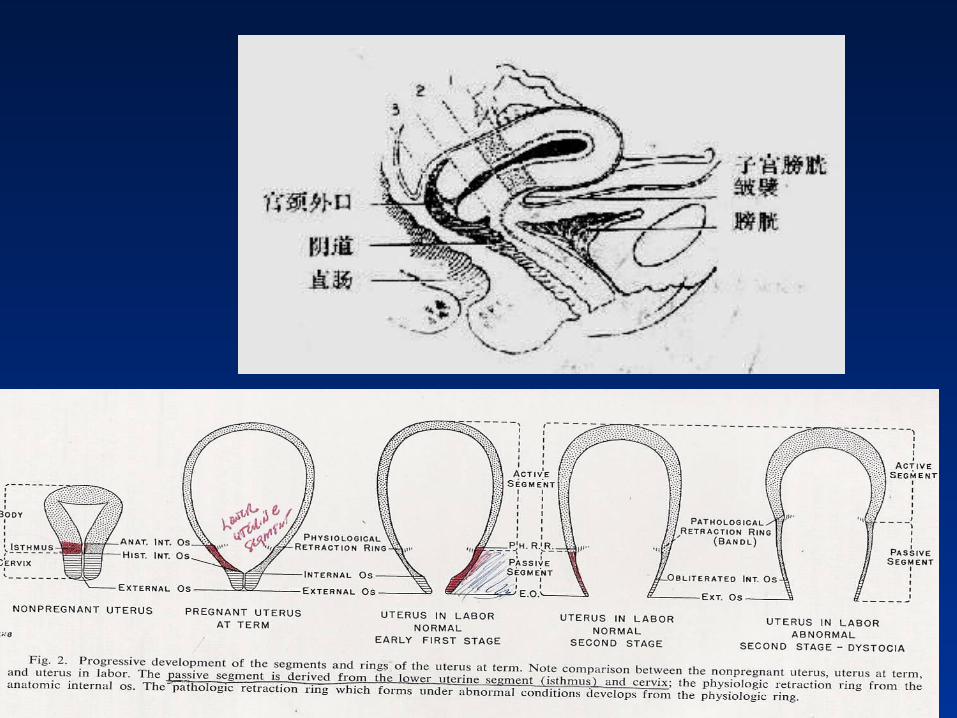
2) Isthmus separate cervix and body
3) Corpus cornu fundus
long 7-8cm; wide 4-5cm; thick 2-3cm
The ratio of the body to the cervix
Before puberty 1:2; after puberty 2:1

The uterus is surported by the following ligaments
Round ligamentBroad ligamentCardinal ligamentUtero-sacral ligament
Histology of uterusHistology of uterusThere are three layers of the bodyThe inner is endometrium which is
consisted of the simple columnar epithelium
Beneath the mucosa is the muscular layer,covered by peritoneal serosa

3)Fallopian tubes3)Fallopian tubesLength: 8-14cmThe tube is divided four portions Interstitial isthmus Ampulla:the centre portion Infundibulum:surround the ovary
and help to collect the oocyte at the time of ovulation
The innner layer is covered by ciliate columnar epitheliun
The cilia beat toward the uterus assisting in oocyte transport

4. ovaries4. ovaries• Be located on either
side of the uterus• Volume:4*3*1cm• Weight:5-6g• be almond-shaped• Is divided two portion: cortex(outer portion): contains amount of
follicles medulla(central portion): blood vessels,
nerve fibres, lymphatics

blood supplyblood supply
Arterial systemOvarian artery:
from abdominal aorta (left may be from kedney artery)
Uterine arteryVaginal arteryInternal pudendal
artery from internal iliac artery


Lymphatic drainageLymphatic drainage
外生殖器淋巴 腹股沟浅 上组 下组 腹股沟深盆腔淋巴 髂淋巴组 骶前淋巴组 腰淋巴组
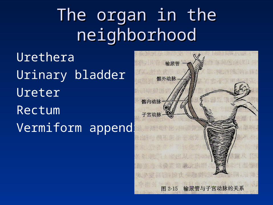
The organ in the neighborhoodThe organ in the neighborhood
Urethera
Urinary bladder
Ureter
Rectum
Vermiform appendix

Teng YinchengTeng Yincheng M.D., Ph.D., ProfessorM.D., Ph.D., Professor
Department of Obstetrics & GynecologyDepartment of Obstetrics & Gynecology
Renji Hospital Affiliated to SJTU School of MedicineRenji Hospital Affiliated to SJTU School of Medicine
Thanks for Your AttentionThanks for Your Attention