anatomical variations of the vascular supply of the
TRANSCRIPT
ONLINE FIRST
This is a provisional PDF only. Copyedited and fully formatted version will be made available soon.
ISSN: 0015-5659
e-ISSN: 1644-3284
Anatomical variations of the vascular supply of the cutaneouscomponent of the serratus anterior myocutaneous flap: a
systematic review
Authors: C. Gakis, D. Chrysikos, A. Samolis, V. Protogerou, G. Tsourouflis, T.Troupis
DOI: 10.5603/FM.a2021.0111
Article type: Review article
Submitted: 2021-09-06
Accepted: 2021-10-13
Published online: 2021-10-26
This article has been peer reviewed and published immediately upon acceptance.It is an open access article, which means that it can be downloaded, printed, and distributed freely,
provided the work is properly cited.Articles in "Folia Morphologica" are listed in PubMed.
Powered by TCPDF (www.tcpdf.org)

Anatomical variations of the vascular supply of the cutaneous component of the
serratus anterior myocutaneous flap: a systematic review
Christos Gakis et al., Anatomy of serratus anterior myocutaneous flap vascular supply
C. Gakis1, D. Chrysikos1, A. Samolis1, V. Protogerou1, G. Tsourouflis2, T.Troupis1
1Department of Anatomy, School of Medicine, National and Kapodistrian University
of Athens, Greece
2Second Department of Propedeutic Surgery, Medical School, National and
Kapodistrian University of Athens, Greece
Address for correspondence: Prof. T. Troupis, Department of Anatomy, School of
Medicine, Faculty of Health Sciences, National and Kapodistrian University of
Athens, 75 Mikras Asias str., Goudi, 11527 Athens, Greece, tel: +30-210-7462388,
fax: +30-210-7462398, e-mail: [email protected]
ABSTRACT
Although appealing from a reconstructive standpoint, the incorporation of the
overlying skin in a serratus anterior muscle flap has not yet seen widespread use, due
to considerations with its blood supply. In the present study, a systematic review of
the literature has been performed, evaluating studies that investigated the vascular
anatomy and variations of serratus anterior myocutaneous flap. The anatomy of the
cutaneous blood supply, the size of the cutaneous territory, the design of the skin
paddle and the reconstructive goals were analyzed. The results showed that the main
blood supply originates from the intramuscular anastomoses between intercostal
artery perforators and the serratus artery branch in the form of choke vessels.
Complementary perfusion from true intramuscular vessel anastomoses or from direct
serratus artery cutaneous perforators could contribute to the skin blood supply but
only in 25% of the cases. The design of the flap is elliptical with its long axis over the
harvested muscle slips and maximum width is approximately 6-8 cm. Α

myocutaneous serratus anterior flap could be applied in a variety of reconstructive
fields, most commonly for head and neck defects. A delay procedure would
considerably enhance the perfusion of the cutaneous component and improve the
overall viability of the flap.
Key words: serratus anterior, flap skin, cutaneous component, blood supply
INTRODUCTION
Myocutaneous flaps, flaps consisting of muscle and their overlying skin, are
frequently used in reconstructive surgery, especially when large tissue bulk is
required. When a skin island is transposed, attached to the underlying muscle, the
subdermal vascular plexus is discontinued. Cutaneous blood supply then stems from
perforator vessels, arteries and accompanying veins, that arise from the muscle
surface [1]. This kind of cutaneous blood supply is very reliable in body areas where
muscles and overlying skin are firmly connected, such as Latissimus Dorsi muscle,
but becomes questionable when skin and muscle are loosely attached together, such as
Serratus anterior [2].
ΕMBRYOLOGY
Serratus anterior muscle belongs to the superficial thoracic muscles. Its origin
and development is different from those of limb muscles. It is believed that it is
formed from myotomes but its exact origin is not certain, and it is not clarified how
myotome cells attach to the scapula to create these muscle. At first in the 9 mm
embryo it resembles a premuscle mass with no attachments neither to the scapula nor
to the ribs. It becomes more defined and attaches with digitations to the ribs in the
11mm embryo. Finally it develops to its adult form with insertion into the scapula in
the 14mm embryo [3],[4],[5].
SURGICAL ANATOMY
Serratus anterior muscle is a fan shaped muscle, originating from the upper 8
to 9 ribs at the lateral chest wall, which inserts into the superior angle, the medial
border and the inferior angle of the scapula. Its blood supply stems from the superior

thoracic artery, the lateral thoracic artery and the serratus artery branch of the
thoracodorsal artery. The latter perfuses the inferior part of the muscle which is
commonly used as a free or pedicled flap [3].
A serratus anterior myocutaneous flap, although first reported in 1982 [6], has
not yet seen widespread use due to considerations with the viability of the cutaneous
component. It was even considered unacceptable to include the overlying skin,
because until then a direct communication of the cutaneous perforators with the
serratus arterial network had yet not been identified [7].
An anatomic investigation of the cutaneous blood supply over the muscle and
its relation with the arterial axis of the serratus anterior flap, the definition of the
shape and size of the flap and the possible technical pitfalls, would delineate the
likelihood of harvesting a myocutaneous flap, which would be a useful option in the
armamentarium of a Reconstructive Surgeon.
MATERIALS AND METHODS
Search strategy
This systematic review was performed in accordance with the PRISMA
(Preferred Reporting Items for Systematic Reviews and Meta-Analyses) guidelines.
The protocol of this systematic review has been submitted to the Institutional Review
Board of Department of Anatomy, National and Kapodistrian, University of Athens,
Greece, and is available upon request. Eligible articles were identified by a search of
the Medline, the Cochrane Library and the Google Scholar bibliographical databases
for the period from April 1982 up to May 2019. The study protocol was agreed by all
co-authors. The search strategy included the following keywords: (“serratus anterior
flap” OR “skin” OR “cutaneous” OR “myocutanous” OR “musculocutaneous” AND
(“anatomical variants” OR “anomalies”)). Language restrictions were applied (only
articles in English, French, and German were considered eligible). Two investigators
(CG and DC), working independently, searched the literature and extracted data from
each eligible study. Reviews were not eligible, while all prospective and retrospective
studies, as well as case reports, were eligible for this systematic review. Manuscripts
that did not state the names of the authors were excluded. Case reports of a serratus
anterior flap with a cutaneous component without an anatomic investigation of the

skin blood supply were also excluded. In addition, we checked all the references of
relevant reviews and eligible articles that our search retrieved, so as to identify
potentially eligible conference abstracts. Titles of interest were further reviewed by
abstract. Finally, reference lists of eligible studies were manually assessed in order to
detect any potential relevant article (“snowball” procedure).
The literature review resulted in 10 relevant anatomical studies. Data was
collected and analyzed based on the anatomic basis of the cutaneous blood supply, the
cutaneous island dimensions (theoretical in cadavers and applied in patients), the
suggested design of a myocutaneous flap and the defect area where each one of them
was applied on.
RESULTS
The flow chart of the study depicts that the investigated subject is not an
intesively reviewed field (Figure 1).
Four studies proved the reliance of the blood supply of the cutaneous surface
on the serratus branch by a cause and effect experiment (intra-arterial dye injection
caused skin staining). Additionally, these studies calculated the mean cutaneous island
surface stained. Typically the procedure included the catheterization of the
thoracodorsal artery and the injection of variable volumes of a dye (methylene blue or
black ink). Perrot in his study used a mixture of a dye and a radiopaque substance for
radiologic co-evaluation [8]. All but one studies [9] stated that the arterial branch to
LD muscle was ligated prior to the injection of the dye.
The calculation of the stained surface was accomplished following a computer
assisted planimetric method [10], using a photo-processing software [9] or with a
simple estimation based on length and width [6]. Estimated width ranged between 5.5
and 21cm, length between 6 and 20.5 cm and surface between 38.4 and 223.6 cm2.
The boundaries of the stained skin in relation to anatomic landmarks were also taken
into consideration (Table I).
Apart from the cutaneous staining experiment, Perrot performed a cadaver
dissection and recognized multiple cutaneous perforators arising from the muscle

fascia, but did not proceed with a further investigation of their intramuscular course
[8].
Innoue noticed intraoperatively multiple short muscular branches arising from
the muscle fascia, during the harvest of pedicled myocutaneous Serratus Anterior
flaps. He speculated that the surface fascia of Serratus Anterior muscle receives its
blood supply from the thoracodorsal artery and perfuses the overlying skin through
these short perforators [11].
Pittet in her cadaver study injected a mixture of a dye, gelatin and barium for
radiologic co-evaluation. She recognized intercostal artery perforators reaching the
skin, as well as giving branches to Serratus Anterior during their intramuscular
course. However, she did not observe any anastomosis between these muscular
branches and the Serratus Artery branch [12].
Infact, it was Godat’s study that uncovered an intramuscular arterial
anastomosis. Following a large number of cadaver dissections, a detailed description
of the vascular anatomy of the Serratus Anterior muscle was achieved. Contributions
to the blood supply from the intercostal artery perforators were found through an
intramuscular anastomosis, however these were inconstant [13].
Hamdi as part of his cadaver dissection study, made a detailed mapping of the
intercostal perforators in the lateral thoracic wall (number of intercostal perforators
and their mean distance from the anterior border of Latissimus Dorsi muscle) trying to
delineate the anatomy of a lateral intercostal perforator fasciocutaneous flap (LICAP).
In 21% of the cases, a vascular anastomosis between Serratus Anterior muscle arteries
and intercostal artery perforators was found. The most frequent positions were in the
6th(30%) and 7th(38%) intercostal spaces. Based on this finding he suggested that in
the presence of that anastomosis, a Serratus Anterior perforator (fasciocutaneous) flap
could be raised [14].
Park found in his study (cadaver dissection and angiography) a similar
frequency of intramuscular anastomosis of the Serratus Anterior artery branch and
intercostal perforators (25%), although the sample in question was small. The
perforators arose from the 6th and 7th intercostal spaces in one case and from the 4th
intercostal space in another [15].
Tamburino in her cadaver dissection study discovered a direct cutaneous
perforator branching from the serratus artery branch in 25% of the cases. She
emphasized that these perforators had no intramuscular course and lay superficial to

the muscle. This perforator was found arising more frequently from the 6th (33%) and
7th (41.6%) intercostal spaces. In her radiologic study, occurrence was reported less
frequently (18%). The author attributed this to the radiological protocol used, since
the samples had been derived from CT chest angiographies of patients suspected for
pneumonic embolism. She believes that a special radiological protocol would have
probably revealed more perforators [16], (Table II).
The suggested design of the flap typically centers the axis of the flap over the
selected muscle slips. Lateral border varies from 2 to 3 cm over the Latissimus Dorsi
muscle to the anterior axillary line. Medial border most commonly reaches the
midclavicular line but a medial extension of 2cm is also described. The superior
border extends to the 5thor the 6th rib and the inferior border to the 8th or the 9th rib.
Maximum width is kept approximately to 6-8 cm to facilitate direct closure. A
different description was given by Janik who defined the vertical anatomic borders of
the flap in relation to the height of the hemithorax [9]. In Park’s study the suggested
orientation of the flap is parallel to the anterior border of Latissimus Dorsi muscle and
not to the Serratus Anterior [15], (Table III).(Figure 2)
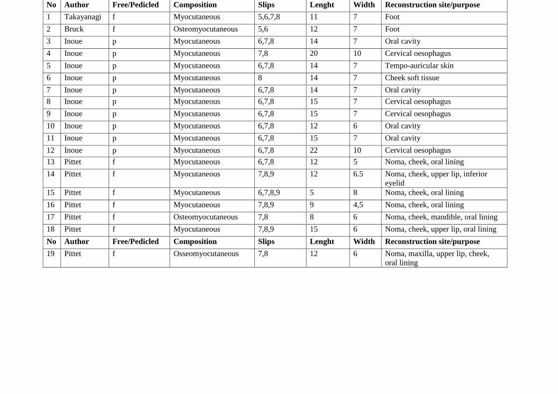
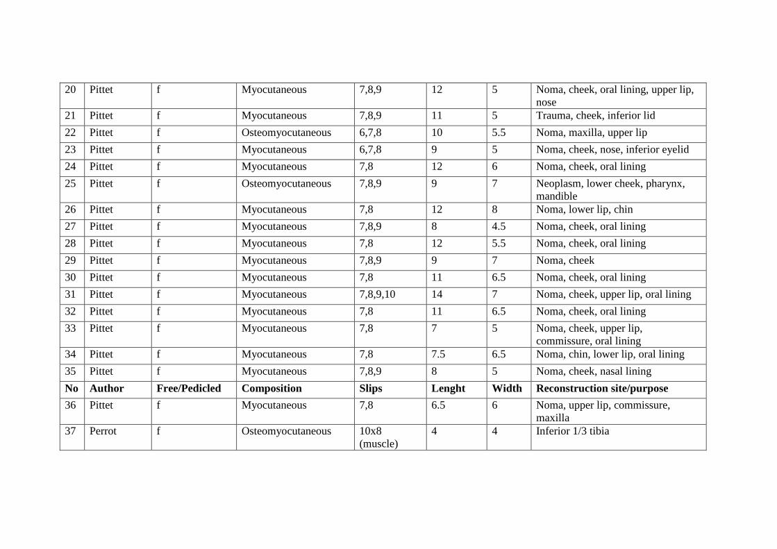
The studies included in our review, reported 54 successful flaps with a
cutaneous component. The flaps were either pedicled or free and were aimed to
reconstruct defects in various anatomic areas, most commonly head and neck defects
[6],[9],[11],[12],[25],,[26] (Table IV).
DISCUSSION
Serratus anterior muscle flap is a useful alternative option for the
reconstruction of various anatomical areas. The muscle is quite thin which makes it
superior compared to other workhorse muscle flaps such as Latissimus Dorsi and
Rectus Abdominis, in cases where a large muscle bulk is not desired. In cases where
only the lower slips are harvested, the resulting functional deficit is negligible [17]. It
is worth mentioning that the overlying skin is generally delicate and pliable.
Additionally the intervening subcutaneous fat in individuals with normal Body Mass
Index is thin. The incorporation of the skin in a myocutaneous Serratus Anterior flap
adds cosmetic superiority and durability while it allows for a shorter healing period in
comparison with a skin graft coverage. Flap monitoring is another convenience that a

skin paddle offers. Clinical examination of a muscle flap, is not as simple as of a
myocutaneous flap, and is even harder if the muscle is covered with a skin graft.
Capillary refill test and flap colour estimation are easily done on a cutaneous paddle
and reflect the viability of the whole flap. Monitoring of the skin island permits an
early recognition of a microvascular compromise and a timely surgical reexploration.
This has been shown to lead to higher salvage rates [27].
These advantages have, for the most part, encouraged the research on the
cutaneous blood supply anatomy.
The skin staining observed after the injection of a dye in the thoracodorsal
artery has proven that a myocutaneous flap could be perfused by this vascular axis.
On the other hand, the estimation of skin surface size using this method is considered
inefficient. Several small cutaneous perforators arise along the course of the
thoracodorsal artery, around the anterior border of the Latissimus Dorsi muscle[18].
In addition a direct dominant perforator branches off the thoracodorsal artery in 55-
60% with three branching patterns being reported, arising from the thoracodorsal
artery(50%), the Latissimus Dorsi branch(30%) and the Serratus branch(20%) [19],
[20]. When placing the catheter proximal to the origin of the thoracodorsal artery,
these perforators are also perfused. Moreover, in the study with the biggest stained
skin territory, it was not clarified whether the branch to the Latissimus Dorsi was
ligated [7]. Through this branch the perforators arising from the Latissimus Dorsi
muscle surface would also perfuse their overlying surface as well as adjacent
territories. Considering the close relationship of Latissimus Dorsi and Serratus
muscles, this could lead to an overestimation of the possible cutaneous territory.
Incising the perimetry of the flap to the level of the muscle fascia first, and then
injecting the dye, would preclude the perfusion of the skin from perforators that do
not arise from the Serratus Anterior muscle.
The lateral thoracic wall has been recognized as a well perfused area receiving
blood from the intercostal artery perforators [21]. Multiple reviewed anatomic studies
confirmed that several intercostal artery perforators pierce the Serratus Anterior
muscle reaching the overlying skin. Further dissection has led to recognition of
intramuscular anastomoses between intercostal artery perforators and the serratus
artery branch in approximately 20-25% [14], 13], [15]. These anastomoses were most
frequently found in the sixth and seventh intercostal spaces (typically the 6,7,8
serratus muscle slips overlying the 6th and 7th intercostal spaces are raised when

harvesting a serratus anterior muscle flap). Interestingly in one study, a cutaneous
perforator arising directly from the serratus artery branch and not from the intercostal
artery, was found in 25% [16]. The low consistency of these anastomoses does not
however correspond to the high success rates of Pittet and Inoue (almost 90% of
complete flap survival) [11], [12].
A reasonable theory was reported by Pittet. In a cadaveric study it was
recognized that intercostal artery perforators give cutaneous and muscular branches.
Although an intramuscular connection between intercostal artery perforators and the
Serratus Anterior muscle arterial network was not directly visualized in her cadaveric
study, she hypothesized the existence of this connection through choke vessels. As the
angiosome theory suggests, three dimensional blocks of tissue (angiosomes) are
perfused by a source artery. Neighboring angiosomes are linked with true vascular
anastomosis (same vessel calibre) or with choke vessels (vessels of smaller size) [22].
Choke vessels distend to the calibre of true anastomosis in cases of blood supply
insufficiency, such as when a flap is delayed, with a peak effect after 2 to 3 days [23].
A clinical indication of the existence of these choke vessels in the case series of Pittet
was that immediately postoperatively, no cutaneous perforators of the myocutaneous
flaps were traceable with a doppler examination. They became progressively audible,
suggesting the opening of the choke vessels. It is likely that the reviewed anatomic
studies have not recognized these choke vessels because of their minor diameter.
In cases where a preoperative recognition of a direct cutaneous perforator is
not feasible, as suggested by Tamburino [16], a reasonable approach would be to
delay the flap in order to enhance its vascularity. Flap delay is a preliminary
procedure of incompletely raising a flap, thus partially obscuring its blood supply.
During the delay period, choke vessels are allowed to dilate and as a result the flap
becomes more tolerant to relevant ischemia. That process is extremely beneficial to
the surgical outcome [24].
Serratus anterior is a thin muscle. For that reason, the point of bifurcation of
the intercostal artery perforator to a muscular and a cutaneous branch would not be far
from its undersurface. This bifurcation would be susceptible to thermal injury from
heat dissipation, if a cautery was uncontrollably used. In that case, blood would not be
allowed to run in a retrograde fashion, (serratus artery branch-choke vessels-muscular
branch of intercostal perforator-cutaneous branch of intercostal perforator) resulting
in flap failure. From a clinical standpoint, it should be noted that Pittet in her surgical

technique description highlighted that she paid attention not to damage the intercostal
artery perforators with the cautery [12]. Most likely her meticulous harvesting
technique preserves greater number of intercostal artery perforators, therefore
achieving high success rates. Consequently it would be advisable to avoid using a
monopolar cautery in the undersurface of the serratus anterior muscle. Instead
bipolar cautery or ligaclips should be used to isolate the perforators.
When the proposed flap designs were compared, homogeneity was observed.
The flaps are commonly oriented over the muscle slips to be used with slight
differences in their medial and lateral extent. On the contrary, Park positions the long
axis of the flap almost vertically against the serratus anterior muscle. Five to six
muscle slips are incorporated compared to authors who only harvest two or three [15].
Although not mentioned in this study, harvesting more muscle slips could have
possibly impaired function in a greater degree. That would be a concern when dealing
with reconstruction in young, active individuals.
CONCLUSIONS
Serratus anterior myocutaneous flap is a useful option for reconstruction in
various anatomical sites.
The intramuscular anastomoses of intercostal artery perforators with the
Serratus artery branch, in the form of choke vessels, is the basis of cutaneous blood
supply, while direct serratus artery cutaneous perforators and true anastomoses of
same size vessels contribute in a less predictable manner, in almost 25% of the cases.
Flap delay and meticulous dissection with avoidance of cautery induced
damage to the intercostal artery perforators, are considered important for a successful
outcome.
Conflicts of interest: None declared
REFERENCES
1 Carvey M, Yang G, Hage R. Skin flaps: A novel approach to medical student
instruction on integumentary arterial blood supply. Transl. Res. Anat., 23 (2021),
Article 100104, 10.1016/j.tria.2020.100104

2 Gurtner G, Neligan P. Plastic Surgery: Volume 1: Principles, 3rd Edition, Elsevier,
2013
3 Lung K, St Lucia K, Lui F. Anatomy, Thorax, Serratus Anterior Muscles. In:
StatPearls [Internet]. Treasure Island (FL): StatPearls Publishing; 2021
4 Pu Q, Huang R, Brand-Saberi B. Development of the shoulder girdle musculature.
Dev Dyn. 2016 ;245(3):342-50. doi: 10.1002/dvdy.24378.
5 Valasek P, Theis S, Krejci E,et al. Somitic origin of the medial border of the
mammalian scapula and its homology to the avian scapula blade. J Anat.
2010;216(4):482-8. doi: 10.1111/j.1469-7580.2009.01200.
6 Takayanagi S, Tsukie T. Free serratus anterior muscle and myocutaneous flaps.
Ann Plast Surg. 1982 ; 8(4):277-83. doi: 10.1097/00000637-198204000-00003.
7 Merle, M. Grandes pertes de substance de la main. In P. Banzet and J. M. Servant
(Eds.), Chirurgie Plastique, Reconstructive et Esthetique. Paris: Flammarion, 1994. P.
580
8 Perrot P, Duteille F, Leaute F et al. Etude anatomique et applications cliniques des
caractéristiques de la palette cutanée du lambeau libre de grand dentelé , Ann Chir
Plast Esthet. 2006 ;51(6):494-8.. doi: 10.1016/j.anplas.2006.02.003.
9 Janik SJ, Paraszti I, Hirtler L et al. Design of skin islands for a myocutaneous
serratus anterior free flap-An anatomical study and clinical implication for pharyngeal
reconstruction after laryngopharyngectomy. Clin Otolaryngol. 2019;44(3):227-234.
doi: 10.1111/coa.13257.
10 Mijatović D, Bulić K, Dzepina I et al. The supply of blood in the skin territory
above the lower part of the serratus anterior muscle. Coll Antropol. 2006;30(3):543-7.
PMID: 17058521.
11 Inoue T, Ueda K, Hatoko M, et al. The pedicled extended serratus anterior
myocutaneous flap for head and neck reconstruction. Br J Plast Surg. 1991;44(4):259-
65. doi: 10.1016/0007-1226(91)90067-t.

12 Pittet B, Mahajan AL, Alizadeh N,et al. The free serratus anterior flap and its
cutaneous component for reconstruction of the face: a series of 27 cases. Plast
Reconstr Surg. 2006;117(4):1277-88. doi: 10.1097/01.prs.0000208297.02556.a5.
13 Godat DM, Sanger JR, Lifchez SD et al. Detailed neurovascular anatomy of the
serratus anterior muscle: implications for a functional muscle flap with multiple
independent force vectors. Plast Reconstr Surg. 2004;114(1):21-9; discussion 30-1.
doi: 10.1097/01.prs.0000129072.11466.c3.
14 Hamdi M, Spano A, Landuyt KV et al. The lateral intercostal artery perforators:
anatomical study and clinical application in breast surgery. Plast Reconstr Surg.
2008;121(2):389-396. doi: 10.1097/01.prs.0000298317.65296.cf.
15 Park SO, Chang H, Imanishi N. The free serratus anterior artery perforator flap-A
case report and anatomic study. Microsurgery. 2016;36(4):339-344. doi:
10.1002/micr.30038.
16 Tamburino S, Menez T, Laloze J et al. Free serratus anterior artery perforator flap:
a case report with an anatomic and radiological study. Surg Radiol Anat.
2017;39(8):837-842. doi: 10.1007/s00276-017-1824-0.
17 Whitney TM, Buncke HJ, Alpert BS et al. The serratus anterior free-muscle flap:
experience with 100 consecutive cases. Plast Reconstr Surg. 1990;86(3):481-90;
discussion 491.
18 Miyamoto S, Arikawa M, Kagaya Y et al. Septocutaneous thoracodorsal artery
perforator flaps: a retrospective cohort study. J Plast Reconstr Aesthet Surg.
2019;72(1):78-84. doi: 10.1016/j.bjps.2018.08.026.
19 Thomas BP, Geddes CR, Tang M et al. The vascular basis of the thoracodorsal
artery perforator flap. Plast Reconstr Surg. 2005;116(3):818-22. doi:
10.1097/01.prs.0000176253.42394.7c
20 Heitmann C, Guerra A, Metzinger SW et al. The thoracodorsal artery perforator
flap: anatomic basis and clinical application. Ann Plast Surg. 2003;51(1):23-9. doi:
10.1097/01.SAP.0000054189.14799.F3.
21 Palmer JH, Taylor GI. The vascular territories of the anterior chest wall. Br J
Plast Surg. 1986;39(3):287-99. doi: 10.1016/0007-1226(86)90037-8.
22 Taylor GI, Palmer JH. The vascular territories (angiosomes) of the body:
experimental study and clinical applications. Br J Plast Surg. 1987;40(2):113-41. doi:
10.1016/0007-1226(87)90185-8.
23 Taylor GI, Corlett RJ, Dhar SC et al. The anatomical (angiosome) and clinical
territories of cutaneous perforating arteries: development of the concept and designing
safe flaps. Plast Reconstr Surg. 2011;127(4):1447-1459. doi:
10.1097/PRS.0b013e318208d21b.
24 Kevin C. Chung . Grabb and Smith’s Plastic Surgery. Wolters Kluwer 2019
25 Bruck JC, Bier J, Kistler D. The serratus anterior osteocutaneous free flap. J
Reconstr Microsurg. 1990;6(3):209-13. doi: 10.1055/s-2007-1006820
26 Janik S, Pyka J, Stanisz I, et al. Use of the myocutaneous serratus anterior free flap
for reconstruction after salvage glossectomy. Eur Arch Otorhinolaryngol.
2019;276(2):559-566. doi: 10.1007/s00405-018-5245-4.
27 Stranix JT, Jacoby A, Lee ZH, et al. Skin Paddles Improve Muscle Flap Salvage
Rates After Microvascular Compromise in Lower Extremity Reconstruction. Ann
Plast Surg. 2018 ;81(1):68-70. doi: 10.1097/SAP.0000000000001425.

Table I. Surface of cutaneous staining after dye injection in cadavers
Author Dye and injected volume
Width (mean) [cm]
Length (mean) [cm]
Territory (mean) [sqcm]
Relation to anatomic landmarks
Bruck (12 hemithoraces) 10 ml of methylene blue
range 5.5-7 12-15 No data Lateraly: anterior axillary line Medially: not specified Superiorly: 5th rib Inferiorly: 7th rib
Perrot (26 hemithoraces) 2 ml of methylene blue mixed with 30ml iodinated contrast
10.75 range 9-12
11.75 (range 10-14)
125 range:110-140
Lateraly: anterior axillary line Medially: nipple Superiorly:5th rib Inferiorly: 7th rib
Mijatovic (50 hemithoraces)
40 ml of black ink No data No data 143.79 range:131,8-211.4
Not specified
*Janik (20 hemithoraces) 40 to 60 ml of methylene blue
15.5 range 10-21
10.9 (range 6-20.5)
85.6 range 38.4-223.6
Lateraly: posterior axillary line Medially: nipple Superiorly: 29.3% of hemithorax Inferiorly: 51.7% of hemithorax (in craniocaudal direction calculated from axilla),
*Janik injects TDA without ligating LD branch

Table II. Summary of anatomic studies
Author Type of anatomic study Number of hemithoraces
Observations Conclusions
Bruck (1990)
Fresh cadaver: dye injection in TDA, (LD branch ligated)
12 Overlying skin of SAM as well as periosteum of 5,6,7 ribs were stained
There is a connection between serratus anterior branch and intercostals perforators (though not visualized) since both skin and periosteum were stained
Inoue (1991)
Intraoperative observations during myocutaneous serratus anterior flap harvest
11 Multiple short muscular branches arising from muscle fascia were observed
The carrier of the blood supply is considered to be the surface fascia of SAM in continuity to the serratus branch of the thoracodorsal artery
Godat (2004)
Cadaver, latex+dye injection, anatomic dissection of the muscle and its arterial network
50 There were variable intramuscular connections between serratus branch, intercostals perforators and lateral thoracic artery
Mijatovic (2006)
Fresh cadaver: dye injection in TDA, (LD branch ligated)
50 Overlying skin of SAM was stained.
Perrot (2006)
Fresh cadaver: dye+ iodized product injection in TDA, (LD branch ligated), macroscopic and radiographic evaluation
25 Overlying skin of SAM was stained. Multiple cutaneous perforators arising from the surface fascia of the SAM were observed
The cutaneous perforators rely on the blood supply of the serratus branch of the thoracodorsal artery
Pittet (2006)
Fresh cadaver: blue dye+ barium+gelatin injection in TDA, anatomic dissection + radiographic evaluation
Not mentioned
The study confirmed the presence of i) perforator vessels from intercostals reaching the skin; ii) intercostals perforators giving muscular branches
Skin blood supply is derived from perforator branches from intercostals, which also give muscular branches. This intramuscular connection with the

thoracodorsal system could not be visualized
Hamdi (2008)
Fresh formalin preserved cadavers, anatomic dissection
47 A vascular connection between intercostal perforators and serratus Branch was found in 21% overall, 38% in 7th intercostal space, 30% in 6th intercostal space. Dominant intercostal perforators were found approximately 3.5 cm medial to LD
There is only a 21% overall incidence of vascular connection between serratus branch and intercostal perforators permitting the harvesting of overlying skin based on TDA
Park (2016) Fresh cadaver, angiography+dissection
8 (4 angiography, 4 dissection)
A connection between intercostal perforators and serratus Branch via an intramuscular branch was observed in 25%
A Serratus Anterior Perforator Flap feasible in 25% of patients
Tamburino (2017)
Fresh cadaver, latex injection in TDA + dissection CT chest angiographies (for suspected PE)
8 33
In 25% of cadaver dissections a cutaneous perforator was found arising directly from the serratus branch(no intramuscular course) The ’direct’ cutaneous perforator was found in 18%(always bilaterally) of the cases
A Serratus Anterior Perforator Flap is possible with suitable preoperative perforator mapping
Janik (2018)
Fresh cadaver,dye injection (LD branch not ligated)
20 Cutaneous staining measurement, definition of anatomical landmarks
Suggested design: superior border = 29.3% of hemithorax, inferior border = 51.7% of hemithorax (in craniocaudal direction calculated from axilla), posterior border not passing LD, anterior border = anterior axillary line

Table III. Suggested design
Author Suggested design
Bruck(1990) Superiorly above the fifth rib
Inferiorly below the seventh rib
Laterally anterior axillary line
Medially: not specified
flap is centered over the sixth rib
5th,6th slips and ribs are harvested
skin island is approximately 6 by 12 cm
Inoue(1991) Laterally midaxillary line
Medially hypochondrium (medial extent is the end of the 7,8 slips)
centered over 7th,8th ribs
Pittet(2006) Superiorly inframammary crease
Inferiorly 9th rib
Laterally anterior border of LD
Medially 2cm medially to midclavicular line
Skin paddle can be extended 2-3cm over LD and harvested with
underlying fascia. Usually 2 slips of serratus harvested but no more
than 3
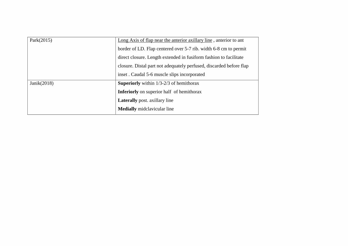
Park(2015) Long Axis of flap near the anterior axillary line , anterior to ant
border of LD. Flap centered over 5-7 rib. width 6-8 cm to permit
direct closure. Length extended in fusiform fashion to facilitate
closure. Distal part not adequately perfused, discarded before flap
inset . Caudal 5-6 muscle slips incorporated
Janik(2018) Superiorly within 1/3-2/3 of hemithorax
Inferiorly on superior half of hemithorax
Laterally post. axillary line
Medially midclavicular line
No Author Free/Pedicled Composition Slips Lenght Width Reconstruction site/purpose 1 Takayanagi f Myocutaneous 5,6,7,8 11 7 Foot 2 Bruck f Osteomyocutaneous 5,6 12 7 Foot 3 Inoue p Myocutaneous 6,7,8 14 7 Oral cavity 4 Inoue p Myocutaneous 7,8 20 10 Cervical oesophagus 5 Inoue p Myocutaneous 6,7,8 14 7 Tempo-auricular skin 6 Inoue p Myocutaneous 8 14 7 Cheek soft tissue 7 Inoue p Myocutaneous 6,7,8 14 7 Oral cavity 8 Inoue p Myocutaneous 6,7,8 15 7 Cervical oesophagus 9 Inoue p Myocutaneous 6,7,8 15 7 Cervical oesophagus 10 Inoue p Myocutaneous 6,7,8 12 6 Oral cavity 11 Inoue p Myocutaneous 6,7,8 15 7 Oral cavity 12 Inoue p Myocutaneous 6,7,8 22 10 Cervical oesophagus 13 Pittet f Myocutaneous 6,7,8 12 5 Noma, cheek, oral lining 14 Pittet f Myocutaneous 7,8,9 12 6.5 Noma, cheek, upper lip, inferior
eyelid 15 Pittet f Myocutaneous 6,7,8,9 5 8 Noma, cheek, oral lining 16 Pittet f Myocutaneous 7,8,9 9 4,5 Noma, cheek, oral lining 17 Pittet f Osteomyocutaneous 7,8 8 6 Noma, cheek, mandible, oral lining 18 Pittet f Myocutaneous 7,8,9 15 6 Noma, cheek, upper lip, oral lining No Author Free/Pedicled Composition Slips Lenght Width Reconstruction site/purpose 19 Pittet f Osseomyocutaneous 7,8 12 6 Noma, maxilla, upper lip, cheek,
oral lining
20 Pittet f Myocutaneous 7,8,9 12 5 Noma, cheek, oral lining, upper lip, nose
21 Pittet f Myocutaneous 7,8,9 11 5 Trauma, cheek, inferior lid 22 Pittet f Osteomyocutaneous 6,7,8 10 5.5 Noma, maxilla, upper lip 23 Pittet f Myocutaneous 6,7,8 9 5 Noma, cheek, nose, inferior eyelid 24 Pittet f Myocutaneous 7,8 12 6 Noma, cheek, oral lining 25 Pittet f Osteomyocutaneous 7,8,9 9 7 Neoplasm, lower cheek, pharynx,
mandible 26 Pittet f Myocutaneous 7,8 12 8 Noma, lower lip, chin 27 Pittet f Myocutaneous 7,8,9 8 4.5 Noma, cheek, oral lining 28 Pittet f Myocutaneous 7,8 12 5.5 Noma, cheek, oral lining 29 Pittet f Myocutaneous 7,8,9 9 7 Noma, cheek 30 Pittet f Myocutaneous 7,8 11 6.5 Noma, cheek, oral lining 31 Pittet f Myocutaneous 7,8,9,10 14 7 Noma, cheek, upper lip, oral lining 32 Pittet f Myocutaneous 7,8 11 6.5 Noma, cheek, oral lining 33 Pittet f Myocutaneous 7,8 7 5 Noma, cheek, upper lip,
commissure, oral lining 34 Pittet f Myocutaneous 7,8 7.5 6.5 Noma, chin, lower lip, oral lining 35 Pittet f Myocutaneous 7,8,9 8 5 Noma, cheek, nasal lining No Author Free/Pedicled Composition Slips Lenght Width Reconstruction site/purpose 36 Pittet f Myocutaneous 7,8 6.5 6 Noma, upper lip, commissure,
maxilla 37 Perrot f Osteomyocutaneous 10x8
(muscle) 4 4 Inferior 1/3 tibia
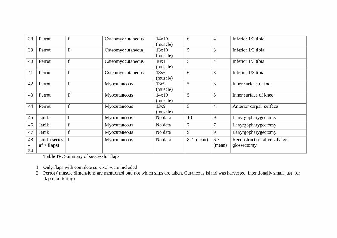
Table IV. Summary of successful flaps
1. Only flaps with complete survival were included 2. Perrot ( muscle dimensions are mentioned but not which slips are taken. Cutaneous island was harvested intentionally small just for
flap monitoring)
38 Perrot f Osteomyocutaneous 14x10 (muscle)
6 4 Inferior 1/3 tibia
39 Perrot F Osteomyocutaneous 13x10 (muscle)
5 3 Inferior 1/3 tibia
40 Perrot f Osteomyocutaneous 18x11 (muscle)
5 4 Inferior 1/3 tibia
41 Perrot f Osteomyocutaneous 18x6 (muscle)
6 3 Inferior 1/3 tibia
42 Perrot F Myocutaneous 13x9 (muscle)
5 3 Inner surface of foot
43 Perrot F Myocutaneous 14x10 (muscle)
5 3 Inner surface of knee
44 Perrot f Myocutaneous 13x9 (muscle)
5 4 Anterior carpal surface
45 Janik f Myocutaneous No data 10 9 Lanyrgopharygectomy 46 Janik f Myocutaneous No data 7 7 Lanyrgopharygectomy 47 Janik f Myocutaneous No data 9 9 Lanyrgopharygectomy 48-54
Janik (series of 7 flaps)
f Myocutaneous No data 8.7 (mean) 6.7 (mean)
Reconstruction after salvage glossectomy

Figure 1. Flow chart
329 Records identified from
Databases 305 Medline 23 Google Scholar 1 Cochrane Library
4 Duplicate records removed
325 Records screened
314 Records excluded
11 Reports sought for retrieval
1 Report not retrieved
10 Reports assessed for eligibility
10 Studies included in review
Iden
tific
atio
n Sc
reen
ing
In
clud
ed
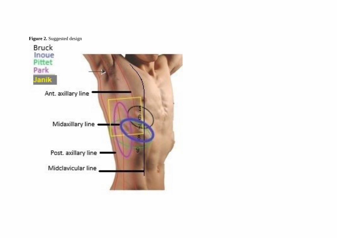
Figure 2. Suggested design