anatomical and physiological studies on the aqueous … · anatomical and physiological studies on...
TRANSCRIPT

Jpn J Ophthalmol 43, 262–271 (1999)© 1999 Japanese Ophthalmological Society 0021-5155/99/$–see front matterPublished by Elsevier Science Inc. PII S0021-5155(99)00017-9
Anatomical and Physiological Studies on the Aqueous Sinus Artery of the Pigeon Eye
Masao Nakabayashi
Department of Ophthalmology, Osaka University Medical School, Osaka, Japan
Purpose:
To test the hypothesis that ischemia of the Schlemm’s canal endothelium is in-volved in the control of intraocular pressure (IOP).
Methods:
Experiments were conducted on pigeons. The course of the aqueous sinus and theaqueous sinus artery was determined by examining serial sections and the distribution of poly-merized resin in the limbal region of the pigeon eye. Ischemia was produced by blocking twomajor arteries to the aqueous sinus artery and then determining the effect on the IOP.
Results:
The anatomy and course of the pigeon aqueous sinus and aqueous sinus artery indi-cated that they are homologous to the human Schlemm’s canal and Schlemm’s canal artery,respectively. When the arterial inflow to the aqueous sinus artery was blocked by laser treat-ment, injected resin was absent in the artery but a small amount invaded the aqueous sinus.Blockage of the two major arteries to the aqueous sinus artery did not alter the IOP.
Conclusions:
The similarity of the pigeon aqueous sinus and aqueous sinus artery to the hu-man Schlemm’s canal and Schlemm’s canal artery indicates that the pigeon eye can be usedto study the relationship between ischemia of the endothelial meshwork cells and IOP. Thelack of a change in IOP in pigeons after ischemia induction was probably the result of anasto-motic arterial blood inflow to the aqueous sinus directly, and thus ischemia was not producedby blocking the two major arteries to the aqueous sinus artery.
Jpn J Ophthalmol1999;43:262–271
© 1999 Japanese Ophthalmological Society
Key Words:
Aqueous sinus, aqueous sinus artery, ischemic theory, open-angle glaucoma
etiology, pigeon eye.
Introduction
We have shown previously that severing all of theanterior ciliary arteries induced a transient ocularhypertension in monkeys and suggested that theblockage of blood flow to the endothelial cells ofSchlemm’s canal altered their physiology, which inturn upset the movement of fluid out of the eye. Thisaltered outflow then resulted in an increased intraoc-ular pressure (IOP).
1
These observations supportedour hypothesis first published in 1981, that open-angle glaucoma of human eyes is caused by ischemiaof the cells of Schlemm’s canal.
2–4
In the human eye, a small artery runs in the outerwall of Schlemm’s canal, not completely circumfer-
entially, but for a considerable distance.
5,6
The ar-tery, called Schlemm’s canal artery, is a branch ofthe penetrating anterior ciliary artery and branchesin either the sclera or the episclera.
7
We proposedthat blockage of Schlemm’s canal artery, whichnourishes the cells of the endothelium, would alterthe physiology of these cells and thus alter the move-ment of fluid out of the eye.
To obtain additional data to support our ischemiahypothesis, a search was made for an animal morereadily available than monkeys, whose vascularanatomy of the anterior segment of the eye is similarto that of the human eye. In general, mammalianeyes are not equipped with a distinct vessel likeSchlemm’s canal for aqueous drainage, except in thehigher primates,
Simiae
. However, the avian eye hasa distinct trunk vessel for aqueous drainage that iscalled the aqueous sinus. The aqueous sinus is con-sidered to be homologous to the Schlemm’s canal of
Received: March 31, 1998Correspondence and reprint requests to: Masao NAKABA-
YASHI, MD, Department of Ophthalmology, Osaka UniversityMedical School, 2-2 Yamadaoka, Suita, Osaka 565, Japan

M. NAKABAYASHI
263
AQUEOUS SINUS ARTERY OF PIGEON EYE
higher primates. Just as the Schlemm’s canal arteryruns in the outer wall of Schlemm’s canal, the aque-ous sinus artery in pigeons is very large and is situ-ated in the lumen of the aqueous sinus.
6–8
Thus, thepigeon aqueous sinus artery appears to be analogousto Schlemm’s canal artery of the human eye.
The purpose of this study was to determinewhether the pigeon eye can be used to study the fac-tors that control the IOP of the eye, and thus be usedto obtain evidence for the ischemia hypothesis. Toaccomplish this, it was necessary to determine thecourse of the pigeon aqueous sinus artery and aque-ous sinus and compare them to the course ofSchlemm’s canal and Schlemm’s canal artery. Thisstudy shows that the aqueous sinus and aqueoussinus artery are morphologically homologous toSchlemm’s canal and Schlemm’s canal artery andconcludes that the pigeon can be used to study the isch-emia hypothesis. To test the ischemia hypothesis inpigeons, this study attempted to induce ischemia byblocking the two major inflow routes to the aqueoussinus artery and measure the IOP before and afterthe blockage.
Materials and Methods
Animals
The experiments were conducted on white pi-geons,
Streptoperia risoria
, of both sexes and weigh-ing 130–180 grams each. The pigeons were obtainedfrom a local vendor (Sakura Co., Aichi). They weremaintained in the laboratory and given free access toavian chow (Ishibashi Co., Osaka) and drinking wa-ter. This study was conducted in accordance with theARVO resolution on the use of animals in research.
Anatomical Studies
To determine the normal morphology of the aque-ous sinus and the aqueous sinus artery, serial sec-tions of the anterior segment of the eye were madeusing the 26 eyes of 13 pigeons. The animals werekilled with an intramuscular injection of 50 mg ofketamine hydrochloride (Ketalar
™
; Sankyo, To-kyo), and the anterior segments were removed bycutting the sclera circumferentially with a scalpel anda pair of scissors. For tissue orientation, a hole wasmade on the edge of the segment facing the animal’sbill. The anterior segments were immersed immedi-ately in 10% formalin and the central part of the lenswas “cut out” by making a circumferential series ofholes with a 30-gauge needle that penetrated fromthe lens to the cornea. A photograph was taken ofthe remaining portion that included the periphery of
the lens, called the annular pad. After 24 hours offixation, each segment was dissected radially (merid-ionally) into eight pieces. The pieces were dehydratedand embedded in hydrophilic resin (Technovit 7100
™
;Kulzer, Wehrheim, Germany). Meridional, 7-
m
m-thickserial sections were cut with a microtome (HM350;Microm, Heidelberg, Germany) and stained with0.1% toluidine blue. The stained sections were ex-amined by light microscopy.
To determine the relationship between the aque-ous sinus and aqueous sinus artery to the IOP, westudied the aqueous sinus and aqueous sinus arteryin eyes in which the IOP was reduced in vivo. Fiveeyes of five pigeons were used with the contralateraleyes as controls. To accomplish this, the animal wasanesthetized by an intramuscular injection of 15 mgketamine, and the lids were spread apart by a lid re-tractor without exerting pressure on the globe (Fig-ure 1). An incision was made through the cornea toreduce the IOP, and the animal was then killed by anadditional intramuscular injection of ketamine. Toinsure rapid exposure of the anterior segment to thefixative, the lid retractor was removed, the lower lidwas excised, and then the head was immediately im-mersed in a 10% acrolein solution for 1 hour. Theanterior segments were then excised and furtherfixed in 10% formalin solution for 24 hours. The lenswas removed intact, and the ciliary body and iriswere peeled off along the deep ciliary cleft with for-ceps. These segments were studied in formalin solu-tion with a stereomicroscope.
Another technique used to study the anatomy ofthe aqueous sinus and aqueous sinus artery was tomake a cast of the aqueous sinus artery network inthe 32 eyes of 16 pigeons. To accomplish this, the an-
Figure 1. Handmade applanators and lid retractor.

264
Jpn J OphthalmolVol 43: 262–271, 1999
imals were killed by an overdose of ketamine and 20mL of a casting resin monomer (Mercox
™
; Dai Nip-pon Ink, Tokyo) mixed with 0.2 mL of polymerizingagent was injected into the left ventricle of the heart.After polymerization (15–60 min), the anterior seg-ments of the eyes were removed as described andimmersed into an
a
-chymotrypsin (Zonolysin
™
, Mo-chida, Tokyo) solution of 125 NFU/mL at 37
8
C for20 hours to remove the lens and retinal pigment epi-thelium (RPE). After 3 hours of supplemental fixa-tion in 10% formalin, the anterior segments werecleared in glycerol at 4
8
C for 48 hours. The resin-filled vessels were then examined in relation to theother tissues using a stereomicroscope.
Ischemia and IOP
To test the ischemia hypothesis, the following fiveprocedures were performed consecutively on six pi-geons, with three pigeons treated in the right eye andthree in the left eye.
Measurement of the control IOP.
The pneumotono-graph (PTG) tonometer (Alcon, Fort Worth, Texas,USA) was selected to measure the IOP of the eyesbecause measurement of the IOP by cannulating theanterior chamber did not give reproducible results.This was due to the thinness of the pigeon cornea,which could not support the cannulating needle (30-gauge). In preliminary studies with the PTG tonom-eter, we found that the IOP decreased considerablyafter placing the applanators (described below) onthe cornea, and a 10-minute recovery period was re-quired for the IOP to return to the preapplanationlevel. Therefore, tonometry was repeated only aftera 20-minute recovery period.
To record the IOP without using a lid retractor,the bill of the pigeon was held by one hand, and adrop of 0.4®% oxybuprocain (Benoxyl
™
; Santen,Osaka) was instilled. The lower lid was pushed asideby the tip of the sensor held in the other hand, andthe IOP was recorded by placing the tip of the sensoron the cornea.
Rupturing Anastomotic Artery by YAG Laser.
After the measurement of the control IOPs, a plasticplate was placed between the bill to maintain a clearbreathing passage, the bill was then fixed by pincers.The body was placed in the supine position, and adrop of oxybuprocain was instilled. To examine thelower chamber angle, an applanator, made of a pieceof microscope slide and an 18-gauge hypodermicneedle (Figure 1), was placed on the upper section ofthe cornea. The ciliary cleft was identified through
the applanator with a slit-lamp microscope equippedwith a YAG laser (Nidek, Aichi) (Figure 2). Theanastomotic artery and accompanying white bridgewere found crossing over the ciliary cleft (Figure 3A).
If the anastomotic artery could not be found, thebody was returned to its normal position, and astrong light from a 75 W halogen lamp was projectedthrough a fiber optic light guide into the contralat-eral eye. With this illumination, the pupil of the ex-perimental eye had a reddish glow, and the “navel”(see below) was easily seen through the sclera (Fig-ure 3B).
Returning the animal to the supine position, theanastomotic artery was found by using the navel andciliary cleft as landmarks. The anastomotic arterywas then exposed to 3.4 mJ of the YAG laser. Theappearance of bleeding was a sign that sufficienttreatment had been given.
Coagulation of penetrating artery by DYE laser.
Returning the bird to the normal position, additionalanesthetic eyedrops were instilled on the same eye,and another applanator, which was constructed of anacrylate plate and a needle (Figure 1), was placed onthe lower limbus of the same eye. This 3-mm-thickapplanator was sufficient to keep the lower lid open.To distinguish arteries from veins, the applanatorwas pressed gently on the limbus to collapse theveins, whereas the arteries remained patent. One ex-posure of the DYE laser (Nidek) at 595 nm, 0.2-mmdiameter, at 1.4 W, and for 0.3 seconds, whitened theartery; but additional shots, at least 10, were givenalong the artery to insure occlusion of the penetrat-ing artery. After the treatment with the YAG andDYE laser, the restraints on the bird were removed
Figure 2. View of lower chamber angle.
M. NAKABAYASHI
265
AQUEOUS SINUS ARTERY OF PIGEON EYE
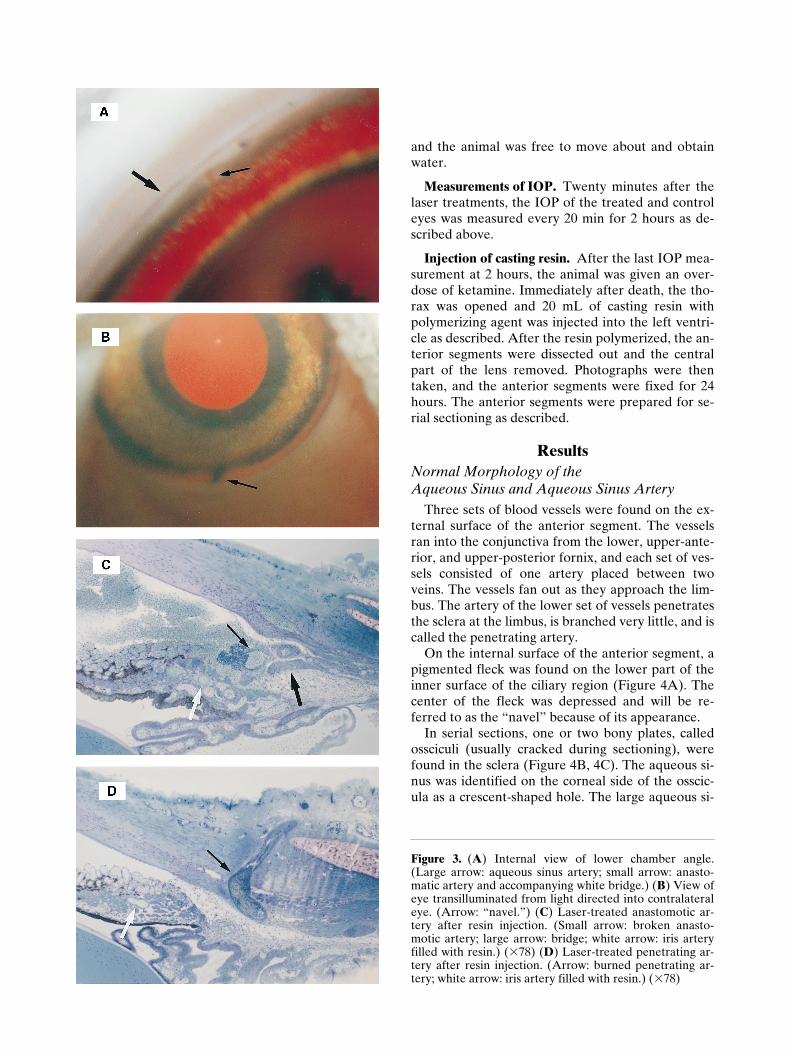
and the animal was free to move about and obtainwater.
Measurements of IOP.
Twenty minutes after thelaser treatments, the IOP of the treated and controleyes was measured every 20 min for 2 hours as de-scribed above.
Injection of casting resin.
After the last IOP mea-surement at 2 hours, the animal was given an over-dose of ketamine. Immediately after death, the tho-rax was opened and 20 mL of casting resin withpolymerizing agent was injected into the left ventri-cle as described. After the resin polymerized, the an-terior segments were dissected out and the centralpart of the lens removed. Photographs were thentaken, and the anterior segments were fixed for 24hours. The anterior segments were prepared for se-rial sectioning as described.
Results
Normal Morphology of the Aqueous Sinus and Aqueous Sinus Artery
Three sets of blood vessels were found on the ex-ternal surface of the anterior segment. The vesselsran into the conjunctiva from the lower, upper-ante-rior, and upper-posterior fornix, and each set of ves-sels consisted of one artery placed between twoveins. The vessels fan out as they approach the lim-bus. The artery of the lower set of vessels penetratesthe sclera at the limbus, is branched very little, and iscalled the penetrating artery.
On the internal surface of the anterior segment, apigmented fleck was found on the lower part of theinner surface of the ciliary region (Figure 4A). Thecenter of the fleck was depressed and will be re-ferred to as the “navel” because of its appearance.
In serial sections, one or two bony plates, calledossciculi (usually cracked during sectioning), werefound in the sclera (Figure 4B, 4C). The aqueous si-nus was identified on the corneal side of the osscic-ula as a crescent-shaped hole. The large aqueous si-
Figure 3.
(
A
) Internal view of lower chamber angle.(Large arrow: aqueous sinus artery; small arrow: anasto-matic artery and accompanying white bridge.) (
B
) View ofeye transilluminated from light directed into contralateraleye. (Arrow: “navel.”) (
C
) Laser-treated anastomotic ar-tery after resin injection. (Small arrow: broken anasto-motic artery; large arrow: bridge; white arrow: iris arteryfilled with resin.) (
3
78) (
D
) Laser-treated penetrating ar-tery after resin injection. (Arrow: burned penetrating ar-tery; white arrow: iris artery filled with resin.) (
3
78)

266
Jpn J OphthalmolVol 43: 262–271, 1999
nus artery was found in the lumen of the aqueoussinus and divided the lumen into two sections (Fig-ure 5, left panel). Each section was connected tomany aqueous draining paths that ran through thesclera near the cornea in front of the osscicula. Thesepaths merged into the subconjunctival veins. In se-rial sections of the lower-anterior region of the ante-rior segment near the bill, the aqueous sinus wassmaller and sometimes not found at all. However,the aqueous sinus artery was found in all sections of theeight blocks and was larger in the lower than in theupper half of the anterior segment. There were sev-eral small branches from the aqueous sinus artery to-ward the sclera but never toward the cornea (Figure5, fifth panel on left).
A distinct morphological feature of the pigeon eyewas the two junctions made by the aqueous sinus ar-tery with the two inflow arteries in the sections ob-tained from the lower polar area. One of these junc-tions was an anastomotic junction with the iris arterythat crossed over the ciliary cleft (Figure 4B). Thisanastomotic artery was accompanied by connectivetissue that formed a bridge between the sclera andthe iris over the cleft. The ciliary region at this posi-tion formed the “navel” (Figure 4A). At the otherjunction, the aqueous sinus artery merged with thepenetrating artery that came from the lower limbus(Figure 4C). These junctions were situated near eachother and, in all cases, the junction with the anasto-motic artery, which forms the navel, was locatedmore anteriorly (toward the bill) than the junctionwith the penetrating artery (Figure 6).
In a few specimens, a small, tortuous artery wasfound that merged into the aqueous sinus artery andappeared as an appendix in the upper or posteriorpart. An appendix to the anastomotic artery wasnever found.
Physical Effect of the IOP on the Aqueous Sinus
When the IOP was reduced by the corneal inci-sion, venous blood flowed retrogradely into theaqueous sinus from downstream veins. The blood,
Figure 4.
(
A
) Internal surface of anterior segment. (Ar-row: “navel” (see text); white arrow: toward bill of pi-geon.) (
3
6) (
B
) Section of lower polar region of limbus.(Small arrow: anastomotic artery; large arrow: “navel.”)(
3
78) (
C
) Section of lower polar region of limbus. (Arrow:penetrating artery.) (
3
78) (
D
) Fixed blood in aqueous si-nus following in vivo lowered IOP. (Arrow: retrogradeflow of venous blood into aqueous sinus; white arrow: to-ward bill.) (
3
7)

M. NAKABAYASHI
267
AQUEOUS SINUS ARTERY OF PIGEON EYE
Figure 5. Resin distribution after unilateral laser treatment. (Small arrow: aqueous sinus artery [ossiculum in left cornershows right eye and in right corner shows left eye]; large arrow: resin.) (349)

268
Jpn J OphthalmolVol 43: 262–271, 1999
fixed by the rapid acrolein fixative, was trapped inthe aqueous sinus (Figure 4D). This view showedthat the lumen of the aqueous sinus was smaller orsometimes not present in the vicinity of the bill.There were also many drainage paths from the aque-ous sinus (Figure 4D).
Figure 6. Resin-filled artery in glycerin-treated anteriorsegment without lens and pigment epithelium. Bottompanel is diagram of this circulatory system. (A) Aqueoussinus artery; (B) anastomotic artery; (C) penetrating ar-tery; (D) iris artery; (E) sphincteric capillary plexus; and(F) ossicula. (Arrow: toward bill.)
Examination of Casts of the Aqueous Sinus Artery
The polymerized resin traced the course of the ar-teries running through the anterior segment (Figure6). The osscicula, which overlapped and roughly de-lineated the corneoscleral border, were visible throughthe semitransparent sclera in the glycerol-clearedpreparation. The sphincteric capillary plexus, which isinvolved in accommodation,
9
appeared as a darkring in the iris. The upper and lower ciliary arteriesmerged with the plexus at each of two points.
The aqueous sinus artery formed an apparentlycomplete circle although the aqueous sinus wassometimes absent in the vicinity of the bill. In thelower part of the aqueous sinus artery circle, the twolarge junctions found in the serial sections wereidentified, the junction of the aqueous sinus arterywith the anastomotic artery and the junction with thepenetrating artery.
Effect of the Blockage of the Inputto the Aqueous Sinus Artery on the IOP
Both the laser-treated and the untreated con-tralateral eyes maintained the same IOP of 24–26mm Hg as before treatment for as long as 2 hours af-ter the laser treatments (Table 1). Thus, there wasno significant change in the IOP after the laser treat-ments.
The Aqueous Sinus in the Lower Polar Region After Laser Treatments
Serial sections of the untreated contralateral eyeshowed that resin filled the entire aqueous sinus ar-tery, the penetrating artery, the anastomotic artery,and the iris arteries. In the treated eye, the rupturedand deformed anastomotic artery was found in theregion of the YAG laser burn (Figure 3C). Evidenceof bleeding was seen in the anterior chamber. In theregion of the DYE laser coagulation, a blood coagu-lum was found in the penetrating artery (Figure 3D).The presence of the coagulated blood 2 hours afterthe treatment demonstrated that the arterial bloodflow had not been restored. In both areas, resin filledonly the iris arteries (Figure 3C, 3D).
The Aqueous Sinus After Laser Treatments (Except for the Lower Polar Region)
In the untreated eye, the entire aqueous sinus ar-tery and ciliary artery were filled with resin (Figure5, left panels). The small tortuous appendix artery

M. NAKABAYASHI
269
AQUEOUS SINUS ARTERY OF PIGEON EYE
did not fill with resin most likely because of its size.In the treated eye, on the other hand, the aqueous si-nus artery was thin and had no resin throughout,indicating that the arterial flow was completelyblocked to this vessel (Figure 5, right panels). Sur-prisingly, resin was found in the aqueous sinus itselfin either the upper or posterior part for 10%–30% ofits length. In a few sections, a piece of resin was alsofound along the aqueous draining path that hadhorn-like projections (Figure 5, right side, fifth panel).A careful tracing in successive sections did not re-veal any connections of these projections to any ar-tery. In addition, the resin in the aqueous sinus alsodid not show a connection to the episcleral arteries.Thus, we cannot presently explain whether the horn-like projections of a piece of resin represent inflowto or outflow from the aqueous sinus.
The aqueous sinus of the treated eyes appearedvery deformed when compared with the aqueous si-nus of the contralateral eyes. This was partly due tothe shrinkage of aqueous sinus artery induced by theischemia and partly due to the outward pressure ofthe resin. These sections also showed that the posi-tion of the aqueous sinus artery in the aqueous sinuslumen was not fixed in the middle. These findingswere found uniformly in all of the treated and non-treated eyes of all six pigeons from the ischemia test.
Discussion
IOP Measurements of the Pigeon Eye
To the best of our knowledge, the intraocularpressure of the pigeon eye has not been published;although the pressure of the chicken eye was mea-sured by the cannulation method. The IOP of thechicken eye was 15 mm Hg and was not affected byintravenous acetazolamide.
10
The PTG tonometer,designed for human eyes, gave reproducible valuesfor the pigeon eye, although it measured only therelative IOP. Repeated measurements of the IOPshowed the so-called “tonography effect,” ie, re-peated determinations lowered the IOP signifi-cantly. Therefore, only a minimal number of mea-surements were made.
It is known that bleeding into the anterior cham-ber leads to an increase in the IOP in the human eye.In the pigeon, however, I found that bleeding intothe anterior chamber after YAG laser treatment didnot alter the IOP significantly or affect the return ofthe IOP to the preapplanator application level, ie,the IOP recovered relatively quickly and never ex-ceeded the level before the treatments. This suggeststhat the blood coagulation mechanism is very rapidin the pigeon, and hemostasis is completed even be-fore removing the applanators.
Table 1.
Course of IOP (mm Hg) After Laser Treatment
Minutes After Treatment
Case No. Side Before Treatment 20 40 60 80 100 120
1 R* 26 25 25 26 26 26 25L 25 25 25 26 25 25 25
2 R* 24 25 25 25 24 24 24L 24 24 25 24 24 24 24
3 R* 26 25 25 25 26 25 24L 25 25 25 25 25 26 25
4 R 26 25 25 26 26 25 25L* 26 25 25 25 25 25 25
5 R 24 24 25 25 24 24 24L* 24 24 24 24 24 24 24
6 R 26 25 25 26 25 25 25L* 25 26 25 25 25 25 25
Average and Standard Deviation in IOP in Treated Eyes
Average 25.2 25.0 24.8 25.0 25.0 24.8 24.5SD 0.98 0.63 0.41 0.63 0.89 0.75 0.55
Average and Standard Deviation in IOP in Control Eyes
Average 25.0 24.7 25.0 25.3 24.8 24.8 24.7SD 0.89 0.51 0.0 0.82 0.75 0.75 0.51
*: Treated eyes.

270
Jpn J OphthalmolVol 43: 262–271, 1999
Our observations showed that the IOP of theunanesthetized pigeon eye was 24–26 mm Hg by mytechnique and apparatus. Blockage of blood flow tothe aqueous sinus artery by laser treatments of theanastomotic and penetrating artery did not alter theIOP of the eye. Thus, these findings do not supportour ischemia hypothesis for ocular hypertension.However, as shown, resin was found in the aqueoussinus of the treated eye. Therefore, complete isch-emia had not been produced. Further experimentswill be needed to produce complete ischemia of theaqueous sinus to obtain a definitive answer to ourhypothesis.
Inflow Into Aqueous Sinus
Both the aqueous sinus and Schlemm’s canal areblind sacks opening only to veins, and the IOP isvery important in controlling what flows into them.When the IOP is low, the inflow is very smooth be-cause the trabecular meshwork moves toward theanterior chamber as discussed previously.
1
Figure4D shows that venous blood flowed retrogradelyinto the aqueous sinus after a decrease in IOP result-ing from venous pressure as a physical phenomenon.
When resin was injected into the heart immedi-ately after death, without laser treatment, resin wasnever found in the aqueous sinus. Occasionally, itwas found macroscopically only in the very largetrunk vein. Other laboratories using the casting resintechnique reported on the vascular cast of the duck-ling eye.
11
In their method, the animal was first bledand then perfused with Ringer’s solution before in-jection of the resin. Based on their observations, wechose to inject the resin: (a) immediately after deathwhen the IOP was still maintained, (b) without priorbleeding and washing out of the blood, (c) only insmall quantity (20 mL), (d) using the same condi-tions for both eyes, and (e) comparing both eyes postfactum. Under these conditions, retrograde flow ofthe resin from a trunk vein into the aqueous sinusshould not be possible.
As shown, resin invaded the upper and posteriorparts but not the lower polar region of the aqueoussinus in the treated eye after treatment by YAG andDYE laser. The YAG-ruptured anastomotic arterydid not leak resin (Figure 3C). Moreover, resin wasnever found in the region of the YAG-rupturedpoint or the DYE-burned point except for the iris ar-tery (Figure 3C, 3D). These findings were as ex-pected, because resin could not flow into these re-gions without blood flow.
From these observations, we suggest that the resininvaded the aqueous sinus in the treated eyes throughsome anastomotic routes, viz, an arterio-sinus or ar-terio-venous anastomosis, which might function un-der ischemic conditions. The two arteries of thethree sets of vessels on the front surface of anteriorsegment, but not the penetrating artery of the lowerset, may be involved in the collateral arterial route.Similar arterio-venous anastomoses have been re-ported in the episclera of dogs
12
and monkeys,
13
andthe search for these anastomoses in pigeons is beingundertaken. The small tortuous appendix arteryfound in the serial sections, never filled by resin per-haps because of the viscosity of the resin, may have asimilar function, although it is an arterio–arterial (A-A) anastomosis.
Relationship to the Previous Study on Monkey Eyes
As stated, we succeeded previously in inducing atransient ocular hypertension in monkeys by sever-ing all anterior ciliary arteries that induced ischemiaof Schlemm’s canal.
1
These results supported our hy-pothesis that open-angle glaucoma in the human eyeis caused by ischemia of the endothelium of Schlemm’scanal.
2–4
However, in the monkey eye the distancebetween the severed points and Schlemm’s canal waslarge, and the possibility existed that Schlemm’s ca-nal was nourished by the long posterior ciliary arteryvia the major arterial circle and the rest of the ante-rior ciliary artery. To produce an absolute ischemiaof the endothelium of the Schlemm’s canal, blockageof the arterial inputs should be made close to the ca-nal. This was the rationale for the site of the lesionsin the pigeon eyes, ie, treating the aqueous sinus ar-tery near the junction with the two inflow arteries. Ifischemia of the endothelium of Schlemm’s canal isthe underlying mechanism for the rise in IOP, thereshould have been a rise in the IOP after the lasertreatments of the penetrating and anastomotic arter-ies in pigeons. As shown, the IOP did not rise but theexperiments proved to be inconclusive as resin wasfound in the aqueous sinus indicating that completeischemia was not produced. In retrospect, an up-stream occlusion of arterial blood may have beenmore effective in inducing ischemia of the aqueoussinus because it should also eliminate anastomoticcollateral routes.
The author thanks Kenichi Kishida, MD, PhD for his generous
support.

M. NAKABAYASHI
271
AQUEOUS SINUS ARTERY OF PIGEON EYE
References
1. Nakabayashi M, Kishida K, Okazaki H. Effect of experimen-tal ocular hypertension on macrovacuolation in Schlemm’s ca-nal endothelial cells of monkey eyes. Ophthalmic Res 1991;23:177–86.
2. Nakabayashi M. Nuclear cataract as a cause of senile glau-coma. Ann Ophthalmol 1981;13:597–602.
3. Nakabayashi M, Takatsuki R. Subconjunctival administrationof oxygen for glaucomatous eyes. Folia Ophthalmol Jpn 1979;30:1811–6.
4. Nakabayashi M, Takatsuki R. Effect of ouabain on aqueousdrainage. Folia Ophthalmol Jpn 1980;31:321–6.
5. Ashton N. Anatomical study of Schlemm’s canal and aqueousveins by means of neoprene casts. I. Aqueous veins. Br J Oph-thalmol 1951;35:291–303.
6. Rohen JW, Rentsch FJ. Elektronenmikroskopische Untersu-chungen über den Bau der Aussenwand des SchlemmschenKanals unter besonderer Berücksichtigung der Abflusskanäleund Altersveränderungen. Graefes Arch für Ophthalmol1969;177:1–17.
7. Hogan MJ, Alvardo JA, Weddel JE. Histology of the humaneye. Philadelphia, PA: Saunders. 1971;121,140,188.
8. Tripathi RC, Tripathi BJ. The mechanism of aqueous outflowin birds. I. An ultrastructural study of normal eyes. Exp EyeRes 1973;15:409–23.
9. Duke-Elder S. System of ophthalmology. I. The eye in evolu-tion. St. Louis, MO: Mosby Co., C.V. 1958;407.
10. Sears ML. Intraocular pressure of the unanesthetized hen. ArchOphthalmol 1960;63:212–6.
11. Hossler FE, Olson KR. Microvasculature of the avian eye:studies on the eye of the duckling with microcorrosion cast-ing, scanning electron microscopy, and stereology. Am J Anat1984;170:205–21.
12. Rohen J. Arterivenöse Anastomosen im Limbusreich desHundes. Graefes Arch für Ophthalmol 1956;157:361–7.
13. Gaasterland DE, Jocson VL, Sears ML. Channels of aqueousoutflow and related blood vessels. II. Episcleral arteriovenousanastomoses in the rhesus monkey eye. Arch für Ophthalmol1970;84:770–5.