analytical pyrolysis as diagnostic tool in the
TRANSCRIPT

Analytical Pyrolysis as Diagnostic Toolin the Investigation of Works of Art
G. Chiavari1* / S. Prati2
1 Chemistry Department G.Ciamician, University of Bologna, via Selmi 2, 48126 Bologna, Italy; E-Mail: [email protected] Laboratory of Chemistry, CIRSA, University of Bologna, via Marconi 48100 Ravenna, Italy
Key Words
Gas chromatography-mass spectrometryPyrolysisWork of artReview
Summary
This paper is a review of the activity of our research group in the last decade. A discussionabout the application of analytical pyrolysis for the examination of objects of art during thisperiod is presented.
Introduction
Cultural Heritage can be defined as
‘‘Every material evidence of civilisation’’
and for this reason its safeguard is a
‘‘must’’ for our society.
The commitment of scientists is cru-
cial for the protection, the restoration
and the exploitation of Cultural Heritage.
Scientists are asked to transfer to this
field technologies developed in different
areas and to develop new scientific tools
suitable for specific areas of Cultural
Heritage. Art conservation is a new dis-
cipline which utilizes modern analytical
techniques in the study and preservation
of works of art: in the last few years new
materials and technologies have been
developed which are continually being
evaluated by conservators for their
potential application to preservation and
restoration [1].
Several analytical techniques have
been developed for the characterisation
of samples in the artistic field [1–3].
Spectroscopic techniques are widely
applied due to the fact that they are
mainly conservative non-destructive
techniques and the sample can be exam-
ined with other complementary tech-
niques. Whenever possible non-
destructive methods are used because
they can be applied directly to the objects
under investigation. Moreover portable
instruments have been produced to avoid
sampling even for frescoes or other large
objects. These techniques are useful to
obtain preliminary information that of-
ten must be confirmed or clarified by
other analyses.
The examination of a work of art
generally begins with a visual inspection
by using special lighting to facilitate the
distinction of differences in colour, gloss
and texture. At this stage photographic
techniques can be used [1]. Optical tech-
niques are important for studying the
morphology of the samples and give
information about the state of conserva-
tion or the technique used in the pro-
duction. FTIR, Raman and X ray
techniques are particularly useful for the
identification of the inorganic compo-
nents (pigments, salts and composition of
mortars or stone). The first two tech-
niques can also give information about
the organic components, but generally
the samples are complex mixtures and
identification can be difficult. In the field
of organic characterisation the separation
techniques such as HPLC and GC (in
particular coupled with mass spectrome-
try) are mainly applied. They are
destructive techniques, but pre-treatment
procedures which use very small quan-
tities of material have been developed
[4–32].
HPLC is mainly used for the charac-
terisation of proteins, by quantification
of the amino acids [4–7] or for the anal-
ysis of organic colorants [8–14]. GC is
used for less polar compounds such as
natural binders and protectives [15–32].
However artistic samples are mixtures of
organic and inorganic substances which
have been chosen for their chemical sta-
bility. Some artistic materials decrease
in solubility and volatility with ageing
DOI: 10.1365/s10337-003-0094-7
2003, 58, 543–554
0009-5893/03/11 $03.00/0 � 2003 Friedr. Vieweg & Sohn Verlagsgesellschaft mbH
Review Chromatographia 2003, 58, November (No. 9/10) 543

and the pre-treatments can be difficult
to apply.
Analytical pyrolysis can be useful for
complex, high molecular weight and polar
compounds which can hardly be analysed
by traditional gas chromatography. It
consists of a thermal pre-treatment of the
sample in an inert atmosphere. The com-
pounds are fragmented by the high tem-
perature (600–800 �C) to produce more
volatile molecules. The pyrogram displays
the decomposition fragments and is a
fingerprint of the original matrix, because
the fragmentation pathway under the
same conditions (pyrolysis temperature,
heating rate, time of pyrolysis, interface
temperature) is reproducible [33].
Pyrolysis has been used since the end
of XIXth century, but only reached its
full potential with the introduction of gas
chromatography-mass spectrometry.
Pyrolysis has been used to characterise
artistic materials for thirty years [34–67].
Several studies have been performed to
build up a data base for the detection of
the organic compounds used in the
artistic field, for the evaluation of the
degradation status and for authentication
purposes [34–36].
Pyrolysis coupled to GC-MS allows
the characterisation of materials such as
waxes, resins, oils, proteinaceous binders,
amber, synthetic polymers and organic
colorants. The characterisation of the
organic composition of a paint layer can
give information about the best restora-
tion operations to be employed or about
the authentication of the work under
examination. For instance the presence of
a synthetic resin in some paintings
attributed to Pieter De Hooghs and to
Vermeer, indicated that the paintings
were fakes [37–38].
Pyrolysis is particularly useful in the
artistic field because even if it is a
destructive technique, it needs only a
small quantity of sample (<1 mg). Three
different types of pyrolyzers are com-
mercially available: filament, Curie point
and furnace pyrolyzers.
Filament and Curie point units are
used mainly for flash pyrolysis with the
sample kept at high temperature for a few
seconds.
In the first type, pyrolysis is performed
on a platinum resistance coil which can
reach temperatures of 600–800 �C in a
few microseconds. The sample is intro-
duced in a quarz capillary which fits in
the platinum coil. With this system
pyrolysis temperature can be set at any
desired temperature in a large range.
However as the resistance may modify in
time, the correspondence between the set
operating temperature and the actual
temperature will change during the life of
the filament. Moreover the filament may
be not uniformly heated over its length
and this aspect can lead to reproducibility
problems.
In the Curie point apparatus the
thermal treatment is produced by a fer-
romagnetic conductor which can be rap-
idly heated by interaction with a high
radio frequency. The increase in temper-
ature is limited to the Curie point tem-
perature, a temperature specific for each
material where the transition from ferro-
magnetic to paramagnetic properties oc-
curs. The end temperature is, for this
reason, well defined and it depends on the
composition of the ferromagnetic metal
or alloy. The sample can be placed in
close contact with the conductor that can
be shaped in different forms such as a
wire, ribbon, cylinder to properly hold
the sample. Commonly the sample is
holded in a ribbon shaped ferromagnetic
alloy which is subsequently inserted into
the middle of the RF coil. With the Curie
point pyrolyzers the contact between the
sample and the heated alloy assures that
the heat transfer to the sample is rapid
and uniform and the reproducibility of
the pyrolysis temperature is more accu-
rate, but it is not possible to change this
temperature without changing the com-
position of the wire [68].
In the furnace pyrolyzer the sample
is inserted in the pyrolysis chamber via a
steel tube. The unit is kept at the pyrolysis
temperature and the degradation process
generally lasts a few minutes. However
flash pyrolysis can be performed at the
same way. In this system the condensa-
tion processes are reduced as well as the
memory effects. In some systems a cryo-
genic trap is used to focus the decompo-
sition fragments. After the focussing
phase, the trap is rapidly heated to
desorbe the products into the GC system.
The major problem of the system is
the poor contact between the sample and
the hot source: the sample, in fact, may
reach a lower temperature than that of
the furnace wall. Moreover generally the
furnaces can reach lower temperature
respect to the other apparatus [68].
The gas chromatographic behaviour
of the thermal decomposition fragments
depends on their volatility and polarity.
To obtain better chromatographic
behaviour, derivatisation techniques have
been introduced. The derivatisation
reaction is achieved together with pyro-
lysis by adding a reagent to the solid
sample.
The procedure introduced by Chall-
inor [69], the so-called thermally assisted
hydrolysis and methylation (THM),
consisting of pyrolysis in combination
with the methylating reagent TMAH
(tetramethylammonium hydroxide), has
shown to be advantageous for the anal-
ysis of organic binders and protectives.
However, in the strong alkaline environ-
ment of TMAH, side reactions, such as
isomerisation of double bonds of unsat-
urated fatty acids [70] and methylation at
the carbon atom in the 2-position [71],
may occur at high temperature and the
use of milder methylating reagents has
been proposed.
Trimethylsilyl derivatisation is em-
ployed as an alternative to methylation
for converting compounds containing
active hydrogens, such as carboxylic acids
and alcohols, into less polar derivatives in
order to improve GC analysis. For this
reason we have proposed hexamethyldi-
silazane as a silylating reagent in pyroly-
sis analyses. The method is fast as it
requires no sample preparation apart
from the application of the silylating
solution and operates under milder con-
ditions. This condition is an important
feature which distinguishes pyrolysis –
silylation from THM with methylating
reagents. Figure 1 reports the derivatisa-
tion reactions.
In this paper a review of the experi-
ence in the application of analytical
pyrolysis to the diagnostic of works of art
achieved by our research group in the last
decade is presented.
Experimental
Pyrolysis were carried out at 600–700 �Cfor 10s using a Chemical Data System
(CDS) 1000 pyroprobe heated filament
pyrolyser (Oxford, USA) directly con-
nected to the injection port of a Varian
3400 gas chromatograph coupled to a
Saturn II ion trap spectrometer (Varian
Analytical Instruments, Walnut Creek,
USA). A Supelco SPB5 capillary column
(30 m, 0,25 mm I.D., 0,25 lm film
thickness) was used.
Samples (0,1-1 mg depending on the
type of the sample and in particular of
the organic material percentage) were
544 Chromatographia 2003, 58, November (No. 9/10) Review

inserted into a pre-pyrolysed quartz
capillary tube. To achieve derivatisation
reaction 3–5 lL of the reactant are added
to the sample in the quartz capillary
before pyrolysis. Tetramethylammonium
hydroxide 25% in water, hexamethyldi-
silazane 99% were used for methylation
and silylation respectively.
Egg
Egg is composed of yolk, an emulsion
between a colloidal solution of phos-
phorilated proteins together with lipids,
and glair, an aqueous colloidal solution
of proteins, mainly albumins, with low
quantities of fats. Albumins are globular
proteins readily soluble in water [72–73].
The lipidic fraction of egg is composed of
triglycerides, cholesterol and phospho-
lipids. Phospholipids are triglycerides
in which a phosphoric group esterifies
one hydroxy group of glycerol. When
the phosphoric acid is esterified with
choline the resulting group of com-
pounds are called lecithins. The phos-
phorous content represents 0,9% of the
dried whole egg.
Egg can be detected through the
presence of lipidic and proteic markers.
Under normal pyrolysis conditions a
pattern of fatty acids similar to that ob-
tained from drying oils is detected.
Moreover, the presence of hexadecanit-
rile can often be noted as a possible
marker of the simultaneous presence of
lipidic and proteic fractions and indole
and methyl indole as degradation frag-
ments of triptophane [74–76]. These
markers are of low intensity with respect
to fatty acids and for this reason it is hard
to distinguish egg from a drying oil. A
pattern of cholesterol derivatives has
been detected in some samples. In other
samples they are completely absent,
indicating their low stability with ageing
and for this reason they cannot be con-
sidered as markers.
Under methylating conditions the
main peaks are the methyl esters palmitic
and stearic acids, while azelaic acid
dimethyl ester is in low concentration
[75–78].
Recent papers report results obtained
by pyrolysis-silylation [79–80]. Besides
the fatty acid trimethylsilyl esters [81] the
tris trimethylsilyl ester of phosphoric acid
has been detected [80]. This compound
derives from lecithin, but it has not been
detected for instance from casein, which
Figure 1. Supposed mechanism reaction of a) methylation with TMAH, b) silylation with HMDS.
Figure 2. Reconstructed ion chromatogram obtained from a) pyrolysis of a egg painting layer b)pyrolysis-methylation of a egg painting layer c) pyrolysis-silylation of a egg painting layer. DKPS isthe abbrevation of diketopiperazines.
Review Chromatographia 2003, 58, November (No. 9/10) 545

is a phospho-protein. Under methylating
conditions the corresponding methylated
compound has not been detected owing
to its low molecular weight. A study is
now being carried out on the effect of the
influence of inorganic pigments, which
seem to strongly influence the detection
of this compound. For this reason at the
moment it cannot be considered as a
marker.
In this study we have given attention
to the proteic component and in par-
ticular the distribution of diketopiper-
azine, characteristic of glue (see below).
It has been stated that even in egg there
are some diketopiperazine compounds,
but with a different distribution, than
in glue pyrograms, and of very low
intensity.
In Figure 2 the pyrograms of an egg
painting layer under the three pyrolytical
conditions are shown, while Figure 3
shows the mass spectra of palmitic acid
and of the corresponding methyl and
trimethylsilyl esters.
Drying Oils
Drying or siccative oils are mainly
mixture of triglycerides. Linseed, walnut
and poppy oils are the mostly used in
painting.
Normal pyrolysis conditions lead to
the formation of fatty acids, mainly
palmitic acid, and stearic acid, but also
a sequence of shorter chain acids. The
thermal treatment also produces a se-
quence of alkenes coming from decar-
boxylation of the fatty acids. The
pyrograms are characterised by fatty
acid tailing peaks with a distribution
similar to that obtained from the lipidic
fraction of egg. It is known [73] that
during the siccative process dicarboxilic
acids, in particular the C9 azelaic acid,
are formed by the reaction of oxygen
with the polyunsaturated chains of tri-
glycerides (in particular linoleic and li-
nolenic acid). Azelaic acid allows to
distinguish a drying oil from the egg
lipidic fraction but; under normal
pyrolysis conditions, owing to its strong
polarity, it is difficult to detect.
Under methylating conditions the
methyl esters of palmitic, stearic, oleic
(C18: 1: 6) and the dimethyl esters of
azelaic and suberic (C8) acids are the
main products. The sequence of the low
molecular weights fatty acids methyl
esters is also present [75, 78].
In silylating conditions the same
pattern of peaks is obtained [79]. At the
moment we are studying the effect of
the inorganic pigments on the detection
of carboxylic acids. Figure 4 gives the
pattern obtained by a linseed oil painting
layer under three pyrolytical conditions.
Glue
Animal glue is obtained from connective
tissues, skin, muscle tissues, bones and
hides of animals and fish as boiling water
extracts. Collagen, present in these tis-
sues, is a fibrous protein formed by three
strands strongly held together by hydro-
gen bonds between the hydroxy group of
hydroxyproline and the hydrogen of
adjacent glycine groups. When boiled for
a long period it is partially degraded due
to the separation of the three strands to
form a sort of gelatine. On cooling, the
material has powerful adhesive property
[73].
In our first papers [74–78, 81] we
identified as pyrolysis markers of animal
glue fragments arising from hydroxypro-
line such as pyrrole (with its methyl
derivatives) and diketodipyrrole. Diketo-
dipyrrole is derived from the cyclisation
and dehydratation of the dipeptide
hydroxyproline-hydroxyproline. The
resulting diketopiperazine (see the reac-
tion scheme in Figure 5) can rearrange
losing two other molecules of waters to
form this very stable compound [82].
In a recent paper we have applied the
silylation reaction to study proteinaceus
binders. As regards glue we have not
obtained derivatised molecules, but we
have focused our attention on the char-
acterisation of the other diketopiperazine
derivatives [80]. We have found that be-
sides diketodipyrrole there is a charac-
teristic distribution of compounds which
contain proline and other amino acids.
The most important are the PRO-GLY
and the PRO-PRO diketopiperazines.
Figure 6 shows the profile obtained by
pyrolytic silylation.
Figure 3. Mass spectrum a) palmitic acid b) palmitic acid methyl ester c) palmitic acid silyl ester.
546 Chromatographia 2003, 58, November (No. 9/10) Review

Milk
Milk typically contains 5.5% of fat, 4.9%
of lactose and between 3 and 5% of
proteins. Casein, the principal protein, is
a phosphoprotein, which contains about
1% of phosphorous in the form of
phosphoric acid combined with serine
and glutamic acid [73].
Milk and casein are the most difficult
binders to be detected by pyrolysis. The
proteic fragmentation gives rise to very
poor pyrograms and the markers are of
such low intensity that in real samples they
are difficult to observe.
In particular in our first investigations
under normal pyrolytical conditions we
focused our attention on milk and found
two markers of the saccharidic fractions,
furanmethanol and maltol. These two
compounds contain hydroxy groups and
so they give rise to broad peaks [75–76].
In our recent paper on the pyrolytic
silylation of milk [80] we have found a
better marker of the saccharidic fraction
(trimethyl-silyl oxymethyl)-2-furaldehyde
[83]. Also in milk a characteristic distri-
bution of diketopiperazines containing
proline has been detected. In Figure 7 the
pyrolysis-silylating profile is illustrated.
Beeswax
Natural waxes are composed of esters of
saturated fatty acids, saturated long
chains (C14–C33) monoalcohols and, in
lower amounts, free fatty acids and long
chain hydrocarbons [73].
As an example of the application of
pyrolysis of wax, a beeswax pyrogram is
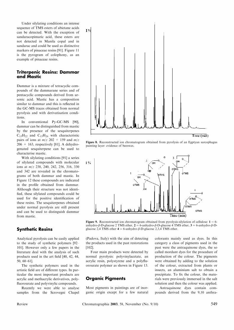
given in Figure 8 [84]. The sample was
taken from an Egyptian sarcophagus;
Egyptians often used the encaustic tech-
nique which consists of using a saponifi-
cated wax obtained after treatment with
lime as a painting binder.
Under normal pyrolysis conditions a
sequence of n)1 alkenes is obtained. Near
each peak corresponding to the alkene,
Figure 4. Reconstructed ion chromatogram obtained from a) pyrolysis of alinseed oil painting layer b) pyrolysis-methylation of the layer c) pyrolysis-silylation of the layer.
Figure 5. Formation scheme of diketopiperazines during pyrolysis.
Review Chromatographia 2003, 58, November (No. 9/10) 547

smaller quantities of the homologous al-
kane and diene are present. As a natural
product, the odd carbon hydrocarbons
dominate, while in an industrial paraffin
wax the odd hydrocarbon content is
equal to the even content.
Challinor analysed waxes under
methylating conditions obtaining a char-
acteristic distribution of fatty acid methyl
esters and fatty alcohols [85].
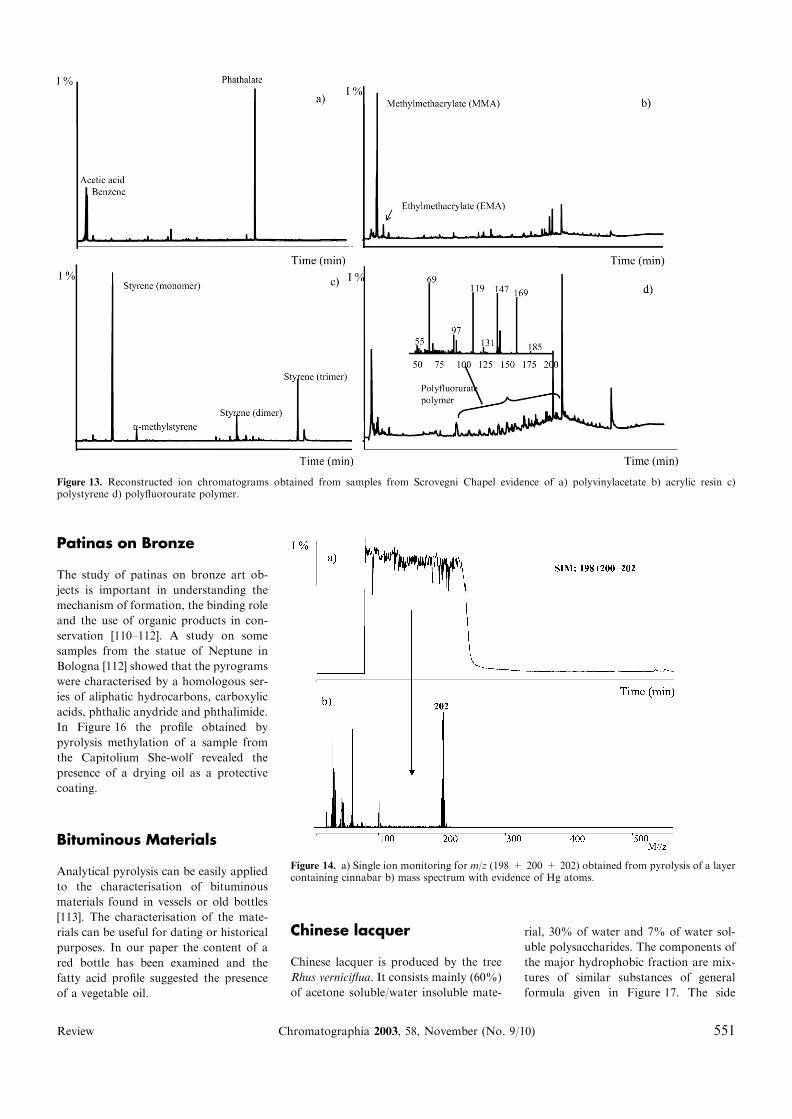
Polysaccharide Compounds
Up to now we have focussed our atten-
tion on cellulose and have tested the dif-
ferent pyrolysis techniques on its
characterisation [83, 86–87].
Pyrolysis of cellulose leads to the for-
mation of levoglucosan (1,6-anhydro-b-D-glucose), as one of the main peaks. This
is a typical anhydrosugar formed by other
carbohydrates containing glucose. How-
ever, its detection can be difficult because
of its polarity which may cause adsorp-
tion in the Py-GC interface. Reactive
pyrolysis could be used to improve GC/
MS performance of these polar markers,
through the derivatisation of OH groups.
Pyrolysis of cellulose under methylat-
ing conditions using tetramethylammo-
nium hydroxide produces mainly
methylated deoxyaldonic acids [83] with
the permethylated anhydrosugars only
being detected as trace amount [88].
Methylated deoxyaldonic acids are
distinctive markers for carbohydrates
capable of providing structural informa-
tion [83]. However, they are not specific
markers for monosaccharides like
anhydrosugars as the formation of deox-
yaldonic acids occurs with racemisation
of C-2 and reduction of C-3 with loss of
structural information. In addition the
strong alkaline environment created by
TMAH favours the production of deox-
yaldonic acids with shorter and larger
chain lengths so that different monosac-
charides produce the same deoxyaldonic
acid. For instance glycolaldehyde and
glycerinaldehyde give rise to the same
products as C6 sugars [89].
Recently, pyrolysis/silylation with
hexamethyldisilazane (HMDS) has been
proposed as an alternative to pyrolysis/
methylation for the characterisation of
carbohydrates [87]. Silylated derivatives
of levoglucosan have been obtained as
important products of cellulose by on-
line pyrolysis-GC/MS with HMDS along
with silylated hydroxylated furans and
pyranones [87]. However, levoglucosan is
found as a minor derivative, while the 4-
monoTMS, 2-monoTMS and 2,4-diTMS
derivatives, are found as major products
(Figure 9).
Diterpenic Resins: Sandaracand Copal
Sandarac is mainly composed of com-
munic acid (a dienic compound which
can easily polymerise), sandaracopimaric
acid (an abietane compound) and some
phenolic compounds.
Under normal pyrolysis conditions
totarol, a phenolic molecule, was identi-
fied as marker of sandarac, while for
copal a series of sesquiterpenoid com-
pounds characterised by 204 as molecular
weight and 161 as base peak in the
chromatogram [90].
The principal identified products un-
der silylating conditions were trimethyl-
silyl esters of various substituted
hydrogenated naphthalene carboxylic
acids (m/z 240, 308, 306), and the TMS
ester of sandaracopimaric acid [93] as can
be seen in Figure 10.
Manila copal exhibits a pattern of
abietane compounds similar to that of
sandarac, with the TMS ester of sandar-
acopimaric acid present in a small
amount. However it can be differentiated
from sandarac by the presence of addi-
tional unidentified compounds with
characteristic mass spectra [92].
Diterpenic Resins: Pinaceae
Under normal pyrolysis these resins are
characterised by the presence of frag-
ments of abietane compounds [90].
Figure 6. Reconstructed ion chromatogram obtained from pyrolysis-silylation of a glue paintinglayer. The dipeptide abbreviations on the tops of the peaks are related to the corresponding
Figure 7. Reconstructed ion chromatogram obtained from pyrolysis-silylation of a milk paintinglayer. The dipeptide abbreviations on the tops of the peaks are related to the correspondingdiketopiperazine compounds.
548 Chromatographia 2003, 58, November (No. 9/10) Review
Under silylating conditions an intense
sequence of TMS esters of abietane acids
can be detected. With the exception of
sandaracopimaric acid, these esters are
not detected in Manila copal and in
sandarac and could be used as distinctive
markers of pinaceae resins [91]. Figure 11
is the pyrogram of colophony, as an
example of pinaceae resins.
Triterpenic Resins: Dammarand Mastic
Dammar is a mixture of tetracyclic com-
pounds of the dammarane series and of
pentacyclic compounds derived from ur-
sonic acid. Mastic has a composition
similar to dammar and this is reflected in
the GC-MS traces obtained from normal
pyrolysis and with derivatisation condi-
tions.
In conventional Py-GC-MS [90],
dammar can be distinguished from mastic
by the presence of the sesquiterpenes
C15H22 and C15H26 with characteristic
pairs of ions at m/z 202 + 159 and m/z
206 + 163, respectively [81]. A dehydro-
genated sesquiterpene can be used to
characterise mastic.
With silylating conditions [91] a series
of silylated compounds with molecular
ions at m/z 238, 240, 242, 256, 316, 330
and 342 are revealed in the chromato-
grams of both dammar and mastic. In
Figure 12 these compounds are indicated
in the profile obtained from dammar.
Although their structure was not identi-
fied, these silylated compounds could be
used for the positive identification of
these resins. The sesquiterpenes obtained
under normal pyrolysis are still present
and can be used to distinguish dammar
from mastic.
Synthetic Resins
Analytical pyrolysis can be easily applied
to the study of synthetic polymers [92–
101]. However only a few papers in the
literature deal with the analysis of such
products used in the art field [40, 42, 44,
50, 60–61].
The synthetic polymers used in the
artistic field are of different types. In par-
ticular the most important products are
acrylic and methacrylic derivatives, poly-
fluorourate and polyvinylic compounds.
Recently we were able to analyse
samples from the Scrovegni Chapel
(Padova, Italy) with the aim of detecting
the products used in the past restorations
[102].
Four main products were detected by
normal pyrolysis: polyvinylacetate, an
acrylic resin, polystyrene and a polyflu-
orourate polymer as shown in Figure 13.
Organic Pigments
Most pigments in paintings are of inor-
ganic origin except for a few natural
colorants mainly used as dyes. In this
category a class of pigments used in the
past were the antraquinone dyes, the so
called mordant dyes for the procedure of
production of the colour. The pigments
were obtained by adding to the solution
of the colour, extracted from plants or
insects, an aluminium salt to obtain a
precipitate. To fix the colour, the mate-
rials were previously immersed in the salt
solution and then the colour was applied.
Antraquinone dyes contain com-
pounds derived from the 9,10 anthra-
Figure 8. Reconstructed ion chromatogram obtained from pyrolysis of an Egptyan sarcophaguspainting layer: evidence of beeswax.
Figure 9. Reconstructed ion chromatogram obtained from pyrolysis-silylation of cellulose: 1 ¼ 6-anhydro-b-D-glucose 2 TMS ether, 2 ¼ 6-anhydro-b-D-glucose 4 TMS ether, 3 ¼ 6-anhydro-b-D-glucose 2,4 TMS ether 4 ¼ 6-anhydro-b-D-glucose 2,3,4 TMS ether.
Review Chromatographia 2003, 58, November (No. 9/10) 549

cenedione. Alizarin and purpurin are the
most important of the plant extracts,
while in the insect extracts carminic acid
and chermesic acid are the most abun-
dant.
Pyrolysis of alizarin under methylat-
ing conditions produces the partially or
totally derivatised compound, while from
purpurin only benzoic acid methyl ester
and dimethylphtalate are obtained. The
same results for purpurin have been ob-
tained by analysing the natural extracts
treated with alum [103].
Another important historical dye is
indigo which belongs to the vat dyes. The
material was immersed in the colourless
solution obtained by extracting the plant
(Indigofira tinctoria). Standing the mate-
rial in the sun gives rise to a blue insol-
uble coloration thanks to the oxidation of
the indoxile present in the solution to
indicano [103].
Pyrolysis with methylation produces
characteristic fragments such as 2-
(amino)-benzoic acid methyl ester and
2-(methylamino)-benzoic acid methyl
ester.
Inorganic Pigments
Some inorganic compounds can be anal-
ysed by means of analytical pyrolysis as
they produce volatile derivatives under
thermal treatment. Cinnabar, for instance
(HgS, red pigment) gives metallic Hg
atoms which can be detected. A broad
peak appears in the chromatogram with a
characteristic mass spectrum showing the
isotopic pattern of mercury [104] (Fig-
ure 14), deriving from the thermal
decomposed salt.
Arsenic compounds such the yellow
pigment orpiment (As2S3) give As4 on
heating and this appears as a regular
peak. Its mass spectrum is characterised
by m/z 300 (As4 molecular weight) as the
base peak and following peaks with losses
of 75 (As) [84, 105] (Figure 15).
Patinas on Stones or Bricks
Degradation patinas on stone or brick
buildings show the presence of aromatic
hydrocarbons deriving from the incom-
plete combustion and alkanes and alk-
enes whose distribution can give
information about the biogenic or
anthropogenic origin of the deposition
[106–109].
Figure 10. Reconstructed ion chromatogram obtained from pyrolysis-silylation of a sandaraclayer.
Figure 11. Reconstructed ion chromatogram obtained from pyrolysis-silylation of colophonylayer: 1 ¼ Pimaric acid TMS ester, 2 ¼ Sandaracopimaric acid TMS ester, 3 ¼ Isopimaric acidTMS ester, 4 ¼ 6 Dehydrodehydroabietic acid TMS ester, 5 ¼ Dehydroabietic acid TMS ester,6 ¼ Abietic acid TMS ester.
Figure 12. Reconstructed ion chromatogram obtained from pyrolysis-silylation of dammar.Numbers on top of the peaks indicate characteristic ions (M+) in the mass spectrum.
550 Chromatographia 2003, 58, November (No. 9/10) Review
Patinas on Bronze
The study of patinas on bronze art ob-
jects is important in understanding the
mechanism of formation, the binding role
and the use of organic products in con-
servation [110–112]. A study on some
samples from the statue of Neptune in
Bologna [112] showed that the pyrograms
were characterised by a homologous ser-
ies of aliphatic hydrocarbons, carboxylic
acids, phthalic anydride and phthalimide.
In Figure 16 the profile obtained by
pyrolysis methylation of a sample from
the Capitolium She-wolf revealed the
presence of a drying oil as a protective
coating.
Bituminous Materials
Analytical pyrolysis can be easily applied
to the characterisation of bituminous
materials found in vessels or old bottles
[113]. The characterisation of the mate-
rials can be useful for dating or historical
purposes. In our paper the content of a
red bottle has been examined and the
fatty acid profile suggested the presence
of a vegetable oil.
Chinese lacquer
Chinese lacquer is produced by the tree
Rhus verniciflua. It consists mainly (60%)
of acetone soluble/water insoluble mate-
rial, 30% of water and 7% of water sol-
uble polysaccharides. The components of
the major hydrophobic fraction are mix-
tures of similar substances of general
formula given in Figure 17. The side
Figure 13. Reconstructed ion chromatograms obtained from samples from Scrovegni Chapel evidence of a) polyvinylacetate b) acrylic resin c)polystyrene d) polyfluorourate polymer.
Figure 14. a) Single ion monitoring for m/z (198 + 200 + 202) obtained from pyrolysis of a layercontaining cinnabar b) mass spectrum with evidence of Hg atoms.
Review Chromatographia 2003, 58, November (No. 9/10) 551

chains are mainly of 15 and 17 carbon
atoms and may be saturated or have 1,2,3
double bonds [73].
Pyrolysis with methylathion of a ter-
racotta jar sample has showed evidence
of Chinese Lacquer [106] by the presence
of substituted dimethoxy phenols.
Conclusions
Pyrolysis both in normal and in derivat-
isation conditions is a useful procedure
for the GC-MS analysis of the organic
fraction in works of art. The main
advantages are the limited amount of
required sample and the possibility to
simultaneously analyse organic compo-
nents belonging to different classes, while
with traditional techniques specific pre-
treatments for different matrices are nec-
essary. Analysis of polar compounds can
be performed thanks to the use of deri-
vatisation reactants.
This technique allows the characteri-
sation of different organic matrices and
of some inorganic pigments.
Our research group is engaged in the
experimentation of this technique and it
is our intention to verify its applicability
field and its limit testing it with new
derivatisation reagent or to the new
materials used in modern art.
References
1. Johnson BB, Cairns T (1972) Anal Chem44:24–34
2. Mirti P (1989) Ann Chim 79:455–4773. May RW, Porter J (1975) J Forens Sci
Soc 15:137–1464. Halpine SM (1998) Chromatogr Sci Ser
78:903–9275. Halpine SM (1995) ACS Symp Ser
617:234–2546. Sinkai T, Sugisita R (1990) Kobunkazai
no Kagaku 35:1–127. Halpine SM (1992) Stud Conservation
37(1):22–388. Halpine SM (1996) Stud Conservation
41(2):76–94
Figure 15. a) Single ion monitoring for m/z 300 obtained from pyrolysis of a layer containingorpiment b) mass spectrum of As4.
Figure 16. Reconstructed ion chromatogram obtained from pyrolysis-methylathion of a samplefrom the bronze statue She-wolf.
Figure 17. General formula of the compoundspresent in the major hydrophobic fraction ofChinese lacquer.
552 Chromatographia 2003, 58, November (No. 9/10) Review

9. Wouters J, Verhecken, A (1991) J SocDyers and Colourists 107(7–8):266–269
10. Shibayama N, Yamaoka R, Sato M, IidaJ (1991) Shitsuryo Bunseki 39(3):123–131
11. Yamaoka R, Shibayama N, Yamada T,Sato M (1989) Shitsuryo Bunseki37(4):249–253
12. Wouters J, Verhecken A (1989) StudConservation 34(4):189–200
13. Wouters J (1985) Stud Conservation30(3):119–128
14. Taylor GW (1983) Stud Conservation28(4):153–160
15. Colombini MP, Modugno F, Fuoco R,Tognazzi A (2002) Microchem J 73(1–2):175–185
16. van den Berg JDJ, van den Berg KJ,Boon JJ (2002) J Chromatogr A 950(1–2):195–211
17. Gimeno-Adelantado JV, Mateo-CastroR, Domenech-Carbo MT, Bosch-Reig F,Domenech-Carbo A, De la Cruz-Caniz-ares J, Casas-Catalan MJ (2002) Talanta56(1):71–77
18. Colombini MP, Modugno F, GiannarelliS, Fuoco R, Mattini M (2000) MicrochemJ 67(1–3):385–396
19. Van den Berg KJ, Boon JJ, Pastorova I,Spetter LFM (2000) J Mass Spectrom35(4):512–533
20. Colombini MP , Modugno F, GiacomelliM, Francesconi S (1999) J Chromatogr A846(1 + 2):113–124
21. Van Der Doelen GA, Van Den Berg KJ,Boon JJ (1998) Stud Conservation43(4):249–264
22. Van Den Berg JDJ, Van Den Berg KJ,Boon JJ (1998) Adv Mass Spectrom14:D052100/1–D052100/8
23. Colombini MP, Fuoco R, Giacomelli A,Muscatello B (1998) Stud Conservation43(1):33–41
24. Casoli A, Musini PC, Palla G (1996)J Chromatogr A 731(1 + 2):237–246
25. Galletti GC, Bocchini P, Chiavari G,Fabbri D (1996) Fresenius J Anal Chem354(3):381–383
26. Nowik W (1995) Stud Conservation40(2):120–126
27. Casoli A, Mirti P, Palla G (1995) Frese-nius J Anal Chem 352(3–4):372–379
28. White R (1978) Stud Conservation23(2):57–68
29. Birshtein VY, Tul’chinskii VM (1976)Khimiya Prirodnykh Soedinenii 1:15–19
30. Gimeno-Adelantado JV, Mateo-CastroR, Domenech-Carbo MT, Bosch-Reig F,Domenech-Carbo A, Casas-Catalan MJ,Osete-Cortina L (2001) J Chromatogr A922(1–2):385–390
31. Aruga R, Mirti P, Casoli A, Palla G(1999) Fresenius J Anal Chem365(6):559–566
32. Schneider U, Kenndler E (2001) Frese-nius J Anal Chem 371(1):81–87
33. Irwins J (1982) In: Analytical pyrolysis: acomprehensive guide, Marcel Dekker Inc,New York
34. Wampler TP (1999) J Chromatogr A842:207–220
35. Shedrinsky AM, Wampler TP, IndicatorN, Baer NS (1989) J Anal Appl Pyrolysis15:393–412
36. Shedrinsky AM, Baer NS (1995) AppliedPyrolysis Handbook 6,125–155
37. Breek R, Froentjes W (1975) Stud Con-servation 20:183–189
38. Froentjes W, Breek R (1977) ChemischWeekblad Magazine 583–589
39. Chiantore O, Scalarone D, Learner T(2003) Int J Polym Anal Charact 8(1):67–82
40. Languri GM, van der Horst J, Boon JJ(2002) J Anal Appl Pyrolysis 63(1):171–196
41. Boon JJ, Learner T (2002) J Anal ApplPyrolysis 64(2):327–344
42. Nakamura S, Takino M, Daishima S(2001) J Chromatogr A 912(2):29–334.
43. Learner T (2001) Stud Conservation46(4):225–241
44. Nakamura S, Takino M, Daishima S(2001) J Chromatogr A 912(2):329–334
45. Cerven JF 222nd ACS National Meeting(2001) Chicago, IL, United States, Au-gust 26–30
46. Scalarone D, Lazzari M, Chiantore O(2001) J Anal Appl Pyrolysis 58–59:503–512
47. Van den Berg KJ, Boon JJ, Pastorova I,Spetter LFM (2000) J Mass Spectrom35(4):512–533
48. Burns DT, Doolan KP (2000) Anal ChimActa 422(2):217–230
49. Sonoda N (1999) Stud Conservation44(3):195–208
50. Bocchini P, Traldi P (1998) J MassSpectrom 33(11):1053–1062
51. Pastorova I, van der Berg KJ, Boon JJ,Verhoeven JW (1997) J Anal Appl Pyro-lysis 43(1):41–57
52. Van den Berg KJ, van der Horst J, BoonJJ, Shibayama N, de la Rie ER (1998)Adv Mass Spectrom 14:563–573
53. Lehrle RS (1997) J Anal Appl Pyrolysis40–41:3–19
54. Stevanato R, Rovea M, Carbini M,Favretto D, Traldi P (1997) Rapid Com-mun Mass Spectrom 11(3):286–294
55. Carbini M, Stevanato R, Rovea M,Traldi P, Favretto D (1996) Rapid Com-mun Mass Spectrom 10(10):1240–1242
56. Wilcken H, Schulten HR (1996) FreseniusJ Anal Chem 355(2):157–163
57. Carbini M, Volpin S, Traldi P (1994) OrgMass Spectrom 29(10):561–565
58. Challinor JM (1990) Chemistry in Aus-tralia 57(4):90–92
59. Sonoda N, Rioux JP, Duval AR (1993)Stud Conservation 38(2):99–127
60. Sonoda N, Rioux JP (1990) Stud Con-servation 35(4):189–204
61. Wampler TP (1989) J Anal Appl Pyrolysis16(4):291–322
62. Burke P, Curry CJ, Davies LM, CousinsDR (1985) Forensic Sci Int 28(3–4):201–19
63. Challinor JM (1984) Report of Investi-gations – Western Australia, GovernmentChemical Laboratories 1–29
64. Simunkova E, Zelinger J (1983) Vlast-nosti a Zpracovani S10:33–48
65. Wa C, Wang Z, Peng J (1983) TuliaoGongye 75:32–34
66. deWitt RG (1976) Conserv Restor PictArt Butterworth, London RG
67. Schultz BW, Perros TP (1975) J—AssocOff Anal Chem 58(6):1150–1153
68. Moldoveanu SC (2998) Analytical Pyro-lysis of Natural Organic Polymers Else-vier, Amsterdam
69. Challinor JM (1991) J Anal Appl Pyro-lysis 20:15–24
70. Asperger A, Engewald W, Fabian G(1999) J Anal Appl Pyrolysis 52:51–63
71. Ishida Y, Wakamatsu S, Yokoi H, OthaniH, Tsuge S (1999) J Anal Appl Pyrolysis49:267–276
72. Matteini M, Moles A (1989) Chemistry inRestoration. Painting materials. NardiniEditori, Firenze (in italian)
73. Mills JS, White RW (1996) The OrganicChemistry of Museum Objects, Butter-worth-Heinemann, UK
74. Chiavari G, Galletti GC, Lanterna G,Mazzeo R (1993) J Anal Appl Pyrolysis24:227–242
75. Chiavari G, Ravanelli M (1999) OPDRestoration 11:130–136 (in italian)
76. Chiavari G, Gandini N, Russo P, FabbriD (1998) Chromatographia 47:420–426
77. Chiavari G, Galletti GC, Bocchini P,Russo P, Gandini N, Fabbri D (1998) SciTechnol Cult Herit 7(1):19–25
78. Chiavari G, Bocchini P, Galletti GC(1992) Sci Technol Cult Herit 1:153–158
79. Chiavari G, Fabbri D, Prati S (2001)Chromatographia 53:311–314
80. Chiavari G, Lanterna G, Luca C,Matteini M, Prati S, Sandu ICA (2003)Chromatographia 57:645–648
81. Chiavari G, Fabbri D, Prati S, Mazzeo R,Bikiaris D, Daniilia S, Chryssoulakis Y(1999) Proceedings of 6� internationalConference on ‘‘Non destructive andmicroanalysis for the diagnostics andconservation of the cultural and environ-mental heritage’’ Art 99 Rome 2:1147–1162
82. Moldoveaunu SC (1998) AnalyticalPyrolysis of Natural Organic Polymers,Elsevier, Amsterdam
83. Fabbri D, Helleur R (1999) J Anal ApplPyrolysis 49:277–293
84. Chiavari G, Fabbri D, Galletti GC,Mazzeo R (1995) Chromatographia40:594–600
85. Challinor JM (1996) J Anal Appl Pyro-lysis 37:185–197
86. Fabbri D, Chiavari G (2001) Anal ChimActa 449:271–280
87. Fabbri D, Chiavari G, Prati S, Vassura I,Vangelista M (2002) Rapid CommunMass Spectrom 16:2349–2355
88. Schwarzinger C, Tanczos I, Schmidt H(2002) J Anal Appl Pyrolysis 62:179–196
89. Schwarzinger C (2002) 15th InternationalSymposium on Analytical and AppliedPyrolysis, Leoben 43
90. Chiavari G, Fabbri D, Mazzeo R, Boc-chini P, Galletti GC (1995) Chromatog-raphia 41:273–281
91. Chiavari G, Fabbri D, Prati S (2002)Chromatographia 55:611–616
92. Haken JK, Iddamalgoda PI (1996)J Chromatogr A 756:1–20
93. Haken JK (1998) J Chromatogr A825:171–187
94. Hiltz AJ (2000) J Anal Appl Pyrolysis55:135–150
95. Fabbri D, Tartari D, Trombini C (2000)Anal Chim Acta 413:3–11
96. Jinxin Li, Haihong Xu, Jing Shi, Chun-man Li, Chenlin Bao (1999) Anal ChimActa 402:311–318
97. Haken JK (1999) Prog Org Coat 36:1–10
98. Cheng-YuWang F, Burleson AD (1999) JChromatogr A 833:111–119
99. Fabbri D (2001) J Anal Appl Pyrolysis58–59:361–370
Review Chromatographia 2003, 58, November (No. 9/10) 553

100. Tienpont B, David F, Vanwalleghem F,Sandra P (2001) J Chromatogr A911:235–247
101. Dolezal Z, Pacakova V, Kovaova J(2001) J Anal Appl Pyrolysis 57:177–185
102. Chiavari G, Ioele M, Prati S, SantopadreP (2002) Chromatographia 56:763–767
103. Fabbri D, Chiavari G, He Ling (2000) JAnal Appl Pyrolysis 56:167–178
104. Chiavari G, Mazzeo R (1999) Chroma-tographia 49:268–272
105. Chiavari G, Fabbri D, Galletti GC (1995)Rapid Commun Mass Spectrom 9:559–562
106. Schiavon N, Chiavari G, Fabbri D,Schiavon G (1994) III InternationalSymposium on the Conservation ofMonuments in the Mediterranean BasinVenice, 93–99
107. Schiavon N, Chiavari G, Fabbri D,Schiavon G (1994) Science and CulturalHeritage 10:540–549 (in italian)
108. Schiavon N, Chiavari G, Schiavon G,Fabbri D (1995) Sci Total Environ167:87–101
109. Galletti GC, Bocchini P, Cam D, Chiav-ari G (1996) Fresenius J Anal Chem354:381–383
110. Chiavari G, Mazzeo R (1997) Proceed-ings 4th International Symposium on theConservation of Monuments in theMediterranean Basin Rhodes 111–123
111. Chiavari G, Colucci A, Mazzeo R,Ravanelli M (1999) Chromatographia49:35–41
112. Chiavari G, Ferretti S, Galletti GC,Mazzeo R (1991) J Anal Appl Pyrolysis20:253–261
Received: May 13, 2003Revised manuscriptreceived: Jul 14, 2003Accepted: Jul 23, 2003
554 Chromatographia 2003, 58, November (No. 9/10) Review