analysis of the interaction between adenine nucleobase and ...tautomeric form of the nucleobase and...
TRANSCRIPT
FULL PAPER
DOI: 10.1002/ejic.200900124
Analysis of the Interaction between Adenine Nucleobase and Metal-MalonatoComplexes
Sonia Pérez-Yáñez,[a] Oscar Castillo,*[a] Javier Cepeda,[a] Juan P. García-Terán,[a]
Antonio Luque,*[a] and Pascual Román[a]
Keywords: Bioinorganic chemistry / Nucleobases / Supramolecular chemistry / Magnetic properties
The synthesis, crystal structure and variable-temperaturemagneticmeasurementsof compounds [M2(μ-Hade)2(μ-mal)2-(H2O)2]·2H2O [M = Ni (1), Co (2)], [Co2(μ-Hade)2(μ-mal)2-(H2O)2] (3), {(H2ade)2[Cu(μ-mal)2]·2H2O}n (4) and (H2ade)2-[Cu(mmal)2(H2O)] (5; H2ade = adeninium, Hade = adenine,mal = malonato, mmal = methylmalonato) are reported. Com-pounds 1–3 contain neutral paddle-wheel [M2(μ-Hade)2(μ-mal)2(H2O)2] [M = Ni (1), Co (2 and 3)] units where the 7H-tautomeric form of the nucleobase and the malonate dianionact as μ-κN3:κN9 and μ-κ2O1,O2:κO1 bridging ligands,respectively. The supramolecular crystal structures of 1–3 areessentially maintained by hydrogen-bonding interactions in-volving the nucleobases, the carboxylate groups and crystal-lisation water molecules (in compounds 1 and 2). Compounds4 and 5 show a hybrid inorganic-organic lamellar structurecontaining layers of anionic metal-malonato complexes andsupramolecular aggregates of the 1H,9H-adeninium cation.Their overall three-dimensional architectures are controlled,
IntroductionOver the past decades substantial research effort has
been invested in the rational design and elaboration of bio-mimetic systems[1] based on the interaction of nucleic acidsand their building units with a wide range of both organicand inorganic frameworks.[2] The interest in these systemsnot only stems from the desire to better understand thecomplex interactions often present in a great diversity ofmolecular biorecognition processes[3] but also to afford apowerful tool for the improvement of pharmaceuticalagents[4] and the development of artificial receptors used asspecific nucleotide sensors or even for the determination oflow concentrations of biological and therapeutic agents.[5]
Our research group has recently demonstrated the highefficiency of several metal-oxalato systems to act as recep-tors of adenine and cytosine (neutral, cationic and supra-molecular aggregates) by means of the covalent anchoringof nucleobases to the metal centres and/or by the establish-
[a] Departamento de Química Inorgánica, Facultad de Ciencia yTecnología, Universidad del País Vasco,Apartado 644, 48080 Bilbao, SpainFax: +34-94-601-3500E-mail: [email protected]
Eur. J. Inorg. Chem. 2009, 3889–3899 © 2009 Wiley-VCH Verlag GmbH & Co. KGaA, Weinheim 3889
in addition to electrostatic forces, by supramolecular recogni-tion processes between the inorganic and organic frame-works. Compound 4 contains one-dimensional anionic cop-per-malonato chains in which the copper atoms are doublybridged by μ-κ2O1,O2:κO2� malonato ligands and the cat-ionic nucleobases form dimeric entities sustained by Hoogs-teen-Hoogsteen hydrogen-bonding interactions. Compound5 is built up of anionic [Cu(mmal)2(H2O)]2– monomeric enti-ties and planar, hydrogen-bonded, one-dimensional, ribbon-like aggregates of protonated adenine. Variable-temperaturemagnetic susceptibility measurements of compounds 1–4show the preponderance of ferromagnetic interactions be-tween the paramagnetic centres. DFT calculations have beenperformed to evaluate the magnetic behaviour of these com-pounds and to analyze the interaction between two adenin-ium cations in compound 4.(© Wiley-VCH Verlag GmbH & Co. KGaA, 69451 Weinheim,Germany, 2009)
ment of complex hydrogen-bonding recognition patternsbetween the organic and inorganic frameworks.[6] Now wehave extended this work to the malonate anion, a ligandwith a greater flexibility that gives a more extended struc-tural diversity. This ligand has a remarkable versatility inadopting several different modes of bonding, includingmonodentate, chelating and bridging, with more than oneof these modes sometimes occurring in the same com-pound.[7]
We report herein the synthesis, crystal structure and vari-able-temperature magnetic measurements of five com-pounds containing the nucleobase adenine and a malonatoligand, namely [M2(μ-Hade)2(μ-mal)2(H2O)2]·2H2O [M =Ni (1), Co (2)], [Co2(μ-Hade)2(μ-mal)2(H2O)2] (3), {(H2ade)2-[Cu(μ-mal)2]·2H2O}n (4) and (H2ade)2[Cu(mmal)2(H2O)](5) (H2ade = adeninium, Hade = adenine, mal = malonato,mmal = methylmalonato), and demonstrate that the metal-malonato frameworks, as well as the metal-oxalato systems,act as adenine receptors.
Compounds 1–3 contain pseudo-structural “paddle-wheel” dinuclear units in which the nucleobase is in the 7H-adenine form. Since the first example of this kind of ar-rangement, [Cu2(μ-adenine)4(H2O)2]·5H2O, was reportedby Sletten,[8] a great deal of attention has been paid to the

O. Castillo, A. Luque et al.FULL PAPERdesign of compounds containing [M2(μ-adenine)4–x(L)x]frameworks with different ligands due both to their poten-tial ability to act as building blocks in the construction ofsupramolecular architectures[9] and because they are pos-sible candidates for anticancer treatment, as has been dis-cussed both theoretically and experimentally.[10] Com-pounds 4 and 5 are lamellar hybrid structures formed byalternating inorganic and organic layers. Hybrid inorganic-organic compounds have attracted an increasing interestover the last few years due to the possibility of combiningthe different characteristic of the components to achieveunusual structures, properties or applications (catalysis, gasseparation and storage) which are not commonly observedin purely inorganic or organic phases.[11]
Results and Discussion
[M2(μ-Hade)2(μ-mal)2(H2O)2]·2H2O [M = Ni (1), Co (2)]and [Co2(μ-Hade)2(μ-mal)2(H2O)2] (3)
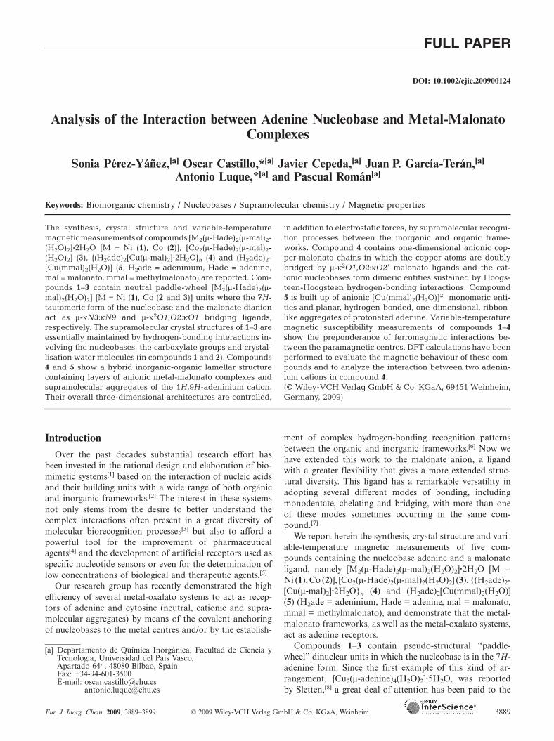
X-ray diffraction analysis of compounds 1–3 showed thattheir crystal structures consist of centrosymmetric [M2(μ-Hade)2(μ-mal)2(H2O)2] [M = Ni (1), Co (2), Co (3)] unitsand crystallisation water molecules (in compounds 1 and2). A perspective view of the structural units of compound1, with the atomic numbering scheme, is given in Figure 1.
Figure 1. Structural units of compound 1 with the atomic number-ing scheme.
The metal centres within the dinuclear entities arebridged by two malonate anions and two neutral adeninemolecules to give a paddle-wheel motif. Each metal is coor-dinated to three oxygen atoms from the malonato ligandsand two nitrogen atoms of adenine nucleobases. The octa-hedral distorted polyhedron is completed with a watermolecule to give an N2O3Ow donor set. Selected bondlengths for the coordination polyhedron are listed inTable 1.
www.eurjic.org © 2009 Wiley-VCH Verlag GmbH & Co. KGaA, Weinheim Eur. J. Inorg. Chem. 2009, 3889–38993890
Table 1. Selected bond lengths [Å] for the coordination polyhedronof compounds 1–3.[a]
1 2 3
M1–N3a 2.082(3) 2.145(4) 2.191(4)M1–N9 2.074(3) 2.144(5) 2.108(5)M1–O11 2.021(2) 2.061(4) 2.144(4)M1–O11a 2.073(2) 2.115(5) 2.105(4)M1–O21 2.003(2) 2.037(5) 2.032(4)M1–O1w 2.047(2) 2.093(3) 2.018(3)
[a] Symmetry codes: (a) 1 – x, –y, 1 – z (for 1, 2); (a) 1 – x, –y, –z(for 3).
The malonate anion shows an unusual tridentate μ-κ2O1,O2:κO1 coordination mode where O11 links bothmetal atoms and O21 is only bonded to one of these metalcentres, thus forming a six-membered chelate ring.[12] Thetwo carboxylato groups of the malonato ligands in com-pound 3 are tilted 40.8°. This distortion can be attributedto more restrictive requirements of the molecular interac-tions due to the absence of crystallisation water moleculesto mediate them. The adenine bridging ligand is attachedto one metal centre by means of the pyrimidinic N3 atomand to the other one through the N9 atom of the imidazolicring to give a μ-κN3:κN9 coordination mode. Unlike incompounds 1 and 2, the two bridging adenine ligands incompound 3 are not coplanar (0.371 Å between their meanplanes). The M···M distances [2.997(1), 3.047(1) and3.051(1) Å for 1–3, respectively] are similar to those re-ported for analogous “paddle-wheel” [Cu2(μ-adenine)4(X)2]dinuclear units containing this bridging ligand.[8,13] Al-though there are no reported examples of the adenine μ-κN3:κN9 coordination mode with nickel or cobalt, Klan-icová et al. have recently reported the crystal structure of acompound containing a paddle-wheel dimeric fragment inwhich cobalt(II) atoms are joined by two 6-(4-fluoroben-zylamino)purinato bridging ligands with a Co···Co distanceof 2.667 Å.[14] The metal atoms in the above cited “paddle-wheel” copper complexes are pentacoordinate and the M–X axial bonds (X = Cl, H2O) are almost collinear with theCu···Cu axis. The metal centres in compounds 1–3 are hexa-coordinate and the angles between the M–O1w bond andthe metal···metal axis are 44.9°, 44.3° and 34.5°, respec-tively. This bending allows the formation of an intramolecu-lar O1w–H11w···O12a hydrogen bond in compounds 1 and2, or may be a consequence thereof.
Coordination of the adenine through N9 of the pyrimid-inic ring results in stabilisation of the 7H-adenine tautomer,which is its most usual form in complexes where the nucleo-base behaves as either an N9-monodentate or N3,N9-bridg-ing ligand. The 7H-adenine tautomer is additionally stabi-lized in compounds 1 and 2 by the formation of an R2
2(9)ring formed by doubly hydrogen-bonding interactions be-tween the Hoogsteen face and the carboxylate group of anadjacent dimeric unit. Similar nine-membered rings involv-ing the Hoogsteen face of the adenine nucleobase are com-mon in both artificial and biological systems, but most ofthem imply O/N–H···N7 interactions with the N7 site asH-bond acceptor.[15] Hoogsteen AT base-pairings found in

Nucleobase–Metal-Malonato Complex Interactions
different structures of DNA and RNA[16] and the Asn/Glu-adenine interaction in protein-nucleic acid complexes[17] aresustained by this kind of hydrogen-bonded ring.
The Watson–Crick face of the nucleobase in compounds1 and 2 is hydrogen-bonded to an oxygen atom of anotherdinuclear entity as donor, and to a crystallisation watermolecule as acceptor. This water molecule establishes a sec-ond hydrogen bond with a coordinated water molecule ofan adjacent dimer, thus giving rise to layers of complexunits in the bc plane (Figure 2a). The dimeric units withinthese layers adopt two different orientations, with an angleof around 40°, thus forming a rhombic net. The dimericlayers are joined together through a hydrogen-bonding in-teraction involving O22 of a malonato ligand and a crystal-lisation water molecule of an adjacent layer. No significantπ–π interactions have been observed.
Figure 2. Supramolecular structures of compounds 1 and 2 (a) and compound 3 (b).
Eur. J. Inorg. Chem. 2009, 3889–3899 © 2009 Wiley-VCH Verlag GmbH & Co. KGaA, Weinheim www.eurjic.org 3891
The supramolecular crystal structure of compound 3 isdifferent to that described for compounds 1–2 due to theabsence of crystallisation water molecules. In this case, theHoogsteen face is hydrogen-bonded to the O12 oxygenatom of the carboxylate group of an adjacent dimeric unit,which results in the formation of an R1
2(7) hydrogen-bond-ing ring. These interactions lead to unidimensional chainsthat run along the [100] direction. The interaction of theWatson–Crick face, which acts only as a hydrogen-bond do-nor (N6–H61···O21), together with those involving the co-ordinated water molecules, gives rise to layers joined to-gether through hydrogen-bonding interactions (Figure 2b).
{(H2ade)2[Cu(μ-mal)2]·2H2O}n (4)
The crystal structure of complex 4 is made up of copper-malonato chains, adeninium cations and crystallisation

O. Castillo, A. Luque et al.FULL PAPERwater molecules held together by electrostatic and hydro-gen-bonding interactions (Figure 3).
Figure 3. Structural units of compound 4 showing the 1D anionicchain and the hydrogen-bonding interactions (dashed lines).
The copper-malonato chains are made up of [Cu-(mal)2]2– anionic units that stack up along the [100] direc-tion. The copper atoms are placed on an inversion centreand are surrounded by six oxygen atoms from four differentmalonato groups to form an elongated octahedral environ-ment. Four coplanar oxygen atoms from two bidentate ma-lonato ligands form the equatorial plane around the copperatom, giving rise to a six-membered chelate ring, while theapical positions are filled by two carboxylate oxygen atoms,which are not involved in the chelation, from two neigh-bouring [Cu(mal)2]2– units. The apical Cu–O bond lengthis significantly longer [2.705(2) Å] than the equatorial ones[1.923(2) and 1.928(2) Å]. Selected bond lengths and anglesfor the coordination polyhedron are listed in Table 2. Eachmalonate anion acts as a bidentate μ-κ2O1,O2:κO2� bridg-ing ligand and connects an equatorial site of one copperatom with the apical site of an adjacent one with acopper···copper separation through the carboxylate bridgeof 5.081 Å.
Table 2. Selected bond lengths [Å] and angles [°] for the coordina-tion polyhedron of compound 4.[a]
Cu1–O11 1.923(2) Cu1–O22b 2.705(2)Cu1–O21 1.928(2)O11–Cu1–O11a 180.0(–) O21–Cu1–O21a 180.0(–)O11–Cu1–O21 93.30(8) O21–Cu1–O22b 89.78(7)O11–Cu1–O21a 86.70(8) O21–Cu1–O22c 90.22(7)O11–Cu1–O22b 86.99(7) O22b–Cu1–O22c 180.0(–)O11–Cu1–O22c 93.01(7)
[a] Symmetry codes: (a) 1 – x, –y, 1 – z; (b) x – 1, y, z; (c) 2 – x,–y, 1 – z.
The polymeric chains are hydrogen-bonded to the crys-tallisation water molecules, which leads to the formation ofanionic inorganic layers that propagate in the ab plane. Theoutside of these layers is made up of the oxygen atoms ofthe malonato ligands and the crystallisation water mole-cules, which make them optimum acceptors for moleculeswith a high number of potential hydrogen-bonding donorpositions, as is the case of the 1H,9H-adeninium cations.These cations are inserted between the anionic sheets, andthey interact through their Watson–Crick face with one ofthese sheets and through the N9 atom of the imidazolicring with the other one (Figure 4). The organic-inorganic
www.eurjic.org © 2009 Wiley-VCH Verlag GmbH & Co. KGaA, Weinheim Eur. J. Inorg. Chem. 2009, 3889–38993892
interaction is reinforced by weak C–H···O hydrogen bonds.The Watson–Crick face establishes an R2
3(8) hydrogen-bonded ring similar to that found in the crystalline struc-ture of 1H,9H-adeninium chloroanilate dihydrate[18] but ex-changing the water molecule and C=O group positions. Italso resembles that found in the end cap of the bindingpocket of the vaccinia VP39 virus protein used to discrimi-nate between alkylated and nonalkylated nucleobases.[19]
Figure 4. Insertion of the protonated nucleobases between the inor-ganic layers.
As a result of the interaction with the inorganic sheets,the Hoogsteen face of adeninium is free to establish adouble N6–H6···N7 interaction with an adjacent cation toform dimeric aggregates. These aggregates spread out alongthe [210] direction in such a way that the lone pairs of thenitrogen N3 atoms from adjacent aggregates lie face to face,at a distance of 3.24 Å, without any other interaction be-tween the aggregates that could explain this unusual dispo-sition. A similar arrangement of the pyrimidinic N3 atomin adenine in relation to the aromatic nitrogen atoms hasbeen described when bis(benzimidazole) groups of organicmolecules anchor to polynucleotides in a non-planar dispo-sition of nucleobase and aromatic ring,[20] although in thiscase it is due to the presence of additional non-covalentinteractions. The observed disposition of the adeniniun cat-ions facing their N3 atoms is therefore probably due tostructural requirements of the crystal packing of compound4, which is sustained by the hydrogen-bonding network in-volving the adeninium cations and the anionic [Cu(μ-mal)2]n2n– complex entities.
B3LYP geometric optimisations were performed to gaina deeper insight into the adeninium-adeninium interactions.The optimisation of a model based on the crystallographicstructural data with only two adeninium cations facing theirN3 atoms, without any symmetry restraints, moves the cat-ions away. However, the initial disposition is maintained ifwe simulate the interactions that are present in the crystalstructure between two opposing cations by means of theformaldehyde molecule, although the N3···N3 distance in-creases (3.8 Å). This fact seems to point to the crystal

Nucleobase–Metal-Malonato Complex Interactions
framework being responsible for the approach between cat-ions instead of an interaction between the lone pairs.
In conclusion, the supramolecular structure of 4 is builtup by three types of molecular recognition patterns: a) be-tween metal-malonato chains and water molecules, b) be-tween anionic chains and adeninium cations and c) betweenadeninium cations (Figure 5). Anionic chains and watermolecules are bound by hydrogen-bonding interactions,which gives rise to an anionic layer. Secondly, organic cat-ions, which interact non-covalently with these chains, oc-cupy the interlayer space. Finally, the molecular recognitionamong the adeninium cations results in double-faced hydro-gen-bonded aggregates.
Figure 5. Packing of compound 4 showing the alternating inor-ganic and organic layers along the [210] direction.
(H2ade)2[Cu(mmal)2(H2O)] (5)
The crystallographic analysis of complex 5 showed thatits structure consists of monomeric [Cu(mmal)2(H2O)]2–
entities and adeninium cations held together by electrostaticand hydrogen-bonding interactions (Figure 6a). The CuII
atom within the complex unit exhibits a square-pyramidal
Figure 6. (a) Structural units of compound 5 with the atomic numbering scheme; (b) view along the [001] direction showing the lamellarstructure; (c) hydrogen-bonding interactions between the inorganic and organic frameworks.
Eur. J. Inorg. Chem. 2009, 3889–3899 © 2009 Wiley-VCH Verlag GmbH & Co. KGaA, Weinheim www.eurjic.org 3893
environment (4+1) with four oxygen atoms belonging totwo chelating methylmalonate anions in the basal plane andone water molecule filling the apical position (Table 3). Thesteric hindrance of the methyl group of the malonato li-gand, which is oriented outwards from the anionic complex,causes the non-planarity of the dicarboxylate.[21]
Table 3. Selected bond lengths [Å] and angles [°] for compound 5.[a]
Cu1–O11 1.960(1) Cu1–O1w 2.265(2)Cu1–O21 1.927(1)O11–Cu1–O11a 91.68(7) O21–Cu1–O21a 91.20(8)O11–Cu1–O21 86.57(5) O11–Cu1–O21a 164.87(6)O11–Cu1–O1w 99.67(5) O21–Cu1–O1w 95.44(6)
[a] Symmetry code: (a) x, –y + 1/2, z.
The monomers are connected by means of [O1w–H11w···O12] hydrogen bonds between the coordinatedwater molecule and the oxygen atoms of one of the methyl-malonato ligand that are not implicated in the metal chela-tion, leading to zigzag ribbons running along the [100] di-rection (Figure 6b).
The 1H,9H-adeninium cations generate cationic ribbonsrunning along the [001] direction by means of R2
2(8) hydro-gen-bonding interactions between the Hoogsteen face of acation and the C2 and N3 atoms of a neighbouring adenin-ium cation (Figure 6c). These cationic ribbons are insertedbetween the anionic layers with a crossing angle betweenthe inorganic and organic ribbons of 90°.
The outer face of the cationic ribbons alternately exposesthe sugar and Watson–Crick sides of the protonated nucleo-bases. This arrangement allows a strong interaction withthe complex anionic ribbons that involves the Watson–Crick side and the O–Cu–O fragment, which establish anR2
2(8) hydrogen-bonded ring, and on the other side the N9donor-site and the non-coordinated oxygen atom of a meth-ylmalonato ligand belonging to a second anionic ribbon. Asimilar interaction involving the Watson–Crick side and O–Cu–O group has been described previously to be the moststable among the possibilities where the oxalato anion actsas metal-bonding ligand.[22] This overall hydrogen-bonding

O. Castillo, A. Luque et al.FULL PAPERscheme leads to an inorganic-organic lamellar arrangement,although no significant interaction is observed between rib-bons belonging to the same sheet (Figure 6c).
Magnetic Properties
The magnetic behaviour of 1 in the form of a χMT vs. Tplot [χM being the magnetic susceptibility per nickel(II) ion]is shown in Figure 7. Upon cooling, χMT increases pro-gressively to reach a maximum at 10 K, and then decreasesquickly. This behaviour is characteristic of a nickel dimerwith a weak ferromagnetic interaction. The decrease ob-served at low temperatures could be due to either zero fieldsplitting and/or the presence of antiferromagnetic interac-tions along the dimeric entities. However, taking into ac-count the long intermolecular Ni···Ni distance (greater than7.1 Å), this decrease is probably due to zero field splitting.The room-temperature χMT value is equal to 1.33 cm3
mol–1, a value which is close to that expected for a magneti-cally isolated spin doublet (1.00 cm3 mol–1, with g = 2). Themagnetic data were successfully fitted by the numerical ex-pression proposed by Ginsberg for dimers of S = 1 ions,[23]
which includes the zero field splitting effect in the paramag-netic centres. The best fit of the magnetic data leads to J =+14.4 cm–1, g = 2.18 and |D| = 5.5 cm–1.
Figure 7. Thermal dependence of χMT (ο) for compound 1. (–) besttheoretical fit (see text).
It is well known that non-linear NCN bridges cause anti-ferromagnetic coupling[24] while μ-oxo bridges are able totransmit both ferromagnetic and antiferromagnetic interac-tions depending on the M–O–M angle and the orientationof the metal-centred magnetic orbitals.[25] However, the co-existence of these two types of bridges requires a moreexhaustive analysis than when there is only one type. Infact, when the bridging ligands are different, the two brid-ges may either add or counterbalance their effects. Thisproblem has been treated by Nishida et al.[26] and McKeeet al.,[27] and these phenomena are known as orbital com-plementarity and countercomplementarity, respectively. Inthe present case, the splitting of the molecular magnetic or-bitals is reversed for each type of bridging ligand, thus lead-ing an almost negligible energy difference between them(see qualitative MO diagram in Scheme 1). This small en-ergy difference is responsible for the observed ferromagneticbehaviour of compound 1.
www.eurjic.org © 2009 Wiley-VCH Verlag GmbH & Co. KGaA, Weinheim Eur. J. Inorg. Chem. 2009, 3889–38993894
This conclusion was reinforced by DFTUB3LYP calcula-tions of the J parameter performed on an isolated dimermodel based on the crystal structure, which gives a value of+20.0 cm–1. This value fits fairly well the experimental one(+14.4 cm–1) with an acceptable deviation for this kind ofcalculation. The calculated spin-density distribution for thesinglet state of compound 1 is shown in Figure 8.
The magnetic behaviours of 2 and 3 in the form of χMTvs. T plots [χM is the magnetic susceptibility per cobalt(II)ion] are shown in Figure 9. At room temperature, χMT isequal to 2.7 (2) and 2.9 cm3 mol–1 K (3), values which areslightly greater than that expected for two magnetically iso-lated spin triplets (χMT = 2.43 cm3 mol–1 K). The experi-mental magnetic data for compounds 2 and 3 were fittedwith the Lines theory[28] for polynuclear cobalt(II) com-pounds, which takes into account the orbital reduction fac-tor (κ) and the spin-orbit coupling parameter (λ) togetherwith the magnetic coupling (J). Unfortunately, the fittingprocess did not reproduce the experimental curves satisfac-torily and, as a consequence, we are unable to provide reli-able magnetic coupling constants for these two complexes.Nevertheless, the characteristics of the curve, which clearlyshows an increase of χMT with temperature decrease forcompound 2, indicate a net ferromagnetic interactionwithin the dimeric entity. The absence of an increase in theχMT curve in compound 3 could be compatible with anti-ferromagnetic or even weak ferromagnetic interactions hid-den by the spin-orbit coupling effect. In order to providefurther insights, DFTUB3LYP calculations of the J param-eter performed on dimeric models based on the crystallo-graphic data lead to values of J = +6.0 cm–1 (2) and J =+1.09 cm–1 (3), in good agreement with those reported pre-viously. These values can be interpreted as a consequenceof the previously described counterbalance effect betweenthe adenine and malonato bridging ligands.
The magnetic behaviour of 4 in the form of a χMT vs. Tplot [χM is the magnetic susceptibility per copper(II) ion]is shown in Figure 10. The room-temperature χMT value(0.42 cm3 mol–1) is close to that expected for a magneticallyisolated spin doublet (0.375 cm3 mol–1). Upon cooling, χMTremains practically constant down to 50 K, and then in-creases smoothly at lower temperatures, reaching a value of0.50 cm3 mol–1 at 4 K. This curve reveals the occurrence ofan overall weak ferromagnetic interaction. As the structureof 4 is made up of uniform chains of [Cu(mal)2]n2n– unitswhich are well separated from each other by large 1H,9H-adeninium cations, its magnetic data can be analysedthrough the numerical expression for a ferromagneticallycoupled regular chain of local spin doublets.[29] The best fitof the magnetic data leads to J = +0.6 cm–1 and g = 2.11.
In order to gain a deeper insight into the magnetic prop-erties of compound 4, DFTUB3LYP calculations were per-formed on an isolated dimer model based on its crystalstructure where the axial oxygen atoms are replaced bywater molecules. The obtained value (J = +1.1 cm–1) is ingood agreement with the experimental one. In good agree-ment with the elongated six-coordinate environment ob-served for the Cu1 atom in compound 4, the unpaired elec-

Nucleobase–Metal-Malonato Complex Interactions
Scheme 1. Energy splitting of the dx2–y2-based magnetic orbitals.
Figure 8. Spin-density distribution for the singlet state of com-pound 1.
Figure 9. Thermal dependence of χMT for compounds 2 (�) and 3(Δ).
Eur. J. Inorg. Chem. 2009, 3889–3899 © 2009 Wiley-VCH Verlag GmbH & Co. KGaA, Weinheim www.eurjic.org 3895
Figure 10. Thermal dependence of χMT (ο) for compound 4. (–)best theoretical fit (see text).
tron is mainly located in the equatorial plane, which is ofthe dx2–y2 type (magnetic orbital) [the x and y axes areroughly defined by the Cu1–O21 and Cu1–O11 bonds]. Aweak spin is thus expected in the apical positions. As theintrachain exchange pathway in 4 involves the equatorial–apical connection through a carboxylato bridge [Cu1–O21–C13–O22···Cu1d], the overlap (S) between the magnetic or-bitals of Cu1 and Cu1d is predicted to be very small or zero(accidental orthogonality). Given that the magnitude of theantiferromagnetic interaction in a copper(II) dimer is pro-portional to S2,[30] the ferromagnetic contribution is ex-pected to be dominant, which means that the resulting mag-netic coupling in 4 is most likely ferromagnetic, as ob-served. In addition, previous experimental and theoreticalstudies of carboxylato-bridged copper(II) complexes haveshown that the value of the exchange coupling between cop-per(II) ions through the bridging carboxylato is strongly de-pendent on the bridging mode of the carboxylate (syn–syn,anti–anti and anti–syn) and the type of Cu–O–C–O–Cu
O. Castillo, A. Luque et al.FULL PAPERpathway involved (equatorial–equatorial or equatorial–api-cal). Finally, the value of the ferromagnetic coupling ob-served in compound 4 (J = +0.6 cm–1) is similar to thosereported for the same orientations of the bridge and mag-netic orbitals.[31]
Conclusions
This work has proved that metal-malonato fragments aregood receptors for neutral (1–3) and protonated adenine (4and 5) molecules. The obtained compounds reveal that theneutral nucleobase is able to coordinate the metal centreand gives rise to dimeric complexes in which both the mal-onato and adenine act as bridging ligands. The presence ofadenine bridges imposes a metal···metal distance which isappropriate for the unusual bridging mode of the malonateanion.
The large number of hydrogen-bond acceptor positionsof the metal-malonato fragments, together with the high ca-pacity of the adeninium cations to act as hydrogen-bonddonors, allows their self-anchoring.
The observed ferromagnetic behaviour of these com-pounds is attributed to an orbital countercomplementarityof the adenine and malonato bridges for nickel and cobaltdimers, and to an accidental orthogonality between themagnetic orbitals for the 1D copper complex.
Experimental SectionReagents: All chemicals were of reagent grade and were used ascommercially obtained.
Physical Measurements: Elemental analyses (C,H,N) were per-formed with a Perkin–Elmer Analyst 100 microanalytical analyser.Metal content was determined by absorption spectrometry. The IRspectra (KBr pellets) were recorded with a FTIR 8400S Shimadzuspectrometer in the 4000–400 cm–1 spectral region. Magnetic mea-surements were performed on polycrystalline samples of the com-plexes taken from the same uniform batches used for the structuraldeterminations with a Quantum Design SQUID susceptometercovering the temperature range 5.0–300 K at a magnetic field of1000 G. The susceptibility data were corrected for the diamagne-tism, as estimated from Pascal’s tables,[32] the temperature-indepen-dent paramagnetism and the magnetisation of the sample holder.
Synthesis of [Ni2(μ-Hade)2(μ-mal)2(H2O)2]·2H2O (1): An aqueoussolution (3 mL) of Ni(NO3)2·6H2O (0.0596 g, 0.2 mmol) was addeddropwise to an aqueous solution (13 mL) containing malonic acid(0.1061 g, 1 mmol) and adenine (0.0274 g, 0.2 mmol) with con-tinuous stirring at 80 °C. The resulting solution (pH 1.9) was neu-tralised with NaOH to obtain a pH of 6.0. Light green X-ray qual-ity single-crystals were obtained by slow evaporation after threedays. Yield (based on metal): 39.8 mg (0.12 mmol, 60%).C8H11N5NiO6 (331.91): calcd. C 28.95, H 3.34, N 21.10, Ni 17.68;found C 29.06, H 3.27, N 20.91, Ni 17.73. Selected IR data (KBrpellet): ν̃ = 3416 s [ν(O–H)]; 3366 s [ν(NH2) + 2δ(NH2)]; 3148 m[ν(C8–H + C2–H)]; 1685 s [νas(O–C–O)]; 1623 s, 1600 s [ν(C=C) +δ(NH2)]; 1561 s, 1525 sh [ν(C4–C5) + ν(N3–C4–C5)]; 1475 m,1427 m [δ(C2–H + C8–N9) + ν(C8–H)]; 1408 m [δ(N1–C6–H6)];1366 m [ν(C5–N7–C8)]; 1283 m [ν(N9–C8 + N3–C2) + δ(C–H) +νs(O–C–O)]; 1260 m, 1180 w, 1124 w [δ(C8–H) + ν(N7–C8)]; 1025 m
www.eurjic.org © 2009 Wiley-VCH Verlag GmbH & Co. KGaA, Weinheim Eur. J. Inorg. Chem. 2009, 3889–38993896
[τ(NH2)]; 955 m, 924 m [ν(N1–C6) + τ(NH2)]; 853 w, 779 w [δ(O–C–O)]; 725 m, 610 m (ring deformation); 550 w [ν(M–O + M–N)].
Synthesis of [Co2(μ-Hade)2(μ-mal)2(H2O)2]·2H2O (2) and [Co2(μ-Hade)2(μ-mal)2(H2O)2] (3): An aqueous solution (2 mL) ofCo(NO3)2·6H2O (0.0586 g, 0.2 mmol) was added dropwise to anaqueous solution (18 mL) containing Na2C3H2O4·H2O (0.0505 g,0.3 mmol) and adenine (0.0542 g, 0.4 mmol) with continuous stir-ring at 80 °C (pH 6.7). A mixture of light pink (2) and dark pink(3) single-crystals suitable for X-ray determination was obtainedafter two months by slow evaporation. Due to their colour andmorphological differences, they were manually separated and thepurity of the samples obtained was analysed by X-ray powder dif-fraction.
Compound 2: Yield 13.3 mg (0.04 mmol, 20%). C8H11CoN5O6
(332.15): calcd. C 28.93, H 3.34, Co 17.74, N 21.09; found C 29.07,H 3.43, Co 17.81, N 20.81. Selected IR data (KBr pellet): ν̃ =3416 s [ν(O–H)]; 3366 s [ν(NH2) + 2δ(NH2)]; 3155 m [ν(C8–H +C2–H)]; 1685 s [νas(O–C–O)]; 1625 s, 1601 s [ν(C=C) + δ(NH2)];1564 s, 1521 sh [ν(C4–C5) + ν(N3–C4–C5)]; 1471 m, 1425 m [δ(C2–H + C8–N9) + ν(C8–H)]; 1406 m [δ(N1–C6–H6)]; 1365 m [ν(C5–N7–C8)]; 1284 m [ν(N9–C8 + N3–C2) + δ(C–H) + νs(O–C–O)]; 1261 m,1183 w, 1126 w [δ(C8–H) + ν(N7–C8)]; 1023 m [τ(NH2)]; 955 m,920 m [ν(N1–C6) + τ(NH2)]; 854 w, 795 w [δ(O–C–O)]; 721 m,609 m (ring deformation); 550 w [ν(M–O + M–N)].
Compound 3: Yield 31.4 mg (0.10 mmol, 50%). C8H9CoN5O5
(314.13): calcd. C 30.59, H 2.89, Co 18.76, N 22.30; found C 30.71,H 2.95, Co 18.47, N 21.94. Selected IR data (KBr pellet): ν̃ =3413 s [ν(O–H)]; 3368 s [ν(NH2) + 2δ(NH2)]; 3150 m [ν(C8–H +C2–H)]; 1682 s [νas(O–C–O)]; 1621 s, 1600 s [ν(C=C) + δ(NH2)];1561 s, 1523 sh [ν(C4–C5) + ν(N3–C4–C5)]; 1475 m, 1423 m [δ(C2–H + C8–N9) + ν(C8–H)]; 1408 m [δ(N1–C6–H6)]; 1362 m [ν(C5–N7–C8)]; 1281 m [ν(N9–C8 + N3–C2) + δ(C–H) + νs(O–C–O)]; 1260 m,1189 w, 1128 w [δ(C8–H) + ν(N7–C8)]; 1027 m [τ(NH2)]; 957 m,918 m [ν(N1–C6) + τ(NH2)]; 850 w, 797 w [δ(O–C–O)]; 726 m,606 m (ring deformation); 549 w [ν(M–O + M–N)].
Synthesis of {(H2ade)2[Cu(μ-mal)2]·2H2O}n (4): An aqueous solu-tion (3 mL) of Cu(NO3)2·3H2O (0.0485 g, 0.2 mmol) was addeddropwise to an aqueous solution (13 mL) containing malonic acid(0.1059 g, 1 mmol) and adenine (0.0273 g, 0.2 mmol) with con-tinuous stirring at 80 °C. The resulting solution (pH 2.0) was neu-tralised with NaOH to obtain a pH of 4.2. Light blue X-ray qualitysingle-crystals were obtained by slow evaporation after two weeks.Yield 51.8 mg (0.09 mmol, 45 %). C16H20CuN10O10 (575.97): calcd.C 33.37, H 3.50, Cu 11.03, N 24.32; found C 33.20, H 3.61, Cu10.99, N 24.36. Selected IR data (KBr pellet): ν̃ = 3416 s [ν(O–H)];3222 s [ν(NH2) + 2δ(NH2)]; 3026 s [ν(C8–H + C2–H)]; 1702 s[νas(O–C–O)]; 1652 w [ν(C=C) + δ(NH2)]; 1556 vs [ν(C4–C5) +ν(N3–C4–C5)]; 1448 m [δ(C2–H + C8–N9) + ν(C8–H)]; 1412 m[δ(N1–C6–H6)]; 1364 m [ν(C5–N7–C8]; 1275 m [ν(N9–C8 + N3–C2)+ δ(C–H) + νs(O–C–O)]; 1234 m, 1191 m, 1117 w [δ(C8–H) +ν(N7–C8)]; 993 m [τ(NH2]; 941 m, 886 m [ν(N1–C6) + τ(NH2)];799 m, 742 m [δ(O–C–O]; 709 m, 644 w, 617 w (ring deformation);560 m, 539 m, 520 m, 458 w [ν(M–O + M–N)].
Synthesis of (H2ade)2[Cu(mmal)2(H2O)] (5): An aqueous solution(5 mL) of Cu(NO3)2·3H2O (0.0483 g, 0.2 mmol) was added drop-wise to an aqueous solution (20 mL) containing methylmalonicacid (0.0484 g, 0.4 mmol) and adenine (0.0271 g, 0.2 mmol) withcontinuous stirring at 80 °C. The resulting solution (pH 2.5) wasneutralised with NaOH to obtain a pH of 5.4. Light blue X-rayquality single-crystals were obtained by slow evaporation afterthree weeks. Yield 41.0 mg (0.07 mmol, 35%). C18H22CuN10O9
(585.98): calcd. C 36.89, H 3.78, Cu 10.84, N 23.90; found C 36.95,

Nucleobase–Metal-Malonato Complex Interactions
Table 4. Single-crystal data and structure refinement details for compounds 1–5.
1 2 3 4 5
Formula C8H11N5NiO6 C8H11CoN5O6 C8H9CoN5O5 C16H20CuN10O10 C18H22CuN10O9
Weight [gmol–1] 331.91 332.15 314.13 575.97 586.01Crystal system monoclinic monoclinic monoclinic triclinic orthorhombicSpace group P21/c P21/c P21/c P1̄ Pnmaa [Å] 7.1286(2) 7.144(1) 9.2324(12) 5.0810(2) 9.0641(3)b [Å] 10.4120(4) 10.587(2) 7.3330(9) 9.4290(7) 19.2927(5)c [Å] 15.9463(5) 16.108(2) 17.669(2) 11.3840(7) 12.9887(3)α [°] 90(–) 90(–) 90(–) 91.738(5) 90(–)β [°] 89.980(3) 89.86(1) 115.204(10) 96.965(4) 90(–)γ [°] 90(–) 90(–) 90(–) 96.172(4) 90(–)V [Å3] 1183.58(7) 1218.3(3) 1082.3(2) 537.71(6) 2271.35(11)Z 4 4 4 1 4ρcalcd. [g cm–3] 1.863 1.811 1.928 1.779 1.714ρobsd [gcm–3] 1.85(1) 1.80(2) 1.91(2) 1.80(2) 1.70(1)μ [mm–1] 1.677 1.445 1.614 1.097 1.037Reflections collected 6533 4819 5019 4264 15545Unique data/parameters 3451/176 3282/176 2324/172 1692/169 3409/181Rint 0.0381 0.1029 0.0815 0.0287 0.0447Reflections with I�2σ(I) 2115 1143 907 1126 2129Goodness of fit (S)[a] 0.941 0.946 0.955 1.030 0.874R1
[b]/wR2[c] [I�2σ(I)] 0.0460/0.1099 0.0475/0.0602 0.0459/0.0832 0.0313/0.0743 0.0383/0.0825
R1/wR2 [all data] 0.0712/0.1145 0.0943/0.0814 0.1454/0.0951 0.0384/0.0759 0.0729/0.0895
[a] S = [∑w(Fo2 – Fc
2)2/(Nobs – Nparam)]1/2. [b] R1 = ∑||Fo| – |Fc||/∑|Fo|. [c] wR2 = [∑w(Fo2 – Fc
2)2/∑wFo2]1/2; w = 1/[σ2(Fo
2) + (aP)2] whereP = [max(Fo
2,0) + 2Fc2]/3 with a = 0.0649 (for 1), 0.0150 (for 2), 0.0375 (for 3), 0.0488 (for 4) and 0.0514 (for 5).
H 3.71, Cu 10.89, N 23.88. Selected IR data (KBr pellet): 3422 s[ν(O–H]; 3247 s [ν(NH2) + 2δ(NH2)]; 3054 s [ν(C8–H + C2–H];1685 s [νas(O–C–O]; 1647 w, 1610 s [ν(C=C) + δ(NH2)]; 1588 s[ν(C4–C5) + ν(N3–C4–C5)]; 1450 m [δ(C2–H + C8–N9) + ν(C8–H)];1400 m [δ(N1–C6–H6]; 1274 m [ν(N9–C8 + N3–C2) + δ(C–H) +νs(O–C–O)]; 1240 w, 1180 w, 1109 w [δ(C8–H) + ν(N7–C8)]; 936 m,914 m, 887 m [ν(N1–C6) + τ(NH2)]; 835 m [δ(O–C–O]; 715 m,639 m, 618 m (ring deformation); 583 w, 561 w, 539 w, 473 w [ν(M–O + M–N)].
X-ray Structural Studies: Diffraction data were collected at293(2) K on Oxford Diffraction Xcalibur (1, 3, 4, 5) and STOEIPDS (2) diffractometers with graphite-monochromated Mo-Kα ra-diation (λ = 0.71073 Å). Data reduction was performed with theCrysAlis RED[33] and X-RED[34] programs, respectively. Structureswere solved by direct methods using the SIR92 program[35] andrefined by full-matrix least-squares on F2 including all reflections(SHELXL97).[36] All calculations were performed using theWINGX crystallographic software package.[37] Crystal parametersand details of the final refinements of compounds 1–5 are summa-rised in Table 4.
CCDC-719156 (for 1), -719157 (for 2), -719158 (for 3), -719159 (for4) and -719160 (for 5) contain the supplementary crystallographicdata for this paper. These data can be obtained free of charge fromThe Cambridge Crystallographic Data Centre via www.ccdc.cam.a-c.uk/data_request/cif.
Computational Details: All quantum-mechanical calculations ofoptimised geometries were carried out in the gas phase using den-sity functional theory with Becke’s three-parameter exchange func-tional[38] along with the Lee–Yang–Parr nonlocal correlation func-tional (B3LYP).[39] The standard 6-31G(d) basis set was used asimplemented in the Gaussian 03 program.[40] It is well known thatalthough the B3LYP functional method might not be suitable forthe consistent study of the whole range of the DNA base interac-tions due to its insufficiency in describing the dispersion interac-
Eur. J. Inorg. Chem. 2009, 3889–3899 © 2009 Wiley-VCH Verlag GmbH & Co. KGaA, Weinheim www.eurjic.org 3897
tions, it predicts reliable interaction energies for hydrogen-bondedsystems.[41] DFT methods have been shown to give good estimatesof the magnetic interactions.[42] A detailed description of the com-putational strategy adopted in this work to compute the magneticcoupling constant (Jcalc) values has been described elsewhere.[43]
Density functional theory was used to carry out two separate calcu-lations to evaluate the coupling constant of each compound. Onecalculation was performed to determine the high-spin state and an-other to determine the low-spin broken symmetry state. The cor-rectness of the latter state was ensured by means of its spin densitydistribution. The hybrid B3LYP method[38] was used in all calcula-tions as implemented in Gaussian 03,[40] so that the exact Hartree–Fock-type exchange was mixed with Becke’s expression for the ex-change functional[44] and the Lee–Yang–Parr correlation func-tional[39] was used. The Gaussian implemented 6-31G(d) basis setwas employed throughout this work.
Acknowledgments
This work was supported by the Ministerio de Ciencia e Innovación(MAT2008-05690/MAT) and the Gobierno Vasco (IT-280-07). So-nia Pérez-Yáñez and Javier Cepeda thank the Universidad del PaísVasco/Euskal Herriko Unibertsitatea for a predoctoral fellowship(PIFA01/2007/021). The SGI/IZO-SGIker UPV/EHU, financed bythe National Program for the Promotion of Human Resourceswithin the National Plan of Scientific Research, Development andInnovation – “Ministerio de Ciencia e Innovación”, “Fondo SocialEuropeo (FSE)” and “Gobierno Vasco/Eusko Jaurlaritza, Di-rección de Política Científica” is gratefully acknowledged for gener-ous allocation of computational, X-ray diffraction and magneticresources.
[1] V. J. Derose, S. Burns, N.-K. Kim, M. Vogt, in: ComprehensiveCoordination Chemistry II (Eds: J. A. McCleverty, T. J. Meyer),

O. Castillo, A. Luque et al.FULL PAPERElsevier, University of Bern, Switzerland, 2003, vol. 8, pp. 787–813.
[2] a) A. D. Richards, A. Rodger, Chem. Soc. Rev. 2007, 36, 471–483; b) H. T. Chifotides, K. R. Dunbar, Acc. Chem. Res. 2005,38, 146–156; c) J. A. R. Navarro, B. Lippert, Coord. Chem. Rev.2001, 222, 219–250.
[3] M. J. Hannon, Chem. Soc. Rev. 2007, 36, 280–295.[4] M. Legraverend, D. S. Grierson, Bioorg. Med. Chem. 2006, 14,
3987–4006.[5] a) P. X. Rojas-González, A. Castiñeiras, J. M. González-Pérez,
D. Choquesillo-Lazarte, J. Niclós-Gutiérrez, Inorg. Chem.2002, 41, 6190–6192; b) F. Zamora, M. Kunsman, M. Sabat,B. Lippert, Inorg. Chem. 1997, 36, 1583–1587.
[6] a) J. P. García-Terán, O. Castillo, A. Luque, U. García-Co-uceiro, G. Beobide, P. Román, Cryst. Growth Des. 2007, 7,2594–2600; b) J. P. García-Terán, O. Castillo, A. Luque, U.García-Couceiro, G. Beobide, P. Román, Inorg. Chem. 2007,46, 3593–3602; c) J. P. García-Terán, O. Castillo, A. Luque, U.García-Couceiro, G. Beobide, P. Román, Dalton Trans. 2006,7, 902–911.
[7] C. Ruiz-Pérez, Y. Rodríguez-Martín, M. Hernández-Molina,F. S. Delgado, J. Pasán, J. Sanchiz, F. Lloret, M. Julve, Polyhe-dron 2003, 22, 2111–2123.
[8] E. Sletten, Acta Crystallogr., Sect. B 1969, 25, 1480–1491.[9] a) P. J. Sanz Miguel, B. Lippert, Dalton Trans. 2005, 1679–
1686; b) A. M. Beatty, Coord. Chem. Rev. 2003, 246, 131–143.[10] a) J. V. Burda, J. Gu, J. Inorg. Biochem. 2008, 102, 53–62; b)
H. T. Chifotides, K. R. Dunbar, Acc. Chem. Res. 2005, 38, 146–156.
[11] a) G. Kickelbick, in Hybrid Materials: Synthesis Characterisa-tion, and Applications, Wiley-VCH: Weinheim, 2007; b) P. Hor-cajada, C. Serre, M. Vallet-Regi, M. Sebban, F. Taulelle, G.Férey, Angew. Chem. Int. Ed. 2006, 45, 5974–5978; c) J. T.Hupp, K. R. Poeppelmeier, Science 2005, 309, 2008–2009.
[12] a) M. Fondo, N. Ocampo, A. M. García-Deibe, M. Corbella,M. S. El Fallah, J. Cano, J. Sanmartin, M. R. Bermejo, DaltonTrans. 2006, 4905–4913; b) A. Pajunen, E. Nasakkala, Finn.Chem. Lett. 1977, 4, 100–103; c) M. Fondo, A. M. García-De-ibe, N. Ocampo, J. Sanmartin, M. R. Bermejo, A. L. Llamas-Saiz, Dalton Trans. 2006, 4260–4270.
[13] a) A. Terzis, A. L. Beauchamp, R. Rivest, Inorg. Chem. 1973,12, 1166–1170; b) P. de Meester, A. C. Skapski, J. Chem. Soc.A 1971, 13, 2167–2169; c) J. M. González-Pérez, C. Alarcón-Payer, A. Castiñeiras, T. Pivetta, L. Lezama, D. Choquesillo-Lazarte, G. Crisponi, J. Niclós-Gutiérrez, Inorg. Chem. 2006,45, 877; d) J. P. García-Terán, O. Castillo, A. Luque, U. García-Couceiro, P. Román, L. Lezama, Inorg. Chem. 2004, 43, 4549.
[14] A. Klanicová, Z. Travnicek, I. Popa, M. Cajan, K. Dolezal,Polyhedron 2006, 25, 1421–1432.
[15] A. Travnicek, Z. Smekal, J. Marel, Z. Kristallogr. 1997, 212,123–124.
[16] E. Cubero, N. G. A. Abrescia, J. A. Subirana, F. G. Luque, M.Orozco, J. Am. Chem. Soc. 2003, 125, 14603–14612.
[17] A. C. Cheng, A. D. Frankel, J. Am. Chem. Soc. 2004, 126, 434–435.
[18] K. Gotoh, R. Ishikawa, H. Ishida, Acta Crystallogr., Sect. E2007, 63, o4433–o4433.
[19] G. Hu, P. D. Gershon, A. E. Hodel, F. A. Quiocho, Proc. Natl.Acad. Sci. 1999, 96, 7149–7154.
[20] J. Mann, A. Baron, Y. Opoku-Boahen, E. Johansson, G. Park-inson, L. R. Kelland, S. Neidle, J. Med. Chem. 2001, 44, 138–144.
[21] a) M. E. Curry, D. S. Eggleston, D. J. Hodgson, J. Am. Chem.Soc. 1985, 107, 8234–8238; b) J. M. Tercero, A. Matilla, M. A.Sanjuan, C. F. Moreno, J. D. Martin, J. A. Walmsley, Inorg.Chim. Acta 2003, 342, 77–87; c) J. Pasán, J. Sanchiz, F. Lloret,M. Julve, C. Ruiz-Pérez, CrystEngComm 2007, 9, 478–487.
[22] J. P. García-Terán, O. Castillo, A. Luque, U. García-Couceiro,G. Beobide, P. Román, Inorg. Chem. 2007, 46, 3593–3602.
www.eurjic.org © 2009 Wiley-VCH Verlag GmbH & Co. KGaA, Weinheim Eur. J. Inorg. Chem. 2009, 3889–38993898
[23] A. P. Ginsberg, R. L. Martin, R. W. Brookes, R. C. Sherwood,Inorg. Chem. 1972, 11, 2884–2889.
[24] D. Sonnenfroh, R. W. Kreilick, Inorg. Chem. 1980, 19, 1259–1262.
[25] a) F. S. Delgado, M. Hernández-Molina, J. Sanchiz, C. Ruiz-Pérez, Y. Rodríguez-Martín, T. López, F. Lloret, M. Julve, Cry-stEngComm 2004, 6, 106–111; b) L. Cañadillas-Delgado, O.Fabelo, J. Pasán, F. S. Delgado, F. Lloret, M. Julve, C. Ruiz-Pérez, Inorg. Chem. 2007, 46, 7458–7465.
[26] Y. Nishida, S. Kida, J. Chem. Soc., Dalton Trans. 1986, 2633–2640.
[27] V. McKee, M. Zvagulis, C. A. Reed, Inorg. Chem. 1985, 24,2914–2919.
[28] M. E. Lines, J. Chem. Phys. 1971, 55, 2977–2984.[29] G. A. Baker, G. S. Rushbrooke, H. E. Gilbert, Phys. Rev. 1964,
135, A1272–A1277.[30] O. Kahn, in: Molecular Magnetism, VCH Publishers, New
York, 1993.[31] a) F. S. Delgado, J. Sanchiz, C. Ruiz-Pérez, F. Lloret, M. Julve,
Inorg. Chem. 2003, 42, 5938–5948; b) J. Sanchiz, Y. Rodríguez-Martín, C. Ruiz-Pérez, A. Mederos, F. Lloret, M. Julve, NewJ. Chem. 2002, 26, 1624–1628; c) C. Ruiz-Pérez, J. Sanchiz, M.Hernández-Molina, F. Lloret, M. Julve, Inorg. Chem. 2000, 39,1363–1370; d) D. Chattopadhyay, S. K. Chattopadhyay, P. R.Lowe, J. Chem. Soc., Dalton Trans. 1993, 913–916.
[32] A. Earnshaw, in: Introduction to Magnetochemistry, AcademicPress, London, 1968.
[33] CrysAlis RED, version 1.170; Oxford Diffraction: Wroclaw, Po-land, 2003.
[34] Stoe & Cie, STADI4 and X-RED, Stoe & Cie GmbH, Darm-stadt, Germany, 2002.
[35] A. Altomare, M. Cascarano, C. Giacovazzo, A. Guagliardi, J.Appl. Crystallogr. 1993, 26, 343–350.
[36] G. M. Sheldrick, SHELXL97, University of Göttingen, Ger-many, 1997.
[37] L. J. Farrugia, WINGX, A Windows Program for CrystalStructure Analysis, University of Glasgow, UK, 1998.
[38] A. D. Becke, J. Chem. Phys. 1993, 98, 5648–5652.[39] a) C. Lee, W. Yang, R. G. Parr, Phys. Rev. B 1988, 37, 785–
789; b) B. Miehlich, A. Savin, H. Stoll, H. Preuss, Phys. Lett.1989, 157, 200–206.
[40] M. J. Frisch, G. W. Trucks, H. B. Schlegel, G. E. Scuseria,M. A. Robb, J. R. Cheeseman, J. A. Montgomery Jr., T.Vreven, K. N. Kudin, J. C. Burant, J. M. Millam, S. S. Iyengar,J. Tomasi, V. Barone, B. Mennucci, M. Cossi, G. Scalmani, N.Rega, G. A. Petersson, H. Nakatsuji, M. Hada, M. Ehara, K.Toyota, R. Fukuda, J. Hasegawa, M. Ishida, T. Nakajima, Y.Honda, O. Kitao, H. Nakai, M. Klene, X. Li, J. E. Knox, H. P.Hratchian, J. B. Cross, V. Bakken, C. Adamo, J. Jaramillo, R.Gomperts, R. E. Stratmann, O. Yazyev, A. J. Austin, R.Cammi, C. Pomelli, J. W. Ochterski, P. Y. Ayala, K. Morok-uma, G. A. Voth, P. Salvador, J. J. Dannenberg, V. G. Zakrzew-ski, S. Dapprich, A. D. Daniels, M. C. Strain, O. Farkas, D. K.Malick, A. D. Rabuck, K. Raghavachari, J. B. Foresman, J. V.Ortiz, Q. Cui, A. G. Baboul, S. Clifford, J. Cioslowski, B. B.Stefanov, G. Liu, A. Liashenko, P. Piskorz, I. Komaromi, R. L.Martin, D. J. Fox, T. Keith, M. A. Al-Laham, C. Y. Peng, A.Nanayakkara, M. Challacombe, P. M. W. Gill, B. Johnson, W.Chen, M. W. Wong, C. Gonzalez, J. A. Pople, Gaussian 03, re-vision C.02, Gaussian, Inc., Wallingford, CT, 2004.
[41] a) A. K. Rappe, E. R. Bernstein, J. Phys. Chem. A 2000, 104,6117–6128; b) O. S. Sukhanov, O. V. Shiskin, L. Gorb, Y. Podo-lyan, J. Leszczynskii, J. Phys. Chem. B 2003, 107, 2846–2852;c) A. V. Morozov, T. Kortemme, K. Tsemekhman, D. Baker,Proc. Natl. Acad. Sci. USA 2004, 101, 6946–6951.
[42] a) J. Cano, P. Alemany, S. Álvarez, M. Verdaguer, E. Ruiz,Chem. Eur. J. 1998, 4, 476–484; b) M. D. Santana, G. García,M. Julve, F. Lloret, J. Pérez, M. Liu, F. Sanz, J. Cano, G.López, Inorg. Chem. 2004, 43, 2132–2140; c) E. Ruiz, A.

Nucleobase–Metal-Malonato Complex Interactions
Rodríguez-Fortea, S. Álvarez, Inorg. Chem. 2003, 42, 4881–4884.
[43] a) E. Ruiz, J. Cano, S. Álvarez, P. Alemany, J. Comput. Chem.1999, 20, 1391–1400; b) E. Ruiz, P. Alemany, S. Álvarez, J.Cano, J. Am. Chem. Soc. 1997, 119, 1297–1303; c) E. Ruiz, A.Rodríguez-Fortea, J. Cano, S. Álvarez, P. Alemany, J. Comput.
Eur. J. Inorg. Chem. 2009, 3889–3899 © 2009 Wiley-VCH Verlag GmbH & Co. KGaA, Weinheim www.eurjic.org 3899
Chem. 2003, 24, 982–989; d) E. Rudberg, P. Salek, Z. Rinkevic-ius, H. Agren, J. Chem. Theory Comput. 2006, 2, 981–989.
[44] A. D. Becke, Phys. Rev. A 1988, 38, 3098–3100.Received: February 5, 2009
Published Online: April 29, 2009