analysis of the apoc-ii gene in apoc-ii deficient patients
TRANSCRIPT
Vol. 124, No. 1, 1984 BIOCHEMICAL AND BIOPHYSICAL RESEARCH COMMUNICATIONS
October 15, 1984 Pages 308-313
ANALYSIS OF THE ApoC-II GENE IN ApoC-II DEFICIENT PATIENTS
S.S. Fojo, S.W. Law, D.L. Sprecher, R.E. Gregg, G. Baggio* and H.B. Brewer, Jr.
Molecular Disease Branch National Heart, Lung, and Blood Institute
National Institutes of Health aethesda, Maryland
* University of Padua, Padua, Italy
Received September 7, 1984
Apolipoprotein C-II (apoC-II), a 79 amino acid protein, is a cofactor for lipoprotein lipase, the enzyme which catalyzes the lipolysis of triglycerides on plasma chylonicrons and VLDL. Patients with apoC-II deficiency have marked elevations in plasma triglycerides, chylomicrons, VLDL, and a type I hyper- lipoproteinemia. In order to evaluate the molecular defect in apoC-II deficiency, genomic DNA was analyzed using Southern Blot from 2 independent apoC-II deficient patients and compared to normal controls. Restriction digests of genomic DNA were performed with five different enzymes and the restriction fragments analyzed utilizing a 354 base pair nick-translated apoC- II probe for hybridization following Southern blotting. The restriction fragments varied from U.8 to 21 Kb, and the pattern with normal DNA was identical to that of the two apoC-II deficient patients. The present study reveals that the apoC-II gene is present in patients with apoC-II deficiency. In addition, no insertional or deletional polymorphism was detected in the apoC-II gene of apoC-II deficient patients. 01984 Academic press. rnc.
Human plasma apolipoprotein+C-II, a 79 amino acid protein, plays a
pivotal role in lipid metabolism as a cofactor for lipoprotein lipase, the
enzyiue which hydrolyzes the triglycerides in plasma chylomicrons and VLDL
(1-j). Recently we have reported the complete nucleic acid sequence of
preapoc-II (4). The derived amino acid sequence contains a 22 residue
prepeptide which is co-translationally cleaved prior to secretion, as well as
a 79 amino acid mature protein with a sequence identical to that previously
described (5). The apoC-II gene has now been localized to chromosome 19 (6,7)
which also contains the apoE (8) and LDL receptor (9) genes.
'Abbreviations: VLDL, very low density lipoproteins; LDL, low density lipoproteins; HDL, high density lipoproteins; apo, apolipoprotein; Kb, kilobase; THE, Tris borate EDTA, ptI 8.3; SSC, standard saline citrate, pH 7.1; rJaDodSO4, sodium dodecylsulfate; BRL, Bethesda Research Laboratories.
0006-291X/84 $1.50 Copyright 0 1984 by Academic Press, Inc. All rights of reproduction in any form reserved. 308
Vol. 124, No. 1, 1984 BIOCHEMICAL AND BIOPHYSICAL RESEARCH COMMUNICATIONS
,ipoC-Ii is synthesized primarily by the liver (10) and secreted into
plasma where it is reversibly bound principally to chylomicrons, VLUI,, and
dDL (II). i\poc;-II in IWL rapidly associates with newly secreted triglyceride
rich lipoproteins synthesized by the liver and intestine. Following
lipolysis, triglyceride ricn lipoproteins are converted to remnants and apoC-
Ii dissociates r'rom these particles and reassociates with tt3L (12). The
distribution of ape<-II on plasma lipoproteins is continually changing between
tii~L and triglyceride rich lipoproteins as a result of the secretion, metabolic
conversion, and catabolism of the plasma lipoproteins (11).
The importance of apoC-II as an activator of lipoprotein lipase has been
establisned by the absence of lipoprotein lipase activity in patients with
apoC-II deficiency. This syndrome, which is inherited as an autosomal
recessive trait, has been described in various independent families (13-18).
Clinical presentation in homozygous individuals may include xanthelasna,
hepatospienomegaly and pancreatitis. Because the clearance of chylomicrons is
greatly impaired, these patients have marked elevation of plasma triglycer-
ides, chylomicrons, decreased LDL and HDL concentrations, and a type I
lipoprotein phenotype. Infusions of normal plasma (13,16,1&S), or isolated
dpoC-II fractions (18) result in transient normalization of plasma
triglycerides and lipoproteins. ApoC-II deficiency is documented by the
absence of apoC-II by radioimmunoassay or immunoelectrophoresis, and
indirectly ay an absence of lipoprotein lipase activity corrected by the
audition of ayoC-II containing plasma. At the present time, the molecular
defect that results in apoC-II deficiency is unknown. In order to study the
nature of the underlying defect in apoC-II deficiency, we have utilized
Soutnern blot techniques to determine the presence of major insertional or
deletional polymorphisms in the DNA of 2 patients with apoC-II aeficiency.
tlATEKIALS AND ISTHODS
Apolipoprotein C-II cDtJA probe: The cDNA probe used in this study was obtained irom an apoC-II CWA clone which contained the nucleic acid sequence encoding for tne entire mature apoC-II protein and the 22 amino acid containing prepeptide (4). A 354 base pair long cDNA insert which encoded for most of the structural sequence of apoC-II as well as part of the 3' untrans-
309

Vol. 124, No. 1, 1984 BIOCHEMICAL AND BIOPHYSICAL RESEARCH COMMUNICATIONS
lated end (see Fig. 1) was obtained by cleavage with Alu 1 (HRL, 3U of enzyme/u:: IMA for 2 hours at 37°C) and purified by agarose gel electrophoresis. Pour hundrru nanograms of purified DIVA was radiolabeled to a speciiic activity of 1 x 10y cp13/pg by nick-translation (19).
D:dA preparation: aiood for DNA isolation was collected from normolipidenic volunteers into tubes containing a final concentration of 3ih"i IJa EDTA. ApoC- li deficient blood was obtained from an 11 yo American white female and a 41 yo italian white male previously documented to have apoC-II deficiency ClS,ZU>. DNA was isolated from whole blood cell nuclei as described by Bell et al. (21), and yields of DiJA were 200-500 ug/ml per 10 ml of blood.
Soutnern blot analysis: Ten micrograms of DNA were digested with 40 units of tiai1til (SKL) , Bgl 1 (bRL), EcoRl (BRL), Hind III (SRI,) and SST 1 (BKL) for 4 hrs at 37°C. 'The incubation was terminated by the addition of 20 ctil EDTA, and tne reaction mixture was heated in a 65'C bath for 10 min. The restriction fragments were separated by 0.7% agarose slab gel electrophoresis (0.7 x 11 x I+ cmj at 35 V for lb nr in T8E. DNA was transferred to nitrocellulose mrmsrane filter paper (Schleicher and Schuell) bidirectionally as described by Souctlern (22) and baked for 2 hr dt 80°C. Filters were incubated for at least 6 nr in 20 ml of pre-hybridization solution containing 5X SSI,, 5X Denhardt's solution, (1.12 liaDodSU4 arm 100 &ml salnon sperm DNA at 65°C in a sealed oag . Hybridization was pertormed for 20 In- at 65°C in 20 ml of buff r containing Si( SSC, 5i; 0enhardt's solution, 0.1% NaDodS04, and 1 X 10 f cpm niCK--translated apoC-11 insert DNA.
KESULTS
Figure 1 illustrates the restriction endonuclease cleavage map of the
apoC-II cL)Iix insert in the recombinant plasmid clone. The 354 base pair long
Alu 1 fragment used to prepare the nick-translated probe contained 15 amino
Alu I Alu I
I GLU i+791
Pst I I361 1)
fi, ure 1 . Hap of the apoC-II cD!JA insert of the recombinant plasmid clone. The insertion of apoC-II cDNA (open bar) and its orientation in pBR322 plasmid ljNA (solid bar) are indicated. The mRNA sequence is represented by the upper DIVA strand with the 5' and 3' termini as indicated. Asterisks indicate the sites of the dC-dC tails. Sites of cleavage by Alu 1 restriction endonuclease are indicated. The stippled area represents the 354 base pair Alu 1 fragment used to prepare the nick-translated probe. Its orientation with respect to the preapoC-II agino acid sequence is indicated.
310
Vol. 124, No. 1, 1984 BIOCHEMICAL AND BIOPHYSICAL RESEARCH COMMUNICATIONS
Barn HI Bgl I EcoRl Hind III
23 -
9.4 -
6.5 -
4.4 -
2.3 -
1.35 -
1.08 -
0.87 -
1231231231 23 123
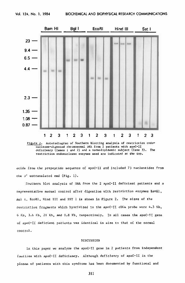
Figure 2. Autoradiogram of Southern blotting analysis of restriction endo- nuclease-digested chromosomal DNA from 2 patients with apoC-II deficiency (lanes 1 and 2j and a normolipidemic subject (lane 3). The restriction endonuclease enzymes used are indicated at the top.
acids from the prepeptide sequence of apoC-II and included 73 nucleotides from
the 3' untranslated end (Fig. 1).
Southern blot analysis of DNA from the 2 apoC-II deficient patients and .a
representative normal control after digestion with restriction enzymes BamHl,
41 1, EcoKl, Hind III and SST 1 is shown in Figure 2. The sizes of the
restriction fragments which hybridized to the apoC-11 cDlJA probe were 4.3 Kb,
6 KD, 3.6 Kb, 21 Kb, and 0.8 Kb, respectively. In all cases the apoC-II gene
of apoC-II deficient patients was identical in size to that of the normal
control.
DISCUSSION
In this paper we analyze the apoC-II gene in 2 patients from independent
families with apoC-II deficiency. Although deficiency of apoC-II in the
plasma of patients with this syndrome has been documented by functional and
311

Vol. 124, No. 1, 1984 BIOCHEMICAL AND BIOPHYSICAL RESEARCH COMMUNICATIONS
immunologic techniques, the basic molecular defect in apoC-II deficiency is
not understood (13-18). Alteration of the apoC-II gene or of the genes
reguiclting apoC-II expression or processing could result in the deficiency.
Cur study of 2 independent patients with apoC-II deficiency failed to
detect any gross deletions or insertions in their apoC-II genes when analyzed
by Southern blotting. Tne 354 base pair probe utilized for hybridization
contains 44 nucleotides from the prepeptide sequence as well as 73 nucleotides
from tne 3' untranslated end, thus encompassing most of the structural gene
ior preapoC-II. In all cases, the size of the restriction fragments obtained
from the DNA of the apoC-II deficient patients and normal controls was
identical, ruling out the presence of major rearrangements in the structural
gene of apoC-II in these patients. However, minor changes in the nucleotide
sequence of the apoC-II gene cannot be excluded by our present study. These
findings are similar to those seen in one kindred with apoA-I and C-III
deficiency where no restriction enzyme polymorphism was detected (23) but
ditferent from a second kindred with apoA-I and C-III deficiency where a 7.5
Kh ONA insertion in one of the exons of the apoA-I gene results in absence of
apo,r-1 and apoC-III (24).
i'ne analysis of the underlying molecular defect in apoC-II deficiency may
provide new information on factors which normally modulate apoC-II
biosynthesis and processing.
ACKNOWLEDGMENT
This work was supported in part by grants from P.S. CNR; Ingegneria genetica e basimolecolari delle malattie ereditarie - sotto progetto basimolecolari delle malattie ereditarie.
REFERENCES
1. LaKosa, J.C., Levy, K.I., Herbert, P., Lux, S.E. and Fredrickson, D.S. (1971)) Uiochem. Biophys. Kes. Comm.
2. 41, 45-62.
Havel, K.J., Shore, J.G., Shore, B. and Bier, D.M. (1970) Circ. Res. 27, 537-000.
3. Smith, L.C., Pownall, H. and Gotto, A.il. (1978) Ann. Rev. Riochem. 47, 751-777.
4. Fojo, S.!;., Law, S.W. and Brewer, H.B. Jr. (1984) Proc. Natl. Acad. Sci. lJSA (in press).
5. Hospattankar, A.V., Fairwell, T., Konan, R. and Brewer, H.B. Jr. (1983) J. Mol. Chem. 25y, 318-322.
312
Vol. 124, No. 1, 1984 BIOCHEMICAL AND BIOPHYSICAL RESEARCH COMMUNICATIONS
b.
7.
8.
9.
1u. 11.
12.
13.
14.
1s.
16.
17.
18.
1Y.
20.
21.
22. 23.
24.
FOJO, S.S., Law, S.W., Brewer, H.B., Jr., Sakaquchi, A.Y. and Naylor, S.L. (1984) Biochem. Biophys. Res. Comm. 122, 687-693. Jackson, C.L., Bruns, G.A. and Breslow, J.L. (1984) Proc. Natl. Acad. Sci. USA 81, 2945-2949. Olaisen, B., Teisburg, P. and Gedde-Dahl, T. Jr. (1982) Human Genet. fi'& 233-236. Francke, U., Brown, M.S. and Goldstein, J.L. (1984) Proc. Natl. Acad. Sci. USA 81, 2826-2830. Wu, A.L. and Windmueller, H.G. (1979) J. Biol. Chem. 254, 7316-7322. Osborne, J.C. Jr. and Brewer, H.B. Jr. (1977) Adv. Protein Chem. 31, 253-337. Eisenberg, S., Bilheimer, D.W., Levy, R.I. and Lindgren, F.T. (1973) Biochem. Biophys. Acta. 326, 361-377. Breckenridge, W.C., Little, J.A., Steiner, G., Chow, A. and Poapst, M. (1978) N. Engl. J. Med. 298, 1265-1273. Yamamura, T., Sudo, H., Ishikawa, K. and Yamamoto, A. (1979) Atherosclerosis 34, 53-65. Crepaldi, G., Fellin, R., Bag&o, G., Augustin, J. and Greten, H. (1980) In: Atherosclerosis L (A.M. Gotto, Jr., L.C. Smith and 5. Allen, eds.) Springer-Verlag, New York, pp. 250-254. Miller, N.E., Rao, S.N., Alaupovic, P., Nobel, N., Slack, J., Brunzell, J.D. and Lewis, B. (1981) Europ. 3. Clin. Invest. 2, 69-76. Stalenhalf, A.F.H., Caspaire, A.F., Demacher, P.N.M., Stouten, J.T.J., Lutterman, J.A. and Van't Laar, A. (1981) Metabolism 2, 919-926. Catapano, A.I., Mills, G.L., Roma, P., LaRosa, M. and Capurso, A. (1983) Clin. Chem. Acta 130, 317-327. Rigby, R.W.J., Dieckmann, M., Rhodes, C. and Berg, P. (1977) J. Mol. Biol. 113, 237-251. Sprecher, D.L., Taam, L. and Brewer, H.B. Jr. (1984) Clin Chem. (in press). Bell, C.I., Karan, J.H. and Rutter, W.J. (1979) Proc. Natl. Acad. Sci. USA 78, 5759-5763. Southern, E.M. (1975) J. Mol. Biol. 98, 503-517. Schaefer, E.J., Ordovas, J.M., Law, S.L., Ghiselli, G., Kashyap, M.L., Srivastava, L.S., Heaton, W.H., Albers, J.J., Connor, W.E., Lindgren, F.T., Lemeshev, Y, Segrest, J.P. and Brewer, H.B., Jr. (1984) 14anuscript in preparation for publication. Karathanasis, S.K., Norum, R.A., Xannis, V.I. and Breslow, J.L. (1983) Nature 301, 718-720.
313