analysis of germ line development in the chick embryo usin ...leitz dialux 20 microscope equippe...
TRANSCRIPT

Development 103, 299-304 (1988)Printed in Great Britain © The Company of Biologists Limited 1988
299
Analysis of germ line development in the chick embryo using an anti-
mouse EC cell antibody
LANCE E. URVEN1, CAROL A. ERICKSON2, URSULA K. ABBOTT1 and JOHN R. McCARREY3
^Department of Avian Sciences and ^Department ofZoology, University of California, Davis, CA 95616, USA3Division of Reproductive Biology, Department of Population Dynamics, The Johns Hopkins University School of Hygiene and PublicHealth, Baltimore, MD 21205, USA
Summary
We have found that EMA-1, a monoclonal antibodyoriginally raised against mouse embryonal carcinoma(Nulli SCC1) cells (Hahnel & Eddy, 1982), also labelschick primordial germ cells (PGCs). We have used thisantibody in immunohistological studies to follow thedevelopment of PGCs in the chick embryo from thetime of their initial appearance beneath the epiblast,through their migratory phase and subsequent colon-ization of the germinal epithelium. During hypoblastformation, individual EMA-1-labelled cells appearedto separate from the basal surface of the epiblast andenter the blastocoel, coincident with the appearance ofmorphologically identifiable PGCs in this same area.EMA-1 continued to label germ cells until the in-
itiation of gametogenesis in each sex; specifically,labelling was absent by 7-8 days of incubation infemales and started to decrease at 11 days of incu-bation in males. There was a recurrence of the epitopeon oogonia at 15 days of incubation, but not onspermatogonia during the remainder of developmentthrough hatching. These observations are consistentwith an epiblast origin for the avian germ line, and arestrikingly similar to those reported for the early mouseembryo using the same antibody (Hahnel & Eddy,1986).
Key words: primordial germ cells, EMA-1, monoclonalantibody, cell marker, chick embryo, germ line.
Introduction
The germ line, whether viewed as a continuum oftotipotent cells or a newly formed cell line arisingfrom somatic elements of each embryo, is, by defirnition, the only link between subsequent generations.Because of their unique role in the life cycle, thedevelopment of germ-line cells is particularly in-triguing, especially with regard to understanding theirdetermination, origin and relationship to somaticcells. Addressing these topics requires the ability torecognize and, ultimately, manipulate germ cellsearly in development.
Neither morphology nor the PAS marker candistinguish avian primordial germ cells (PGCs) priorto gastrulation, when they appear in the germinalcrescent. Investigation of PGC origin has been lim-ited largely to indirect in vitro studies in whichchick-quail chimaeras are constructed, using epiblastand hypoblast from different species (Eyal-Giladi etal. 1981) or by culture of isolated hypoblasts and
epiblasts until PGCs could be recognized by PASstaining (Sutasurya et al. 1983; Ginsburg & Eyal-Giladi, 1986). These studies all concluded that PGCsderive from the epiblast at stages prior to those atwhich they are identifiable by either morphological orhistochemical criteria, and that the germinal crescentendoderm hypoblast is a secondary location in PGCdevelopment.
In order to investigate directly the origin of PGCsin situ, a germ cell marker is required that candistinguish PGCs at stages prior to the time whenthey become morphologically distinct. In this study,we use the antibody EMA-1 as a PGC marker in chickembryos. EMA-1, a monoclonal IgM antibody pro-duced against a glycoprotein cell surface antigen ofthe embryonal carcinoma Nulli SCC1 (Hahnel &Eddy, 1982), recognizes fucosylated polylactosaminecarbohydrate groups and has been previously shownto label murine PGCs (Hahnel & Eddy, 1986). Wedemonstrate here that EMA-1 labels chick germ cellsat an earlier stage and for a longer period than PAS
300 L. E. Urven and others
staining does, and we provide additional evidenceconsistent with an epiblast origin of PGCs in thechick.
Materials and methods
Fertile White Leghorn eggs were purchased from a localcommercial supplier (Donsing, Rio Linda, California).Eggs were incubated at 37-5°C and 55 % relative humidity.
Immunohistochemical or immunofluorescent studiesusing EMA-1 antibody (kindly provided by Dr E. M. Eddy)employed a total of 100 chick embryos and hatchlingsranging from preincubation embryos to newly hatchedchicks. Both cryostat and paraffin sections were prepared.Each embryo was staged according to criteria establishedby Eyal-Giladi & Kochav (1976) or by Hamburger &Hamilton (1951).
Cryostat sectionsEmbryos were prepared for cryosectioning by fixing for15min in 4 % paraformaldehyde, infiltrating at room tem-perature for 2 h each in 15 % and 30 % sucrose and freezingin OCT compound (Miles Laboratories) under liquid nitro-gen. 12 jim sections were cut on a Bright Cryostat andpostfixed by dipping the slides in 0-4 % paraformaldehyde.A 1:10000 dilution of EMA-1 in phosphate-buffered saline(PBS) was applied to the sections for i h, followed by a PBSrinse. Sections were then incubated in fluorescein- orrhodamine-conjugated goat anti-mouse immunoglobulinsecondary antibody (1:100 and 1:200 dilutions, respect-ively; Cooper Biomedical). Sections were mounted in 2%n-propyl gallate (Giloh & Sedat, 1982) and examined with aLeitz Dialux 20 microscope equipped for epifluorescence.
Paraffin sectionsEmbryos were fixed in Bouin's fixative, with care taken toremove and fix the entire area vasculosa in embryosyounger than stage 18. They were then dehydrated, embed-ded in Paraplast Plus (Monoject Scientific), serially sec-tioned at 10 pm, deparaffinized and stained as describedabove. Alternatively, paraffin sections were secondarilylabelled with the avidin/biotin-conjugated-alkalinephosphatase system (Vectastain ABC-AP kit, Vector Lab-oratories) according to manufacturer's suggestions. Sec-tions were counterstained with the cytoplasmic stain FastGreen. Avidin-biotin system-stained slides were mountedin Pro-Texx (Lerner Laboratories). Controls for non-specific antibody binding substituted monoclonal mouseIgM anti-human IgG (1:500 dilution) for EMA-1 on sec-tions taken at intervals from representative stages of chick.These were consistently negative with the exception ofblood cells and occasional yolky endoderm cells, whichshowed nonspecific staining under the conditions describedabove.
PAS stainingSections from 23 chick embryos ranging in age fromunincubated embryos to 13 days of incubation were stainedwith PAS, providing a demonstration of PGC location
independent of EMA-1 labelling. Whole embryos orgonads were fixed in Rossman's fluid, double-embedded incelloidin and paraffin (Humason, 1972), and cut at 10/im.Sections were deparaffinized and hydrated, oxidized for20min in 1 % periodic acid (Sigma), rinsed with water,stained with Schiff s reagent (Sigma) counterstained withhaematoxylin, cleared and mounted in Pro-Texx. In orderto determine directly whether individual germ cells stainedwith both PAS and EMA-1, two chick embryos at stage 20(3i days of incubation) were fixed in high alcohol fixatives(Rossman's in one case and Gendre's in the other),embedded in paraffin and sectioned at 5 and 7^m, respect-ively. Alternate sections were stained with PAS or labelledwith EMA-1. All embryos stained with ABC-AP or PASwere studied and photographed using a Zeiss Photomicro-scope III. Cell size was measured with an ocular mi-crometer.
Results
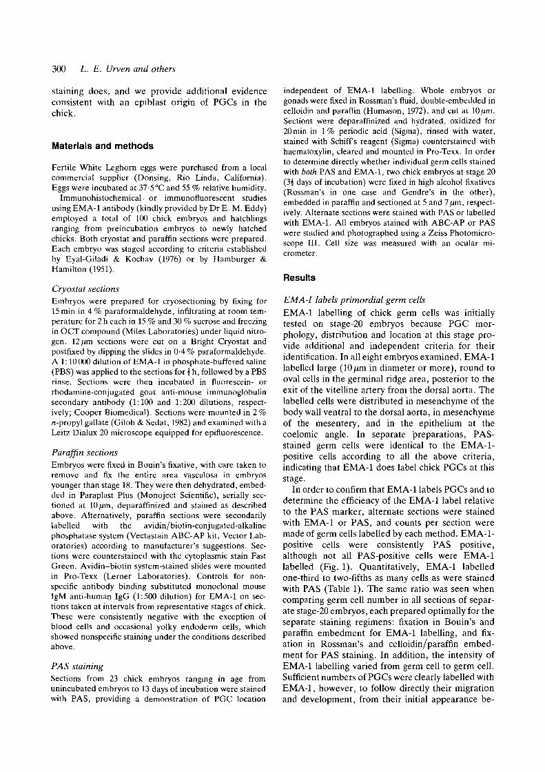
EMA-1 labels primordial germ cellsEMA-1 labelling of chick germ cells was initiallytested on stage-20 embryos because PGC mor-phology, distribution and location at this stage pro-vide additional and independent criteria for theiridentification. In all eight embryos examined, EMA-1labelled large (10/im in diameter or more), round tooval cells in the germinal ridge area, posterior to theexit of the vitelline artery from the dorsal aorta. Thelabelled cells were distributed in mesenchyme of thebody wall ventral to the dorsal aorta, in mesenchymeof the mesentery, and in the epithelium at thecoelomic angle. In separate preparations, PAS-stained germ cells were identical to the EMA-1-positive cells according to all the above criteria,indicating that EMA-1 does label chick PGCs at thisstage.
In order to confirm that EMA-1 labels PGCs and todetermine the efficiency of the EMA-1 label relativeto the PAS marker, alternate sections were stainedwith EMA-1 or PAS, and counts per section weremade of germ cells labelled by each method. EMA-1-positive cells were consistently PAS positive,although not all PAS-positive cells were EMA-1labelled (Fig. 1). Quantitatively, EMA-1 labelledone-third to two-fifths as many cells as were stainedwith PAS (Table 1). The same ratio was seen whencomparing germ cell number in all sections of separ-ate stage-20 embryos, each prepared optimally for theseparate staining regimens: fixation in Bouin's andparaffin embedment for EMA-1 labelling, and fix-ation in Rossman's and celloidin/paraffin embed-ment for PAS staining. In addition, the intensity ofEMA-1 labelling varied from germ cell to germ cell.Sufficient numbers of PGCs were clearly labelled withEMA-1, however, to follow directly their migrationand development, from their initial appearance be-
Analysis of chick germ line development 301
da - ' (da• »'* *
1A
», da ^ J da
B
Fig. 1. Confirmation of PGC labelling with EMA-1. In aGendre-fixed, paraffin-embedded stage 20 (3-5 days ofincubation) chick embryo, EMA-1 labelled a subset ofchick primordial germ cells in the germinal ridge area.These alternate sections show a PGC simultaneouslylabelled with EMA-1 (A) and with PAS (arrows) (B), aswell as PAS-positive, EMA-1-negative PGCs(arrowheads), mg, midgut; da, dorsal aorta. Bars, 50fim.
neath the epiblast through their migration to thegerminal epithelium and their subsequent entry intogametogenesis.
Distribution of EMA-1 epitope(1) Somatic tissue labelling
Unincubated embryos often had only a few labelled
cells in the epiblast. Progressively older embryos hadincreasing numbers of epiblast cells labelled withEMA-1. By gastrulation, all of the epiblast cells in thecentre of the area pellucida were strongly labelledwith EMA-1, whereas labelled cells became graduallyless common peripherally until none were seen in thearea opaca. Mesenchymal cells moving through theprimitive streak showed reduced intensity of labelcompared to the epiblast cells from which they hadjust detached. The head process stained slightly, andthe notochord, paraxial, intermediate and lateralplate mesoderm all showed scattered antibody label-ling immediately anterior to the regressing primitivestreak. Labelling of the ectoderm derived from theepiblast ceased by the end of gastrulation, with theexception of some localized staining in the diencepha-lon and spinal cord. Between stages 13 and 19,portions of the medial midgut began to label withEMA-1. Progressively more foregut and midgut en-doderm labelled in older embryos. Many of themesonephric tubules stained after stage 25 (5 days ofincubation).
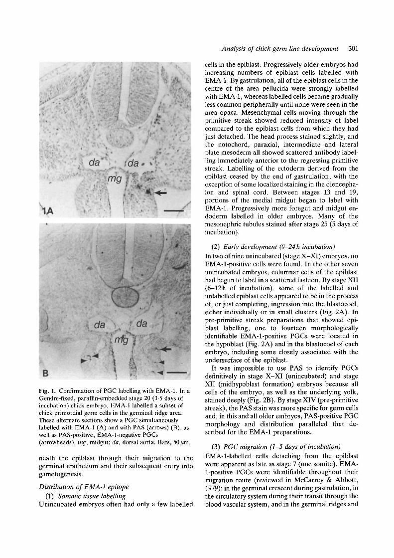
(2) Early development (0-24 h incubation)In two of nine unincubated (stage X-XI) embryos, noEMA-1-positive cells were found. In the other sevenunincubated embryos, columnar cells of the epiblasthad begun to label in a scattered fashion. By stage XII(6-12h of incubation), some of the labelled andunlabelled epiblast cells appeared to be in the processof, or just completing, ingression into the blastocoel,either individually or in small clusters (Fig. 2A). Inpre-primitive streak preparations that showed epi-blast labelling, one to fourteen morphologicallyidentifiable EMA-1-positive PGCs were located inthe hypoblast (Fig. 2A) and in the blastocoel of eachembryo, including some closely associated with theundersurface of the epiblast.
It was impossible to use PAS to identify PGCsdefinitively in stage X-XI (unincubated) and stageXII (midhypoblast formation) embryos because allcells of the embryo, as well as the underlying yolk,stained deeply (Fig. 2B). By stage XIV (pre-primitivestreak), the PAS stain was more specific for germ cellsand, in this and all older embryos, PAS-positive PGCmorphology and distribution paralleled that de-scribed for the EMA-1 preparations.
(3) PGC migration (1-5 days of incubation)EMA-1-labelled cells detaching from the epiblastwere apparent as late as stage 7 (one somite). EMA-1-positive PGCs were identifiable throughout theirmigration route (reviewed in McCarrey & Abbott,1979): in the germinal crescent during gastrulation, inthe circulatory system during their transit through theblood vascular system, and in the germinal ridges and

302 L. E. Urven and others
Table 1.
Section width (fim)Alternate sectionsPGCs
PAS(+)/EMA-l(+)
Gendre
PAS
764
136
2-96
fix
EMA-1
76446
Rosman fix
PAS EMA-1
5 5122 122433 175
2-47
RossmanPAS
10
238
2-40
BouinEMA-1
10
99
cl
h
2A
Fig. 2. EMA-1 labelling in embryos after approximately12h of incubation. (A) A Bouin's-fixed, paraffin-embedded stage-XII, embryo shows EMA-1 stainingsome cells of the epiblast (e) in cross section, includingindividual cells apparently in the process of ingression (ic)and some cells in a cluster (cf). A PGC (arrow) is locatedin the hypoblast (h). (B) PAS stains cells nonspecificallyin stage XII embryos. This embryo was prepared byfixation in Rossman's fluid and celloidin/paraffinembedment. Bars, 50fim.
adjacent mesenchyme during the period of germinalridge invasion.
(4) Gonadal development and differentiation (5—18 days incubation)PGCs continued to be labelled by EMA-1 throughoutthe period of growth of the germinal ridge andformation of the sexually indifferent gonad. In later
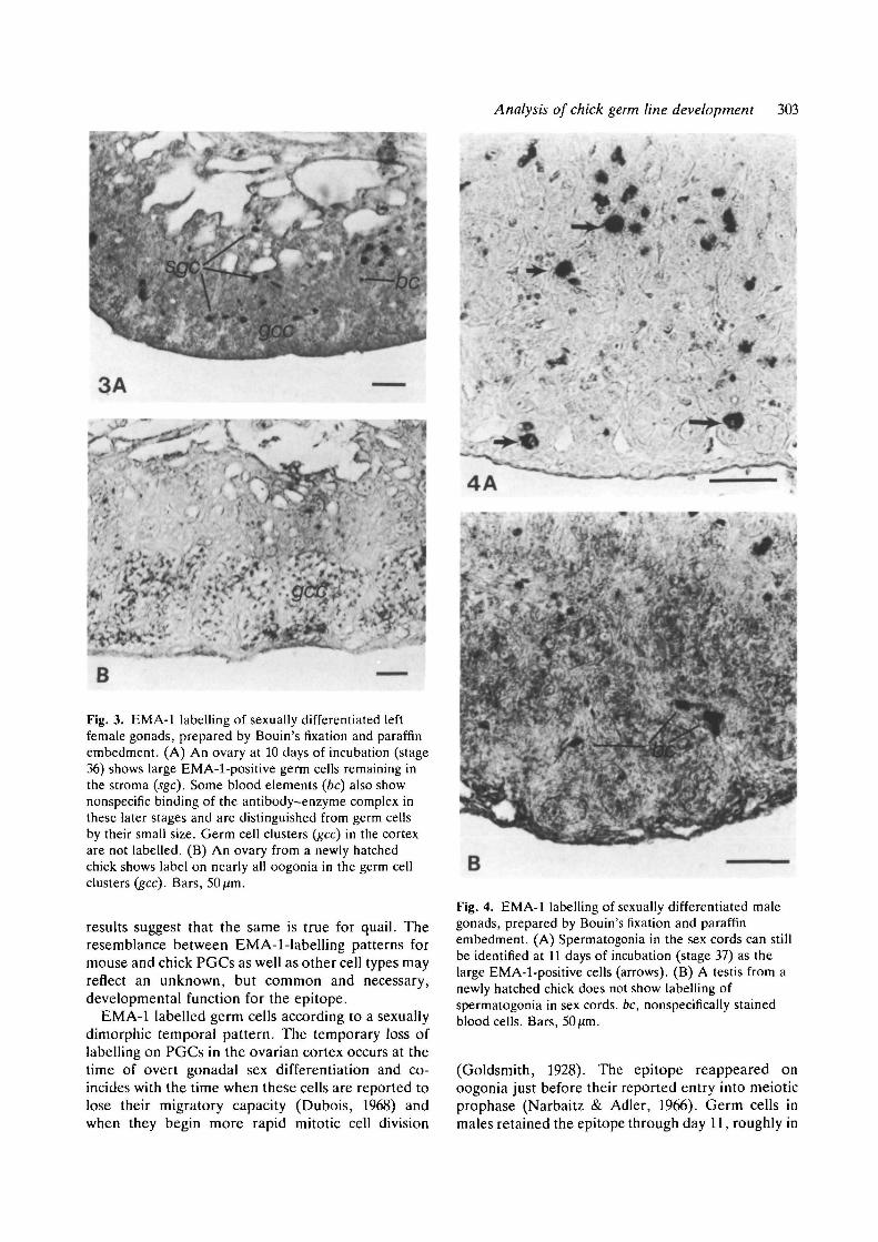
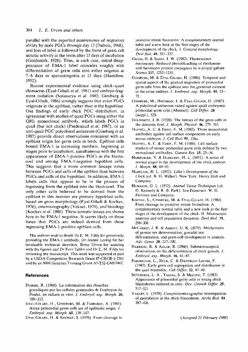
stages, EMA-1 reactivity showed a marked sexualdimorphism. In females, germ cells lost the antigen atthe time that overt gonadal sexual differentiationbegan at 7-8 days of incubation. Few labelled germcells were seen in the germ cell clusters of the ovariancortex between days 7 and 14 days of incubation(stages 32 and 40), although some germ cells in thestroma were labelled (Fig. 3A). The EMA-1 epitopereappeared on cortical oogonia by 15 days of incu-bation, with nearly 100% labelling efficiency of cellsin germ cell clusters through the time of hatching andwith few, if any, labelled germ cells still present in thestroma (Fig. 3B). Unlike germ cells in the female,germ cells in the male retained EMA-1 antigenicityuntil after gonadal sexual differentiation, with agradual decrease in numbers of labelled cells becom-ing apparent at 11 days of incubation (stage 37)(Fig. 4A) and continuing until essentially all sper-matogonia were unreactive with EMA-1 at the timeof hatching (Fig. 4B).
Discussion
EMA-1, a monoclonal antibody raised against mouseembryonal carcinoma (Nulli SCC1) cell surface anti-gens (Hahnel & Eddy, 1986), identifies germ cells inchick embryos throughout the development of thegerm line. EMA-1 is the only monoclonal antibodythus far reported in the literature that can serve toidentify chick PGCs. In addition, EMA-1 representsa direct marker that effectively extends the temporallimits of specific germ cell labelling in the chickembryo both back into the early stages of hypoblastformation, and ahead, beyond the period of gonadalcolonization. Like PAS, EMA-1 allows easy identifi-cation of PGCs from the germinal crescent stagesthrough the migratory phase. However, EMA-1 rep-resents the first chick germ cell marker reported tolabel subsequent to the beginning of primary sexdifferentiation.
The EMA-1 epitope appears in both somatic andgerm line tissues in the chick embryo in a patternstrikingly similar to that reported for the mouseembryo (Hahnel & Eddy, 1986), and preliminary
Analysis of chick germ line development 303
3A
-*rv iA . V -
• * < - ,
t'**
• - * '
B
Fig. 3. EMA-1 labelling of sexually differentiated leftfemale gonads, prepared by Bouin's fixation and paraffinembedment. (A) An ovary at 10 days of incubation (stage36) shows large EMA-1-positive germ cells remaining inthe stroma (sgc). Some blood elements (be) also shownonspecific binding of the antibody-enzyme complex inthese later stages and are distinguished from germ cellsby their small size. Germ cell clusters (gec) in the cortexare not labelled. (B) An ovary from a newly hatchedchick shows label on nearly all oogonia in the germ cellclusters (gcc). Bars, 50 jim.
results suggest that the same is true for quail. Theresemblance between EMA-1-labelling patterns formouse and chick PGCs as well as other cell types mayreflect an unknown, but common and necessary,developmental function for the epitope.
EMA-1 labelled germ cells according to a sexuallydimorphic temporal pattern. The temporary loss oflabelling on PGCs in the ovarian cortex occurs at thetime of overt gonadal sex differentiation and co-incides with the time when these cells are reported tolose their migratory capacity (Dubois, 1968) andwhen they begin more rapid mitotic cell division
B
Fig. 4. EMA-1 labelling of sexually differentiated malegonads, prepared by Bouin's fixation and paraffinembedment. (A) Spermatogonia in the sex cords can stillbe identified at 11 days of incubation (stage 37) as thelarge EMA-1-positive cells (arrows). (B) A testis from anewly hatched chick does not show labelling ofspermatogonia in sex cords, be, nonspecifically stainedblood cells. Bars, 50fim.
(Goldsmith, 1928). The epitope reappeared onoogonia just before their reported entry into meioticprophase (Narbaitz & Adler, 1966). Germ cells inmales retained the epitope through day 11, roughly in
304 L. E. Urven and others
parallel with the reported maintenance of migratoryability by male PGCs through day 12 (Dubois, 1968),and loss of label is followed by the burst of germ cellmitotic activity in the testis after 15 days of incubation(Goldsmith, 1928). Thus, in each case, initial disap-pearance of EMA-1 label coincides roughly withdifferentiation of germ cells into either oogonia at7—8 days or spermatogonia at 13 days (Hamilton,1952).
Recent experimental evidence using chick-quailchimaeras (Eyal-Giladi et al. 1981) and embryo-frag-ment isolation (Sutasurya et al. 1983; Ginsburg &Eyal-Giladi, 1986) strongly suggests that avian PGCsoriginate in the epiblast, rather than in the hypoblast.Our findings of early chick PGC distribution, inagreement with studies of quail PGCs using either theQH1 monoclonal antibody, which labels PGCs inquail (but not chick) (Pardenaud et al. 1987), or ananti-quail PGC polyclonal antiserum (Ginsburg et al.1987) provide direct observations consistent with anepiblast origin for germ cells in birds. Epiblast cellsbound EMA-1 in increasing numbers, beginning atstages prior to incubation, simultaneous with the firstappearance of EMA-1-positive PGCs in the blasto-coel and among EMA-1-negative hypoblast cells.This suggests that a more common lineage existsbetween PGCs and cells of the epiblast than betweenPGCs and cells of the hypoblast. In addition, EMA-1labels cells that appear to be in the process ofingressing from the epiblast into the blastocoel. Theonly other cells believed to be derived from theepiblast in this manner are primary hypoblast cells,based on gross morphology (Eyal-Giladi & Kochav,1976), cinemicrography (Vakaet, 1970), and histology(Kochav et al. 1980). These somatic tissues are shownhere to be EMA-1 negative. It seems likely on thesebases that PGCs are indeed derived from theseingressing EMA-1-positive epiblast cells.
The authors wish to thank Dr E. M. Eddy for generouslyproviding the EMA-1 antibody, Dr Jeanne Loring for herinvaluable technical direction, Betsy Urven for assistingwith the figures and Dr Fern Tablin and Dr E. M. Eddy forreviewing the manuscript. This work was supported in partby a USDA Competitive Research Grant 87-CRCR-1-2301and by an NTH Genetics Training Grant A5-T32-GM07467.
References
DUBOIS, R. (1968). La colonization des e'bauchesgonadiques par les cellules germinales de l'embryon dePoulet, en culture in vitro. J. Embryol. exp. Morph. 20,189-213.
EYAL-GILADI, H., GINSBURG, M. & FARBAROV, A. (1981).Avian primordial germ cells are of epiblastic origin. J.Embryol. exp. Morph. 65, 139-147.
EYAL-GILADI, H. & KOCHAV, S. (1976). From cleavage to
primitive streak formation: A complementary normaltable and a new look at the first stages of thedevelopment of the chick. I. General morphology.Devi Biol. 49, 321-337.
GILOH, H. & SEDAT, J. W. (1982). Fluorescencemicroscopy: Reduced photobleaching of rhodamineand fluorescein protein conjugates by n-propyl gallate.Science 217, 1252-1255.
GINSBURG, M. & EYAL-GILADI, H. (1986). Temporal andspatial aspects of the gradual migration of primordialgerm cells from the epiblast into the germinal crescentin the avian embryo. /. Embryol. exp. Morph. 95, 53-71.
GINSBURG, M., HOCHMAN, I. & EYAL-GILADI, H. (1987).A polyclonal antiserum raised against quail embryonicprimordial germ cells (Abstract). Cell Differ. 20(suppl.), 52S.
GOLDSMITH, J. B. (1928). The history of the germ cells inthe domestic fowl. /. Morph. Physiol. 46, 275-315.
HAHNEL, A. C. & EDDY, E. M. (1982). Three monoclonalantibodies against cell surface components on earlymouse embryos. J. Cell Biol. 95, 156a.
HAHNEL, A. C. & EDDY, E. M. (1986). Cell surfacemarkers of mouse primordial germ cells defined by twomonoclonal antibodies. Gamete Res. 15, 25-34.
HAMBURGER, V. & HAMILTON, H. L. (1951). A series ofnormal stages in the development of the chick embryo.J. Morph. 88, 49-92.
HAMILTON, H. L. (1952). Lillie's Development of theChick (ed. B. H. Willier). New York: Henry Holt andCompany.
HUMASON, G. L. (1972). Animal Tissue Techniques (ed.D. Kennedy & R. B. Park). San Francisco: W. H.Freeman and Company.
KOCHAV, S., GINSBURG, M. & EYAL-GILADI, H. (1980).From cleavage to primitive streak formation: Acomplementary normal table and a new look at the firststages of the development of the chick. II. Microscopicanatomy and cell population dynamics. Devi Biol. 79,296-308.
MCCARREY, J. R. & ABBOTT, U. K. (1979). Mechanismsof genetic sex determination, gonadal sexdifferentiation, and germ-cell development in animals.Adv. Genet. 20, 217-290.
NARBAITZ, R. & ADLER, R. (1966). Submicroscopicalobservations on the differentiation of chick gonads. J.Embryol. exp. Morph. 16, 41-47.
PARDENAUD, L., BUCK, C. & DIETERLEN-LIEVRE, F.
(1987). Early germ cell segregation and distribution inthe quail blastodisc. Cell Differ. 22, 47-60.
SUTASURYA, L. A., YASUGI, S. & MIZUNO, T. (1983).
Appearance of primordial germ cells in young chickblastoderms cultured in vitro. Dev. Growth Differ. 25,517-521.
VAKAET, L. (1970). Cinephotomicrographic investigationsof gastrulation in the chick blastoderm. Archs Biol. 81,387-426.
(Accepted 23 February 1988)