anaesthetic monitoring equipment for small...
TRANSCRIPT
In Practice ● NO V E M B E R/DE C E M B E R 2005512
THE CASE FOR ANAESTHETIC MONITORING
In 1990, Clarke and Hall carried out a survey of anaesthet-ic mortality in small animal practice. Their findings sug-gested that approximately 1 in 679 healthy dogs and catsdied as a result of anaesthesia, while the figure increasedto 1 in 31 for sick patients. More recently, the preliminaryresults of a Confidential Enquiry into Perioperative SmallAnimal Fatalities (CEPSAF) indicated that the risk ofanaesthetic death was 1 in 1849 for healthy dogs, 1 in 895for healthy cats, 1 in 75 for sick dogs and 1 in 71 for sickcats (Brodbelt and others 2005). Thus, anaesthetic-relateddeaths appear to have decreased somewhat in veterinarypractice over the past 15 years. In comparison, although itis difficult to establish exact equivalent figures, mortalityassociated with anaesthesia in humans appears to beapproximately 1 in 10,000 to 1 in 100,000.
There is little doubt that the lower mortality figures inhumans are due in part to the administration of anaesthe-sia primarily by specialist anaesthetists, but it is highlylikely that access to electronic monitoring equipmentplays a role. Indeed, minimum standards of monitoringare now well established for human anaesthesia (see boxon the right), whereas the majority of patients anaes-thetised in veterinary practice are not afforded this luxury.This is not to say that the provision of electronic monitor-ing equipment, in itself, will necessarily improve the standards of anaesthesia in veterinary patients: a thoroughunderstanding of the equipment in use, and the ability tointerpret the results produced, are absolutely fundamentalto achieve any benefit. However, such equipment certain-ly provides an ‘early warning’, and helps alert the anaes-thetist to the development of potentially critical situations,
CO
MP
AN
ION
A
NIM
AL
P
RA
CT
ICE
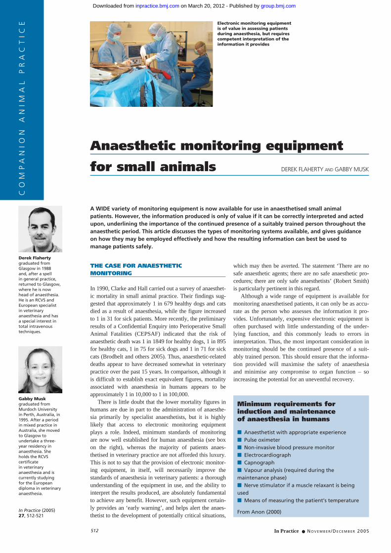
A WIDE variety of monitoring equipment is now available for use in anaesthetised small animal patients. However, the information produced is only of value if it can be correctly interpreted and acted upon, underlining the importance of the continued presence of a suitably trained person throughout theanaesthetic period. This article discusses the types of monitoring systems available, and gives guidanceon how they may be employed effectively and how the resulting information can best be used tomanage patients safely.
Anaesthetic monitoring equipment
for small animals DEREK FLAHERTY AND GABBY MUSK
Electronic monitoring equipment is of value in assessing patientsduring anaesthesia, but requirescompetent interpretation of theinformation it provides
In Practice (2005)27, 512-521
which may then be averted. The statement ‘There are nosafe anaesthetic agents; there are no safe anaesthetic pro-cedures; there are only safe anaesthetists’ (Robert Smith)is particularly pertinent in this regard.
Although a wide range of equipment is available formonitoring anaesthetised patients, it can only be as accu-rate as the person who assesses the information it pro-vides. Unfortunately, expensive electronic equipment isoften purchased with little understanding of the under-lying function, and this commonly leads to errors ininterpretation. Thus, the most important consideration inmonitoring should be the continued presence of a suit-ably trained person. This should ensure that the informa-tion provided will maximise the safety of anaesthesiaand minimise any compromise to organ function – soincreasing the potential for an uneventful recovery.
Derek Flahertygraduated fromGlasgow in 1988 and, after a spell in general practice,returned to Glasgow,where he is now head of anaesthesia.He is an RCVS andEuropean specialist in veterinaryanaesthesia and has a special interest intotal intravenoustechniques.
Gabby Muskgraduated fromMurdoch University in Perth, Australia, in1995. After a periodin mixed practice inAustralia, she movedto Glasgow toundertake a three-year residency inanaesthesia. Sheholds the RCVScertificate in veterinaryanaesthesia and iscurrently studying for the Europeandiploma in veterinaryanaesthesia.
Minimum requirements for induction and maintenance of anaesthesia in humans
■ Anaesthetist with appropriate experience■ Pulse oximeter■ Non-invasive blood pressure monitor■ Electrocardiograph■ Capnograph■ Vapour analysis (required during the maintenance phase)■ Nerve stimulator if a muscle relaxant is beingused■ Means of measuring the patient’s temperature
From Anon (2000)
group.bmj.com on March 20, 2012 - Published by inpractice.bmj.comDownloaded from

In Practice ● NO V E M B E R/DE C E M B E R 2005 513
The target site of action for all anaesthetic agents is the central nervous system (CNS), which becomesincreasingly depressed with progressive ‘deepening’ ofanaesthesia. While monitoring systems are available fordirectly assessing the degree of CNS depression, theseare relatively complex and expensive, and are unlikely to be used in veterinary practice for many years. Instead,during anaesthesia, emphasis is placed mainly on elec-tronic monitoring of the ‘indirect’ effects of the CNSdepression on cardiopulmonary function. That is to say,with progressive deepening of anaesthesia, as the CNSbecomes more depressed, increasing cardiopulmonarydepression will also be evident. Monitoring the cardio-vascular and respiratory systems therefore provides ‘ahandle’ on the depth of anaesthesia, and gives someinformation on tissue perfusion and oxygenation.
MONITORING CARDIOVASCULAR FUNCTION
OESOPHAGEAL STETHOSCOPEAn oesophageal stethoscope consists of a blind-endingplastic tube with a number of side openings at the distalend, which are covered with a thin plastic membrane.The device is placed into the oesophagus until it overliesthe heart, and the cardiac and respiratory sounds canthen be detected by attaching the proximal end to anordinary stethoscope from which the bell part has beenremoved. Unfortunately, unless the anaesthetist is will-ing to wear stethoscope earpieces throughout the periodof anaesthesia, this device provides non-continuousinformation. However, oesophageal stethoscopes canalso be attached to electronic monitors that amplify thecardiac sounds, thus permitting a continuous audible signal and freeing the anaesthetist to move around thetheatre.
There is evidence from human studies that the inten-sity of the heart sounds shows some correlation to systemic blood pressure (Sakamoto and others 1965),although this has been more difficult to prove in veteri-nary patients.
Oesophageal stethoscopes are available in small,medium and large sizes, and are cheap and reliable monitors.
ELECTROCARDIOGRAPHYElectrocardiography is relatively standard for monitoringanaesthetised patients, but actually gives limited infor-mation. The machine displays the electrical activity of
the heart, but there is no correlation between this and the cardiac output. For example, the condition termedelectromechanical dissociation (EMD) produces near-normal electrical activity of the heart but no contraction;under these circumstances, electrocardiography providesa false sense of security, as it implies that cardiac activi-ty is normal. If an oesophageal stethoscope is in place,however, or if a pulse is being palpated, it should beobvious that there is no output from the heart (heartsounds are absent during EMD).
Alterations in cardiac rate and rhythm are commonduring anaesthesia, with an incidence of 50 to 80 percent in humans undergoing surgery (Bertrand and others1971). Bradycardia, tachycardia and ventricular prema-ture complexes (VPCs) are most frequently encountered.While abnormalities of heart rate are readily detectablein the absence of monitoring equipment, VPCs and otherarrhythmias are difficult to assess without electrocardio-graphy. For the purposes of anaesthetic monitoring, precise placement of the electrocardiographic leads isnot necessary, and a basic three-lead system (leads I, IIand III) is sufficient.
Although abnormalities of heart rate or rhythm maybe caused by a number of factors (see box below), theirdevelopment during anaesthesia is usually due to an inadequate depth of anaesthesia or quality of analgesia, or the development of hypercapnia or hypoxaemia. Thesefactors should therefore be ruled out before more directpharmacological intervention is undertaken. Agents suchas halothane sensitise the myocardium to catecholamines,and it is relatively common to observe VPCs during themaintenance of anaesthesia. When this occurs, the temp-tation is to reduce the quantity of vapour being inspiredby the patient but, in many circumstances, it may be moreappropriate to increase the vaporiser dial setting, as thearrhythmia is often due to inadequate anaesthesia.
Small, medium and large oesophageal stethoscopes
ECG showing ventricularpremature complexes. Picture, Dr J. Dukes McEwan
Common causes of anaesthetic-related arrhythmias
■ Inadequate anaesthesia/analgesia■ Hypoxaemia■ Hypercapnia■ Hypotension■ Hypo/hyperthermia■ Electrolyte abnormalities
group.bmj.com on March 20, 2012 - Published by inpractice.bmj.comDownloaded from

In Practice ● NO V E M B E R/DE C E M B E R 2005514
Electrocardiography can also be a useful guide to certain electrolyte abnormalities, particularly alterationsin extracellular potassium concentration; reasonablycharacteristic changes are seen, especially in the case ofhyperkalaemia (as illustrated above). In humans, electro-cardiography is also widely used to detect myocardialhypoxia during anaesthesia (which is common due to thehigh incidence of coronary arterial disease); elevation ordepression of the S-T segment of the electrocardiogram(ECG) is suggestive of inadequate myocardial perfusionor oxygenation. However, S-T segment changes appearto occur commonly in anaesthetised animals, and do notseem to reliably indicate myocardial hypoxia.
R WAVE MONITORSThese electronic monitors detect (but do not display)electrical activity of the heart, and ‘bleep’ in response tothe presence of the R wave on the ECG. These devicessuffer from the same disadvantage as electrocardio-graphs in that they fail to detect EMD. In addition, theymay give a signal during periods of electrical interfer-ence (eg, during the use of diathermy), which may bemisinterpreted as indicating an arrhythmia.
MONITORS OF ARTERIAL BLOOD PRESSUREArterial blood pressure is the product of cardiac output andtotal peripheral resistance (systemic vascular resistance):
Although monitoring systems for direct measurementof cardiac output are now more widely available, theseare both expensive and invasive, and it is likely that arte-rial blood pressure monitoring – despite the limitationsoutlined above – will continue to be the standard methodof assessing the adequacy of cardiac output and bloodflow for many years to come.
General opinion suggests that, during general anaes-thesia, systolic arterial pressure should be maintainedabove 80 to 90 mmHg, and mean pressure above 60 to70 mmHg, in order to ensure sufficient perfusion pres-sure for the brain and heart. Diastolic pressures of lessthan 40 mmHg are associated with impaired coronaryartery perfusion in humans (most myocardial perfusionoccurs during diastole), but no comparative studies havebeen performed in animals; as there is a lower incidenceof coronary artery disease in veterinary patients, lowerdiastolic pressures may be acceptable.
Intraoperative hypotension may impair organ perfu-sion and increase perioperative morbidity, thus measure-ment and support of arterial blood pressure is important.Arterial blood pressure can be measured by: ■ Direct (invasive) monitoring;■ Indirect (non-invasive) monitoring.
Direct monitoringDirect arterial blood pressure monitoring gives moreaccurate and continuous information compared withindirect methods. It is performed by cannulation of anartery and connection of the cannula to a device thatgives a reading of arterial blood pressure. The cannula iscommonly connected to a transducer, which converts thepressure signal from the artery into an electrical signal,and then to an electronic monitor, which provides a dis-play of the arterial blood pressure trace, as well as valuesfor systolic, mean and diastolic pressure. Alternatively,the cannula can be connected to an aneroid manometerto give mean arterial blood pressure values.
Technical skill is required for successful cannulationof a peripheral artery, especially in smaller patients.However, because this method of blood pressure mea-surement gives beat-to-beat information, it is preferableto non-invasive techniques if large swings in blood pressure are anticipated during surgery, or if the opera-tion has the potential to induce excessive haemorrhage.Direct blood pressure measurement carries a small risk ofischaemic damage to the area supplied by the cannulatedartery, but this is less common in veterinary patients thanin humans due to the lower incidence of peripheral vascu-lar disease in animals. Haematoma formation must beprevented when the arterial cannula is removed by apply-ing firm pressure to the area for five minutes.
Arterial blood pressure = Cardiac output x Total peripheral resistance(ABP) (CO) (TPR)
ECG trace from a dog withhyperkalaemia. Note thespiked T waves and theabsence of P waves. Picture, Dr I. Ramsey
Consequently, arterial pressure is often monitored dur-ing general anaesthesia to provide information on cardiacoutput, which is a major determinant of tissue perfusion.However, arterial pressure is also dependent on thedegree of vascular tone. Therefore, it is possible for apatient with normal or high arterial blood pressure tohave low cardiac output but high peripheral resistance.Under these circumstances, tissue blood flow may wellbe impaired despite reasonable blood pressure. Thus, theresults of arterial blood pressure monitoring, while poten-tially providing information on cardiac output and organperfusion, cannot be viewed in isolation. A rough evalua-tion of vascular tone may be made by assessing mucousmembrane colour and capillary refill time. In the absenceof anaemia, pale mucous membranes generally suggestperipheral vasoconstriction. Cannula placed in the dorsal pedal artery
group.bmj.com on March 20, 2012 - Published by inpractice.bmj.comDownloaded from

In Practice ● NO V E M B E R/DE C E M B E R 2005 515
Indirect monitoringIndirect arterial blood pressure monitoring is less techni-cally demanding than direct methods and is associatedwith lower morbidity because arterial cannulation is notrequired. However, the technique is also less accurateand does not give continuous readings. In addition, thepresence of cardiac arrhythmias may provoke dubiousresults from certain indirect blood pressure monitors,and this can be problematic even if the patient is exhibit-ing a normal rhythm variation (eg, sinus arrhythmia).
There are two indirect methods for arterial bloodpressure monitoring:■ Doppler ultrasonic flow;■ Oscillometry;
DOPPLER ULTRASONIC FLOW METHOD
The Doppler ultrasonic flow method involves positioninga small probe, which emits an ultrasonic beam, over aperipheral artery (usually on the tail or paw) and applyingultrasound coupling gel between the probe and the skin.As blood flows along the vessel under the probe, a‘whooshing’ noise is emitted by the monitor. If an inflat-able cuff, connected to an aneroid manometer, is placedfurther up the appendage and sufficiently inflated, it willocclude the artery and the noise will disappear. If the cuffis then slowly deflated, the sound will recur at systolicarterial pressure, which can be read off the manometer.
Arterial cannula connectedto a transducer (circled).Note that the transducer is positioned level with the heart (point of theshoulder in dorsalrecumbency)
Arterial blood pressure trace displayed on a monitor (blue trace, above), giving values of 100 mmHg, 49 mmHgand 64 mmHg for systolic, diastolic and mean arterialpressures, respectively
Doppler ultrasonic flow probe
Doppler ultrasonic flow monitor. The probe is connected at A, and the noise is amplified through B
A
B
Inflatable cuff connected to an aneroid manometer
In dogs, good correlation has been observed betweenmeasured Doppler systolic arterial pressure and that pro-vided by direct femoral arterial cannulation (Weiser andothers 1977) but, in cats, the system tends to under-readthe true systolic pressure and it has been suggested that a
group.bmj.com on March 20, 2012 - Published by inpractice.bmj.comDownloaded from

In Practice ● NO V E M B E R/DE C E M B E R 2005516
correction factor of approximately 14 mmHg has to beadded to the observed reading (Grandy and others 1992).However, Caulkett and others (1998) demonstratedgreater correlation in cats between directly measuredmean arterial pressure and that measured by the Dopplersystem. Thus, although this technique is used to assesssystolic arterial pressure, due to inherent inaccuracies inthe system the value obtained in cats probably correlatesmore closely to mean arterial pressure. Doppler monitor-ing provides only a vague (and inconsistent) indicationof diastolic pressure in all species.
The Doppler flow probe can also be used as a pulsemonitor to provide an audible beat-to-beat signal.
OSCILLOMETRIC METHOD
The oscillometric method involves connecting a cuffsystem to an electronic monitor. The cuff is placed over a peripheral artery and the machine automaticallyinflates the cuff to occlude the artery before slowlyreleasing the pressure. As the cuff deflates, the machinedetects oscillations in the artery as the blood begins toflow back through; these oscillations begin at systolicpressure, reach a maximum at mean arterial pressure,and gradually disappear at diastolic pressure. Thus,unlike the Doppler system, the oscillometric methodgives readings at all three blood pressure points. It canalso be set to cycle automatically, thereby giving regularreadings, whereas the Doppler technique has to be performed manually each time. With the oscillometricmethod, the mean reading is the most reliable, followedby the systolic measurement; the diastolic reading shouldonly be considered moderately accurate. Cuffs are com-monly placed over the dorsal pedal artery at the metatar-sus, over the radial artery just above the carpus, or overthe coccygeal artery in the ventral tail.
URINE OUTPUTUrine production depends on adequate renal perfusion.As blood flow to the kidneys declines and subsequentlyceases at a mean arterial blood pressure of less than 60mmHg approximately, urine output can give an indirectindication of the adequacy of arterial blood pressure.
Normal urine output is 1 to 2 ml/kg/hour, with values ofless than 0·5 ml/kg/hour representing oliguria. Therefore,if urine output is measured and proves to be greater than 1 ml/kg/hour, this suggests that mean arterial bloodpressure is greater than 60 mmHg. Unfortunately, manyanaesthetic drugs influence urine production, and the
An oscillometric blood pressure monitor
Cuff placed around themetatarsal area. Note that, unlike the Dopplertechnique, the oscillometricsystem will generallyprovide readings if the cuff is in the vicinity of theunderlying artery, even if itis not optimally positioned.With Doppler, locating theartery with the ultrasonicprobe can occasionally beproblematic
Doppler and oscillometric monitoring compared
Older oscillometric machines were extremelyunreliable in small dogs and cats, often fail-ing to display any blood pressure readingwhatsoever. However, newer machines have,to some extent, overcome this problem(Pedersen and others 2002, Sawyer and oth-ers 2004). Since oscillometric devices general-ly record the pulse rate as well as the arterialblood pressure, it is always worth checkingthat the displayed value is equivalent to amanually recorded pulse rate before placingany reliance on the blood pressure reading.Similarly, inaccurate oscillometric arterialblood pressure results commonly occur if cardiac arrhythmias are present – even in the case of normal variants such as sinusarrhythmia. In these cases, Doppler or directarterial blood pressure monitoring will pro-vide more accurate results.
With both Doppler and oscillometricmethods of blood pressure measurement,the size of the occluding cuff, and its positionrelative to the heart, are critical in obtainingaccurate results. The width of the cuff shouldbe approximately 40 to 60 per cent of the cir-cumference of the area it is placed around;cuffs that are too small will result in an over-reading of the blood pressure, and vice versa.In addition, the occluding cuff should bepositioned level with the heart; arterial pressure will be erroneously high if the cuff is below the heart, and erroneously low ifabove it. Applying cuffs too tightly aroundthe appendage will result in an under-read-ing of the blood pressure, while applyingthem too loosely will cause an over-reading.
While direct arterial blood pressure monitoring gives reliable results on a beat-
to-beat basis, indirect techniques are con-sidered more useful for following trends inpressure rather than for providing absolutevalues. Despite being less accurate, indirecttechniques are particularly convenientbecause of their ease of use and limiteddependence on the technical skills of theanaesthetist. Of the two types available, theDoppler system is the more reliable in thepresence of cardiac arrhythmias or low arterial pressure.
As well as providing some informationon tissue perfusion, monitoring of trends inarterial blood pressure can give a useful indication of altering anaesthetic depth. Inthe absence of marked surgical blood loss, agradual decline in arterial blood pressuresuggests deepening of anaesthesia, and viceversa.
group.bmj.com on March 20, 2012 - Published by inpractice.bmj.comDownloaded from
In Practice ● NO V E M B E R/DE C E M B E R 2005 517
stress response associated with both anaesthesia andsurgery increases the release of antidiuretic hormone,which may acutely decrease urine output. Thus, manynormal patients may exhibit reduced urine flow duringanaesthesia, even in the face of adequate blood pressureand renal perfusion.
Monitoring urine production is recommended inpatients with renal disease or conditions predisposing torenal shutdown (eg, sepsis, major trauma), to ensureappropriate treatment can be undertaken rapidly if urineoutput falls. It is important that the bladder is drained ofurine at the start of the procedure before an assessmentof urine production is made. As monitoring of urine out-put relies on catheterisation of the bladder, an aseptictechnique is essential.
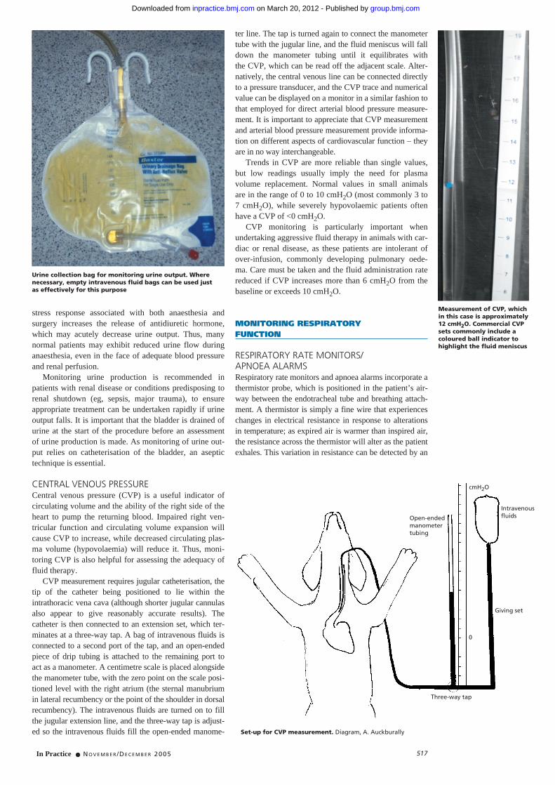
CENTRAL VENOUS PRESSURECentral venous pressure (CVP) is a useful indicator ofcirculating volume and the ability of the right side of theheart to pump the returning blood. Impaired right ven-tricular function and circulating volume expansion willcause CVP to increase, while decreased circulating plas-ma volume (hypovolaemia) will reduce it. Thus, moni-toring CVP is also helpful for assessing the adequacy offluid therapy.
CVP measurement requires jugular catheterisation, thetip of the catheter being positioned to lie within theintrathoracic vena cava (although shorter jugular cannulasalso appear to give reasonably accurate results). Thecatheter is then connected to an extension set, which ter-minates at a three-way tap. A bag of intravenous fluids isconnected to a second port of the tap, and an open-endedpiece of drip tubing is attached to the remaining port to act as a manometer. A centimetre scale is placed alongsidethe manometer tube, with the zero point on the scale posi-tioned level with the right atrium (the sternal manubriumin lateral recumbency or the point of the shoulder in dorsalrecumbency). The intravenous fluids are turned on to fillthe jugular extension line, and the three-way tap is adjust-ed so the intravenous fluids fill the open-ended manome-
ter line. The tap is turned again to connect the manometertube with the jugular line, and the fluid meniscus will falldown the manometer tubing until it equilibrates with the CVP, which can be read off the adjacent scale. Alter-natively, the central venous line can be connected directlyto a pressure transducer, and the CVP trace and numericalvalue can be displayed on a monitor in a similar fashion tothat employed for direct arterial blood pressure measure-ment. It is important to appreciate that CVP measurementand arterial blood pressure measurement provide informa-tion on different aspects of cardiovascular function – theyare in no way interchangeable.
Trends in CVP are more reliable than single values,but low readings usually imply the need for plasma volume replacement. Normal values in small animals are in the range of 0 to 10 cmH2O (most commonly 3 to7 cmH2O), while severely hypovolaemic patients oftenhave a CVP of <0 cmH2O.
CVP monitoring is particularly important whenundertaking aggressive fluid therapy in animals with car-diac or renal disease, as these patients are intolerant ofover-infusion, commonly developing pulmonary oede-ma. Care must be taken and the fluid administration ratereduced if CVP increases more than 6 cmH2O from thebaseline or exceeds 10 cmH2O.
MONITORING RESPIRATORY FUNCTION
RESPIRATORY RATE MONITORS/APNOEA ALARMSRespiratory rate monitors and apnoea alarms incorporate athermistor probe, which is positioned in the patient’s air-way between the endotracheal tube and breathing attach-ment. A thermistor is simply a fine wire that experienceschanges in electrical resistance in response to alterationsin temperature; as expired air is warmer than inspired air,the resistance across the thermistor will alter as the patientexhales. This variation in resistance can be detected by an
Urine collection bag for monitoring urine output. Wherenecessary, empty intravenous fluid bags can be used just as effectively for this purpose
Measurement of CVP, which in this case is approximately12 cmH2O. Commercial CVPsets commonly include acoloured ball indicator tohighlight the fluid meniscus
Set-up for CVP measurement. Diagram, A. Auckburally
Three-way tap
Giving set
0
IntravenousfluidsOpen-ended
manometertubing
cmH2O
group.bmj.com on March 20, 2012 - Published by inpractice.bmj.comDownloaded from

In Practice ● NO V E M B E R/DE C E M B E R 2005518
attached monitor, which can be programmed to bleep eachtime the patient takes a breath. In addition to providing a numerical display of the respiratory rate, most of thesemonitors can also be set to emit an alarm if there is no airmovement for a pre-assigned period.
Although fairly widely used in veterinary practice,respiratory rate monitors/apnoea alarms are relativelyexpensive for the amount of information they provide, asexactly the same endpoint can be achieved by observingthe movement of the reservoir bag on the anaestheticbreathing system.
PULSE OXIMETRYNinety-eight per cent of oxygen transported by the bloodis bound to haemoglobin, and only a small proportion is carried dissolved in the plasma. Thus, measuring theamount of haemoglobin that is saturated with oxygen
will give an idea of how well oxygenated the patient is.Deoxygenated and oxygenated haemoglobin absorb dif-ferent wavelengths of light and, very simplistically, thisis the basis of pulse oximetry.
There are two types of pulse oximeter probe – trans-mittance probes and reflectance probes. Transmittanceprobes consist of, on one side, red and infrared (660 nmand 940 nm, respectively) light-emitting diodes, whichflash on and off several hundred times a second and, onthe other side, a photodetector to determine the amount ofeach wavelength of light that passes through the tissuebed (usually the tongue, ear, toe or skin flap). Reflectanceprobes have the light-emitting diodes and photodetectorplaced adjacent to each other, and the light is reflected offa tissue rather than transmitted through it; these probesare commonly placed in the rectum in very smallpatients, but may also be wrapped around digits.
With both types of probe, the light is absorbed notonly by the haemoglobin in the arterial blood, but alsoby haemoglobin in capillary and venous blood, and by the tissue itself. The pulse oximeter is, however, able to ignore absorption from these other sources, and concentrate only on the arterial haemoglobin. It does thisby assessing only pulsatile absorption (arterial blood),and ignoring static, non-pulsatile absorption (tissue,venous blood, and so on). By comparing the amount ofthe two wavelengths of light that has been absorbedacross the tissue, the pulse oximeter can indicate the percentage of haemoglobin saturation. As pulse oximetrydepends on pulsatile blood flow, it not only gives anindication of oxygenation but also that cardiac output isreaching the peripheral tissues. Thus, it can be consid-ered as a monitor of both respiratory and cardiovascularsystems.
In general, arterial haemoglobin saturation should be >95 per cent in anaesthetised patients breathing an oxygen-enriched gas although, provided haemoglobinsaturation is >90 per cent, there is no immediate cause for concern. Values of between 90 and 95 per cent in anaesthetised patients may indicate an underlying prob-lem that should be identified but, most commonly, thereis a fault with the probe, and adjusting its position will usually result in a return to more normal values.Saturation of <90 per cent should be investigated immediately. Animals premedicated with alpha-2 adreno-ceptor agonists (eg, xylazine, medetomidine, romifidine)develop intense peripheral vasoconstriction and, in these cases, pulse oximeters may be unable to detect asignal.
A thermistor probe (circled) positioned between the endotracheal tube and breathing system
Respiratory rate monitor and apnoea alarm. The thermistorprobe connects to the monitor, which can be adjusted toallow a specified period of apnoea before emitting an alarm
Ear lobe pulse oximeter probe, showing the light-emittingdiodes (A) and photodetector (B)
Pulse oximeter monitor displaying a plethysmographic trace(white pulsatile trace), haemoglobin saturation value (SaO2100 per cent) and pulse rate (143 beats per minute)
A
B
group.bmj.com on March 20, 2012 - Published by inpractice.bmj.comDownloaded from
In Practice ● NO V E M B E R/DE C E M B E R 2005 519
The pulse oximeter probe must be placed at a suitablesite to provide a reliable reading. The best site is a non-pigmented, relatively hairless part of the body. Thetongue is usually the most appropriate, but the pinna,webbing between the digits, vulva or scrotum are oftensuitable alternatives if they are not heavily pigmented.Failure to obtain an accurate reading may be due to: ■ Pigment or fur at the site;■ Lack of perfusion (eg, vasoconstriction associatedwith alpha-2 adrenoceptor agonist administration orhypothermia);■ Ambient lighting overwhelming the light from thelight-emitting diodes;■ Movement obscuring the signal;■ Haemoglobin abnormalities (eg, methaemoglobinaemiaand carboxyhaemoglobinaemia);■ Inappropriate size or shape of probes.
The pulse oximeter provides a measure of haemo-globin saturation and pulse rate and, in some cases, willgenerate a plethysmographic trace (a graph of pulse volume against time), which displays consecutive pulsewaveforms.
CAPNOGRAPHYThe ‘gold standard’ method for assessing the adequacyof ventilation is measuring the partial pressure of carbondioxide (PaCO2) in the arterial blood by blood gas analysis. However, this technique is invasive, technicallydemanding and the equipment required is expensive. Analternative, non-invasive technique is capnography. Inpatients with normal lungs, the carbon dioxide concen-tration in the pulmonary capillaries will have equilibrat-ed with that in the alveoli by the end of expiration.Therefore, if a sample of the end-tidal expired gas can beobtained and its carbon dioxide concentration measured,it will allow the carbon dioxide concentration of the arterial blood to be evaluated with a reasonable degree of accuracy, and so provide a non-invasive tool forassessing ventilatory adequacy.
Capnography relies on the absorption of infrared lightby carbon dioxide molecules in the patient’s respiredgases. A capnograph incorporates a compact probe,which sits between the endotracheal tube and patientbreathing system, and an analyser. The presence of the probe increases the dead space in the breathing system, which may be significant in very small patients,although low dead space adaptors have recently becomeavailable. There are two types of analysers: mainstreamanalysers, which measure carbon dioxide directly at thesite of the probe; and sidestream analysers, which con-tinually divert a small sample of gas from the airway tothe main body of the machine. While mainstream analy-sers have a more rapid response rate, they tend to bebulkier than sidestream versions.
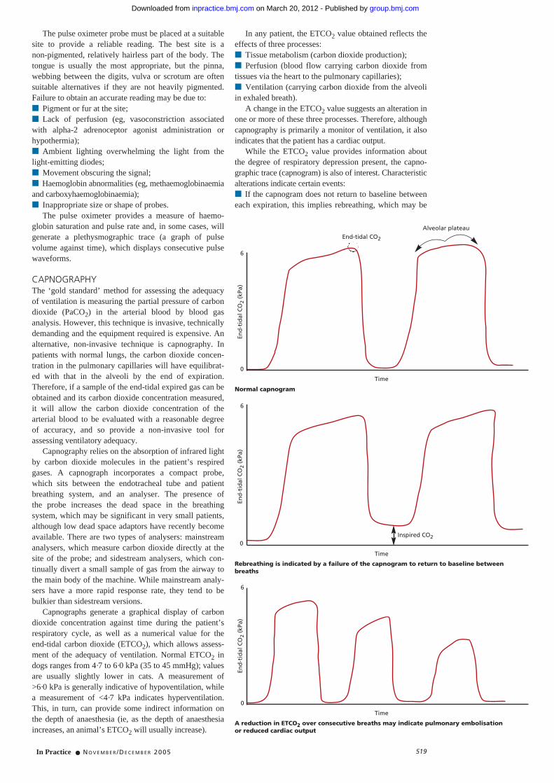
Capnographs generate a graphical display of carbondioxide concentration against time during the patient’srespiratory cycle, as well as a numerical value for theend-tidal carbon dioxide (ETCO2), which allows assess-ment of the adequacy of ventilation. Normal ETCO2 indogs ranges from 4·7 to 6·0 kPa (35 to 45 mmHg); valuesare usually slightly lower in cats. A measurement of >6·0 kPa is generally indicative of hypoventilation, whilea measurement of <4·7 kPa indicates hyperventilation.This, in turn, can provide some indirect information onthe depth of anaesthesia (ie, as the depth of anaesthesiaincreases, an animal’s ETCO2 will usually increase).
In any patient, the ETCO2 value obtained reflects theeffects of three processes: ■ Tissue metabolism (carbon dioxide production);■ Perfusion (blood flow carrying carbon dioxide fromtissues via the heart to the pulmonary capillaries);■ Ventilation (carrying carbon dioxide from the alveoliin exhaled breath).
A change in the ETCO2 value suggests an alteration inone or more of these three processes. Therefore, althoughcapnography is primarily a monitor of ventilation, it alsoindicates that the patient has a cardiac output.
While the ETCO2 value provides information aboutthe degree of respiratory depression present, the capno-graphic trace (capnogram) is also of interest. Characteristicalterations indicate certain events: ■ If the capnogram does not return to baseline betweeneach expiration, this implies rebreathing, which may be
Normal capnogram
Rebreathing is indicated by a failure of the capnogram to return to baseline betweenbreaths
A reduction in ETCO2 over consecutive breaths may indicate pulmonary embolisation or reduced cardiac output
End-tidal CO2
Alveolar plateau
Time
Inspired CO2
End
-tid
al C
O2
(kPa
)
6
0
End
-tid
al C
O2
(kPa
)
6
0
End
-tid
al C
O2
(kPa
)
6
0
Time
Time
group.bmj.com on March 20, 2012 - Published by inpractice.bmj.comDownloaded from

End
-tid
al C
O2
(kPa
)
In Practice ● NO V E M B E R/DE C E M B E R 2005520
due to a failure of the unidirectional flow valves in a circular breathing system, exhaustion of soda lime, inadequate fresh gas flow in a non-rebreathing system orexcessive apparatus dead space;■ Pulmonary embolisation may be suspected if theETCO2 falls over consecutive expirations due to a reduc-tion in pulmonary artery perfusion;■ In cases of chronic obstructive airway disease oracute-onset bronchospasm, a characteristic waveformwith a slow upstroke is produced;■ Patients undergoing intermittent positive pressureventilation may ‘buck’ the ventilator and this will be evi-dent as dips or clefts along the alveolar plateau;■ A normal variation on the usual capnogram is that of cardiogenic oscillations, which appear as a number of‘bumps’ on the downward slope of the trace. Theseoscillations are usually observed with slow respiratoryrates, and are thought to be due to the heart beatingagainst the lungs during the prolonged expiratory phase,which causes small amounts of carbon dioxide to beexpelled.
Capnometers are similar to capnographs, but only display a numerical value for ETCO2. Although thesedevices can be useful, the absence of a graphical displayof carbon dioxide concentration limits the informationthat can be interpreted by the anaesthetist.
OTHER FORMS OF MONITORING
TEMPERATURE MONITORINGHypothermia is perhaps the most ubiquitous complica-tion of general anaesthesia, and has numerous detrimen-tal effects on the patient. It is the most common cause of delayed recovery of consciousness; therefore, temper-ature monitoring is usually performed in the perianaes-thetic period. Although rectal thermometers (eg, digitalor mercury) are accurate in the awake patient, underanaesthesia the rectum tends to balloon, reducing thevalidity of results obtained from this site. Small thermis-tor probes that can be placed into the oesophagus are more useful, as the temperature at this site closelyapproximates to the core temperature.
MULTI-GAS ANALYSISMachines that measure and display inspired and expiredanaesthetic vapour concentrations, as well as oxygen andnitrous oxide, are available. These are particularly usefulwhen using rebreathing anaesthetic systems at low gasflows where there is usually a marked discrepancy between the vaporiser setting and the concentration ofagent inhaled by the patient. Due to their cost, thesemachines are not commonly available outside large vet-erinary referral institutions.
PERIPHERAL NERVE STIMULATIONIn patients that receive neuromuscular blocking agents(muscle relaxants) as part of their anaesthetic regimen,one of the problems encountered is the assessment of thedegree of neuromuscular blockade produced, both intra-operatively and, perhaps more importantly, at the end of anaesthesia prior to tracheal extubation. It is commonpractice, therefore, to use a nerve stimulator to help quan-tify the intensity of relaxation. This is a hand-held devicethat delivers a small electrical current through a pair ofelectrodes that are attached to the skin over a peripheralnerve (commonly the ulnar nerve on the medial aspect ofthe elbow, the peroneal nerve at the lateral cranial tibia,or the facial nerve on the lateral aspect of the head). Theresponse of the muscle groups innervated by these nervesis observed when the nerve stimulator is activated.
The most common form of stimulation in use is train-of-four (TOF), where four electrical pulses are applied to the nerve over a two-second period (ie, 0·5 secondbetween twitches). In the absence of neuromuscularblocking agents, four distinct muscle twitches – each ofidentical strength – will occur. If a non-depolarisingrelaxant is subsequently administered, the fourth twitch inthe TOF will become weaker and eventually disappear,followed by the third twitch, then the second, and eventu-ally the first, if sufficient relaxant is given. This phenom-enon of a gradually decreasing muscle response to nervestimulation, with the onset of non-depolarising inducedrelaxation, is known as fade.
Peripheral nerve stimulation serves two useful purpos-es. First, it has been shown that ideal muscle relaxationfor abdominal surgery is achieved when only one or two
A slow upstroke phase can be due to any form of respiratory obstruction, such as a kinkedendotracheal tube or bronchospasm
A ‘camel hump’ is due to a patient fighting (‘bucking’) imposed mechanicalventilation. The dips in the alveolar plateau are due to the animal attemptingto take spontaneous breaths
Capnogram showing cardiogenic oscillations
Time
0
Time
Time
6En
d-t
idal
CO
2(k
Pa)
0
6
End
-tid
al C
O2
(kPa
)
0
6
group.bmj.com on March 20, 2012 - Published by inpractice.bmj.comDownloaded from

In Practice ● NO V E M B E R/DE C E M B E R 2005 521
Peripheral nerve stimulator with various stimulation pattern options. Hypodermic needles are commonly placed subcutaneously over the peripheral nerve, and the stimulating leads applied to these
Response of a normalanimal (no relaxant) to TOF peripheral nervestimulation. All fourtwitches in the TOF arepresent and of equalstrength
Onset of non-depolarisingblockade. Fade is present inthe TOF, but all the twitchesare still visible
More profound neuromuscular blockade; the fourth twitchin the TOF has now disappeared and the remaining threetwitches are weaker
Twit
ch s
tren
gth
Twit
ch s
tren
gth
Twit
ch s
tren
gth
Time
Time
Time
twitches remain in the TOF. This allows the anaesthetistto titrate the dose of a neuromuscular-blocking drug toachieve suitable surgical conditions. Secondly, at the endof surgery, it permits an assessment of residual neuro-muscular blockade. The TOF should have recovered tofour equal-strength twitches before the animal is allowedto awaken and the trachea extubated.
Suxamethonium-induced relaxation exhibits a slightlydifferent TOF pattern to the non-depolarising typedescribed above, in that twitch strength decreases butfade is not observed (ie, all four twitches become weak-er, but remain equal to each other). Peripheral nervestimulation is less commonly employed in the monitor-ing of suxamethonium neuromuscular blockade due toits short duration and the drug is now seldom used inveterinary anaesthesia.
SUMMARY
The use of electronic monitoring equipment duringanaesthesia supplies some information that would nototherwise be available. It assists the anaesthetist by providing an ‘early warning’ of impending complica-tions and may help to avoid these. However, a thoroughunderstanding of the data presented is essential to gainmaximum benefit from such equipment.
ReferencesANON (2000) Recommendations for Standards of MonitoringDuring Anaesthesia and Recovery, 3rd edn. Association ofAnaesthetists of Great Britain and Ireland. www.aagbi.org/pdf/Absolute.pdfBERTRAND, C. A., STEINER, N. V., JAMESON, A. G. & LOPEZ, M. (1971) Disturbances of cardiac rhythm during anaesthesia and surgery. Journal of the American Medical Association216, 1615-1617BRODBELT, D., BREARLEY, J., YOUNG, L., WOOD, J. & PFEIFFER, D. (2005) Anaesthetic-related mortality risks in small animals in theUK. In Proceedings of the Association of Veterinary Anaesthetists.Spring Meeting, Rimini. p 67CAULKETT, N. A., CANTWELL, S. L. & HOUSTON, D. M. (1998) A comparison of indirect blood pressure monitoring techniques in the anaesthetized cat. Veterinary Surgery 27, 370-377CLARKE, K. W. & HALL, L. W. (1990) A survey of anaesthesia in smallanimal practice: AVA/BSAVA report. Journal of the Association ofVeterinary Anaesthetists 17, 4-10GRANDY, J. L., DUNLOP, C. I., HODGSON, D. S., CURTIS, C. R.
& CHAPMAN, P. L. (1992) Evaluation of the Doppler ultrasonic method of measuring systolic arterialblood pressure in cats. American Journal of Veterinary Research 53, 1166-1169PEDERSEN, K. M., BUTLER, M. A., ERSBOLL, A. K. & PEDERSEN, H. D. (2002) Evaluation of an oscillometricblood pressure monitor for use in anesthetized cats. Journal of the American Veterinary MedicalAssociation 221, 646-650SAKAMOTO, T., KUSUKAWA, R., MacCANON, D. M. & LUISADA, A. A. (1965) Hemodynamicdeterminants of the amplitude of the first heart sound. Circulation Research 16, 45-57SAWYER, D. C., GUIKEMA, A. H. & SIEGEL, E. M. (2004) Evaluation of a new oscillometric blood pressuremonitor in isoflurane-anesthetized dogs. Veterinary Anaesthesia and Analgesia 31, 27-39WEISER, M. G., SPANGLER, W. L. & GRIBBLE, D. H. (1977) Blood pressure measurement in the dog.Journal of the American Veterinary Medical Association 171, 364-368
Further readingDORSCH, J. A. & DORSCH, S. E. (1988) Understanding Anaesthetic Equipment. Philadelphia, LippincottWilliams & WilkinsJOHNSON, C. (1999) Patient monitoring. In Manual of Small Animal Anaesthesia and Analgesia.Cheltenham, BSAVA. pp 43-55MOYLE, J. T. B. (2002) Pulse Oximetry. London, BMJ Publishing GroupO’FLAHERTY, D. (1994) Capnography. London, BMJ Publishing GroupWARD, C. S., DAVEY, J., MOYLE, J. T. B. & DAVEY, A. (1997) Ward’s Anaesthetic Equipment.Philadelphia, W. B. Saunders
group.bmj.com on March 20, 2012 - Published by inpractice.bmj.comDownloaded from

doi: 10.1136/inpract.27.10.512 2005 27: 512-521In Practice
Derek Flaherty and Gabby Musk animalsAnaesthetic monitoring equipment for small
http://inpractice.bmj.com/content/27/10/512Updated information and services can be found at:
These include:
serviceEmail alerting
the box at the top right corner of the online article.Receive free email alerts when new articles cite this article. Sign up in
Notes
http://group.bmj.com/group/rights-licensing/permissionsTo request permissions go to:
http://journals.bmj.com/cgi/reprintformTo order reprints go to:
http://group.bmj.com/subscribe/To subscribe to BMJ go to:
group.bmj.com on March 20, 2012 - Published by inpractice.bmj.comDownloaded from