anaerobic oxidation of hg(0) and methylmercury … · anaerobic oxidation of hg(0) and...
TRANSCRIPT
Available online at www.sciencedirect.com
www.elsevier.com/locate/gca
Geochimica et Cosmochimica Acta 112 (2013) 166–177
Anaerobic oxidation of Hg(0) and methylmercury formationby Desulfovibrio desulfuricans ND132
Matthew J. Colombo a, Juyoung Ha a,1, John R. Reinfelder a, Tamar Barkay b,Nathan Yee a,⇑
a Department of Environmental Sciences, Rutgers University, New Brunswick, NJ 08901, United Statesb Department of Biochemistry and Microbiology, Rutgers University, New Brunswick, NJ 08901, United States
Received 3 August 2012; accepted in revised form 3 March 2013; available online 13 March 2013
Abstract
The transformation of inorganic mercury (Hg) to methylmercury (MeHg) plays a key role in determining the amount ofHg that is bioaccumulated in aquatic food chains. An accurate knowledge of Hg methylation mechanisms is required to pre-dict the conditions that promote MeHg production in aquatic environments. In this study, we conducted experiments toexamine the oxidation and methylation of dissolved elemental mercury [Hg(0)] by the anaerobic bacterium Desulfovibrio
desulfuricans ND132. Anoxic cultures of D. desulfuricans ND132 were exposed to Hg(0) in the dark, and samples were col-lected and analyzed for the loss of Hg(0), formation of non-purgeable Hg, and formation of MeHg over time. We found thatD. desulfuricans ND132 rapidly transformed dissolved gaseous mercury into non-purgeable Hg, with bacterial cultures pro-ducing approximately 40 lg/L of non-purgeable Hg within 30 min, and as much as 800 lg/L of non-purgeable Hg after 36 h.Derivatization of the non-purgeable Hg in the cell suspensions to diethylmercury and analysis of Hg(0)-reacted D. desulfuri-
cans ND132 cells using X-ray absorption near edge structure (XANES) spectroscopy demonstrated that cell-associated Hgwas dominantly in the oxidized Hg(II) form. Spectral comparisons and linear combination fitting of the XANES spectra indi-cated that the oxidized Hg(II) was covalently bonded to cellular thiol functional groups. MeHg analyses revealed that D.
desulfuricans ND132 produced up to 118 lg/L of methylmercury after 36 h of incubation. We found that a significant fractionof the methylated Hg was exported out of the cell and released into the culture medium. The results of this work demonstratea previously unrecognized pathway in the mercury cycle, whereby anaerobic bacteria produce MeHg when provided with dis-solved Hg(0) as their sole Hg source.� 2013 Elsevier Ltd. All rights reserved.
1. INTRODUCTION
Mercury (Hg) is a highly toxic element, and its concen-tration in the environment has risen significantly as a resultof human activities (Selin, 2009). Anthropogenic sources ofHg typically enter the environment as inorganic mercury.Anaerobic microbial processes in anoxic sediments subse-quently convert inorganic mercury into neurotoxic methyl-
0016-7037/$ - see front matter � 2013 Elsevier Ltd. All rights reserved.
http://dx.doi.org/10.1016/j.gca.2013.03.001
⇑ Corresponding author.E-mail address: [email protected] (N. Yee).
1 Current address: School of Environmental and Life Sciences,Kean University, Union, NJ 07083, United States.
mercury (MeHg) (Compeau and Bartha, 1985; Gilmouret al., 1992). Because MeHg is bioaccumulated in fish, thetransformation of inorganic Hg to MeHg is a critical stepthat governs the transfer of Hg to aquatic food webs andits biomagnification in higher trophic levels. In order to pre-dict the fate and deleterious effects of mercury contami-nants in the environment, a mechanistic understanding ofMeHg production in aquatic ecosystems is required.
Dissolved elemental mercury [Hg(0)] is a ubiquitousform of inorganic mercury in marine and terrestrial water(Amyot et al., 2000; Andersson et al., 2008). Previous stud-ies have shown that surface water (Siciliano et al., 2002;Poulain et al., 2007), soil (Zhang et al., 2001), and ground-water (Murphy et al., 1994; Walvoord et al., 2008) can
M.J. Colombo et al. / Geochimica et Cosmochimica Acta 112 (2013) 166–177 167
accumulate high concentrations of Hg(0). The formation ofHg(0) in these environments primarily occurs by the reduc-tion of mercuric Hg [Hg(II)], which is mediated by photore-actions with sunlight (Amyot et al., 1997), enzymaticreduction by mercury-resistant microorganisms (Barkayet al., 2003), and geochemical reactions with humic acidsand mineral-associated ferrous iron (Skogerboe and Wilson,1981; Charlet et al., 2002; Wiatrowski et al., 2009). In terres-trial settings, the loss of gaseous Hg(0) to the atmospherefrom soils is an important process that decreases the amountof Hg remaining in watersheds (Selvendiran et al., 2008). Insaturated sediments where gas-exchange is restricted, Hg(0)can remain dissolved in water and be mobilized by ground-water advection. Hg(0) in groundwater has been found todischarge to drinking water wells (Murphy et al., 1994)and has been suggested to contribute to the unexpectedlylow partition coefficient of Hg in confined aquifers due toits low affinity for sediment surfaces (Bone et al., 2007). Be-cause Hg(0) is generally considered to be unreactive (Morelet al., 1998), its formation and transport are thought to limitthe amount of Hg available for methylation (Fitzgeraldet al., 1991). However, the uptake and methylation ofHg(0) by anaerobic bacteria have never been tested.
In aquatic environments, Hg(0) is oxidized to Hg(II) byboth chemical and biological processes. In surficial waters ex-posed to sunlight, Hg(0) oxidation is controlled by photo-chemical reactions. Laboratory investigations of natural andartificial waters have shown that the oxidation of Hg(0) by so-lar UV-B radiation is linked to photochemically producedreactive compounds such as hydroxyl radicals (Lalondeet al., 2001, 2004). In the dark, Amyot et al. (1997) demon-strated that dissolved Hg(0) can also be oxidized by unfilteredseawater, with O2 as the most likely oxidant. Furthermore,experimental studies have shown that drops of liquid Hg(0)in water can be rapidly oxidized in the presence of oxygenand chloride or thiol compounds (Demagalhaes and Tubino,1995; Yamamoto, 1995, 1996; Amyot et al., 2005).
The microbial oxidation of Hg(0) to Hg(II) is alsoknown to occur. To date, aerobic and phototrophic micro-organisms have been implicated as the primary microbialagents catalyzing Hg(0) oxidation reactions. Smith et al.(1998) showed that the catalase enzymes in Escherichia coli
can oxidize dissolved Hg(0) in water. The Hg(0) oxidationactivity in E. coli was associated with the cytosolic cata-lase/hydroperoxidase proteins KatG and KatE. However,a double mutant lacking both the katG and katE genes re-tained the ability to oxidize Hg(0), suggesting the existenceof other bacterial oxidation pathways that are currentlyuncharacterized. The aerobic soil bacteria Bacillus andStreptomyces exhibited high levels of Hg(0) oxidizing activ-ity (Smith et al., 1998), illustrating the potential for micro-bial oxidation in the cycling of Hg in soils. In a field studyat Jack’s Lake and Lake Ontario, Siciliano et al. (2002)showed that a strong correlation exists between microbialHg(0) oxidase activity and the decrease in dissolved elemen-tal Hg concentrations in freshwater lakes. Experiments byPoulain et al. (2007) demonstrated that biogenic organicmaterials produced by marine algae can oxidize Hg(0). Cur-rently, the subsurface microbial processes involved in Hgoxidation remain poorly understood, and the ability of
anaerobic microorganisms to oxidize Hg(0) has not beenexplored.
Anaerobic bacteria play a central role in the Hg biogeo-chemical cycle through their catalysis of Hg methylation.Laboratory and field studies have shown that bacterial up-take of neutral Hg(II) species is an important control onthe methylation process (Barkay et al., 1997; Benoit et al.,1999, 2001; Drott et al., 2007). Neutral Hg(II) complexesdo not adsorb onto negatively charged bacterial cell walls,and can passively diffuse across the cell membrane into thecytoplasm. Benoit et al. (1999, 2001) proposed that dissolvedmercuric sulfide [HgS0
(aq)] is a substrate for Hg methylationby sulfate-reducing bacteria in sulfidic sediments. Recently,Schaefer and Morel (2009) showed that the Hg(II)–cysteine2
complex is an important form of Hg for methylation in iron-reducing bacteria, and that the uptake of these Hg(II)–thiolcomplexes occurs by active cellular transport (Schaefer et al.,2011). Although Hg(0) is a neutral form of dissolved mer-cury, little is known about the interactions of anaerobicmethylating bacteria with Hg(0). If dissolved Hg(0) can beconverted to MeHg, then an important MeHg source maybe missing in current Hg biogeochemistry models.
Previous studies have shown that X-ray AbsorptionNear Edge Structure (XANES) spectroscopy is a usefulanalytical method to determine the speciation of mercuryin environmental and laboratory samples (Xia et al.,1999; Kim et al., 2004; Brigatti et al., 2005; Khwaja et al.,2006; Skyllberg et al., 2006). XANES spectroscopy hasbeen employed successfully to identify the coordination ofHg to sulfhydryl, carboxyl, and amine groups in natural or-ganic matter (NOM) (Xia et al., 1999; Skyllberg et al.,2006). Recently, Mishra et al. (2011) used XANES spectralanalysis to demonstrate that mercury binding onto bacte-rial cells occurs via complexation to sulfhydryl and car-boxyl functional groups in Bacillus subtilis, a grampositive bacterium known to sorb high concentrations ofHg(II) (Daughney et al., 2002). Other applications of HgXANES spectroscopy in biological systems include analy-ses of mercury speciation in swordfish (Harris et al., 2003)and water hyacinth (Rajan et al., 2008).
In this study, we carried out Hg(0) oxidation and meth-ylation experiments using the model anaerobic bacteriumDesulfovibrio desulfuricans ND132. The objectives of thisstudy were (1) to determine if anaerobic bacteria oxidizedissolved Hg(0) to non-purgeable Hg(II) and (2) to deter-mine if anaerobic Hg-methylating bacteria produce MeHgwhen provided with Hg(0) as their sole Hg source. We em-ployed a combination of bulk chemical and XANES spec-troscopic methods to investigate the anaerobic oxidationand methylation of Hg(0). The results of this work provideexperimental evidence for a previously unrecognized path-way in the mercury cycle, whereby MeHg is produced fromdissolved elemental mercury by anaerobic bacteria.
2. METHODS
2.1. Bacterial growth conditions
D. desulfuricans ND132 was grown under strict anaero-bic conditions in a defined medium containing 10 mM
168 M.J. Colombo et al. / Geochimica et Cosmochimica Acta 112 (2013) 166–177
MOPS, 1.5 mM KH2PO4, 4.7 mM NH4Cl, 6.7 mM KCl,3.2 mM MgCl2, 1.4 mM CaCl2, 257 mM NaCl, 10 mL/LWolfe’s vitamins (Balch et al., 1979), 5 mL/L Wolfe’s min-erals (contributing 75 lM sulfate as a sulfur source), and�10 mM NaOH to adjust the pH to �7.3. The mediumwas amended with 25 mM pyruvate as the electron donorand 30 mM fumarate as the electron acceptor, and deoxy-genated by purging with ultra-high purity N2 gas. Addi-tional sulfate was not supplied to the medium to limit theformation of biogenic sulfide by this sulfate-reducing bacte-rium. All media were sealed with butyl rubber stoppers inacid-cleaned serum bottles and autoclaved. D. desulfuricans
cultures were inoculated at a 1:10 (v:v) dilution of an activeculture, incubated at 28 �C, and grown to mid-exponentialphase for experiments. All constant source Hg(0) experi-ments were conducted at an initial optical density(600 nm) of 0.210–0.227.
2.2. Hg(0) oxidation and methylation experiments
Cells were harvested by centrifugation at 10,000 RPMfor 7 min in anaerobic centrifuge tubes. After washing inanoxic 0.5 mM phosphate buffer containing 1 mM NaCl,cells were resuspended in 20 mL of the phosphate buffer.Cell suspensions were then transferred to 40 mL Brooks-Rand� MERX Total Hg certified autosampler bottles fittedwith septum caps and wrapped in aluminum foil. A volumeof Hg(0)-saturated N2 gas containing approximately 2 ng ofHg (for mass balance experiments) or 30 ng of Hg (for ethy-lation experiments) was injected through the septum cap ofthe BrooksRand� vials. After the desired duration of reac-tion, the bottles were removed from the glove box andplaced on the autosampler of a BrooksRand� MERX TotalMercury Analytical System. The remaining Hg(0) wasquantified by purging samples directly through a Brooks-Rand� cold vapor atomic fluorescence spectroscopy(CVAFS) detector with Hg-free N2 as a carrier gas. Sam-ples were purged sequentially for 1 min intervals until nofurther Hg(0) was detected (typically 4–5 rounds of purg-ing). An aliquot of the purged sample was then collected,preserved with 5% bromine monochloride (BrCl), reducedwith SnCl2, and analyzed by CVAFS. A sample of non-purgeable Hg was also collected and used immediately foran ethylation experiment to directly measure the formationof Hg(II). Ethylation experiments were performed by add-ing sodium tetraethylborate to purged samples that werenot oxidized with BrCl. Diethylmercury was then measuredby gas chromatography CVAFS, as described below formethylmercury analysis.
Experiments were also carried out by reacting growingcultures of D. desulfuricans ND132 with a constant sourceof Hg(0) gas. In these experiments, Hg(0) gas was generatedby placing a drop of liquid Hg(0) in an uncapped HPLCvial inside of a 30 mL serum bottle. Before the experiment,the volatilized Hg(0) gas was flushed several times by purg-ing the bottles inside the antechamber of a Coy� glove box,and then the serum bottle was capped with a butyl rubberstopper inside the glove box under 95% N2/5% H2. The re-dox potential of the chamber was monitored by Oxoid�
anaerobic indicators to ensure anoxic conditions were
maintained. The serum bottles were then wrapped in alumi-num foil and the Hg(0)(l) was allowed to equilibrate withthe gas phase for 24 h. Reaction with Hg(0) was initiatedby injecting 3 mL of either live cells harvested at exponen-tial phase, cell filtrates (exponential phase culture passedthrough a 0.2 lm filter), or sterile medium into the serumbottle around the HPLC vial. One parent culture was usedfor the 0–4 h experiments, a second for the 8–24 h experi-ments, and five independent cultures were used to inoculatethe 36 h reactors. The reactors were gently agitated at31 �C.
At periodic intervals, three independent bottles per timepoint were sacrificed for analysis of non-purgeable Hg.Approximately 2.5 mL of the cell suspension was removedfrom the reactor by needle and syringe, placed in an acid-cleaned I-Chem� vial, and then immediately purged withN2 gas at 0.8 L/min for 5 min to remove the unreactedHg(0). This method was validated by purging Hg(0)-reactedbacteria for progressively longer intervals until no furtherdecline in Hg concentration was observed. For some sam-ples, the N2-purged suspensions were syringe-filteredthrough a 0.2 lm polycarbonate filter to determine the con-centrations of non-purgeable Hg in the aqueous phase andassociated with the cells. All samples were preserved with1 mL of concentrated BrCl. For Hg analysis, samples wereprepared by adding hydroxylamine hydrochloride to de-stroy excess free radicals. The concentration of Hg in sam-ples was then determined by SnCl2 reduction and coldvapor atomic absorbance spectroscopy (CVAAS) using aLeeman Labs Hydra AA Mercury Analyzer.
Cell suspensions from the reactors were also collectedfor total, dissolved (0.2 lm filtered), and cell-associatedMeHg analysis. MeHg was analyzed using distillation andethylation-gas chromatography (Bloom, 1989; Horvatet al., 1993). Briefly, the samples were distilled with sulfuricacid, ammonium pyrrolidinedithiocarbamate (APDC), andpotassium chloride followed by ethylation with sodium tet-raethylborate. The MeHg derivative, methylethylmercury,was separated from other Hg species by isothermal gaschromatography, converted to Hg(0) by thermal decompo-sition, and analyzed by CVAFS using a Tekran 2500 detec-tor. MeHg distillation recoveries ranged from 70% to 96%as determined by matrix spikes. The detection limit of thismethod was 10 pg per sample volume (0–24 h samples) or50 pg per sample volume (36 h samples).
2.3. Cell density, microscopy, and sulfide analyses
In addition to non-purgeable Hg and MeHg analyses,aliquots from 36 h experiments were taken for more de-tailed analysis. Optical density (O.D.) was measured atthe beginning and end of the incubation using a Shimadzu�
spectrophotometer set to a wavelength of 600 nm. All mea-surements were normalized to a sterile ultra-pure waterblank. For epifluorescence microscopy, each 36 h samplewas diluted 1:10 and stained with a LIVE/DEAD�
BacLighte Bacterial Viability Kit provided by Life Tech-nologies according to the manufacturer’s instructions.Stained suspensions were vortexed and 5 lL was wetmounted on a slide and examined under the 100� oil
M.J. Colombo et al. / Geochimica et Cosmochimica Acta 112 (2013) 166–177 169
immersion lens of a Zeiss Axioskop 20 microscope. Sulfidewas analyzed in samples passed through a 0.2 lm filter atthe start of 36 h experiments by the Cline assay (Cline,1969).
2.4. X-ray absorption spectroscopy
Hg(0)-reacted D. desulfuricans ND132 cells were ana-lyzed using X-ray absorption near edge structure (XANES)spectroscopy. Cell pellets were collected by centrifuging N2-purged cell suspensions at 10,000 RPM for 20 min. The pel-lets were shipped in sealed, deoxygenated containerswrapped in ice packs to Argonne National Laboratory (Illi-nois, USA) and analyzed at the Advanced Photon Source.Shipment of samples took 1 day, and the samples werestored at 4 �C for 12–24 h at the synchrotron until analysis.Reference compounds included hydrated Hg2+, Hg–cys-teine2, Hg–acetate, cinnabar (HgS), HgCl2, CH3Hg–ace-tate, CH3Hg–glutamate, hydrated CH3Hg+, and Hg(0).Hydrated Hg2+ and Hg–cysteine2 standards were preparedusing a 5 mM (1000 ppm) stock solution of Hg(NO3)2. AllpH adjustments were made with HNO3 or NaOH. Hy-drated Hg2+ was prepared by adjusting the solution topH 2. Hg–cysteine2 was prepared by adding 0.6 mmol ofcysteine to 7 mL of solution and adjusting to pH 5. TheHg–acetate standard was prepared by dissolving 40 mg ofmercuric acetate powder in 4 mL of Milli-Q water and0.25 mL glacial acetic acid and adjusting to pH 5. Cinnabarand HgCl2 were analyzed in solid form. All methylmercurystandards were prepared using 4 mL of a 5 mM (1000 ppm)stock solution of CH3HgCl. CH3Hg–acetate was preparedby adding 0.36 mmol of sodium acetate to solution andadjusting to pH 5. CH3Hg–glutamate was prepared by add-ing 0.36 mmol of sodium glutamate to solution and adjust-ing to pH 5. Hydrated CH3Hg+ ion was prepared byadjusting the solution to pH 3. Previous studies have shownthat Hg(II) is reduced to Hg(0) via reaction with magnetiteunder anaerobic conditions (Wiatrowski et al., 2009; Mishraet al., 2011). Thus, we obtained our Hg(0) reference com-pound by reacting 100 mL of a 0.2 g/L anoxic magnetite sus-pension with Hg(II) at a Fe(II):Hg(II) molar ratio of 10 to 1.The reaction was carried out for 2 h at pH 7 in an aluminumfoil-wrapped serum bottle. The suspension was centrifugedand the mineral pellet was collected for analysis.
Hg LIII-edge XANES spectra were collected at station13BMD, GeoSoilEnviroCARS, which is a bending magnetstation with Si(111) crystals and a 13 element Germaniumdetector. Standard operating conditions were 7-GeV ma-chine energy and had a current typically around 100 mAduring these measurements. Higher order harmonics wererejected by detuning the monochromator 30%. Cell pelletsamples were placed between Kapton� tape and liquid stan-dards were placed in SPEX� SamplePrep X-Cell cups foranalysis. Spectra were collected at ambient temperatureand pressure. Depending on signal-to-noise criteria, 18–25spectra were collected and averaged for each sample, while3–4 individual spectra were collected and averaged for eachreference compound. Energy calibration was performedusing HgCl2 and assigning the first edge inflection pointas 12,284 eV. Each scan took 18 min to complete the collec-
tion of data. All the scans were measured from a single po-sition on the sample during the experiments. The energystep size for XANES scans was 0.2 eV throughout theexperiments. The pre-edge background of each averagedscan was subtracted, and the absorption coefficient wasnormalized to a unit-edge step. Although we cannot com-pletely rule out the possibility of beam damage causingchanges in the Hg XANES spectra during the measure-ments, no significant shifts or changes were observed inthe spectral features between the initial and last XANESmeasurement. Therefore, we expect that any beam damagethat might have been present in our sample is likely to beinsignificant.
The first derivative of the Hg LIII-edge XANES spectrawere used for spectral comparisons and linear combinationfittings. While the raw spectra of Hg LIII-edge XANESspectra often do not have distinctive spectral features, theygenerally have two large main peaks in their first derivativedue to transitions from the core level binding energy of theLIII electron to unoccupied orbitals mainly 6s and 6p incharacter. Therefore, comparing the first derivative of theHg LIII-edge XANES spectra can provide significant cluesto speciation. Spectra were analyzed using the SixPACKinterface to IFEFFIT (Newville, 2001; Webb, 2005). Forthe linear combination fitting analysis, each sample spec-trum was first sequentially fit with one model compoundto find the best 1-component fit. A second reference com-pound was then systematically added to determine the 2-component fit that best explained the remaining variation.Finally, 3- and 4-component fits were tested until the resid-ual values did not show any significant improvement in thefitting analysis. For each multi-component fit, a leastsquares fitting module was used to determine the percent-ages of all the model types in the unknown samples. Similarresidual analysis has been conducted by others and success-fully identified the best fits based on the comparison ofresidual values (Kim et al., 2000; Jew et al., 2011).
3. RESULTS AND DISCUSSION
3.1. Anaerobic oxidation of Hg(0) to Hg(II)
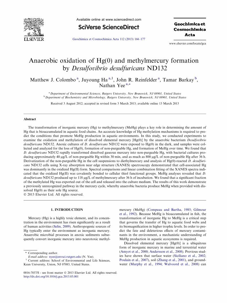
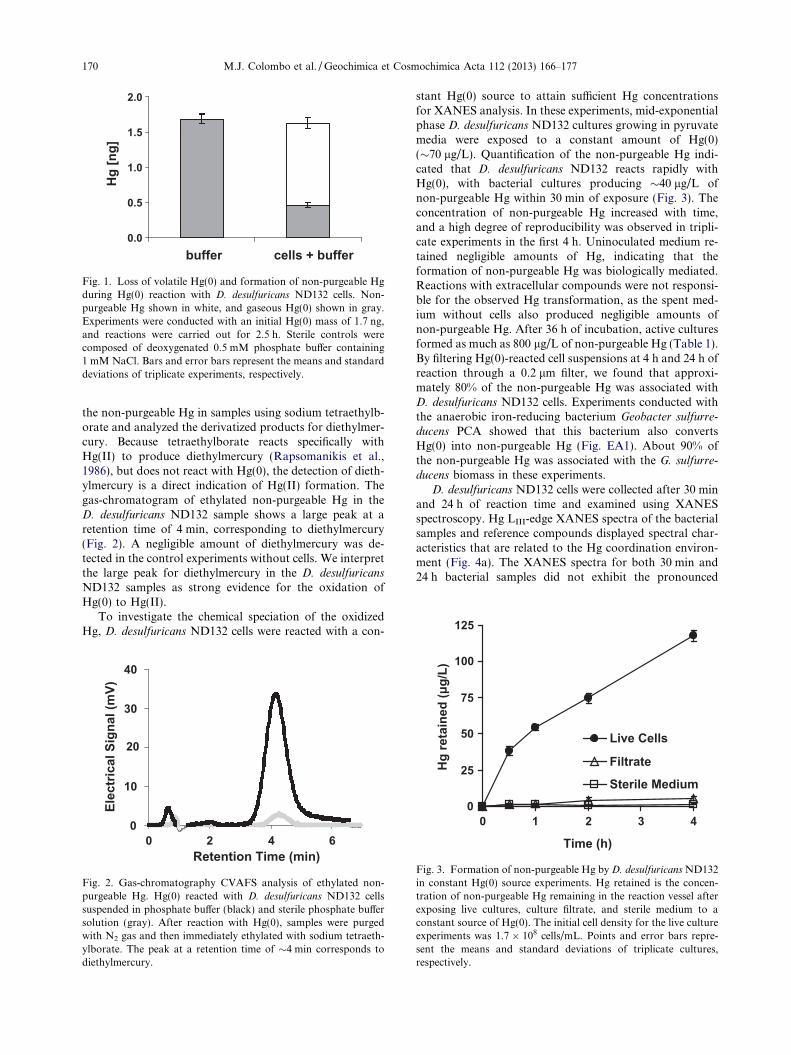
A mass balance experiment was conducted to determineif the anaerobic bacterium D. desulfuricans ND132 formsnon-purgeable Hg when reacted with a known amount ofHg(0). After 2.5 h of reaction with 1.7 ng of Hg(0), D.
desulfuricans ND132 cells suspended in phosphate bufferproduced 1.2 ± 0.1 ng of non-purgeable Hg, while0.46 ± 0.04 of unreacted volatile Hg(0) was recovered bypurging with N2 gas (Fig. 1). We also performed controlexperiments with the phosphate buffer in the absence ofbacterial cells, allowing the buffer solution to react withHg(0) for the same amount of time. In the controls,1.68 ± 0.07 ng of Hg(0) was recovered from the phosphatebuffer, and the concentration of non-purgeable Hg was be-low the detection limit. These data indicate Hg(0) reactionwith D. desulfuricans ND132 results in the loss of Hg(0) andformation of non-purgeable Hg.
To determine if the non-purgeable Hg formed by D.
desulfuricans ND132 was Hg(II), we immediately ethylated
0.0
0.5
1.0
1.5
2.0
buffer cells + buffer
Hg
[ng]
Fig. 1. Loss of volatile Hg(0) and formation of non-purgeable Hgduring Hg(0) reaction with D. desulfuricans ND132 cells. Non-purgeable Hg shown in white, and gaseous Hg(0) shown in gray.Experiments were conducted with an initial Hg(0) mass of 1.7 ng,and reactions were carried out for 2.5 h. Sterile controls werecomposed of deoxygenated 0.5 mM phosphate buffer containing1 mM NaCl. Bars and error bars represent the means and standarddeviations of triplicate experiments, respectively.
125
170 M.J. Colombo et al. / Geochimica et Cosmochimica Acta 112 (2013) 166–177
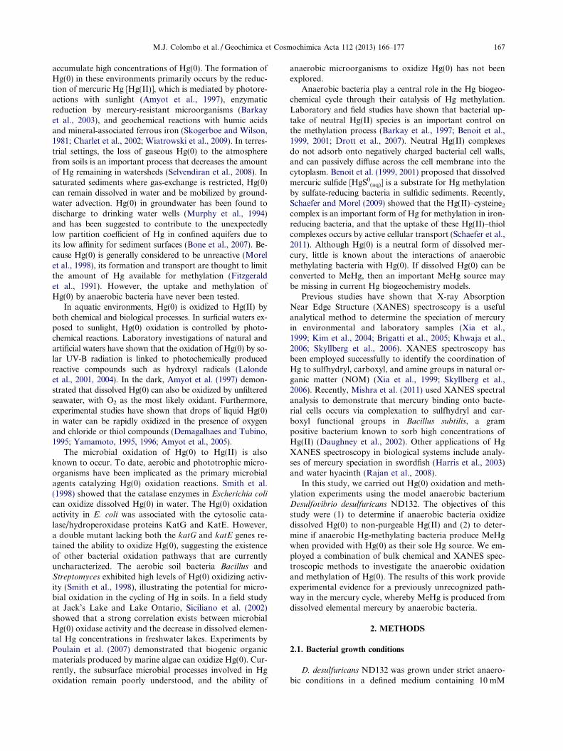
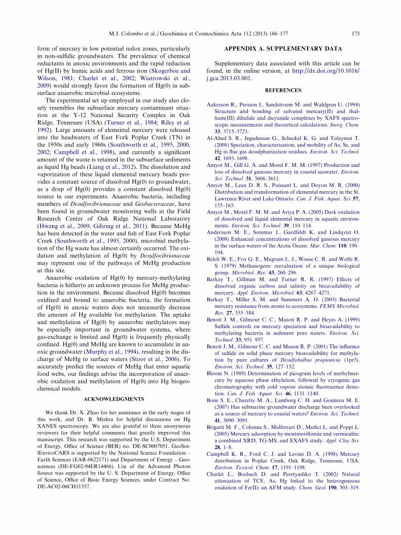
the non-purgeable Hg in samples using sodium tetraethylb-orate and analyzed the derivatized products for diethylmer-cury. Because tetraethylborate reacts specifically withHg(II) to produce diethylmercury (Rapsomanikis et al.,1986), but does not react with Hg(0), the detection of dieth-ylmercury is a direct indication of Hg(II) formation. Thegas-chromatogram of ethylated non-purgeable Hg in theD. desulfuricans ND132 sample shows a large peak at aretention time of 4 min, corresponding to diethylmercury(Fig. 2). A negligible amount of diethylmercury was de-tected in the control experiments without cells. We interpretthe large peak for diethylmercury in the D. desulfuricans
ND132 samples as strong evidence for the oxidation ofHg(0) to Hg(II).
To investigate the chemical speciation of the oxidizedHg, D. desulfuricans ND132 cells were reacted with a con-
0
10
20
30
40
0 2 4 6Retention Time (min)
Elec
tric
al S
igna
l (m
V)
Fig. 2. Gas-chromatography CVAFS analysis of ethylated non-purgeable Hg. Hg(0) reacted with D. desulfuricans ND132 cellssuspended in phosphate buffer (black) and sterile phosphate buffersolution (gray). After reaction with Hg(0), samples were purgedwith N2 gas and then immediately ethylated with sodium tetraeth-ylborate. The peak at a retention time of �4 min corresponds todiethylmercury.
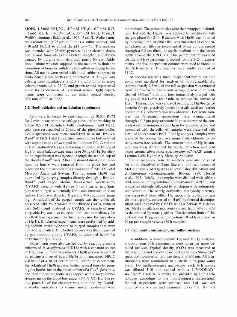
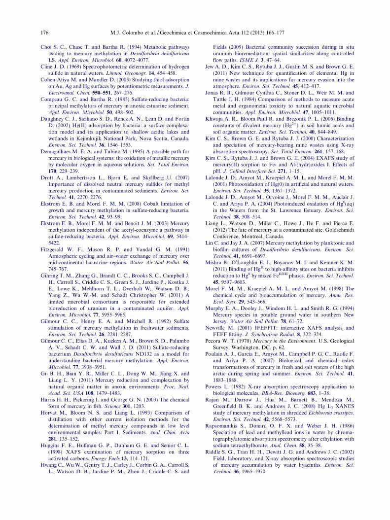
stant Hg(0) source to attain sufficient Hg concentrationsfor XANES analysis. In these experiments, mid-exponentialphase D. desulfuricans ND132 cultures growing in pyruvatemedia were exposed to a constant amount of Hg(0)(�70 lg/L). Quantification of the non-purgeable Hg indi-cated that D. desulfuricans ND132 reacts rapidly withHg(0), with bacterial cultures producing �40 lg/L ofnon-purgeable Hg within 30 min of exposure (Fig. 3). Theconcentration of non-purgeable Hg increased with time,and a high degree of reproducibility was observed in tripli-cate experiments in the first 4 h. Uninoculated medium re-tained negligible amounts of Hg, indicating that theformation of non-purgeable Hg was biologically mediated.Reactions with extracellular compounds were not responsi-ble for the observed Hg transformation, as the spent med-ium without cells also produced negligible amounts ofnon-purgeable Hg. After 36 h of incubation, active culturesformed as much as 800 lg/L of non-purgeable Hg (Table 1).By filtering Hg(0)-reacted cell suspensions at 4 h and 24 h ofreaction through a 0.2 lm filter, we found that approxi-mately 80% of the non-purgeable Hg was associated withD. desulfuricans ND132 cells. Experiments conducted withthe anaerobic iron-reducing bacterium Geobacter sulfurre-
ducens PCA showed that this bacterium also convertsHg(0) into non-purgeable Hg (Fig. EA1). About 90% ofthe non-purgeable Hg was associated with the G. sulfurre-
ducens biomass in these experiments.D. desulfuricans ND132 cells were collected after 30 min
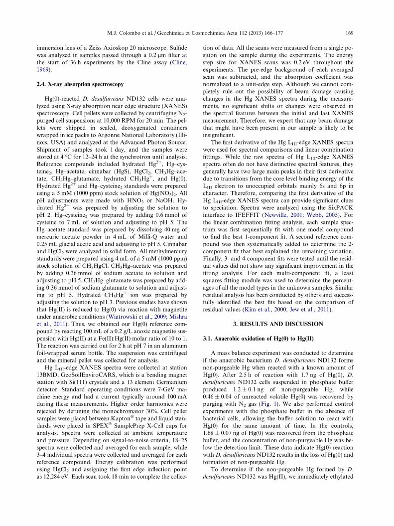
and 24 h of reaction time and examined using XANESspectroscopy. Hg LIII-edge XANES spectra of the bacterialsamples and reference compounds displayed spectral char-acteristics that are related to the Hg coordination environ-ment (Fig. 4a). The XANES spectra for both 30 min and24 h bacterial samples did not exhibit the pronounced
0
25
50
75
100
0 1 2 3 4
Time (h)
Hg
reta
ined
(µg/
L)
Live Cells
Filtrate
Sterile Medium
Fig. 3. Formation of non-purgeable Hg by D. desulfuricans ND132in constant Hg(0) source experiments. Hg retained is the concen-tration of non-purgeable Hg remaining in the reaction vessel afterexposing live cultures, culture filtrate, and sterile medium to aconstant source of Hg(0). The initial cell density for the live cultureexperiments was 1.7 � 108 cells/mL. Points and error bars repre-sent the means and standard deviations of triplicate cultures,respectively.
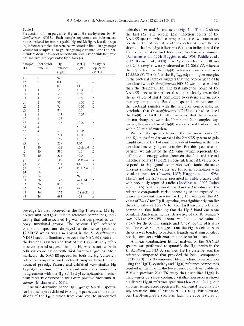
Table 1Production of non-purgeable Hg and Hg methylation by D.
desulfuricans ND132. Each sample represents an independentbottle analyzed for non-purgeable Hg and MeHg. A less than sign(<) indicates samples that were below detection limit (10 pg/samplevolume for samples a1 to g5, 50 pg/sample volume for h1 to h5).Standard deviations are of replicate analyses. Time points that werenot analyzed are represented by a dash (–).
SampleID
Incubationtime (h)
Hgretained(lg/L)
MeHg(lg/L)
Analyticalreplicates(MeHg)
a1 0 0.0 – –a2 0 0.1 – –a3 0 0.0 <1 1b1 1 55 <0.05 1b2 1 57 <0.5 1b3 1 52 <0.5 2c1 2 78 <0.05 2c2 2 75 <0.05 2c3 2 71 <0.1 1d1 4 115 <0.05 1d2 4 123 – –d3 4 116 <0.04 2d4 4 – 2 1d5 4 – <0.05 1e1 8 211 <0.03 1e2 8 192 <0.2 2e3 8 257 0.02 1f1 16 552 1.5 ± 0.4 2f2 16 306 <0.1 2f3 16 354 <0.04 1g1 24 100 10 ± 0.8 2g2 24 774 0.6 2g3 24 108 44 ± 8.8 4g4 24 – 31 1g5 24 – 26 1h1 36 143 54 ± 14 2h2 36 818 <0.7 1h3 36 189 66 1h4 36 172 118 ± 21 2h5 36 693 <0.8 2
M.J. Colombo et al. / Geochimica et Cosmochimica Acta 112 (2013) 166–177 171
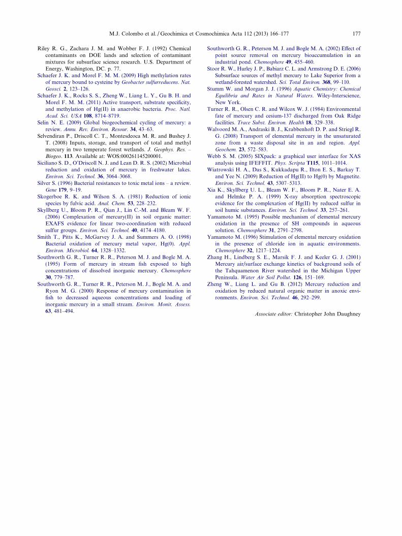
pre-edge features observed in the Hg(II)–acetate, MeHg–acetate and MeHg–glutamate reference compounds, indi-cating that cell-associated Hg was not complexed to car-boxyl functional groups. The aqueous Hg2+ referencecompound spectrum displayed a distinctive peak at12,310 eV which was also absent in the D. desulfuricans
ND132 spectra. Similarity between the XANES spectra ofthe bacterial samples and that of the Hg-(cysteine)2 refer-ence compound suggests that the Hg was associated withcells via coordination with thiol functional groups. Mostmarkedly, the XANES spectra for both the Hg-(cysteine)2
reference compound and bacterial samples lacked a pro-nounced pre-edge feature and exhibited very similar HgLIII-edge positions. This Hg coordination environment isin agreement with the Hg–sulfhydryl complexation mecha-nism recently observed in the Gram positive bacterium B.
subtilis (Mishra et al., 2011).The first derivative of the Hg LIII-edge XANES spectra
for both samples exhibited two major peaks due to the tran-sitions of the LIII electron from core level to unoccupied
orbitals of 6s and 6p character (Fig. 4b). Table 2 showsthe first (E1) and second (E2) inflection points of theXANES spectra, which correspond to the two maximumpoints in the first derivative of the spectra. We used the po-sition of the first edge inflection (E1) as an indication of theHg oxidation state and local coordination environment(Aakesson et al., 1994; Huggins et al., 1998; Riddle et al.,2002; Rajan et al., 2008). The E1 values for both 30 minand 24 h samples were positioned at 12,286.6 eV, whereasthe E1 value for the Hg(0) reference compound was12,285.0 eV. The shift in the Hg LIII-edge to higher energiesin the bacterial samples suggests that the non-purgeable Hgassociated with D. desulfuricans ND132 was more oxidizedthan the elemental Hg. The first inflection point of theXANES spectra for bacterial samples closely resembledthe E1 values of Hg(II) complexed to cysteine and methyl-mercury compounds. Based on spectral comparisons ofthe bacterial samples with the reference compounds, weconcluded that D. desulfuricans ND132 cells had oxidizedthe Hg(0) to Hg(II). Finally, we noted that the E1 valuesdid not change between the 30 min and 24 h samples, sug-gesting that oxidation of Hg(0) was rapid and had occurredwithin 30 min of reaction.
We used the spacing between the two main peaks (E1
and E2) in the first derivative of the XANES spectra to gaininsight into the level of ionic or covalent bonding in the cell-associated mercury–ligand complex. For this spectral com-parison, we calculated the DE value, which represents thedifference in energy values between the first and secondinflection points (Table 2). In general, larger DE values cor-respond to Hg–ligand complexes with ionic characterwhereas smaller DE values are found in complexes withcovalent character (Powers, 1982; Huggins et al., 1998).The E1 and the DE values presented in Table 2 agree wellwith previously reported studies (Riddle et al., 2002; Rajanet al., 2008), and the overall trend in the DE values for thereference compounds varied according to the expected in-crease in covalent character for Hg. For example, the DE
value of 7.2 eV for Hg(II–cysteine2 was significantly smallerthan the value of 13.2 eV for the Hg(II)–acetate referencecompound, thus indicating that the Hg–S bond was morecovalent. Analyzing the first derivative of the D. desulfuri-
cans ND132 XANES spectra, we found a DE value of7.5 eV for the 30 min sample and 7.7 eV for the 24 h sam-ple. These DE values suggest that the Hg associated withthe cells was bonded to bacterial ligands via strong covalentbonds, consistent with coordination to sulfur atoms.
A linear combination fitting analysis of the XANESspectra was performed to quantify the Hg species in theD. desulfuricans ND132 samples. Hg(II)–cysteine2 was thereference compound that provided the best 1-componentfit (Table 3). For 2-component fitting, a linear combinationusing the Hg(II)–cysteine2 and Hg(0) reference compoundsresulted in the fit with the lowest residual values (Table 3).While a previous XANES study that quantified Hg(0) inmine wastes by a slow cooling crystallization process showsa different Hg(0) reference spectrum (Jew et al., 2011), ourambient temperature spectrum for elemental mercury clo-sely resembles that of Mishra et al. (2011). Furthermore,our Hg(0)–magnetite spectrum lacks the edge features of
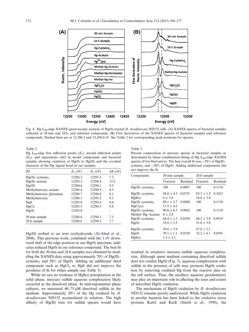
Fig. 4. Hg LIII-edge XANES spectroscopic analysis of Hg(0)-reacted D. desulfuricans ND132 cells. (A) XANES spectra of bacterial samplescollected at 30 min and 24 h, and reference compounds. (B) First derivatives of the XANES spectra of bacterial samples and referencecompounds. Dashed lines are at 12,286.3 and 12,294.0 eV. See Table 2 for corresponding peak positions for spectra.
Table 2Hg LIII-edge first inflection points (E1), second inflection points(E2), and separations (DE) in model compounds and bacterialsamples showing oxidation of Hg(0) to Hg(II) and the covalentcharacter of the Hg–ligand bond in our samples.
ID E1 (eV) E2 (eV) DE (eV)
Hg(II)–cysteine2 12286.3 12293.5 7.2Hg(II)–acetate 12285.2 12298.4 13.2Hg(II) 12284.6 12294.1 9.5Methylmercury acetate 12286.4 12294.9 8.5Methylmercury glutamate 12286.7 12294.8 8.1Methylmercury 12286.1 12295.2 9.1HgS 12285.6 12293.6 8.0HgCl2 12285.5 12294.3 8.8Hg(0) 12285.0 – –
30 min sample 12286.6 12294.1 7.524 h sample 12286.6 12294.3 7.7
Table 3Percent composition of mercury species in bacterial samples asdetermined by linear combination fitting of Hg LIII-edge XANESspectra (First Derivative). The best overall fit was �70% of Hg(II)–cysteine2 and �30% of Hg(0). Adding additional components didnot improve the fit.
Components 30 min sample 24 h sample
Fraction Residual Fraction Residual
Hg(II)–cysteine2 100 0.0887 100 0.1110
Hg(II)–cysteine2 94.0 ± 4.5 0.0755 83.2 ± 1.2 0.1013HgS(s) 6 ± 3.4 16.8 ± 3.4Hg(II)–cysteine2 88.1 ± 5.7 0.0880 100 0.1110HgCl2(s) 11.9 ± 4.2 –Hg(II)–cysteine2 96.0 ± 6.5 0.0802 100 0.1110Methyl–Hg–Acetate 4 ± 2.8 –Hg(II)–cysteine2 64.4 ± 1.2 0.0350 68.2 ± 3.8 0.0519Hg(0) 35.6 ± 2.5 31.8 ± 3.8
Hg(II)–cysteine2 59.6 ± 5.9 67.8 ± 2.1Hg(0) 39.1 ± 1.1 0.0350 32.2 ± 6.1 0.0591HgS(s) 1.3 ± 2.1 –
172 M.J. Colombo et al. / Geochimica et Cosmochimica Acta 112 (2013) 166–177
Hg(II) sorbed to an iron oxyhydroxide (Al-Abed et al.,2008). This previous work, combined with the 1 eV down-ward shift of the edge position in our Hg(0) spectrum, indi-cates reduced Hg(0) in our reference compound. The best fitfor both the 30 min and 24 h samples was obtained by mod-eling the XANES data using approximately 70% of Hg(II)–cysteine2 and 30% of Hg(0). Adding an additional thirdcomponent such as HgCl2 or HgS did not improve thegoodness of fit for either sample (see Table 3).
While we saw no evidence of HgS(s) precipitation in thesolid phase, mercury–sulfide aqueous complexation likelyoccurred in the dissolved phase. In mid-exponential phasecultures, we measured 40–75 lM dissolved sulfide in themedium. Approximately 20% of the Hg oxidized by D.
desulfuricans ND132 accumulated in solution. The highaffinity of Hg(II) ions for sulfide species would have
resulted in extensive mercury–sulfide aqueous complexa-tion. Although spent medium containing dissolved sulfidedoes not oxidize Hg(0) (Fig. 3), aqueous complexation withsulfide in the presence of cells may promote Hg(0) oxida-tion by removing oxidized Hg from the reactive sites onthe cell surface. Thus, the ancillary aqueous geochemistrymay play an important role in affecting the rates and extentof microbial Hg(0) oxidation.
The mechanism of Hg(0) oxidation by D. desulfuricans
ND132 remains poorly understood. While Hg(0) oxidationin aerobic bacteria has been linked to the oxidative stressproteins KatG and KatE (Smith et al., 1998), the
0
35
70
105
140
0 10 20 30 40Time (h)
MeH
g (µ
g/L)
(A)
(B)R2= 0.9669
0.15
0.2
0.25
0.3
0.35
0 50 100 150O
ptic
al D
enis
ty (6
00 n
m)
MeHg (µg/L)
(C) High MeHg
Low MeHg
Fig. 5. MeHg production from Hg(0). (A) MeHg concentrations inD. desulfuricans ND132 cultures determined at periodic intervalsup to 36 h of incubation. Data points represent individualexperiments and error bars are analytical replicates of a singlesample. (B) Relationship between cell density and MeHg produc-tion in samples collected at 36 h (C) Representative microscopicimages of samples collected at 36 h; (top) a culture of ND132 thatproduced a high MeHg concentration and (bottom) a culture ofND132 that produced low MeHg concentration.
M.J. Colombo et al. / Geochimica et Cosmochimica Acta 112 (2013) 166–177 173
mechanism of anaerobic Hg(0) oxidation is unknown.Inspection of the G. sulfurreducens PCA genome reveals ahomolog of the katG gene, suggesting that cytosolic cata-lase-mediated Hg(0) oxidation in iron-reducing bacteria ispossible. D. desulfuricans ND132 and many other obligateanaerobes do not carry such catalase/hydroperoxidase genes,suggesting that Hg(0) is being oxidized by an alternate mech-anism in this organism. The absence of catalase/hydroperox-idase in D. desulfuricans ND132 is not surprising, as obligateanaerobes are generally not exposed to oxygen in their natu-ral habitats and do not need to decompose damaging hydro-gen peroxide by-products emitted by aerobic respiration.
Our XANES data provide evidence that the oxidized Hgis associated with cells via coordination with thiol func-tional groups, and it is possible that these functional groupsmay be involved in the formation of Hg(II) products. Re-cently, Gu et al. (2011) demonstrated that Hg(0) can com-plex with thiol groups in reduced humic acids andpostulated that Hg(0) bound to organic matter becomesoxidized. Subsequently, these investigators showed thatother thiol-bearing organic compounds such as glutathionecan also bind and oxidize Hg(0) (Zheng et al., 2012). Inter-estingly, electron transfer from Hg(0) to the organic mole-cule occurs even though mercury is coordinated to a –SHfunctional group. This is somewhat surprising as sulfur inthe –SH moiety is in its most reduced oxidation state. Pre-vious potentiometric studies suggest that there is directinteraction between Hg(0) and reduced sulfur that may leadto oxidation (Cohen-Atiya and Mandler, 2003). We specu-late that the reactive thiol functional groups in anaerobicbacteria, either in the cell wall or cytoplasm, may be in-volved in Hg(0) oxidation.
3.2. MeHg production from Hg(0)
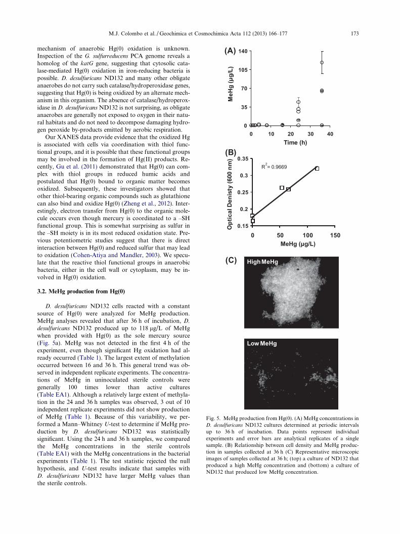
D. desulfuricans ND132 cells reacted with a constantsource of Hg(0) were analyzed for MeHg production.MeHg analyses revealed that after 36 h of incubation, D.
desulfuricans ND132 produced up to 118 lg/L of MeHgwhen provided with Hg(0) as the sole mercury source(Fig. 5a). MeHg was not detected in the first 4 h of theexperiment, even though significant Hg oxidation had al-ready occurred (Table 1). The largest extent of methylationoccurred between 16 and 36 h. This general trend was ob-served in independent replicate experiments. The concentra-tions of MeHg in uninoculated sterile controls weregenerally 100 times lower than active cultures(Table EA1). Although a relatively large extent of methyla-tion in the 24 and 36 h samples was observed, 3 out of 10independent replicate experiments did not show productionof MeHg (Table 1). Because of this variability, we per-formed a Mann–Whitney U-test to determine if MeHg pro-duction by D. desulfuricans ND132 was statisticallysignificant. Using the 24 h and 36 h samples, we comparedthe MeHg concentrations in the sterile controls(Table EA1) with the MeHg concentrations in the bacterialexperiments (Table 1). The test statistic rejected the nullhypothesis, and U-test results indicate that samples withD. desulfuricans ND132 have larger MeHg values thanthe sterile controls.
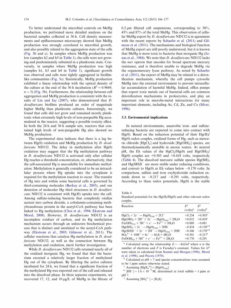
Table 4Standard potentials for the Hg(II)/Hg(0) and other relevant redoxcouples.
Reaction Eo
(volts)aEa
(volts)b
HgCl2 + 2e� ! Hg(0)(aq) + 2Cl� +0.234 +0.503c
Hg(OH)2 + 2H+ + 2e� ! Hg(0)(aq) + 2H2O +0.832 +0.418c
Fe(OH)3(s) + 3H+ + e� ! Fe2+ + 3H2O +0.880 �0.081Hg(SH)2 + 2e� !Hg(0)(aq) + 2HS� �0.434 �0.150c,d
Hg(SH)S� + 2e� + 2H+! Hg(0)(aq) + 2HS� �0.246 �0.170c,d
SO42� + 10H+ + 8e� ! H2S + 4H2O +0.301 �0.217e
FeOOH(s) + 3H+ + e� ! Fe2+ + 2H2O +0.770 �0.291
a Calculated using the relationship E = �DG/nF where n is thenumber of electrons and F is Faraday’s constant. Values for Go
were taken or calculated from Stumm and Morgan (1996), Morelet al. (1998), and Pecora (1970).
b Calculated at pH = 7 and species concentrations were assumedto be 1 ppm unless otherwise stated.
c Assuming [HgXnm] = [Hg(0)(aq)].
d [HS�] = 1.6 � 10�5 M, determined at total sulfide = 1 ppm atpH 7.
e Assuming [SO42�] = [H2S].
174 M.J. Colombo et al. / Geochimica et Cosmochimica Acta 112 (2013) 166–177
To better understand the microbial controls on MeHgproduction, we performed more detailed analyses on thebacterial samples collected at 36 h. Cell density measure-ments and epifluorescence microscopy showed that MeHgproduction was strongly correlated to microbial growth,and also possibly related to the aggregation state of the cells(Fig. 5b and c). In samples where MeHg production waslow (samples h2 and h5 in Table 1), the cells were not grow-ing and predominately subsisted in a planktonic state. Con-versely, in samples where MeHg production was high(samples h1, h3 and h4 in Table 1), significant growthwas observed and cells were tightly aggregated in biofilm-like communities (Fig. 5c). Statistically, MeHg productionexhibited a linear relationship with the optical density ofthe culture at the end of the 36 h incubation (R2 = 0.9669,n = 5) (Fig. 5b). Furthermore, the relationship between cellaggregation and MeHg production is consistent with the re-sults of Lin and Jay (2007), who demonstrated that D.
desulfuricans biofilms produced an order of magnitudehigher MeHg than planktonic cultures. Interestingly, wefound that cells did not grow and remained mostly plank-tonic when extremely high levels of non-purgeable Hg accu-mulated in the reactor, suggesting a possible toxicity effect.In both the 24 h and 36 h sample sets, reactors that con-tained high levels of non-purgeable Hg also showed noMeHg production.
The experimental data indicate that there is a lag be-tween Hg(0) oxidation and MeHg production by D. desul-
furicans ND132. The delay in methylation after Hg(0)oxidation may suggest that the Hg methylation pathwayin D. desulfuricans ND132 is activated only after cellularHg reaches a threshold concentration, or, alternatively, thatthe cell-associated Hg is unavailable for immediate methyl-ation. Methylation is generally considered to be an intracel-lular process where Hg uptake into the cytoplasm isrequired for the methylation reaction to occur. The transferof Hg into and within some bacterial cells is governed bythiol-containing molecules (Barkay et al., 2003), and ourdetection of molecular Hg–thiol structures in D. desulfuri-
cans ND132 is consistent with Hg(II) uptake into the cell.Among sulfate-reducing bacteria that completely oxidizeacetate into carbon dioxide, a cobalamin-containing meth-yltransferase protein in the acetyl-CoA pathway has beenlinked to Hg methylation (Choi et al., 1994; Ekstrom andMorel, 2008). However, D. desulfuricans ND132 is anincomplete oxidizer of carbon, and its Hg methylationmechanism occurs through an unknown biochemical pro-cess that is distinct and unrelated to the acetyl-CoA path-way (Ekstrom et al., 2003; Gilmour et al., 2011). Thecellular reactions that catalyze Hg methylation in D. desul-
furicans ND132, as well as the connection between Hgmethylation and oxidation, merit further investigation.
While D. desulfuricans ND132 cells retained nearly all ofthe oxidized inorganic Hg, we found that that the bacte-rium excreted a relatively larger fraction of methylatedHg out of the cytoplasm. By filtering the active culturesincubated for 24 h, we found that a significant fraction ofthe methylated Hg was exported out of the cell and releasedinto the dissolved phase. In three separate experiments, werecovered 17, 12, and 10 lg/L of MeHg in the filtrate of
0.2 lm filtered cell suspensions, corresponding to 50%,43% and 87% of the total MeHg. This observation of cellu-lar MeHg export by D. desulfuricans ND132 is in agreementwith the recent reports by Schaefer et al. (2011) and Gil-mour et al. (2011). The mechanisms and biological functionof MeHg export are still poorly understood, but it is knownthat MeHg is more toxic to bacteria than inorganic Hg (Jo-nas et al., 1984). We note that D. desulfuricans ND132 lacksthe mer operon that encodes for broad spectrum mercuryresistance, and is therefore unable to degrade MeHg viathe organomercury lyase pathway. As noted by Schaeferet al. (2011), the export of MeHg may be related to a detox-ification mechanism, whereby the cell pumps cytosolicMeHg into the external environment to prevent intracellu-lar accumulation of harmful MeHg. Indeed, efflux pumpsthat export toxic metals out of bacterial cells are commondetoxification mechanisms in prokaryotes, and play animportant role in microbe-metal interactions for manyimportant elements, including As, Cd, Zn, and Co (Silver,1996).
3.3. Environmental implications
In natural environments, anaerobic iron- and sulfate-reducing bacteria are expected to come into contact withHg(0). Based on the reduction potential of their Hg(II)/Hg(0) redox couples, oxidized forms of Hg, such as mercu-ric chloride [HgCl2] and hydroxide [Hg(OH)2] species, arethermodynamically unstable in anoxic waters. At neutralpH, the Eh values of the HgCl2/Hg(0) and Hg(OH)2/Hg(0) couples are +0.503 and +0.418 volts, respectively(Table 4). The dissolved mercuric sulfide species Hg(SH)2
and Hg(SH)S� are more stable under reducing conditions,and convert to Hg(0) at Eh values below �0.170 volts. Incomparison, sulfate and iron oxyhydroxide reduction ex-tends down to �0.217 and �0.291 volts, respectively.According to these redox potentials, Hg(0) is the stable
M.J. Colombo et al. / Geochimica et Cosmochimica Acta 112 (2013) 166–177 175
form of mercury in low potential redox zones, particularlyin non-sulfidic groundwaters. The prevalence of chemicalreductants in anoxic environments and the rapid reductionof Hg(II) by humic acids and ferrous iron (Skogerboe andWilson, 1981; Charlet et al., 2002; Wiatrowski et al.,2009) would strongly favor the formation of Hg(0) in sub-surface anaerobic microbial ecosystems.
The experimental set up employed in our study also clo-sely resembles the subsurface mercury contaminant situa-tion at the Y-12 National Security Complex in OakRidge, Tennessee (USA) (Turner et al., 1984; Riley et al.1992). Large amounts of elemental mercury were releasedinto the headwaters of East Fork Poplar Creek (TN) inthe 1950s and early 1960s (Southworth et al., 1995, 2000,2002; Campbell et al., 1998), and currently a significantamount of the waste is retained in the subsurface sedimentsas liquid Hg beads (Liang et al., 2012). The dissolution andvaporization of these liquid elemental mercury beads pro-vides a constant source of dissolved Hg(0) to groundwater,as a drop of Hg(0) provides a constant dissolved Hg(0)source in our experiments. Anaerobic bacteria, includingmembers of Desulfovibrionaceae and Geobacteraceae, havebeen found in groundwater monitoring wells at the FieldResearch Center of Oak Ridge National Laboratory(Hwang et al., 2009; Gihring et al., 2011). Because MeHghas been detected in the water and fish of East Fork PoplarCreek (Southworth et al., 1995, 2000), microbial methyla-tion of the Hg waste has almost certainly occurred. The oxi-dation and methylation of Hg(0) by Desulfovibrionaceae
may represent one of the pathways of MeHg productionat this site.
Anaerobic oxidation of Hg(0) by mercury-methylatingbacteria is hitherto an unknown process for MeHg produc-tion in the environment. Because dissolved Hg(0) becomesoxidized and bound to anaerobic bacteria, the formationof Hg(0) in anoxic waters does not necessarily decreasethe amount of Hg available for methylation. The uptakeand methylation of Hg(0) by anaerobic methylators maybe especially important in groundwater systems, wheregas-exchange is limited and Hg(0) is frequently physicallyconfined. Hg(0) and MeHg are known to accumulate in an-oxic groundwater (Murphy et al., 1994), resulting in the dis-charge of MeHg to surface waters (Stoor et al., 2006). Toaccurately predict the sources of MeHg that enter aquaticfood webs, our findings advise the incorporation of anaer-obic oxidation and methylation of Hg(0) into Hg biogeo-chemical models.
ACKNOWLEDGMENTS
We thank Dr. X. Zhao for her assistance in the early stages ofthis work, and Dr. B. Mishra for helpful discussions on HgXANES spectroscopy. We are also grateful to three anonymousreviewers for their helpful comments that greatly improved thismanuscript. This research was supported by the U.S. Departmentof Energy, Office of Science (BER) no. DE-SC0007051. GeoSoi-lEnviroCARS is supported by the National Science Foundation –Earth Sciences (EAR-0622171) and Department of Energy – Geo-sciences (DE-FG02-94ER14466). Use of the Advanced PhotonSource was supported by the U. S. Department of Energy, Officeof Science, Office of Basic Energy Sciences, under Contract No.DE-AC02-06CH11357.
APPENDIX A. SUPPLEMENTARY DATA
Supplementary data associated with this article can befound, in the online version, at http://dx.doi.org/10.1016/j.gca.2013.03.001.
REFERENCES
Aakesson R., Persson I., Sandstroem M. and Wahlgren U. (1994)Structure and bonding of solvated mercury(II) and thal-lium(III) dihalide and dicyanide complexes by XAFS spectro-scopic measurements and theoretical calculations. Inorg. Chem.
33, 3715–3723.Al-Abed S. R., Jegadeesan G., Scheckel K. G. and Tolaymat T.
(2008) Speciation, characterization, and mobility of As, Se, andHg in flue gas desulphurization residues. Environ. Sci. Technol.
42, 1693–1698.Amyot M., Gill G. A. and Morel F. M. M. (1997) Production and
loss of dissolved gaseous mercury in coastal seawater. Environ.
Sci. Technol. 31, 3606–3611.Amyot M., Lean D. R. S., Poissant L. and Doyon M. R. (2000)
Distribution and transformation of elemental mercury in the St.Lawrence River and Lake Ontario. Can. J. Fish. Aquat. Sci. 57,155–163.
Amyot M., Morel F. M. M. and Ariya P. A. (2005) Dark oxidationof dissolved and liquid elemental mercury in aquatic environ-ments. Environ. Sci. Technol. 39, 110–114.
Andersson M. E., Sommar J., Gardfeldt K. and Lindqvist O.(2008) Enhanced concentrations of dissolved gaseous mercuryin the surface waters of the Arctic Ocean. Mar. Chem. 110, 190–194.
Balch W. E., Fox G. E., Magrum L. J., Woese C. R. and Wolfe R.S. (1979) Methanogens: reevaluation of a unique biologicalgroup. Microbiol. Rev. 43, 260–296.
Barkay T., Gillman M. and Turner R. R. (1997) Effects ofdissolved organic carbon and salinity on bioavailability ofmercury. Appl. Environ. Microbiol. 63, 4267–4271.
Barkay T., Miller S. M. and Summers A. O. (2003) Bacterialmercury resistance from atoms to ecosystems. FEMS Microbiol.
Rev. 27, 355–384.Benoit J. M., Gilmour C. C., Mason R. P. and Heyes A. (1999)
Sulfide controls on mercury speciation and bioavailability tomethylating bacteria in sediment pore waters. Environ. Sci.
Technol. 33, 951–957.Benoit J. M., Gilmour C. C. and Mason R. P. (2001) The influence
of sulfide on solid phase mercury bioavailability for methyla-tion by pure cultures of Desulfobulbus propionicus (1pr3).Environ. Sci. Technol. 35, 127–132.
Bloom N. (1989) Determination of picogram levels of methylmer-cury by aqueous phase ethylation, followed by cryogenic gaschromatography with cold vapour atomic fluorescence detec-tion. Can. J. Fish. Aquat. Sci. 46, 1131–1140.
Bone S. E., Charette M. A., Lamborg C. H. and Gonneea M. E.(2007) Has submarine groundwater discharge been overlookedas a source of mercury to coastal waters? Environ. Sci. Technol.
41, 3090–3095.Brigatti M. F., Colonna S., Malferrari D., Medici L. and Poppi L.
(2005) Mercury adsorption by montmorillonite and vermiculite:a combined XRD, TG-MS, and EXAFS study. Appl. Clay Sci.
28, 1–8.Campbell K. R., Ford C. J. and Levine D. A. (1998) Mercury
distribution in Poplar Creek, Oak Ridge, Tennessee, USA.Environ. Toxicol. Chem. 17, 1191–1198.
Charlet L., Bosbach D. and Peretyashko T. (2002) Naturalattenuation of TCE, As, Hg linked to the heterogeneousoxidation of Fe(II): an AFM study. Chem. Geol. 190, 303–319.
176 M.J. Colombo et al. / Geochimica et Cosmochimica Acta 112 (2013) 166–177
Choi S. C., Chase T. and Bartha R. (1994) Metabolic pathwaysleading to mercury methylation in Desulfovibrio desulfuricans
LS. Appl. Environ. Microbiol. 60, 4072–4077.Cline J. D. (1969) Spectrophotometric determination of hydrogen
sulfide in natural waters. Limnol. Oceonogr. 14, 454–458.Cohen-Atiya M. and Mandler D. (2003) Studying thiol adsorption
on Au, Ag and Hg surfaces by potentiometric measurements. J.
Electroanal. Chem. 550–551, 267–276.Compeau G. C. and Bartha R. (1985) Sulfate-reducing bacteria:
principal methylators of mercury in anoxic estuarine sediment.Appl. Environ. Microbiol. 50, 498–502.
Daughney C. J., Siciliano S. D., Rencz A. N., Lean D. and FortinD. (2002) Hg(II) adsorption by bacteria: a surface complexa-tion model and its application to shallow acidic lakes andwetlands in Kejimkujik National Park, Nova Scotia, Canada.Environ. Sci. Technol. 36, 1546–1553.
Demagalhaes M. E. A. and Tubino M. (1995) A possible path formercury in biological systems: the oxidation of metallic mercuryby molecular oxygen in aqueous solutions. Sci. Total Environ.
170, 229–239.Drott A., Lambertsson L., Bjorn E. and Skyllberg U. (2007)
Importance of dissolved neutral mercury sulfides for methylmercury production in contaminated sediments. Environ. Sci.
Technol. 41, 2270–2276.Ekstrom E. B. and Morel F. M. M. (2008) Cobalt limitation of
growth and mercury methylation in sulfate-reducing bacteria.Environ. Sci. Technol. 42, 93–99.
Ekstrom E. B., Morel F. M. M. and Benoit J. M. (2003) Mercurymethylation independent of the acetyl-coenzyme a pathway insulfate-reducing bacteria. Appl. Environ. Microbiol. 69, 5414–5422.
Fitzgerald W. F., Mason R. P. and Vandal G. M. (1991)Atmospheric cycling and air–water exchange of mercury overmid-continental lacustrine regions. Water Air Soil Pollut. 56,745–767.
Gihring T. M., Zhang G., Brandt C. C., Brooks S. C., Campbell J.H., Carroll S., Criddle C. S., Green S. J., Jardine P., Kostka J.E., Lowe K., Mehlhorn T. L., Overholt W., Watson D. B.,Yang Z., Wu W.-M. and Schadt Christopher W. (2011) Alimited microbial consortium is responsible for extendedbioreduction of uranium in a contaminated aquifer. Appl.
Environ. Microbiol. 77, 5955–5965.Gilmour C. C., Henry E. A. and Mitchell R. (1992) Sulfate
stimulation of mercury methylation in freshwater sediments.Environ. Sci. Technol. 26, 2281–2287.
Gilmour C. C., Elias D. A., Kucken A. M., Brown S. D., PalumboA. V., Schadt C. W. and Wall J. D. (2011) Sulfate-reducingbacterium Desulfovibrio desulfuricans ND132 as a model forunderstanding bacterial mercury methylation. Appl. Environ.
Microbiol. 77, 3938–3951.Gu B. H., Bian Y. R., Miller C. L., Dong W. M., Jiang X. and
Liang L. Y. (2011) Mercury reduction and complexation bynatural organic matter in anoxic environments. Proc. Natl.
Acad. Sci. USA 108, 1479–1483.Harris H. H., Pickering I. and George G. N. (2003) The chemical
form of mercury in fish. Science 301, 1203.Horvat M., Bloom N. S. and Liang L. (1993) Comparison of
distillation with other current isolation methods for thedetermination of methyl mercury compounds in low levelenvironmental samples: Part 1. Sediments. Anal. Chim. Acta
281, 135–152.Huggins F. E., Huffman G. P., Dunham G. E. and Senior C. L.
(1998) XAFS examination of mercury sorption on threeactivated carbons. Energy Fuels 13, 114–121.
Hwang C., Wu W., Gentry T. J., Carley J., Corbin G. A., Carroll S.L., Watson D. B., Jardine P. M., Zhou J., Criddle C. S. and
Fields (2009) Bacterial community succession during in situuranium bioremediation: spatial similarities along controlledflow paths. ISME J. 3, 47–64.
Jew A. D., Kim C. S., Rytuba J. J., Gustin M. S. and Brown G. E.(2011) New technique for quantification of elemental Hg inmine wastes and its implications for mercury evasion into theatmosphere. Environ. Sci. Technol. 45, 412–417.
Jonas R. B., Gilmour Cynthia C., Stoner D. L., Weir M. M. andTuttle J. H. (1984) Comparison of methods to measure acutemetal and organometal toxicity to natural aquatic microbialcommunities. Appl. Environ. Microbiol. 47, 1005–1011.
Khwaja A. R., Bloom Paul R. and Brezonik P. L. (2006) Bindingconstants of divalent mercury (Hg2+) in soil humic acids andsoil organic matter. Environ. Sci. Technol. 40, 844–849.
Kim C. S., Brown G. E. and Rytuba J. J. (2000) Characterizationand speciation of mercury-bearing mine wastes using X-rayabsorption spectroscopy. Sci. Total Environ. 261, 157–168.
Kim C. S., Rytuba J. J. and Brown G. E. (2004) EXAFS study ofmercury(II) sorption to Fe- and Al-(hydr)oxides I. Effects ofpH. J. Colloid Interface Sci. 271, 1–15.
Lalonde J. D., Amyot M., Kraepiel A. M. L. and Morel F. M. M.(2001) Photooxidation of Hg(0) in artificial and natural waters.Environ. Sci. Technol. 35, 1367–1372.
Lalonde J. D., Amyot M., Orvoine J., Morel F. M. M., Auclair J.C. and Ariya P. A. (2004) Photoinduced oxidation of Hg0(aq)in the Waters from the St. Lawrence Estuary. Environ. Sci.
Technol. 38, 508–514.Liang L., Watson D., Miller C., Howe J., He F. and Pierce E.
(2012) The fate of mercury at a contaminated site. GoldschmidtConference, Montreal, Canada.
Lin C. and Jay J. A. (2007) Mercury methylation by planktonic andbiofilm cultures of Desulfovibrio desulfuricans. Environ. Sci.
Technol. 41, 6691–6697.Mishra B., O’Loughlin E. J., Boyanov M. I. and Kemner K. M.
(2011) Binding of HgII to high-affinity sites on bacteria inhibitsreduction to Hg0 by mixed FeII/III phases. Environ. Sci. Technol.
45, 9597–9603.Morel F. M. M., Kraepiel A. M. L. and Amyot M. (1998) The
chemical cycle and bioaccumulation of mercury. Annu. Rev.
Ecol. Syst. 29, 543–566.Murphy E. A., Dooley J., Windom H. L. and Smith R. G. (1994)
Mercury species in potable ground water in southern NewJersey. Water Air Soil Pollut. 78, 61–72.
Newville M. (2001) IFEFFIT: interactive XAFS analysis andFEFF fitting. J. Synchrotron Radiat. 8, 322–324.
Pecora W. T. (1970) Mercury in the Environment. U.S. GeologicalSurvey, Washington, DC. p. 62.
Poulain A. J., Garcia E., Amyot M., Campbell P. G. C., Raofie F.and Ariya P. A. (2007) Biological and chemical redoxtransformations of mercury in fresh and salt waters of the higharctic during spring and summer. Environ. Sci. Technol. 41,1883–1888.
Powers L. (1982) X-ray absorption spectroscopy application tobiological molecules. BBA-Rev. Bioenerg. 683, 1–38.
Rajan M., Darrow J., Hua M., Barnett B., Mendoza M.,Greenfield B. K. and Andrews J. C. (2008) Hg L3 XANESstudy of mercury methylation in shredded Eichhornia crassipes.Environ. Sci. Technol. 42, 5568–5573.
Rapsomanikis S., Donard O. F. X. and Weber J. H. (1986)Speciation of lead and methyllead ions in water by chroma-tography/atomic absorption spectrometry after ethylation withsodium tetraethylborate. Anal. Chem. 58, 35–38.
Riddle S. G., Tran H. H., Dewitt J. G. and Andrews J. C. (2002)Field, laboratory, and X-ray absorption spectroscopic studiesof mercury accumulation by water hyacinths. Environ. Sci.
Technol. 36, 1965–1970.
M.J. Colombo et al. / Geochimica et Cosmochimica Acta 112 (2013) 166–177 177
Riley R. G., Zachara J. M. and Wobber F. J. (1992) Chemicalcontaminants on DOE lands and selection of contaminantmixtures for subsurface science research. U.S. Department ofEnergy, Washington, DC. p. 77.
Schaefer J. K. and Morel F. M. M. (2009) High methylation ratesof mercury bound to cysteine by Geobacter sulfurreducens. Nat.
Geosci. 2, 123–126.Schaefer J. K., Rocks S. S., Zheng W., Liang L. Y., Gu B. H. and
Morel F. M. M. (2011) Active transport, substrate specificity,and methylation of Hg(II) in anaerobic bacteria. Proc. Natl.
Acad. Sci. USA 108, 8714–8719.Selin N. E. (2009) Global biogeochemical cycling of mercury: a
review. Annu. Rev. Environ. Resour. 34, 43–63.Selvendiran P., Driscoll C. T., Montesdeoca M. R. and Bushey J.
T. (2008) Inputs, storage, and transport of total and methylmercury in two temperate forest wetlands. J. Geophys. Res. –
Biogeo. 113. Available at: WOS:000261145200001.Siciliano S. D., O’Driscoll N. J. and Lean D. R. S. (2002) Microbial
reduction and oxidation of mercury in freshwater lakes.Environ. Sci. Technol. 36, 3064–3068.
Silver S. (1996) Bacterial resistances to toxic metal ions – a review.Gene 179, 9–19.
Skogerboe R. K. and Wilson S. A. (1981) Reduction of ionicspecies by fulvic acid. Anal. Chem. 53, 228–232.
Skyllberg U., Bloom P. R., Qian J., Lin C.-M. and Bleam W. F.(2006) Complexation of mercury(II) in soil organic matter:EXAFS evidence for linear two-coordination with reducedsulfur groups. Environ. Sci. Technol. 40, 4174–4180.
Smith T., Pitts K., McGarvey J. A. and Summers A. O. (1998)Bacterial oxidation of mercury metal vapor, Hg(0). Appl.
Environ. Microbiol. 64, 1328–1332.Southworth G. R., Turner R. R., Peterson M. J. and Bogle M. A.
(1995) Form of mercury in stream fish exposed to highconcentrations of dissolved inorganic mercury. Chemosphere
30, 779–787.Southworth G. R., Turner R. R., Peterson M. J., Bogle M. A. and
Ryon M. G. (2000) Response of mercury contamination infish to decreased aqueous concentrations and loading ofinorganic mercury in a small stream. Environ. Monit. Assess.
63, 481–494.
Southworth G. R., Peterson M. J. and Bogle M. A. (2002) Effect ofpoint source removal on mercury bioaccumulation in anindustrial pond. Chemosphere 49, 455–460.
Stoor R. W., Hurley J. P., Babiarz C. L. and Armstrong D. E. (2006)Subsurface sources of methyl mercury to Lake Superior from awetland-forested watershed. Sci. Total Environ. 368, 99–110.
Stumm W. and Morgan J. J. (1996) Aquatic Chemistry: Chemical
Equilibria and Rates in Natural Waters. Wiley-Interscience,New York.
Turner R. R., Olsen C. R. and Wilcox W. J. (1984) Environmentalfate of mercury and cesium-137 discharged from Oak Ridgefacilities. Trace Subst. Environ. Health 18, 329–338.
Walvoord M. A., Andraski B. J., Krabbenhoft D. P. and Striegl R.G. (2008) Transport of elemental mercury in the unsaturatedzone from a waste disposal site in an and region. Appl.
Geochem. 23, 572–583.Webb S. M. (2005) SIXpack: a graphical user interface for XAS
analysis using IFEFFIT. Phys. Scripta T115, 1011–1014.Wiatrowski H. A., Das S., Kukkadapu R., Ilton E. S., Barkay T.
and Yee N. (2009) Reduction of Hg(II) to Hg(0) by Magnetite.Environ. Sci. Technol. 43, 5307–5313.
Xia K., Skyllberg U. L., Bleam W. F., Bloom P. R., Nater E. A.and Helmke P. A. (1999) X-ray absorption spectroscopicevidence for the complexation of Hg(II) by reduced sulfur insoil humic substances. Environ. Sci. Technol. 33, 257–261.
Yamamoto M. (1995) Possible mechanism of elemental mercuryoxidation in the presence of SH compounds in aqueoussolution. Chemosphere 31, 2791–2798.
Yamamoto M. (1996) Stimulation of elemental mercury oxidationin the presence of chloride ion in aquatic environments.Chemosphere 32, 1217–1224.
Zhang H., Lindberg S. E., Marsik F. J. and Keeler G. J. (2001)Mercury air/surface exchange kinetics of background soils ofthe Tahquamenon River watershed in the Michigan UpperPeninsula. Water Air Soil Pollut. 126, 151–169.
Zheng W., Liang L. and Gu B. (2012) Mercury reduction andoxidation by reduced natural organic matter in anoxic envi-ronments. Environ. Sci. Technol. 46, 292–299.
Associate editor: Christopher John Daughney