an unusual complication during bronchoscopy: hypotension...
TRANSCRIPT
An Unusual Complication During Bronchoscopy:Hypotension, Global ST Segment Elevation, and
Acute Severe Left Ventricular Systolic Dysfunction
Sudeep Kumar DM, Alok Nath MD DM, Satyajeet Singh MD,Tanuj Bhatia MD, and Aditya Kapoor DM
Bronchoscopy is widely performed and generally considered safe. Cardiac complications duringbronchoscopy are uncommon, and usually occur in elderly patients with coexistent coronary arterydisease, hypertension, or severely impaired pulmonary function and resting hypoxemia. We reporta patient who developed sudden onset restlessness, chest discomfort, hypotension, global ST eleva-tion in multiple electrocardiogram leads, and acute severe left ventricular systolic dysfunctionduring a bronchoscopic transbronchial lymph node biopsy. Differential diagnosis included a mas-sive myocardial infarction, apical ballooning (Tako-tsubo syndrome), or coronary vasospasm. Theelectrocardiogram changes resolved spontaneously, and a coronary angiogram 48 hours later re-vealed normal coronary artery anatomy and normal LV function. The patient made an uneventfulrecovery. It is important for physicians to be aware of such unusual complications to be able toappropriately manage these patients in clinical practice. Key words: bronchoscopy; cardiac; myo-cardial infarction. [Respir Care 2013;58(9):e111–e115. © 2013 Daedalus Enterprises]
Introduction
The morbidity and mortality associated with bronchos-copy is low (0.01–0.02%) and cardiovascular compli-cations are extremely rare.1,2 We report a patient who de-veloped sudden-onset chest discomfort, hypotension,ST segment elevation in multiple electrocardiogram (ECG)leads, and acute severe left ventricular (LV) systolic dys-function during a bronchoscopic transbronchial lymph nodebiopsy. The ECG changes and cardiac biomarker assayindicated a massive ST elevation myocardial infarction.There was spontaneous resolution of the ECG changes,
raising the possibility of “acute malignant coronary vaso-spasm” or apical ballooning (Tako-tsubo syndrome) as theunderlying mechanism. A coronary angiogram 48 hourslater revealed normal coronary artery anatomy and normalLV function, without any residual regional wall motionabnormalities. The patient was managed conservativelyand made an uneventful recovery.
Case Report
The patient was a 61-year-old, non-diabetic, non-hypertensive, non-smoker, female who presented withgradually progressive shortness of breath and dry, non-productive cough for the last 1 year. There was no historyof fever, weight loss, or hemoptysis. She denied any his-tory of angina or a cardiac event in the past. Routinehemogram and blood chemistry were normal, while aMantoux test was strongly positive (25 mm induration48 h after purified protein derivative injection). Chest x-rayrevealed bilateral reticulonodular opacities, and computedtomography showed interlobular septal thickening, multi-ple area of bronchiectasis with cystic changes in the rightmiddle lobe, multiple enlarged mediastinal lymph nodes,and bilateral hilar lymphadenopathy. During transbron-
The authors are affiliated with the Department of Cardiology, withthe exception of Dr Nath, who is affiliated with the Department of Pul-monology, Sanjay Gandhi Postgraduate Institute of Medical Sciences,Lucknow, India.
The authors have disclosed no conflicts of interest.
Correspondence:: Aditya Kapoor DM, Department of Cardiology, SanjayGandhi Postgraduate Institute of Medical Sciences, Lucknow 226014,India. E-mail: [email protected].
DOI: 10.4187/respcare.02318
RESPIRATORY CARE • SEPTEMBER 2013 VOL 58 NO 9 e111

chial lymph node biopsy to diagnose the lymphadenopathyin the right hilar lymph node region, the patient developedsudden restlessness, retrosternal chest discomfort, and afeeling of uneasiness. The ECG monitor revealed grossST elevation, and the patient rapidly became diaphoretic,hypotensive, and hypoxemic. The procedure was immedi-ately terminated, and efforts were instituted to maintainher blood pressure with intravenous fluid infusion andinotropic support. She was transferred to the ICU, where a12 lead ECG revealed gross ST elevation on the inferiorand anterior precordial leads (Fig. 1), suggestive of acuteST elevation myocardial infarction, and her blood pressurewas 70/55 mm Hg.
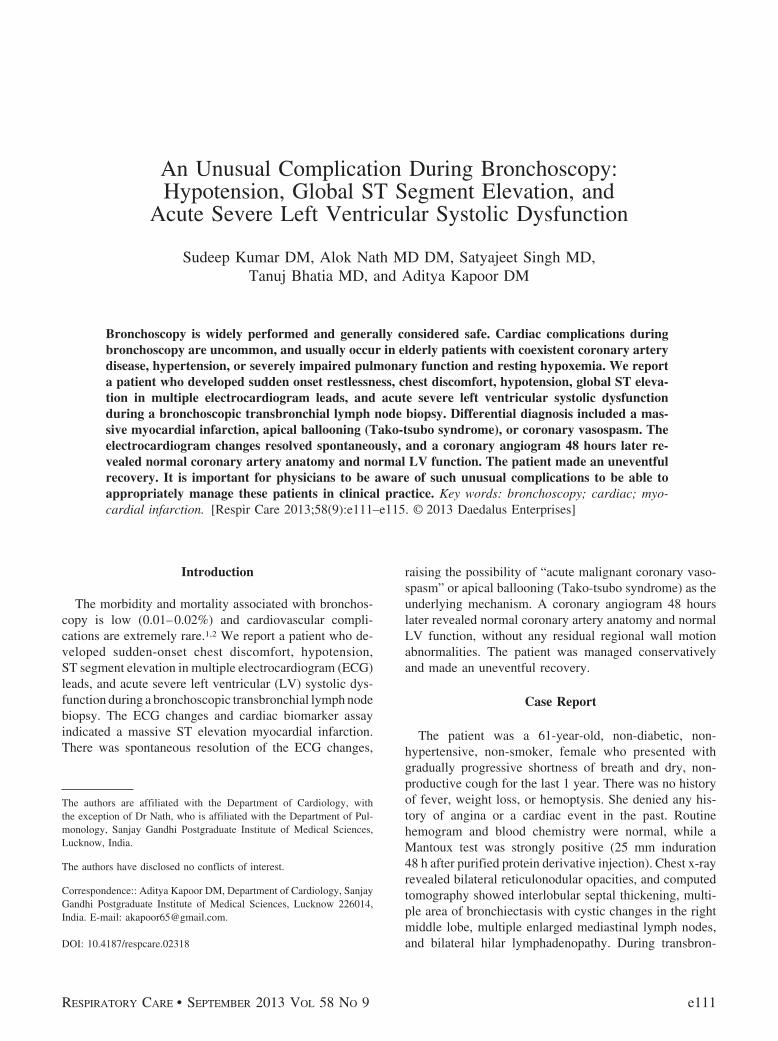
In view of the ECG changes and likelihood of massive,acute myocardial infarction, a cardiology opinion wassought for consideration of transfer to the cardiac cathe-terization laboratory and urgent coronary angiography. Achest x-ray revealed no evidence of pneumothorax or pneu-momediastinum. At the time of examination by the cardi-ology team she was conscious, oriented, and denied anyongoing chest pain. Another 12 lead ECG at this timeinterestingly revealed slight reduction in the ST elevation,especially in the inferior leads and precordial leads V4-V6(Fig. 2).
An urgent bedside echocardiography revealed severeglobal hypokinesia and an overall LV ejection fraction of30%. Although a peri-procedural myocardial infarctionwas still the most likely possibility, in view of the spon-taneous reduction in the ST segment elevation, a likeli-hood of “malignant coronary vasospasm” was also nowconsidered. While the cardiac catheterization laboratory
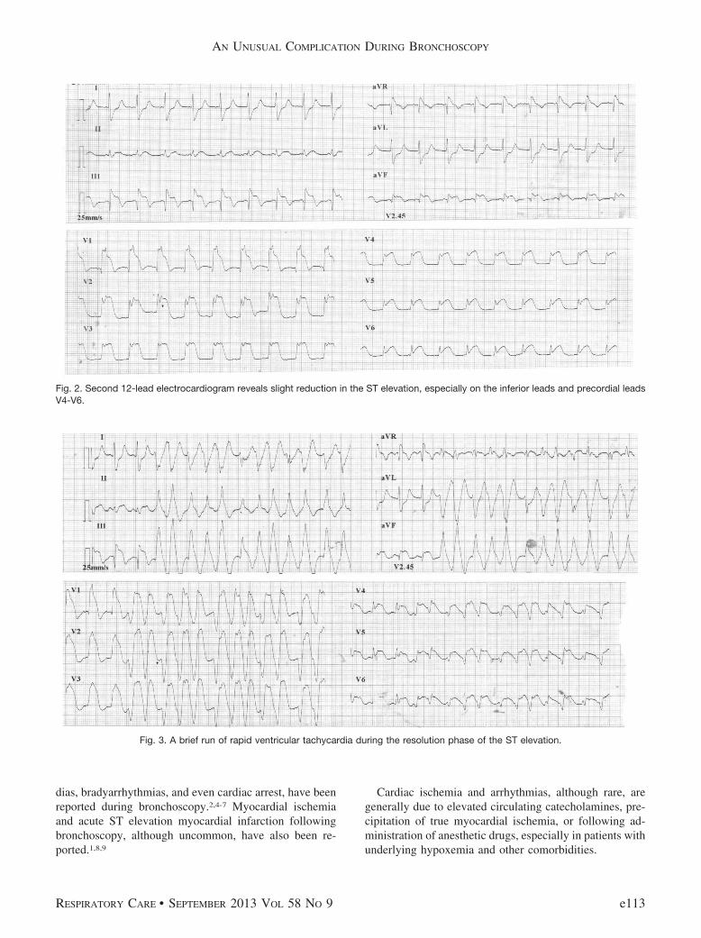
team was on standby, it was decided to perform serialECGs and observe the temporal pattern of ST segmentevolution before performing an urgent coronary angio-gram. As the ST segment elevation gradually resolved, abrief run of rapid ventricular tachycardia was noted (Fig. 3),which was managed successfully with a bolus of amioda-rone (150 mg intravenous). Serial ECGs over the next30 min revealed progressive resolution of the ST segmentelevation and normalization of the ST segment (Fig. 4)
Cardiac biomarker assay expectedly revealed elevatedtroponin-I (9.2 ng/mL) and creatine kinase-myocardialband (CKMB) (12 ng/mL), which further escalated to80 ng/mL and 178 ng/mL, respectively, at 6 hours. Shemade a rapid, uneventful recovery over the next 48 hours.A coronary angiogram 48 hours later revealed normalcoronary arteries (Fig. 5). The LV angiogram also re-vealed normal LV function, without any regional wall mo-tion abnormalities (Fig. 6), therefore suggesting possibleacute coronary vasospasm as the etiology of the global STelevation.
Discussion
Cardiac complications, such as arrhythmia, myocardialischemia, and infarction, during bronchoscopy are un-common, and generally occur in elderly patients with co-morbidities such as preexisting coronary artery disease,hypertension, or severe impairment of pulmonary functionand resting hypoxemia.3,4 Various cardiac arrhythmias,ranging from atrial and ventricular premature complexesor supraventricular tachycardias to ventricular tachycar-
Fig. 1. Gross ST elevation on the inferior and anterior precordial leads of the 12-lead electrocardiogram.
AN UNUSUAL COMPLICATION DURING BRONCHOSCOPY
e112 RESPIRATORY CARE • SEPTEMBER 2013 VOL 58 NO 9
dias, bradyarrhythmias, and even cardiac arrest, have beenreported during bronchoscopy.2,4-7 Myocardial ischemiaand acute ST elevation myocardial infarction followingbronchoscopy, although uncommon, have also been re-ported.1,8,9
Cardiac ischemia and arrhythmias, although rare, aregenerally due to elevated circulating catecholamines, pre-cipitation of true myocardial ischemia, or following ad-ministration of anesthetic drugs, especially in patients withunderlying hypoxemia and other comorbidities.
Fig. 2. Second 12-lead electrocardiogram reveals slight reduction in the ST elevation, especially on the inferior leads and precordial leadsV4-V6.
Fig. 3. A brief run of rapid ventricular tachycardia during the resolution phase of the ST elevation.
AN UNUSUAL COMPLICATION DURING BRONCHOSCOPY
RESPIRATORY CARE • SEPTEMBER 2013 VOL 58 NO 9 e113

The ECG changes in our patient suggested a massivemyocardial infarction, which was corroborated by the el-evated cardiac biomarker levels. We initially planned anurgent coronary angiogram (in view of the ECG findings,hypotension, and echocardiographic evidence of LV dys-function), but because of the spontaneous resolution of theECG changes (in absence of any fibrinolysis or coronaryintervention), the coronary angiogram was deferred, andserial ECGs revealed complete normalization of the STchanges. Had the ECGs not shown this spontaneous res-olution or had her chest pain continued, urgent coronaryangiogram would definitely have been performed, and the
cardiac catheterization laboratory team was on continuousstandby.
The pathogenesis was believed to be either apical bal-looning (Tako-tsubo syndrome) or “acute malignant coro-nary vasospasm.” Apical ballooning is characterized bysudden, severe, transient, segmental dysfunction of the LVdue to a hyper-adrenergic state, usually following intenseemotional or physical stress. The clinical presentation sim-ulates that of an acute myocardial infarction, with ST el-evation in the ECG and rapid development of acute LVsystolic dysfunction.10,11 In these patients coronary an-giography typically reveals normal coronary anatomy andimpaired LV systolic function (apical ballooning), whichrapidly resolves, without any residual impairment. Guer-rero et al recently reported the occurrence of Tako-tsubosyndrome following bronchoscopy.12 However, in contrastto our patient, their patient had no important ECG changesor troponin rise, although coronary angiography confirmednormal coronary anatomy with severe LV dysfunction.
Due to the presence of the completely normal LV func-tion, without any regional wall motion abnormality on theLV angiogram, we hypothesized that “acute malignant cor-onary vasospasm” could be the underlying pathophysiol-ogy of the global ST elevation in our patient. The diffusecoronary vasospasm may have resulted in global myocar-dial ischemia, ST elevation, and acute severe LV systolicdysfunction. It is possible that our patient had Tako-tsubosyndrome and the LV function had completely recoveredby the time of the coronary angiography. However, the LVwas initially globally hypokinetic in our patient, while incases of apical ballooning syndrome, apical akinesia ordyskinesia with preserved or hyperkinetic contractile basalLV segments are more typical.
This case highlights the occurrence of an extensivepseudo-myocardial infarction pattern as a complication ofbronchoscopy, in a patient without underlying coronaryartery disease, possibly secondary to “acute malignant cor-onary vasospasm.” The case highlights the fact that suchlife-threatening complications can occur during bronchos-copy. Since it is not possible to predict the occurrence ofsuch complications, routine ECG (especially for elderly/
Fig. 4. Progressive resolution of the ST segment elevation and normalization of the ST segment.
Fig. 5. Coronary angiogram reveals normal coronary arteries. Left:Left coronary artery. Right: Right coronary artery.
Fig. 6. Left ventricular angiogram shows normal left ventricularfunction, with no regional wall motion abnormalities. Left: Diastolicframe. Right: Systolic frame.
AN UNUSUAL COMPLICATION DURING BRONCHOSCOPY
e114 RESPIRATORY CARE • SEPTEMBER 2013 VOL 58 NO 9

high-risk patients) during bronchoscopy is important forearly diagnosis and appropriate management of such ad-verse effects.
REFERENCES
1. Credle WF Jr, Smiddy JF, Elliot RC. Complications of fiberopticbronchoscopy. Am Rev Respir Dis 1974;109(1):67-72.
2. Surratt PM, Smiddy JF, Gruber B. Deaths and complications asso-ciated with fiberoptic bronchoscopy. Chest 1976;69(6):747-751.
3. Cavaliere S, Foccoli P, Farina PL. Nd:YAG Laser bronchoscopy. Afive-year experience with 1,396 applications in 1,000 patients. Chest1988;94(1):15-21.
4. Davies L, Mister R, Spence DP, Calverley PM, Earis JE, PearsonMG. Cardiovascular consequences of fibreoptic bronchoscopy. EurRespir J 1997;10(3):695-698.
5. D’Aloia A, Faggiano P, Fiorina C, Vizzardi E, Cavaliere S, FoccoliP, et al. Cardiac arrest due to ventricular fibrillation as a complica-tion occurring during rigid bronchoscopic laser therapy. MonaldiArch Chest Dis 2003;59(1):88-90.
6. Elguindi AS, Harrison GN, Abdulla AM, Chaudhary BA, Vallner JJ,Kolbeck RC, Speir WA Jr. Cardiac rhythm disturbances during fi-
beroptic bronchoscopy: a prospective study. J Thorac CardiovascSurg 1979;77(4):557-561.
7. Katz AS, Michelson EL, Stawicki J, Holford FD. Cardiac arryth-mias: frequency during fiberoptic bronchoscopy and correlation withhypoxemia. Arch Intern Med 1981;141(5):603-606.
8. Matot I, Drenger B, Glantz L, Kramer MR. Coronary spasm duringoutpatient fiberoptic laser bronchoscopy. Chest 1999;115(6):1744-1746.
9. Osula S, Stockton P, Abdelaziz MM, Walshaw MJ. Intratrachealcocaine induced myocardial infarction: an unusual complication offibreoptic bronchoscopy. Thorax 2003;58(8):733-734.
10. Gianni M, Dentali F, Grandi AM, Sumner G, Hiralal R, Lonn E.Apical ballooning syndrome or takotsubo cardiomyopathy: a sys-tematic review. Eur Heart J 2006;27(13):1523-1529.
11. Kurowski V, Kaiser A, von Hof K, Killermann DP, Mayer B,Hartmann F. Apical and midventricular transient left ventricular dys-function syndrome (Tako-tsubo cardiomyopathy): frequency, mech-anisms, and prognosis. Chest 2007;132(3):809-816.
12. Guerrero J, Majid A, Ernst A. Cardiogenic shock secondary to Tako-tsubo syndrome after debridement of malignant endobronchial ob-struction. Chest 2009;135(1):217-220.
AN UNUSUAL COMPLICATION DURING BRONCHOSCOPY
RESPIRATORY CARE • SEPTEMBER 2013 VOL 58 NO 9 e115