an overview of several inhibitors for alzheimer s disease
TRANSCRIPT

International Journal of
Molecular Sciences
Review
An Overview of Several Inhibitors for Alzheimer’s Disease:Characterization and Failure
Subramanian Boopathi 1, Adolfo B. Poma 2,3 and Ramón Garduño-Juárez 1,*
�����������������
Citation: Boopathi, S.; Poma, A.B.;
Garduño-Juárez, R. An Overview of
Several Inhibitors for Alzheimer’s
Disease: Characterization and Failure.
Int. J. Mol. Sci. 2021, 22, 10798.
https://doi.org/10.3390/
ijms221910798
Received: 28 August 2021
Accepted: 3 October 2021
Published: 6 October 2021
Publisher’s Note: MDPI stays neutral
with regard to jurisdictional claims in
published maps and institutional affil-
iations.
Copyright: © 2021 by the authors.
Licensee MDPI, Basel, Switzerland.
This article is an open access article
distributed under the terms and
conditions of the Creative Commons
Attribution (CC BY) license (https://
creativecommons.org/licenses/by/
4.0/).
1 Instituto de Ciencias Físicas, Universidad Nacional Autónoma de México, Cuernavaca 62210, Mexico;[email protected]
2 Department of Biosystems and Soft Matter, Institute of Fundamental Technological Research Polish Academyof Science, Pawinskiego 5B, 02-106 Warsaw, Poland
3 International Center for Research on Innovative Biobased Materials (ICRI-BioM)—International ResearchAgenda, Lodz University of Technology, Zeromskiego 116, 90-924 Lodz, Poland;[email protected]
* Correspondence: [email protected]
Abstract: Amyloid beta (Aβ) oligomers are the most neurotoxic aggregates causing neuronal deathand cognitive damage. A detailed elucidation of the aggregation pathways from oligomers tofibril formation is crucial to develop therapeutic strategies for Alzheimer’s disease (AD). Althoughexperimental techniques rely on the measure of time- and space-average properties, they face severedifficulties in the investigation of Aβ peptide aggregation due to their intrinsically disorder character.Computer simulation is a tool that allows tracing the molecular motion of molecules; hence itcomplements Aβ experiments, as it allows to explore the binding mechanism between metal ionsand Aβ oligomers close to the cellular membrane at the atomic resolution. In this context, integratedstudies of experiments and computer simulations can assist in mapping the complete pathways ofaggregation and toxicity of Aβ peptides. Aβ oligomers are disordered proteins, and due to a rapidexploration of their intrinsic conformational space in real-time, they are challenging therapeutictargets. Therefore, no good drug candidate could have been identified for clinical use. Our previousinvestigations identified two small molecules, M30 (2-Octahydroisoquinolin-2(1H)-ylethanamine)and gabapentin, capable of Aβ binding and inhibiting molecular aggregation, synaptotoxicity,intracellular calcium signaling, cellular toxicity and memory losses induced by Aβ. Thus, werecommend these molecules as novel candidates to assist anti-AD drug discovery in the near future.This review discusses the most recent research investigations about the Aβ dynamics in water, closecontact with cell membranes, and several therapeutic strategies to remove plaque formation.
Keywords: Alzheimer’s disease; amyloid β peptide; plaque formation; small molecules; M30;gabapentin; MD simulation
1. Introduction
Approximately 50 million people are globally affected by Alzheimer’s disease (AD) [1,2].This number will increase to 150 million by 2050 unless new prevention treatments becomeavailable [1]. Amyloid plaques and neurofibrillary tangles in brain tissue are the main hall-marks of AD. Amyloid plaques are composed of amyloid β (Aβ) peptides. Neurofibrillarytangles are composed of hyperphosphorylated tau proteins. In 1992, Hardy and Higgins [2]developed the amyloid cascade hypothesis; Aβ aggregates are transformed into Aβ fibrilsthat accumulate in the brain and finally trigger neurodegeneration.
Amyloid precursor protein (APP) gene generates three variant APP695, APP751 andAPP770, which are produced in neurons, endothelial cells, and platelets, respectively. In theaggregation pathway, transmembrane APP695 is cleaved by β- and γ-secretase to generatesAβ1-40 and Aβ1-42 peptides. During the APP695 cleavage, two processes occur (Figure 1).(1) Aβ is released to the extracellular hydrophobic environment, where Aβ monomers
Int. J. Mol. Sci. 2021, 22, 10798. https://doi.org/10.3390/ijms221910798 https://www.mdpi.com/journal/ijms

Int. J. Mol. Sci. 2021, 22, 10798 2 of 31
assemble into dimers, trimers, tetramers, oligomers, and fibrils [3–6], Aβ fibrils constitutethe amyloid plaques considered a major pathological hallmark of AD. (2) A small amount ofAβ remains above the cellular membrane and can form membrane-associated Aβ oligomersthat disrupt the shape of the membrane [7]. The amyloid fibrils are generally insoluble andtransform into plaques. The Aβ oligomers are soluble and mainly spread throughout ADaffected brain. Soluble Aβ oligomers deposited approximately 3–4 kDa in the AD braincould affect the calcium ion channel activity in synapsis through disrupting nerve signaltransmission and damage mitochondrial causes to increase free radial lead to cell death.Soluble oligomers reach 10–100 kDa, which is considered more cytotoxicity than amyloidfibril aggregation; thus, the soluble oligomers exposed higher toxicity when comparedwith an insoluble fibril structure [8]. The water-mediated attraction in Aβ peptides andhigh propensity favor the formation of insoluble amyloid fibrils. The oligomers and fibrilsconformation are recognized as a generic antibody epitope [9]. Although the relationshipbetween oligomers and fibrils is still under debate, soluble and insoluble Aβ structureshave been targeted to develop a cure for AD.
Figure 1. Plaques are around the neurons and tangles inside the neurons. Plaques and tangles are involved in killingthe neurons resulting in drastic shrinking of the brain compared to normal brain. β-secretase cleaves APP (composed of695 amino acids) into the membrane-tethered C-terminal fragments β (CTFβ or C99) and N-terminal sAPPβ. Aβ is obtainedafter the sequential cleavage of CTFβ by γ-secretases. Upon APP cleavage, two mechanism pathways have been proposed,in the first, Aβ is released to the extracellular environment, due to its hydrophobic nature, formed Aβ aggregates, in thesecond, a small amount of Aβ remains in the membrane evolving into membrane-associated Aβ aggregates. Aβ peptidehas divided into five regions, N-terminus or hydrophilic region represent in grey, Central hydrophobic region in green,Loop region in red, second hydrophobic region in blue and C-terminal in orange. AD brain shrunken as compared withHealthy one.
Molecular insight into aggregation pathways from oligomers to fibril formation re-mains an open problem in amyloidogenesis. A recent study reported [10] that during theaggregation, a large majority of oligomer structures are unstable and dissociate into theirmonomers instead of forming a new fibril structure, while the minority of the oligomers
Int. J. Mol. Sci. 2021, 22, 10798 3 of 31
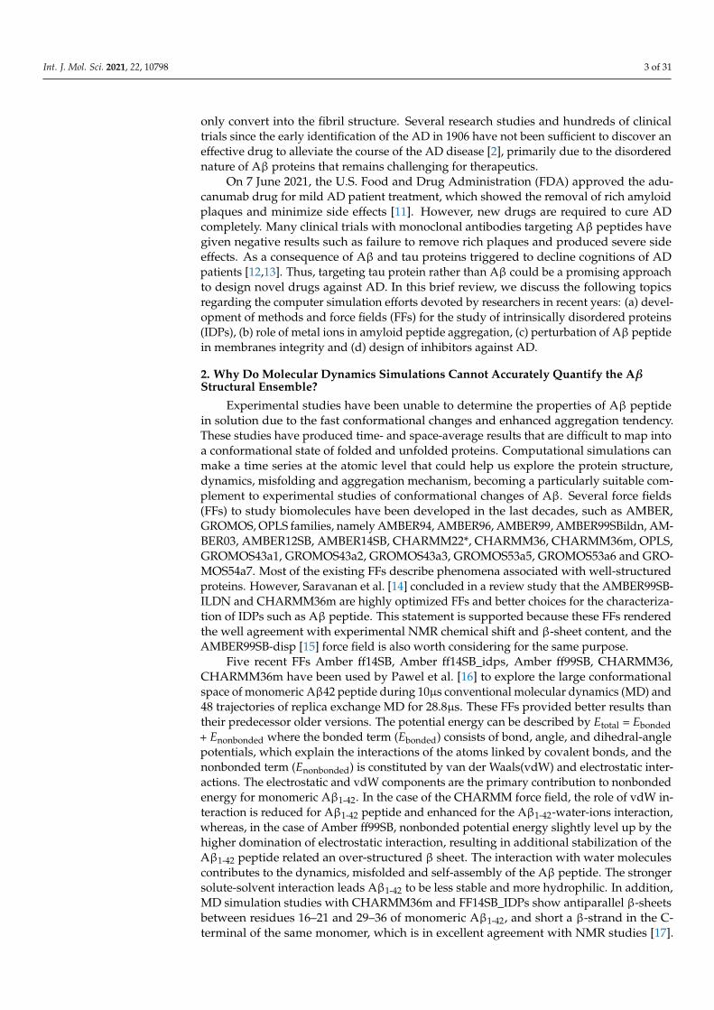
only convert into the fibril structure. Several research studies and hundreds of clinicaltrials since the early identification of the AD in 1906 have not been sufficient to discover aneffective drug to alleviate the course of the AD disease [2], primarily due to the disorderednature of Aβ proteins that remains challenging for therapeutics.
On 7 June 2021, the U.S. Food and Drug Administration (FDA) approved the adu-canumab drug for mild AD patient treatment, which showed the removal of rich amyloidplaques and minimize side effects [11]. However, new drugs are required to cure ADcompletely. Many clinical trials with monoclonal antibodies targeting Aβ peptides havegiven negative results such as failure to remove rich plaques and produced severe sideeffects. As a consequence of Aβ and tau proteins triggered to decline cognitions of ADpatients [12,13]. Thus, targeting tau protein rather than Aβ could be a promising approachto design novel drugs against AD. In this brief review, we discuss the following topicsregarding the computer simulation efforts devoted by researchers in recent years: (a) devel-opment of methods and force fields (FFs) for the study of intrinsically disordered proteins(IDPs), (b) role of metal ions in amyloid peptide aggregation, (c) perturbation of Aβ peptidein membranes integrity and (d) design of inhibitors against AD.
2. Why Do Molecular Dynamics Simulations Cannot Accurately Quantify the AβStructural Ensemble?
Experimental studies have been unable to determine the properties of Aβ peptidein solution due to the fast conformational changes and enhanced aggregation tendency.These studies have produced time- and space-average results that are difficult to map intoa conformational state of folded and unfolded proteins. Computational simulations canmake a time series at the atomic level that could help us explore the protein structure,dynamics, misfolding and aggregation mechanism, becoming a particularly suitable com-plement to experimental studies of conformational changes of Aβ. Several force fields(FFs) to study biomolecules have been developed in the last decades, such as AMBER,GROMOS, OPLS families, namely AMBER94, AMBER96, AMBER99, AMBER99SBildn, AM-BER03, AMBER12SB, AMBER14SB, CHARMM22*, CHARMM36, CHARMM36m, OPLS,GROMOS43a1, GROMOS43a2, GROMOS43a3, GROMOS53a5, GROMOS53a6 and GRO-MOS54a7. Most of the existing FFs describe phenomena associated with well-structuredproteins. However, Saravanan et al. [14] concluded in a review study that the AMBER99SB-ILDN and CHARMM36m are highly optimized FFs and better choices for the characteriza-tion of IDPs such as Aβ peptide. This statement is supported because these FFs renderedthe well agreement with experimental NMR chemical shift and β-sheet content, and theAMBER99SB-disp [15] force field is also worth considering for the same purpose.
Five recent FFs Amber ff14SB, Amber ff14SB_idps, Amber ff99SB, CHARMM36,CHARMM36m have been used by Pawel et al. [16] to explore the large conformationalspace of monomeric Aβ42 peptide during 10µs conventional molecular dynamics (MD) and48 trajectories of replica exchange MD for 28.8µs. These FFs provided better results thantheir predecessor older versions. The potential energy can be described by Etotal = Ebonded+ Enonbonded where the bonded term (Ebonded) consists of bond, angle, and dihedral-anglepotentials, which explain the interactions of the atoms linked by covalent bonds, and thenonbonded term (Enonbonded) is constituted by van der Waals(vdW) and electrostatic inter-actions. The electrostatic and vdW components are the primary contribution to nonbondedenergy for monomeric Aβ1-42. In the case of the CHARMM force field, the role of vdW in-teraction is reduced for Aβ1-42 peptide and enhanced for the Aβ1-42-water-ions interaction,whereas, in the case of Amber ff99SB, nonbonded potential energy slightly level up by thehigher domination of electrostatic interaction, resulting in additional stabilization of theAβ1-42 peptide related an over-structured β sheet. The interaction with water moleculescontributes to the dynamics, misfolded and self-assembly of the Aβ peptide. The strongersolute-solvent interaction leads Aβ1-42 to be less stable and more hydrophilic. In addition,MD simulation studies with CHARMM36m and FF14SB_IDPs show antiparallel β-sheetsbetween residues 16–21 and 29–36 of monomeric Aβ1-42, and short a β-strand in the C-terminal of the same monomer, which is in excellent agreement with NMR studies [17].
Int. J. Mol. Sci. 2021, 22, 10798 4 of 31
AMBER_ff14SB and AMBER_ff99SB overestimated α-helical and β-contents, respectively.Pawel et al. [16] strongly recommended using CHARMM36m force field for the study ofthe Aβ42-water-ion complex system over the AMBER FFs.
It is a big challenge to determine an accurate description of the structure of IDPsthrough MD simulations based only on FFs. In this perspective, Chong et al. [18] reviewedadvanced computational methods that employ protein configuration entropy and rendera thermodynamic connection between structural disorder and protein properties. Forexample, the CHARMM and OPLS FFs exhibit lower average β-sheet content in dimers ofAβ42 than that obtained with GROMOS 53a6 force field [18]. Subsequently, the averageβ-sheet content of the Aβ42 dimers was found to be greater in OPLSAA [19] than in AM-BERFF99SB [20]. Interestingly, both AMBER99SB-ILDN and OPLS/L FFs have producedresults of the average secondary structure of Aβ42 tetramer similar to each other [21].
The structural and thermodynamics properties of IDPs are susceptible to solute-solvent interaction compared to the folded protein. The choice of a reliable water model isnecessary to characterize the Aβ42 peptide. Chong et al. [22] performed an MD simulationto investigate the structural properties of Aβ1-42 peptide by employing AMBER ff99SBforce field with different solvent water models. They demonstrated that TIP4P-Ew exposedmore Aβ1-42-water interaction than conventional TIP3P water model [18]. They stronglyencouraged using the TIP4P-Ew water model to investigate the Aβ1-42 peptide structuralproperties. In a review, Chong et al. [18] reported that existing FFs are insufficient toexpose Aβ protein to water. Recently, the same problem has been addressed by a coupleof groups. The first group [23] opted to scale the Lennard-Jones potential between atomsin proteins and oxygen atoms in water by factor 1.1 without disturbing water-water andwater-protein interaction. The second group [24] introduced a new water model, TIP4P-D, which included an additional parameter in the TIP4P water model to overcome thedeficiencies in water dispersion interaction.
Recently developed FFs and their default water model are tabulated in Table 1.Rahman et al. [25] have evaluated the accuracy of recent developed FFs ff99IDPs, ff14IDPs,ff14IDPSFF, ff03w, CHARMM36m, and CHARMM22* by performing MD simulationsfor two short peptides (HEWL19 and RS), five IDPs (HIV-rev, Aβ40, Aβ42
1Z0Q, Aβ42model,
and pdE-γ) and two folded proteins (CspTm and ubiquitin) using trajectories of 1, 1.5, 5or 10 µs for each system. They have compared J-coupling between MD simulation andNMR experiment for folded and disordered protein using different FFs. The J-coupling(J3-HNH2 and J3-HαC) parameter measures the secondary structure distribution basedon ϕ backbone dihedral angle. Three IDPs FFs, ff99IDPs, ff14IDPSFF, ff14IDPs were ingood agreement with the experimental J-coupling constant compared with tested FFs ff03w,CHARMM36m, and CHARMM22*. The balance between the local structural property(NMR chemical shift) and global structural property (Rg) is still a challenging issue formolecular simulations of IDPs. Rahman et al., [25] noted two observations: (1) averageRg for Aβ421Z0Q is 12.1 Å which is in close agreement with experimental Rg 12.4 Å, whileff03w showed Rg equal to 10.53 Å and CHARMM36m displayed Rg about 13 Å, whichsuggests highly divergence among those FFs; (2) the three IDPs FFs render a good balancebetween secondary structures contents for both Aβ40 and Aβ42
model. While Amberff03w,CHARMM36m and CHARMM22* overestimated the α-helical structure for IDPs, thusfavouring folded protein structures. Therefore, the three specific IDPs FFs were developedby incorporating the changes made in the pre-existing FFs (Table 1) to enable an accuratedescription of the folded and misfolded proteins [15,26–35].
Int. J. Mol. Sci. 2021, 22, 10798 5 of 31
Table 1. Latest developed force field for intrinsically disordered protein and water model.
Force Field Parameter Set Developments Water Model Reference
AMBER
ff99IDPsUpdated from ff99SBildn by adding a set of
backbone torsion parameters of eightdisordered promoting amino acids.
TIP3P Wei Y et al. [26] 2015
ff14IDPsUpdated from ff14SB by embedding a set of
backbone torsion parameters of eightdisordered promoting amino acids.
TIP3P Song et al. [28] 2017
ff14IDPSFFUpdated from ff14SB by introducing a set of
backbone torsion parameters for 20amino acids
TIP3P Song et al. [29] 2017
ff03CMAP Updated from ff03 by introducing a correctionmaps (CMAP)-optimized force field
TIP4PD(Modified the
dispersion interactionof the TIP4P)
Zhang et al. [30] 2019
ff14SBUpdated from ff99SB by improving the
Accuracy of Protein Side Chain andBackbone Parameters
TIP3P Maier et al. [31] 2015
ff03w Updated from ff03 by adding slightbackbone modification TIP4P/2005 Best et al. [32] 2010
A99SB_dispUpdate from a99SB-ILDN by an introducingsmall change in the protein and water vdW
interaction termsTIP4P-D Robustelli et al. [15] 2018
CHARMM
CHARMM36m Updated from CHARMM36 by a refinedbackbone correction map potential CHARMM-modified TIP3P Huang et al. [33] 2017
CHARMM36IDPSFFUpdated from CHARMM36m by CMAP
corrections made for all 20 naturally occurringamino acids
CHARMM-modified TIP3P Liu H et al. [34] 2019
CHARMM22* Updated from CHARMM by introducingmodifications in backbone torsion potential CHARMM-modified TIP3P Stefano Piana et al. [35] 2011
CHARMM36mW Van der Waals interaction between proteinand water are included in CHARMM36m CHARMM-modified TIP3P Samantray et al. [27] 2020
The inconsistency of empirical physical models used in MD techniques can impactFFs and water models that affect the simulation result’s accuracy. Researchers have strivedhard to develop perfect FFs to improve IDPs description; they aim to describe the highflexibility of these proteins, thus enlarging the conformational ensemble and increase thepossibility of locating them in different local minima. Mu et al. [36] reported a couple ofideas to improve the accuracy of FFs for IDPs structural characterization, (1) Modificationof force field parameters aided by global optimization, and (2) Maintaining a good balancebetween secondary structure via reparameterization (backbone dihedral parameters andvdW interaction between water-protein interaction) of existing FFs. One of the most com-mon problems among the IDPs force field is over-stabilizing protein-protein interactionthat impacts the aggregation mechanism of IDPs. Due to the IDPs force field’s inaccuracy,Mu et al. [36] encouraged improving backbone dihedral parameters and Lennard-Jonespotential parameter (protein-water interaction) in the existing IDPs FFs and obtained train-ing data from experimental observation and quantum chemical calculation. Undoubtedly,both reparameterization and training strategies may assist in new FFs development.
Another option to the atomistic FFs is to employ state-of-the-art coarse-grained (CG)models that not only sample more efficiently the entire space of protein conformationsfor large systems but also allow simulations for longer time scales of hundreds of µs [37],generally forbidden by brute force all-atom MD simulation and very relevant for biologicalprocesses [38] (e.g., folding, allosteric communications, conformational changes undermutations, self-assembly process, etc). Some popular CG FFs such as UNRES has beenemployed to study the fibril formations initiated by templates of Aβ40 fragments [39]capturing the dock-lock mechanism and similar the crowding effect of fragments wasstudied by PRIMO CG FF [40]. MARTINI 2 was employed to unveil the aggregation andorganization of short Aβ16-22 peptides in lipid membranes [41]. The abovementioned CGFFs capture processes in time scales inaccessible atomic FFs, but yet they were restrictedby system size or a lack of flexibility in secondary structure transitions. In this regard, the

Int. J. Mol. Sci. 2021, 22, 10798 6 of 31
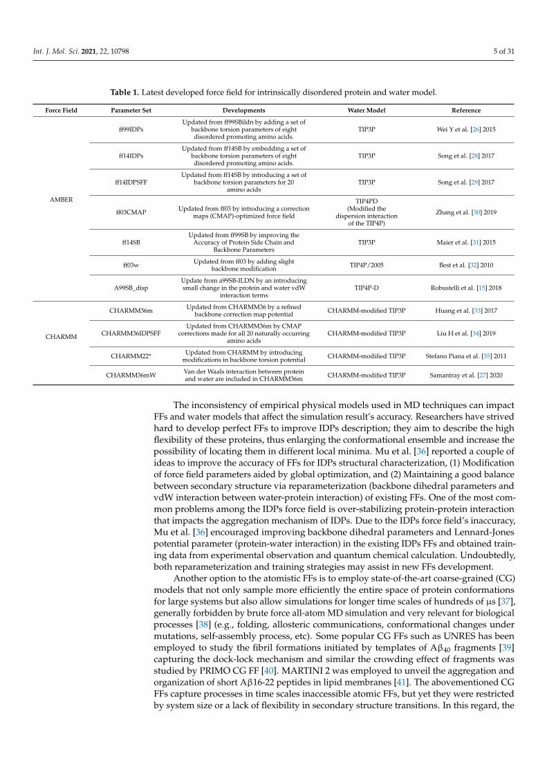
new release of MARTINI 3 force field aided by Go-Like model [42–45] can become a newtool for realistic exploration of full-length Aβ peptide aggregation in contact with complexlipid-cholesterol membranes. Marrink group has made large efforts to develop a libraryof different lipid species (i.e., about 63 types) consistent with human plasma cells [46].Following this idea, Poma et al. [47] have employed a very simplified CG model [48] tounveil the mechanical properties of Aβ40 and Aβ42 fibrils under different mechanicaldeformation process (e.g., tensile, indentation and shearing stresses) [49] and more recentlythey investigated [50] the change in mechanical stability between the oligomer and maturedfibrils. The soluble oligomers are characterized by a length size about 3 to 5 nm, whereasAβ fibrils typically reach hundreds of nm (see Figure 2). It is well-known the inverserelationship between toxicity and the length of the Aβ assembly [51]. Oligomers areconsidered more toxic than fibrils because of their high degree of flexibility toward lowmolecular weights, and the possibility of forming hydrophobic structures that may impaircell functions. Instead, fibrils are more thermodynamic stable and stiffer and less capableto undergo transitions to smaller and more toxic assemblies. Over the years, still somequestions still remain open in terms of the mechanical characterization in amyloidogenesisand Aβ aggregate maturation: (a) what is the major role of the mechanical stability duringAβ oligomerization (e.g., tetramer, hexamer, etc)? (b) is the toxicity of oligomers whichare closer to the membrane correlated by a minimum of mechanostability of Aβ peptidecomplexes, (c) how is the gain in mechanical stability from oligomers to fibrils involvedin disease progression? and (d) Can we devise new strategies to reduce the mechanicalstability of oligomer-to-fibril step through small molecular breakers (i.e., drugs recognitionprocess)? To provide new answers to those open problems, new research combiningversatile CG FFs and single molecule force spectroscopy is highly advised. For instance,in MD simulations, one can trace hydrophobic native interactions in equilibrium andsimultaneously during a deformation process. Hence, the idea of hydrophobic structurescan find support in molecular simulations as the main driving force for cell damage.Furthermore, molecular pathways could elucidate the critical conformation that maintainsthe mechanical stability of the Aβ assembly in the single-molecule force spectroscopyexperiment. Figure 2 depicts the most relevant structure during aggregation and currentmethodologies used for its computational characterization [50,52].
Samantray et al. [35] have examined recently developed IDPs FFs, namely AMBER99SB-disp,CHARMM36m, and CHARMM36 with enhanced protein-water interactions (CHARMM36mW)for the study of Aβ16-22 (wildtype) aggregation and its mutation F19L Aβ16−22 (mutation 1) andF19 V/F20 V Aβ16−22 (mutation2), as model systems for testing purpose. In AMBER99-disp,the peptide-water interactions are increased too much resulting in an inhibition of the Aβ16-22aggregation. The same trend has been observed in the simulation with the AMBERFF03w forcefield [53]. The difference between CHARMM36m and CHARMM36mW is a reparameterizationof the protein-water interaction. In contrast, an experimental study [54] reported the followingaggregation order mutation1>wildtype>mutation2≈0. AMBER99-disp does not apply for Aβaggregation process because the interactions between peptide and water are drastically increased,leading to inhibition of the aggregation pathway. The CHARMM36mW can provide aggre-gation rate in the order of wildtype>mutation1>mutation2. Thus, FFs cannot reproduce theaggregation of Aβ peptide observed in experiments, but they maintain a better balance betweenpeptide-water and peptide-peptide interaction. These results imply that improving the force fieldsignificantly impacted the simulation aggregation pathway than modifying the protein sequence.Samantray et al. [35] strongly encourage the use of CHARMM36mW for studying a full-lengthAβ, even though it is not a perfect force field, since it yielded a promising result for aggregationbenchmark. Nevertheless, reparameterization of this specific force field is still required.
Int. J. Mol. Sci. 2021, 22, 10798 7 of 31
Figure 2. Representation of Aβ structures during aggregation: free Aβ peptide, oligomers, fibrils and plaques. Typicallengths are given for peptide (<3 nm), oligomers (3–5 nm) and fibrils (20–100 nm). All-atom MD (AA-MD), coarse-grainedMD (CG-MD) and continuum models have been employed to unveil the mechanical stability for each structure. Youngmodulus (Y) defined by the ratio of applied tensile stress to a given strain provides an idea of the elastic regime. Y valuesfor each structure is taken from ref (50 and 51). Some images are modified with permission of BrightFocus Foundation.
Lockhart et al. [55] performed REMD simulations to examine the impact of the threepopular FFs, CHARMM22 (protein FF) with CHARMM36 (lipid FF), CHARMM36m (pro-tein FF) with CHARMM36 (lipid FF), and Amber14SB (protein FF) with Lipid14 (lipidFF), for the binding mechanism between the Aβ10-40 and the Dimyristoylgylcerophos-phocholine (DMPC) bilayers. These three FFs have shown similar results in subjects like(a) stable helix formed in C-terminal of the peptide, (b) C-terminal of the peptide insertedinto the bilayer hydrophobic core, (c) the thickness of the bilayer induced by the peptideabout 10 Å and d) the disordered effect induced by the peptide on the fatty acid tails in theDMPC lipids. Nevertheless, these three FFs yielded different conformation ensembles ofthe peptide and bilayer that do not disturb the binding of the peptide with the bilayers.
Coskuner et al. [56] have reviewed several studies extensively and suggested thatwidely used FFs CHARMM, AMBER, GROMOS and OPLS, cannot produce accurate resultsfor disordered entities. Even more, all existing computational techniques were designedto describe phenomena in ordered protein systems rather than in disordered proteinstructures. Important to mention that, Density Functional Theory (DFT) suffered from anumber of errors that originated in the approximation of exchange-correlation functionals.These errors have been identified as the underestimation of barriers in describing chemicalreactions, the band gaps, charge transfer excitation energies, and binding energies of chargetransfer species in a biomolecule. DFT is the basis for constructing FFs such as CHARMM,AMBER, GROMOS and OPLS for intrinsically disordered protein and their complexeswith ligands. These FFs are associated with an approximate exchange-correlation functionthat may lead to mistakes during prediction of the structural properties of IDPs. Notably,overcoming deficiencies of the exchange-correlation functional in DFT will help to improvethe accuracy of IDP FFs.
3. Why Does Aβ Peptide Perturb Membrane Integrity?
Three models [13] have been proposed for small protein insertion in lipid bilayers,(a) membrane pore formation model: Aβ oligomers formed within the membrane facilitateswell-defined pores that lead to an unbalanced flow of ions in and out of the membranes,
Int. J. Mol. Sci. 2021, 22, 10798 8 of 31
(b) carpeting model: Aβ peptide contacts the membrane surface to produce asymmetricpressure between two lipid layers of the membrane, which causes small molecules leakage,(c) detergent-like effect model: the lipids leave from the cellular membranes interactswith the Aβ peptide in resulting from perturbing membrane integrity. In this context,these models perturb the membrane through the interaction of Aβ peptide that yieldsto cell toxicity. Computer simulations assist in examining these models to unveil themolecular mechanism of the Aβ-membranes interaction. In general, three fundamentalquestions have not been elucidated yet, (1.) How does Aβ oligomeric intermediate perturbmembrane integrity? (2.) What are the specific residues involved in membranes-Aβinteraction? (3.) What is the impact of metal ions in the mechanism of interaction betweenAβ and membranes?
3.1. Aβ Monomer-Membrane
Although the Aβ peptides monomers were dissolved in non-polar environment, wa-ter solution and micelles environment using NMR techniques, there are no experimentalstudies on the 3D structure of monomers within the membrane environment [57]. In thisperspective, computer simulations have been employed to unveil the interaction mecha-nism between truncated or full-length monomers and various types of membranes. Forinstance, MD simulations [58] subjected to examine the α-helical conformation (pdb id:1Z0Q) and β-sheet conformation (pdb id: 2BEQ) of the Aβ1-42 peptide behavior on zwitte-rionic dipalmitoylphosphatidylcholine (DPPC) and anionic dioleoyl phosphatidylserine(DOPS) membranes, demonstrated that both membrane surfaces attract Aβ1-42 peptide,the attraction promotes dual mechanism, (a) the peptide perturbs the membrane surfaceand b) then Aβ1-42 follows aggregation by peptide-peptide interactions. Subsequently,REMD simulations were recruited to explore the same peptide in the same membranes [59],showing that no β-hairpin was found in the peptide except unstable β-hairpin on theanionic DOPS membrane, and the salt bridge Asp23-Lys28 gives significant contribution inthe β-hairpin conformation of Aβ1-42 fibril and it is not formed in the membrane surfacedue to the Lys28 made electrostatic interaction with the charged lipid of the membranes.
Lockhart et al., [60] have employed REMD simulations to study the binding betweenAβ10-40 monomer and the DMPC bilayer in the presence of calcium ions. Results suggestedtwo observations (a) the Asp23-Lys28 salt bridge is destabilized by the calcium ions andit is compelled to Lys28 interacts with the bilayers and (b) calcium ions reinforce theinteraction between the monomer and the membranes by robust electrostatic interaction ofcharged amino acids to lipid polar head groups. These driving forces seemed to assistedthe monomer in penetrating the lipid membranes.
In vitro studies reported faster aggregation of Aβ25-35 compared to the full-lengthof Aβ peptide [61,62]. Smith et al. [63] probed the binding character of the Aβ25-35 andAβ10-40 with the zwitterionic DMPC bilayer using REMD simulation and discovered twostates when Aβ25-35 binding to bilayers, stable state-bound: peptide binding to surfacepolar head groups of the membrane, and less stable state: peptide inserted in the bilayerhydrophobic core. Free energy calculations confirm the Aβ25-35 transition between surface-bound and insertion state. In the case of the inserted state, the Aβ25-35 induces minordepletion in the lipid structure. In contrast, the C-terminal of the Aβ10-40 penetrates thebilayer deeply, inducing severe damage to membrane integrity, and thus, it confirmed thebinding mechanism of Aβ25-35 and Aβ10-40 as different from each other.
3.2. Aβ Dimer-Membrane
The dimer of Aβ17-42 peptide insertion-membrane-pathway was examined usingexplicit solvent molecular dynamic simulations [64], with the structure extracted fromthe NMR data of Aβ1-42 fibril (PDB code: 2BEG) and with a U-shaped conformation andβ-strand-turn-β-strand motif. The five different dimer configurations were generated,constructed the dimer on and in the lipid bilayer namely dimer1, dimer 2, dimer3, dimer4and dimer5. Dimer1 was placed at the bilayer surface in the upper bilayer leaflet and is
Int. J. Mol. Sci. 2021, 22, 10798 9 of 31
barely interacting with the bilayer leaflets. The dimer2 to dimer4 are partially embeddedin the bilayers, and dimer5, fully immersed into the bilayers. The five different initialconformations of the dimers show different behavior on the DOPC (Dioleoyl, 1,2-dioleoyl-sn-glycero-3-phosphocholine) bilayers during the simulation. Jang et al. [64] have foundthat dimer structures reached equilibration more quickly in the membrane-embeddedstate than when dimers interact with the surface of the bilayer. Dimer1 slightly diffusedinto the bilayers with decreased beta-sheet contents, Dimer2 got inserted into the bilayerwith modified initial conformation, Dimer3 managed to get inserted in the bilayer withloss of beta-sheet, Dimer4 was partially inserted with U-shaped conformation deviatedsignificantly from initial conformation and finally, Dimer5 was deeply inserted into thebilayer preserving the U-shaped. Dimer 2 and 5 have maintained the U-shaped. Theseresults demonstrated that U-shaped dimer is a more stable conformation observed both insolution and in the membrane. These structures are capable of penetrating the membranesthat lead to the proposed mechanism of membrane toxicity.
Davis et al. [65] performed MD simulations to examine the dimer formation of Aβ1-42peptide on zwitterionic DPPC and anionic DOPS membrane surfaces and found thatboth membranes promoted Aβ1-42 dimer formation. The DOPS membrane promotes thestrong peptide-peptide interaction within the dimer exposed to solvent for the peptideaggregation. An opposite trend is followed by DPPC membranes in which peptide-peptideinteraction becomes weaker during dimerization due to the strong interaction betweenpeptide and lipid. These observations supported that DOPS rather than DPPC manifestedthe dimer process rapidly for Aβ1-42 peptide aggregation and in addition, DOPS served asa catalyst in the peptide aggregation.
Researchers have thought that two proteins, tau and amyloid, cause the AD dis-ease for long decades. In contrast, Snowden et al. [66] proposed the hypothesis thatOmega-3 and Omega-6 fatty acids are responsible for protective and pathogenic effectsin AD. Lu et al. [67] have performed MD simulations to explore the structural characterof Aβ29-42 dimer within the Omega-3 (docosahexaenoic acid) and Omega-6 (arachidonicacid) fatty acids membranes. Their results are compared with the Aβ29-42 dimer insertedinto 1-palmitoyl-2-oleoyl-sn-glycero-3-phosphocholine (POPC) membrane system. MDsimulations show that omega-3 membranes induced a higher population of the dimerdisordered than in omega-6 membrane, mainly due to the decrease of beta-sheet andincrease of helical content. Both fatty acid membranes yielded new conformations andorientation of the dimer compared to the POPC membrane.
3.3. Aβ Trimer/Tetramer-Membrane
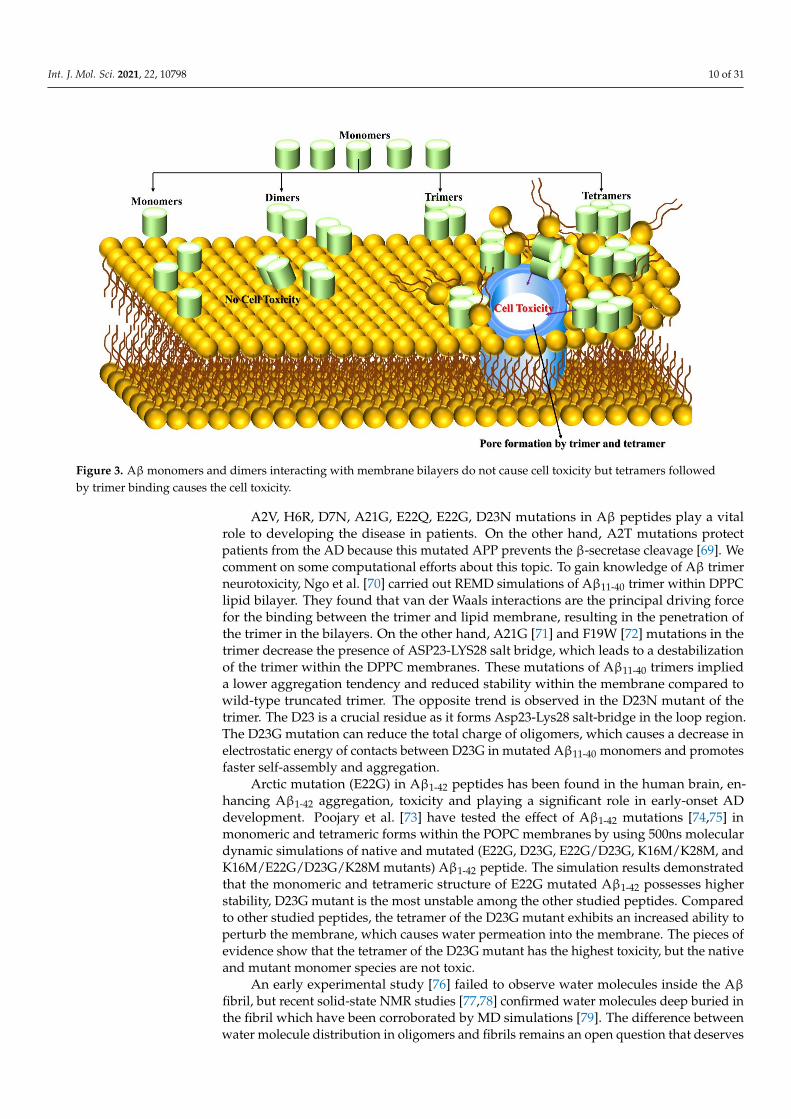
Aβ toxicity is significantly linked to its binding to the cell membrane and causesneuronal cell death. Jana et al. [68] have used mouse cortical neuronal culture to test thecorrelation between Aβ oligomer (up to tetramer level) binding and cell viability. Mousecortical was treated with 5µm concentration of Aβ peptide for 96 h, Aβ42 peptide bindingto neurons is 7-fold to 10-fold higher than that of Aβ1-40, which binds neurons after 24 h.Surprisingly, tetramers and trimers are observed after 48 h with less than 5% of totalbound Aβ, and the peptide has not become toxic up to 48 h with 590 femtograms Aβ/cellbound. The peptide concentration was increased up to 15 µm and significant changes wereobserved when Aβ42 binding rate reached 777–923 femtogram Aβ/cell at 72 and 96 h,being neurotoxic triggered cell death and allowing trimers and tetramers to bind graduallyto neurons, increasing over time for Aβ1-42 and contributing 15–20% of the total bound.The cell-bound trimer and tetramer (not monomer and dimer) play a vital role in Aβ1-42toxicity (Figure 3). Small efforts exist on this matter, and researchers are highly encouragedto explore the Aβ trimer and tetramer peptides on the surface of membranes and withinmembranes using multiscale MD simulation to unveil cell toxicity mechanisms.
Int. J. Mol. Sci. 2021, 22, 10798 10 of 31
Figure 3. Aβmonomers and dimers interacting with membrane bilayers do not cause cell toxicity but tetramers followedby trimer binding causes the cell toxicity.
A2V, H6R, D7N, A21G, E22Q, E22G, D23N mutations in Aβ peptides play a vitalrole to developing the disease in patients. On the other hand, A2T mutations protectpatients from the AD because this mutated APP prevents the β-secretase cleavage [69]. Wecomment on some computational efforts about this topic. To gain knowledge of Aβ trimerneurotoxicity, Ngo et al. [70] carried out REMD simulations of Aβ11-40 trimer within DPPClipid bilayer. They found that van der Waals interactions are the principal driving forcefor the binding between the trimer and lipid membrane, resulting in the penetration ofthe trimer in the bilayers. On the other hand, A21G [71] and F19W [72] mutations in thetrimer decrease the presence of ASP23-LYS28 salt bridge, which leads to a destabilizationof the trimer within the DPPC membranes. These mutations of Aβ11-40 trimers implieda lower aggregation tendency and reduced stability within the membrane compared towild-type truncated trimer. The opposite trend is observed in the D23N mutant of thetrimer. The D23 is a crucial residue as it forms Asp23-Lys28 salt-bridge in the loop region.The D23G mutation can reduce the total charge of oligomers, which causes a decrease inelectrostatic energy of contacts between D23G in mutated Aβ11-40 monomers and promotesfaster self-assembly and aggregation.
Arctic mutation (E22G) in Aβ1-42 peptides has been found in the human brain, en-hancing Aβ1-42 aggregation, toxicity and playing a significant role in early-onset ADdevelopment. Poojary et al. [73] have tested the effect of Aβ1-42 mutations [74,75] inmonomeric and tetrameric forms within the POPC membranes by using 500ns moleculardynamic simulations of native and mutated (E22G, D23G, E22G/D23G, K16M/K28M, andK16M/E22G/D23G/K28M mutants) Aβ1-42 peptide. The simulation results demonstratedthat the monomeric and tetrameric structure of E22G mutated Aβ1-42 possesses higherstability, D23G mutant is the most unstable among the other studied peptides. Comparedto other studied peptides, the tetramer of the D23G mutant exhibits an increased ability toperturb the membrane, which causes water permeation into the membrane. The pieces ofevidence show that the tetramer of the D23G mutant has the highest toxicity, but the nativeand mutant monomer species are not toxic.
An early experimental study [76] failed to observe water molecules inside the Aβfibril, but recent solid-state NMR studies [77,78] confirmed water molecules deep buried inthe fibril which have been corroborated by MD simulations [79]. The difference betweenwater molecule distribution in oligomers and fibrils remains an open question that deserves

Int. J. Mol. Sci. 2021, 22, 10798 11 of 31
further exploration as water leakage has a crucial role in neurotoxicity. In this perspective,Nguyen et al. [21] address why the oligomers are more toxic than fibrils. They haveidentified the conformational space of Aβ1-42 tetramer (smallest stable oligomers) usingcoarse-grained FFs. They employed MD simulations to examine Aβ structural stabilityand compared them to NMR fibril structures. Thus, they proposed that the water densityinside the oligomer is greater than in fibril, causing it to enhance the toxicity of oligomers.The interaction mechanism between tetramer and membranes interaction has yet beenelusive, the open challenge question to the researcher is to investigate the smallest oligomer(Aβ1-42 tetramer) dynamic behavior on or within membranes (water-lipid interaction) byusing computer simulations. This important piece of information will assist in unveilingneurotoxicity more quantitatively.
3.4. Aβ Oligomer-Membrane
Despite many efforts made in the amyloid pore hypothesis, none of the studiesprovides the atomic structure of Aβ oligomers. Without this information, it is impossibleto elucidate the mechanism of Aβ oligomers toxicity. In this regard, Serra-Batiste et al. [80]examined the Aβ behavior within the membrane to elucidate the neurotoxicity in ADand found that Aβ1-40 and Aβ1-42 exhibited different physical behavior, Aβ1-40 aggregatedinto amyloid fibril while Aβ1-42 assembled into oligomers. The Aβ1-42 oligomers adoptedβ-barrel arrangement and formed well-defined pore into a membrane that named byβ-barrel-pore-forming Aβ1-42 oligomers (βPFOSAβ42). In comparison with Aβ1-40, Aβ1-42has a significant role in AD by the higher propensity of βPFOSAβ42.
Carulla’s group further investigated [81] the atomic structures of Aβ1-42 tetramersand octamers within the membrane using experimental and MD simulations studies. TheAβ1-42 tetramer consisted of the six-stranded β-sheet core, Aβ1-42 octamers constitutedby two Aβ1-42 tetramers, and both tetramer and octamers surrounded by a membrane.In the simulation study, the hydrophilic residues placed in both tetramers and octamersedges are unfavourably exposed to the hydrophilic lipid tails of the membrane. Thehead group of the membrane is reoriented towards hydrophilic edges of tetramers andoctamers leading to stabilizing of the lipid-protein complex and resulting in lipid-stabilizedpores. A high degree of water permeation is observed in the membrane and higher solventaccessible surface area in the octamers than tetramers. The water and ion penetration in themembrane was identified experimentally. Simulation and experimental results togetherproposed that water and ion penetration occurred through lipid-stabilized pores, mediatedby hydrophilic amino acids in the β-sheet edges of the oligomers that could be responsiblefor neurotoxicity in AD.
Smith et al. [82] used replica exchange MD simulation for studying the aggregation ofthe Aβ25-35 peptide within the DMPC bilayer using CHARMM22/CMAP correction andCHARMM36 FFs for peptide and lipid FFs, respectively. In general, the Aβ25-35 peptideis characterized by two regions N-terminal (residues 25–28) and C-terminal (residues29–35). The hydrophobic and electrostatic interactions between the Aβ25-35 dimers drivesto produced parallel out-of-registry aggregation, which manifested pore formation inthe bilayer. In particular, the aggregation within the membrane is constituted by threeconcentric rings, two outer rings located in the upper and lower leaflets are in contactthrough hydrophobic C-terminal with the bilayer core and N-terminal pointing towardsthe lipid head groups. The third rings are closer towards the pore centre, N-terminaldirected the pore center to create pore lining. The peptide aggregation increases theextent of bilayer thinning that is more than four-fold more significant than the monomericspecies; mainly because the dimers reduce the thickness of the DMPC bilayer from 40 Å to24 Å. Thus, extensive damage to the membrane bilayers was mediated by uncontrollableCa2+ permeation.
Conventional and steered MD simulations [83] have been employed to explore thezwitterionic POPC and palmytoil-oleoyl-phosphatidylethanolamine (POPE) head groupsthat influence the interaction between Aβ9-40 octamers and lipids. The results demon-

Int. J. Mol. Sci. 2021, 22, 10798 12 of 31
strated that the POPC head groups form weaker electrostatic interaction with the Aβoctamer which have shorter-lived contact with the bilayers. Immediately, the head groupsreorganized themselves, resulting in the lipid tail moving upwards to enhance electrostaticcontact with the C-terminal of the octamer, which led to Aβ insertion into the membrane.This process is called a detergent-like effect on membranes for amyloid peptide forma-tion. Whereas in the case of POPE-Aβ9-40 octamers, the head group repels the peptideinsertion into membrane, this barrier is overcome by the stronger electrostatic interactionsthat persisted between charged residues in the Aβ9-40 and lipids bilayer and causing theC-terminal of the peptide to be inserted in the bilayer.
Qiang et al. [84] have used solid-state NMR spectroscopy to probed the phospholipidsdynamics and interactions between lipid and peptide in a POPC bilayer fused with Aβ1-40oligomers. At physiological conditions, lipids show changes in terms of lipid motionand reorganization. The stronger lipid-Aβ1-40 interactions restrict the lipid motion andinter-strand interaction between the loop and C-terminal of the oligomers. This effect isweakened by the lipid molecules inserted into oligomers that form rapid aggregates alongwith membranes intercalated by a hydrogen bonding network. The loop region of theoligomer could interact with the lipid head group to severely disturb membrane integrityby weakened interactions between lipid bilayers.
Fernández-Pérez experimental study [85] examines the relationship between choles-terol and Aβ oligomers behavior in a membrane. The membrane perforation induced byAβ is much faster when a low concentration of cholesterol is present in the membrane.In contrast, a high concentration of cholesterol blocks the perforation effect of Aβ. In-terestingly, neurons treated with cholesterol significantly increased Aβ association in themembranes compared with the free cholesterol. This evidence supports that the perforationeffect of Aβ depends on the amount of cholesterol in the membrane and that cholesterolhas a protective effect for Aβ toxicity. A large number of studies [13] provided evidencethat cholesterol promotes Aβ aggregation and neurotoxicity.
Neurotoxicity of the Aβ has been elucidated by studies of the different forms of theAβ peptide that interact with phosphatidylcholine (PC) and cholesterol. A dual mechanismhas been observed [86], (a) The low concentration of cholesterol in the membrane interactswith Aβ peptide to promote the peptide insertion into the membrane and (b) opposingthe peptide permeation at elevated cholesterol concentration through membrane stiffnesseffect. It gives evidence that Cholesterols manifested Aβ neurotoxicity [87–91]. In contrast,PC inhibits neurotoxicity by blocking aggregation and β-sheet formation [92,93].
The impact of cholesterol on the binding of Aβ (Aβ17-42 and Aβ11-42) fibrils withDPPC bilayers was investigated through MD simulations using Martini 2.0 force field [94].The MD results supported the electrostatic interaction as the major driving force for thefibril binding to the membranes, along with the elevated level of cholesterol present in themembrane, which modulates this interaction by a dual mechanism, i.e., to the increasedbinding of positive residues with the lipid head groups and enhanced Ca2+ binding withthe bilayers. The high concentration of cholesterol promotes Aβ1-42 peptide binding tothe bilayer, in contrast, the cholesterol prevents the insertion of Aβ10-40 peptide into thebilayer. The relationship between Aβ peptide and membrane co-incubated with cholesterolis unclear, and computer simulations are still required to shed light onto this interaction.
4. How Do Metal Ions Govern Aβ Peptide Behavior?
The senile plaques are enriched by the presence of Zn2+, Cu2+, Fe3+ and Al3+ metalions, it has been observed that in the case of a postmortem AD brain [95] their concentra-tions are 0.4 mM of Cu2+, 1 mM of Zn2+, and 1 mM of Fe3+; Al3+ has been also found ingreater concentration in isolated core of senile plaques [96]. We will describe computationalsimulation efforts of metal interaction with Aβ peptides in the following sections.

Int. J. Mol. Sci. 2021, 22, 10798 13 of 31
4.1. Cu and Zn Ions Interactions with Aβ Peptides
In general, metal ion binding to peptide has been divided into three approaches,namely (a) bonded, (b) non-bonded, and (c) cationic dummy atomic models. Our previousresearch elucidated the dynamics of Aβ peptides upon metal interaction with bonded andnon-bonded approaches [97,98]. For the non-bonded, we found Aβ1-40-Zn2+ and Aβ1-42-Zn2+ adopted a β-hairpin structure. For the bonded approach, the conformational space ofAβ1-42–Zn2+ is more heterogeneous compared to that of Aβ1-42–Cu2+ and Aβ1-42 becauseof the large number of basins present in the free energy surface (FES) of Aβ1-42–Zn2+ (seeFigure 4). It confirmed that Aβ1-42–Zn2+ aggregates lead to a more amorphous state com-pared to other cases. In addition, we found that Zn2+ rather than Cu2+ binding promotesgreater hydrophobicity in Aβ1-42. Our result agrees with Miller et al. [99] studies. Theymonitored the role of Zn2+ ions on Aβ oligomers by using MD and REMD simulationswith the CHARMM27 force field. They observed that Zn2+ binding decreased the solva-tion energy (increase hydrophobicity) of Aβ oligomer, which enhanced the aggregationpropensity, and that a higher concentration of Zn2+ could reduce aggregation kinetics. Incontrast, the aggregation process became much faster than in metal-free solutions.
Figure 4. Free energy landscape for (A). Aβ42, (B). Aβ42-Cu2+, and (C). Aβ42-Zn2+ peptides as a function of RMSD andthe gyration of radius. Results are based on the whole ensemble of trajectories. Representative structures in the free energyminimum basins are displayed. Reproduced with permission from the work of Boopathi et al. [98]. Copyright 2020 JohnWiley and Sons.
Lee et al. [100] performed MD simulations with the CHARMM27 force field andreported three significant insights: (a) Zn2+ ion mediated to increase the hydrogen bondnetwork between Aβ1-42 layers of the oligomers, which caused to reinforce the stabilizationof Aβ1-42 oligomers, in contrast, the same ion decreases the stabilization of Aβ1-42 fibrils byreducing the hydrogen bond network, (b) in comparison to Cu2+, Zn2+ destabilizes Aβ1-42fibrils more effectively, Cu2+ does not reduce the hydrogen bond network in the fibril and

Int. J. Mol. Sci. 2021, 22, 10798 14 of 31
(c) in comparison to Cu2+, Zn2+ binding did significant attenuate the energy of the saltbridge, which is an essential role in the formation of Aβ1-42 aggregation.
It is known that two residues are oxidized in Aβ, met35 is oxidized to a sulfoxideand Tyr10 is oxidized to form dityrosine covalent dimer; these oxidations impact the fibrilformation. Met35 is oxidized by H202 alone without the presence of oxidation radicals. Theoxidation of Tyr10 by radicals to form a Dityrosine covalent Aβ dimer has five- and eight-fold higher concentration in the hippocampus and neocortical region of the AD brain [101].Cu2+ has a higher concentration in the AD patient brain, and H202 commonly exists atphysiologically and mild acidic conditions. Miao et al. [102] have curiously monitoredthe relation between the concentration of Cu2+ + H202 and the amount of Dityrosineproduction by using experimental studies, noted 5 µm of Cu2+ concentration and a higherconcentration of H202 from 0.4 mM to 1.6 mM that can enhance the amount of Dityrosineformation. By contrast, they found that the total dityrosine production is not affected bytwo-fold higher concentration Cu2+ ions (10 µm) with the same H202 concentration. Theseresults supported H202 rather than Cu2+ as the main catalyzer in dityrosine productions.In addition, the Cu2+ +H202 redox cycling system oxidized most of the monomers within100 h to produce a Dityrosine dimer. This effect accelerates the preformed Aβ1-40 fibrilformation and reduces fibril length from 800 nm to 150 nm.
Santis et al. [103] utilized an X-ray Absorption spectroscopy to obtain the data forAβ1-42 with Cu2+ and Zn2+ complex samples and reported that Zn2+ binding to Aβ1-42could affect the Cu2+ coordination to the same peptide. If Zn2+ is added to the Aβ1-42solution first, it prevents Cu2+ binding to the Aβ1-42 peptide. In contrast, if Cu2+ is addedfirst does not inhibit Zn2+ binding to the Aβ1-42 peptide.
In general, the Aβ peptide could not adopt a unique conformation because of theirdisordered nature. Its binding to metal ions is quickly is equilibrated in an aqueousenvironment compared with wild-type Aβ peptide. Srivastava et al. [104] answered thefollowing fundamental question. Does metal ion affect the formation of aggregates andfibril formation? Of course, a high concentration of metal ions triggered fibril formation. Forinstance, Zn2+ and Cu2+ inhibit Aβ1-42 fibrillization and promote non-fibrillar aggregates.Later studies by Electron microscopy have shown that amorphous (non-fibrillar) aggregatesconvert into fibrils by binding metal ions. This result implies that amorphous aggregatesare not the final point in the aggregation process. In other words, there are two cases,fibrils and small aggregation, for instance, metal ions are present in “amorphous” and“mature fibril” aggregates in which His6, His13, and His14 are coordinated with the metalions. In particular, metal-bound His13 and His14 (beta-sheet conformation) side chainsare found in opposite sides of the peptide. Later, the opposite trend followed by the smallaggregation in which monomeric Aβ binds to metal ions through H6, H13, H14 and aminoacid terminus, but H13 and H14 not bound with metal ions on opposite sides of the peptidechains resulting in around H13 and H14 regions could not convert β-sheet conformation.In some cases, His6 also does not turn into a β-sheet conformation in most of the fibrilssince metal ions mediate a lesser binding effect. We commented that high concentrations ofmetal ions could mediate the amorphous Aβ aggregation, while low concentrations havetriggered the Aβ fibril structure. It is noteworthy that the positive-charged metal ions canreduce the net-negative charge of Aβ, resulting in an enhancement of the aggregation rateof the peptide.
What metal ions induce Aβ cytotoxicity?Aβ peptide can reduce Cu2+ to Cu+, and Fe3+ to Fe2+, facilitating the generation of
reactive oxygen species H202 and OH• radical. Tyr10 residue of the Aβ peptide loses oneelectron and produces reactive tyrosine radicals, which bind to the peptide to form a dimer,causing the killing of the brain cells [105]. On the other hand, (a) metal ions can induceconformational changes and are more aggregation-prone structures than the monomericAβ peptide and (b) they can allow via molecular bridging the formation of aggregates.
To elucidate the role of Tyr10 residue on the Aβ self-assembly mechanism, Coskunerand Uversky [106] have utilized AMBER FF14SB and CHARMM22/CMAP FFs, employed

Int. J. Mol. Sci. 2021, 22, 10798 15 of 31
REMD (7.2 µs) with thermodynamics calculation to study wild-type and Tyr10Ala mutationof Aβ1-42 monomer in the explicit aqueous environment. They have observed two issues(a) Tyr10 residue promotes higher toxic β-sheet formation in the structural ensemble ofAβ1-42 monomer; however, (b) an opposite trend followed by Tyr10Ala mutation decreasesthe β-sheet formation in the monomer. In general, the β-sheet formation in the peptideplays a vital role in self-assembly and fibril formation. This result revealed that Tyr10regulates the toxic β sheet structure formation in the monomer causing faster self-assembly,but Tyr10Ala mutation impedes the self-assembly by decreasing the β sheet content. Anopen question is the impact of Tyr10 in Aβ in the presence of metal ions has not beenelucidated yet.
Notably, the Cu2+ binding to Aβ generates less radial oxygen species in the case ofmurine than human Aβ, same in rats and mice that have not developed AD pathologybecause Aβ contained a lack of His13, a crucial residue in the binding of metal ions. TheHis13 residue mutation in human Alzheimer’s Aβ peptide is one of the ongoing researchfields [107–111].
4.2. Fe Interaction with Aβ Peptide
So far, one study reported in the literature deals with Fe2+ interaction with Aβ pep-tide. We conducted 200 ns MD simulations with DFT study [112] to probe the structuraldynamics of Aβ1-42-Zn2+, Aβ1-42-Cu2+ and Aβ1-42-Fe2+. Our results suggested that Fe2+
binding generates a U-shaped structure in Lys16-Met35 with the turn in the loop region andβ-sheet extended over central hydrophobic and C-terminal regions, which is corroboratedby the NMR study [76,113]. Overall, this result implies that Fe2+ binding enhances thefibril aggregation of the Aβ1-42.
4.3. Al Interaction with Aβ Peptide
Matthew Turner et al. [114] have employed five-microsecond MD simulation withAMBER ff14SB forcefield and study the process of how Al3+ governs the structural dynamicbehaviour of Aβ1-40 and Aβ1-42 peptides. They observed Al3+ forming robust coordinationwith negatively charged E3, D7 and E11 residues, with coordination number of four. Suchcoordination persisted throughout the simulations despite metal ion bindings being mod-eled as a non-bonded model. This description has significantly impacted the structure anddynamics of both Aβ1-40 and Aβ1-42 peptides changes by promoting the helical structureformation by severely disrupting the salt-bridge network. Surprisingly, why is Al3+-Aβbinding coordination not disrupted throughout the simulation, even though a non-bondedmetal binding model describes it? It is yet unclear. Is it an artifact of the chosen forcefield or simulation protocol that maintains the metal coordination? In this connection,Platts [115] utilized the semi-empirical tight-binding method GFN2-XTB for modelling theinteraction of Al3+ with truncated Aβ1-16 peptide; the outcome of the optimized geometryis in agreement with DFT benchmark data. Metadynamics simulation has been used toexplore exhaustively the coordination pattern of the same peptide [115]. The studies showthat Al3+ binding to the Aβ1-16 is highly fluxional due to all acidic sidechains and severalbackbone oxygens involving coordination. The estimated coordination number on averageis 5.2 atoms per ion. This evidence confirmed that the metadynamics approach does notmatch the microsecond MD simulation regarding the metal coordination number. Themain reason is that in the case of non-bonded, atomic FFs could neglect charge transferand polarization for peptide-ion interaction. In the bonded approach, atomic chargesare obtained based on electrostatic potential (RESP) derived by B3LYP/6-31G (d) level oftheory, amino acid backbone of oxygen to Al3+ is more electronegative −0.79 e as comparedwith MD simulations of non-bonded counterparts −0.40 to 0.58 e. These differences leadmetadynamics to promote higher coordination number (5.2) than molecular dynamicscoordination number (~4). MD simulation permits conformational changes of the Aβwithout disrupting metal coordination, while metadynamics accounts for the differentmetal coordination mode.
Int. J. Mol. Sci. 2021, 22, 10798 16 of 31
Roldán-Martín et al. [116] have examined the structural features of Aβ1-42 peptideupon Cu2+ and Al3+ binding by using Gaussian accelerated MD simulation with AMBERFF14SB force field. They noted the following: (a) Cu2+ binding increased α-helical contentin the Aβ1-42 peptide causes U-shaped conformations which is promoted by Glu22/Asp23-Ser26/Lys28 turn region that links two helices and arranges them in parallel through inter-helical hydrophobic contacts, (b) by contrast, Al3+ binding can convert the helical contentsinto the extended structure, which are more favourable for β-sheet formation, in otherwords, presence of Al3+ could have manifested aggregation-prone Aβ1-42 structures, and(c) the total charge of Aβ1-42 is neutralized by +3 charge of Al3+ metal, Aβ1-42-Al3+ complexbecomes zero total charge, and the complex accelerates their aggregation propensity fasteras compared to Aβ1-42-Cu2+ (charge, −1) and Aβ1-42 (charge, −3).
4.4. Ag Interaction with Aβ Peptide
Wallin et al. [117] used Fluorescence spectroscopy, solid-state AFM imaging, circulardichroism spectroscopy, and NMR spectroscopy to probe the Aβ fibrillization pathwaysin the presence of silver (Ag+) metal ions. The Aβ peptide from the fibril incorporatesother monomeric Aβ to elongate the fibril end. The Ag+ ion made weak binding tothe N-terminal of the monomeric Aβ that lead to weakens the Aβ fibrillization kineticsby reducing fibril end elongation rate. Therefore, Ag+ bound peptide is insufficient ofmaintaining aggregation by decreasing the Aβ for fibril extension and the same observationis applied by the case Zn2+ binding with the Aβ peptide. These systems confirmed Ag+
crucial role in suffering the bulk aggregation by retardation of the fibril end elongation.
4.5. Pb Interaction with Aβ Peptide
The NMR studies confirmed the presence of Pb in the AD brain, this molecule inducesan increase in the concentration of Aβ in rats [118] and enhances plaque formation inmonkeys [119]. Wallin et al. [120] inspect the impact of the metal ions Cd+, Cr2+, Pb2+
and Pb4+ on Aβ aggregation by employed experimental studies. They found a couple ofsignificant observations such as: (a) Pb4+ binds H6, Y10, E11, H13, and H14 residues ofAβ1-40 monomer and it can mediate faster aggregation rate, as compared with Cd+, Cr2+,and Pb2+ metal ions, which typically establishes electrostatic interaction with a monomer.The NMR and Fluorescence spectroscopy showed that Pb4+ binding to Aβ is slightlydifferent from the Cu2+ and Zn2+ binding residues of D1, H6, H13, and H14, (b) Pb4+
binding induces M35 residue oxidation, facilitating the release of harmful reactive oxygenspecies (ROS) in the plaques that damage the neuron activities.
4.6. Hg Interaction with Aβ Peptide
Most humans are exposed to mercury by eating fish and shellfish contaminated withmethylmercury. In general, Hg2+ ions cannot pass the blood-brain barrier (BBB), butmetallic vapor mercury penetrated BBB to oxidized Hg2+ trapped inside the brain. Earlystudies [121] suggested the Hg2+ binds to thiol(-SH) and selenohydryl (-SeH) groupscontained residue only. Thus, it does not bind the Aβ1-42 peptide because cysteine residuesare absent. Nevertheless, recent experiments reported [122] Hg2+ directly interactingwith Aβ peptide and strengthen the hypothesis of Hg2+ risk factor in AD. Subsequently,the molecular interaction of the Aβ1-42 with Hg2+ [123] has been explored using NMR,Fluorescence Spectroscopy and AFM, the outcome implies that Hg2+ binds to H6, H13,and H14 which can impede the Aβ1-40 and Aβ1-42 fibrillization and facilitate amorphousaggregation at a 1:1 Hg2+/Aβ ratio.
4.7. Mn Interaction with Aβ Peptide
Several AD symptoms appear suddenly after severely exposed manganese [124], therelationship between Mn and AD is an ongoing debate. Wallin et al. [125] have used NMR,CD, AFM, Fluorescence spectroscopy and MD simulation to monitor the effect of Mn2+
ions on Aβ aggregation and fibrils. The NMR data [126] reported that Aβ1-16-Zn2+ complex
Int. J. Mol. Sci. 2021, 22, 10798 17 of 31
with Zn2+ is coordinated by H6, H13, H14, and E11 residues. When Zn2+ manually wasreplaced by Mn2+ in the same complex and modelled Aβ1-16-Mn2+ complex with Mn2+binds result in similar binding to the same residues: H6, H13, H14, and E11. This complexwas used treated as an input structure for microsecond MD simulations. During MDsimulation, the Mn2+ dissociated from initial binding residues, as it preferred to coordinatenegatively charged residues of D1, E3, D7, and E11. These amino acids prevent the Hisresidue from binding Mn2+ ion.
Consequently, NMR data proved that D1, H13, D22, and E23 residues are morefavourable for Mn2+ binding. Simulations and experiments predicted different bindingsites for Mn2+, the exact details of Mn2+ coordination mode are still under investigation.Furthermore, CD spectroscopy and THT fluorescence data imply random coil (in thestarting structure of Aβ peptide) transitions to β-sheets. In addition, the AFM imageshowed Aβ40 peptide aggregation mediates the formation of fibril structures, but Mn2+
does not influence at all on aggregation kinetics and fibril morphology of Aβ40 due tothe weak interaction with the peptide. However, more simulations are required to modeltruncated Aβ1-16 or the full length of Aβ bound to Mn2+ ions.
Geng Lin et al. [127] have tested eight-month-old male mice divided into Mn-treatedmice and controlled mice groups. Mn concentration was measured using mass spectroscopyat the end of the fifth-month treatment. The measurement ensured Mn level in similarconcentration in blood and the brain. The qRT-PCR and western blot analysis confirmedin the case of Mn-treatment increased β- and γ-secretases cleavages at APP, an enormousamount of Aβ1-42 peptides production in the cerebral cortex and hippocampus of brains,while α-secretase cleavage activity was significantly reduced. These data suggested thatMn-treatment is a risk factor for the development of AD pathogenesis. However, no studiesin the literature reported the effect of Mn on Aβ1-42 productions at atomic level details. Thebeforementioned studies supported the Aβ aggregation modified by Fe2+, Mn2+, Pb4+ andZn2+, as well as the Hg2+ binding to specific residues of the Aβ peptide.
4.8. Li Interaction with Aβ Peptide
The AD symptoms are significantly reduced by the Li+ ions treatment [57,128,129],which showed a substantial improvement regarding Aβ clearance [130], enhance spa-tial memory [131], reduced oxidative stress level [132] and decreased the amount of Aβplaques [133] in AD brain. Berntsson et al. [134] used biophysical techniques, NMR, Flu-orescence quenching, CD, and IR, to monitor the elusive in vitro interaction between Li+
and Aβ peptide. They found that Li+ displayed weak interaction with the Aβ, resultingin unaltered secondary structures of Aβ1-40 monomers and Aβ1-42 oligomers. At elevatedLi+ concentration, Li+ can manifest minor perturbation-effect on the morphology of aggre-gated Aβ1-40 fibril, Aβ1-40 aggregation kinetics and Aβ1-42 oligomeric stability. Overall, theresults suggested Li+ ions are not able to modulate the Aβ aggregation and toxicity. TheLi+ treatment on AD progression is not caused by direct interaction of Li+ with Aβ.
5. What Is the Role of Small Molecules in Inhibition Mechanism of Aβ Aggregation?
Researchers have widely used four novel therapeutic approaches to reduce plaqueformation, (a) secretase inhibitors and modulators, (b) immunotherapeutic strategies,(c) peptide-based inhibitors, and (d) small molecular inhibitors.
5.1. Secretase Inhibitors and Modulators
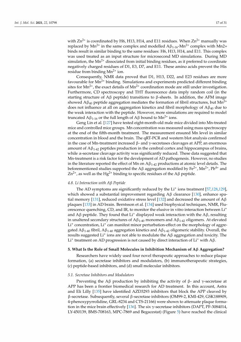
Preventing the Aβ production by inhibiting the activity of β- and γ-secretase atAPP has been a frontier biomedical research for AD treatment. In this account, Astraand Eli Lilly [135] have identified AZD3293 inhibitors that block the APP cleaved byβ-secretase. Subsequently, several β-secretase inhibitors (OM99-2, KMI-429, GSK188909,4-phenoxypyrrolidine, GRL-8234 and CTS-21166) were shown to attenuate plaque forma-tion in the mice brain effectively [136]. The six γ-secretase inhibitors (DAPT, PF-3084014,LY-450139, BMS-708163, MPC-7869 and Begacestat) (Figure 5) have reached the clinical
Int. J. Mol. Sci. 2021, 22, 10798 18 of 31
trial [136–138]. In 2001, an in-vivo study demonstrated for the first time that γ-secretaseinhibitor DAPT (N-[N-(3,5-difluorophenacetyl)- l-alanyl]-S-phenylglycine t-butyl ester)blocked the Aβ production in the AD mice brain, but the lymphocyte development andthe intestine symptoms were still observed [138]. During the prenatal period, Lympho-cyte development occurs in humans. The new-born immune system contains functionalT (thymus-derived) and B (born-marrow derived) lymphocytes. T and B lymphocytesare responsible for the function of antibody production and cell-mediated immune re-sponses, respectively. Symptoms of intestine problems are stomach pain, vomiting, nausea,dehydration, a feeling of illness, and difficulty passing gas.
Figure 5. The γ-secretase inhibitors used in clinical trial for the treatment of AD and taken from PubChem.
The PF-3084014 (Nirogacestat) has significantly reduced the Aβ level in the brain,plasma and cerebrospinal fluid in guinea pigs and Tg2576 transgenic mice. Since it waslinked to a highly lipophilic nature, the clinical trial was discontinued due to an unfa-vorable pharmacokinetic and pharmacodynamic profile. The avagacestat (BMS-708163)significantly reduces Aβ1-40 levels in female rats, and significantly reduce Aβ1-40 in thebrain and cerebrospinal fluid (CSF) of male beagle dogs [139]. The semagacestat (LY-450139)could reduce the plasma Aβ40 concentration by 58% and 65% for the 100-mg and 140-mggroups, respectively [140]. The avagacestat and semagacestat inhibitors blocked γ-secretaseactivity at APP in the cell membrane. However, these inhibitors failed in a phase 2 andphase3 of clinical trials because they generated a degeneration in cognitive ability in ADpatients along with other severe side effects.
During the semagacestat treatment, side effects were shown to increase the risk of skincancer. Begacestat (GSI-953) decreased Aβ levels in plasma, brain, and cerebrospinal fluidin Tg2576 mice, but it failed to reduce Aβ concentration in the AD patient brain [141]. It wasdiscontinued in the phase 1 trial in 2010. Tarenflurbil (MPC-7869) produced Aβ38 insteadof Aβ42 by modifying the γ-secretase at APP. The Aβ38 manifested lower neurotoxicityand phase 2 trials showed encouraging results. The Phase 3 trial terminated in 2009 sincethe agent penetrate the blood-brain barrier insufficiently [137]. However, those inhibitorshave decreased Aβ concentrations in the animal brain, but they showed severe side effectsin AD patients.

Int. J. Mol. Sci. 2021, 22, 10798 19 of 31
5.2. Immunotherapeutic Strategies
In the immunotherapeutic approach, immunotherapy induces the host cell to rec-ognize and fight Aβ in order to reduce Aβ aggregation in the brain. It is divided intotwo categories (a) active immunization and (b) passive immunization. The primary isto generate a vaccine for Aβ production to reduce plaques. The latter is to producemonoclonal antibodies and administration of immunoglobulins. The following five drugcandidates, bapineuzumab, solanezumab, gantenerumab, aducanumab and Lecanemab,are humanized monoclonal antibodies except for solanezumab, which is another mon-oclonal antibody [142–144]. The first four drugs have significantly improved cognitiveabilities and are undergoing clinical phase2 or 3 trials, while Lecanemab binds soluble Aβand potential diseases attenuated by removing plaques.
The monomers, oligomers and fibrils of Aβ have exposed epitopes; typically, they arethe same and antibodies can recognize and bind the epitopes of Aβ. Muller et al. [145]revealed that 20 amino acid SDPM1 proteins bind to the structure of the tetramers ofAβ40 and Aβ42 peptides, blocking Aβ association and removing plaques in AD brain.Solanezumab, Gantenerumab, and IVIG antibodies can improve cognitive skills by bindingsoluble Aβ peptides in the clinical trial [146,147]. The solanezumab and crenezumabhave higher contact with the Aβ via KLVFF epitope [148]. The human anti-Aβmonoclonalantibody, gantenerumab, crossed the blood-brain barrier, binding aggregates of Aβ throughthe N-terminal and central regions of the peptide resulting in a removal of Aβ plaques,which was observed in phase 3 clinical study [143].
The drugs mainly target amyloid peptide, tau protein, mitochondrial dysfunction,oxidation stress and metal dysregulation to reverse the AD progression. Although agood amount (approximately 111 drugs) of drug candidates released between 2010 and2020 [136], the majority of the candidates failed in phase 2 trials primarily due to the lackof the ability to reduce toxicity, cognitive loss and plaques formation, and more than 50candidates reached phase 3 trial. 40% of the candidates fell into repurposed drugs. Notably,the four repurpose drugs, escitalopram, brexipiperazole, masitinib, and losartan, are un-dergoing phase 3 clinical trials. However, today, only one drug, aducanumab, successfullypassed the phase 3 clinical trial and came to the market for mild AD patient’s treatment.
The FDA approved two types of drugs (Figure 6A–D): (a) cholinesterase inhibitorsand (b) memantine [1]. The primary can boost the amount of cholinesterase, which isresponsible for memory functions. The latter inhibitor regulates the activity of glutamate,which is a messenger chemical involved in brain function, including learning and memory.There are three cholinesterase inhibitors: Donepezil (Aricept), Galantamine (Razadyne),and Rivastigmine (Exelon). Donepezil is used in all stages of AD patients, Galantamineserves to treat mild to moderate disease, and Rivastigmine in case of severe AD patients.These inhibitors reduce the symptoms of the disease with side effects including nausea,vomiting and diarrhea. Thus, these therapeutics can alleviate some of AD symptoms, butnot the course of the disease.
Int. J. Mol. Sci. 2021, 22, 10798 20 of 31
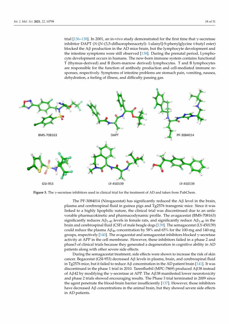
Figure 6. The U.S FDA approved drugs for AD (A) Donepezil, (B) Galantamine, (C) Rivastigmineand (D) Memantine. (E) fragment antigen-binding region of aducanumab (AduFab) -Aβ1-11 peptide.AduFab depicted by van-der-Waals surface. The chemical structures taken from PubChem andprotein data bank.
In this perspective, Sevigny et al. [11] utilized positron emission tomography (PET)to monitor the efficacy of an aducanumab in mice and human brains. Aducanumab(Figure 6E) is an IgG1 monoclonal antibody and recombinant antibody derived from slowor absent cognitive decline patients. Three separated patient groups were subjected inthis study, double-blind, randomized and placebo-controlled. The patients were clinicallydiagnosed with mild AD, and 1, 3, 6 and 10 mg kg−1 doses of aducanumab were given to31, 32, 30 and 32 patients respectively for one year. In the end, the result was comparedwith the placebo-controlled group (40 patients). The PET measurement showed that theAβ plaques formation has significantly reduced in subcortical white matter and corticalregions of the brain at 6 and 10 mg kg−1 within 54 weeks. In the placebo-controlled cases,no changes occurred at amyloid plaques. In addition, they gave 0.3, 1, 3, 10 and 30 mg kg−1
doses to mice for 9.5 to 15.5 months and discovered that 63% of Aβ plaques in the cortexand hippocampus of mice brains disappeared at 10 or 30 mg kg−1 doses. This evidencesupports the strong effect of aducanumab entering the brain and reducing plaque formationby binding with the Aβ. All the data revealed aducanumab as a potential inhibitor fordisease-modifying by decreasing soluble and insoluble Aβ [149,150]. Due to the removalof rich amyloid plaques and minimizing side effects, a milestone in AD therapy occurredon June 7th, 2021, the U.S. FDA approved aducanumab for the AD treatment [151].
Arndt et al. [152] deposited a crystal structure of AduFab (the fragment antigen-binding region of aducanumab)-Aβ1-11 peptide complex in the protein data bank (PDBid:6co3). In this structure, aducanumab binds to 3–7 residues of Aβ in an extended confor-mation. Furthermore, Frost and Zacharias [153] have performed MD simulation to explorethe interaction between AduFab and Aβ1-40 peptide in the form of monomers, dimers,oligomers, and fibrils. The result suggested that AduFab binding affinity is increased inoligomers and fibrils compared to monomers.

Int. J. Mol. Sci. 2021, 22, 10798 21 of 31
5.3. Peptide-Based Inhibitors
Calcium ions flow in the membrane to increase the release of synaptic vesicles inhippocampal neurons that generate neurotransmission. In contrast, the Aβ plays a role indecreasing the number of synaptic vesicles that cause neurotransmission failure. Subse-quently, the Aβ oligomer directly interacts with the membrane generating pore structurein which the flow of calcium is increased and leads to cell death. In this connection,Peter et al. [154] derived pentapeptide(G33LMVG37) from the glycine zipper region of C-terminal Aβ. Experiments characterized the Aβ activity upon the pentapeptide interaction,which confirmed G33LMVG37 involvement in three main activities: (a) blocking the in-crease of the calcium level in the neurons (b) inhibiting the perforation in the membrane(c) preventing the association of Aβ peptide. Finally, this pentapeptide supports neuro-transmission by blocked Aβ induced membrane pore formation. In other words, the smallsize of the hydrophobic entity crosses the blood-brain barrier to reduce plaque forma-tion. Other pentapeptides, G25SNKG29 and G29AIIG33, could not have any effect on Aβpeptide aggregation.
Zheng et al. [155] have employed multiple approaches such as ion mobility spec-troscopy, mass spectroscopy, and MD simulation to characterize the interaction betweenthe full length Aβ1-42 and two modified Aβ39-42 derivatives: VVIA-NH2 and Ac-VVIA. Themass spectroscopy revealed VVIA-NH2 binding to Aβ1-42 monomers, dimers, tetramers,hexamers, or oligomers, while Ac-VVIA only binds to monomers. Ion-mobility spec-troscopy results showed that VVIA-NH2 prevents dodecamers formation and generatesnon-toxic oligomers, which form fibrils. On the other hand, Ac-VVIA mediated toxicoligomers eventually led to a fibril. MD simulation results suggested VVIA-NH2 has aweak binding affinity to the C-terminal region of monomers. In contrast, Ac-VVIA showeda strong binding effect to multiple regions of Aβ1-42. Overall, this data supported Ac-VVIAbinding to oligomers as a crucial step for the inhibition of Aβ1-42 toxicity.
5.4. Small Molecular Inhibitors
Drug repositioning and repurposing are promising therapeutic strategies for drugdiscovery of anti-AD. Cramer et al. [156] have demonstrated that the anti-cancer drug,bexarotene, it can reduce 50% Aβ plaques in mice models of AD within 72 h. The followingquestion is obscure: bexarotene destroys plaques by a direct interaction with Aβ. In thisaccount, In this account, Pham et al. [157] employed in-silico and in-vitro experiments toinspect the role of bexarotene in the Aβ aggregation and reported that bexarotene showedweek interaction affinity to Aβ fibril, which does not affect amyloid aggregation. The Aβproduced by the cleavage of APP by β- and γ-secretase, whose activity is tuned by thedrug that is ongoing research to remove plaque formation. Subsequently, they also usedin-silico and in-vitro experiments [158] to explore the possibility of preventing the Aβassociation by bexarotene binding to β-secretase. The results imply that plaques could notbe reduced due to the weak interaction between β-secretase and bexarotene. However,one more possible investigation remains obscure, examining the interaction mechanismbetween γ-secretase and bexarotene to understand the reduction of plaques.
Mei et al. [159] have addressed the following question: How will the interactionbetween either negatively charged (ER) or neutral TS0/TS1 small molecules and Aβ42oligomers affect the aggregation of oligomers? They have identified, in a comparativestudy with a small neutral molecule, ER (charged -2), as a suitable candidate to inhibit theneurotoxicity of Aβ42 aggregates through strong binding, decreasing aggregates hydropho-bicity, degrading the β sheet contents and inter-and intra-molecular hydrogen bond on themain chain, and perturbation of K23-L28 salt bridges and the vdW interactions. Hence,they recommend developing negatively charged inhibitors for the target Aβ42 oligomersto prevent plaque formation.
As shown in Figure 7, the intrinsically disordered Aβ aggregation pathway hasbeen characterized into four types [160]: (a) Primary nucleation, oligomers formed frommonomeric species; (b) Elongation, oligomers and fibrils size increased by adding monomers;

Int. J. Mol. Sci. 2021, 22, 10798 22 of 31
(c) Fragmentation, fibrils break into several oligomers and small fibrils that are capableof elongation; (d) Secondary nucleation, the surface of fibril aggregates promoting a newformation of oligomers species. Heller et al. [161] identified a small molecule, 10074-g5 (biphenyl-2-yl-(7-nitro-benzo [1,2,5] oxadiazol-4-yl)-amine), which interacts with themonomeric state of Aβ1-42 peptide using experiments, computational simulation and math-ematical modelling. They observed (a) this molecule binds to monomeric Aβ1-42 peptideresulting in a significantly delayed the primary- and secondary-nucleation pathways inAβ1-42 aggregation, (b) this molecule can decrease the hydrophobicity of the Aβ1-42 pep-tide and c) this molecule inhibits the formation of the Aβ1-42 association and toxicity byenhancing the conformation entropy.
Figure 7. Schematic illustration of the aggregation pathways. There are characterized into four types: Primary nucleation,monomers formed dimers, trimers, hexamers and then oligomers. Elongation, the oligomers are joined together to generatedthe fibrils. Fragmentations, the fibrils break into the number of small fibrils and oligomers. Secondary nucleation, onlyminority of small fibrils convert into a new fibril by adding monomers, but due to the addition of monomers, the majorityof the small fibrils turn into the monomers or oligomers.
Pagano et al. [162] have reviewed the role of the natural compound in the Aβ pep-tide aggregation pathways using in-silico and in-vitro studies. The thirteen compounds(Brazilin, Curcumin, Epigallocatechin gallate, Ginnalin A, wgx-50, Myricetin, Oleuropein,Oleuropein aglycone, Reserveratol, Rosmarinic acid, Sclerotiorin, Tanshinone and Uncar-inic acid C) were extracted from plants and observed that they cross the blood-brain-barrier.Since the solvent exposure of the hydrophobic surface is regulated Aβ toxicity, inhibiting ormodulating the toxicity of Aβ aggregation is one of the main approaches against AD. Con-siderable evidence coming from the experiments and computer simulations have confirmedthat those thirteen compounds can directly interact with Aβ affecting amyloid aggregationpathway by (a) inhibiting formation of oligomers from monomers, (b) preventing thesecondary nucleation of aggregation and (c) reducing the toxicity of aggregates.
Using REMD simulations, Mohamoudinobar et al. [163] have characterized the effectof NaCl, scylloinositol and 4-aminophenol in Aβ1-42 aggregation pathway in an aqueousenvironment. In the case of pure water, β-sheet is formed in the residue segments 16–22 (central hydrophobic region), 23–29 (central hydrophilic region) and the N-terminus,

Int. J. Mol. Sci. 2021, 22, 10798 23 of 31
whereas in the case of NaCl contained water, the β-sheet is formed at N- and C-terminal, butthe β-sheet formation in the central hydrophilic region is inhibited. Furthermore, in purewater, frequent residue contacts have been observed between the central hydrophobic andthe C-terminus which were reduced by increasing the interaction between C-terminus andcentral hydrophilic region in the presence of inhibitors (scylloinositol and 4-aminophenol).Aβ1-42 peptide adopted a U-shaped conformation with strand-loop-strand structure inNaCl and pure water, but not in water-containing inhibitors. The U-shaped structure iswell corroborated with the NMR fibril structure [164] and promotes Aβ1-42 aggregates.This result implies that the inhibitors prevent the Aβ1-42 aggregates, in contrast to theNaCl case.
In the past ten years of our research investigations, we have reached four main conclu-sions using in-silico, in-vitro and in-vivo experiments. (1) Nicotine molecule interactioncauses the β-sheet of Aβ transformation into the alpha-helical structure that prohibits theaggregation of Aβ peptides [165]. (2) A venom of black mamba snake could inhibit the fibrilformation of Aβ peptides by destroying the inter-peptide Asp23-Lys28 salt-bridge [166].(3) U.S. FDA approved drug, gabapentin (GBP), binding to Aβ1-42 peptides and blockedmultiple actions: aggregation, oligomers toxicity, membrane poration, intracellular calciumand synaptotoxicity induced by Aβ1-42 [167]. We recommend evaluating the GBP in aclinical trial for mild AD patients with a low dose of 400 mg/day to avoid risk associ-ated with higher doses. (4) We identified a new low molecular weight compound M30(2-Octahydroisoquinoline-2(1H)-ylethanamine) which generated a neuroprotective effectby removing the Aβ1-42 toxicity in hippocampal neurons and significantly improved spatialmemory of AD affected mice [168].
6. Conclusion and Future Perspective
Why do researchers fail to identify drugs against AD? Although hundreds of moleculesare present in the human brain, we believe Aβ and Tau aggregation are the main hallmarksof AD [169]. The hundreds of clinical trials targeting Aβ and Tau proteins have faileddue to other molecules excluding Aβ and Tau proteins, which are invoking in initiatingAD pathology. On the other hand, Aβ or Tau proteins interact with several biomoleculessuch as the membranes, transition metal ions, cholesterol, apoE3, apoE4, prion, Na+, K+,omega3, omega6, and other molecules. The experimental studies could not render thoseinteractions. In addition, experiments have faced two main hurdles with predicting anti-AD drugs: unable to mimic the brain membrane exactly, prepared oligomers and fibrilsstructures are not resembling those presented in AD patients’ brains. They cannot providethe high-resolution structure of the Aβ oligomers. The atomic structural details of Aβaggregates are in great need. Computer simulation studies are not only prepared basedon the supplementary details of NMR experiments, but they also provide more insight onAβ-Membrane and Aβ-drug interactions when experiments studies are challenging. In thisaccount, it is highly encouraged to pursue new investigations using computer simulations.
1. Omega3 and Omega6 polyunsaturated fatty acids are buried in the neuronal mem-branes and are linked to a slowing down and increasing the risk of AD, respectively.Thus, it is essential to explore the dynamic mechanism of Aβ1-40/Aβ1-42 oligomerswith and without interaction of metal ions in omega3 and omega6 phospholipids asthese details are lacking in the AD research.
2. Tyr10 residue in Aβ1-42 regulates the toxic β-sheet formation and leads to aggregation,but none of the studies in the literature regarding the role of Tyr10 in metal-boundAβ1-42 peptide addressed this issue. In this perspective, investigating the interactionmechanism between metal-bound Aβ1-42 peptides and lipid membranes will be acrucial step to describe the toxicity of Aβ1-42 peptides.
3. The inhibitory effect of the number of natural compounds in the aggregation path-ways has been explained in the manuscript. However, their actions remain to beelucidated. The transition metal ions, Cu2+ and Zn2+, and their bindings induced ahigher tendency of β-sheet formation in the Leu17-Met35 regions of Aβ1-42 peptide
Int. J. Mol. Sci. 2021, 22, 10798 24 of 31
that can decrease solvent exposure (increase hydrophobicity) of the peptide whichcan lead to regulate toxicity [98]. To attenuate the hydrophobicity propensity of thepeptide by the interaction of natural compounds, it is an essential investigation foranti-AD drug discovery.
4. Large evidence [170,171] implicated the Aβ1-42 peptide aggregation controlled bylipid membranes, but in contrast, cholesterol in the lipid membrane significantlyenhances the peptide aggregation by cholesterol, showing a high affinity for thepeptide. However, the detailed interaction and its connection between the lipid bilayerand membrane remains elusive. The new idea is to study how free and metal ionsbound to Aβ1-42 during aggregation changes in cholesterol-contained membranes.
5. The Aβ25-35 cytotoxicity is well explained by the lipid bilayer mediated truncatedAβ25-35 peptide aggregation, which has made perforation in the membrane causinguncontrollable permeation of Ca2+ ions [82]. So far, exploring the full-length of Aβ1-42peptide aggregation in the presence or absence of Cu2+, Zn2+ and Fe2+ metal ionsin the membrane remains an open research problem. Here one can describe thecytotoxicity of the full-length Aβ1-42.
6. Advanced MD simulation, aided by the improved version of coarse-grained forcefields (e.g., MARTINI 3, UNRES, PRIMO, etc), can describe large-scale systems andovercome time-scale limitations, and improve our understanding of the connectionbetween AD progression and initial stages of the disease by tracing the molecularpathways of full-length Aβ aggregation in complex with lipid bilayers. Similarly,it will help to unveil the fundamental role played by mechanical stability duringamyloidogenesis and Aβ aggregate maturation.
7. Our previous investigations have envisaged two molecules, M30 and gabapentin,blocking multiple steps of the Aβ cascade: Aggregation, Synaptotoxicity, membranepore formation, calcium dyshomeostasis and memory impairment. Thus, we stronglyrecommend using M30 and gabapentin as part of the clinical trial as an alternativeAD treatment.
Author Contributions: S.B. and A.B.P. contributed equally in writing the manuscript. R.G.-J. re-viewed, edited and made grammar corrections. All authors have read and agreed to the publishedversion of the manuscript.
Funding: This research received fund from Dirección General de Asuntos del Personal Académicode la Universidad Nacional Autónoma de México (DGAPA-UNAM). This research financial sup-ported by the National Science Centre, Poland, under Grant 2017/26/D/NZ1/00466, and from theFoundation for Polish Science under the grant MAB PLUS/11/2019.
Data Availability Statement: Not applicable.
Acknowledgments: S.B. acknowledged Dirección General de Asuntos del Personal Académico dela Universidad Nacional Autónoma de México (DGAPA-UNAM) for a postdoctoral Fellowship.A.B.P. acknowledges the National Science Centre, Poland, and the Foundation for Polish Science.Research was supported by PL-Grid Infrastructure and the TUL Computing & Information ServicesCenter infrastructure.
Conflicts of Interest: The authors declare no conflict of interest.
Abbreviations
Aβ Amyloid betaAD Alzheimer’s diseaseMD Molecular dynamicsDFT Density Functional TheoryIDP Intrinsically disordered proteinsDMPC DimyristoylgylcerophosphocholineDPPC Dipalmitoylphosphatidylcholine

Int. J. Mol. Sci. 2021, 22, 10798 25 of 31
DOPS Dioleoyl phosphatidylserinePOPC 1-palmitoyl-2-oleoyl-sn-glycero-3-phosphocholinePOPE Palmytoil-oleoyl-phosphatidylethanolamine
References1. Alzheimer’s Association. 2021 Alzheimer’s Disease Facts and Figures. Alzheimer’s Dement. 2021, 17, 327–406. [CrossRef]2. Hardy, J.A.; Higgins, G.A. Alzheimer’s Disease: The Amyloid Alzheimer’s Disease. Science 1992, 256, 184–185. [CrossRef]3. Bai, X.C.; Yan, C.; Yang, G.; Lu, P.; Ma, D.; Sun, L.; Zhou, R.; Scheres, S.H.W.; Shi, Y. An Atomic Structure of Human γ-Secretase.
Nature 2015, 525, 212–217. [CrossRef] [PubMed]4. Colvin, M.T.; Silvers, R.; Ni, Q.Z.; Can, T.V.; Sergeyev, I.; Rosay, M.; Donovan, K.J.; Michael, B.; Wall, J.; Linse, S.; et al. Atomic
Resolution Structure of Monomorphic Aβ42 Amyloid Fibrils. J. Am. Chem. Soc. 2016, 138, 9663–9674. [CrossRef] [PubMed]5. Wälti, M.A.; Ravotti, F.; Arai, H.; Glabe, C.G.; Wall, J.S.; Böckmann, A.; Güntert, P.; Meier, B.H.; Riek, R. Atomic-Resolution
Structure of a Disease-Relevant Aβ(1-42) Amyloid Fibril. Proc. Natl. Acad. Sci. USA 2016, 113, E4976–E4984. [CrossRef]6. Gremer, L.; Schenk, C.; Reinartz, E.; Ravelli, R.B.G.; Tusche, M.; Lopez-iglesias, C.; Hoyer, W.; Heise, H.; Willbold, D. Fibril
Structure of Amyloid-b (1-42) by Cryo–Electron Microscopy. Science 2017, 119, 116–119. [CrossRef]7. Roberts, B.R.; Lind, M.; Wagen, A.Z.; Rembach, A.; Frugier, T.; Li, Q.X.; Ryan, T.M.; McLean, C.A.; Doecke, J.D.; Rowe, C.C.; et al.
Biochemically-Defined Pools of Amyloid-β in Sporadic Alzheimer’s Disease: Correlation with Amyloid PET. Brain 2017, 140,1486–1498. [CrossRef] [PubMed]
8. Kayed, R.; Head, E.; Thompson, J.L.; McIntire, T.M.; Milton, S.C.; Cotman, C.W.; Glabel, C.G. Common Structure of SolubleAmyloid Oligomers Implies Common Mechanism of Pathogenesis. Science 2003, 300, 486–489. [CrossRef] [PubMed]
9. O’Nuallain, B.; Wetzel, R. Conformational Abs Recognizing a Generic Amyloid Fibril Epitope. Proc. Natl. Acad. Sci. USA 2002, 99,1485–1490. [CrossRef]
10. Michaels, T.C.T.; Šaric, A.; Curk, S.; Bernfur, K.; Arosio, P.; Meisl, G.; Dear, A.J.; Cohen, S.I.A.; Dobson, C.M.; Vendruscolo, M.;et al. Dynamics of Oligomer Populations Formed during the Aggregation of Alzheimer’s Aβ42 Peptide. Nat. Chem. 2020, 12,445–451. [CrossRef]
11. Sevigny, J.; Chiao, P.; Bussière, T.; Weinreb, P.H.; Williams, L.; Maier, M.; Dunstan, R.; Salloway, S.; Chen, T.; Ling, Y.; et al. TheAntibody Aducanumab Reduces Aβ Plaques in Alzheimer’s Disease. Nature 2016, 537, 50–56. [CrossRef] [PubMed]
12. Hanseeuw, B.J.; Betensky, R.A.; Jacobs, H.I.L.; Schultz, A.P.; Sepulcre, J.; Becker, J.A.; Cosio, D.M.O.; Farrell, M.; Quiroz, Y.T.;Mormino, E.C.; et al. Association of Amyloid and Tau with Cognition in Preclinical Alzheimer Disease: A Longitudinal Study.JAMA Neurol. 2019, 76, 915–924. [CrossRef] [PubMed]
13. Nguyen, P.H.; Ramamoorthy, A.; Sahoo, B.R.; Zheng, J.; Faller, P.; Straub, J.E.; Dominguez, L.; Shea, J.E.; Dokholyan, N.V.; deSimone, A.; et al. Amyloid Oligomers: A Joint Experimental/Computational Perspective on Alzheimer’s Disease, Parkinson’sDisease, Type II Diabetes, and Amyotrophic Lateral Sclerosis. Chem. Rev. 2021, 121, 2545–2647. [CrossRef]
14. Saravanan, K.M.; Zhang, H.; Zhang, H.; Xi, W.; Wei, Y. On the Conformational Dynamics of β-Amyloid Forming Peptides: AComputational Perspective. Front. Bioeng. Biotechnol. 2020, 8, 1–19. [CrossRef]
15. Robustelli, P.; Piana, S.; Shaw, D.E. Developing a Molecular Dynamics Force Field for Both Folded and Disordered Protein States.Proc. Natl. Acad. Sci. USA 2018, 115, E4758–E4766. [CrossRef]
16. Krupa, P.; Pham, D.Q.H.; Li, M.S. Properties of Monomeric Aβ42 Probed by Different Sampling Methods and Force Fields: Roleof Energy Components. J. Chem. Phys. 2019, 151, 055101. [CrossRef]
17. Kirkitadze, M.D.; Condron, M.M.; Teplow, D.B. Identification and Characterization of Key Kinetic Intermediates in Amyloidβ-Protein Fibrillogenesis. J. Mol. Biol. 2001, 312, 1103–1119. [CrossRef] [PubMed]
18. Chong, S.H.; Chatterjee, P.; Ham, S. Computer Simulations of Intrinsically Disordered Proteins. Annu. Rev. Phys. Chem. 2017, 68,117–134. [CrossRef]
19. Viet, M.H.; Nguyen, P.H.; Derreumaux, P.; Li, M.S. Effect of the English Familial Disease Mutation (H6R) on the Monomers andDimers of Aβ40 and Aβ42. ACS Chem. Neurosci. 2014, 5, 646–657. [CrossRef]
20. Huy, P.D.Q.; Vuong, Q.V.; La Penna, G.; Faller, P.; Li, M.S. Impact of Cu(II) Binding on Structures and Dynamics of Aβ42 Monomerand Dimer: Molecular Dynamics Study. ACS Chem. Neurosci. 2016, 7, 1348–1363. [CrossRef]
21. Nguyen, H.L.; Krupa, P.; Hai, N.M.; Linh, H.Q.; Li, M.S. Structure and Physicochemical Properties of the Aβ42 Tetramer:Multiscale Molecular Dynamics Simulations. J. Phys. Chem. B 2019, 123, 7253–7269. [CrossRef]
22. Chong, S.H.; Ham, S. Assessing the Influence of Solvation Models on Structural Characteristics of Intrinsically Disordered Protein.Comput. Theor. Chem. 2013, 1017, 194–199. [CrossRef]
23. Best, R.B.; Zheng, W.; Mittal, J. Balanced Protein-Water Interactions Improve Properties of Disordered Proteins and Non-SpecificProtein Association. J. Chem. Theory Comput. 2014, 10, 5113–5124. [CrossRef]
24. Piana, S.; Donchev, A.G.; Robustelli, P.; Shaw, D.E. Water Dispersion Interactions Strongly Influence Simulated StructuralProperties of Disordered Protein States. J. Phys. Chem. B 2015, 119, 5113–5123. [CrossRef] [PubMed]
25. Rahman, M.U.; Rehman, A.U.; Liu, H.; Chen, H.F. Comparison and Evaluation of Force Fields for Intrinsically DisorderedProteins. J. Chem. Inf. Model. 2020, 60, 4912–4923. [CrossRef]
26. Ye, W.; Ji, D.; Wang, W.; Luo, R.; Chen, H.F. Test and Evaluation of Ff99IDPs Force Field for Intrinsically Disordered Proteins. J.Chem. Inf. Model. 2015, 55, 1021–1029. [CrossRef] [PubMed]
Int. J. Mol. Sci. 2021, 22, 10798 26 of 31
27. Song, D.; Wang, W.; Ye, W.; Ji, D.; Luo, R.; Chen, H.F. Ff14IDPs Force Field Improving the Conformation Sampling of IntrinsicallyDisordered Proteins. Chem. Biol. Drug Des. 2017, 89, 5–15. [CrossRef] [PubMed]
28. Song, D.; Luo, R.; Chen, H.F. The IDP-Specific Force Field Ff14IDPSFF Improves the Conformer Sampling of IntrinsicallyDisordered Proteins. J. Chem. Inf. Model. 2017, 57, 1166–1178. [CrossRef]
29. Zhang, Y.; Liu, H.; Yang, S.; Luo, R.; Chen, H.F. Well-Balanced Force Field Ff03 CMAP for Folded and Disordered Proteins. J.Chem. Theory Comput. 2019, 15, 6769–6780. [CrossRef] [PubMed]
30. Maier, J.A.; Martinez, C.; Kasavajhala, K.; Wickstrom, L.; Hauser, K.E.; Simmerling, C. Ff14SB: Improving the Accuracy of ProteinSide Chain and Backbone Parameters from Ff99SB. J. Chem. Theory Comput. 2015, 11, 3696–3713. [CrossRef]
31. Best, R.B.; Mittal, J. Protein Simulations with an Optimized Water Model: Cooperative Helix Formation and Temperature-InducedUnfolded State Collapse. J. Phys. Chem. B 2010, 114, 14916–14923. [CrossRef] [PubMed]
32. Huang, J.; Rauscher, S.; Nawrocki, G.; Ran, T.; Feig, M.; De Groot, B.L.; Grubmüller, H.; MacKerell, A.D. CHARMM36m: AnImproved Force Field for Folded and Intrinsically Disordered Proteins. Nat. Methods 2016, 14, 71–73. [CrossRef] [PubMed]
33. Liu, H.; Song, D.; Zhang, Y.; Yang, S.; Luo, R.; Chen, H.F. Extensive Tests and Evaluation of the CHARMM36IDPSFF Force Fieldfor Intrinsically Disordered Proteins and Folded Proteins. Phys. Chem. Chem. Phys. 2019, 21, 21918–21931. [CrossRef]
34. Piana, S.; Lindorff-Larsen, K.; Shaw, D.E. How Robust Are Protein Folding Simulations with Respect to Force Field Parameteriza-tion? Biophys. J. 2011, 100, L47–L49. [CrossRef]
35. Samantray, S.; Yin, F.; Kav, B.; Strodel, B. Different Force Fields Give Rise to Different Amyloid Aggregation Pathways in MolecularDynamics Simulations. J. Chem. Inf. Model. 2020, 60, 6462–6475. [CrossRef]
36. Mu, J.; Liu, H.; Zhang, J.; Luo, R.; Chen, H.F. Recent Force Field Strategies for Intrinsically Disordered Proteins. J. Chem. Inf.Model. 2021, 61, 1037–1047. [CrossRef]
37. Kmiecik, S.; Gront, D.; Kolinski, M.; Wieteska, L.; Dawid, A.E.; Kolinski, A. Coarse-Grained Protein Models and Their Applications.Chem. Rev. 2016, 116, 7898–7936. [CrossRef]
38. Seo, M.; Rauscher, S.; Pomès, R.; Tieleman, D.P. Improving Internal Peptide Dynamics in the Coarse-Grained MARTINI Model:Toward Large-Scale Simulations of Amyloid- and Elastin-like Peptides. J. Chem. Theory Comput. 2012, 8, 1774–1785. [CrossRef][PubMed]
39. Rojas, A.; Maisuradze, N.; Kachlishvili, K.; Scheraga, H.A.; Maisuradze, G.G. Elucidating Important Sites and the Mechanism forAmyloid Fibril Formation by Coarse-Grained Molecular Dynamics. ACS Chem. Neurosci. 2017, 8, 201–209. [CrossRef]
40. Latshaw, D.C.; Cheon, M.; Hall, C.K. Effects of Macromolecular Crowding on Amyloid Beta (16-22) Aggregation Using Coarse-Grained Simulations. J. Phys. Chem. B 2014, 118, 13513–13526. [CrossRef] [PubMed]
41. Sahoo, A.; Xu, H.; Matysiak, S. Pathways of Amyloid-Beta Absorption and Aggregation in a Membranous Environment. Phys.Chem. Chem. Phys. 2019, 21, 8559–8568. [CrossRef]
42. Souza, P.C.T.; Alessandri, R.; Barnoud, J.; Thallmair, S.; Faustino, I.; Grünewald, F.; Patmanidis, I.; Abdizadeh, H.; Bruininks,B.M.H.; Wassenaar, T.A.; et al. Martini 3: A General Purpose Force Field for Coarse-Grained Molecular Dynamics. Nat. Methods2021, 18, 382–388. [CrossRef]
43. Mahmood, M.I.; Poma, A.B.; Okazak, K.I. Optimizing Go-MARTINI Coarse-Grained Model for F-BAR Protein on Lipid Membrane.Front. Mol. Biosci. 2021, 8, 1–10. [CrossRef]
44. Poma, A.B.; Cieplak, M.; Theodorakis, P.E. Combining the MARTINI and Structure-Based Coarse-Grained Approaches for theMolecular Dynamics Studies of Conformational Transitions in Proteins. J. Chem. Theory Comput. 2017, 13, 1366–1374. [CrossRef][PubMed]
45. Poma, A.B.; Li, M.S.; Theodorakis, P.E. Generalization of the Elastic Network Model for the Study of Large ConformationalChanges in Biomolecules. Phys. Chem. Chem. Phys. 2018, 20, 17020–17028. [CrossRef] [PubMed]
46. Ingólfsson, H.I.; Melo, M.N.; Van Eerden, F.J.; Arnarez, C.; Lopez, C.A.; Wassenaar, T.A.; Periole, X.; De Vries, A.H.; Tieleman,D.P.; Marrink, S.J. Lipid Organization of the Plasma Membrane. J. Am. Chem. Soc. 2014, 136, 14554–14559. [CrossRef]
47. Poma, A.B.; Guzman, H.V.; Li, M.S.; Theodorakis, P.E. Mechanical and Thermodynamic Properties of Aβ42, Aβ40, and α-Synuclein Fibrils: A Coarse-Grained Method to Complement Experimental Studies. Beilstein J. Nanotechnol. 2019, 10, 500–513.[CrossRef] [PubMed]
48. Chwastyk, M.; Bernaola, A.P.; Cieplak, M. Statistical Radii Associated with Amino Acids to Determine the Contact Map: Fixingthe Structure of a Type i Cohesin Domain in the Clostridium Thermocellum Cellulosome. Phys. Biol. 2015, 12, 46002. [CrossRef]
49. Poma, A.B.; Chwastyk, M.; Cieplak, M. Elastic Moduli of Biological Fibers in a Coarse-Grained Model: Crystalline Cellulose andβ-Amyloids. Phys. Chem. Chem. Phys. 2017, 19, 28195–28206. [CrossRef]
50. Poma, A.B.; Thu, T.T.M.; Tri, L.T.M.; Nguyen, H.L.; Li, M.S. Nanomechanical Stability of Aβ Tetramers and Fibril-like Structures:Molecular Dynamics Simulations. J. Phys. Chem. B 2021, 125, 7628–7637. [CrossRef]
51. Sengupta, U.; Nilson, A.N.; Kayed, R. The Role of Amyloid-beta oligomers in Toxicity, Propogation, and Immunotherapy.EBioMedicine 2016, 6, 42–49. [CrossRef] [PubMed]
52. Hall, C.M.; Moeendarbary, E.; Sheridan, G.K. Mechanobiology of the Brain in Ageing and Alzheimer’s Disease. Eur. J. Neurosci.2021, 53, 3851–3878. [CrossRef] [PubMed]
53. Carballo-Pacheco, M.; Ismail, A.E.; Strodel, B. On the Applicability of Force Fields to Study the Aggregation of AmyloidogenicPeptides Using Molecular Dynamics Simulations. J. Chem. Theory Comput. 2018, 14, 6063–6075. [CrossRef] [PubMed]
Int. J. Mol. Sci. 2021, 22, 10798 27 of 31
54. Senguen, F.T.; Doran, T.M.; Anderson, E.A.; Nilsson, B.L. Clarifying the Influence of Core Amino Acid Hydrophobicity, SecondaryStructure Propensity, and Molecular Volume on Amyloid-β 16-22 Self-Assembly. Mol. Biosyst. 2011, 7, 497–510. [CrossRef]
55. Lockhart, C.; Smith, A.K.; Klimov, D.K. Three Popular Force Fields Predict Consensus Mechanism of Amyloid β Peptide Bindingto the Dimyristoylgylcerophosphocholine Bilayer. J. Chem. Inf. Model. 2020, 60, 2282–2293. [CrossRef] [PubMed]
56. Akbayraka, I.Y.; Caglayanb, S.I.; Ozcanb, Z.; Uverskyc, V.N.; Coskuner-Weberb, O. Current Challenges and Limitations in theStudies of Intrinsically Disordered Proteins in Neurodegenerative Diseases by Computer Simulations. Curr. Alzheimer Res. 2020,17, 805–818. [CrossRef]
57. Press-Sandler, O.; Miller, Y. Molecular Mechanisms of Membrane-Associated Amyloid Aggregation: Computational Perspectiveand Challenges. Biochim. Biophys. Acta -Biomembr. 2018, 1860, 1889–1905. [CrossRef] [PubMed]
58. Davis, C.H.; Berkowitz, M.L. Interaction between Amyloid-β (1-42) Peptide and Phospholipid Bilayers: A Molecular DynamicsStudy. Biophys. J. 2009, 96, 785–797. [CrossRef] [PubMed]
59. Davis, C.H.; Berkowitz, M.L. Structure of the Amyloid-β (1-42) Monomer Absorbed to Model Phospholipid Bilayers: A MolecularDynamics Study. J. Phys. Chem. B 2009, 113, 14480–14486. [CrossRef]
60. Lockhart, C.; Klimov, D.K. Calcium Enhances Binding of AβMonomer to DMPC Lipid Bilayer. Biophys. J. 2015, 108, 1807–1818.[CrossRef]
61. Pike, C.J.; Burdick, D.; Walencewicz, A.J.; Glabe, C.G.; Cotman, C.W. Neurodegeneration Induced by β-Amyloid Peptides inVitro: The Role of Peptide Assembly State. J. Neurosci. 1993, 13, 1676–1687. [CrossRef]
62. Millucci, L.; Ghezzi, L.; Bernardini, G.; Santucci, A. Conformations and Biological Activities of Amyloid Beta Peptide 25–35. Curr.Protein Pept. Sci. 2009, 999, 1–6. [CrossRef]
63. Smith, A.K.; Klimov, D.K. Binding of Cytotoxic Aβ25-35 Peptide to the Dimyristoylphosphatidylcholine Lipid Bilayer. J. Chem.Inf. Model. 2018, 58, 1053–1065. [CrossRef]
64. Jang, H.; Connelly, L.; Teran Arce, F.; Ramachandran, S.; Kagan, B.L.; Lal, R.; Nussinov, R. Mechanisms for the Insertion of Toxic,Fibril-like β-Amyloid Oligomers into the Membrane. J. Chem. Theory Comput. 2013, 9, 822–833. [CrossRef]
65. Davis, C.H.; Berkowitz, M.L. A Molecular Dynamics Study of the Early Stages of Amyloid-β(1-42) Oligomerization: The Role ofLipid Membranes. Proteins Struct. Funct. Bioinform. 2010, 78, 2533–2545. [CrossRef] [PubMed]
66. Snowden, S.G.; Ebshiana, A.A.; Hye, A.; An, Y.; Pletnikova, O.; O’Brien, R.; Troncoso, J.; Legido-Quigley, C.; Thambisetty, M.Association between Fatty Acid Metabolism in the Brain and Alzheimer Disease Neuropathology and Cognitive Performance: ANontargeted Metabolomic Study. PLoS Med. 2017, 14, e1002266. [CrossRef] [PubMed]
67. Lu, Y.; Shi, X.F.; Nguyen, P.H.; Sterpone, F.; Salsbury, F.R.; Derreumaux, P. Amyloid-β(29-42) Dimeric Conformations inMembranes Rich in Omega-3 and Omega-6 Polyunsaturated Fatty Acids. J. Phys. Chem. B 2019, 123, 2687–2696. [CrossRef]
68. Jana, M.K.; Cappai, R.; Pham, C.L.L.; Ciccotosto, G.D. Membrane-Bound Tetramer and Trimer Aβ Oligomeric Species Correlatewith Toxicity towards Cultured Neurons. J. Neurochem. 2016, 136, 594–608. [CrossRef]
69. Doig, A.J.; Castillo-frias, M.P.; Berthoumieu, O.; Tarus, B.; Nasica-labouze, J.; Sterpone, F.; Nguyen, P.H.; Hooper, N.M.; Faller, P.;Derreumaux, P. Why Is Research on Amyloid-β Failing to Give New Drugs for Alzheimer’s Disease? ACS Chem. Neurosci. 2017,8, 1435–1437. [CrossRef] [PubMed]
70. Ngo, S.T.; Hung, H.M.; Tran, K.N.; Nguyen, M.T. Replica Exchange Molecular Dynamics Study of the Amyloid Beta (11-40)Trimer Penetrating a Membrane. RSC Adv. 2017, 7, 7346–7357. [CrossRef]
71. Ngo, S.T.; Nguyen, M.T.; Nguyen, N.T.; Vu, V.V. The Effects of A21G Mutation on Transmembrane Amyloid Beta (11-40) Trimer:An in Silico Study. J. Phys. Chem. B 2017, 121, 8467–8474. [CrossRef] [PubMed]
72. Tran, T.T.; Pan, F.; Tran, L.; Roland, C.; Sagui, C. The F19W Mutation Reduces the Binding Affinity of the TransmembraneAβ11-40trimer to the Membrane Bilayer. RSC Adv. 2021, 11, 2664–2676. [CrossRef]
73. Poojari, C.; Strodel, B. Stability of Transmembrane Amyloid β-Peptide and Membrane Integrity Tested by Molecular Modeling ofSite-Specific Aβ42 Mutations. PLoS ONE 2013, 8, e78399. [CrossRef]
74. Murakami, K.; Irie, K.; Morimoto, A.; Ohigashi, H.; Shindo, M.; Nagao, M.; Shimizu, T.; Shirasawa, T. Neurotoxicity andPhysicochemical Properties of Aβ Mutant Peptides from Cerebral Amyloid Angiopathy: Implication for the Pathogenesis ofCerebral Amyloid Angiopathy and Alzheimer’s Disease. J. Biol. Chem. 2003, 278, 46179–46187. [CrossRef]
75. Luheshi, L.M.; Tartaglia, G.G.; Brorsson, A.C.; Pawar, A.P.; Watson, I.E.; Chiti, F.; Vendruscolo, M.; Lomas, D.A.; Dobson, C.M.;Crowther, D.C. Systematic in Vivo Analysis of the Intrinsic Determinants of Amyloid β Pathogenicity. PLoS Biol. 2007, 5, e290.[CrossRef]
76. Petkova, A.T.; Yau, W.M.; Tycko, R. Experimental Constraints on Quaternary Structure in Alzheimer’s β-Amyloid Fibrils.Biochemistry 2006, 45, 498–512. [CrossRef]
77. Yung, S.K.; Liu, L.; Axelsen, P.H.; Hochstrasser, R.M. 2D IR Provides Evidence for Mobile Water Molecules in β-Amyloid Fibrils.Proc. Natl. Acad. Sci. USA 2009, 106, 17751–17756. [CrossRef]
78. Wang, T.; Jo, H.; DeGrado, W.F.; Hong, M. Water Distribution, Dynamics, and Interactions with Alzheimer’s β-Amyloid FibrilsInvestigated by Solid-State NMR. J. Am. Chem. Soc. 2017, 139, 6242–6252. [CrossRef] [PubMed]
79. Xi, W.; Hansmann, U.H.E. Ring-like N-Fold Models of Aβ42 Fibrils. Sci. Rep. 2017, 7, 1–14. [CrossRef] [PubMed]80. Serra-Batiste, M.; Ninot-Pedrosa, M.; Bayoumi, M.; Gairí, M.; Maglia, G.; Carulla, N. Aβ42 Assembles into Specific β-Barrel
Pore-Forming Oligomers in Membrane-Mimicking Environments. Proc. Natl. Acad. Sci. USA 2016, 113, 10866–10871. [CrossRef]

Int. J. Mol. Sci. 2021, 22, 10798 28 of 31
81. Ciudad, S.; Puig, E.; Botzanowski, T.; Meigooni, M.; Arango, A.S.; Do, J.; Mayzel, M.; Bayoumi, M.; Chaignepain, S.; Maglia, G.;et al. Aβ(1-42) Tetramer and Octamer Structures Reveal Edge Conductivity Pores as a Mechanism for Membrane Damage. Nat.Commun. 2020, 11, 1–14. [CrossRef] [PubMed]
82. Smith, A.K.; Klimov, D.K. De Novo Aggregation of Alzheimer’s Aβ25-35 Peptides in a Lipid Bilayer. Sci. Rep. 2019, 9, 1–12.[CrossRef] [PubMed]
83. Tofoleanu, F.; Brooks, B.R.; Buchete, N.V. Modulation of Alzheimers Aβ Protofilament-Membrane Interactions by Lipid Head-groups. ACS Chem. Neurosci. 2015, 6, 446–455. [CrossRef]
84. Qiang, W.; Doherty, K.E.; Klees, L.M.; Tobin-Miyaji, Y. Time-Dependent Lipid Dynamics, Organization and Peptide-LipidInteraction in Phospholipid Bilayers with Incorporated β-Amyloid Oligomers. J. Phys. Chem. Lett. 2020, 11, 8329–8336. [CrossRef][PubMed]
85. Fernández-Pérez, E.J.; Sepúlveda, F.J.; Peters, C.; Bascuñán, D.; Riffo-Lepe, N.O.; González-Sanmiguel, J.; Sánchez, S.A.; Peoples,R.W.; Vicente, B.; Aguayo, L.G. Effect of Cholesterol on Membrane Fluidity and Association of Aβ Oligomers and SubsequentNeuronal Damage: A Double-Edged Sword. Front. Aging Neurosci. 2018, 10, 1–14. [CrossRef] [PubMed]
86. D’Errico, G.; Vitiello, G.; Ortona, O.; Tedeschi, A.; Ramunno, A.; D’Ursi, A.M. Interaction between Alzheimer’s Aβ(25-35) Peptideand Phospholipid Bilayers: The Role of Cholesterol. Biochim. Biophys. Acta -Biomembr. 2008, 1778, 2710–2716. [CrossRef]
87. Di Scala, C.; Troadec, J.D.; Lelièvre, C.; Garmy, N.; Fantini, J.; Chahinian, H. Mechanism of Cholesterol-Assisted OligomericChannel Formation by a Short Alzheimer β-Amyloid Peptide. J. Neurochem. 2014, 128, 186–195. [CrossRef]
88. Di Scala, C.; Chahinian, H.; Yahi, N.; Garmy, N.; Fantini, J. Interaction of Alzheimer’s β-Amyloid Peptides with Cholesterol:Mechanistic Insights into Amyloid Pore Formation. Biochemistry 2014, 53, 4489–4502. [CrossRef] [PubMed]
89. Di Scala, C.; Yahi, N.; Garmy, N.; Chahinian, H.; Fantini, J. Biochemical Identi Fi Cation of a Linear Cholesterol-Binding Domainwithin Alzheimer’ s β Amyloid Peptide. ACS Chem. Neurosci. 2013, 4, 509–517. [CrossRef]
90. Avdulov, N.A.; Chochina, S.V.; Igbavboa, U.; Warden, C.S.; Vassiliev, A.V.; Wood, W.G. Lipid Binding to Amyloid β-PeptideAggregates: Preferential Binding of Cholesterol as Compared with Phosphatidylcholine and Fatty Acids. J. Neurochem. 1997, 69,1746–1752. [CrossRef]
91. Yu, X.; Zheng, J. Cholesterol Promotes the Interaction of Alzheimer β-Amyloid Monomer with Lipid Bilayer. J. Mol. Biol. 2012,421, 561–571. [CrossRef] [PubMed]
92. Zhao, L.N.; Chiu, S.W.; Benoit, J.; Chew, L.Y.; Mu, Y. Amyloid β Peptides Aggregation in a Mixed Membrane Bilayer: A MolecularDynamics Study. J. Phys. Chem. B 2011, 115, 12247–12256. [CrossRef] [PubMed]
93. Abramov, A.Y.; Ionov, M.; Pavlov, E.; Duchen, M.R. Membrane Cholesterol Content Plays a Key Role in the Neurotoxicity ofβ-Amyloid: Implications for Alzheimer’s Disease. Aging Cell 2011, 10, 595–603. [CrossRef] [PubMed]
94. Dias, C.L.; Jalali, S.; Yang, Y.; Cruz, L. Role of Cholesterol on Binding of Amyloid Fibrils to Lipid Bilayers. J. Phys. Chem. B 2020,124, 3036–3042. [CrossRef]
95. Lovell, M.A.; Robertson, J.D.; Teesdale, W.J.; Campbell, J.L.; Markesbery, W.R. Copper, Iron and Zinc in Alzheimer’s DiseaseSenile Plaques. J. Neurol. Sci. 1998, 158, 47–52. [CrossRef]
96. Candy, J.M.; Klinowski, J.; Perry, R.H.; Perry, E.K.; Fairbairn, A.; Oakley, A.E.; Carpenter, T.A.; Atack, J.R.; Blessed, G.; Edwardson,J.A. Aluminosilicates and Senile Plaque Formation in Alzheimer’S Disease. Lancet 1986, 327, 354–356. [CrossRef]
97. Boopathi, S.; Kolandaivel, P. Role of Zinc and Copper Metal Ions in Amyloid β-Peptides Aβ 1–40 and Aβ 1–42 Aggregation. RSCAdv. 2014, 4, 38951–38965. [CrossRef]
98. Boopathi, S.; Huy, P.D.Q.; González, W.; Theodorakis, P.E.; Li, M.S. Zinc Binding Promotes Greater Hydrophobicity in Alzheimer’sAβ1–42 Peptide than Copper Binding: Molecular Dynamics and Solvation Thermodynamics Studies. Proteins 2020, 88, 1285–1302.[CrossRef]
99. Miller, Y.; Ma, B.; Nussinov, R. Zinc Ions Promote Alzheimer A Aggregation via Population Shift of Polymorphic States. Proc.Natl. Acad. Sci. USA 2010, 107, 9490–9495. [CrossRef]
100. Lee, M.; Kim, J.I.; Na, S.; Eom, K. Metal Ions Affect the Formation and Stability of Amyloid β Aggregates at Multiple LengthScales. Phys. Chem. Chem. Phys. 2018, 20, 8951–8961. [CrossRef]
101. Hensley, K.; Maidt, M.L.; Yu, Z.; Sang, H.; Markesbery, W.R.; Floyd, R.A. Electrochemical Analysis of Protein Nitrotyrosine andDityrosine in the Alzheimer Brain Indicates Region-Specific Accumulation. J. Neurosci. 1998, 18, 8126–8132. [CrossRef] [PubMed]
102. Gu, M.; Bode, D.C.; Viles, J.H. Copper Redox Cycling Inhibits Aβ Fibre Formation and Promotes Fibre Fragmentation, WhileGenerating a Dityrosine Aβ Dimer. Sci. Rep. 2018, 8, 1–14. [CrossRef] [PubMed]
103. De Santis, E.; Minicozzi, V.; Proux, O.; Rossi, G.; Silva, K.I.; Lawless, M.J.; Stellato, F.; Saxena, S.; Morante, S. Cu(II)−Zn(II)Cross-Modulation in Amyloid−Beta Peptide Binding: An X-ray Absorption Spectroscopy Study. J. Phys. Chem B 2015, 119,15813–15820. [CrossRef] [PubMed]
104. Srivastava, A.K.; Pittman, J.M.; Zerweck, J.; Venkata, B.S.; Moore, P.C.; Sachleben, J.R.; Meredith, S.C. β-Amyloid Aggregationand Heterogeneous Nucleation. Protein Sci. 2019, 28, 1567–1581. [CrossRef]
105. Vázquez De La Torre, A.; Gay, M.; Vilaprinyó-Pascual, S.; Mazzucato, R.; Serra-Batiste, M.; Vilaseca, M.; Carulla, N. DirectEvidence of the Presence of Cross-Linked Aβ Dimers in the Brains of Alzheimer’s Disease Patients. Anal. Chem. 2018, 90,4552–4560. [CrossRef]
106. Coskuner, O.; Uversky, V.N. Tyrosine Regulates β-Sheet Structure Formation in Amyloid-B42: A New Clustering Algorithm forDisordered Proteins. J. Chem. Inf. Model. 2017, 57, 1342–1358. [CrossRef]

Int. J. Mol. Sci. 2021, 22, 10798 29 of 31
107. Orr, M.E.; Garbarino, V.R.; Salinas, A.; Buffenstein, R. Sustained High Levels of Neuroprotective, High Molecular Weight,Phosphorylated Tau in the Longest-Lived Rodent. Neurobiol. Aging 2015, 36, 1496–1504. [CrossRef]
108. Bush, A.I.; Pettingell, W.H.; Multhaup, G.; Paradis, M.D.; Vonsattel, J.P.; Gusella, J.F.; Beyreuther, K.; Masters, C.L.; Tanzi, R.E.Rapid Induction of Alzheimer Aβ Amyloid Formation by Zinc. Science 1994, 265, 1464–1467. [CrossRef]
109. Kozin, S.A.; Mezentsev, Y.V.; Kulikova, A.A.; Indeykina, M.I.; Golovin, A.V.; Ivanov, A.S.; Tsvetkov, P.O.; Makarov, A.A. Zinc-Induced Dimerization of the Amyloid-β Metal-Binding Domain 1–16 Is Mediated by Residues 11–14. Mol. Biosyst. 2011, 7,1053–1055. [CrossRef]
110. Cherny, R.A.; Atwood, C.S.; Xilinas, M.E.; Gray, D.N.; Jones, W.D.; McLean, C.A.; Barnham, K.J.; Volitakis, I.; Fraser, F.W.; Kim,Y.S.; et al. Treatment with a Copper-Zinc Chelator Markedly and Rapidly Inhibits β-Amyloid Accumulation in Alzheimer’sDisease Transgenic Mice. Neuron 2001, 30, 665–676. [CrossRef]
111. Guilloreau, L.; Combalbert, S.; Sournia-Saquet, M.; Mazarguil, H.; Faller, P. Redox Chemistry of Copper-Amyloid-β: TheGeneration of Hydroxyl Radical in the Presence of Ascorbate Is Linked to Redox-Potentials and Aggregation State. ChemBioChem2007, 8, 1317–1325. [CrossRef] [PubMed]
112. Boopathi, S.; Kolandaivel, P. Fe2+ Binding on Amyloid β-Peptide Promotes Aggregation. Proteins Struct. Funct. Bioinform. 2016,84, 1257–1274. [CrossRef]
113. Tycko, R. Molecular Structure of Amyloid Fibrils: Insights from Solid-State NMR. Q. Rev. Biophys. 2006, 39, 1–55. [CrossRef][PubMed]
114. Turner, M.; Mutter, S.T.; Kennedy-Britten, O.D.; Platts, J.A. Molecular Dynamics Simulation of Aluminium Binding to Amyloid-βand Its Effect on Peptide Structure. PLoS ONE 2019, 14, e0217992. [CrossRef] [PubMed]
115. Platts, J.A. Quantum Chemical Molecular Dynamics and Metadynamics Simulation of Aluminium Binding to Amyloid-β andRelated Peptides. R. Soc. Open Sci. 2020, 7, 191562. [CrossRef]
116. Roldán-Martín, L.; Peccati, F.; Sciortino, G.; Sodupe, M.; Maréchal, J.-D. Impact of Cu(II) and Al(III) on the ConformationalLandscape of Amyloidb1-42. Phys. Chem. Chem. Phys. 2021, 23, 13023–13032. [CrossRef]
117. Wallin, C.; Jarvet, J.; Biverstål, H.; Wärmländer, S.; Danielsson, J.; Gräslund, A.; Abelein, A. Metal Ion Coordination DelaysAmyloid-β Peptide Self-Assembly by Forming an Aggregation-Inert Complex. J. Biol. Chem. 2020, 295, 7224–7234. [CrossRef]
118. Basha, M.R.; Wei, W.; Bakheet, S.A.; Benitez, N.; Siddiqi, H.K.; Ge, Y.W.; Lahiri, D.K.; Zawia, N.H. The Fetal Basis of Amyloidoge-nesis: Exposure to Lead and Latent Overexpression of Amyloid Precursor Protein and β-Amyloid in the Aging Brain. J. Neurosci.2005, 25, 823–829. [CrossRef]
119. Wu, J.; Basha, M.R.; Brock, B.; Cox, D.P.; Cardozo-Pelaez, F.; McPherson, C.A.; Harry, J.; Rice, D.C.; Maloney, B.; Chen, D.; et al.Alzheimer’s Disease (AD)-like Pathology in Aged Monkeys after Infantile Exposure to Environmental Metal Lead (Pb): Evidencefor a Developmental Origin and Environmental Link for AD. J. Neurosci. 2008, 28, 3–9. [CrossRef]
120. Wallin, C.; Sholts, S.B.; Österlund, N.; Luo, J.; Jarvet, J.; Roos, P.M.; Ilag, L.; Gräslund, A.; Wärmländer, S.K.T.S. Alzheimer’sDisease and Cigarette Smoke Components: Effects of Nicotine, PAHs, and Cd(II), Cr(III), Pb(II), Pb(IV) Ions on Amyloid-βPeptide Aggregation. Sci. Rep. 2017, 7, 1–14. [CrossRef]
121. Charlet, L.; Chapron, Y.; Faller, P.; Kirsch, R.; Stone, A.T.; Baveye, P.C. Neurodegenerative Diseases and Exposure to theEnvironmental Metals Mn, Pb, and Hg. Coord. Chem. Rev. 2012, 256, 2147–2163. [CrossRef]
122. Meleleo, D.; Notarachille, G.; Mangini, V.; Arnesano, F. Concentration-Dependent Effects of Mercury and Lead on Aβ42: PossibleImplications for Alzheimer’s Disease. Eur. Biophys. J. 2019, 48, 173–187. [CrossRef] [PubMed]
123. Wallin, C.; Friedemann, M.; Sholts, S.B.; Noormägi, A.; Svantesson, T.; Jarvet, J.; Roos, P.M.; Palumaa, P.; Gräslund, A.;Wärmländer, S.K.T.S. Mercury and Alzheimer’s Disease: Hg(II) Ions Display Specific Binding to the Amyloid-β Peptide andHinder Its Fibrillization. Biomolecules 2020, 10, 44. [CrossRef] [PubMed]
124. Banta, R.G.; Markesbery, W.R. Elevated Manganese Levels Associated with Dementia and Extrapyramidal Signs. Neurology 1977,27, 213–216. [CrossRef] [PubMed]
125. Wallin, C.; Kulkarni, Y.S.; Abelein, A.; Jarvet, J.; Liao, Q.; Strodel, B.; Olsson, L.; Luo, J.; Abrahams, J.P.; Sholts, S.B.; et al.Characterization of Mn(II) Ion Binding to the Amyloid-β Peptide in Alzheimer’s Disease. J. Trace Elem. Med. Biol. 2016, 38,183–193. [CrossRef] [PubMed]
126. Zirah, S.; Kozin, S.A.; Mazur, A.K.; Blond, A.; Cheminant, M.; Ségalas-Milazzo, I.; Debey, P.; Rebuffat, S. Structural Changesof Region 1-16 of the Alzheimer Disease Amyloid β-Peptide upon Zinc Binding and in Vitro Aging. J. Biol. Chem. 2006, 281,2151–2161. [CrossRef] [PubMed]
127. Lin, G.; Li, X.; Cheng, X.; Zhao, N.; Zheng, W. Manganese Exposure Aggravates β-Amyloid Pathology by Microglial Activation.Front. Aging Neurosci. 2020, 12, 1–15. [CrossRef]
128. Hampel, H.; Lista, S.; Mango, D.; Nisticò, R.; Perry, G.; Avila, J.; Hernandez, F.; Geerts, H.; Vergallo, A. Lithium as a Treatment forAlzheimer’s Disease: The Systems Pharmacology Perspective. J. Alzheimer’s Dis. 2019, 69, 615–629. [CrossRef]
129. Velosa, J.; Delgado, A.; Finger, E.; Berk, M.; Kapczinski, F.; de Azevedo Cardoso, T. Risk of Dementia in Bipolar Disorder and theInterplay of Lithium: A Systematic Review and Meta-Analyses. Acta Psychiatr. Scand. 2020, 141, 510–521. [CrossRef] [PubMed]
130. Pan, Y.; Short, J.L.; Newman, S.A.; Choy, K.H.C.; Tiwari, D.; Yap, C.; Senyschyn, D.; Banks, W.A.; Nicolazzo, J.A. CognitiveBenefits of Lithium Chloride in APP/PS1 Mice Are Associated with Enhanced Brain Clearance of β-Amyloid. Brain Behav. Immun.2018, 70, 36–47. [CrossRef] [PubMed]

Int. J. Mol. Sci. 2021, 22, 10798 30 of 31
131. Habib, A.; Shytle, R.D.; Sawmiller, D.; Koilraj, S.; Munna, S.A.; Rongo, D.; Hou, H.; Borlongan, C.V.; Currier, G.; Tan, J. Comparingthe Effect of the Novel Ionic Cocrystal of Lithium Salicylate Proline (LISPRO) with Lithium Carbonate and Lithium Salicylate onMemory and Behavior in Female APPswe/PS1dE9 Alzheimer’s Mice. J. Neurosci. Res. 2019, 97, 1066–1080. [CrossRef]
132. Xiang, J.; Cao, K.; Dong, Y.T.; Xu, Y.; Li, Y.; Song, H.; Zeng, X.X.; Ran, L.Y.; Hong, W.; Guan, Z.Z. Lithium Chloride Reduced theLevel of Oxidative Stress in Brains and Serums of APP/PS1 Double Transgenic Mice via the Regulation of GSK3β/Nrf2/HO-1Pathway. Int. J. Neurosci. 2020, 130, 564–573. [CrossRef]
133. Liu, M.; Qian, T.; Zhou, W.; Tao, X.; Sang, S.; Zhao, L. Beneficial Effects of Low-Dose Lithium on Cognitive Ability and PathologicalAlteration of Alzheimer’s Disease Transgenic Mice Model. Neuroreport 2020, 31, 943–951. [CrossRef] [PubMed]
134. Berntsson, E.; Paul, S.; Vosough, F.; Sholts, S.B.; Jarvet, J.; Roos, P.M.; Barth, A.; Gräslund, A.; Wärmländer, S.K.T.S. Lithium IonsDisplay Weak Interaction with Amyloid-Beta (Aβ) Peptides and Have Minor Effects on Their Aggregation. Acta Biochim. Pol.2021, 68, 169–179. [CrossRef] [PubMed]
135. Eketjäll, S.; Janson, J.; Kaspersson, K.; Bogstedt, A.; Jeppsson, F.; Fälting, J.; Haeberlein, S.B.; Kugler, A.R.; Alexander, R.C.; Cebers,G. AZD3293: A Novel, Orally Active BACE1 Inhibitor with High Potency and Permeability and Markedly Slow Off-Rate Kinetics.J. Alzheimer’s Dis. 2016, 50, 1109–1123. [CrossRef] [PubMed]
136. Athar, T.; Al Balushi, K.; Khan, S.A. Recent Advances on Drug Development and Emerging Therapeutic Agents for Alzheimer’sDisease. Mol. Biol. Rep. 2021, 48, 5629–5645. [CrossRef] [PubMed]
137. Hassan, M.; Abbas, Q.; Seo, S.Y.; Shahzadi, S.; Al Ashwal, H.; Zaki, N.; Iqbal, Z.; Moustafa, A.A. Computational Modeling andBiomarker Studies of Pharmacological Treatment of Alzheimer’s Disease (Review). Mol. Med. Rep. 2018, 18, 639–655. [CrossRef][PubMed]
138. Imbimbo, B.P. Alzheimer’s Disease: γ-Secretase Inhibitors. Drug Discov. Today Ther. Strateg. 2008, 5, 169–175. [CrossRef]139. Hopkins, C.R. ACS Chemical Neuroscience Molecule Spotlight on BMS-708163. ACS Chem. Neurosci. 2012, 3, 149–150. [CrossRef]140. Hopkins, C.R. ACS Chemical Neuroscience Molecule Spotlight on Semagacestat (Ly450139). ACS Chem. Neurosci. 2010, 1, 533–534.
[CrossRef]141. Hopkins, C.R. ACS Chemical Neuroscience Molecule Spotlight on Begacestat (GSI- 953). ACS Chem. Neurosci. 2012, 3, 3–4.
[CrossRef] [PubMed]142. Siemers, E.R.; Sundell, K.L.; Carlson, C.; Case, M.; Sethuraman, G.; Liu-seifert, H.; Dowsett, S.A.; Pontecorvo, M.J.; Dean, R.A.;
Demattos, R. Phase 3 Solanezumab Trials: Secondary Outcomes in Mild Alzheimer ’ s Disease Patients. Alzheimer’s Dement. 2016,12, 110–120. [CrossRef]
143. Ostrowitzki, S.; Lasser, R.A.; Dorflinger, E.; Scheltens, P.; Barkhof, F.; Nikolcheva, T.; Ashford, E.; Retout, S.; Hofmann, C.; Delmar,P.; et al. A Phase III Randomized Trial of Gantenerumab in Prodromal Alzheimer’ s Disease. Alzheimer’s Res. Ther. 2017, 9, 1–15.[CrossRef]
144. Tucker, S.; Möller, C.; Tegerstedt, K.; Lord, A.; Laudon, H.; Sjödahl, J.; Söderberg, L.; Spens, E.; Sahlin, C.; Waara, E.R.; et al.The Murine Version of BAN2401 (MAb158) Selectively Reduces Amyloid-β Protofibrils in Brain and Cerebrospinal Fluid ofTg-ArcSwe Mice. J. Alzheimer’s Dis. 2015, 43, 575–588. [CrossRef] [PubMed]
145. Mueller-Steiner, S.; Zhou, Y.; Arai, H.; Roberson, E.D.; Sun, B.; Chen, J.; Wang, X.; Yu, G.; Esposito, L.; Mucke, L.; et al.Antiamyloidogenic and Neuroprotective Functions of Cathepsin B: Implications for Alzheimer’s Disease. Neuron 2006, 51,703–714. [CrossRef] [PubMed]
146. DeMattos, R.B.; Bales, K.R.; Cummins, D.J.; Dodart, J.C.; Paul, S.M.; Holtzman, D.M. Peripheral Anti-Aβ Antibody Alters CNSand Plasma Aβ Clearance and Decreases Brain Aβ Burden in a Mouse Model of Alzheimer’s Disease. Proc. Natl. Acad. Sci. USA2001, 98, 8850–8855. [CrossRef] [PubMed]
147. Lannfelt, L.; Relkin, N.R.; Siemers, E.R. Amyloid-ß-Directed Immunotherapy for Alzheimer’s Disease. J. Intern. Med. 2014, 275,284–295. [CrossRef] [PubMed]
148. Crespi, G.A.N.; Hermans, S.J.; Parker, M.W.; Miles, L.A. Molecular Basis for Mid-Region Amyloid-β Capture by LeadingAlzheimer’s Disease Immunotherapies. Sci. Rep. 2015, 5, 2–6. [CrossRef]
149. Schneider, L. A Resurrection of Aducanumab for Alzheimer’s Disease. Lancet Neurol. 2020, 19, 111–112. [CrossRef]150. Cummings, J.; Aisen, P.; Lemere, C.; Atri, A.; Sabbagh, M.; Salloway, S. Aducanumab Produced a Clinically Meaningful Benefit in
Association with Amyloid Lowering. Alzheimer’s Res. Ther. 2021, 13, 10–12. [CrossRef]151. FDA Grants Accelerated Approval for Alzheimer’s Drug. Available online: https://doi.org/https://www.fda.gov/news-events/
press-announcements/fda-grants-accelerated-approval-alzheimers-drug (accessed on 7 June 2021).152. Arndt, J.W.; Qian, F.; Smith, B.A.; Quan, C.; Kilambi, K.P.; Bush, M.W.; Walz, T.; Pepinsky, R.B.; Bussière, T.; Hamann, S.; et al.
Structural and Kinetic Basis for the Selectivity of Aducanumab for Aggregated Forms of Amyloid-β. Sci. Rep. 2018, 8, 1–16.[CrossRef] [PubMed]
153. Frost, C.V.; Zacharias, M. From Monomer to Fibril: Abeta-Amyloid Binding to Aducanumab Antibody Studied by MolecularDynamics Simulation. Proteins Struct. Funct. Bioinform. 2020, 88, 1592–1606. [CrossRef] [PubMed]
154. Peters, C.; Fernandez-Perez, E.J.; Burgos, C.F.; Espinoza, M.P.; Castillo, C.; Urrutia, J.C.; Streltsov, V.A.; Opazo, C.; Aguayo,L.G. Inhibition of Amyloid Beta-Induced Synaptotoxicity by a Pentapeptide Derived from the Glycine Zipper Region of theNeurotoxic Peptide. Neurobiol. Aging 2013, 34, 2805–2814. [CrossRef] [PubMed]
Int. J. Mol. Sci. 2021, 22, 10798 31 of 31
155. Zheng, X.; Wu, C.; Liu, D.; Li, H.; Bitan, G.; Shea, J.E.; Bowers, M.T. Mechanism of C-Terminal Fragments of Amyloid β-Protein asAβ Inhibitors: Do C-Terminal Interactions Play a Key Role in Their Inhibitory Activity? J. Phys. Chem. B 2016, 120, 1615–1623.[CrossRef]
156. Cramer, P.E.; Cirrito, J.R.; Wesson, D.W.; Lee, C.Y.D.; Karlo, J.C.; Zinn, A.E.; Casali, B.T.; Restivo, J.L.; Goebel, W.D.; James,M.J.; et al. ApoE-Directed Therapeutics Rapidly Clear β-Amyloid and Reverse Deficits in AD Mouse Models. Science 2012, 335,1503–1506. [CrossRef]
157. Huy, P.D.Q.; Thai, N.Q.; Bednarikova, Z.; Phuc, L.H.; Linh, H.Q.; Gazova, Z.; Li, M.S. Bexarotene Does Not Clear Amyloid BetaPlaques but Delays Fibril Growth: Molecular Mechanisms. ACS Chem. Neurosci. 2017, 8, 1960–1969. [CrossRef]
158. Pham, H.D.Q.; Thai, N.Q.; Bednarikova, Z.; Linh, H.Q.; Gazova, Z.; Li, M.S. Bexarotene Cannot Reduce Amyloid Beta Plaquesthrough Inhibition of Production of Amyloid Beta Peptides: In Silico and in Vitro Study. Phys. Chem. Chem. Phys. 2018, 20,24329–24338. [CrossRef]
159. Mei, J.; Yang, H.; Sun, B.; Liu, C.; Ai, H. Small-Molecule Targeted Aβ42Aggregate Degradation: Negatively Charged SmallMolecules Are More Promising than the Neutral Ones. ACS Chem. Neurosci. 2021, 12, 1197–1209. [CrossRef] [PubMed]
160. Cohen, S.I.A.; Linse, S.; Luheshi, L.M.; Hellstrand, E.; White, D.A.; Rajah, L.; Otzen, D.E.; Vendruscolo, M.; Dobson, C.M.;Knowles, T.P.J. Proliferation of Amyloid-B42 Aggregates Occurs through a Secondary Nucleation Mechanism. Proc. Natl. Acad.Sci. USA 2013, 110, 9758–9763. [CrossRef]
161. Heller, G.T.; Aprile, F.A.; Michaels, T.C.T.; Limbocker, R.; Perni, M.; Ruggeri, F.S.; Mannini, B.; Löhr, T.; Bonomi, M.; Camilloni, C.;et al. Small-Molecule Sequestration of Amyloid-β as a Drug Discovery Strategy for Alzheimer’s Disease. Sci. Adv. 2020, 6, 1–16.[CrossRef]
162. Pagano, K.; Tomaselli, S.; Molinari, H.; Ragona, L. Natural Compounds as Inhibitors of Aβ Peptide Aggregation: ChemicalRequirements and Molecular Mechanisms. Front. Neurosci. 2020, 14, 1–18. [CrossRef]
163. Mahmoudinobar, F.; Nilsson, B.L.; Dia, C.L. Effects of Ions and Small Compounds on the Structure of Aβ42 Monomers. J. Phys.Chem. B 2021, 125, 1085–1097. [CrossRef]
164. Ritter, C.; Adrian, M.; Riek-loher, D.; Bohrmann, B.; Do, H.; Schubert, D.; Riek, R. 3D Structure of Alzheimer’s Amyloid-Beta(1-42) Fibrils. Proc. Natl. Acad. Sci. USA 2005, 102, 17342–17347.
165. Boopathi, S.; Kolandaivel, P. Targeted Studies on the Interaction of Nicotine and Morin Molecules with Amyloid β-Protein. J. Mol.Model. 2014, 20, 2109. [CrossRef]
166. Boopathi, S.; Kolandaivel, P. Study on the Inter- and Intra-Peptide Salt-Bridge Mechanism of Aβ 23–28 Oligomer Interaction withSmall Molecules: QM/MM Method. Mol. Biosyst. 2015, 11, 2031–2041. [CrossRef]
167. Aguayo, L.G.; Gonzalez-Sanmiguel, J.; Burgos, C.F.; Bascunan, D.; Fernandez-Perez, E.J.; Riffo-Lepe, N.; Boopathi, S.; Fernandez-Perez, A.; Bobadilla-Azocar, C.; Gonzalez, W.; et al. Gabapentin Inhibits Multiple Steps in the Amyloid Beta Toxicity Cascade.ACS Chem. Neurosci. 2020, 11, 3064–3076. [CrossRef]
168. Peters, C.; Bascuñán, D.; Burgos, C.F.; Bobadilla, C.; González-Sanmiguel, J.; Boopathi, S.; Riffo, N.; Fernández-Pérez, E.J.; Tarnok,M.E.; Aguilar, L.F.; et al. Characterization of a New Molecule Capable of Inhibiting Several Steps of the Amyloid Cascade inAlzheimer’s Disease. Neurobiol. Dis. 2020, 141, 104938. [CrossRef]
169. Ciuperca, I.S.; Dumont, M.; Lakmeche, A.; Mazzocco, P.; Pujo-Menjouet, L.; Rezaei, H.; Tine, L.M. Alzheimer’s Disease and Prion:An in Vitro Mathematical Model. Discret. Contin. Dyn. Syst. -Ser. B 2019, 24, 5225–5260. [CrossRef]
170. Habchi, J.; Chia, S.; Galvagnion, C.; Michaels, T.C.T.; Bellaiche, M.M.J.; Ruggeri, F.S.; Sanguanini, M.; Idini, I.; Kumita, J.R.;Sparr, E.; et al. Cholesterol Catalyses Aβ42 Aggregation through a Heterogeneous Nucleation Pathway in the Presence of LipidMembranes. Nat. Chem. 2018, 10, 673–683. [CrossRef] [PubMed]
171. Banerjee, S.; Hashemi, M.; Zagorski, K.; Lyubchenko, Y.L. Cholesterol in Membranes Facilitates Aggregation of Amyloid β Proteinat Physiologically Relevant Concentrations. ACS Chem. Neurosci. 2021, 12, 506–516. [CrossRef] [PubMed]