an overview of green plant phylogeny - bio-nica
TRANSCRIPT

1
he word plant is commonly used to refer to any auto-trophic eukaryotic organism capable of converting lightenergy into chemical energy via the process of photosynthe-
sis. More specifically, these organisms produce carbohydratesfrom carbon dioxide and water in the presence of chlorophyllinside of organelles called chloroplasts. Sometimes the term plant
is extended to include autotrophic prokaryotic forms, especiallythe (eu)bacterial lineage known as the cyanobacteria (or blue-green algae). Many traditional botany textbooks even include thefungi, which differ dramatically in being heterotrophic eukaryoticorganisms that enzymatically break down living or dead organicmaterial and then absorb the simpler products. Fungi appear to bemore closely related to animals, another lineage of heterotrophscharacterized by eating other organisms and digesting them inter-nally.
In this chapter we first briefly discuss the origin and evolution ofseveral separately evolved plant lineages, both to acquaint youwith these important branches of the tree of life and to help put thegreen plant lineage in broad phylogenetic perspective. We thenfocus attention on the evolution of green plants, emphasizing sev-eral critical transitions. Specifically, we concentrate on the origins ofland plants (embryophytes), of vascular plants (tracheophytes), of
An Overview of Green Plant Phylogeny
UNCORRECTED PAGE PROOFS

seed plants (spermatophytes), and of flowering plants(angiosperms).
Although knowledge of fossil plants is critical to adeep understanding of each of these shifts and some keyfossils are mentioned, much of our discussion focuses onextant groups. In Chapter 8 you will find detaileddescriptions of the major extant groups of vascularplants and of seed plants, along with much more infor-mation on the biology of these plants. Likewise, Chapter9 focuses on the attributes of flowering plant lineages,and their phylogenetic relationships.
Our main aim is to chronicle the evolutionary eventsleading up to angiosperms. We therefore pay rather littleattention to major branches such as the chlorophytes, themosses, the lycophytes, and the ferns and their allies.From a phylogenetic standpoint we could just as well“tell the story” of green plant evolution as leading up tothe evolution of the mosses, the horsetails, or any othergroup (O’Hara 1992), but we follow the path leading toangiosperms simply because they are the focus of thisbook.
Before we proceed, it is important comment on thetaxonomic names we will use in this chapter. Ourknowledge of phylogenetic relationships among themajor plant lineages has long been uncertain, and this isreflected in the existence of many contrasting classifica-tion systems. Sometimes the same name has been usedto refer to different groups. For example, the nameChlorophyta is sometimes applied to the entire greenplant clade, and sometimes to a branch within the greenplants that includes all or most of the “green algae.” Inother cases different names have been used for the samegroup: The green plants have been called Chlorophytaby some authors and Viridiplantae by others.
To a large extent these differences reflect the attemptsof different authors to assign taxonomic ranks to groupsin what they believe to be an internally consistent man-ner. However, as we have stressed elsewhere (see Chap-ter 2), the assignment of taxonomic ranks is basicallyarbitrary, and typically it reflects only the traditions ofthe relevant taxonomic community. Thus, taxa assignedto a particular taxonomic rank (such as class, order, orfamily) are not necessarily equivalent with respect toage, species diversity, or ecological breadth.
Other problems relate to changes in our knowledge ofphylogeny. Progress in discerning relationships has quiteoften resulted in the realization that traditionally recog-nized groups are not, in fact, clades. For example, thename Bryophyta has long been applied to a group thatincludes the liverworts, hornworts, and mosses. Inrecent years, however, it has become clear that thesegroups do not form a clade; instead, “bryophytes” refersto a grade, or paraphyletic group, at the base of theembryophytes (land plants).
As we will emphasize, the same is true of severalother traditional groups, including “green algae,” “seed-less vascular plants,” “gymnosperms,” and “dicotyle-
dons.” In some cases it is possible to abandon suchnames entirely, but in others it is tempting to retainthem, either as common names for certain forms of orga-nization (e.g., the “bryophytic” life cycle), or to refer to aclade (e.g., applying “gymnosperms” to a hypothesizedclade including just the extant “naked seed plants”).
In this chapter we simply do not refer to taxonomicranks. Elsewhere in the text, major clades within vascu-lar plants are referred to orders and families, and we usethe same names here. Likewise, standard genus andspecies names are used. However, whether a taxon isconsidered to be a class or an order by a particularauthor is not important in our discussion of green plantphylogeny.
Our choice of names reflects our sense of which onesare most commonly used in the literature and will there-fore create the least confusion. Where possible, we havechosen names with rank-neutral endings, especially theending -phytes, which means “plants.” In addition, weavoid using names that refer to non-monophyleticgroups, but when we do use such names (e.g., to clarifyhistorical usage) we put them in quotation marks.
Endosymbiotic EventsThe chloroplasts found in eukaryotes are endosymbioticorganelles derived ultimately from cyanobacteria. Thisview of the origin of plastids is now firmly establishedon the basis of structural evidence (e.g., the form andnumber of membranes) and molecular studies establish-ing that the DNA in plastids is more closely related tofree-living cyanobacterial DNA than it is to DNA in thenucleus of the same cell.
Endosymbiosis entailed massive reduction in the sizeand gene content of the plastid genome relative to free-living cyanobacteria (see Chapter 5) (Palmer and Del-wiche 1998; Delwiche 1999; Palmer 2000). An averagecyanobacterium has a genome size of about 3600 kilo-bases and some 3200 genes. By contrast, a red algalchloroplast has on the order of 190 kilobases and onlyabout 250 genes. Green algal chloroplasts are even small-er in most cases: about 120 kilobases and 120 genes. Thisreduction has involved the complete loss of some genesand the transfer of others from the chloroplast to thenucleus (e.g., Baldauf and Palmer 1990). There are manymore proteins active within plastids (from 500 to 5000)than there are genes, meaning that some of these areproducts of genes that reside outside of the chloroplast,which then need to be imported into the plastid.
How many endosymbiotic events have there been?Recent phylogenetic evidence is consistent with just asingle primary endosymbiotic event. For example, arecent analysis of eukaryote phylogeny (Baldauf et al.2000) recovered a clade containing viridophytes (greenplants), rhodophytes (red algae), and glaucophytes (Fig-ure 7.1). This result, combined with evidence on thenumber of membranes and other morphological charac-
2 C H A P T E R S E V E NUNCORRECTED PAGE PROOFS

ters, suggests that a primary endosymbiotic event occur-red in the common ancestor of this clade. In the glauco-phytes the cyanobacterial cell wall is still present sur-rounding the plastid, but the wall was been lost in thelineage that includes red algae and green plants.
Plastids in red algae and in green plants differ signifi-cantly from one another, which makes it possible to dis-tinguish with considerable confidence between a redplastid lineage and a green plastid lineage (Delwiche1999). This is important because it helps us identify situ-ations in which plastids have been acquired by perma-nent incorporation of either red or green eukaryotes. Itappears that red algal chloroplasts were acquired viasuch secondary endosymbiosis in stramenopiles (includ-ing brown algae, golden algae, and diatoms, which arediscussed in the next section) and several other lineages.Endosymbiotic events involving the uptake of greenalgal eukaryotes appear to account for euglenas and sev-eral other groups. Dinoflagellates include a mixture ofdifferent types and may even (in one lineage) involve atertiary symbiotic event. The origin of the remnant plas-tids found in apicomplexans (including Plasmodium, the
malaria parasite) is controversial, though recent analysessuggest a secondary endosymbiotic event involving amember of the red algal line.
Miscellaneous “Algae”The term algae is applied to a wide variety of aquaticphotosynthetic organisms belonging to several lineagesthat are not directly related to one another (see Figure7.1). Before we provide brief descriptions of several ofthe major groups of “algae,” it is first important tobriefly review life cycle diversity. In humans and otheranimals, the diploid phase of the life cycle is the domi-nant phase, and the only haploid cells are the gametes(produced by meiosis). This kind of life cycle occursamong plants but is very rare. Some plants have lifecycles that are basically the opposite of ours: A multicel-lular haploid organism is the dominant phase and givesrise to gametes by mitosis; syngamy (fusion of gametes)yields a diploid zygote that undergoes meiosis to yieldhaploid spores. Most autotrophic life cycles lie some-where in the middle of these two extremes and exhibit
A N O V E R V I E W O F G R E E N P L A N T P H Y L O G E N Y 3UNCORRECTED PAGE PROOFS
fpo
Bac
teri
a (i
ncl.
cyan
obac
teri
a)
Arc
haea
Dip
lom
onad
s, e
tc.
Ani
mal
s
Fung
i
Slim
e m
old
s
Alv
eola
tes
(inc
l. d
inof
lage
llate
s)
Stra
men
opile
s (i
ncl.
phae
ophy
tes,
dia
tom
s)
Eug
leno
ids
Gla
ucop
hyte
s
Rho
dop
hyte
s
Vir
idop
hyte
s (g
reen
pla
nts)
>1 BYBP
>1 BYBP
>2 BYBP
>3.5 BYBP
Life
Eukaryotes
Primary endosymbiosis(cyanobacterium)
Membrane-bound nucleus,organelles, etc.
Chlorophyll b, starch storage, “stellate” flagellar structure, gene transfers
Figure 7.1 Phylogenetic tree of life,showing the position of green plants(viridophytes) and various “algae” amongeukaryotes, as well as characters markingseveral major clades (see text). Grayarrows represent endosymbiotic events.BYBP, billion years before present.(Adapted from Baldauf et al. 2000.)
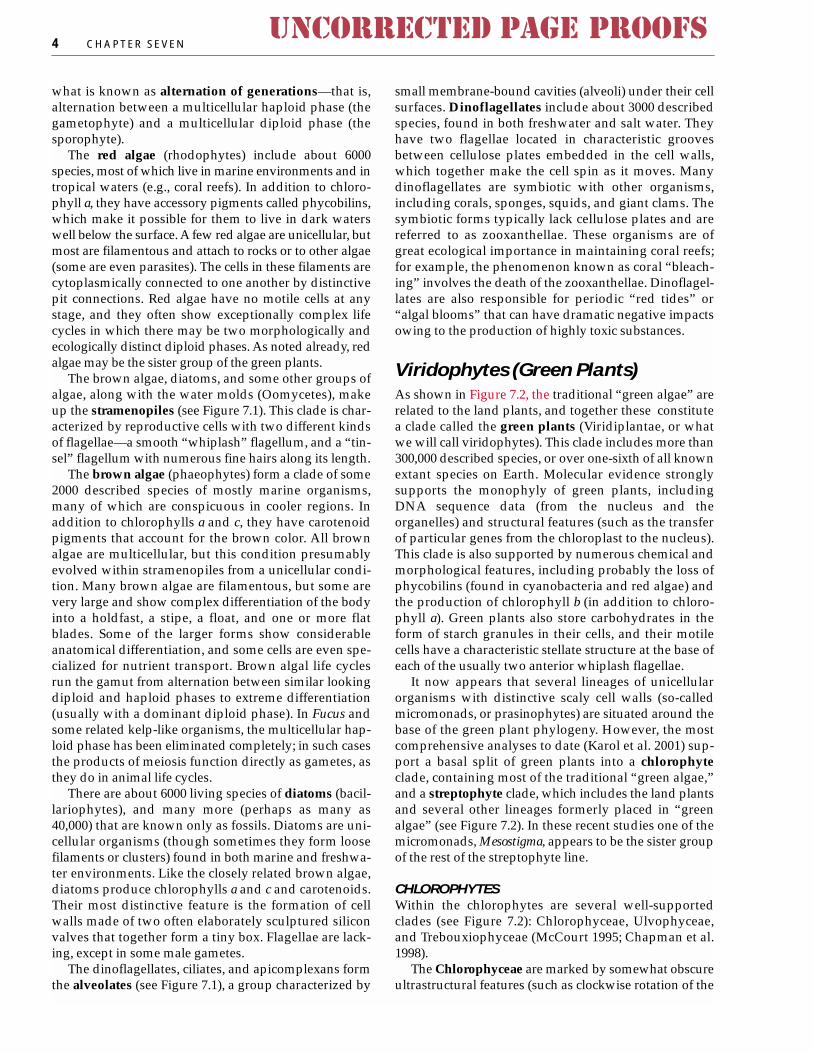
what is known as alternation of generations—that is,alternation between a multicellular haploid phase (thegametophyte) and a multicellular diploid phase (thesporophyte).
The red algae (rhodophytes) include about 6000species, most of which live in marine environments and intropical waters (e.g., coral reefs). In addition to chloro-phyll a, they have accessory pigments called phycobilins,which make it possible for them to live in dark waterswell below the surface. A few red algae are unicellular, butmost are filamentous and attach to rocks or to other algae(some are even parasites). The cells in these filaments arecytoplasmically connected to one another by distinctivepit connections. Red algae have no motile cells at anystage, and they often show exceptionally complex lifecycles in which there may be two morphologically andecologically distinct diploid phases. As noted already, redalgae may be the sister group of the green plants.
The brown algae, diatoms, and some other groups ofalgae, along with the water molds (Oomycetes), makeup the stramenopiles (see Figure 7.1). This clade is char-acterized by reproductive cells with two different kindsof flagellae—a smooth “whiplash” flagellum, and a “tin-sel” flagellum with numerous fine hairs along its length.
The brown algae (phaeophytes) form a clade of some2000 described species of mostly marine organisms,many of which are conspicuous in cooler regions. Inaddition to chlorophylls a and c, they have carotenoidpigments that account for the brown color. All brownalgae are multicellular, but this condition presumablyevolved within stramenopiles from a unicellular condi-tion. Many brown algae are filamentous, but some arevery large and show complex differentiation of the bodyinto a holdfast, a stipe, a float, and one or more flatblades. Some of the larger forms show considerableanatomical differentiation, and some cells are even spe-cialized for nutrient transport. Brown algal life cyclesrun the gamut from alternation between similar lookingdiploid and haploid phases to extreme differentiation(usually with a dominant diploid phase). In Fucus andsome related kelp-like organisms, the multicellular hap-loid phase has been eliminated completely; in such casesthe products of meiosis function directly as gametes, asthey do in animal life cycles.
There are about 6000 living species of diatoms (bacil-lariophytes), and many more (perhaps as many as40,000) that are known only as fossils. Diatoms are uni-cellular organisms (though sometimes they form loosefilaments or clusters) found in both marine and freshwa-ter environments. Like the closely related brown algae,diatoms produce chlorophylls a and c and carotenoids.Their most distinctive feature is the formation of cellwalls made of two often elaborately sculptured siliconvalves that together form a tiny box. Flagellae are lack-ing, except in some male gametes.
The dinoflagellates, ciliates, and apicomplexans formthe alveolates (see Figure 7.1), a group characterized by
small membrane-bound cavities (alveoli) under their cellsurfaces. Dinoflagellates include about 3000 describedspecies, found in both freshwater and salt water. Theyhave two flagellae located in characteristic groovesbetween cellulose plates embedded in the cell walls,which together make the cell spin as it moves. Manydinoflagellates are symbiotic with other organisms,including corals, sponges, squids, and giant clams. Thesymbiotic forms typically lack cellulose plates and arereferred to as zooxanthellae. These organisms are ofgreat ecological importance in maintaining coral reefs;for example, the phenomenon known as coral “bleach-ing” involves the death of the zooxanthellae. Dinoflagel-lates are also responsible for periodic “red tides” or“algal blooms” that can have dramatic negative impactsowing to the production of highly toxic substances.
Viridophytes (Green Plants)As shown in Figure 7.2, the traditional “green algae” arerelated to the land plants, and together these constitutea clade called the green plants (Viridiplantae, or whatwe will call viridophytes). This clade includes more than300,000 described species, or over one-sixth of all knownextant species on Earth. Molecular evidence stronglysupports the monophyly of green plants, includingDNA sequence data (from the nucleus and theorganelles) and structural features (such as the transferof particular genes from the chloroplast to the nucleus).This clade is also supported by numerous chemical andmorphological features, including probably the loss ofphycobilins (found in cyanobacteria and red algae) andthe production of chlorophyll b (in addition to chloro-phyll a). Green plants also store carbohydrates in theform of starch granules in their cells, and their motilecells have a characteristic stellate structure at the base ofeach of the usually two anterior whiplash flagellae.
It now appears that several lineages of unicellularorganisms with distinctive scaly cell walls (so-calledmicromonads, or prasinophytes) are situated around thebase of the green plant phylogeny. However, the mostcomprehensive analyses to date (Karol et al. 2001) sup-port a basal split of green plants into a chlorophyteclade, containing most of the traditional “green algae,”and a streptophyte clade, which includes the land plantsand several other lineages formerly placed in “greenalgae” (see Figure 7.2). In these recent studies one of themicromonads, Mesostigma, appears to be the sister groupof the rest of the streptophyte line.
CHLOROPHYTESWithin the chlorophytes are several well-supportedclades (see Figure 7.2): Chlorophyceae, Ulvophyceae,and Trebouxiophyceae (McCourt 1995; Chapman et al.1998).
The Chlorophyceae are marked by somewhat obscureultrastructural features (such as clockwise rotation of the
4 C H A P T E R S E V E NUNCORRECTED PAGE PROOFS

basal bodies), but they have been supported as a clade inmost molecular studies as well. Included within this lineis the so-called volvocine lineage, within which progres-sively more complex colonies (from 4 cells in Gonium toas many as 500–50,000 cells in the hollow sphericalcolonies of Volvox) were presumed to have been derivedfrom unicells not unlike the model organism Chlamy-domonas (Figure 7.3A, B). Recent studies indicate that thestory is more complex, with several colonial lines derivedindependently, perhaps from within Chlamydomonasitself, which has hundreds of species (Larsen et al. 1991;Buchheim et al. 1994; Chapman et al. 1998).
The Ulvophyceae include many marine forms and ismarked by the production of multinucleate cells (Figure7.3D–F). In some, the entire body is a coenocytic thallus,lacking walls between the nuclei except in the case ofreproductive cells. Included in this group is the modelorganism Acetabularia (Figure 7.3F).
Finally, the Trebouxiophyceae contain forms with fla-gellate spores, but most are small round forms (appar-
ently derived several times independently) that com-pletely lack motile cells at any stage. Many of the non-motile forms live in terrestrial habitats, often in associa-tion with lichen-forming fungi or invertebrate animals.Lichen associations appear to have originated and tohave been lost multiple times (Lutzoni et al. 2001).
STREPTOPHYTESThe discovery of the streptophyte lineage began in thelate 1960s, when detailed ultrastructural studies of celldivision first revealed a major difference in the orienta-tion of the spindle microtubules among the organismsthat had traditionally been classified as “green algae”(Pickett-Heaps 1979; Mattox and Stewart 1984; McCourt1995). Some of these were found to have the phragmo-plast orientation found in all land plants, in which thespindle is oriented perpendicular to the formation of thecell wall. A thorough survey showed that the phragmo-plast condition occurred in the so-called charophyceanalgae (Coleochaetales and Charales). These plants show
A N O V E R V I E W O F G R E E N P L A N T P H Y L O G E N Y 5UNCORRECTED PAGE PROOFS
fpo
Chl
orop
hyce
ae
Tre
boux
ioph
ycea
e
Ulv
ophy
ceae
Mes
osti
gma
Chl
orok
ybus
Kle
bsor
mid
iale
s
Zyg
nem
atal
es
Col
eoch
aeta
les
Cha
rale
s
Em
bryo
phyt
es(l
and
pla
nts)
>1000 MYBP
>470 MYBP
Streptophytes
Viridophytes (green plants)
Chlorophytes
“Green algae”
Chlorophyll b, starch storage, “stellate”flagellar structure, gene transfers
Multicellular sporophyte, embryo, gametangia, sporangium, cuticle
Multilayered structure offlagellae and cytoskeleton
Filamentous growth
“Micromonads”
Phragmoplast
Branched, apical growth, oogamy,plasmodesmata, retention of egg
Encasement of egg, sperm morphology, numerous chloroplasts per cell
Figure 7.2 The base of green plant phy-logeny, showing the separation of chloro-phytes from streptophytes, the relationshipof some former “green algae” to embryo-phytes, and characters marking major clades(see text). MYBP, million years before present.(Adapted from Karol et al. 2001.)

a range of different life forms (including upright, branch-ing forms, as in Chara and Nitella, and flattened forms, asin Coleochaete) and live in near-shore, freshwater habitats(Figure 7.4A–C) As these organisms were studied inmore detail, the idea emerged that they were actuallymore closely related to land plants than they were toother “green algae.” has since become clear that severalother former green algal ineages belong in the strepto-phyte clade, including Klebsormidiales and Zygne-matales (McCourt 1995; Chapman et al. 1998). Of these,the Zygnematales may be familiar as the group thatincludes Spirogyra and its relatives (Figure 7.4D–E).These are the so-called conjugating “green algae,” in ref-erence to a form of sexual reproduction that involves theformation of a tubular connection between cells of adja-cent filaments, passage of the protoplast from one cell toanother, and the eventual fusion of nuclei to form azygote.
The relationships among the streptophyte groupsshown in Figure 7.2 have been confirmed by molecularstudies (Karol et al. 2001), including some structural
molecular data. For example, the protein elongation fac-tor tufA appears to have moved from the chloroplast tothe nucleus in the ancestor of the “charophyte” lineagesand the land plants (Baldauf and Palmer 1990).Coleochaetales and Charales possess some functionallyimportant traits that are found otherwise only in landplants, such as flavonoids and the chemical precursors ofcuticle. Most important from the standpoint of the evo-lution of the land plant life cycle is the fact that theyretain the egg and sometimes even the zygote (after fer-tilization) on the body of the haploid plant (Graham1993).
These phylogenetic results have many importantimplications for our understanding of green plant evolu-tion. For instance, they imply that there were severalindependent originations of multicellularity. As we havenoted, the volvocine forms explored a lifestyle in whichthe cells became aggregated into colonies. The larger ofthese colonies also show cytoplasmic interconnectionsand a division of labor, with some cells specialized forreproduction. Other chlorophytes formed filaments ormembranous parenchymatous bodies of much largersize (such as the sea lettuce, Ulva, and its relatives),which show a more complex morphological integrationand differentiation of cell functions. The Ulvophyceaefollowed a separate path involving multinucleate cells,sometimes forming filaments, and sometimes (as in
6 C H A P T E R S E V E NUNCORRECTED PAGE PROOFS
(A)
(F)
Figure 7.3 Morphology ofchlorophytes. (A–C) Chlorophyceae: (A) Chlamydomonas, show-ing flagellae (f ); (B) Eudorina, a colonial “volvocine” form; (C)Stigeoclonium, a branched filamentous form. (D–F) Ulvophyceae:(A) Ulva; (B) Codium, showing a coenocytic diploid thallus; (C)Acetabula. (From Scagel et al. 1969.)
(B)
(C)
(D)
(E)
Flagellae

Codium) forming a thallus by densely intertwining thefilaments. Finally, multicellularity evolved separately inthe streptophyte line. Many Zygnematales are filamen-tous, and parenchymatous forms (with plasmodesmataconnecting adjacent cells) are found in the two charo-phyte lineages and the land plants.
Among the early-diverging lineages of green plants,we also encounter a wide variety of life cycles. Alterna-tion of similar haploid gametophyte and diploid sporo-phyte generations (as in Ulva) is quite common. In con-trast, Codium (Ulvophyceae) evolved a life cycle like thatof humans, in which gametes are the only haploid cells.In stark contrast, in charophytes the plants are haploid,and the only diploid cell in the life cycle is the zygote,which results from fertilization of a large nonmotile eggby a swimming sperm (oogamy).
Embryophytes (Land Plants)The land plants are depicted as stemming from a singlecommon ancestor in Figure 7.2, a finding that is stronglysupported by both molecular and morphological evi-
dence (Kenrick and Crane 1997a, 1997b; Karol et al.2001). Land plants are also called embryophytes becausethey have a resting embryo stage early in the life of thesporophyte. Embryophyte is the preferable term in thiscase because several algal lineages have independently(though less conspicuously) also made the transition tolife on land (e.g., in Trebouxiophyceae). In addition tothe embryo, embryophytes are characterized by produc-tion of a multicellular sporophyte, multicellular repro-ductive structures (antheridia, archegonia, and sporan-gia), cuticle, and thick-walled spores with characteristictrilete marks (see Figure 7.7C).
Traditionally, embryophytes have been classified aseither bryophytes or vascular plants. There are threemajor lineages of bryophytes—mosses, hornworts, andliverworts—which we will characterize briefly in thenext few paragraphs (see also Shaw and Goffinet 2000).As we shall discuss, however, it has become increasinglyclear that “bryophytes” are paraphyletic with respect tovascular plants (see Figure 7.6).
MOSSESMosses are probably the most familiar bryophytic plantsand, with some 15,000 species, they are also the mostdiverse. The upright and leafy gametophyte is the domi-nant phase in the moss life cycle (Figure 7.5A–C). Thesporophyte forms a single unbranched stalk terminatedby a sporangium (or capsule). Haploid spores, producedvia meiosis, are released from the sporangium; typically,dehiscence of the sporangium occurs by the detachmentof a lid or operculum.
When a spore germinates, it forms a protonemalstage, which resembles a green algal filament. The pro-
A N O V E R V I E W O F G R E E N P L A N T P H Y L O G E N Y 7UNCORRECTED PAGE PROOFS
(A)
Figure 7.4 Morphology of basal streptophytes. (A)Coleochaete, showing a haploid discoidal thallus, with setae. (B,C) Charales: (B) Chara, showing a node with an egg-bearingstructure (above) and a sperm-producing structure (below)’ (C)Nitella habit, showing node (n) and internode (in) construction.(D, E) Zygnematales: (D) Spirogyra, a filamentous form, showinghelical chloroplasts (ch). (E) Staurastrum, a unicellular desmid,forming two mirror-image semicells. (A from Taylor and Taylor1993; B–E from Scagel et al. 1969.)
Chloroplast
(D)
(B) (C)
(E)
n
in

tonema produces one or more upright leafy gameto-phytes, which ultimately produce sperm and eggs inantheridia and archegonia, respectively. Fusion of thegametes yields the zygote, which develops through aseries of mitotic divisions into the embryo and eventual-ly into the mature sporophyte.
Analyses of relationships within mosses have gener-ally supported the idea that Sphagnum (peat moss) is sit-uated near the base of the tree and that Andreaea and afew close relatives also form an early branch (see Ken-rick and Crane 1997a). The enigmatic Takakia, which wasconsidered to be a liverwort until the recent discovery ofthe sporophyte phase, is also probably situated near the
base of the moss tree. The sporangium in Andreaea opensby four vertical slits, and in Takakia by a single helical slit,as opposed to the lid-like operculum found in the vastmajority of mosses. The operculum of most mosses isalso characterized by a distinctive row of tooth-likestructures, which together make up the peristome.
LIVERWORTSThere are about 9000 species of liverworts, which comein a thalloid form or, more commonly, a leafy form (Fig-ure 7.5D,E). Unlike mosses and hornworts, liverwortslack stomates, although some have epidermal poreswithout true guard cells. They also lack a characteristiccolumnar mass of sterile tissue (the columella) in thesporangium, which is present in mosses, hornworts, andearly vascular plant lineages.
These liverwort features have sometimes been inter-preted as ancestral within land plants, but this is nolonger clear. Sex in liverworts involves the production of sperm-producing antheridia and egg-containingarchegonia. The sporophyte phase, with its terminal spo-rangium, is rather small and inconspicuous. The capsuletypically opens through four valves, and sterile hy-groscopic cells (elaters) among the spores may aid in dispersal.
8 C H A P T E R S E V E NUNCORRECTED PAGE PROOFS
Figure 7.5 Morphology of “bryophytes.” (A–C) Mosses: (A)Dawsonia superba habit, showing leafy gametophyte andunbranched sporophyte with terminal sporangium; (B) spo-rangium (capsule) of a moss prior to dehiscence; (C) apex of thedehiscing sporangium of a moss, Fontinalis antipyretica, showingthe peristome teeth. (D, E) Liverworts: (D) A leafy liverwort,Lepidozia reptans, showing dehiscence of the sporangium byfour valves; (E) portion of a thalloid liverwort, Monoclea forsteri,showing sporangia with longitudinal dehiscence. (F, G)Hornworts: (F) Phaeoceros laevis habit, showing the thalloidgametophyte and dehiscing sporangia of the sporophyte; (G)stomate, with guard cells, from the sporangium wall ofAnthoceros. (A from Barnes 1998; B–G from Scagel et al. 1969.)
(A)
(B) (E)
(F)
(G)(D)
(C)

HORNWORTSThere are only about 100 species of hornworts (Figure7.5A,B), which are encountered much more rarely thaneither mosses or liverworts. One presumably derivedfeature of this entirely thalloid group is the presence ofan intercalary meristem in the sporophyte located at thebase of the capsule. The activity of this meristemaccounts for the continued upward growth of the cap-sule, which is quite extensive in some groups (e.g.,Anthoceros).
PHYLOGENETIC RELATIONSHIPS OF EMBRYOPHYTESAll recent phylogenetic analyses of land plants have con-cluded that “bryophytes” are paraphyletic. However,the exact relationships are still controversial (Figure 7.6).Initially, morphological analyses supported a basal splitbetween the liverwort lineage and everything else, andplaced the mosses as the sister group to the vascularplants (Mishler and Churchill 1984, 1985). Under thisview, stomates are considered to be an innovation link-ing hornworts, mosses, and vascular plants, to the exclu-sion of liverworts. Likewise, specialized cells in thestems of mosses (in both the gametophyte and sporo-phyte of some species), called hydroids and leptoids,have been interpreted as precursors of the water- andnutrient-conducting cells found in vascular plants.Mosses and vascular plants have sporophytes that
increase in height through cell divisions in an apicalmeristem, and the first vascular plants also had uprightgametophytes, as do mosses. These interpretations havebeen upheld by some molecular studies, including ananalysis showing the gain of three mitochondrial intronsin all land plants except liverworts (Qiu et al. 1998).
Several recent studies of molecular data, however—alone and in combination with a variety of morphologi-cal and ultrastructural characters (especially spermultrastructure)—support an alternative hypothesis (Fig-ure 7.6B). In these trees, hornworts are the sister group ofall other extant land plants, and a clade containing moss-es and liverworts is the sister group of the vascularplants (Nickrent et al. 2000; Renzaglia et al. 2000).
This hypothesis is consistent with detailed studiesindicating that the hydroids and leptoids in mossesprobably are not homologous with tracheids in thexylem and sieve cells in the phloem of vascular plants(Ligrone et al. 2000). However, such relationships wouldimply either the loss or the independent evolution of sto-mates and apical meristems. Although this issue remains
A N O V E R V I E W O F G R E E N P L A N T P H Y L O G E N Y 9UNCORRECTED PAGE PROOFS
Hor
nwor
ts
Liv
erw
orts
Mos
ses
Hor
nwor
ts
Liv
erw
orts
Mos
ses
Hor
neop
hyto
n
Agl
aoph
yton
Tra
cheo
phyt
es(v
ascu
lar
plan
ts)
>450 MYBP
>425 MYBP
Embryophytes (land plants)
Polysporangiophytes“Bryophytes”
“Bryophytes”
Multicellular sporophyte, embryo,gametangia, sporangium, cuticle
Sperm ultrastructure
Sporophyte independent,branching
Vascular tissue, xylemwith tracheids
Sporophyteapical meristem
Stomata
(A)
(B)
Figure 7.6 Phylogenetic relationships at the base of theembryophytes (land plants), showing characters that mark majorclades under two hypotheses of how the “bryophyte” groups (horn-worts, liverworts, and mosses) are related to vascular plants (seetext). MYBP, million years before present. (A adapted from Mishlerand Churchill 1984; B adapted from Nickrent et al. 2000 andRenzaglia et al. 2000.)

unsettled, note that all recent analyses support the viewthat “bryophytes” are paraphyletic with respect to vas-cular plants.
TRANSITION TO LANDThis phylogenetic knowledge illuminates the origin ofseveral key adaptations to life on land (Graham 1993).Cuticle and sporopollenin (present in the thick sporewall) appear to be responses to desiccation. Gasexchange is facilitated by small pores in the epidermis orby genuine stomates with guard cells that can open orclose the stomate depending on environmental condi-tions, thereby regulating water loss. Flavonoids helpabsorb damaging long-wavelength UV radiation. A gly-colate oxidase system helps ameliorate the fact that car-bon dioxide fixation is inhibited in the presence of oxy-gen, which is in much higher concentrations in the airthan in water. The first land plants probably dependedon symbiotic relationships with fungi to obtain nutrientsfrom the soil, and such relationships have been docu-mented in the major bryophytic lineages, as well as invascular plants (where they are ubiquitous). The precur-sors of many of these adaptations can be found amongthe closely related Coleochaetales and Charales, andthese plants therefore appear to have been preadapted tomake the transition to land (Delwiche et al. 1989; Gra-ham et al. 1991; Graham 1993).
Appreciation that both the traditional “green algae”and the “bryophytes” are paraphyletic has also helpedus understand the origin of the characteristic land plantlife cycle, involving an alternation of multicellular game-tophyte and sporophyte phases (Mishler and Churchill1985; Graham 1993). As noted earlier, in Coleochaete andChara the egg is retained on the haploid parent plant. InColeochaete the zygote (the only diploid stage) alsoremains on the parent plant until it undergoes meiosis togive rise to haploid spores. A key innovation in the linethat includes the charophyte lineages and the embryo-phytes was the establishment of nutrient transportbetween haploid and diploid phases through a placentaltransfer tissue (Graham and Wilcox 2000). The landplant life cycle was probably derived from a charophyte-like ancestral condition by simple delay of meiosis andinterpolation of a multicellular diploid phase via a seriesof mitotic divisions of the zygote.
In the embryophytes, the egg—and after fertilization,the embryo—is protected by a multicellular structurecalled an archegonium (plural archegonia). Sperm areproduced and protected by a multicellular structurecalled an antheridium (plural antheridia). Initially, thegametophyte phase was dominant, as it is today in horn-worts, liverworts, and mosses, and the sporophyteremained attached to, and was nutritionally dependenton, the gametophyte. In vascular plants, the sporophytebecame dominant and nutritionally independent, andthere was progressive reduction in gametophyte size(Kenrick and Crane 1997a, 1997b).
These findings also help us interpret the absolute tim-ing of events (see Figures 7.1, 7.2, and 7.6). Green plantsmay be a billion or more years old, and it is possible thatsome major green plant lineages existed in the Precam-brian (Heckman et al. 2001). From the Cambrian (about550 million years ago), a variety of chlorophyte fossilshave been found, including well-preserved lime-secret-ing Ulvophyceae, such as relatives of Acetabularia.Charophytes (in the form of calcified Charales) do notappear in the fossil record until the mid-Silurian, but thewholesale occupation of land by green plants probablytook place beginning in the mid-Ordovician, about 450million years ago. Starting at that time (and possiblyeven earlier, in the Cambrian), dispersed spores arefound, sometimes in envelope-enclosed tetrads or diads(sets of four or two, respectively) resembling those seentoday in some liverworts. Tiny bits of cuticle and tubularstructures of plant origin also appear in the Ordovician,and individual spores with the characteristic triletemarks of land plants (see Figure 7.7C) have been recov-ered from the early Silurian.
It is probable, therefore, that hornworts, liverworts,mosses, and vascular plants were all in existence by thelate Ordovician. Somewhat later, beginning in the mid-Silurian, there are well-preserved macrofossils represent-ing the vascular plant lineage. The occupation of landwas certainly in full swing by then.
Tracheophytes (Vascular Plants)All indications are that the first land plants were smalland very simple in structure. In the case of the vascularplant lineage, the sporophyte was basically a dichoto-mously branching stem, about the height of a matchstickat first, with the sporangia (the site of meiosis yieldinghaploid spores) produced at the tips of the branches(Figure 7.7A). These plants had no leaves or roots. Insome cases (e.g., Rhynia, from the Rhynie chert in Scot-land), the preservation of these plants is spectacular, andit is possible to discern many anatomical details, includ-ing stomates, spores, and the vascular tissue inside thestem.
On the basis of such fossils, it was recently discoveredthat the first polysporangiophytes —plants withbranching sporophytes—did not actually produce bonafide water-conducting cells (tracheids) in the xylem tis-sue and must therefore have depended on turgor pres-sure to remain upright. True water-conducting cellsevolved somewhat later and characterize a clade knownas the true vascular plants, or the tracheophytes (Ken-rick and Crane 1997a, 1997b).
Tracheids are elongate cells with thickened walls thatare dead at maturity. Where one tracheid connects to thenext, there are characteristic openings or pits, but a pitmembrane (primary cell wall) remains intact, and watermust pass through it as it moves from one cell to thenext. In the first tracheophytes (represented by Rhynia),
10 C H A P T E R S E V E NUNCORRECTED PAGE PROOFS

the tracheids were of a distinctive type, in which decayresistance (conferred by lignification of cellulose fibers)was present only as a very thin layer. Cell walls that aremuch more decay-resistant characterize a eutracheo-phyte clade, which includes all extant vascular plants(Kenrick and Crane 1997a). In these species, the stronglylignified tracheids allow more efficient water conductionand provide internal support, allowing the plants togrow much taller.
In recent years, careful paleobotanical studies haverevealed that some early land plant fossils are actuallyhaploid gametophytes, bearing antheridia and archego-nia (Remy 1982; Remy et al. 1993). These fossils areremarkable because they are rather large, upright, andbranched, and in general they resemble the sporophytephase of the life cycle (see Figure 7.7E). This finding hasled to the view that the first members of the vascularplant lineage exhibited alternation of more or less similargenerations. Thus, in relation to the bryophytic groups, itseems that both the gametophyte and the sporophytephases were elaborated.
This knowledge allows us to piece together asequence of events leading to the life cycle that we see invascular plants today. This life cycle includes a dramaticreduction in the gametophyte phase and an equallyimpressive elaboration of the sporophyte phase. In thefirst vascular plants, the gametophyte was nutritionallyindependent of the sporophyte, and this condition isretained today in the “free-spore” lineages such as ferns
A N O V E R V I E W O F G R E E N P L A N T P H Y L O G E N Y 11UNCORRECTED PAGE PROOFS
Figure 7.7 Fossils of early tracheophytes (vascular plants). (A)Reconstruction of Rhynia major, showing the dichotomouslybranching stem (without leaves or roots) and terminal sporan-gia. (B) Enlarged terminal sporangium of R. major, with sporesinside. (C) R. major spores in tetrad, and a single thick-walledspore with the trilete mark characteristic of land plants. (D)Reconstruction of Uskiella spargens, showing the dichotomouslybranched stem and terminal sporangia with distal dehiscence.(E) Reconstruction of a Devonian gametophyte, Sciadophytonsp.; gametangia are present on the terminal disk-shaped struc-tures. (A–C from Stewart 1983; D and E from Kenrick and Crane1997a.)
(A)
(B)
(C)
(D)
(E)

and lycophytes. With the evolution of seed plants, how-ever, the gametophyte became much further reducedand eventually became completely dependent on thesporophyte.
Viewed in this context, the bryophytic groups (espe-cially the mosses) and the vascular plants appear to haveexplored two different mechanisms to increase the num-ber of spores produced per fertilization event (Mishlerand Churchill 1985). In mosses this increase in spore pro-duction was accomplished by intercalation of a filamen-tous protonemal stage that could produce numerousunbranched leafy gametophytes, each bearing a singleunbranched sporophyte terminated by a single spo-rangium. In contrast, in the vascular plant lineage thenumber of sporangia was increased by branching of thesporophyte so that each branch tip could bear a spo-rangium.
What factors might have favored the elaboration ofthe sporophyte phase as opposed to the gametophyte
phase (which became increasingly specialized for sexualreproduction)? One hypothesis holds that diploid organ-isms are buffered against deleterious mutations. But analternative is that the sporophyte was free to becomelarger (which was advantageous in competing for lightand may also have enhanced spore dispersal), whereasthe gametophyte was dependent on water for fertiliza-tion as long as the sperm needed to swim to the egg.
Phylogenetic relationships among the major lines ofextant vascular plants are shown in Figure 7.8. Theseconclusions are based on morphological and molecularevidence and are now quite strongly supported (Doyle1998; Pryer et al. 2001). The basal split, which occurred inthe early to mid-Devonian (before 400 million yearsago), separated a clade that included the modern lyco-phyte lineage from a clade, known as the euphyllo-phytes, that contains all of the other extant vascularplant lineages. This split is marked by a variety of mor-phological features. One noteworthy feature is the pres-
12 C H A P T E R S E V E NUNCORRECTED PAGE PROOFS
fpo
Isoe
tes
Lep
idod
end
rale
s†
Sela
gine
lla
Lyc
opod
iace
ae
Zos
tero
phyt
es†
Coo
kson
ia†
Psilo
tace
ae
Oph
iogl
ossa
ceae
Equ
iset
ophy
tes
Mar
atti
ales
Poly
pod
iale
s
Ane
urop
hyte
s†
Arc
haeo
pter
is†
Sper
mat
ophy
tes
(see
d p
lant
s)
>420 MYBP
>410 MYBP>380 MYBP
Tracheophytes (vascular plants)
Lycophytes Euphyllophytes
Monilophytes Lignophytes
Vascular tissue, xylemwith tracheids
Tracheids strongly lignified
Seed
Heterospory
Secondary xylem (wood)
Chloroplast inversion,multiflagellate sperm, megaphylls
Over-topping branches(pseudomonopodial)
Loss of roots
Stellar anatomy
Psilophyton†
Rhyniophytes†
Secondary xylem
Heterostyly, ligule
Microphylls
Lateral reniformsporangia
Transverse dehiscence
Figure 7.8 Phylogeny of tracheophytes(vascular plants) showing the basal splitbetween lycophytes and euphyllophytes,the relationship of some former “seedlessvascular plants” (monilophytes and “pro-gymnosperms”) to spermatophytes, andcharacters marking major clades (see text).MYBP, million years before present; †, extincttaxon. (After Pryer et al. 2001.)

ence of multiflagellate sperm in the euphyllophytes, asopposed to biflagellate sperm in the bryophytic lineagesand in lycophytes (except in Isoetes and Phylloglossum,where multiflagellate sperm evolved independently).One compelling bit of molecular evidence is the presencein the euphyllophytes of a 30-kilobase inversion in thechloroplast DNA (Raubeson and Jansen 1992); lyco-phytes and the bryophytic plants lack this inversion.
LYCOPHYTESThe lycophyte lineage (see Figures 7.8 and 7.9; see alsoFigures 8.2 and 8.3) appeared in the fossil record verysoon after the first appearance of vascular plants and ismarked by the lateral position, reniform shape, andtransverse dehiscence of the sporangia. Small, micro-phyllous leaves with a single vascular strand evolvedwithin this lineage (possibly through modification of lat-eral sporangia), as did distinctive dichotomouslybranching roots. During the Carboniferous period lyco-phytes were especially diverse and abundant, and theremains of these plants account for our major coaldeposits. In particular, they dominated coastal swampsof tropical lowlands (DiMichele and Skog 1992; Batemanet al. 1998).
Some lycophytes became large trees, with secondarygrowth allowing an increase in girth (Figure 7.9). Thestems of these plants were covered by leaves, which leftthe distinctive leaf bases seen in fossils. These plants alsoevolved so-called stigmarian root systems; these are pre-sumed to have been derived from rhizomes, in whichcase the spirally arranged rootlets may be modifiedleaves. Patterns of growth in these large plants are stillpoorly understood (Bateman 1994; Bateman et al. 1998),but they may have grown very slowly in height at first(while the root system became established) and laterelongated rapidly. They may have died after simultane-ously producing strobili at the tips of all the branches.
Today there are over 1200 species of lycophytes,belonging to several major lines (see Figures 7.8 and 7.9).Of these, rhizomatous species of Huperzia, Lycopodiella,and Lycopodium (club mosses) are commonly encoun-tered in forests of the Northern Hemisphere. Theseplants and their tropical relatives are homosporous,meaning that they produce just a single kind of spore,which gives rise to a bisexual gametophyte, producingboth sperm and eggs.
The other living lycophytes (Selaginella, Isoetes) areheterosporous, producing microspores, which give riseto male gametophytes, and megaspores, which give riseto female gametophytes. The heterosporous taxa form aclade, which is also united by the association of a leaf-like flap of tissue, the ligule, with the adaxial side of theleaf base.
Selaginella (spike mosses) (see Figure 7.9F–I), withover 700 species, is most diverse in the tropics, wheremany species grow as epiphytes. Isoetes (quillworts, withperhaps 150 species) is the only living remnant of the
clade that included the giant lycopods of the Carbonifer-ous, though it may have been derived from plants in thislineage that never attained the size of Lepidodendron (seeFigure 7.9D) and the other very large lycophyte trees.Isoetes has retained the cambium and some secondarygrowth, and it has rootlets that resemble those of theextinct trees (see Figure 7.9J).
EUPHYLLOPHYTESAn advance of the euphyllophytes (see Figure 7.8) wasdifferentiation between a main axis and side branches(pseudomonopodial growth), an arrangement found ina variety of Devonian fossils known as trimerophytes(Figure 7.10A). According to the “telome theory” (Zim-mermann 1952, 1965), large megaphyllous leaves charac-teristic of the euphyllophytes were derived from flat-tened lateral branch systems. This derivation involvedplanation of the branch system and then webbing toform the leaf blade. It seems clear that leaves evolvedindependently, and by very different pathways, in thelycophyte line versus the euphyllophyte line.
Living euphyllophytes appear to belong to two majorclades (see Figure 7.8): the seed plants (spermatophytes)and a clade that includes several “fern” lineages, thehorsetails (equisetophytes), and the whisk ferns (themonilophytes; Moniliformopses of Kenrick and Crane1997a). This new view of relationships is well supportedin combined analyses of morphological characters andboth chloroplast and nuclear genes (Pryer et al. 2001).Within the monilophytes (ferns and their allies), thereare five major lineages, each discussed briefly here: (1)leptosporangiate ferns (Polypodiales), (2) Ophioglos-saceae, (3) Psilotaceae, (4) Marattiales, and (5) equiseto-phytes (see also Chapter 8).
The common name fern is applied to the members ofthree of these major lineages: Polypodiales, Marattiales,and Ophioglossaceae. These plants are superficially sim-ilar in usually having large (often highly dissected)frond-like leaves that unfold from a “fiddlehead” (so-called circinate vernation). These three lineages are usu-ally divided into two groups on the basis of the structureand development of the sporangia. The Marattiales andthe Ophioglossaceae are so-called eusporangiate ferns(see Figure 7.10). These appear to have retained theancestral condition, in which the sporangium developsfrom several initial cells and the mature wall of the spo-rangium is more than one cell layer thick. Eusporangiaalso tend to contain large numbers of haploid spores atmaturity.
In contrast, the Polypodiales are characterized by aderived development in which the sporangium arisesfrom a single cell and the mature wall is only one cellthick. These leptosporangia are borne on a distinct stalkand have a characteristic annulus consisting of a row ofcells with thickened inner walls and thin outer walls (seeFigure 8.12). The leptosporangia of most species containa relatively small and definite number of haploid spores
A N O V E R V I E W O F G R E E N P L A N T P H Y L O G E N Y 13UNCORRECTED PAGE PROOFS

14 C H A P T E R S E V E NUNCORRECTED PAGE PROOFS
(A)
(B)
(D)
Figure 7.9 Morphology of lycophytes. (A) Reconstruction ofthe extinct Zosterophyllum deciduum, showing prostrate rhizomebearing leafless upright axes with lateral reniform sporangia. (B)Reconstruction of the extinct Asteroxylon mackiei, showingupright dichotomizing stems covered by microphyllous leaves,and rootlike axes. (C) A. mackiei, showing part of a fertile axiswith reniform sporangia and tranverse dehiscence. (D)Reconstruction of an extinct Lepidodendron sp., showing thedichotomously branching root system, the massive trunk withdichotomous branching above, and terminal strobili. (E) Portionof the surface of a stem of a Lepidodendron sp., showing three
attached leaves and the scars left by the abscission of five oth-ers. (F) Tip of a branch of Selaginella, showing microphyllousleaves and a terminal strobilus. (G) Microsporangium ofSelaginella in the axil of a microsporophyll. (H) Megasporangiumof Selaginella in the axil of a megasporophyll. (I) Longitudinalsection through a strobilus of Selaginella harrisiana, showingmegasporangia (me) with four large megaspores, microsporan-gia (mi) with many tiny microspores, and ligules. (J) Isoetesbolanderi habit, showing leaves and roots. (A and J from Kenrickand Crane 1997a; B, C, and I from Stewart 1983; D and E fromGifford and Foster 1989; F–H from Barnes 1998.)
(J)
(C)
(F) (G) (H)
(E)
(I)
Ligule
memi

(e.g., 16, 32, 64), which are ejected from the sporangia bya mechanism driven by changes in moisture content inthe annulus cells.
Probably the most familiar monilophytes are thePolypodiales, of which there are more than 12,000 livingspecies (see Figures 8.1, and 8.8 through 8.14). Many ofthese plants have highly dissected pinnate leaves, of thetype we commonly associate with ferns, but leaf form isactually extremely variable, and some even have simple,undissected leaves. The sporangia are typically pro-duced in small clusters (each cluster is called a sorus, col-lectively the sori) on the undersides of the leaves. Thesori are often covered by a flap of tissue called an indusi-um, though some are “naked.” The structure and posi-tion of the sori and the indusium vary enormously fromone fern group to another, and this variation has beenemphasized in taxonomic treatments (see Chapter 8).
Fern gametophytes are often small, heart-shaped struc-tures, with the archegonia present near the notch andantheridia situated among the characteristic rhizoids.There is considerable variation, however, and in someferns the gametophyte is even filamentous.
Within the leptosporangiate line, recent morphologi-cal and molecular studies have identified several note-worthy clades (Pryer et al. 1995; Wolf et al. 1998). As hasbeen long suggested on the basis of sporangium devel-opment (sporangia not in sori, rudimentary annulus,large number of spores), Osmundaceae (cinnamon ferns)are seen to be the sister group of the rest. One distinctiveleptosporangiate clade includes the large tree ferns(Cyatheaceae), and another contains all of the het-erosporous aquatic fern groups (placed in Marsileaceaeand Salviniaceae). Although the aquatic ferns are mor-phologically quite different from one another (e.g.,Salvinia and Azolla with small floating leaves, versusMarsilea with leaves resembling those of a four-leafclover; see Figure 8.11), the existence of fossil intermedi-ates also supports the monophyly of the group (Roth-well 1999; Lupia et al. 2000). Another interesting resultconcerns the Polypodiaceae. Despite earlier views thatthis diverse group was polyphyletic, recent studies indi-cate that its members form a clade.
The Marattiales are mainly plants of the wet tropicsthat tend to have very large pinnate fronds with thick-walled eusporangia in distinctive clusters (sometimesfused) on the lower surfaces (see Figure 7.10D). Thereare perhaps 300 living species in this clade, most of
A N O V E R V I E W O F G R E E N P L A N T P H Y L O G E N Y 15UNCORRECTED PAGE PROOFS
(A)
Figure 7.10 Morphology of various euphyl-lophytes. (A) The extinct trimerophytePsilophyton forbesii, showing pseudo-monopodial growth (differentiation between amain trunk and side branches). (B) Schematicrepresentation of an extinct treelike equiseto-phyte, Calamites, showing the stout rhizomeand tall, upright, branching shoot. (C) Largearching leaves of Angiopteris (Marratiales). (D)The lower (abaxial) surface of a fertile leaflet ofAngiopteris, showing a cluster of eusporangia.(A from Stewart 1983; B and D from Giffordand Foster 1989; C from Barnes 1998.)
(B)
(C)
(D)

which belong to Angiopteris (over 100 species) or Marra-tia (about 60 species), but they have a long fossil record,and extinct relatives (especially Psaronius) were impor-tant components of Carboniferous swamps. Consistentwith their relative morphological stasis, these plantsmay have a decelerated rate of molecular evolution(Soltis et al. 2002).
The Ophioglossaceae (with perhaps a total of 80species) are characterized by fronds that are divided intoa flattened vegetative portion (or sterile segment) and asporangium-bearing fertile segment (see Figure 8.6).This peculiar arrangement may have been derived froma dichotomous branch system. The gametophytes aresubterranean, achlorophyllous, tuber-like structures thatare associated with an endophytic fungus.
The Psilotaceae includes about 15 species placed inPsilotum (the widespread whisk ferns) and Tmesipteris(from Australia and the South Pacific) (see Figure 8.5).Because the plant body consists of dichotomouslybranching stems, psilophytes have often been viewed asthe last remnants of the first vascular plants. An alterna-tive theory, based mainly on their subterranean gameto-phytes, which are associated with fungi, has been thatthey are reduced leptosporangiate ferns (possibly relatedto Gleicheniaceae) (Bierhorst 1977). Recent molecularphylogenetic studies have established with considerablecertainty that neither of these ideas is correct (Pryer et al.2001). Instead, it appears that Psilotaceae are most close-ly related to the Ophioglossaceae, with which they sharesome similarities in gametophytes and in the develop-ment and position of the sporangia. Under this view, thetiny leaves and the absence of true roots in thePsilotaceae are considered to be derived conditions.
Today there are only about 15 species of equiseto-phytes, or horsetails, all placed in Equisetum (Equise-taceae; see Figure 8.7). Equisetophytes have jointed, hol-low stems, with distinct ridges where the epidermal cellsdeposit silica on their surfaces. The leaves are generallyreduced to small scales and are borne in a whorl at eachnode. The haploid spores are produced in sporangia thatare attached on the undersides of unusual peltate spo-rangiophores and clustered in strobili at the tips of thestems. Although the modern equisetophytes are homo-sporous, there is controversy over whether the gameto-phytes have separate sexes. Some gametophytes startout producing just antheridia and some only archegonia,but at least the female forms later become bisexual.
Equisetophytes are well known as fossils, which caneasily be identified by the characteristic stem architec-ture. Like the lycophytes, these plants were present inthe Devonian but became much more abundant anddiverse in the Carboniferous, when some of them alsohad much larger leaves, evolved heterospory, andbecame impressive trees. The position of equisetophyteswithin monilophytes is uncertain (see Figure 7.8), butthere is some indication of a connection to Marattiales(Pryer et al. 2001).
Spermatophytes (Seed Plants)Spermatophytes, or seed plants, are by far the mostdiverse lineage within the vascular plants, with about270,000 living species. Most of this diversity is accountedfor by just one subclade: the flowering plants, or angio-sperms. Morphological evidence for the monophyly ofseed plants includes the seed habit itself, but also the factthat the major extant seed plant lineages all share (atleast ancestrally) the production of wood (secondaryxylem) through the activity of a secondary meristemcalled the cambium. Another noteworthy vegetativecharacteristic is axillary branching, as compared with theunequal dichotomous branching that preceded it withineuphyllophytes.
MAJOR CHARACTERISTICS OF SEED PLANTSTo understand the seed, it helps to think about how itevolved (Figure 7.11). Seed plants are nested well withina lineage characterized by homospory (one kind ofspore, bisexual gametophytes). A critical step in the evo-lution of the seed was the evolution of heterospory—theproduction of two kinds of spores (microspores andmegaspores), which produce two kinds of gametophytes(male or microgametophytes, which ultimately producesperm; and female or megagametophytes, which pro-duce one or more eggs).
Heterospory evolved several times within separatevascular plant lineages, including the lycophytes, the
16 C H A P T E R S E V E NUNCORRECTED PAGE PROOFS
Figure 7.11 Archaeopteris and early seed plants. (A)Reconstruction of the habit of Archaeopteris, an extinct “progym-nosperm” with a large trunk and flattened lateral branch sys-tems. (B) Reconstruction of an extinct “seed fern,” Medullosa noei(3.5–4.5 m high), showing the large compound leaves. (C)Probable steps in the evolution of the seed: (i) homospory in adistant ancestor; (ii) heterospory, with differentiation betweensporangia that produce microspores and megaspores; (iii) reduc-tion of the number of functional megaspores to one, and itsdevelopment inside of the sporangium (endospory); (iv) envel-opment of the megasporangium by integument tissue, leaving amicropyle at the apex (f gam, female gametophyte; int, integu-ment; mega, megaspores; micro, microspores; nuc, nucellus ormegasporangium wall; spor, sporangium). (D) Stages in the evo-lution of the integument in early seeds (all extinct): (i)Genomosperma kidstoni, (ii) G. latens, (iii) Eurystoma angulare, (iv)Stamnostoma huttonense. (E) Pollen-receiving structures at theapex of the ovule in early seeds (all extinct): (i) Physostoma ele-gans; (ii) P. elegans, longitudinal section showing pollen chamberwithin; (iii) Eurystoma angulare, showing cup-shaped opening. (F)Portion of long shoot and spur shoot of the extant ginkgophyte,Ginkgo biloba, showing axillary microsporangiate strobili; detailof axis and four microsporangium-bearing structures at right. (G)Portion similar to that in F of an ovule-bearing plant of G. biloba,showing axillary stalks, each bearing a pair of ovules; detail ofthe tip of a stalk at right. (H) Longitudinal section of the seed ofG. biloba with young embryo (emb, embryo; ii, inner layer ofintegument; mi, middle layer of integument; oi, outer layer ofintegument). (A, F, and G from Bold et al. 1967; B and D fromGifford and Foster 1989; C and H from Scagel et al. 1969; E fromStewart 1983.)

A N O V E R V I E W O F G R E E N P L A N T P H Y L O G E N Y 17UNCORRECTED PAGE PROOFS
(A)
(C) Probable steps of seed evolution
(F)
(G)
(H)
(E) Pollen-receiving structures
(D) Evolution of the integument
(i) (ii) (ii) (iv)
(iii)(ii)(i)
(iv)(iii)(ii)(i)
(B)
Spor
MicroMega
Mega
f gam
nuc
int
Embryo
iioimi

leptosporangiate ferns, the equisetophytes, and the lineincluding the seed plants (Bateman and DiMichele1994). In several of these cases, the evolution of het-erospory was followed by a reduction in the number offunctional megaspores. In the line leading to seed plantsthe number was reduced to just one by abortion of allbut one of the four haploid products of a single meioticdivision. The single remaining megaspore was retainedwithin the megasporangium and went on to produce afemale gametophyte within the spore (endosporic devel-opment). Finally, the megasporangium becameenveloped by sterile sporophyte tissue known as integu-ment (Figure 7.11D), but leaving open a little hole at theapex called the micropyle. In seed plants except angio-sperms, the micropyle serves as the entrance for one ormore pollen grains, which are microspores within whichthe male gametophyte has developed.
It is helpful to look at the developmental events lead-ing to a mature seed in a plant like a cycad or a pine tree.A single meiotic division occurs within the ovule (youngseed), three of the resulting haploid products disinte-grate, and within the remaining spore the female game-tophyte develops. Eventually the female gametophytemay contain thousands of cells, with one or more eggcells differentiated near the micropylar end of the seed.Microspores are produced in microsporangia, which areborne elsewhere on the same plant (monoecy) or on sep-arate plants (dioecy).
One or more pollen grains are transported to thevicinity of the micropyle—presumably by wind in thefirst seed plants. In many cases a drop of liquid (a pollendroplet) is exuded from the micropyle, which pullsadhering pollen grains inside when it retracts. A pollengrain germinates and sends out a tubular male gameto-phyte, which eventually delivers sperm to the vicinity ofthe egg. In modern cycads and ginkgos (discussed laterin this section), the pollen tube is haustorial, ramifyingslowly through the megasporangium wall, and two verylarge multiflagellate sperm are eventually produced. Incontrast, in the remaining modern seed plant lineages apair of nonmotile sperm are delivered directly to thefemale gametophyte by the pollen tube. Following fertil-ization, the diploid zygote develops into a new sporo-phyte embryo, and the female gametophyte serves asthe nutritive tissue.
The second major characteristic of seed plants is theproduction of wood, or secondary xylem, which (alongwith the evolution of a mechanism to regenerate theouter covering of the stem—the periderm) allows thedevelopment of a substantial trunk. Understanding howwood is produced requires some basic knowledge ofhow vascular plants develop. They grow in lengththrough the activity of primary apical meristems at thetip of each shoot and of each root. These apical meris-tems are populated by undifferentiated cells that under-go mitotic cell divisions, leaving behind derivative cellsthat go on to differentiate into all of the different cell
types and tissues in the plant body. Shoot apical meris-tems are also the site of initiation of new buds andleaves.
Some of the cells produced by the apical meristemdifferentiate within the stem into distinct strands of tis-sue that ultimately will function as vascular tissue. With-in these strands, or vascular bundles, one sees the differ-entiation of the first (primary) xylem, situated towardthe inside of the plant axis, and of phloem, situatedtoward the outside. Between the xylem and the phloemthere remains an undifferentiated layer of cells called thecambium. The cambium acts as a secondary meristem,giving rise to new cells both toward the inside and theoutside of the stem, which then go on to differentiateinto new xylem cells (such as tracheids) and newphloem cells (such as sieve cells).
The tissues that are produced through this process arereferred to as secondary xylem and secondary phloem,respectively. Secondary xylem builds up over the years,forming wood, which is made up of dead, thick-walledcells that are quite sturdy and resistant to decay. Sec-ondary phloem does not build up because phloem cellsare not so thick-walled and they have to be alive to carryout their function of transporting carbohydrates andnutrients up and down in the plant body.
It is interesting to note that in contrast to the bifacialcambium of seed plants, the giant lycophytes and equi-setophytes of the Carboniferous seem to have had unifa-cial cambia, producing secondary xylem internally butnot phloem. They also lacked the ability to substantiallyincrease the size of the cambial ring, so wood productionin these plants was actually quite limited (Cichan andTaylor 1990; Bateman et al. 1998).
EARLY EVOLUTIONWith this background on the seed and on wood, let usbriefly consider the origin and early evolution of seedplants (see Figures 7.8, 7.11, and 7.12). Our knowledge ofthe relevant events relies heavily on well-preserved fos-sils from the late Devonian and early Carboniferous,which have been called “progymnosperms” and “seedferns” (see Figure 7.11).
Recall that the differentiation of a main trunk and sidebranches had already evolved in the euphyllophyte lin-eage. One first sees the appearance of very large trunks,with wood rather similar in structural detail to that ofmodern conifers, in the late Devonian. These trunkswere connected to large frond-like branch systems bear-ing many small leaves (see Figure 7.11A). Archaeopteris,as this plant is now called, was found to be het-erosporous, yet without seeds.
The accurate reconstruction and phylogenetic place-ment of Archaeopteris and other “progymnosperms,”such as Aneurophyton (Beck 1981, 1988; Beck and Wight1988), was fundamental in establishing that both het-erospory and the production of wood pre-dated the evo-lution of the seed. The clade containing the seed plants
18 C H A P T E R S E V E NUNCORRECTED PAGE PROOFS

plus “progymnosperms” has been called the lignophytes(Doyle and Donoghue 1986), in reference to the produc-tion of wood (see Figure 7.8).
The term seed fern is applied to a wide variety of earlyseed plants with large, frond-like leaves, resemblingthose seen in ferns today but bearing bona fide seeds(Stewart and Rothwell 1993; Taylor and Taylor 1993). It isclear that these are not all most closely related to oneanother and that a series of Paleozoic seed fern groupsform a paraphyletic grade at the base of the seed plantradiation.
Careful analyses (e.g., Serbet and Rothwell 1992) haverevealed that the first seeds were situated in “cupules”and that each seed had an elaborate outgrowth of thesporangium wall that formed a specialized pollen cham-ber. This structure presumably functioned in pollen graincapture through secretion of a pollination droplet. Integu-ment tissue may have been derived from a series of steril-ized sporangia, which initially formed lobes at the apexas opposed to a distinct micropyle (see Figure 7.11D).
Through much of the last century, extant and extinctseed plant lineages were commonly divided into twomajor groups: the cycadophytes and the coniferophytes.The cycadophytes, including modern cycads, were distin-guished by rather limited production of wood with widerays (manoxylic wood) and by large frond-like leaves andradially symmetrical seeds. In contrast, in coniferophytes,including the ginkgos and the conifers, the wood is welldeveloped and dense (pycnoxylic), the leaves are simpleand often needle-like, and the seeds are biradially sym-metrical (platyspermic, or flattened). Here this distinctionsuggested to some workers that seed plants actually orig-inated twice. Under this view, the cycadophyte line wasderived from a progymnospermous ancestor by the mod-ification of flattened lateral branch systems into large,frond-like leaves. In coniferophytes, on the other hand,the individual leaves of a precursor like Archaeopterismight have been modified into needle-like leaves. Thisscenario implies that the seed itself evolved twice, corre-sponding to the two different symmetries.
A N O V E R V I E W O F G R E E N P L A N T P H Y L O G E N Y 19UNCORRECTED PAGE PROOFS
fpo
Lygi
nopt
eris
†
Med
ullo
sa†
Eph
edra
Wel
wit
schi
a
Gne
tum
Con
ifer
s
Gin
kgos
Cyc
ads
Ang
iosp
erm
s(f
low
erin
g pl
ants
)
>360 MYBP
>325 MYBP
Spermatophytes (seed plants)
“Gymnosperms”
Gnetophytes
Seed, axillary branching
Loss of cupule, loss oflagenostome column
Carpel, endosperm,reduced gametophytes
Biradial seed symmetry,sealed micropyle
Reticulate venation,reduced gametophytes
Opposite leaves, multiple buds, micropylar tube
Mesozoic seed ferns, etc.†
Figure 7.12 Phylogenetic relationships amongmajor extant lineages of seed plants, and severalrelevant extinct groups, with characters markingmajor clades (see text). MYBP, million years beforepresent; †, extinct taxon.
However, phylogenetic analyses that have includedthe extant lineages along with representative fossils havegenerally supported the relationships shown in Figures7.8 and 7.12 (e.g., Crane 1985; Doyle and Donoghue1986; Nixon et al. 1994; Rothwell and Serbet 1994). Thesestudies imply that the seed evolved just once, and thatthe first seed plants were more cycadophytic, at least inhaving large dissected leaves and radially symmetricalseeds. Specifically, it appears that a series of Devonian-Carboniferous “seed ferns” (Lyginopteris and medul-losans) are situated at the base of the seed plant phyloge-ny and that coniferophytes are variously nested wellwithin the tree, in a platyspermic clade. This arrange-ment implies a later shift to small, needle-like leaves andto smaller flattened seeds—both perhaps as adaptationsto arid environments.
EXTANT LINEAGES OF SPERMATOPHYTESToday there are five major lineages of seed plants: cycads(Cycadales), ginkgos (Ginkgoales), conifers(Coniferales), gnetophytes (Gnetales), and floweringplants (angiosperms). The first four groups are oftencalled gymnosperms, in reference to having nakedseeds, as opposed to angiosperms, in which the seedsare enclosed inside of a carpel. Despite many efforts toresolve the phylogenetic relationships among these linesusing morphological and molecular data, they remainquite uncertain (see Figure 7.12).
Some recent molecular analyses have indicated thatthe extant groups of “naked seed plants” form a clade,which is sister to the angiosperms (see the next section).However, note that even if this were true, the “gym-nosperms” as a whole would not be monophyletic. Theyare paraphyletic when one takes into account the early-diverging fossil lineages already mentioned (see Figure7.12), as well as several other “seed fern” lineages fromthe later Permian and Mesozoic, at least some of whichare probably on the line leading to modern angiosperms.We will return to a discussion of these relationships fol-lowing a brief introduction to each of the major groups(see also Chapter 8).
CycadsCycads (Cycadales) were most abundant and diverseduring the Mesozoic. Today there are perhaps 130species left. Cycads generally produce squat trunks, withlimited secondary xylem, and large compound leavesresembling those of ferns or palms (see Figure 8.15).They are dioecious, meaning that some plants bear stro-bili producing only seeds, whereas others bear onlypollen strobili. Both types of strobili are typically verylarge, and in some cases brightly colored. Likewise theseeds are generally large and usually have a fleshy andcolorful seed coat, presumably to attract vertebrate dis-persal agents.
Several cycad features may be ancestral within seedplants, such as haustorial pollen and gigantic multifla-
gellate sperm. However, cycads are united by severalapparently derived morphological characters, includingthe loss of axillary branching, the presence of “girdling”leaf traces, and the production of coralloid roots thathouse nitrogen-fixing cyanobacteria.
Within cycads, phylogenetic analyses indicate that thefirst split divides Cycas from the remaining groups. Cycashas retained the presumed ancestral condition (seen insome fossil relatives, such as Taeniopteris) of having sev-eral ovules borne on the rather leaf-like megasporo-phylls, which are not clustered into strobili. The derivedcondition, seen in the other line, is a reduction to twoovules borne on a peltate megasporophyll, with theovules pointing in toward the axis of the strobilus.
GingkosThere is just one surviving species of ginkgo (Ginkgoa-les): Ginkgo biloba (see Figure 7.11F–H). This species ishardly known in the wild, but it has been maintained forcenturies around temples in China, and in moderntimes it has been spread by humans as a street tree. Per-haps the most characteristic feature of the modern gink-go is the production of deciduous, fan-shaped leaveswith dichotomous venation. Ginkgos are well known inthe fossil record, where a greater diversity of leaf shapesis seen.
Like cycads, ginkgos are dioecious (see Figure 7.11F,G). The ovules are borne in pairs on axillary stalks,thought to be reduced strobili. The integument tissuedifferentiates into a fleshy (and smelly) outer layer and ahard inner layer that encloses the female gametophyte(Figure 7.11H). Again like cycads, ginkgos retain severalancestral characteristics, including haustorial pollen andswimming sperm.
ConifersThere are approximately 600 living species of conifers(Coniferales) (see Figures 8.17–8.20). These are shrubs orsmall trees with well-developed wood and often needle-like leaves. Normally the leaves are borne singly alongthe stem, but in pines (Pinus) they are clustered in shortshoots. The needles often display additional adaptationsto drought, such as sunken stomates. In some of theSouthern Hemisphere conifers (e.g., Podocarpus, Agathis),however, the leaves are rather broad and flat, and inPhyllocladus the flattened branches resemble leaves.
Many conifers are monoecious, with both pollen-pro-ducing and seed-producing strobili borne on the sameplant. Dioecy is found in other groups, such as in thejunipers (Juniperus), yews (Taxus), and podocarps(Podocarpus). In the pollen cones, microsporophylls bearmicrosporangia on the abaxial surface. The pollen grainsoften have a pair of sac-like appendages, but these seemto have been lost in several lineages.
Receptive ovules are situated on the upper side ofeach cone scale. Meiosis occurs inside each ovule, andthe one remaining haploid cell gives rise to the female
20 C H A P T E R S E V E NUNCORRECTED PAGE PROOFS

gametophyte, which eventually produces one or moreeggs at the micropylar end. A pollen tube grows downthrough the wall of the megasporangium to deliver twosperm. The phenomenon of “polyembryony” is fairlycommon in conifers, with multiple embryos producedeither through separate fertilization events (dependingon the number of eggs and pollen tubes) or, more com-monly, by a characteristic subdivision of a single embryointo several genetically identical embryos early in devel-opment.
In modern conifers the pollen strobili are said to besimple, whereas the seed cones are compound. Thepollen strobilus is interpreted as a modified branch, andthe microsporophylls as modified leaves. The seed cone,in contrast, was derived through modification of abranch that bore lateral branches in the axils of a series ofleaves. This view is supported by fossils showing aseries of steps in the reduction of a lateral branch bearinga number of seeds to the highly modified cone scale thatwe see in the modern groups (Figure 7.13A–E) (Florin1951, 1954). It also follows from the observation that eachcone scale is subtended by a bract, which represents themodified leaf. In a few conifers the subtending bract isnoticeable, sticking out from between the cone scales.This is the case, for example, in the Douglas fir (Pseudot-suga mensiezii), where the cone scale is produced in theaxil of a prominent three-pronged bract (Figure 7.14C).In many conifers, however, the bract is quite reduced. InCupressaceae, such as Taxodium or Cryptomeria, the bractis fused to the cone scale, which still shows evidence of“leaves” (visible as small teeth or bumps).
Phylogenetic studies have yielded some importantinsights into the evolution of conifers (e.g., Stefanovic etal. 1998). Molecular data imply a basal split between thePinaceae and a clade including all of the other conifers.The Pinaceae are distinguished by several features,including inversion of the ovules (micropyle facing theaxis of the cone) and the wing of the seed being derivedduring development from the cone scale.
Within the other clade of conifers, the two majorSouthern Hemisphere groups—Podocarpaceae andAraucariaceae—form a clade, perhaps united by a shiftto one ovule per cone scale. The Cupressaceae aremarked by several potential apomorphies, such asfusion of the cone scale and the subtending bract. Inturn, this group may be linked with the Taxaceae (theyews), which have highly reduced cones bearing justone terminal seed surrounded by a colorful fleshy aril.
GnetophytesThe fourth major extant lineage of seed plants is the gne-tophytes (Gnetales) (Figure 7.13F–I; see also Figure 8.21).This group contains only about 80 living species, whichbelong to three quite distinct lineages (see Doyle 1996;Friedman 1996; Price 1996). Ephedra (with about 40species in deserts around the world) has very reducedscale-like leaves (see Figure 8.21). Gnetum (with about 35
species in tropical forests of the Old and New Worlds)has broad leaves (Figure 7.13G–I), like those seen in mostflowering plants. Finally, Welwitschia (with only onespecies, W. mirabilis, in southwestern Africa) producesjust two (rarely four) functional leaves during its life-time, which grow from the base and gradually fray outat the tips (Figure 7.13F).
Although these plants look very different from oneanother, they share some unusual features, such asopposite leaves, multiple axillary buds, vessels with cir-cular openings between adjoining cells, compoundpollen and seed strobili, and ancestrally ellipsoid pollenwith characteristic striations running from tip to tip. Theseeds also have two integumentary layers—the innerone forming a micropylar tube that exudes the pollendroplet, and the outer one derived from a fused pair ofbracts. Molecular studies also strongly support themonophyly of this group.
Within the gnetophytes, Gnetum and Welwitschia forma well-supported clade. Morphological synapomorphiesinclude reticulate leaf venation, further reduction of themale gametophyte, and aspects of female gametophytestructure (tetrasporic development, loss of archegonia,free nuclei functioning as eggs). The characteristic striatepollen found in Ephedra and Welwitschia was apparentlylost along the line leading to Gnetum (which has spinygrains with no apertures).
Aside from fossil pollen, the fossil record of this cladeis rather poor; only a few macrofossils have beendescribed (Crane 1996). Although gnetophyte pollengrains are found as far back as the Triassic, it appearsthat the clade containing the modern groups diversifiedmost significantly during the mid-Cretaceous, alongwith the angiosperms (see the next section).
Like the angiosperms, the gnetophytes shortened thelife cycle (and probably became herbaceous) andevolved insect pollination (found in some living species).In marked contrast to flowering plants, however, gneto-phytes never became significant components of the veg-etation at mid and high paleolatitudes, and they under-went a dramatic decline during the Late Cretaceous(Crane et al. 1995; Crane 1996).
Angiosperms (Flowering Plants)With over 257,000 extant species, flowering plantsaccount for most of green plant, land plant, and seedplant diversity. Strong evidence for the monophyly ofangiosperms comes from molecular studies and frommany shared derived morphological characters. Ofthese, some of the more obvious and important repro-ductive features are (1) seeds produced within a carpelwith a stigmatic surface for pollen germination; (2) avery reduced female gametophyte, consisting in mostcases of just eight nuclei in seven cells; and (3) doublefertilization, leading to the formation of a typicallytriploid nutritive tissue called endosperm.
A N O V E R V I E W O F G R E E N P L A N T P H Y L O G E N Y 21UNCORRECTED PAGE PROOFS

22 C H A P T E R S E V E NUNCORRECTED PAGE PROOFS
(B)
(A)
(D)
(G)
(H)
(E) Steps in ovuliferous cone scale evolution
(I)
(F)
(C)
(i)
os
b os
o
b
(ii) (iii)
ob
intib
mt
o
ss
ds
br
o ss
br
ds
os
o
br

Several derived vegetative characteristics are alsonoteworthy. Almost all angiosperms produce vessels inthe xylem tissue, though this feature may have evolvedwithin the group. Vessels differ from tracheids in thatwater can flow from one vessel element (an individualcell, evolutionarily derived from a tracheid) to the nextwithout traversing a pit membrane (see Figure 4.31).Vessels are extremely efficient with respect to watertransport but may be more prone to damage (especiallythrough air embolisms) when subjected to droughtstress. Angiosperm phloem differs from that of all otherplants in that the sieve tube elements (living but enucle-ate cells functioning in the transport of carbohydrates)are accompanied by one or more “companion cells” thatare derived from the same mother cell.
FLOWERS AND THE ANGIOSPERM LIFE CYCLEThe production of flowers is commonly considered thediagnostic feature of angiosperms, but the term flower isactually a bit nebulous. If flowers are short reproductiveaxes with closely aggregated sporophylls, then gneto-phytes and Bennettitales might be said to have flowers.It is the particular construction and arrangement of theflower parts that sets the angiosperms apart from allother seed plants (see Figure 4.16). Most angiosperm sta-mens have a stalk (filament) and a tip portion (anther)bearing usually two pairs of microsporangia (pollensacs). The angiosperm carpel is typically differentiatedinto a lower portion (ovary) that encloses the ovules andan elongate portion (style) that elevates a surface recep-tive to pollen (stigma). The angiosperm ovule is unusualin several ways (see Figures 4.39 and 4.40). It generallybecomes curved over (anatropous) during development,so that the micropyle lies near the stalk of the ovule (in
contrast to the orthotropous condition in other seedplants, where the micropyle faces away from the stalk).In addition, whereas non-angiosperm seeds have onelayer of integument tissue (sometimes differentiated intofleshy and hard layers), angiosperms typically have twodistinct integuments (bitegmic ovules).
The angiosperm life cycle is also remarkably derived(see Figure 4.17). The male gametophyte has just threenuclei, or sometimes just two at the time the pollen isshed. A pollen grain that lands on the right stigma sendsout a pollen tube that delivers the sperm directly to thefemale gametophyte inside the ovule. In the develop-ment of a typical angiosperm female gametophyte,meiosis is followed by the abortion of three products,and the remaining haploid nucleus undergoes a verysmall series of mitotic divisions (see Figure 4.40).
Ultimately the egg is situated toward the micropylarend, along with two other cells (synergids), whichappear to play a critical role in orienting the pollen tubeand delivering the sperm nuclei. There are usually threecells (antipodals) at the opposite end of the female game-tophyte, and two nuclei (polar nuclei) situated in a largecell in the middle. One of the two sperm nuclei fuseswith the egg to give rise to the diploid zygote, and theother fuses with the two polar nuclei. This process iscalled double fertilization. The resulting triploid productundergoes a series of mitotic divisions to produceendosperm, which serves as the nutritive tissue in theseed (as opposed to the female gametophyte).
TIME OF ORIGINWhen did the flowering plants originate and radiate? Itappears from the fossil record (pollen, leaves, flowers,and fruits) that angiosperms underwent a major radia-tion starting in the Early Cretaceous (Doyle and Hickey1976; Friis et al. 1987; Doyle and Donoghue 1993; Craneet al. 1995). The oldest unequivocal angiosperm fossilsare pollen grains from about 130 million years ago.Extraordinarily complete fossils from China were firstdescribed as being from the Late Jurassic (Sun et al. 1998,2002), but now they are interpreted as Early Cretaceousin age. Many major lineages can be recognized by themid-Cretaceous (water lilies, Chloranthaceae, Winter-aceae, and eudicots are present by 120 million yearsago), and by the end of the Cretaceous they had diversi-fied extensively and were the dominant plants in manyterrestrial environments (see Magallon and Sanderson2001).
In discussing the age of the angiosperms (or any othergroup), it is important to distinguish clearly between theorigin of the line leading to the modern group (i.e., whenthis lineage split from its sister lineage) and the origin ofthe least inclusive clade that contains all of the extantmembers (the crown clade). The clade that includes the“stem” lineage has been referred to as the “angio-phytes,” to help distinguish it from crown-clade angio-sperms (Doyle and Donoghue 1993).
A N O V E R V I E W O F G R E E N P L A N T P H Y L O G E N Y 23UNCORRECTED PAGE PROOFS
Figure 7.13 Morphology of conifers and gnetophytes. (A)Pseudotsuga taxifolia, showing a branch with a first-year seedcone. (B) Single ovuliferous cone scale of Pseudotsuga, showingtwo ovules on the upper surface. (C) A single bract-scale com-plex of Pseudotsuga, showing the excreted three-pronged bract(b) subtending the ovuliferous scale (os). (D) Longitudinal sec-tion through two bract-scale complexes in an ovulate cone ofPinus strobus, showing an ovule (o) with micropyle directedtoward the cone axis, the ovuliferous scale (os), and the subtend-ing bract (b). (E) Probable evolutionary steps in the origin of theovuliferous cone scale of conifers: (i) the extinct Cordaites, withseveral ovules (o) and sterile scales (ss) attached to a dwarfshoot (ds) in the axil of a bract (br); (ii) the extinct plant Lebachia,in which the number of ovules is reduced; (iii) extant Pinus, withtwo ovules attached to the upper surface of the ovuliferousscale. (F) General habit of the gnetophyte Welwitschia mirabilis,showing the short woody stem with two large leaves, axillaryposition of the multiple strobili, and taproot. (G) Leaves andcompound microsporangiate strobili of Gnetum. (H) Gnetumwith mature seeds. (I) Longitudinal section through a youngseed of Gnetum, showing the inner integument (int) extendedinto a micropylar tube (mt), surrounded by inner and outerbracteoles (ib, ob). (A–D from Stewart 1983; E, G, and I fromScagel et al. 1969; F from Barnes 1998.)

It is possible that the angiophytes are quite ancient,whereas the crown angiosperms originated much morerecently, perhaps not long before the radiation seen inthe fossil record. That the angiophyte line may be quiteold is suggested by the fact that all of the likely close rel-atives of angiosperms have fossil records going back atleast to the Triassic. We might, therefore, expect to findthem as fossils before the Cretaceous, though perhapswithout the full complement of characters found inmodern angiosperms. So far, however, putative angio-sperm fossils from the Triassic and Jurassic have either
turned out not to be related or are equivocal on the basisof available material.
Estimates based on molecular data are faced with theproblem of shifts in the rate of molecular evolution, pos-sibly independently in different lineages. Early molecu-lar clock studies (e.g., Martin et al. 1989) yielded implau-sibly early ages for crown angiosperms. However,progress has been made in “relaxing” the molecular-clock assumption, and recent estimates place the originof the angiosperm crown clade between 140 and 190 mil-lion years ago (Sanderson and Doyle 2001).
24 C H A P T E R S E V E NUNCORRECTED PAGE PROOFS
(B)
(D)
(i)
(i)
(ii)
Figure 7.14 Reconstructions of Mesozoic fossils that may beclosely related to angiosperms. (A, B) Bennettitales: (A) Habit ofWilliamsonia sewardiana, showing cycad-like trunk and com-pound leaves; (B) longitudinal section of a flower-like strobilus ofWilliamsoniella. B, bracts; M, microsporophyll with microsporan-gia; OS, stalked ovules and sterile scales borne on a central axis.(C) Caytoniales (Mesozoic “seed ferns”): (i) palmate leaf, Sagen-opteris phillipsi; (ii) portion of a microsporophyll, Caytonanthus
kochi; (iii) megasporophyll of Caytonia nathorsti, showing tworows of cupules; (iv) longitudinal section of a cupule of Caytoniathomasi, showing ovules within. (D) Glossopteridales: (i) Ovulteportion of Denkania indica, showing six cupule-like structuresattached to a leaf; (ii) Lidettonia mucronata, showing seedsattached on the lower surfaces of stalked disks borne on a leaf.(A from Taylor and Taylor 1993; B, C:ii–iv, and D from Gifford andFoster 1989; C:i from Stewart 1983.)
(A)
(C)
(ii)
(iv)(iii)
OS
B
M
S

RELATIONSHIP OF ANGIOSPERMS TO OTHER GROUPS
Botanists have long puzzled over the relationship ofangiosperms to other seed plants. As noted earlier, thisproblem is complicated because, in addition to the otherextant clades of seed plants (cycads, ginkgos, conifers,and gnetophytes), several extinct groups bear directly onthe problem (see Stebbins 1974; Beck 1988; Stewart andRothwell 1993; Taylor and Taylor 1993). In particular, ithas long been hypothesized that flowering plants aremost closely related to some group of Mesozoic “seedferns” (e.g., Caytonia, glossopterids), or perhaps to theBennettitales (also known as “cycadeoids” because oftheir resemblance to cycads; Figure 7.14A). Bennettitaleshave been especially attractive candidates because someof them produced large, flower-like reproductive struc-tures with pollen-producing organs surrounding a cen-tral stalk that bears the naked seeds (Figure 7.14B).
Regarding the five extant lineages, ideas on relation-ships have shifted over the years. In the early 1900s (e.g.,Arber and Parkin 1907), Gnetales (along with the extinctBennettitales) were widely believed to be related toangiosperms on the basis of several morphological simi-larities, such as vessels in the wood, net-veined leaves inGnetum, and flower-like reproductive organs. Theseviews changed by the middle of the twentieth centurywith the reinterpretation of these characters. For exam-
ple, vessels were interpreted as being derived indepen-dently in Gnetales (from tracheids with circular bor-dered pits) and in angiosperms (from tracheids withscalariform pits). This character, and several others, sug-gested instead that gnetophytes were related to conifers.
In the mid-1980s, several phylogenetic studies of seedplants were carried out using morphological characteris-tics (Crane 1985; Doyle and Donoghue 1986). Theseanalyses concluded that angiosperms formed a cladewith Bennettitales and Gnetales—a clade referred to asthe “anthophytes” to highlight the flower-like reproduc-tive structures (Figure 7.15A). Since then, a number ofindependent morphological analyses have yielded thesame basic result. In some of these studies, the gneto-phytes have even emerged as paraphyletic with respectto angiosperms (Taylor and Hickey 1992; Nixon et al.1994). The characters that have served to unite the antho-phytes vary among analyses, but they are mostly ratherobscure and in some cases unknown in fossil groups—for example, lignin chemistry, the layering of cells in theapical meristems, and pollen and megaspore features(Donoghue and Doyle 2000).
However, the repeated recovery of the anthophyteclade favored a return to the view that gnetophytes andangiosperms are closely related. In turn, this conclusioninfluenced the interpretation of morphological evolu-tion. Perhaps most notably, double fertilization (reported
A N O V E R V I E W O F G R E E N P L A N T P H Y L O G E N Y 25UNCORRECTED PAGE PROOFS
fpo
Gin
kgos
Cyc
ads
Con
ifer
s
Gne
toph
ytes
Ang
iosp
erm
s
Gin
kgos
Cyc
ads
Con
ifer
s
Gne
toph
ytes
Ang
iosp
erm
s
(A) Anthophyte hypothesis (B) Gnetifer hypothesis
Gin
kgos
Oth
er c
onif
ers
Cyc
ads
Pina
ceae
Gne
toph
ytes
Ang
iosp
erm
s
Gin
kgos
Cyc
ads
Con
ifer
s
Gne
toph
ytes
Ang
iosp
erm
s
(C) Gnepine hypothesis (D) Alternative rooting
Figure 7.15 Alternative hypothesesof relationships among the fivemajor extant lineages of seed plants.(A) In the anthophyte hypothesis,gnetophytes are most closely relatedto angiosperms. (B) In the gnetiferhypothesis, gnetophytes are mostclosely related to conifers. (C) In thegnepine hypothesis, gnetophytes aremost closely related to Pinaceaewithin the conifers. (D) An exampleof a tree that is difficult to reject withcurrent data (see text).

for Ephedra in the early and mid-1900s, but recentlyclearly documented in Ephedra and Gnetum) (Friedman1990; Carmichael and Friedman 1996) was interpreted ashaving evolved in the common ancestor of gnetophytesand angiosperms, with polyploid endosperm evolvinglater in the angiosperm line (see Donoghue and Scheiner1992; Friedman and Floyd 2001).
The first molecular phylogenetic studies of the prob-lem yielded a variety of results and were viewed as pos-sibly being consistent with the anthophyte hypothesis(see Donoghue and Doyle 2000). Within the last fewyears, however, several major studies (especially thosebased on mitochondrial genes or on a combination ofgenes from different genomes) have cast serious doubton the existence of an anthophyte clade (e.g., Goremykinet al. 1996; Bowe et al. 2000; Chaw et al. 2000; Frohlichand Parker 2000; but see Rydin et al. 2002). These analy-ses suggest instead that extant gymnosperm groupsform a clade that is sister to the angiosperms, and thatGnetales are related more directly to conifers (thegnetifer hypothesis; see Figure 7.13B) or may even benested within the conifers as the sister group of thePinaceae (the gnepine hypothesis; see Figure 7.13C).Detailed analyses of the molecular data sets (e.g., Gra-ham and Olmstead 2000; Sanderson et al. 2000) haverevealed several different signals; some partitions of thedata even favor placing the gnetophytes as sister to allother extant seed plant groups.
Unfortunately, at the present time these issues remainunresolved. It has become clear, however, that there areseveral potentially separate issues at stake. One impor-tant question is whether an anthophyte clade exists, orwhether, instead, gnetophytes may be directly related toconifers. A second issue is the rooting of the seed planttree, especially of the portion that includes the extant lin-eages. One possibility is a basal split into the angio-sperms on the one hand and the extant gymnospermson the other. Other possibilities are difficult to rule outon the basis of presently available data, such as place-ment of the root in the vicinity of cycads and ginkgos(see Figure 7.15D).
There is a distinct possibility that no living group ofseed plants is very closely related to angiosperms.Recent results therefore accentuate the importance of fit-ting fossils into the picture, which will depend on moreand better fossils and more attention to the phylogeneticanalysis of morphological characters (Donoghue andDoyle 2000; Frohlich and Parker 2000).
Enormous progress has recently been made in under-standing phylogenetic relationships at the base of theangiosperms themselves (Figure 7.16). Until very recent-ly, the problem of identifying the root of the angio-sperms and relationships among the basal brancheslooked intractable (Donoghue 1994). Over the last fewyears, however, several different lines of evidence haveconverged on the same answer. These new findings arehaving a major impact on our interpretation of early
angiosperm evolution and the factors that account forthe enormous success of flowering plants.
Most students of angiosperm evolution have heldthat the first flowering plants were among the “Magnoli-idae”—a paraphyletic group including magnolias, avo-cados, water lilies, and black peppers, among others(Cronquist 1988; Thorne 1992; Takhtajan 1997). Even iftrue, however, this conclusion is not very helpful inderiving an image of the first flowering plants becausethese plants display an impressive range of morphologi-cal forms. Some are woody plants and some are smallherbs. Moreover, some, like magnolias, have large flow-ers, with many flower parts (stamens, carpels) spirallyarranged on an elongate axis, while others, such as blackpeppers, have tiny flowers with few parts in distinctwhorls. In fact, some phylogenetic results have suggest-ed that the first flowering plants were woody with largeflowers; others have implied that they were herbaceouswith tiny flowers (see Doyle and Donoghue 1993).
Since 1999, several independent molecular studieshave concluded that the first split within modern angio-sperms was between a lineage that now includes just thesingle species Amborella trichopoda (and possibly also thewater lilies, Nymphaeales) and all the rest of the approx-imately 257,000 species (Mathews and Donoghue 1999;Qiu et al. 1999; Soltis et al. 1999; Barkman et al. 2000;Zanis et al. 2002; see also Zimmer et al. 2000). Amborellatrichopoda is a shrubby plant from the island of NewCaledonia with rather small flowers and a limited num-ber of spirally arranged parts (Endress and Igersheim2000b). Pollen-producing flowers are borne on someplants and seed-producing flowers on others. The pres-ence of staminodes in the carpellate flowers, however,implies that this species evolved from ancestors withbisexual (perfect) flowers. Unlike almost all other angio-sperms, the water-conducting cells in the xylem ofAmborella are tracheids (Feild et al. 2000), supporting theview that the first angiosperms lacked vessels (see Fig-ure 7.16).
The water lilies (Nymphaeales) form another veryearly branch of the angiosperm tree (Friis et al. 2001), asdo Austrobaileyales (including Illiciaceae). Interestingly,the female gametophytes of plants in these lineages havejust four cells and form diploid endosperm tissue, whichmay prove to be ancestral for angiosperms (Williamsand Friedman 2002). Along with Amborella, these twolineages subtend a clade including all of the rest of theflowering plants, which we refer to as the core angio-sperms.
Whereas in the basal-most lineages the carpels aretypically sealed by a secretion, in members of the coreclade the carpels usually are sealed by postgenital fusionof epidermal layers (Endress and Igersheim 2000a). Inthe three basal lineages, and also in Chloranthaceae(which may be at the base of the core angiosperms;Doyle and Endress 2000), the carpels are ascidiate, mean-ing that the primordium is U-shaped at first, and grows
26 C H A P T E R S E V E NUNCORRECTED PAGE PROOFS

up like a tube, whereas in core angiosperms the carpelsare plicate, like a leaf folded down the middle. Althoughthese observations clarify the basal carpel condition inangiosperms, they leave open the controversial issue ofwhether the carpel was derived from a leaf or instead isa compound structure derived from a reduced branchand its subtending leaf (see Doyle 1994).
Within the core angiosperm clade, relationships arestill poorly resolved, with the placement of several enig-matic groups still uncertain, especially the Chloran-thaceae and Ceratophyllum. However, several majorclades are rather well supported. First, a restricted mag-noliid clade includes the Magnoliales plus Laurales, andthe Canellales plus Piperales (including the Aristolochi-aceae and Lactoridaceae). Winteraceae, a vessel-lessgroup, is in the Canellales, implying that vessels mayactually have been lost in some cases (see also “Tro-chodendraceae” in Chapter 9).
A second major lineage of core angiosperms, contain-ing the remainder of the former dicotyledons, has beencalled the eudicots. This lineage was first recognized inmorphological analyses and was initially called the tri-colpate clade (Donoghue and Doyle 1989), in referenceto the main morphological character marking thegroup—namely, the production of pollen grains withthree colpi, or germinal furrows (and a variety of deriva-tive forms; see Figure 4.46). Many eudicots also haveflowers with parts in fours or fives, or in multiples ofthese numbers.
Altogether there are perhaps 160,000 species of eudi-cots. This huge group contains a number of very spe-ciose lineages, including legumes (about 16,000 species)and composites (about 20,000 species), as well as butter-cups, roses, oaks, mustards, tomatoes, mints, and snap-dragons, to list only a few familiar names among thegroups discussed in detail in Chapter 9.
A N O V E R V I E W O F G R E E N P L A N T P H Y L O G E N Y 27UNCORRECTED PAGE PROOFS
fpo
Am
bore
lla
Nym
phae
ales
Aus
trob
aile
yale
s
Mag
nolia
les
Lau
rale
s
Can
ella
les
Pipe
rale
s
Chl
oran
thac
eae
Cer
atop
hyllu
m
Eud
icot
s
Mon
ocot
s
>140 MYBP
>125 MYBP
Core angiosperms
Angiosperms (flowering plants)
“Dicotyledons”
Magnoliids
Carpel, endosperm,reduced gametophytes
Vessels (?)
1 cotyledonTricolpatepollen
Postgenital fusion of carpel margins (?),plicate carpels (?)Figure 7.16 Phylogenetic relationships at
the base of the angiosperms, and charactersmarking major clades (see text and Chapter9). A question mark (?) means the postion ofthe character changes is uncertain. MYBP,million years before present. (Adapted fromZanis et al. 2002.)

A third major clade, with some 65,000 species, corre-sponds to the traditional monocotyledons. Almost halfof the species of monocots are either orchids (about20,000 species) or grasses (about 9000 species), but thisgroup also includes palms, bromeliads, bananas, aroids,lilies, irises, and many other familiar and importantplants (see Chapter 9).
Many of the features traditionally cited in support ofthe monocot clade may be ancestral angiosperm condi-tions, including flower parts in threes and monosulcatepollen. Other features may unite the monocots, such asscattered vascular bundles and loss of vascular cambi-um, parallel leaf venation, and development of the leafblade from the basal part of the leaf primordium, butthis depends on exactly what their relatives turn out tobe and on relationships within the monocots. In the end,the presence of one seed leaf, or cotyledon, may be themorphological character that best distinguishes themonocotyledons (see Figure 4.42).
Note that the view of relationships we have just out-lined is at odds with standard classifications in whichflowering plants are divided into two major groups: themonocotyledons and the dicotyledons. Instead, themonocots make up a clade that is nested within theparaphyletic “dicots.”
POLLINATION, DISPERSAL, AND HABITMuch of the diversity of flowers is related to pollinationbiology (see Chapter 4). Insect pollination is known fromseveral other seed plant lineages: the modern cycads andthe gnetophytes, as well as the fossil Bennettitales andpossibly some Mesozoic “seed ferns.” Insect pollinationwas apparently established by the origin of crown-groupangiosperms and was probably first carried out bypollen-eating or pollen-collecting insects, especially bee-tles and flies. Flowers pollinated by nectar-collectinginsects evolved later. This conclusion is supported by themorphology of early angiosperm fossils, as well as byknowledge of pollination mechanisms in extant mem-bers of basal lineages (Friis et al. 1987; Thien et al. 2000).
It is unclear how much pollination by insects stimu-lated the early diversification of angiosperms, but theevolution of flowering plants apparently did not have amajor impact on the origin of the major insect lineages,which evolved much earlier. It is abundantly clear, how-ever, that diversification within some angiosperm andinsect lineages has been causally linked.
Variation in fruit morphology is largely related to theuse of different dispersal agents (see Chapter 4). Creta-ceous fossil fruits and seeds are generally quite small,and there is no direct evidence of specialized dispersalby mammals or birds (see Friis et al. 1987). Adaptationsfor dispersal by frugivorous and granivorous animalsapparently did not appear until later in the Cretaceous,and in most lineages probably originated in the Tertiary.Not until the Early Tertiary do we first see evidence ofcomplex rain forests, and the major radiation of birds
and mammals occurred during the same time. The evo-lution of large colorful fruits and seeds was linked to theevolution of these groups.
Finally, it is interesting to contemplate the evolutionof growth form within angiosperms and what effectsthis might have had on diversification. Most recent stud-ies position woody plant lineages near the base of thetree. Amborella and Austrobaileyales are mostly shrubs orsmall trees, though they show a tendency toward vine-like growth. Modern representatives, at least, live in for-est understory environments, and they show variousadaptations to rather low-light environments (Feild et al.2001). A major exception among the early lineages is thewater lily clade, in which the plants are herbaceous andlive in high-light aquatic environments. The extinctArchaefructus, whose relationships are poorly resolved,was also probably an aquatic plant (Sun et al. 2002).
The herbaceous habit evolved early in angiospermevolution, probably several times independently—forexample, in Nymphaeales, Chloranthaceae, Piperales,Ceratophyllum, and monocotyledons. In several cases thisdevelopment appears to be correlated with movementinto aquatic habitats. Larger woody forms have also re-evolved from herbaceous plants on some occasions,though the evolution of “normal” wood was precludedin the monocots by the loss of the cambium. Within themonocots, large stature has instead been attained in sev-eral other ways—for example, through a specializedthickening mechanism in the apical meristem of palms;enlarged, stiffened leaf bases in the bananas and theirrelatives; and an anomalous form of cambial activity inthe Ruscaceae, Agavaceae, and a few of their relatives(see Chapter 9).
Within the eudicots we see enormous variation inhabit, but again there have been many shifts fromwoody to herbaceous growth form, some of these quiteearly in the evolution of the group. For example, herba-ceous poppies (Papaveraceae) and buttercups (Ranuncu-laceae) may have evolved early, and independently,within one of the first major eudicot branches, theRanunculales. Nelumbo, the water lotus, presents anotherearly example involving a shift to the aquatic environ-ment.
An important trend within eudicots has been thederivation of herbaceous lineages of temperate climatezones from within tropical woody plant lineages (Juddet al. 1994). These transitions often appear to be correlat-ed with upward shifts in the rate of diversification (Juddet al. 1994; Magallon and Sanderson 2001), and takentogether they have had a profound impact on angio-sperm diversity.
SummaryThe tremendous progress made over the last fewdecades in establishing phylogenetic relationships isnow having a major impact on our understanding of
28 C H A P T E R S E V E NUNCORRECTED PAGE PROOFS

green plant evolution. Recent phylogenetic analyseshave shown that some traditionally recognized groupsare not monophyletic. For example, we appreciate that“plants” (autotrophic eukaryotes) originated indepen-dently through several separate endosymbiotic events.Within the green plant clade, traditional “green algae”are paraphyletic with respect to land plants, as are“bryophytes” with respect to vascular plants, “seedlessvascular plants” with respect to seed plants, “gym-nosperms” with respect to flowering plants, and “dico-tyledons” with respect to monocots. As such groups aredismantled, major new clades are being identified, suchas the streptophytes (some “green algae” plus embryo-phytes) and euphyllophytes (some “seedless vascularplants” plus spermatophytes).
A variety of long-standing phylogenetic questionshave also recently been answered with considerable con-fidence. For example, the whisk ferns (Psilotaceae) arenot remnants of the first vascular plants but instead arepart of the monilophyte clade. Perhaps most surprising-
ly, the very base of the angiosperm tree is finally beingresolved, with the Amborella and water lily branchesdiverging before a core angiosperm clade that includesthe eudicots and the monocots.
Although phylogenetic progress has been rapid,many key questions remain unresolved. For example,we are more uncertain today than we were a decade agoabout relationships at the base of the embryophytes, aswell as among the major seed plant lineages. Where dothe gnetophytes fit, and what really are the closest rela-tives of the flowering plants? And within core angio-sperms, what are the closest relatives of the monocotsand the eudicots?
These important questions are proving to be very dif-ficult to resolve, but the successes of the last few decadessuggest that answers will eventually be forthcoming.Experience also implies that analyses integrating evi-dence from a wide variety of sources—molecular data,morphology, development, and the fossil record—standthe best chance of lasting success.
A N O V E R V I E W O F G R E E N P L A N T P H Y L O G E N Y 29UNCORRECTED PAGE PROOFS
Literature Cited and Suggested Reading
References marked with an asterisk (*) are other reviews focused on phylogenetic relation-ships and are especially recommended for additional information and viewpoints. Refer-ences marked with a number symbol (#) are standard texts on comparative plant anato-my, morphology, physiology, and paleobotany that can be consulted for backgroundinformation on specific characters and groups of organisms. These sources are not specifi-cally cited in the text of this chapter, but they provide the basis for discussions of life cyclesand morphological evolution. The symbol @ identifies recent symposia on gnetophyte andangiosperm phylogeny.
Arber, E. A. N. and J. Parkin. 1907. On the ori-gin of angiosperms. J. Linnean Soc. Bot. 38:29–80.
Baldauf, S. L. and J. D. Palmer. 1990.Evolutionary transfer of the chloroplasttufA gene to the nucleus. Nature 344:262–265.
Baldauf, S. L, A. J. Roger, I. Wenk-Siefert andW. F. Doolittle. 2000. A kingdom-levelphylogeny of eukaryotes based on com-bined protein data. Science 290: 972–977.
Barkman, T. J., G. Chenery, J. R. McNeal, J.Lyons-Weiler and C. W. de Pamphilis.2000. Independent and combined analy-ses of sequences from all three genomiccompartments converge on the root offlowering plant phylogeny. Proc. Natl.Acad. Sci. USA 97: 13166–13171.
Barnes, R. S. K. (ed.) 1998. The diversity of liv-ing organisms. Blackwell Science, Oxford.
*Bateman, R. M. 1994. Evolutionary-develop-mental change in the growth architectureof fossil rhizomorphic lycopsids:Scenarios constructed on cladistic foun-dations. Biol. Rev. 69: 527–597.
Bateman, R. M. and W. A. DiMichele. 1994.Heterospory: The most iterative key inno-vation in the evolutionary history of theplant kingdom. Biol. Rev. 69: 345–417.
Bateman, R. M., P. R. Crane, W. A. DiMichele,P. R. Kenrick, N. P. Rowe, T. Speck and W.E. Stein. 1998. Early evolution of landplants: Phylogeny, physiology and ecolo-
gy of the primary terrestrial radiation.Annu. Rev. Ecol. Syst. 29: 263–292.
Beck, C. B. 1981. Archaeopteris and its role invascular plant evolution. In Paleobotany,paleoecology, and evolution, K. J. Niklas(ed.), Vol. 1, 193–230. Praeger, New York.
Beck, C. B. (ed.). 1988. Origin and evolution ofgymnosperms. Columbia University Press,New York.
Beck, C. B. and D. C. Wight. 1988. Progym-nosperms. In Origin and evolution of gym-nosperms, C. Beck (ed.), 1–84. ColumbiaUniversity Press, New York.
Bierhorst, D. W. 1977. The systematic posi-tion of Psilotum and Tmesipteris. Brittonia29: 3–13.
Bold, H. C. 1967. Morphology of plants, 2nd ed.Harper & Row, New York.
#Bold, H. C. and M. J. Wynne. 1985. Intro-duction to the algae: Structure and reproduc-tion. Prentice Hall, Englewood Cliffs, NJ.
Bowe, L. M., G. Coat and C. W. dePamphilis.2000. Phylogeny of seed plants based onall three genomic compartments: Extantgymnosperms are monophyletic andGnetales’ closest relatives are conifers.Proc. Natl. Acad. Sci. USA 97: 4092–4097.
Buchheim, M. A., M. A. McAuley, E. A.Zimmer, E. C. Theriot and R. L. Chap-man. 1994. Multiple origins of colonialgreen flagellates from unicells: Evidencefrom molecular and organismal charac-ters. Mol. Phylogenet. Evol. 3: 322–343.
Carmichael, J. S. and W. E. Friedman. 1996.Double fertilization in Gnetum gnemon(Gnetaceae): Its bearing on the evolutionof sexual reproduction within theGnetales and the anthophyte clade. Am. J.Bot. 83: 767–780.
*Chapman, R. L., M. L. Buchheim, C. F.Delwiche, T. Friedl, V. A. R. Huss, K. G.Karol, L. A. Lewis, J. Manhart, R. M.McCourt, J. L. Olsen and D. A. Waters.1998. Molecular systematics of greenalgae. In Systematics of plants II, D. E.Soltis, P. S. Soltis and J. J. Doyle (eds.),508–540. Kluwer, Norwell, MA.
Chaw, S. M., C. L. Parkinson, Y. C. Cheng, T.M. Vincent and J. D. Palmer. 2000. Seedplant phylogeny inferred from all threeplant genomes: Monophyly of extantgymnosperms and origin of Gnetalesfrom conifers. Proc. Natl. Acad. Sci. USA97: 4086–4091.
Cichan, M. A. and T. N. Taylor. 1990.Evolution of cambium in geologicaltime—A reappraisal. In The vascular cam-bium, M. Iqbal (ed.), 213–228. Wiley,Somerset, UK.
Crane, P. R. 1985. Phylogenetic analysis ofseed plants and the origin of angio-sperms. Ann. Missouri Bot. Gard. 72: 716–793.
Crane, P. R. 1996. The fossil history of theGnetales. Int. J. Plant Sci. 157: S50–S57.

Crane, P. R. and S. Lidgard. 1989. Angio-sperm diversification and paleolatitudi-nal gradients in Cretaceous floristicdiversity. Science 246: 675–678.
*Crane, P. R., E. M. Friis and K. R. Pedersen.1995. The origin and early diversificationof angiosperms. Nature 374: 27–33.
Cronquist, A. 1988. The evolution and classifi-cation of flowering plants. New YorkBotanical Garden, New York.
Dahlgren, R. M. T., H. T. Clifford and P. F.Yeo. 1985. The families of the monocotyle-dons: Structure, evolution, and taxonomy.Springer, Berlin.
*Delwiche, C. F. 1999. Tracing the thread ofplastid diversity through the tapestry oflife. Am. Nat. 154: S164–S177.
Delwiche, C. F., L. E. Graham, and N.Thomson. 1989. Lignin-like compoundsand sporopollenin in Coleochaete, an algalmodel for land plant ancestry. Science 245:399–401.
DiMichele, W. A. and J. E. Skog. 1992. TheLycopsida: A symposium. Ann. MissouriBot. Gard. 79: 447–449.
*Donoghue, M. J. 1994. Progress and pros-pects in reconstructing plant phylogeny.Ann. Missouri Bot. Gard. 81: 405–418.
*Donoghue, M. J. 2002. Plants. In Encyclopediaof evolution, Vol. 2, M. Pagel (ed.), 911–918.Oxford University Press, Oxford, UK.
Donoghue, M. J. and J. A. Doyle. 1989. Phylo-genetic analysis of angiosperms and therelationships of Hamamelidae. In Evolu-tion, systematics and fossil history of theHamamelidae, Vol. 1, P. Crane and S.Blackmore (eds.), 17–45. Clarendon,Oxford, UK.
Donoghue, M. J. and J. A. Doyle. 2000.Demise of the anthophyte hypothesis?Curr. Biol. 10: R106–R109.
Donoghue, M. J. and S. M. Scheiner. 1992.The evolution of endosperm: A phyloge-neticaccount. In Ecology and evolution of plantreproduction: New approaches, R. Wyatt(ed.), 356–389. Chapman and Hall, NewYork.
Doyle, J. A. 1994. Origin of the angiospermflower: A phylogenetic perspective. PlantSyst. Evol. 8: 7–29.
Doyle, J. A. 1996. Seed plant phylogeny andthe relationships of Gnetales. Int. J. PlantSci. 157: S3–S39.
*Doyle, J. A. 1998. Phylogeny of the vascularplants. Annu. Rev. Ecol. Syst. 29: 567–599.
Doyle, J. A. and M. J. Donoghue. 1986. Seedplant phylogeny and the origin of angio-sperms: An experimental cladisticapproach. Bot. Rev. 52: 321–431.
Doyle, J. A. and M. J. Donoghue. 1993.Phylogenies and angiosperm diversifica-tion. Paleobiology 19: 141–167.
Doyle, J. A. and P. K. Endress. 2000.Morphological phylogenetic analysis ofbasal angiosperms: Comparison andcombination with molecular data. Int. J.Plant Sci. 161: S121–S153.
Doyle, J. A. and L. J. Hickey. 1976. Pollen andleaves from the mid-Cretaceous PotomacGroup and their bearing on angiospermevolution. In Origin and early evolution ofangiosperms, C. Beck (ed.), ColumbiaUniversity Press, New York.
Endress, P. K. and A. Igersheim. 2000a.Gynoecium structure and evolution inbasal angiosperms. Int. J. Plant Sci. 161:S211–S223.
Endress, P. K. and A. Igersheim. 2000b. Thereproductive structures of the basal
angiosperm Amborella trichopoda (Am-borellaceae). Int. J. Plant Sci. 161: S237–S248.
#Esau, K. 1977. Anatomy of seed plants. Wiley,New York.
Feild, T. S., T. Brodribb, T. Jaffre and N. M.Holbrook. 2001. Acclimation of leafanatomy, photosynthetic light use, andxylem hydraulics to light in Amborella tri-chopoda (Amborellaceae). Int. J. Plant Sci.162: 999–1008.
Feild, T. S., M. A. Zweiniecki, T. Brodribb, T.Jaffre, M. J. Donoghue and N. M.Holbrook. 2000. Structure and function oftracheary elements in Amborella trichopo-da. Int. J. Plant Sci. 161: 705–712.
Florin, R. 1951. Evolution of cordaites andconifers. Acta Hort. Berg. 15: 285–388.
Florin, R. 1954. The female reproductiveorgans of conifers and taxads. Biol. Rev.29: 367–389.
Friedman, W. E. 1990. Double fertilization inEphedra, a non-flowering seed plant: Itsbearing on the origin of angiosperms.Science 247: 951–954.
@Friedman, W. E. 1996. Introduction to biol-ogy and evolution of the Gnetales. Int. J.Plant. Sci. 157: S1–S2.
Friedman, W. E. and S. K. Floyd. 2001.Perspective: The origin of floweringplants and their reproductive biology—Atale of two phylogenies. Evolution 55:217–231.
Friis, E. M., W. G. Chaloner, and P. R. Crane.1987. The origins of angiosperms and theirbiological consequences. Cambridge Uni-versity Press, Cambridge, UK.
Friis, E. M., K. R. Pederson and P. R. Crane.2001. Fossil evidence of water lilies(Nymphaeales) in the early Cretaceous.Nature 410: 356–360.
Frohlich, M. W. and D. S. Parker. 2000. Themostly male theory of flower evolution-ary origins: From genes to fossils. Syst.Bot. 25: 155–170.
#Gifford, E. M. and A. S. Foster. 1989.Morphology and evolution of vascular plants,3rd ed. Freeman, New York.
Goremykin, V., V. Bobrova, J. Pahnke, A.Troitsky, A. Antonov and W. Martin. 1996.Noncoding sequences from the slowlyevolving chloroplast inverted repeat inaddition to rbcL data do not support gne-talian affinities of angiosperms. Mol. Biol.Evol. 13: 383–396.
Graham, L. E. 1993. Origin of the land plants.Wiley, New York.
Graham, L. E. and L. W. Wilcox. 2000. Theorigin of alternation of generations inland plants: A focus on matrotrophy andhexose transport. Philos. Trans. R. Soc.London [Biol.] 355: 757–767.
Graham, L. E., C. F. Delwiche and B. D.Mishler. 1991. Phylogenetic connectionsbetween ‘green algae’ and the‘bryophytes.’ Adv. Bryol. 4: 213–244.
Graham, S. W. and R. G. Olmstead. 2000.Utility of 17 chloroplast genes for infer-ring the phylogeny of the basal angio-sperms. Am. J. Bot. 87: 1712–1730.
Heckman, D. S., D. M. Geiser, B. R. Eidell, R.L. Stauffer, N. L. Kardos and S. B. Hedges.2001. Molecular evidence for the earlycolonization of land by fungi and plants.Science 293: 1129–1133.
Hoot, S. B., S. Magallón and P. R. Crane 1999.Phylogeny of basal eudicots based onthree molecular data sets: atpB, rbcL, and18S nuclear ribosomal DNA sequences.Ann. Missouri Bot. Gard. 86: 1–32.
Judd, W. S., R. W. Sanders and M. J.Donoghue. 1994. Angiosperm familypairs—Preliminary phylogenetic analy-ses. Harvard Pap. Bot. 5: 1–51.
Karol, K. G., R. M. McCourt, M. T. Ciminoand C. F. Delwiche. 2001. The closest liv-ing relatives of land plants. Science 294:2351–2353.
Kenrick, P. 2000. The relationships of vascu-lar plants. Philos. Trans. R. Soc. London[Biol.] 355: 847–855.
*Kenrick, P. and P. R. Crane. 1997a. The originand early diversification of land plants: Acladistic study. Smithsonian InstitutionPress, Washington, DC.
*Kenrick, P. and P. R. Crane. 1997b. The ori-gin and early evolution of plants on land.Nature 389: 33–39.
Larsen, A., M. M. Kirk and D. C. Kirk. 1991.Molecular phylogeny of the volvocineflagellates. Mol. Biol. Evol. 9: 85–102.
Ligrone, R., J. G. Duckett and J. S. Renzaglia.2000. Conducting tissues and phyleticrelationships of bryophytes. Philos. Trans.R. Soc. London [Biol.] 355: 795–813.
Lupia, R., H. Schneider, G. M. Moeser, K. M.Pryer and P. R. Crane. 2000. Marsileaceaesporocarps and spores from the lateCretaceous of Georgia, U.S.A. Int. J. PlantSci. 161: 975–988.
Lutzoni, F., M. Pagel and V. Reeb. 2001. Majorfungal lineages are derived from lichensymbiotic ancestors. Nature 411: 937–940.
Magallon, S., P. R. Crane and P. S. Herendeen.1999. Phylogenetic pattern, diversity, anddiversification of eudicots. Ann. MissouriBot. Gard. 86: 297–372.
Magallon, S. and M. J. Sanderson. 2001.Absolute diversification rates in angio-sperm clades. Evolution 55: 1762–1780.
Manhart, J. R. and J. D. Palmer. 1990. Thegain of two chloroplast tRNA intronsmarks the green algal ancestor of landplants. Nature 345: 268–270.
Martin, W., A. Gierl and H. Saedler. 1989.Molecular evidence for pre-Cretaceousangiosperm origins. Nature 339: 46–48.
Mathews, S. and M. J. Donoghue. 1999. Theroot of angiosperm phylogeny inferredfrom duplicate phytochrome genes.Science 286: 947–950.
Mattox, K. R. and K. D. Stewart. 1984. Theclassification of green algae: A conceptbased on comparative cytology. In Thesystematics of the green algae, D. Irvine andD. Johns (eds.), 29–72. Academic Press,London.
*McCourt, R. M. 1995. Green algal phyloge-ny. Trends Ecol. Evol. 10: 159–163.
Mishler, B. D. and S. P. Churchill. 1984. Acladistic approach to the phylogeny of the“bryophytes.” Brittonia 36: 406–424.
Mishler, B. D. and S. P. Churchill. 1985.Transition to a land flora: Phylogeneticrelationships of the green algae andbryophytes. Cladistics 1: 305–328.
Mishler, B. D., L. A. Lewis, M. A. Buchheim,K. S. Renzaglia, D. L. Garbary, et al. 1994.Phylogenetic relationships of the “greenalgae” and “bryophytes.” Ann. MissouriBot. Gard. 81: 451–483.
Nickrent, D., C. L. Parkinson, J. D. Palmerand R. J. Duff. 2000. Multigene phylogenyof land plants with special reference tobryophytes and the earliest land plants.Mol. Biol. Evol. 17: 1885–1895.
Niklas, K. J. 1997. The evolutionary biology ofplants. University of Chicago Press,Chicago.
30 C H A P T E R S E V E NUNCORRECTED PAGE PROOFS

UNCORRECTED PAGE PROOFSA N O V E R V I E W O F G R E E N P L A N T P H Y L O G E N Y 31
Niklas, K. J., B. H. Tiffney, and A. H. Knoll.1983. Patterns in vascular plant diversifi-cation. Nature 303: 614–616.
Nixon, K. C., W. L. Crepet, D. Stevenson andE. M. Friis. 1994. A reevaluation of seedplant phylogeny. Ann. Missouri Bot. Gard.81: 484–533.
O’Hara, R. J. 1992. Telling the tree: Narrativerepresentation and the study of evolu-tionary history. Biol. Philos. 7: 135–160.
Palmer, J. D. 2000. A single birth of all plas-tids? Nature 405: 32–39.
Palmer, J. D. and C. F. Delwiche. 1998. Theorigin and evolution of plastids and theirgenomes. In Systematics of plants II, D. E.Soltis, P. S. Soltis and J. J. Doyle (eds.),375–409. Kluwer, Norwell, MA.
Parkinson, C. L., K. L. Adams and J. D.Palmer. 1999. Multigene analyses identifythe three earliest lineages of extant flow-ering plants. Curr. Biol. 9: 1485–1488.
Pickett-Heaps, J. D. 1979. Electron micro-scopy and the phylogeny of green algaeand land plants. Am. Zool. 19: 545–554.
Price, R. A. 1996. Systematics of Gnetales: Areview of morphological and molecularevidence. Int. J. Plant Sci. 157: S40–S49.
Pryer, K. M., H. Schneider, A. R. Smith, R.Cranfill, P. G. Wolf, J. S. Hunt and S. D.Sipes. 2001. Horsetails and ferns are amonophyletic group and the closest liv-ing relatives to seed plants. Nature 409:618–622.
Pryer, K. M., A. R. Smith and J. E. Skog. 1995.Phylogenetic relationships of extant fernsbased on evidence from morphology andrbcL sequences. Am. Fern J. 85: 205–282.
Qiu, Y.-L., Y. Cho, J. C. Cox and J. D. Palmer.1998. The gain of three mitochondrialintrons identifies liverworts as the earli-est land plants. Nature 394: 671–674.
Qiu, Y.-L., J. H. Lee, F. Bernasconi-Quadroni,D. E. Soltis, P. S. Soltis, M. Zanis, E. A.Zimmer, Z. D. Chen, V. Savolainen andM. W. Chase. 1999. The earliest angio-sperms: Evidence from mitochondrial,plastid and nuclear genomes. Nature 402:404–407.
Raubeson, L. A. and R. K. Jansen. 1992.Chloroplast DNA evidence on the ancientevolutionary split in vascular plants.Science 255: 1697–1699.
#Raven, P. H., R. F. Evert, and S. E. Eichhorn.1992. Biology of plants, 5th ed. Worth, NewYork.
Remy, W. 1982. Lower Devonian gameto-phytes: Relation to the phylogeny of landplants. Science 215: 1625–1627.
Remy, W., P. G. Gensel, and H. Hass. 1993.The gametophyte generation of someearly Devonian land plants. Int. J. PlantSci. 154: 35–58.
Renzaglia, K. S., R. J. Duff, D. L. Nickrent andD. J. Garbary. 2000. Vegetative and repro-ductive innovations of early land plants:Implications for a unified phylogeny.Philos. Trans. R. Soc. London [Biol.] 355:769–793.
Rothwell, G. W. 1999. Fossils and ferns in theresolution of land plant phylogeny. Bot.Rev. 65: 188–218.
Rothwell, G. W. and R. Serbet. 1994.Lignophyte phylogeny and the evolutionof spermatophytes: A numerical cladisticanalysis. Syst. Bot. 19: 443–482.
Rydin, C., M. Kallersjo and E. M. Friis. 2002.Seed plant relationships and the system-atic position of Gnetales based on nuclearand chloroplast DNA: Conflicting data,rooting problems, and the monophyly ofconifers. Int. J. Plant Sci. 163: 197–214.
Sanderson, M. J. and M. J. Donoghue. 1994.Shifts in diversification rate with the ori-gin of angiosperms. Science 264: 1590–1593.
Sanderson, M. J. and J. A. Doyle. 2001.Sources of error and confidence intervalsin estimating the age of angiospermsfrom rcbL and 18S rDNA data. Am. J. Bot.88: 1499–1516.
Sanderson, M. J., M. F. Wojciechowski, J.-M.Hu, T. Sher Khan and S. G. Brady. 2000.Error, bias, long-branch attraction in datafor two chloroplast photosystem genes inseed plants. Mol. Biol. Evol. 17: 782–797.
Savolainen, V., M. W. Chase, S. B. Hoot, C. M.Morton, D. E. Soltis, C. Bayer, M. F. Fay,A. Y. De Bruijn, S. Sullivan and Y. L. Qiu.2000. Phylogenetics of flowering plantsbased on combined analysis of plastidatpB and rbcL gene sequences. Syst. Biol.49: 306–362.
Scagel, R. F., R. J. Bandoni, G. E. Rouse, W. B.Schofield, J. R. Stein and T. M. C. Taylor.1969. Plant diversity: An evolutionaryapproach. Wadsworth Publishing Co.,Belmont, CA.
Serbet, R. and G. W. Rothwell. 1992.Characterizing the most primitive seedferns. I. A reconstruction of Elkinsia poly-morpha. Int. J. Plant Sci. 153: 602–621.
#Shaw, A. J. and B. Goffinet (eds.). 2000. Thebiology of bryophytes. CambridgeUniversity Press, New York.
Soltis, P. S., D. E. Soltis and M. W. Chase.1999. Angiosperm phylogeny inferredfrom multiple genes as a tool for compar-ative biology. Nature 402: 402–404.
Soltis, P. S., D. E. Soltis, V. Savolainen, P. R.Crane and T. G. Barraclough. 2002. Rateheterogeneity among lineages of tracheo-phytes: Integration of molecular and fos-sil data and evidence for molecular livingfossils. Proc. Natl. Acad. Sci. USA 99:4430–4435.
Stebbins, G. L. 1974. Flowering plants:Evolution above the species level. HarvardUniversity Press, Cambridge, MA.
Stefanovic, S., M. Jager, J. Deutsch, J. Broutinand M. Masselot. 1998. Phylogenetic rela-tionships of conifers inferred from partial28S rRNA gene sequences. Am. J. Bot. 85:688–697.
Stewart, W. N. 1983. Paleobotany and the evo-lution of plants. Cambridge UniversityPress, Cambridge.
#Stewart, W. N. and G. W. Rothwell. 1993.Paleobotany and the evolution of plants, 2nded. Cambridge University Press, NewYork.
Sun, G., D. L. Dichler, S. Zheng and Z. Zhou.1998. In search of the first flower: AJurassic angiosperm, Archaefrutus, fromNortheast China. Science 282: 1692–1695.
Sun, G., Q. Ji, D. L. Dilcher, S. Zheng, K. C.Nixon and X. Wang. 2002. Archae-fructaceae, a new basal angiosperm fami-ly. Science 296: 899–904.
Takhtajan, A. L. 1997. Diversity and classifica-tion of flowering plants. ColumbiaUniversity Press, New York.
Taylor, D. W. and L. J. Hickey. 1992.Phylogenetic evidence for the herbaceousorigin of angiosperms. Plant Syst. Evol.180: 137–156.
#Taylor, T. N. and E. L. Taylor. 1993. The biol-ogy and evolution of fossil plants. PrenticeHall, Englewood Cliffs, NJ.
Thien, L. B., H. Azuma and S. Kawano. 2000.New perspectives on the pollination biol-ogy of basal angiosperms. Int. J. Plant Sci.161: S225–S235.
Thorne, R. F. 1992. An updated phylogeneticclassification of the flowering plants.Aliso 13: 365–389.
Williams, J. H. and W. E. Friedman. 2002.Identification of diploid endosperm in anearly angiosperm lineage. Nature 415:522–526.
Wolf, P. G., K. M. Pryer, A. R. Smith and M.Hasebe. 1998. Phylogenetic studies ofextant pteridophytes. In Systematics ofplants II, D. E. Soltis, P. S. Soltis and J. J.Doyle (eds.), 541–556. Kluwer, Norwell,MA.
Zanis, M. J., D. E. Soltis, P. S. Soltis, S.Mathews and M. J. Donoghue. 2002. Theroot of the angiosperms revisited. Proc.Natl. Acad. Sci. USA. 99: 6848–6853.
@Zimmer, E. A., Y. L. Qiu, P. K. Endress andE. M. Friis. 2000. Current perspectives onbasal angiosperms: Introduction. Int. J.Plant Sci. 161: S1–S2.
Zimmermann, W. 1952. Main results of the“telome theory.” Paleobotanist 1: 456–470.
Zimmermann, W. 1965. Die Telomtheorie.Fischer, Stuttgart, Germany.

32 C H A P T E R S E V E NUNCORRECTED PAGE PROOFS