an introduction to the skeleton system and the … introduction to the skeleton system and the...
TRANSCRIPT

Chapter 8
An Introduction to the Skeleton System and the Anatomy of the Boney Spinal Column

Overview of the Skeleton
• The two regions of the skeleton
– axial skeleton = forms the central supporting axis of the body
• skull, auditory ossicles, hyoid bone, vertebral column, and thoracic cage (ribs and sternum)
– appendicular skeleton
• pectoral girdle and the bones of the upper limbs
• pelvic girdle and bones of the lower limbs

Overview of the Skeleton
Number of bones changes throughout life (270 bonesat birth, decreases with fusion)
206 in typical adult skeleton (many bones fuse!)
• varies with development of sesamoid bones(e.g. patella)
– bones that form within some tendons in response to stress
• Bone count number also varies with presence of sutural (wormian) bones in skull
– extra bones that develop in skull suture lines
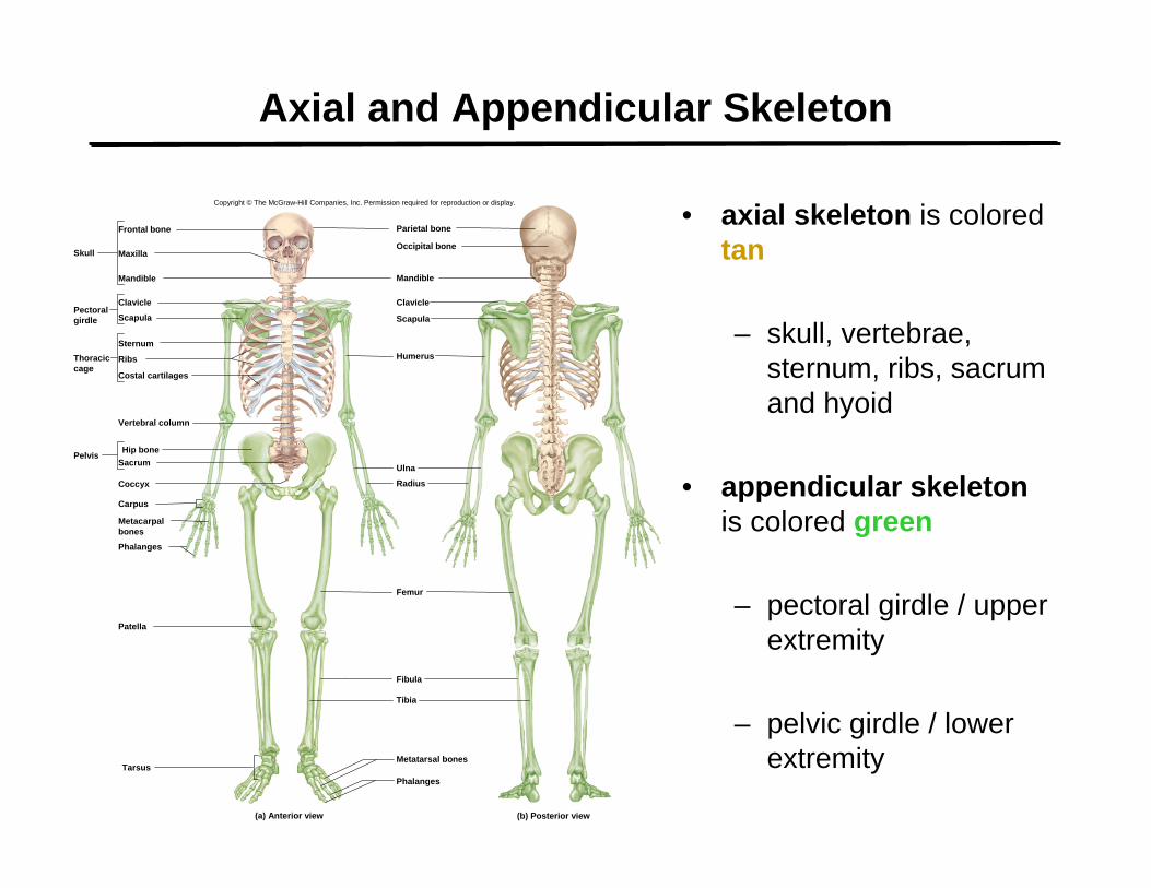
Axial and Appendicular Skeleton
• axial skeleton is colored tan
– skull, vertebrae, sternum, ribs, sacrum and hyoid
• appendicular skeletonis colored green
– pectoral girdle / upper extremity
– pelvic girdle / lower extremity
Copyright © The McGraw-Hill Companies, Inc. Permission required for reproduction or display.
Skull
Frontal bone
Clavicle
Maxilla
Parietal bone
Mandible
Occipital bone
Mandible
Humerus
Femur
Fibula
Ulna
Radius
Scapula
Clavicle
Scapula
Hip boneSacrum
Patella
Carpus
Pelvis
Sternum
Ribs
Costal cartilages
Phalanges
Metatarsal bones
Phalanges
Coccyx
(b) Posterior view
Pectoralgirdle
Thoraciccage
Vertebral column
Metacarpalbones
Tarsus
(a) Anterior view
Tibia


Shapes of Bones
• long bones– longer than wide– rigid levers acted upon by
muscles
• short bones – equal in length and width– glide across one another in
multiple directions
• flat bones– protect soft organs – curved but wide & thin
• irregular bones– elaborate shapes that don’t
fit into the other categories
Copyright © The McGraw-Hill Companies, Inc. Permission required for reproduction or display.
Femur
Scapula
Sternum
Sphenoid bone
Radius
Ulna
Irregular bonesFlat bones
Short bones Long bones
Vertebra
Capitate(carpal) bone
Talus
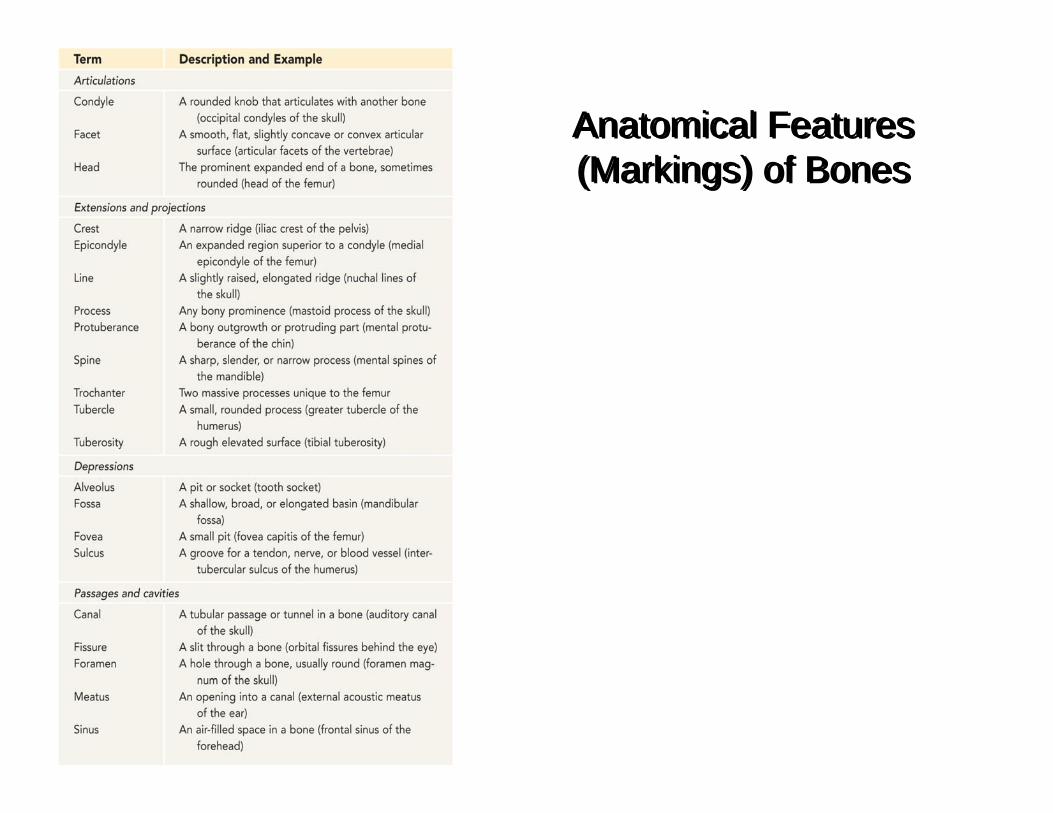
Anatomical Features(Markings) of BonesAnatomical Features(Markings) of Bones

Anatomical Features of Bones
Sinuses
Crest
Foramen
Foramen
Fossae
HeadTubercle
Crest
Tuberosity
Line
HeadFovea
Trochanters
Fossae
(a) Skull (lateral view)
Epicondyles
Condyles
AlveolusSpineCondyleProcess
Lines
Meatus
(b) Scapula (posterior view)(c) Femur
(posterior view)(d) Humerus
(anterior view)
ProcessSpine

Structure of a Long Bone
• epiphyses and diaphysis
• compact and spongy bone
• marrow cavity
• articular cartilage
• periosteum(a) Living (b) Dried
Marrow cavity
Periosteum
Nutrient foramen
Site of endosteum
Compact bone
Spongy bone
Epiphysis
Epiphysis
Diaphysis
Articularcartilage
Epiphysealline
Red bonemarrow
Yellow bone marrow
Epiphysealline
Articularcartilage

The Osteon and the Blood Vessels of Bone
• nutrient foramina –on bone surface
• perforating (Volkmann’s) canals – transverse or diagonal canals
• central canals –vertical canals
• circumferential lamellae
• interstitial lamellae
Periosteum
Endosteum
Perforating fibers
Perforating canal
Osteon
Lacuna
NerveBlood vessel
(b)
Spongy bone
Spicules
Centralcanal
Collagenfibers
Concentriclamellae
Circumferentiallamellae
Trabeculae
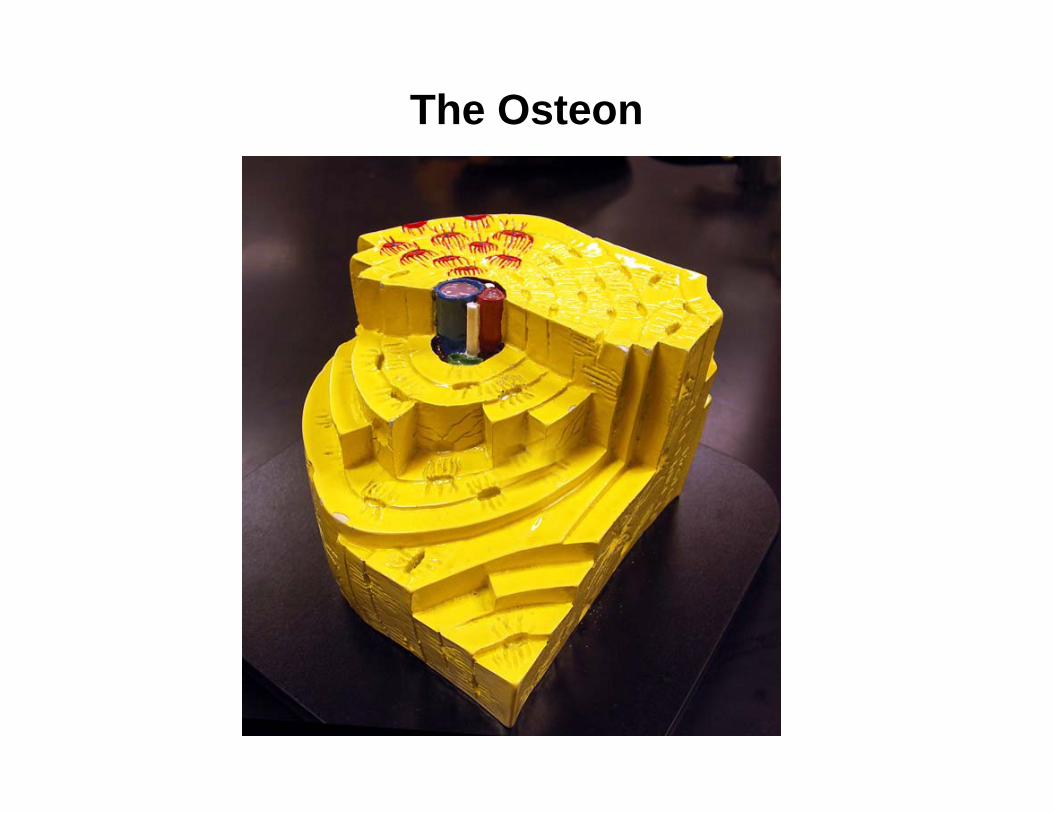
The Osteon

The Osteon
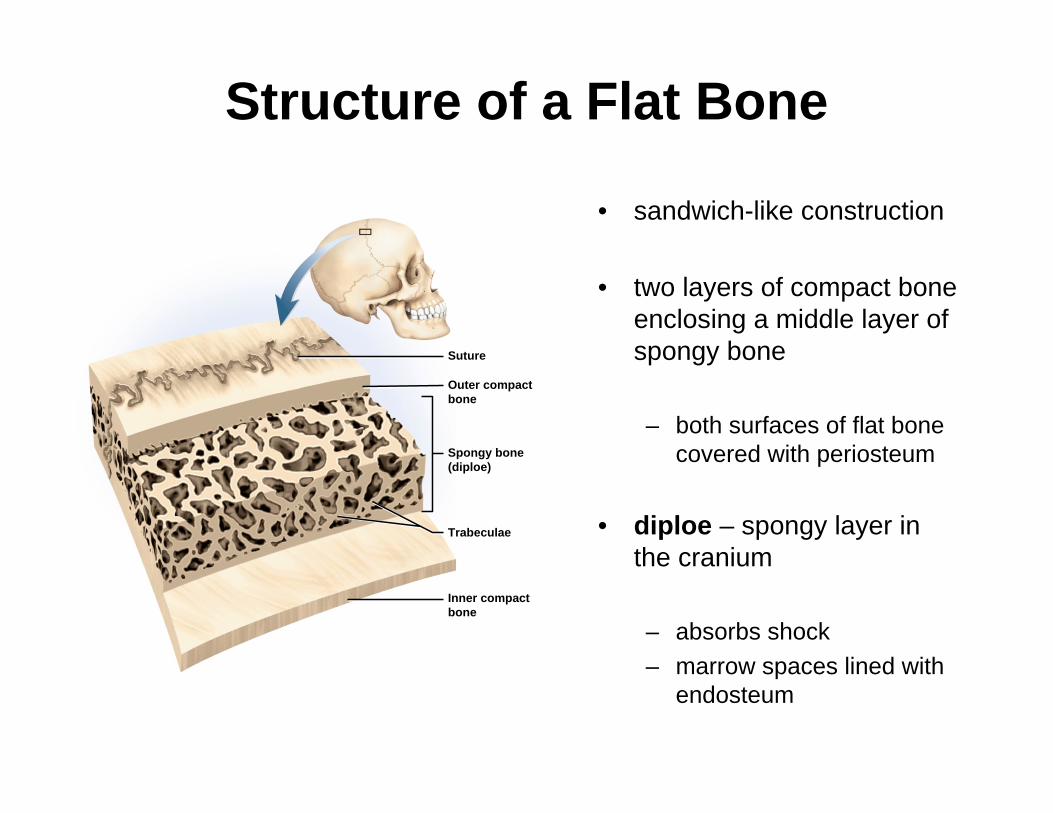
Structure of a Flat Bone
• sandwich-like construction
• two layers of compact bone enclosing a middle layer of spongy bone
– both surfaces of flat bone covered with periosteum
• diploe – spongy layer in the cranium
– absorbs shock– marrow spaces lined with
endosteum
Suture
Outer compactbone
Spongy bone(diploe)
Inner compactbone
Trabeculae

Major Skull Cavities
Frontal bone
Ethmoid bone
MiddleSuperior
Inferior Maxilla
Nasal cavity
Mandible
Vomer
Orbit
Cranial cavity
Nasalconchae
Oralcavity
Maxillarysinus
Zygomaticbone
Ethmoidair cells

Cranium (Braincase)
– protects the brain and associated sense organs
– swelling of the brain inside the rigid cranium may force tissue through foramen magnum resulting in death
– consists of two parts: • the calvaria (skullcap)• and the cranial base
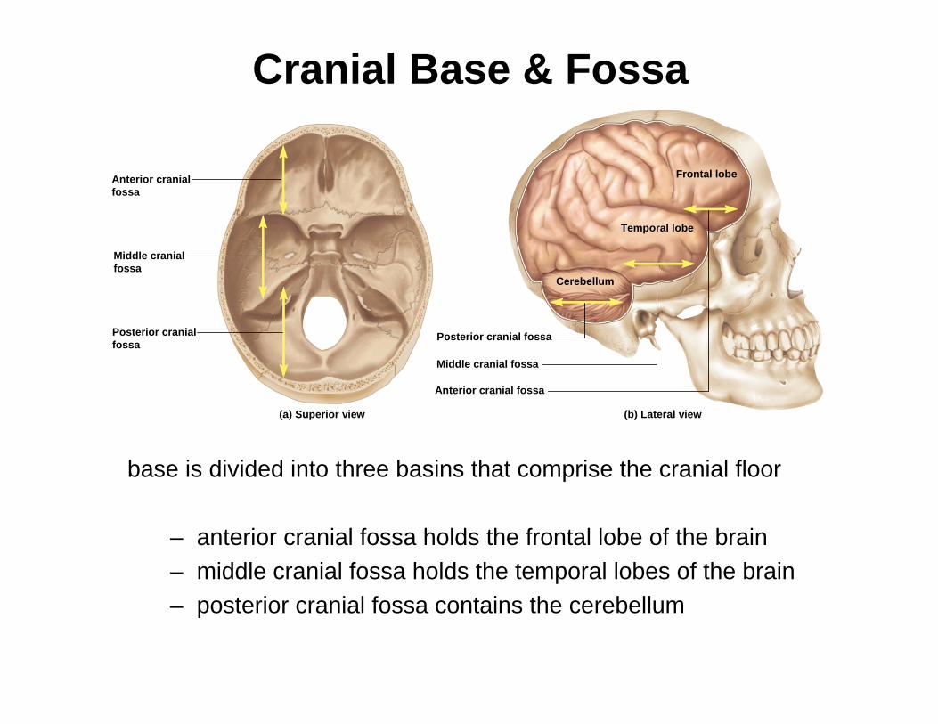
Cranial Base & Fossa
base is divided into three basins that comprise the cranial floor
– anterior cranial fossa holds the frontal lobe of the brain– middle cranial fossa holds the temporal lobes of the brain– posterior cranial fossa contains the cerebellum
(a) Superior view
Posterior cranial fossa
Middle cranial fossa
Anterior cranial fossa
Cerebellum
Temporal lobe
Frontal lobe
(b) Lateral view
Anterior cranialfossa
Middle cranialfossa
Posterior cranialfossa
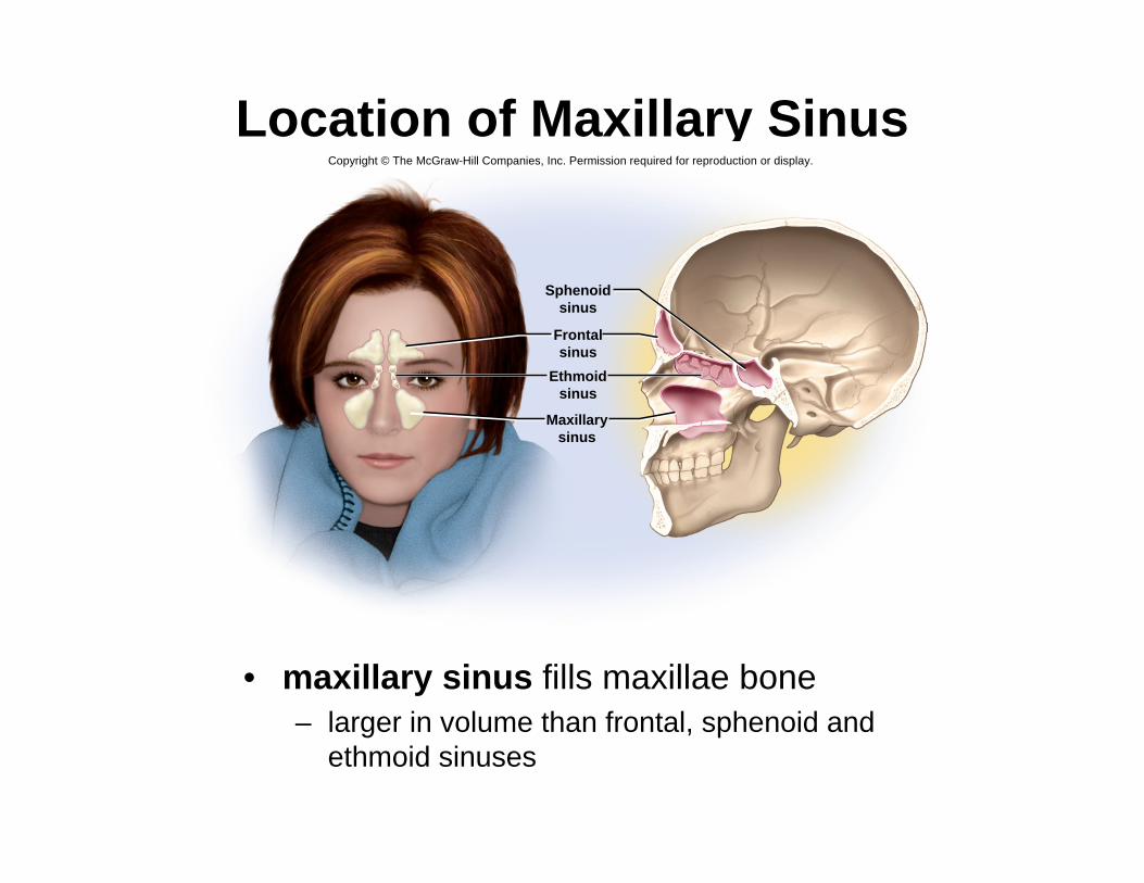
Location of Maxillary Sinus
• maxillary sinus fills maxillae bone– larger in volume than frontal, sphenoid and
ethmoid sinuses
Figure 8.8
Copyright © The McGraw-Hill Companies, Inc. Permission required for reproduction or display.
Sphenoidsinus
Frontalsinus
Ethmoidsinus
Maxillarysinus
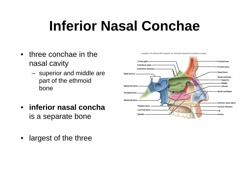
Inferior Nasal Conchae
• three conchae in the nasal cavity– superior and middle are
part of the ethmoidbone
• inferior nasal conchais a separate bone
• largest of the three
Frontal sinus
Inferior
Anterior nasal spine
Maxilla
Incisive foramenLacrimal bone
Nasal bone
Nasal cartilages
SuperiorMiddle
Sphenoid sinus
Palatine bone
Sphenoid bone
Crista galli
Sella turcica
Cribriform plate
Cribriform foramina
Occipital bone
Lip
Frontal bone
Incisor
Nasal conchae:
Copyright © The McGraw-Hill Companies, Inc. Permission required for reproduction or display.

• auditory ossicles– three in each middle-ear
cavity– malleus, incus, and stapes
• hyoid bone– slender u-shaped bone
between the chin and larynx– does not articulate with any
other bone– suspended from styloid
process of skull by muscle and ligament
– body and greater and lesser horns (cornua)
– fractured hyoid bone is evidence of strangulation
Bones Associated With Skull
Hyoid
Larynx
Styloid process
Stylohyoid muscle
Greater hornLesser horn
Body

Skull in Infancy and Childhood• fontanels - spaces between unfused
bones– filled with fibrous membrane– allow shifting of bones during birth
and growth of brain – anterior, posterior, sphenoid
(anterolateral), and mastoid(posterolateral fontanels
– feel pulse– allow insight about hydration
• two frontal bones fuse by age 6(metopic suture)
• skull reaches adult size by 8 or 9 years of age
Parietal bone
Occipital boneMaxilla
Frontal bone
Anterior fontanel
Sagittal suture
Posterior fontanel
Mandible
(b) Superior view
(a) Lateral view
Lambdoidsuture
Squamoussuture
Mastoidfontanel
Temporal bone
Parietalbone
Sphenoidbone
CoronalsutureFrontalboneSphenoidfontanel
Nasalbone
Zygomaticbone

Newborn Spinal Curvature
• Newborn’s spine exhibits one continuous C-shaped curve at birth
• known as primary curvature
Copyright © The McGraw-Hill Companies, Inc. Permission required for reproduction or display.
© The McGraw-Hill Companies, Inc./Bob Coyle, photographer

Adult Spinal Curvatures
• s-shaped vertebral column with four normal curvatures
– cervical– thoracic– lumbar– pelvic
• primary curvatures – present at birth
– thoracic and pelvic
• secondary curvatures – develop later
– cervical and lumbar– lifting head as it begins to crawl
develops cervical curvature– push up with arms before walking
start to develop lumbar– walking upright develops lumbar
curvature
Cervical curvature
Thoracic curvature
Lumbar curvature
Pelvic curvature
C7T1
T12L1
S1L5
C1


Abnormal Spinal Curvatures• from disease, paralysis of trunk
muscles, poor posture, pregnancy, or congenital defect
• scoliosis – abnormal lateral curvature– most common– usually in thoracic region– particularly of adolescent girls– developmental abnormality in which
the body and arch fail to develop on one side of the vertebrae
• kyphosis (hunchback) – exaggerated thoracic curvature
– usually from osteoporosis, also osteomalacia or spinal tuberculosis, or wrestling or weightlifting in young boys
• lordosis (swayback) – exaggerated lumbar curvature
– is from pregnancy or obesity
KeyNormalPathological
(b) Kyphosis(“hunchback”) (c) Lordosis
(“hunchback”)
(a) Scoliosis
Copyright © The McGraw-Hill Companies, Inc. Permission required for reproduction or display.