an intracellular nanotrap redirects proteins and ...mbio.asm.org/content/6/1/e02117-14.full.pdf ·...
TRANSCRIPT
An Intracellular Nanotrap Redirects Proteins and Organelles in LiveBacteria
Sarah Borg,a Felix Popp,a Julia Hofmann,a* Heinrich Leonhardt,a Ulrich Rothbauer,c Dirk Schülera,b
Department of Biology, Ludwig Maximillians University Munich, LMU Biozentrum, Martinsried, Germanya; Department of Microbiology, University of Bayreuth, Bayreuth,Germanyb; Department of Natural Science and Medicine, University of Tübingen, Reutlingen, Germanyc
* Present address: Julia Hofmann, Sequiserve GmbH, Vaterstetten, Germany.
S. Borg and F. Popp contributed equally to this work.
ABSTRACT Owing to their small size and enhanced stability, nanobodies derived from camelids have previously been used forthe construction of intracellular “nanotraps,” which enable redirection and manipulation of green fluorescent protein (GFP)-tagged targets within living plant and animal cells. By taking advantage of intracellular compartmentalization in the magneticbacterium Magnetospirillum gryphiswaldense, we demonstrate that proteins and even entire organelles can be retargeted alsowithin prokaryotic cells by versatile nanotrap technology. Expression of multivalent GFP-binding nanobodies on magnetosomesectopically recruited the chemotaxis protein CheW1-GFP from polar chemoreceptor clusters to the midcell, resulting in a grad-ual knockdown of aerotaxis. Conversely, entire magnetosome chains could be redirected from the midcell and tethered to one ofthe cell poles. Similar approaches could potentially be used for building synthetic cellular structures and targeted protein knock-downs in other bacteria.
IMPORTANCE Intrabodies are commonly used in eukaryotic systems for intracellular analysis and manipulation of proteinswithin distinct subcellular compartments. In particular, so-called nanobodies have great potential for synthetic biology ap-proaches because they can be expressed easily in heterologous hosts and actively interact with intracellular targets, for instance,by the construction of intracellular “nanotraps” in living animal and plant cells. Although prokaryotic cells also exhibit a consid-erable degree of intracellular organization, there are few tools available equivalent to the well-established methods used in eu-karyotes. Here, we demonstrate the ectopic retargeting and depletion of polar membrane proteins and entire organelles to dis-tinct compartments in a magnetotactic bacterium, resulting in a gradual knockdown of magneto-aerotaxis. This intracellularnanotrap approach has the potential to be applied in other bacteria for building synthetic cellular structures, manipulating pro-tein function, and creating gradual targeted knockdowns. Our findings provide a proof of principle for the universal use of fluo-rescently tagged proteins as targets for nanotraps to fulfill these tasks.
Received 14 November 2014 Accepted 1 December 2014 Published 13 January 2015
Citation Borg S, Popp F, Hofmann J, Leonhardt H, Rothbauer U, Schüler D. 2015. An intracellular nanotrap redirects proteins and organelles in live bacteria. mBio 6(1):e02117-14. doi:10.1128/mBio.02117-14.
Editor Sang Yup Lee, Korea Advanced Institute of Science and Technology
Copyright © 2015 Borg et al. This is an open-access article distributed under the terms of the Creative Commons Attribution-Noncommercial-ShareAlike 3.0 Unported license,which permits unrestricted noncommercial use, distribution, and reproduction in any medium, provided the original author and source are credited.
Address correspondence to Dirk Schüler, [email protected].
Intrabodies are functional fragments derived from full-lengthantibodies that can be expressed in heterologous hosts and
which specifically recognize their antigen within cells. In variouseukaryotic systems, they have been demonstrated to be powerfultools that enable the intracellular analysis and manipulation ofprotein functions (1–5). Among the various types of intrabodies,so-called nanobodies have proven to be particularly useful due totheir small size, solubility, enhanced stability, and the relative easeof screening, cloning, and expression procedures (6–9). Nano-bodies are routinely derived from camelid heavy-chain antibod-ies, which lack the light chains present in conventional IgG anti-bodies and recognize their target by interaction with single VHH(variable domain of heavy chain antibodies) domains (10). Afterthe genetic repertoire of B cells is extracted from an immunizedcamelid, antigen-binding VHHs can be selected and expressed ashighly robust single-domain antibodies. Because of their special
topology, nanobodies preferentially bind to concave surfaces ofantigens which are often inaccessible to conventional antibodies(11). Examples for nanobody-based applications in living plantand animal cells include the inhibition of enzyme activity throughspecific binding to the active site (7, 12), modulation of spectralproperties of fluorescent proteins (13), and the construction ofnanobody-mediated synthetic regulatory circuits (14). Further-more, different strategies for nanobody-based protein knock-downs have been reported, either by targeting nanobody-boundproteins to degradation pathways (15) or by artificially retargetinginteraction partners to specific intracellular localizations (16–18).Artificial relocalization of targeted proteins was either caused bytrapping of nanobody-bound proteins in the cytoplasm due tointerference with protein translocation to cellular compartments(18) or by specifically anchoring the nanobody to distinct struc-tures and compartments of the eukaryotic cell, such as distinct
RESEARCH ARTICLE crossmark
January/February 2015 Volume 6 Issue 1 e02117-14 ® mbio.asm.org 1
m
bio.asm.org
on May 7, 2018 - P
ublished by m
bio.asm.org
Dow
nloaded from

DNA regions, plasma membranes, or the centrioles of animalcells, resulting in ectopic recruitment of green fluorescent protein(GFP)-tagged targets (16, 17, 19). The application of anchorednanobodies against GFP (GFP-binding protein [GBP]) as an in-tracellular nanotrap is a particularly versatile tool because of thewidespread use of derivatives of this fluorescent tag.
It has been realized only rather recently that prokaryotic cellsalso contain highly organized subcellular structures (20). Bacteriapossess, for example, structural homologs to eukaryotic cytoskel-etal elements that define cell shape, structure, and function (21,22). In addition, they form large supramolecular protein com-plexes, contain microcompartments, and even biosynthesize ca-nonical membrane-enveloped organelles that show distinct sub-cellular localization patterns (22–24).
The ability to target proteins intracellularly and possibly evenredirect macromolecular complexes to defined subcellular loca-tions in bacteria has great potential for synthetic intracellular scaf-folding and targeting of proteins or protein complexes (25, 26).For instance, such techniques could be used for protein knock-downs through spatial separation of interaction partners. Otherpossible applications are specific targeting of proteins to bacterialsubcellular compartments (27–29), the setup of synthetic intracel-lular gradients (30), or even artificially compartmentalizing anddistributing different cellular processes and organelles to distinctsubcellular localizations. However, so far there are few tools avail-able that are equivalent to the well-established methods used ineukaryotic cells and that would efficiently fulfill these tasks in bac-teria.
One of the most intricate examples of natural compartmental-ization in prokaryotic cells is magnetosomes, which are nano-sized ferromagnetic crystals synthesized within intracellularmembrane vesicles by magnetotactic bacteria such as Magnetospi-rillum gryphiswaldense. These organelles are attached to a cyto-skeletal filament formed by the actin-like protein MamK and ar-ranged in a chain that is positioned at the midcell (31, 32). Theresulting magnetic dipole moment rotates the bacterial cell intoalignment with the geomagnetic field, thereby enhancing themovement of the bacteria toward growth-favoring oxygen levels(33). Recently, our lab demonstrated the display of nanobodies onmagnetosomes that were functional in recognizing their antigennot only in vitro but also in vivo. Expression of MamC–red fluo-rescent protein (RFP)-binding protein (RBP) fusions resulted inthe recruitment of cytoplasmic RFP to the magnetosomes (34),showing that intracellular localization of soluble heterologousproteins can be manipulated in bacteria. This motivated us tofurther investigate whether magnetosome anchors can also beused to trap proteins with distinct functions from other cellularcompartments. For this purpose, we chose the chemotaxis proteinCheW, which is part of the chemoreceptor clusters that are uni-versally found in chemotactic bacteria and typically display a dis-tinct polar localization (35). We demonstrate that CheW1 fused toenhanced GFP (EGFP) can be depleted from cell poles by expres-sion of multivalent GBP nanobodies fused to the magnetosomeprotein MamC on endogenous levels, resulting in ectopic recruit-ment of CheW1 to the magnetosome chain of M. gryphiswaldense.Depletion of CheW1 from polar clusters resulted in a gradual im-pairment of aerotaxis. Intriguingly, the interaction between themagnetosome anchor and polar CheW1-EGFP also led to artificialrepositioning of the entire magnetosome chain from its midcellposition toward one of the cell poles, indicating that entire organ-
elles can be redirected by nanobodies and tethered to ectopic po-sitions. Our study establishes the application of nanotrap technol-ogy for artificial targeting of proteins and even entire organelles tobacterial cells. Similar approaches could be used for building tai-lored subcellular structures in synthetic biology and for gradualprotein knockdowns in other prokaryotic systems.
RESULTSRecruitment of CheW1-EGFP to magnetosomes with a GBPnanotrap. In M. gryphiswaldense, CheW1 is encoded within thecheOp1 chemotaxis operon, which was recently demonstrated tocontrol magneto-aerotactic swimming polarity (36). It is well es-tablished in various bacteria that CheW acts as a linker and inter-acts both with the chemoreceptor and the histidine kinase CheAproteins, thereby enhancing the polar chemoreceptor clusteringand function (37). First, we replaced the native cheW1 gene withcheW1-egfp via chromosomal insertion. Similarly as observed inother bacteria (38–40), spot-like fluorescent signals originatingfrom EGFP-tagged CheW1 were exclusively found at both cellpoles in the wild-type background in fluorescence micrographs(Fig. 1ai and f). This is consistent with previous results of cryo-electron microscopy of M. gryphiswaldense cells, where chemore-ceptor complexes were identified near the poles (31). Only inelongated cells close to completion of cell division, two new clus-ters were formed at the midcell (see Fig. S1 in the supplementalmaterial). In mutant backgrounds either forming magnetosomeclusters instead of chains (�mamJ mutant) (32) or entirely lackingany magnetite particles (�mamM mutant) (41), the same CheW1-EGFP fluorescence localization pattern as in the wild-type back-ground was observed (Fig. 1di; see also Fig. S2b), indicating thatpolar chemoreceptor localization was independent of the pres-ence and configuration of magnetosome chains, as expected.
Next, we asked whether the localization of CheW1-EGFP wasaffected by coexpression of a GBP nanobody that had been iden-tified by Rothbauer and colleagues before (42). To trap CheW1-EGFP, GBP was expressed either alone in the cytoplasm (MagG-BPcyt) or fused to the abundant magnetosome membrane proteinMamC (43), which has routinely been used as a magnetosomeanchor for immobilization of various functional moieties, such asEGFP, enzymes, or an RBP (34, 43–46). In addition to the nativegbp gene, we used a synthetic allele that was specifically optimizedfor the expression in M. gryphiswaldense (Magnetospirillum-optimized green-binding protein [maggbp]). MamC was fused toeither one single copy of GBP connected to mCherry (mCherry-GBP, also referred to as “chromobody”) (42), and the resultingMamC-mCherry-GBP fusion is referred to as MamC-1�GBPhere, or to a tandem copy of maggbp-gbp (resulting in MamC-MagGBP-GBP, referred to as MamC-2�GBP here). All differentgbp constructs were inserted into chromosomes of parent strainscoexpressing CheW1-EGFP. Western blot analysis of cell extractsof all strains carrying the generated fusions revealed reacting pro-tein bands with expected sizes, indicating that the mono- andbivalent GBP nanobodies were stably expressed on magnetosomes(see Fig. S3 in the supplemental material).
Cytoplasmic expression of unfused MagGBPcyt alone had noeffect on the localization of CheW1-EGFP fluorescence in thewild-type background (see Fig. S2d). However, upon coexpres-sion of MamC-1�GBP and CheW1-EGFP, we detected weaker,secondary fluorescent foci at approximately the midcell positionin addition to the two polar CheW1-EGFP signals (Fig. 1bi). We
Borg et al.
2 ® mbio.asm.org January/February 2015 Volume 6 Issue 1 e02117-14
m
bio.asm.org
on May 7, 2018 - P
ublished by m
bio.asm.org
Dow
nloaded from

scored the number of fluorescent foci in four equidistant sectorsalong lengths of a representative set of cells and calculated therelative abundance of fluorescence intensity in each of the sectors(see Materials and Methods for details). In contrast to the wild-type background, which displayed only polar foci, about 30% offluorescence intensity was detected within the cytoplasm uponcoexpression of MamC-1�GBP (a representative cell is shown inFig. 1bi and f). Recruitment of CheW1-EGFP was likely due tointeraction with GBP expressed on magnetosomes, as green(CheW1-EGFP) and red (mCherry-tagged magnetosomes) fluo-rescence signals coincided in all analyzed cells, indicating that di-rect GBP-EGFP interaction caused the observed redirection ofCheW1 (see Fig. S2e and f). In cells coexpressing two GBP copiesin tandem (MamC-2�GBP), a single large, nonpolar fluorescencesignal was detected in the vast majority of cells. Ninety percent ofthe CheW1-EGFP fluorescence intensity was shifted toward themidcell (Fig. 1ci and 4), while only 10% of the fluorescence signalremained at the cell pole (Fig. 1f). Instead of the spot-like, exclu-sively polar foci of the parent strain, a linear fluorescence signalwas present near the midcell in most MamC-2�GBP-expressingcells, demonstrating efficient redirection of membrane complex-associated GFP-tagged proteins (Fig. 1ci; see also Fig. S4).
Next, we investigated whether the absence of magnetic nano-particles would affect the recruitment of CheW1-EGFP throughMamC-GBP fusions by analyzing nonmagnetic cells. Due to lossof the magnetosomal iron transporter MamM, �mamM cells lackany magnetite crystals but still produce empty magnetosomemembrane vesicles (41). CheW1-EGFP fluorescence was shiftedtoward the midcell in the �mamM strain coexpressing MamC-2�GBP to the same extent as in the magnetite-containing strains(see Fig. S2c). To analyze whether the configuration of magneto-some chains had an effect on CheW1-EGFP recruitment, we alsoexpressed MamC-1�GBP in the �mamJ background, in whichthe physical interaction of magnetosome chains with the actin-like MamK filaments is abolished (32), resulting in agglomeratedclusters rather than linear, well-ordered chains of magnetosomes(Fig. 1dii and eii). In the vast majority of analyzed �mamJ MamC-1�GBP cells, the major proportion of CheW1-EGFP fluorescence(�85% of all foci) was located at only 1 cell pole (Fig. 1f) andappeared to be slightly distorted longitudinally (Fig. 1ei).
Effect of CheW1-EGFP recruitment on magnetosome local-ization. We noticed that all strains which showed strong CheW1
mislocalization were increasingly affected in their magnetic align-ment, as indicated by reduced magnetic response (Cmag) values(e.g., MamC-2�GBP, 0.60 � 0.07; wild type, 1.24 � 0.20). TheCmag value provides an optical measure of the relative alignmentof cells in a cuvette by applying a strong magnetic field eitherparallel or perpendicular to the light beam of a photometer.Transmission electron microscopy (TEM) analysis revealed thatwild-type cells expressing CheW1-EGFP alone displayed the samemagnetosome localization pattern as that of their parent strain(Fig. 1aii). Both automated image analysis by the Chain AnalysisProgram (CHAP) (47) and manual scoring of magnetosome po-sition (see Materials and Methods for details; Fig. 1g) indicatedthat the linear chains of magnetosomes were consistently posi-tioned at the midcell and displayed the same configuration as typ-ically observed for the M. gryphiswaldense parent strain (48, 49),with approximately 35 particles per cell that had an average crystalsize of 35 to 47 nm (49). Additional cytoplasmic expression ofMagGBPcyt in the same background did not affect magnetosome
chain configuration (see Fig. S5b). Coexpression of MamC-1�GBP and CheW1-EGFP did not affect the midcell position ofmagnetosome chains either, but chains were less compact, i.e.,particles were more widely spaced, as indicated by the fuzzier,slightly stretched appearance of magnetosome chains in CHAPanalysis heat maps (Fig. 1biii). TEM analysis of �mamJ cells ex-pressing CheW1-EGFP alone revealed the same magnetosome lo-calization pattern as that of their parent strain (Fig. 1dii). Consis-tent with the observed shift of the CheW1-EGFP fluorescencetoward one pole in the �mamJ MamC-1�GBP strain, 90% ofmagnetosome clusters detected in TEM micrographs were local-ized at a single cell pole only, while clusters were no longer ob-served at the midcell or along the cell length, as commonly foundin the �mamJ parent strain (32, 50) (Fig. 1g). Moreover, the loosemagnetosome assemblies observed at the poles were slightly elon-gated compared to the compact, rounded magnetosome clustersof the parent strain (Fig. 1diii and eiii). This indicated that tar-geted recruitment and partial rearrangement of magnetosomeswere facilitated in cells in which magnetosome particles were nolonger bound to the MamK filament by their molecular connectorMamJ (33). As observed for mislocalization of CheW1-EGFP flu-orescence, in wild-type cells coexpressing divalent tandem fusionsof GBP (MamC-2�GBP), magnetosome chains were predomi-nantly drawn to one of the cell poles (Fig. 1cii; see also Fig. S6).Magnetosome chains were even less compact than in the presenceof the monovalent nanobody, as reflected by the rather scatteredpattern of poorly aligned magnetosome chains (Fig. 1ciii). Con-sistent with the overall shift of the chain, the mean fraction ofmagnetosome particles located at one of the cell poles increasedfrom 7 to 36% (Fig. 1g).
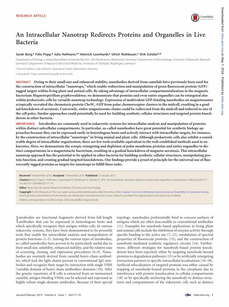
Effect of CheW1-EGFP recruitment on chemotaxis of M. gry-phiswaldense. The observed mislocalization of chains also af-fected the magnetic alignment of swimming cells. While wild-typecells expressing only CheW1-EGFP predominantly swam alignedto the ambient magnetic field, as did the parent strain, a largefraction of cells coexpressing MamC-2�GBP displayed trajecto-ries that were oriented at random angles to the ambient magneticfield (Fig. 2a). As indicated by video microscopy, motility andmean swimming speed were not affected in any of the analyzedstrains. Compared to the �cheW1 and �cheOp1 control strains, inwhich aerotaxis was entirely abolished, as indicated by the forma-tion of small aerotactic halos in swim plate assays (see Fig. S7)(36), coexpression of cytoplasmic MagGBPcyt and CheW1-EGFPin the wild-type background did not affect the size of swim halosthat were virtually identical to those of the parent strain (Fig. 2band c).
While the �cheW1 strain did not show any response whenshifted from anaerobic conditions to 2% oxygen in a microscopicgas perfusion chamber (Fig. 2d) and displayed a straight-swimming phenotype comparable to that of the �cheOp1 deletionstrain, wild-type cells expressing CheW1-EGFP showed a reactionvery similar to that observed in the parent strain (36). The reversalfrequency instantaneously rose from less than 0.1 s�1 to morethan 0.2 s�1 after microoxic upshift. This was followed by a rapiddrop in reversal frequency below prestimulus levels within 15 s(Fig. 2d). However, coexpression of MamC-1�GBP and CheW1-EGFP led to slightly reduced halo sizes in swim plates and a lowernumber of reversals in response to the oxygen shift. The maxi-mum reversal frequency remained below 0.15 s�1 and peaked atapproximately 60% of the wild-type rate. Interestingly, coexpres-
Nanobody-Mediated Magnetosome Recruitment
January/February 2015 Volume 6 Issue 1 e02117-14 ® mbio.asm.org 3
m
bio.asm.org
on May 7, 2018 - P
ublished by m
bio.asm.org
Dow
nloaded from

FIG 1 Analysis of subcellular CheW1-EGFP and magnetosome localization. Fluorescence (i) and TEM (ii) micrographs of representative M. gryphiswaldenseCheW1-EGFP (a), CheW1-EGFP MamC-1�GBP (b), CheW1-EGFP MamC-2�GBP (c), �mamJ CheW1-EGFP (d), and �mamJ CheW1-EGFP MamC-1�GBP(e) cells. Cells were analyzed by CHAP (iii) and scored for the distribution of fluorescence signal, represented by the percentage of fluorescent foci detected within4 equidistant compartments (f), and magnetosomes, represented by the percentage of magnetosomes detected within 4 equidistant compartments (g). Whitescale bar, 2 �m; black scale bar, 1 �m. Twenty cells were aligned by CHAP for each strain, and heat maps display the numbers of magnetosomes. Single cells weresegmented into four compartments, and for each strain 20 cells were scored to obtain fluorescence and magnetosome distributions.
Borg et al.
4 ® mbio.asm.org January/February 2015 Volume 6 Issue 1 e02117-14
m
bio.asm.org
on May 7, 2018 - P
ublished by m
bio.asm.org
Dow
nloaded from
sion of MamC-1�GBP also caused delayed adaptation after theshift, as the reversal frequency remained above prestimulus levelswithin 20 s postshift.
Coexpression of MamC-2�GBP and CheW1-EGFP, whichcompletely depleted CheW1-EGFP from the cell poles as sug-gested by fluorescence microscopy (Fig. 1ci), also had a dramaticeffect on the cells’ switching behavior under anoxic equilibriumconditions and the response elicited by oxygen exposure. The pre-stimulus reversal frequency was comparable to that of the �cheW1
strain and rose only minimally after oxygen upshift to 2% O2,remaining on a very low level (maximum frequency below0.05 s�1). In conclusion, an increase in the copy number of GBPled to gradually stronger impairment of aerotaxis, eventually re-ducing the number of reversals in a strain coexpressing CheW1-EGFP and the divalent MamC-2�GBP fusion to the level of a�cheW1 null mutant.
DISCUSSION
We investigated the interaction between components of the uni-versal bacterial chemotaxis signaling pathway and nanobodies ex-pressed on the magnetosome organelles of M. gryphiswaldense,which enabled us to easily follow the structural and behavioraleffects of artificial recruitment by TEM and fluorescence micros-copy (FM) imaging and video microscopy at the single cell level.We observed that by anchoring GBP to the magnetosome mem-
brane, the localization of CheW1-GFP was shifted from the polesto the midcell, i.e., to the typical position of the magnetosomechain. There are two possible explanations for the observed redi-rection of CheW1 from the polar clusters to the magnetosomes: (i)unbound CheW1, in equilibrium with the receptor-bound form,could be recruited from a cytoplasmic pool, whereas (ii)membrane-bound CheW1 could be directly withdrawn from pre-existing polar clusters. CheW is a soluble protein that lacks trans-membrane domains but in vitro forms ultrastable ternary com-plexes together with CheA and chemoreceptors (51). However, inliving cells, signaling complexes are weakly dynamic and displayslow turnover (of approximately 12 min), as indicated by fluores-cence recovery after photobleaching (FRAP) experiments onCheA and CheW constructs (52). Consistent with these observa-tions, it has been suggested that small amounts of CheA might bepermanently present in an unbound state in the cytoplasm (53).However, the relative copy numbers of all cluster components aretightly regulated, and since overexpression of CheW leads to im-paired chemotactic signaling (due to competitive inhibition ofCheA binding to the chemoreceptors) (51), the pool of free CheWin the cytoplasm must be rather small. Thus, it seems most prob-able that soluble CheW present at low concentrations in the cyto-plasm is sequestered by magnetosome-anchored GBP, and overtime also those molecules initially bound to the chemoreceptor
FIG 2 Magneto-aerotactic swimming behavior of M. gryphiswaldense strains expressing CheW1-EGFP and MamC-GBP fusions. (a) Magnetic alignment ofswimming cells expressing CheW1-EGFP alone or in combination with MamC-2�GBP. A plot of all tracks from a representative video record is shown for eachstrain. Cells swimming in the gas perfusion chamber were exposed to a homogenous vertical magnetic field of 0.26 mT (B). (b) Average halo diameter of strainsexpressing MamC-GBP fusions in swim plates (means � standard deviations [SD] from at least 3 independent replicates). The �cheW1 and �cheOp1 chemotaxisgene deletion mutants were used as controls. Transcomplementation of the �cheW1 mutant strain by constitutive expression of CheW1-EGFP from a plasmidrestored chemotactic efficiency to 80% of the wild-type cells expressing CheW1-EGFP at wild-type levels. (c) Halo formation of wild-type CheW1-EGFP,CheW1-EGFP MamC-1�GBP, CheW1-EGFP MamC-2�GBP, and �cheW cells in 0.2 % motility agar 3 days after inoculation. (d) Aerotactic reversal responseupon abrupt shift from 0% to 2% oxygen in a microscopic gas perfusion chamber. Video records were analyzed by automated tracking software (WimTaxis;Wimasis) to obtain swim tracks and reversal events of individual cells (72), and reversal rates were calculated for 5-s intervals by averaging single-cell data fromat least 3 independent recordings.
Nanobody-Mediated Magnetosome Recruitment
January/February 2015 Volume 6 Issue 1 e02117-14 ® mbio.asm.org 5
m
bio.asm.org
on May 7, 2018 - P
ublished by m
bio.asm.org
Dow
nloaded from

clusters might gradually be released and trapped at ectopic posi-tions by a strong interaction with the nanobody.
The localization of CheW1-GFP was unaffected by coexpres-sion of cytoplasmic (unfused) GBP in the wild-type backgroundbut shifted toward the midcell upon MamC-2�GBP expression inthe nonmagnetic �mamM strain, which lacks any electron-densemagnetic crystals but still forms empty vesicles of the magneto-some membrane (41). This demonstrates that GBP immobilizedon magnetite-free membrane vesicles is sufficiently effective tospecifically redirect localized proteins. Furthermore, this suggeststhat recruitment and retargeting could be achieved in other bac-teria lacking magnetosomes by using different spatial determi-nants as intracellular traps.
Although the presence of magnetic particles was no absoluteprerequisite for efficient recruitment, redistribution of CheW wasstrongly affected by magnetosome chain configuration. Magneto-some clusters were drawn to only one pole in the �mamJ back-ground upon expression of MamC-1�GBP and acted as efficientnanotraps for CheW1-GFP. In contrast to the undefined midcellfluorescence caused by partial depletion from polar clusters ob-served upon expression of MamC-1�GBP in the wild-type back-ground, virtually no CheW1-GFP signal was detected at the mid-cell or the opposite pole in the �mamJ mutant strain. This eithermight be due to increased avidity of nanobodies concentrated inthe tightly clustered magnetosome assemblies or might reflect astochastic shift of CheW diffusion equilibrium due to the concen-tration of two sinks (i.e., the native chemoreceptor cluster and theartificial magnetosomal nanobody cluster) at a single pole (Fig. 3,bottom right).
Interaction of MamC-GBP and CheW1-GFP reciprocally af-fected configuration and positioning of the magnetosome chain.Binding of CheW1-GFP to magnetosome particles disturbed theirproper alignment into regular, densely spaced chains. Increasingthe expression of GBP (MamC-1�GBP and -2�GBP) also grad-
ually increased the interparticle spacing, possibly by additionalprotein bound to the magnetosome surface which might weakenthe magnetostatic interactions between particles. Overexpressionof MamC-2�GBP in the wild-type background caused a nearlycomplete shift of the magnetosome chains toward the poles, withthe majority of magnetosome chains originating at polar or sub-polar positions (see Fig. S6), which was probably caused by redi-recting and tethering the chains to a fraction of membrane-boundCheW1-GFP remaining at the cell pole (Fig. 3, bottom left). Al-though the magnetosome chain of wild-type cells generally occu-pies the midcell position, it becomes mobilized during cell divi-sion, when the chain is split and repositioned by MamK dynamicsto the midcell of daughter cells (50). We found that magnetosomechain localization was most severely impaired in cells lacking theacidic MamJ protein, which is assumed to connect magnetosomeparticles to the cytoskeletal magnetosome filament formed by theactin-like MamK protein (32). In the �mamJ background, over-expression of the monovalent nanobody was already sufficient torearrange (Fig. 1eiii) and recruit (Fig. 1eii) the magnetosome clus-ter to 1 cell pole (Fig. 3, bottom right). The increased intracellularmobility of �mamJ magnetosome clusters might be explained by alack of the presumed MamK-mediated interactions with divisomeconstituents (50). In wild-type cells, these interactions need to beovercome by interaction with polar CheW, whereas in �mamJcells, magnetosome redirection is facilitated because MamK-magnetosome interactions are abolished.
The level of CheW1-GFP recruitment clearly depended on genedosage. While redirection of CheW1-GFP was only partial in cellsexpressing MamC-1�GBP, overexpression of MamC-2�GBPcaused a complete shift of CheW1-GFP localization toward themidcell. There is precedence for significantly increased avidity(500�) of a nanobody consisting of a fusion of two identical do-mains compared to the monovalent nanobody (54). Similarly, inour experiments, the binding of CheW1-GFP to the monovalent
FIG 3 Model of MamC-GBP and CheW1-EGFP interaction. CheW1-EGFP localizes distinctly at the cell poles if expressed chromosomally in the wild-type and�mamJ backgrounds (top right and left). If MamC-GBP is coexpressed in the wild type (bottom left), CheW1-EGFP is depleted completely from the poles.Expression of MamC-GBP in the �mamJ CheW1-EGFP background leads to recruitment of whole magnetosomes to the cell poles (bottom right). Expression ofmonovalent and divalent nanobodies on a magnetosomes and interaction with CheW1-EGFP is illustrated in the inset. Expressed proteins are illustrated in samecolors as genes.
Borg et al.
6 ® mbio.asm.org January/February 2015 Volume 6 Issue 1 e02117-14
m
bio.asm.org
on May 7, 2018 - P
ublished by m
bio.asm.org
Dow
nloaded from

GBP was apparently comparable to the in vivo turnover of thechemoreceptor-CheW complexes, since polar- and midcell-localized CheW1-GFP could be detected. In contrast, the avidity ofthe bivalent nanobody was much stronger, more endogenousCheW1-GFP was bound, and the equilibrium was shifted towardthe GBP-bound state.
In addition to demonstrating efficient redirection of entire or-ganelles to distinct locations, we observed that ectopic redirectionof CheW1-GFP also gradually modulated chemotactic efficiencyof M. gryphiswaldense cells. While chemoreceptors readily formcomplexes in the absence of CheA and CheW, the latter is essentialfor stabilizing native CheA-receptor interactions and lattice for-mation (37, 53). Partial depletion of CheW1 gradually reducedchemotactic efficiency, while expression of the bivalent nanobodyessentially phenocopied the deletion of cheW1 (Fig. 2c). As GBPexpressed in the cytoplasm had no effect on aerotaxis, this was notdue to inactivation of CheW1-GFP but caused by redirection anddepletion from its native polar environment. Although bacteriado not display the same level of compartmentalization as eukary-otic cells, the functionality of many bacterial proteins similarlydepends on their localization. Our results show that testing pro-tein function by manipulating its subcellular localization as ap-plied to eukaryotic systems (2, 18) can be extended to muchsmaller bacterial cells and be used to efficiently modulate proteinfunction by subcellular retargeting.
Compared to other approaches for silencing or manipulatingthe expression of selected genes at the DNA or RNA (55, 56) level,the biggest advantage of regulating gene expression at the proteinlevel is that there is no change of mRNA transcript or native pro-tein expression level (57). Especially for bacterial genes encoded inoperons, gradual knockdown of individual proteins might be dif-ficult to achieve at the transcriptional level, if polar effects ontranscription of downstream genes are to be avoided. Addition-ally, it would be desirable to develop inducible systems, e.g., togradually control in vivo the stoichiometry of proteins in largerclusters. This might facilitate the study of complex regulatorypathways, such as cell division or cell differentiation processes inother bacteria.
Intrabodies are well established as powerful tools in eukaryoticcells for trapping soluble proteins at defined subcellular locations(16–18) or for inhibition of protein function (12). Although re-combinant nanobodies can be produced easily in bacteria such asEscherichia coli (10), to date the use of intrabodies in bacterial cellshas been restricted to only very few studies. Two early publicationsreported the intracellular expression of single-chain Fv antibodyfragments (e.g., to block transcriptional activation) (58, 59), andmore recently nanobodies have been applied in bacteria to inhibitenzyme activity (60). However, in these approaches, intrabodieswere not anchored to defined positions, and inhibition of enzymeswas achieved by neutralization rather than redirection to com-pletely different compartments of the cell. Although for proof ofprinciple we took advantage of the specific compartmentalizationin M. gryphiswaldense, in which the magnetosomes provide a nat-ural anchor for setting up an intracellular nanotrap, this approachcould also be extended and adapted for application in other bac-teria. By using universal tags like GFP for recruitment, many pro-teins can be targeted with the same nanobody by applying thesame strategy, obviating the need of camelid immunization andscreening of whole libraries. Multiple other applications are pos-sible, because GFP fusion proteins can be combined with any cel-
lular anchor point, such as subcellular locations (e.g., poles, mid-cell), specific protein complexes, compartments, organelles, orother spatial determinants. For instance, potential applications ofour approach in bacteria could be building synthetic cellularstructures (e.g., artificial tethering of heterologously expressedbacterial microcompartments) or compartmentalization of bio-synthetic pathways, which can dramatically increase productionby restricting reactions spatially to subcellular compartments (61,62).
MATERIALS AND METHODSBacterial strains, plasmids, and culture conditions. Bacterial strains andplasmids used in this study are listed in Table S1 and S2 in the supplemen-tal material. M. gryphiswaldense strains were grown microaerobically with1% oxygen in modified flask standard medium (FSM) at 30°C (63) andmoderate shaking (120 rpm). E. coli strains were cultivated as previouslydescribed (64). For growth of E. coli WM3064 (W. Metcalf, unpublisheddata) or BW29427 (K. Datsenko and B. L. Wanner, unpublished data),1 mM DL-�,�-diaminopimelic acid (DAP) was added to lysogeny brothmedium (LB). Strains were routinely cultured on plates solidified with1.5% (wt/vol) agar. For strains carrying recombinant plasmids, mediawere supplemented with 25 �g · ml�1 kanamycin and 50 �g · ml�1 am-picillin (Amp) for E. coli strains and 5 �g · ml�1 kanamycin (km) forM. gryphiswaldense strains.
For the preparation of swim plates, only 0.2% agar was used, the con-centration of carbon source (lactate) was lowered to 1.5 mM, and peptonewas omitted from FSM. Five microliters of overnight culture was pipettedinto the swim agar, and plates were incubated under microoxic conditionsfor 2 days (protocol modified from that of Schultheiss et al. [65]).
Molecular and genetic techniques. Oligonucleotides were purchasedfrom Sigma-Aldrich (Steinheim, Germany), and sequences can be sup-plied on request. Plasmids were constructed by standard recombinanttechniques as described in detail below. All constructs were sequenced onan ABI 3730 capillary sequencer (Applied Biosystems, Darmstadt, Ger-many), utilizing BigDye Terminator v3.1. Sequence data were analyzedwith Software Vector NTI Advance 11.5 (Invitrogen, Darmstadt, Ger-many). The GBP nanobody (42) was provided by ChromoTek GmbH(Planegg-Martinsried), and a synthetic GBP was specifically optimized forthe expression in M. gryphiswaldense with respect to its codon usage andpurchased from ATG:biosynthetics (Merzhausen, Germany).
Construction of plasmids for chromosomal gene insertion, dele-tion, and fusion. For chromosomal exchange of cheW1 against cheW1-egfp, the fluorescence marker was fused via overlap extension PCR (66) tocheW1 and to a 1,000-bp downstream fragment of the gene. The fusedproduct was inserted into pORFM, and the native cheW1 copy was ex-changed chromosomally against cheW1-egfp by homologous recombina-tion facilitated by GalK counterselection (67). Deletion of cheW1 wasachieved following a similar strategy by fusion of approximately 1,000-bpfragments upstream and downstream of cheW1 connected by 12 nonsensebp replacing the native cheW1. For complementation of cheW1 deletion,cheW1 was amplified from the genome and inserted into pAP150 (46).
All mamC-gbp fusions were chromosomally introduced by transposi-tion; therefore, all gene fusions created by overlap PCR were inserted intotransposable pSB6 and pSB7 plasmids (46).
Analytical methods. Magnetic reaction of cells was validated by lightmicroscopy applying a bar magnet. Optical density (OD) and magneticresponse (Cmag) of exponentially growing cells were measured photomet-rically at 565 nm as previously reported (68). For Cmag measurement, amagnetic field of approximately 70 mT was used.
Biochemical methods. Polyacrylamide gels were prepared accordingto the method of Laemmli (69). Strains were grown overnight and spundown via centrifugation, OD565 was set to 10, and 20 �l was loaded onto12% (wt/vol) SDS gels and analyzed via immunoblotting. Proteins wereelectroblotted onto polyvinylidene difluoride (PVDF) membranes (Roth,Germany). Membranes were blocked for 1 h at room temperature with
Nanobody-Mediated Magnetosome Recruitment
January/February 2015 Volume 6 Issue 1 e02117-14 ® mbio.asm.org 7
m
bio.asm.org
on May 7, 2018 - P
ublished by m
bio.asm.org
Dow
nloaded from

blocking solution (2.5% [wt/vol] milk powder in Tris-buffered saline[TBS] [50 mM Tris-HCl, pH 7.6, and 150 mM NaCl]) and incubated foranother hour with primary rabbit anti-MamC IgG antibody (1:500 dilu-tion; Santa Cruz, CA). Membranes were washed 4 times with TBS for5 min and incubated with a secondary alkaline phosphatase-labeled goatanti-rabbit IgG antibody (1:2,000 dilution; Promega, United States) for45 min. Membranes were washed 4 times with TBS for 5 min, and immu-noreactive proteins were visualized with nitroblue tetrazolium (NBT)/5-bromo-4-chloro-3-indolylphosphate (BCIP) (Roche kit).
Phase contrast and fluorescence microscopy. Strains with genomicCheW1-EGFP fusions and additional MamC-GBP fusions were grown in1 ml FSM in 24-well plates for 16 h at 30°C and 1% O2 without agitation.For microscopy, cells were immobilized on agarose pads (phosphate-buffered saline [PBS] buffer supplemented with 1% agarose) and imagedwith an Olympus BX81 microscope equipped with a 100UPLSAPO100XO objective (numerical aperture of 1.40) and aHamamatsu Orca AG camera. The Olympus xcellence pro software wasused to capture and analyze images.
To analyze relative positions of fluorescent foci, we manually seg-mented each cell along its long axis into four equal sectors and scored thefluorescent foci within each sector. The strongest fluorescence signal(s)was scored as “��,” and weaker signals were scored as “�.” Since theorientation of imaged cells was random and in many cases the distributionof fluorescent foci was not perfectly symmetric, we rotated the cells wherenecessary so that the sectors with the highest cumulated score were sectors1 and 2. We then calculated relative frequencies of fluorescent focus po-sitions based on the ratio of cumulated scoring points of all analyzed cellsper sector divided by the total number of scoring points in all cells.
Transmission electron microscopy. Magnetosome chain localizationwas examined by transmission electron microscopy (TEM), for whichcells were concentrated via centrifugation and adsorbed onto carbon-coated copper grids. Cells were imaged with an FEI Morgagni 268 (FEI,Eindhoven, Netherlands) electron microscope at an accelerating voltageof 80 kV. For analysis of magnetosome alignment and chain compactness,we used the CHAP script implemented in Matlab and ran the program for20 cells for each strain (48). For analysis of magnetosome position, wemanually segmented each cell along its long axis into four equal sectorsand scored the number of magnetosomes within each sector. Since theorientation of imaged cells was random and in many cases the distribu-tions of magnetosomes were not perfectly symmetric, we rotated the cellswhere necessary so that the sector with the most magnetosomes scoredwas either sector 1 or 2. We then calculated relative frequencies of mag-netosome positions based on the ratio of cumulated magnetosomes of allanalyzed cells per sector divided by the total number of magnetosomes inall cells.
Video microscopy and analysis of swimming parameters. The swim-ming behavior of cells was analyzed and recorded using dark-field micros-copy on an upright Zeiss Axioplan microscope (Zeiss, Jena, Germany) at�100 magnification. All microscopic motility experiments were per-formed within a microscopic gas perfusion chamber (Ludin Chamber,Life Imaging Services) that was equilibrated with variably moisturized andprecisely adjusted O2-N2 gas mixtures containing between 0 and 2% ox-ygen (70).
Homogeneous conditions were maintained by using strongly dilutedcell suspensions (OD of 0.005) and exposing cell suspensions to a constantgas flow of 50 ml · min�1, protected against turbulence by placing a gas-permeable agar sheet on top.
Videos were recorded with a UK1158-M camera (EHD, Damme, Ger-many) at a frame rate of 15 frames per second and a standard resolution of1,360 by 1,024 pixels using VirtualDub software. Dark-field video recordswere analyzed by a custom-made automated tracking software(WimTaxis—Bacteria Tracking; Wimasis GmbH, Munich, Germany)specifically adapted to determine the basic swimming characteristics. Thesoftware automatically detected swimming reversals and provided the x-ycoordinates of every tracked cell for each frame.
The minimum track length was set to be 50 frames. Within the usualtracking times (depending on the time bacteria stayed in the viewing field[usually fewer than 10 s]), reversals generally were too infrequent to sim-ply average the reversal rates for single cells. Therefore, the reversal fre-quency analysis for each experiment was performed at the populationlevel, and all detected reversals were divided by the total respective track-ing time (sum of the temporal length of all tracks) to obtain the popula-tion average.
To analyze the cells’ reaction to oxygen shifts, the gas stream wasmanually switched between oxic and anoxic. For this purpose, weequipped our setup with a three-way valve and a flowmeter to adjust theflow of N2 gas to 50 ml · min�1 (70). Cells were first equilibrated for 3 minunder anoxic conditions before the video recording was started. After 20 s,the gas flow was shifted to 2% O2, and cells were recorded for an addi-tional 20 s. To determine the average reversal frequency over time, thenumbers of detected reversals within 5-s intervals were added from threeindependent video recordings and normalized to the total correspondingtracking time.
SUPPLEMENTAL MATERIALSupplemental material for this article may be found at http://mbio.asm.org/lookup/suppl/doi:10.1128/mBio.02117-14/-/DCSupplemental.
Figure S1, PDF file, 0.1 MB.Figure S2, PDF file, 0.1 MB.Figure S3, PDF file, 0.1 MB.Figure S4, PDF file, 0.01 MB.Figure S5, PDF file, 0.02 MB.Figure S6, PDF file, 0.02 MB.Figure S7, PDF file, 0.2 MB.Table S1, PDF file, 0.01 MB.Table S2, PDF file, 0.03 MB.
ACKNOWLEDGMENTS
We thank ChromoTek GmbH for providing the GBP nanobody. We alsothank M. Eibauer and A. Heins for help with CHAP implementation.
This work was funded by DFG grants Schu1080/9-1, 15-1, and 16-1 toD.S.
S.B., F.P., and D.S. designed the research; S.B., F.P., and J.H. per-formed the research; S.B., F.P., and D.S. analyzed the data; S.B., F.P., andD.S. wrote the paper.
REFERENCES1. Carlson JR, Weissman IL. 1988. Molecular tools for inactivating a yeast
enzyme in vivo. Mol Cell Biol 8:2647–2650.2. Kaiser PD, Maier J, Traenkle B, Emele F, Rothbauer U. 2014. Recent
progress in generating intracellular functional antibody fragments to tar-get and trace cellular components in living cells. Biochim Biophys Acta1844:1933–1942. http://dx.doi.org/10.1016/j.bbapap.2014.04.019.
3. Marasco WA, Haseltine WA, Chen SY. 1993. Design, intracellular ex-pression, and activity of a human anti-human immunodeficiency virustype 1 Gp120 single-chain antibody. Proc Natl Acad Sci U S A 90:7889 –7893. http://dx.doi.org/10.1073/pnas.90.16.7889.
4. Tavladoraki P, Benvenuto E, Trinca S, De Martinis D, Cattaneo A,Galeffi P. 1993. Transgenic plants expressing a functional single-chain Fvantibody are specifically protected from virus attack. Nature 366:469 – 472. http://dx.doi.org/10.1038/366469a0.
5. Wesolowski J, Alzogaray V, Reyelt J, Unger M, Juarez K, Urrutia M,Cauerhff A, Danquah W, Rissiek B, Scheuplein F, Schwarz N, AdriouchS, Boyer O, Seman M, Licea A, Serreze DV, Goldbaum FA, Haag F,Koch-Nolte F. 2009. Single domain antibodies: promising experimentaland therapeutic tools in infection and immunity. Med Microbiol Immu-nol 198:157–174. http://dx.doi.org/10.1007/s00430-009-0116-7.
6. Bodelón G, Palomino C, Fernández LÁ. 2013. Immunoglobulin do-mains in Escherichia coli and other enterobacteria: from pathogenesis toapplications in antibody technologies. FEMS Microbiol Rev 37:204 –250.http://dx.doi.org/10.1111/j.1574-6976.2012.00347.x.
7. Lauwereys M, Arbabi Ghahroudi M, Desmyter A, Kinne J, Hölzer W,De Genst E, Wyns L, Muyldermans S. 1998. Potent enzyme inhibitors
Borg et al.
8 ® mbio.asm.org January/February 2015 Volume 6 Issue 1 e02117-14
m
bio.asm.org
on May 7, 2018 - P
ublished by m
bio.asm.org
Dow
nloaded from

derived from dromedary heavy-chain antibodies. EMBO J 17:3512–3520.http://dx.doi.org/10.1093/emboj/17.13.3512.
8. Pellis M, Pardon E, Zolghadr K, Rothbauer U, Vincke C, Kinne J,Dierynck I, Hertogs K, Leonhardt H, Messens J, Muyldermans S,Conrath K. 2012. A bacterial-two-hybrid selection system for one-stepisolation of intracellularly functional nanobodies. Arch Biochem Biophys526:114 –123. http://dx.doi.org/10.1016/j.abb.2012.04.023.
9. De Meyer T, Muyldermans S, Depicker A. 2014. Nanobody-based prod-ucts as research and diagnostic tools. Trends Biotechnol 32:263–270.http://dx.doi.org/10.1016/j.tibtech.2014.03.001.
10. Muyldermans S. 2013. Nanobodies: natural single-domain antibodies.Annu Rev Biochem 82:775–797. http://dx.doi.org/10.1146/annurev-biochem-063011-092449.
11. De Genst E, Silence K, Decanniere K, Conrath K, Loris R, Kinne J,Muyldermans S, Wyns L. 2006. Molecular basis for the preferential cleftrecognition by dromedary heavy-chain antibodies. Proc Natl Acad Sci U SA 103:4586 – 4591. http://dx.doi.org/10.1073/pnas.0505379103.
12. Jobling SA, Jarman C, Teh MM, Holmberg N, Blake C, Verhoeyen ME.2003. Immunomodulation of enzyme function in plants by single-domainantibody fragments. Nat Biotechnol 21:77– 80. http://dx.doi.org/10.1038/nbt772.
13. Kirchhofer A, Helma J, Schmidthals K, Frauer C, Cui S, Karcher A,Pellis M, Muyldermans S, Casas-Delucchi CS, Cardoso MC, LeonhardtH, Hopfner KP, Rothbauer U. 2010. Modulation of protein properties inliving cells using nanobodies. Nat Struct Mol Biol 17:133–138. http://dx.doi.org/10.1038/nsmb.1727.
14. Tang JC, Szikra T, Kozorovitskiy Y, Teixiera M, Sabatini BL, Roska B,Cepko CL. 2013. A nanobody-based system using fluorescent proteins asscaffolds for cell-specific gene manipulation. Cell 154:928 –939. http://dx.doi.org/10.1016/j.cell.2013.07.021.
15. Caussinus E, Kanca O, Affolter M. 2012. Fluorescent fusion proteinknockout mediated by anti-GFP nanobody. Nat Struct Mol Biol 19:117–121. http://dx.doi.org/10.1038/nsmb.2180.
16. Herce HD, Deng W, Helma J, Leonhardt H, Cardoso MC. 2013.Visualization and targeted disruption of protein interactions in livingcells. Nat Commun 4:2660. http://dx.doi.org/10.1038/ncomms3660.
17. Rothbauer U, Zolghadr K, Muyldermans S, Schepers A, Cardoso MC,Leonhardt H. 2008. A versatile nanotrap for biochemical and functionalstudies with fluorescent fusion proteins. Mol Cell Proteomics 7:282–289.http://dx.doi.org/10.1074/mcp.M700342-MCP200.
18. Schornack S, Fuchs R, Huitema E, Rothbauer U, Lipka V, Kamoun S.2009. Protein mislocalization in plant cells using a GFP-binding chromo-body. Plant J 60:744 –754. http://dx.doi.org/10.1111/j .1365-313X.2009.03982.x.
19. Spira F, Mueller NS, Beck G, von Olshausen P, Beig J, Wedlich-SöldnerR. 2012. Patchwork organization of the yeast plasma membrane into nu-merous coexisting domains. Nat Cell Biol 14:640 – 648. http://dx.doi.org/10.1038/ncb2487.
20. Gitai Z. 2005. The new bacterial cell biology: moving parts and subcellulararchitecture. Cel l 120:577–586. http:/ /dx.doi .org/10.1016/j.cell.2005.02.026.
21. Margolin W. 2009. Sculpting the bacterial cell. Curr Biol 19:R812–R822.http://dx.doi.org/10.1016/j.cub.2009.06.033.
22. Shapiro L, McAdams HH, Losick R. 2009. Why and how bacteria localizeproteins. Science 326:1225–1228. http://dx.doi.org/10.1126/science.1175685.
23. Cornejo E, Abreu N, Komeili A. 2014. Compartmentalization and or-ganelle formation in bacteria. Curr Opin Cell Biol 26:132–138. http://dx.doi.org/10.1016/j.ceb.2013.12.007.
24. Murat D, Byrne M, Komeili A. 2010. Cell biology of prokaryotic organ-elles. Cold Spring Harbor Perspect Biol 2:a000422. http://dx.doi.org/10.1101/cshperspect.a000422.
25. Chen AH, Silver PA. 2012. Designing biological compartmentalization.Trends Cell Biol 22:662– 670. http://dx.doi.org/10.1016/j.tcb.2012.07.002.
26. Bashor CJ, Horwitz AA, Peisajovich SG, Lim WA. 2010. Rewiring cells:synthetic biology as a tool to interrogate the organizational principles ofliving systems. Annu Rev Biophys 39:515–537. http://dx.doi.org/10.1146/annurev.biophys.050708.133652.
27. Choudhary S, Quin MB, Sanders MA, Johnson ET, Schmidt-Dannert C.2012. Engineered protein nano-compartments for targeted enzyme local-i z a t i o n . P L o S O n e 7 : e 3 3 3 4 2 . h t t p : / / d x . d o i . o r g / 1 0 . 1 3 7 1 /journal.pone.0033342.
28. Frank S, Lawrence AD, Prentice MB, Warren MJ. 2013. Bacterial mi-
crocompartments moving into a synthetic biological world. J Biotechnol163:273–279. http://dx.doi.org/10.1016/j.jbiotec.2012.09.002.
29. Kerfeld CA, Heinhorst S, Cannon GC. 2010. Bacterial microcompart-ments. Annu Rev Microbiol 64:391– 408. http://dx.doi.org/10.1146/annurev.micro.112408.134211.
30. Chau AH, Walter JM, Gerardin J, Tang C, Lim WA. 2012. Designingsynthetic regulatory networks capable of self-organizing cell polarization.Cell 151:320 –332. http://dx.doi.org/10.1016/j.cell.2012.08.040.
31. Katzmann E, Scheffel A, Gruska M, Plitzko JM, Schüler D. 2010. Loss ofthe actin-like protein MamK has pleiotropic effects on magnetosome for-mation and chain assembly in Magnetospirillum gryphiswaldense. Mol Mi-crobiol 77:208 –224. http://dx.doi.org/10.1111/j.1365-2958.2010.07202.x.
32. Scheffel A, Gruska M, Faivre D, Linaroudis A, Graumann PL, PlitzkoJM, Schüler D. 2006. Corrigendum: an acidic protein aligns magneto-somes along a filamentous structure in magnetotactic bacteria. Nature441:248. http://dx.doi.org/10.1038/nature04777.
33. Faivre D, Schüler D. 2008. Magnetotactic bacteria and magnetosomes.Chem Rev 108:4875– 4898. http://dx.doi.org/10.1021/cr078258w.
34. Pollithy A, Romer T, Lang C, Müller FD, Helma J, Leonhardt H,Rothbauer U, Schüler D. 2011. Magnetosome expression of functionalcamelid antibody fragments (nanobodies) in Magnetospirillum gryphi-swaldense. Appl Environ Microbiol 77:6165– 6171. http://dx.doi.org/10.1128/AEM.05282-11.
35. Briegel A, Ortega DR, Tocheva EI, Wuichet K, Li Z, Chen S, Müller A,Iancu CV, Murphy GE, Dobro MJ, Zhulin IB, Jensen GJ. 2009. Uni-versal architecture of bacterial chemoreceptor arrays. Proc Natl Acad Sci US A 106:17181–17186. http://dx.doi.org/10.1073/pnas.0905181106.
36. Popp F, Armitage JP, Schüler D. 2014. Polarity of bacterial magnetotaxisis controlled by aerotaxis through a common sensory pathway. Nat Com-mun 5:5398. http://dx.doi.org/10.1038/ncomms6398.
37. Kentner D, Thiem S, Hildenbeutel M, Sourjik V. 2006. Determinants ofchemoreceptor cluster formation in Escherichia coli. Mol Microbiol 61:407– 417. http://dx.doi.org/10.1111/j.1365-2958.2006.05250.x.
38. Maddock JR, Shapiro L. 1993. Polar location of the chemoreceptor com-plex in the Escherichia coli cell. Science 259:1717–1723. http://dx.doi.org/10.1126/science.8456299.
39. Wadhams GH, Warren AV, Martin AC, Armitage JP. 2003. Targeting oftwo signal transduction pathways to different regions of the bacterial cell.Mol Microbiol 50:763–770. http://dx.doi.org/10.1046/j.1365-2958.2003.03716.x.
40. Ringgaard S, Schirner K, Davis BM, Waldor MK. 2011. A family ofParA-like ATPases promotes cell pole maturation by facilitating polar lo-calization of chemotaxis proteins. Genes Dev 25:1544 –1555. http://dx.doi.org/10.1101/gad.2061811.
41. Uebe R, Junge K, Henn V, Poxleitner G, Katzmann E, Plitzko JM,Zarivach R, Kasama T, Wanner G, Pósfai M, Böttger L, Matzanke B,Schüler D. 2011. The cation diffusion facilitator proteins MamB andMamM of Magnetospirillum gryphiswaldense have distinct and complexfunctions, and are involved in magnetite biomineralization and magneto-some membrane assembly. Mol Microbiol 82:818 – 835. http://dx.doi.org/10.1111/j.1365-2958.2011.07863.x.
42. Rothbauer U, Zolghadr K, Tillib S, Nowak D, Schermelleh L, Gahl A,Backmann N, Conrath K, Muyldermans S, Cardoso MC, Leonhardt H.2006. Targeting and tracing antigens in live cells with fluorescent nano-bodies. Nat Methods 3:887– 889. http://dx.doi.org/10.1038/nmeth953.
43. Lang C, Schüler D. 2008. Expression of green fluorescent protein fused tomagnetosome proteins in microaerophilic magnetotactic bacteria. ApplEnviron Microbiol 74:4944 – 4953. http://dx.doi.org/10.1128/AEM.00231-08.
44. Grünberg K, Müller EC, Otto A, Reszka R, Linder D, Kube M, Rein-hardt R, Schüler D. 2004. Biochemical and proteomic analysis of themagnetosome membrane in Magnetospirillum gryphiswaldense. Appl En-viron Microbiol 70:1040 –1050. http: / /dx.doi .org/10.1128/AEM.70.2.1040-1050.2004.
45. Ohuchi S, Schüler D. 2009. In vivo display of a multisubunit enzymecomplex on biogenic magnetic nanoparticles. Appl Environ Microbiol75:7734 –7738. http://dx.doi.org/10.1128/AEM.01640-09.
46. Borg S, Hofmann J, Pollithy A, Lang C, Schüler D. 2014. New vectors forchromosomal integration enable high-level constitutive or inducible mag-netosome expression of fusion proteins in Magnetospirillum gryphiswal-dense. Appl Environ Microbiol 80:2609 –2616. http://dx.doi.org/10.1128/AEM.00192-14.
47. Katzmann E, Eibauer M, Lin W, Pan Y, Plitzko JM, Schüler D. 2013.
Nanobody-Mediated Magnetosome Recruitment
January/February 2015 Volume 6 Issue 1 e02117-14 ® mbio.asm.org 9
m
bio.asm.org
on May 7, 2018 - P
ublished by m
bio.asm.org
Dow
nloaded from
Analysis of magnetosome chains in magnetotactic bacteria by magneticmeasurements and automated image analysis of electron micrographs.Appl Environ Microbiol 79:7755–7762. http://dx.doi.org/10.1128/AEM.02143-13.
48. Jogler C, Schüler D. 2009. Genomics, genetics, and cell biology of mag-netosome formation. Annu Rev Microbiol 63:501–521. http://dx.doi.org/10.1146/annurev.micro.62.081307.162908.
49. Lohsse A, Ullrich S, Katzmann E, Borg S, Wanner G, Richter M, VoigtB, Schweder T, Schüler D. 2011. Functional analysis of the magnetosomeisland in Magnetospirillum gryphiswaldense: the mamAB operon is suffi-cient for magnetite biomineralization. PLoS One 6:e25561. http://dx.doi.org/10.1371/journal.pone.0025561.
50. Katzmann E, Müller FD, Lang C, Messerer M, Winklhofer M, PlitzkoJM, Schüler D. 2011. Magnetosome chains are recruited to cellular divi-sion sites and split by asymmetric septation. Mol Microbiol 82:1316 –1329. http://dx.doi.org/10.1111/j.1365-2958.2011.07874.x.
51. Erbse AH, Falke JJ. 2009. The core signaling proteins of bacterial che-motaxis assemble to form an ultrastable complex. Biochemistry 48:6975– 6987. http://dx.doi.org/10.1021/bi900641c.
52. Schulmeister S, Ruttorf M, Thiem S, Kentner D, Lebiedz D, Sourjik V.2008. Protein exchange dynamics at chemoreceptor clusters in Escherichiacoli. Proc Natl Acad Sci U S A 105:6403– 6408. http://dx.doi.org/10.1073/pnas.0710611105.
53. Briegel A, Wong ML, Hodges HL, Oikonomou CM, Piasta KN, HarrisMJ, Fowler DJ, Thompson LK, Falke JJ, Kiessling LL, Jensen GJ. 2014.New insights into bacterial chemoreceptor array structure and assemblyfrom electron cryotomography. Biochemistry 53:1575–1585. http://dx.doi.org/10.1021/bi5000614.
54. Coppieters K, Dreier T, Silence K, de Haard H, Lauwereys M, CasteelsP, Beirnaert E, Jonckheere H, Van de Wiele C, Staelens L, Hostens J,Revets H, Remaut E, Elewaut D, Rottiers P. 2006. Formatted anti-tumornecrosis factor � VHH proteins derived from camelids show superiorpotency and targeting to inflamed joints in a murine model of collagen-induced arthritis. Arthritis Rheum 54:1856 –1866. http://dx.doi.org/10.1002/art.21827.
55. Man S, Cheng R, Miao C, Gong Q, Gu Y, Lu X, Han F, Yu W. 2011.Artificial trans-encoded small non-coding RNAs specifically silence theselected gene expression in bacteria. Nucleic Acids Res 39:e50. http://dx.doi.org/10.1093/nar/gkr034.
56. Na D, Yoo SM, Chung H, Park H, Park JH, Lee SY. 2013. Metabolicengineering of Escherichia coli using synthetic small regulatory RNAs. NatBiotechnol 31:170 –174. http://dx.doi.org/10.1038/nbt.2461.
57. Melchionna T, Cattaneo A. 2007. A protein silencing switch by ligand-induced proteasome-targeting intrabodies. J Mol Biol 374:641– 654.http://dx.doi.org/10.1016/j.jmb.2007.09.053.
58. Garcillán-Barcia MP, Jurado P, González-Pérez B, Moncalián G,
Fernández LA, de la Cruz F. 2007. Conjugative transfer can be inhibitedby blocking relaxase activity within recipient cells with intrabodies. MolM i c r o b i o l 6 3 : 4 0 4 – 4 1 6 . h t t p : / / d x . d o i . o r g / 1 0 . 1 1 1 1 / j . 1 3 6 5-2958.2006.05523.x.
59. Jurado P, Fernández LA, De Lorenzo V. 2006. In vivo drafting of single-chain antibodies for regulatory duty on the sigma54-promoter Pu of theTOL plasmid. Mol Microbiol 60:1218 –1227. http://dx.doi.org/10.1111/j.1365-2958.2006.05183.x.
60. Zafra O, Fraile S, Gutiérrez C, Haro A, Páez-Espino AD, Jiménez JI, deLorenzo V. 2011. Monitoring biodegradative enzymes with nanobodiesraised in Camelus dromedarius with mixtures of catabolic proteins. Envi-ron Microbiol 13:960 –974. http://dx.doi.org/10.1111/j.1462-2920.2010.02401.x.
61. Avalos JL, Fink GR, Stephanopoulos G. 2013. Compartmentalization ofmetabolic pathways in yeast mitochondria improves the production ofbranched-chain alcohols. Nat Biotechnol 31:335–341. http://dx.doi.org/10.1038/nbt.2509.
62. DeLoache WC, Dueber JE. 2013. Compartmentalizing metabolic path-ways in organelles. Nat Biotechnol 31:320 –321. http://dx.doi.org/10.1038/nbt.2549.
63. Heyen U, Schüler D. 2003. Growth and magnetosome formation bymicroaerophilic Magnetospirillum strains in an oxygen-controlled fer-menter. Appl Microbiol Biotechnol 61:536 –544. http://dx.doi.org/10.1007/s00253-002-1219-x.
64. Sambrook J, Russell D. 2001. Molecular cloning: a laboratory manual.Cold Spring Harbor Laboratory Press, New York, NY.
65. Schultheiss D, Kube M, Schüler D. 2004. Inactivation of the flagellin geneflaA in Magnetospirillum gryphiswaldense results in nonmagnetotacticmutants lacking flagellar filaments. Appl Environ Microbiol 70:3624 –3631. http://dx.doi.org/10.1128/AEM.70.6.3624-3631.2004.
66. Heckman KL, Pease LR. 2007. Gene splicing and mutagenesis by PCR-driven overlap extension. Nat Protoc 2:924 –932. http://dx.doi.org/10.1038/nprot.2007.132.
67. Raschdorf O, Plitzko JM, Schüler D, Müller FD. 2014. A tailored galKcounterselection system for efficient markerless gene deletion and chro-mosomal tagging in Magnetospirillum gryphiswaldense. Appl Environ Mi-crobiol 80:4323– 4330. http://dx.doi.org/10.1128/AEM.00588-14.
68. Schüler D, Rainer U, Bäuerlein E. 1995. A simple light scattering methodto assay magnetism in Magnetospirillum gryphiswaldense. FEMS MicrobiolLett 132:139 –145. http://dx.doi.org/10.1111/j.1574-6968.1995.tb07823.x.
69. Laemmli UK. 1970. Cleavage of structural proteins during assembly ofhead of bacteriophage-T4. Nature 227:680 – 685. http://dx.doi.org/10.1038/227680a0.
70. Popp F, Armitage JP, Schüler D. 2014. Polarity of bacterial magnetotaxisis controlled by aerotaxis through a common sensory pathway. Nat Com-mun 5:5398.
Borg et al.
10 ® mbio.asm.org January/February 2015 Volume 6 Issue 1 e02117-14
m
bio.asm.org
on May 7, 2018 - P
ublished by m
bio.asm.org
Dow
nloaded from