an innovative technique for fabrication of silicone
TRANSCRIPT
An Innovative Technique for Fabrication of Silicone Auricular Prosthesis
International Journal of Prosthodontics and Restorative Dentistry, October-December 2018;8(4):125-129 125
IJOPRD
1,2Postgraduate Resident, 3Associate Professor, 4Assistant Professor1-4Department of Dental Surgery and Oral Health Sciences, Armed Forces Medical College, Pune, Maharashtra, India
Corresponding Author: Pramod K Chahar, Postgraduate Resident, Department of Dental Surgery and Oral Health Sciences, Armed Forces Medical College, Pune, Maharashtra, India, e-mail: [email protected]
ABSTRACTAim: To fabricate a silicone auricular prosthesis for unilateral auricular defect exactly simulating the healthy contralateral ear.
Background: Congenital or acquired loss of any facial structure has a very high negative impact on an individual’s life. The pros-thetic rehabilitation of a patient with unilateral auricular defect is a challenging task to replicate the complex anatomy of the auricle simulating contralateral ear.
Case description: This case report describes an innovative, accurate and simple technique which has not been mentioned in literature till date for fabrication of wax pattern of the auricular prosthesis without the use of any expensive or sophisticated equipment
Conclusion: This case report describes a technique that can be used in routine clinical practice to replicate the anatomy of auricle without using any expensive equipment.
Keywords: Auricular prosthesis, Maxillofacial, Microtia, Mirror image wax pattern, Silicone prosthesis.
How to cite this article: Chahar PK, Sarkar A, Gowda EM, Prakash P. An Innovative Technique for Fabrication of Silicone Auricular Prosthesis. Int J Prosthodont Restor Dent 2018;8(4): 125-129.
Source of support: Nil
Conflict of interest: None
INTRODUCTION
Congenital or acquired loss of any structure or part of the stomatognathic system the body has a negative impact on patient’s social and psychological status. The prosthodon-tist plays an important role by replacing these structures to restore form, function and aesthetics, oral/extraoral parts which in turn enhances the psychosocial well being of the patient. The rehabilitation of patients with a unilateral missing ear is a challenging task because of the complex anatomical structure of the auricle.1 Various methods to mimic the missing ear to normal ear have
CASE REPORT10.5005/jp-journals-10019-1220
An Innovative Technique for Fabrication of Silicone Auricular Prosthesis1Pramod K Chahar, 2Abir Sarkar, 3E Mahesh Gowda, 4Poonam Prakash
IJOPRD
been suggested in the literature which includes donor ear impression of a sibling or other family member having similar morphology, sculpting the wax pattern manu-ally or by using digital technology.2,3 The advancements in technology have simplified the production of exact mirror image of the contralateral ear by using design-ing software and three dimensional (3D) printing, but it requires expensive equipment and materials to print the prototype.4 This case report describes a simple, accurate and innovative technique wherein a mirror image wax pattern can be fabricated using commonly used dental materials.
CLINICAL REPORT
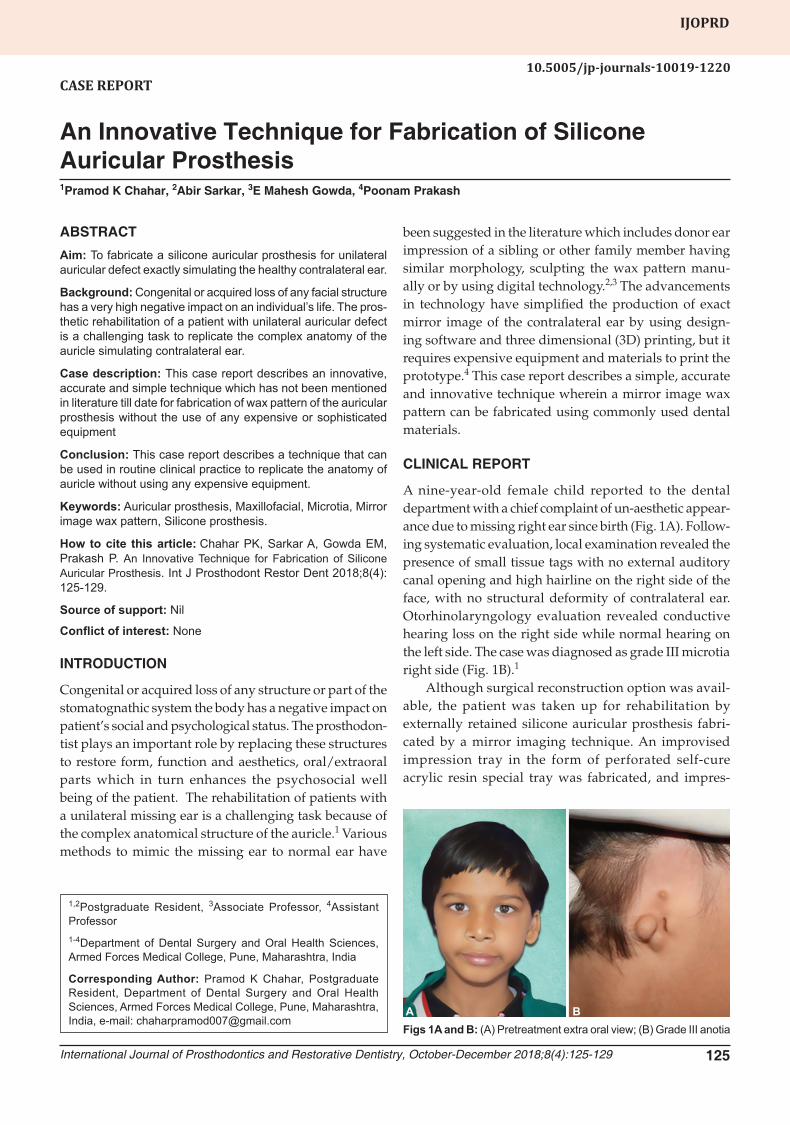
A nine-year-old female child reported to the dental department with a chief complaint of un-aesthetic appear-ance due to missing right ear since birth (Fig. 1A). Follow-ing systematic evaluation, local examination revealed the presence of small tissue tags with no external auditory canal opening and high hairline on the right side of the face, with no structural deformity of contralateral ear. Otorhinolaryngology evaluation revealed conductive hearing loss on the right side while normal hearing on the left side. The case was diagnosed as grade III microtia right side (Fig. 1B).1
Although surgical reconstruction option was avail-able, the patient was taken up for rehabilitation by externally retained silicone auricular prosthesis fabri-cated by a mirror imaging technique. An improvised impression tray in the form of perforated self-cure acrylic resin special tray was fabricated, and impres-
Figs 1A and B: (A) Pretreatment extra oral view; (B) Grade III anotiaA B

Pramod K Chahar et al.
126
The stone model of the unaffected ear was placed in a customized box made of base plate wax sheet, and type III dental stone was poured to obtain a stone block with ear model embedded in it. After finishing the block, orientation markings of 1.5 mm were made on the block using marking pen and metal scale on both anteropos-terior and superior-inferior direction (Fig. 2B).
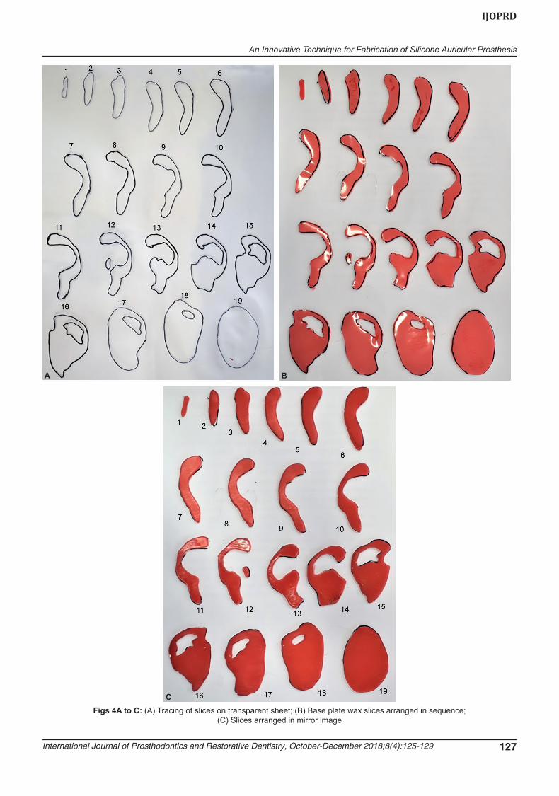
The marked model was trimmed using model trimmer till each successive guide/index marking and simultaneously exposed portion of ear model embedded in the block was traced on a transparent sheet till base. Nineteen slices (Fig. 3) and 19 tracings (Fig. 4A) were obtained from the stone block. This transparent sheet of traced marking was placed over base plate wax sheet (Marc Base Plate wax, Shiva Products, Mumbai) of same thickness, i.e. 1.5 mm, wax sheet was cut along with trans-parent sheet according to the marks of the tracing, slices were numbered and subsequently arranged in the same sequence (Fig. 4B). Slices were reversed at same position and sequence (Fig. 4C). After reversing, the slices were placed one over the other in reverse order sequentially with slice number 19 at the base and slice number 01 on top (Fig. 5A). The margins were merged with a hot spatula. Carving and finer adjustments were done during
sions of both sides (defect side and contralateral ear) were made with irreversible hydrocolloid impression material (Dentsply Zelgan 2002 Alginate, JMD Enter-prises, New Delhi). The cast of defect side was made using in type III dental stone (Kalabhaiultrastone type III, Kalabhai Karson Pvt Ltd, Mumbai) and contralat-eral ear in type IV dental stone (Kalabhaiultrastone type IV, Kalabhai Karson Pvt Ltd, Mumbai) (Fig. 2A).
Figs 2A and B: (A) Model of unaffected ear; (B) Model of unaffected ear embedded in stone block
A B
Fig. 3: Slices obtained after trimming stone block
An Innovative Technique for Fabrication of Silicone Auricular Prosthesis
International Journal of Prosthodontics and Restorative Dentistry, October-December 2018;8(4):125-129 127
IJOPRD
Figs 4A to C: (A) Tracing of slices on transparent sheet; (B) Base plate wax slices arranged in sequence; (C) Slices arranged in mirror image
A B
C

Pramod K Chahar et al.
128
a sequential arrangement of wax slices. This led to the production of the final wax pattern which was an exact mirror image of the contralateral ear (Fig 5B).
Tissue tags present on cast of defect side were relieved by adding a layer of base plate wax, connected to the base of clear self-cure acrylic (DPI self-cure acrylic resin, Bombay Burmah Trading Corporation, Mumbai) and wax pattern of the auricle was attached. The acrylic base along with wax pattern was attached to the hair-band with double-sided tape, and the wax pattern was tried on the patient (Fig. 6A). After confirming the orientation and position of the auricular prosthesis, markings were made on hair-band and wax pattern was processed in the three-piece mold for easy retrieval of the prosthesis. Dewaxing was done, and shade selection was carried out in natural light. Three shades were selected for different parts of the ear and to match with skin tone. Intrinsic stains were added to the RTV silicone (Sai Enterprises) and mold was packed carefully to prevent incorporation of air bubbles and clamping was done to remove excess material. The RTV silicone was processed at room temperature for 24 hours according to the manufacturer’s instructions. After processing, the prosthesis was retrieved, finishing and polishing were done. The prosthesis was attached to the hair band at previously marked position and delivered (Fig 6B). Post-treatment instructions were given to the patient for maintenance of prosthesis. The patient was advised to avoid contact sports and contact of sharp objects or organic solvents and to remove the prosthesis while sleeping or lying down. The patient was advised to clean the prosthesis with mild soap and water.
DISCUSSION
Auricular defects may occur due to hereditary or devel-opmental causes. Congenital defects may arise due to anomalies of the 1st and 2nd branchial arches which results in anotia or microtia. In microtia, there is an absence of external auditory canal and the presence of a
small remnant of deformed cartilage, whereas the absence of the whole ear is called anotia. These abnormalities are usually associated with Treacher Collins syndrome and Goldenhaar’s syndrome. Among others, bilateral absence of ear is seen in less than 10% of all cases.5
Acquired defects may arise due to trauma, burns or as post surgical defects. Some of the common malignancies of head and neck region like squamous cell carcinoma, malignant melanoma, etc. are treated by surgical excision along with or without radiotherapy and chemotherapy. After surgical removal, the size of the defect, location, and desires of the patient decide the mode of rehabilitation, either by surgical reconstruction or prosthetic rehabili-tation. Due to the complexity of the anatomy, multiple surgeries are required to achieve acceptable surgical reconstruction. The rib cartilage is used for major ear reconstruction, which is scaffolded with stainless steel wire and placed under the epidermis.5
The difficulties faced during fabrication of custom-made prosthesis are; to obtain an accurate impression of the defect without distortion of tissue, the positioning of the prosthesis in harmony with healthy auricle on the opposite side, sculpting the exact morphology, to match the shade exactly with the skin color and tone of opposite side of the face. The position of the unilateral prosthetic auricle is determined by observing the association of con-
Figs 5A and B: (A) Addition of slices in reverse order; (B) Finished wax patternA B
A BFigs 6A and B: (A)Wax pattern try-in; (B) Prosthesis in situ
retained with hair band

An Innovative Technique for Fabrication of Silicone Auricular Prosthesis
International Journal of Prosthodontics and Restorative Dentistry, October-December 2018;8(4):125-129 129
IJOPRD
tralateral ear with facial structures and then transferring this position at the proposed reconstruction site.
The retention of the prosthesis is a critical factor for its acceptability. Different methods to retain an auricular prosthesis are by hair bands, eyeglass frames, medical grade adhesives and craniofacial implants with magnets or bars attachments.6 Although, maxillofacial implants can provide superior retention and stability to the pros-thesis in comparison with other modes of retention, but requires surgical intervention and a waiting period of 3 to 4 months for adequate osseointegration. For adhesively retained prosthesis, it is difficult for the patient to place and orient the prosthesis in the correct position and the skin adhesive may deteriorate which would result in decreased strength and bonding with skin over a period of time. Hypersensitive reactions have also been reported with some skin adhesives.7
Silicone elastomeric maxillofacial materials are more frequently used, as they provide better constancy and more life-like appearance, which meets patient’s aesthetic and cosmetic needs. These materials possess excellent physical, mechanical and chemical proper-ties and soft tissue like consistency which provide an added advantage in restoring the defects of movable soft tissues. Silicone materials are available in various shades to exactly resemble the skin texture and com-plexion. The drawback of the silicone prosthesis is that, over a while, the material deteriorates and change when get exposed to different environmental temperatures, moisture, UV light, and sunlight, thus creating a need for replacement by a new prosthesis. To overcome these shortcomings, various newer materials like polyphos-phazenes, silicon block polymers, methacryloxypropyl terminated polydimethylsiloxane have been introduced with enhanced physical, mechanical and chemical prop-erties like better elongation, improved edge strength, and heat stability, good tear strength, chemically inert, low hardness and viscosity for fabrication of maxillo-facial prostheses.8
Another techniques mentioned in literature by Gajdhar et al. include obtaining a reverse image of an ear by transferring parallel lines to the cast by using a vertical camera and tracing paper, dividing the cast of contralateral ear into small cubes to enable sculpting of the missing ear or producing mirror image of contralateral ear by slicing the wax pattern and placing it in reverse order, but it is very difficult to cut slices of wax pattern by saw in thin sections without distortion.2,3 This case report described an innovative, simple and accurate technique to fabricate an auricular prosthesis that is precisely the mirror image of the patient’s normal auricle in frontal and rear views. To add lifelike appearance, the shade
of prosthesis was matched by addition of intrinsic and extrinsic stains. The retention was achieved by attach-ing the prosthesis to the hair-band with self-cure acrylic resin. The anterior margins were strategically hidden by the hairline while the posterior borders were merged well in shade to the skin. The patient was comfortable and highly satisfied with the aesthetic results.
CONCLUSION
Different techniques have been used to fabricate the wax pattern but the modified technique which is used in this case is unique and easily reproducible. It is a simple, innovative, accurate, inexpensive and practically pos-sible technique that does not require any expensive or sophisticated equipment and materials. The only disad-vantage seen in this technique is the loss of some anatomic details while merging the margins of wax slices with a hot spatula, which require finer adjustment by minor carving and finishing of wax pattern.
CLINICAL SIGNIFICANCE
This case report describes a very simple and innovative technique for the fabrication of auricular prosthesis which is the mirror image of the contralateral ear without using any sophisticated or expensive equipment. This technique can be used in routine clinical practice to enhance the aesthetics and acceptance of the patient by fabricating anatomically similar prosthesis.
REFERENCES
1. Beumer J, Curtis TA, Firtell DN (eds). Maxillofacial rehabili-tation: Prosthodontic and surgical considerations. St. Louis; C.V. Mosby; 1979. p.328-340.
2. Gajdhar S, Gajdhar SK, Salakalakonda SR, Vasthare A. A modi-fied technique for fabricating a mirror image wax pattern for an auricular prosthesis. J Prosthet Dent. 2015 Jan 1;113(1):71-73.
3. Ebrahimi A, Kazemi A, Rasouli HR, Kazemi M, Motamedi MH. Reconstructive surgery of auricular defects: An overview. Trauma Mon. 2015 Nov;20(4):68-76.
4. Al Mardini M, Ercoli C, Graser GN. A technique to produce a mirrorimage wax pattern of an ear using rapid prototyping technology. J Prosthet Dent 2005;94:195-198.
5. Madhan R, Nayar S. Prosthetic management of patient with treacher collins syndrome. Indian J Dent Res. 2006 Apr-Jun;17(2):78-81.
6. Singh A, Ghosh S, Kar S, Ahmed I. Silicone prosthesis for a patient with unilateral ear defect: A clinical case report. Eur J Gen Dent. 2013;2:315-319.
7. Kiatamnuay S, Gettleman L, Khan Z, Goldsmith LJ. Effect of adhesive retention on maxillofacial prostheses. Part I: Skin dress-ings and solvent removers. J Prosthet Dent. 2000;84:335-340.
8. Barhate AR, Gangadhar SA, Bhandari AJ, Joshi AD. Materials used in maxillofacial prosthesis: A review. Pravara Med Rev. 2015 Mar 1;7(1):5-8.