an increase in transmission-related traits and in … · an increase in transmission-related traits...
TRANSCRIPT

An increase in transmission-related traits and inphenotypic plasticity is documented during a fungal invasion
MATTEO GARBELOTTO,1 GIANNI DELLA ROCCA,2,� TODD OSMUNDSON,1,3
VINCENZO DI LONARDO,2 AND ROBERTO DANTI2
1Department of ESPM, 54 Hilgard Hall, University of California, Berkeley, California 94720 USA2Institute for Sustainable Plant Protection, CNR, Via Madonna del Piano 10, 50019 Sesto Fiorentino (FI), Italy
Citation: Garbelotto, M., G. Della Rocca, T. Osmundson, V. di Lonardo, and R. Danti. 2015. An increase in transmission-
related traits and in phenotypic plasticity is documented during a fungal invasion. Ecosphere 6(10):180. http://dx.doi.org/
10.1890/ES14-00426.1
Abstract. The adaptive rapid evolution of phenotypic traits is potentially a key contributor to
invasiveness, but has been relatively little studied for the fungi, even though these organisms are
responsible for devastating losses in agriculture and natural resources. In this study, we compare
biologically relevant phenotypic characters of spore-generated individuals from two native and two
invasive populations of the fungal pathogen Seiridium cardinale to infer which traits may be adaptive and
rapidly evolving during an ongoing biological invasion. Results show that: (1) lower growth rate and
smaller spore size are selected for in invasive populations, independent of the stage of invasion; (2) there is
no selection evident towards increased rapid sporulation, but overall reproductive potential increases in
later stages of the invasions; and (3) demographic plasticity of most traits increases during the initial stages
of invasion, but decreases in a later phase. Comparisons against levels of neutral genetic variation (Qst-Fst
comparisons) showed that the decrease in spore size is strongly adaptive, despite the trade-off of reduced
viability. Lesion size of isolates inoculated on the naıve Italian cypress host was not correlated with their
growth rate, and was significantly lower in invasive than in native populations. This last result indicates
that rate of host colonization is a complex trait affected both by host and pathogen, which may not be
necessarily adaptive and/or which may not easily evolve. In summary, the success of S. cardinale as an
invasive in the Mediterranean basin is associated with reduced spore size and increased plasticity of almost
all traits in initial phases, followed by further decreased spore size, increased overall sporulation, and
decreased plasticity in a second phase of the invasion. Interestingly, growth rate by population results
show that invasive populations are well adapted only to moderate temperatures, while native populations
fare well also when exposed to relative extremes in temperature. This different adaptation suggests a
‘‘master-of-some’’ specialization scenario for the invasion by S. cardinale in the Mediterranean.
Key words: adaptation; canker; co-evolution; cypress; disease; epidemic; genetic variability; pathogenicity; selection.
Received 15 November 2014; accepted 23 December 2014; final version received 25 June 2015; published 22 October
2015. Corresponding Editor: M. Allen.
Copyright: � 2015 Garbelotto et al. This is an open-access article distributed under the terms of the Creative Commons
Attribution License, which permits unrestricted use, distribution, and reproduction in any medium, provided the
original author and source are credited. http://creativecommons.org/licenses/by/3.0/3 Present address: Department of Biology, University of Wisconsin, La Crosse, Wisconsin 54601 USA.
� E-mail: [email protected]
INTRODUCTION
As they expand their ranges and encounter
new environments, invasive species provide an
invaluable opportunity to understand the evolu-
tion of adaptation in organisms in real-time
(Suarez and Tsutui 2008). In particular, they
allow for the observation of how trait variation
v www.esajournals.org 1 October 2015 v Volume 6(10) v Article 180

may be correlated to adaptation within themanageable timeframe of an invasion process.Useful observations include not only the mea-surement of changes in phenotypic traits, butalso of the range of their variability during aninvasion, a response that may be driven in partby plasticity (Pigliucci 2001). Because invasionsare often initiated by the long-distance move-ment of a few individuals, the extent of pheno-typic variability in the presence of limited geneticvariability may provide insights into the roleplasticity may play in the invasion process, aconcept that has been often theoretically sug-gested, but little studied (Richards et al. 2006).This is particularly true for clonally reproducingorganisms, in which the genetic make up offounder individuals has the potential to influenceinvasive populations for periods of considerablelength, as mutation-derived adaptation occurs atmuch lower frequency than that driven byrecombination in sexually reproducing species(Fisher 1930, Muller 1932). Although the litera-ture on phenotypic adaptation of invasive organ-isms is ample, there is often no clear consensuson what traits may be driving invasions (re-viewed in van Kleunen et al. 2010). In manycases, the inability to draw solid conclusions isderived from the fact that ‘‘soft’’ traits, moreeasily measurable and hence more frequentlyanalysed, are not a perfect proxy for the ‘‘hard’’traits directly involved in the determination ofinvasiveness (Violle et al. 2007). The limitedamount of published research focusing on traitselection for fungi is also noteworthy. The scantpublication record on this subject for an entireKingdom has been determined not only by thedifficulty in performing analysis of phenotypictraits for microorganisms, but also by an exces-sive focus on the presumed role played by highvirulence levels in non-coevolved host-fungusinteractions, neglecting other ecological andtransmission factors (Garbelotto et al. 2010). Inrecent times, a new impetus has been placed inproviding a more comprehensive framework tounderstand the invasive potential of fungi, whichcan be better modelled and explained bydetermining their transmission potential as afunction of their establishment success, survival,reproductive rate and dispersal, rather than justas a function of their pathogenicity (Gonthier andGarbelotto 2013). A further limitation—this one
common to the study of all invasive species—isthe imperfect knowledge of the exact geographicorigin of most invasive fungi. The inclusion of thecorrect source population is critical for studyingtrait variation of an invasive population throughcomparison with native populations (Keller andTaylor 2008).
In a simulation study, Burton et al. (2010)showed that during range expansion, dispersivetraits are selected for at the front of theexpanding population, whereas traits associatedwith fitness at equilibrium density (i.e., compet-itive ability) show strong declines. However,hard data in support of these predictions forfungi are still scant.
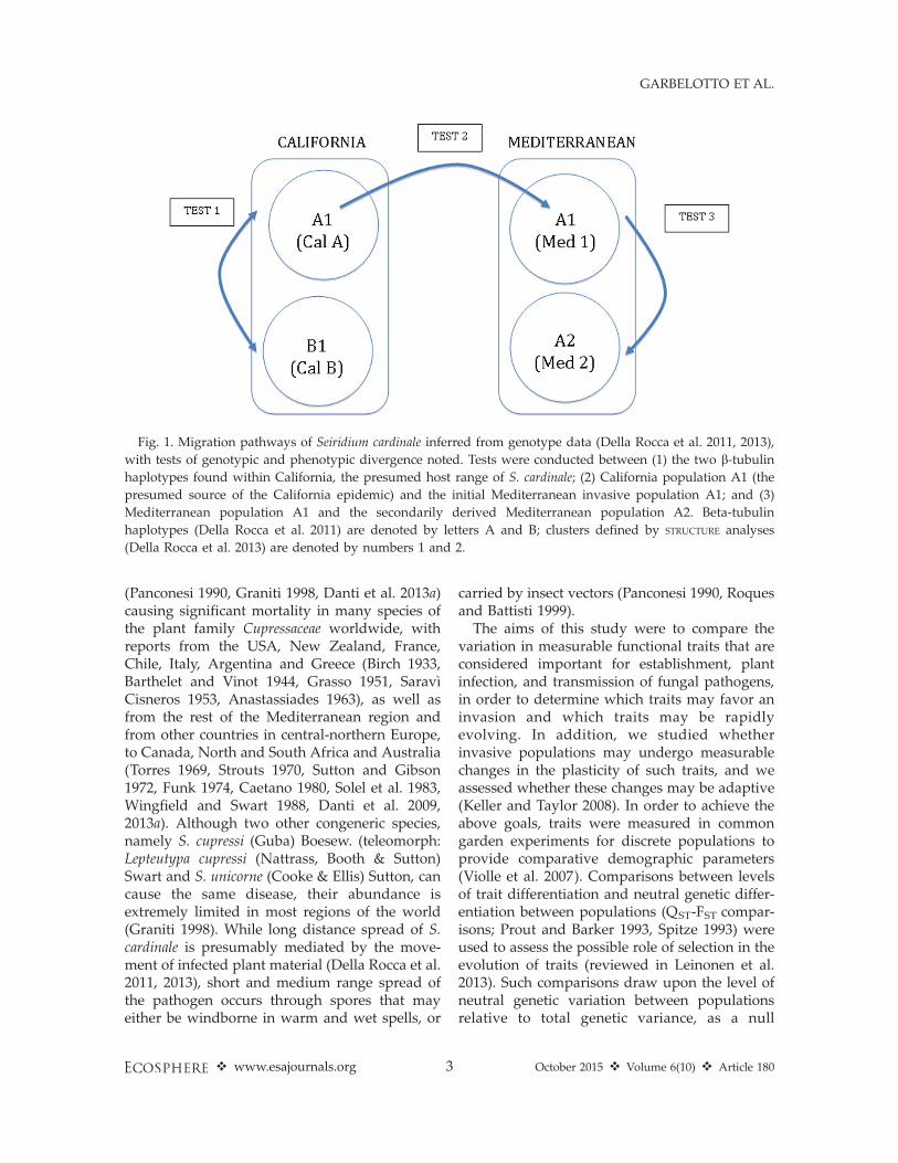
In this study, we analyze and compare traits oftwo native and sexually outcrossing Californiapopulations of the pathogenic fungus Seiridumcardinale (Wagener 1928) Sutton & Gibson (Asco-mycota; class: Coelomycetes; order: Melanconiales)with those of two populations sequentiallyderived from them after the successful movementof the pathogen to southern Europe (Della Roccaet al. 2011, 2013). The two native populations canbe differentiated by sequence differences at the b-tubulin locus, and thus they have been called b-tubulin A and B or, in short, Cal A and Cal B(Della Rocca et al. 2011). The two Europeanpopulations are sequentially derived from theCal A population but can be differentiated usingBayesian analysis of population structure(STRUCTURE; Pritchard et al. 2000) based onAFLP loci. Structure group 1 includes thefounder Mediterranean population (i.e., thatdirectly derived from Cal A) and has been calledMed 1, while Structure group 2—the populationderived from Med 1 in Europe—has been calledMed 2 (Della Rocca et al. 2013). Structure group 1also includes some California individuals (re-ferred to as Cal A1), presumably related to thefounder individuals that were originally trans-ported from California to Southern Europe (seeFig. 1 for a schematic representation of popula-tions in this study). All studies thus far haveshown Mediterranean populations to be geneti-cally rather uniform (Viljoen et al. 1993, Moriccaet al. 2000, Barnes et al. 2001, Krokene et al. 2004,Pedron et al. 2007) and reproducing clonally(Della Rocca et al. 2011, 2013).
S. cardinale is the main causal agent of apandemic tree disease called cypress canker
v www.esajournals.org 2 October 2015 v Volume 6(10) v Article 180
GARBELOTTO ET AL.
(Panconesi 1990, Graniti 1998, Danti et al. 2013a)causing significant mortality in many species ofthe plant family Cupressaceae worldwide, withreports from the USA, New Zealand, France,Chile, Italy, Argentina and Greece (Birch 1933,Barthelet and Vinot 1944, Grasso 1951, Sarav�ıCisneros 1953, Anastassiades 1963), as well asfrom the rest of the Mediterranean region andfrom other countries in central-northern Europe,to Canada, North and South Africa and Australia(Torres 1969, Strouts 1970, Sutton and Gibson1972, Funk 1974, Caetano 1980, Solel et al. 1983,Wingfield and Swart 1988, Danti et al. 2009,2013a). Although two other congeneric species,namely S. cupressi (Guba) Boesew. (teleomorph:Lepteutypa cupressi (Nattrass, Booth & Sutton)Swart and S. unicorne (Cooke & Ellis) Sutton, cancause the same disease, their abundance isextremely limited in most regions of the world(Graniti 1998). While long distance spread of S.cardinale is presumably mediated by the move-ment of infected plant material (Della Rocca et al.2011, 2013), short and medium range spread ofthe pathogen occurs through spores that mayeither be windborne in warm and wet spells, or
carried by insect vectors (Panconesi 1990, Roquesand Battisti 1999).
The aims of this study were to compare thevariation in measurable functional traits that areconsidered important for establishment, plantinfection, and transmission of fungal pathogens,in order to determine which traits may favor aninvasion and which traits may be rapidlyevolving. In addition, we studied whetherinvasive populations may undergo measurablechanges in the plasticity of such traits, and weassessed whether these changes may be adaptive(Keller and Taylor 2008). In order to achieve theabove goals, traits were measured in commongarden experiments for discrete populations toprovide comparative demographic parameters(Violle et al. 2007). Comparisons between levelsof trait differentiation and neutral genetic differ-entiation between populations (QST-FST compar-isons; Prout and Barker 1993, Spitze 1993) wereused to assess the possible role of selection in theevolution of traits (reviewed in Leinonen et al.2013). Such comparisons draw upon the level ofneutral genetic variation between populationsrelative to total genetic variance, as a null
Fig. 1. Migration pathways of Seiridium cardinale inferred from genotype data (Della Rocca et al. 2011, 2013),
with tests of genotypic and phenotypic divergence noted. Tests were conducted between (1) the two b-tubulinhaplotypes found within California, the presumed host range of S. cardinale; (2) California population A1 (the
presumed source of the California epidemic) and the initial Mediterranean invasive population A1; and (3)
Mediterranean population A1 and the secondarily derived Mediterranean population A2. Beta-tubulin
haplotypes (Della Rocca et al. 2011) are denoted by letters A and B; clusters defined by STRUCTURE analyses
(Della Rocca et al. 2013) are denoted by numbers 1 and 2.
v www.esajournals.org 3 October 2015 v Volume 6(10) v Article 180
GARBELOTTO ET AL.

expectation for the degree of between-populationvariance in additive genetic traits.
Our specific hypotheses were as follows: (1)Range expansion should be strongly mediated bytraits facilitating transmission (such as overallfruiting body production) and long-distancespread (e.g., smaller spore sizes), while traitslinked to fitness and virulence (e.g, growth ratein planta and in vitro) should not necessarily befavored. In addition, assuming S. cardinaleinvaded a niche unoccupied by other competi-tors, there should be no measurable shift towardsincreased sporulation in the short term andtowards increased germinability, two traits thatmay confer an advantage to the invasive speciesin the presence of competitors. (2) Demographicplasticity should be favored and should increasein the early stages of invasion, thus allowing theexotic species to adapt to a variety of novelenvironments, but establishment of the exoticorganism in the most favorable habitats shouldreduce plasticity in the second stages of aninvasion. (3) Although measurable shifts maybe identified in various traits, only some will beadaptive, i.e., driven by selection, while othersmay simply shift due to genetic drift. In addition,a delay in the change of a trait (i.e., a traitappears to change only in the second stage of aninvasion) may be an indication that geneticregulation of the trait is complex and that thetrait may be only slowly evolvable. (4) Due to thegenetic bottleneck experienced by the invasivespecies, naturalized invasive populations may bephenotypically distinct from the native ones theyderive from, even after their demographic ex-pansion in the novel region may have increasedtheir genetic variability. As a result, traits such asgrowth in planta and the ability to grow atdifferent temperatures may differ markedlybetween source and invasive populations, withimportant implications related to possible addi-tional introductions of individuals belonging toan exotic species.
MATERIALS AND METHODS
IsolationSeiridium cardinale isolates were obtained in
2007–08 from symptomatic trees across theMediterranean region and California. Diseasedstems, branches, or twigs were excised, put in
plastic bags, and processed in the laboratorywithin 24 hours. The outer bark was carefullyscraped with a sterile scalpel to expose themargin of cankers. Small fragments cut fromthe margin of the necrotic tissues were placedonto PDA (Potato Dextrose Broth 20 g/Lþ 20 g/LAgar) plates maintained at 258C in the dark.
White-greyish, cottony colonies (with olive-green shades) were transferred within 3–5 dayson 1% MEA (Malt Extract Agar 10 g/L þ 20 g/LAgar) supplemented with autoclaved (1208C, 20min) cypress seeds in 60-mm Petri dishes. Plateswere then incubated at 188C under mixed visibleand near-ultraviolet light (NUV, 400–200 nm), setto provide 12-h light/dark cycles to inducesporulation. After 3–4 weeks, acervuli (i.e.,asexual fruiting bodies) of the fungus developedon the seeds and on the agar surface.
The identity of the fungus was confirmedbased on the morphology of the six-celledfusiform conidia under a compound microscopeat 4003magnification (Wagener 1939, Sutton andGibson 1972, Mordue 1976, Chou 1989) and bysequence analysis of a portion of the b-tubulinlocus (Della Rocca et al. 2011, 2013).
Cultures from single S. cardinale conidia wereobtained for each isolate as follows: 2–3 matureacervuli were collected and placed in 1.5-mLmicrocentrifuge tubes where they were gentlycrushed in sterile distilled water using a sterileplastic pestle. Aliquots of 100 ll of conidialsuspension were spread onto the surface of 2%Water Agar (WA) in 90-mm Petri dishes whichwere then maintained at 258C in the dark. After12 h of incubation, germinating conidia weresingly transferred to 1% MEA Petri dishescontaining autoclaved cypress seeds and wereincubated at 188C under mixed white and NUVlight as reported above. For each isolate, seedswith acervuli were collected and transferred inplastic vials and stored at �208C. All isolateswere maintained in the culture collection of theInstitute for Sustainable Plant Protection, IPSP-CNR, Florence, Italy.
Morphological and cultural observationsObservations of morphological and cultural
traits as well as of size of induced cankers oninoculated trees were conducted on 22 S.cardinale isolates from different Mediterraneancountries and 23 isolates from several California
v www.esajournals.org 4 October 2015 v Volume 6(10) v Article 180
GARBELOTTO ET AL.

counties (USA). Isolates were chosen as repre-sentatives of the two b-tubulin haplotypes and ofthe two Structure groups that were evidenced inprevious studies on genetic diversity of thefungus (Della Rocca et al. 2011, 2013; AppendixA).
Radial growth of colonies.—Radial growth ofcolonies was determined on 9 cm diameter Petridishes containing 18 mL 2% MEA, by placing a 4-mm plug taken from the margin of a colonygrown on MEA 2% for 10 d at 258C at the centerof the plate. Plates were then incubated for 14 din the dark at 158, 208, 258 and 308C, beforemeasuring the two perpendicular diameters foreach colony. Five replicates were used for eachisolate and temperature.
Production of acervuli in vitro.—The productionof acervuli, i.e., structures bearing the asexualspores (conidia), was evaluated by obtaining a 4-mm plug of mycelium taken from the margin of acolony grown for 10 d at 258C on 2% MEA andplacing it in the center of a 6 cm diameter Petridish containing 5 mL of 2% Water Agar and 10autoclaved cypress seeds evenly distributed onthe surface. Four replicates were used for eachisolate. Plates were incubated at 188C undermixed white and NUV light, as reported above.After 30 and 90 d, acervuli on the surface of the10 seeds were counted with a dissecting scope.
Conidial size.—For each isolate, conidia extrud-ing from mature acervuli were suspended insterile water in 1.5-mL microcentrifuge tubes.After 10 minutes, the conidial suspension wasplaced on a microscope slide to allow for themeasurement of the length and width of 100conidia using a light microscope (4003; AxioskopZeiss) equipped with a digital camera.
Germination of conidia.—For each isolate, co-nidial suspensions (see above) were diluted withsterile water in 1.5-mL microcentrifuge tubeswater to a concentration of 53 103 conidia mL�1.Suspensions were incubated at 258C in the dark.After 24 h, 100 ll of suspension were placed on amicroscope slide and the number of germinatedconidia and mean number of germination tubesper conidium for 100 random conidia werecounted at 4003 using a light microscope(Axioskop Zeiss).
Artificial inoculationsItalian cypress (Cupressus sempervirens L. var.
horizontalis) plants obtained from seed and grownin 30 L pots containing a mixture of peat, compostand perlite (3:1:1 v/v/v) were placed in agreenhouse at the IPSP experimental farm locatedat Antella, Bagno a Ripoli, Florence, Italy (43844.20
N, 11819.40 E, 139 m a.s.l.). The average maximumtemperature was 258C 6 48C and average relativehumidity was 65% 6 5%. Five-year-old plantswith a mean height of 1.9 m and a mean basalstem diameter of 4.7 cm were inoculated in April2010. Inoculations were made at a point where thestem had a 1–2 cm diameter. A 4 mm circular plugof bark extending into the cambium was removedwith a cork borer and replaced with an equallysized plug of mycelium (top-side-down) takenfrom the margin of a fungal colony grown in thedark for 10 d at 258C on 2% MEA. Inoculationswere covered with cotton and wrapped with tapefor one week. Each S. cardinale isolate wasinoculated on 10 cypress plants, for a total of450 plants. On 10 control cypress plants a sterile2% MEA plug was inserted onto the wound.Plants were arranged in a completely randomizeddesign inside the greenhouse. The entire experi-ment was repeated twice.
Development of cankers was evaluated 6months after the artificial inoculations. The outerbark of inoculated stems was carefully scrapedoff with a sterile scalpel blade near the inocula-tion site in order to expose the margins ofnecrotic lesions. The number of successfullyinduced cankers and length and width ofnecrotic lesions were measured for each isolate.In order to fulfil Koch’s postulates, small pieces ofnecrotic tissues cut from the edge of lesions wereplated onto 2% PDA plates to recover theinoculated fungus.
Statistical analysesAssay results were analyzed using STATISTI-
CA 10.0 software. Differences among isolateswithin groups or between paired groups orpopulations of the fungus and effects of factorswere examined using analysis of variance(ANOVA). Tukey’s HSD (honestly significantdifference) was performed for post-hoc pairwisecomparisons. Percentages were converted usingthe formula y ¼ arcsin
ffiffiffiffiffiffiffiffiffiffiffiffiffip=100
pwhere p is the
percentage value, prior to analysis.Pearson correlation coefficients were calculated
to explore relationships between the mean values
v www.esajournals.org 5 October 2015 v Volume 6(10) v Article 180
GARBELOTTO ET AL.

of the following traits: size of induced cankersand radial growth of colonies at various temper-atures; size of cankers and number of acervuliproduced in vitro by isolates. Size of the necroticlesions was approximated to the area of anellipse and calculated using the formula (D/23 d/2)p, where D is the length and d the width ofcankers, respectively. Correlation between conid-ial size and percentage of germinated conidiaand number of germination tubes per 100 conidiawere also studied.
A discriminant analysis was performed on amatrix containing the mean values calculated foreach isolate for all of the assayed traits to explorethe possibility of a relationship between clustersbased on phenotypic traits and those previouslydetermined based on genetic markers andgeographic provenance, e.g., the two b-tubulinhaplotypes and the two AFLP Structure groups.
Phenotypic plasticity of all examined traits wasevaluated for the California and Mediterraneanpopulations and for the genetic sub-populationsof the fungus using two quantitative estimators:the phenotypic plasticity index (PIv) as (maxi-mum mean� minimum mean)/maximum mean,and the coefficient of variation (CV) as standarddeviation/mean (Valladares et al. 2006).
The following indices of genetic diversity werealso calculated based on the number andfrequency of multilocus genotypes (haplotypes)determined from the SSR analysis of S. cardinalepopulations from the Mediterranean and Cali-fornia reported by Della Rocca et al. (2011),including the isolates used in the present work:(1) Haplotype diversity, H ¼ k/n where k is thenumber of haplotypes, i.e., individuals sharingidentical alleles for all the loci analyzed, and n isthe number of individuals analyzed; (2) genediversity
H ¼ n
n� 1ð1�
Xk
i¼1
p2i Þ
where k is the number of haplotypes, n is thenumber of individuals analyzed and pi is thefrequency of the ith haplotype; and (3) genediversity (expected heterozygosity), estimated as
HE ¼1
m
Xm
l¼1
Xk
i¼1
p2i
where p is the frequency of the ith of k alleles,
averaged over each of m loci.
QST -FST comparisonsTraits exhibiting Pearson product-moment
correlations greater than 0.6 were removed fromsubsequent analyses. Traits analysed were num-ber of acervuli at 1 month, canker area, germi-nation percentage, number of germination tubes,conidial length, growth at 158C, growth at 208C,and growth at 308C. QST values were calculatedfor the phenotypic data collected from thecommon garden experiments using the followingformula:
QST ¼r2
B
r2W þ r2
B
where r2B is the between-group trait variance and
r2W is the within-group trait variance (Spitze
1993). Variance components were determinedusing a single-factor analysis of variance. Meanand single-locus FST values were calculated usingthe hierfstat package (Goudet 2005) for the Rstatistical computing environment (www.r-project.org). Observed QST values were com-pared to a null distribution of QST-FST valuesusing the simulation method of Whitlock andGuillaume (2009) as implemented by Lind et al.(2011; R script provided in Supplemental Infor-mation therein). Values of QST-FST ’ 0 suggestthat observed trait variance between populationsis the result of genetic drift; QST-FST . 0 suggestsdirectional selection on the trait, and QST-FST , 0suggests stabilizing selection on the trait (Leino-nen et al. 2013).
RESULTS
Morphological observationsDefinition of populations.—Although the popu-
lations studied here are either outcrossing (Cal Aand Cal B), nested within one another (Californiaand Mediterranean populations in the Structure 1group, or Med 1 and Med 2 both within the b-tubulin A group), or sequentially derived fromone another (Med 2 from Med 1), and results arepresented for all possible genetically distinctdemographic units, major inferences are drawnfor the following four demes: the Cal A and Cal BCalifornia populations, and the Med 1 and Med 2Mediterranean populations (see Fig. 1). Boxplotsshowing trait differences between groups are
v www.esajournals.org 6 October 2015 v Volume 6(10) v Article 180
GARBELOTTO ET AL.

provided in Appendix B.Radial growth of colonies.—For both geographic
populations of the pathogen (Californian andMediterranean, respectively CAL and MED) themean radial growth rate on 2% MEA was higherat 258C and progressively lower at 208, 158 and308C (Table 1). At each assayed temperature, thegrowth rate of CAL isolates was consistentlyhigher than that of MED isolates (158C: F1, 204 ¼105.4, P , 0.001; 208C: F1, 197 ¼ 53.6, P , 0.001;258C: F1, 192 ¼ 115.4, P , 0.001; 308C: F1, 201 ¼105.5, P , 0.001). Within the California popula-tion, the radial growth rate of isolates belongingto b-tubulin haplotype B was higher than that ofhaplotype A at 158C (F1,90 ¼ 5.71, P , 0.05) and208C (F1,90 ¼ 9.76, P , 0.01) but not at 258C and308C (Table 1).
Within the Mediterranean isolates, those be-longing to the Structure group Med 1 showed asignificantly higher growth rate than those ofStructure group Med 2 only at 158C (F1,98¼ 8.03,P , 0.01; Table 1).
No significant correlation was found betweenradial growth of isolates for any pairs oftemperatures (data not shown) while interactionbetween isolate and temperature was highlysignificant both for the MED (F63, 350 ¼ 21.1, P ,
0.001) and the CAL (F66, 276 ¼ 47.0, P , 0.001)populations. The difference between radialgrowth at 158 and 308C was rather marked forsome isolates that showed a preference for one ofthe two, while others showed similar radialgrowth at these two temperatures.
Production of acervuli in vitro.—The meannumber of acervuli produced in vitro wassignificantly higher for CAL than for MED isolatesboth after 30- and 90-d incubation (F1, 203¼ 28.4, P, 0.001 and F1, 203 ¼ 6.96, P , 0.01, respectively;
Table 2). Within the b-tubulin haplotype group A,isolates from California produced a higher num-ber of fruit bodies both after 30- and 90-dincubation than isolates from the Mediterranean(F1, 135 ¼ 13.9, P , 0.001 and F1, 135 ¼ 3.96, P ,
0.05). No significant difference in production ofacervuli was observed between isolates belongingto the two b-tubulin haplotypes within the CALpopulation; however, within the MED population,isolates of Structure group 2 produced a highernumber of acervuli than Structure group 1 isolates(P , 0.05) after 90 days of incubation (Table 2). Inboth CAL and MED populations, significantdifferences among isolates were observed withregards to the number of acervuli developed 30
Table 1. Mean diameter of colonies (in cm) of S. cardinale isolates developed on 2% MEA after 14-d incubation in
the dark at different temperatures. Isolates were grouped according to their geographic origin or genetic
subpopulations. Means not sharing same letters between paired sub-populations within each row were
significantly different for P ¼ 0.01 or * P¼ 0.05.
T (8C)
Geographicpopulation
b-tubulinhaplotype A
Structuregroup 1
b-tubulin haplotype(California isolates)
Structure group(Mediterranean isolates)
MED CAL Med A Cal A Med 1 Cal 1 Cal A Cal B Med 1 Med 2
158 3.18 a 4.39 b 3.18 a 4.19 b 3.33 a 4.39 b 4.19 a* 4.54 b* 3.33 b 2.77 a208 5.10 a 5.94 b 5.10 a 5.72 b 5.08 a 5.94 b 5.72 a 6.10 b 5.08 a 5.16 a258 5.61 a 6.92 b 5.61 a 7.01 b 5.64 a 6.91 b 7.01 a 6.86 a 5.64 a 5.64 a308 2.04 a 3.39 b 2.04 a 3.46 b 2.08 a 3.39 b 3.46 a 3.34 a 2.08 a 2.11 a
Table 2. Mean number of acervuli produced in vitro on
2% WA after 30-d and 90-d incubation at 188C under
12 h cycles under mixed white and NUV light by
isolates of the two geographic populations (MED
and CAL) and the genetic subpopulations of S.
cardinale. Paired means not sharing same letters
within a same line were different for P¼0.01 or * P¼0.05.
Population
No. acervuli
30 d 90 d
MED 2.9 a 8.6 aCAL 5.1 b 10.3 bHaplotype AMED 2.9 a 8.5 a*CAL 4.6 b 10.3 b*
Structure group 1MED 3.1 b 7.9 aCAL 5.1 a 10.3 a
CAL haplotypeA 4.6 a 10.3 aB 5.4 a 10.4 a
MED structure groupMed 1 3.1 a 8.0 a*Med 2 2.7 a 9.9 b*
v www.esajournals.org 7 October 2015 v Volume 6(10) v Article 180
GARBELOTTO ET AL.

and 90 d after incubation. Nine out of 10 isolatesthat produced the highest number of acervuliwere from California, while 8 out of 10 isolatesexhibiting the lowest number of acervuli after 30-d incubation were from Mediterranean (data notshown). With few exceptions, isolates showed amore or less marked increase in the number ofdeveloped acervuli between 30-d and 90-d incu-bation periods.
Percentage of germinated conidia.—The meanpercentage of conidia germinated in water after24 h incubation at 258C in the dark was high forboth CAL (97.3%) and MED (88.9%) isolates,though the difference between mean germinationwas statistically significant between the twopopulations (F1,43 ¼ 16.8, P , 0.001; Table 3).
Within b-tubulin haplotype A and Structuregroup 1, CAL isolates showed a significantlyhigher percentage of germinated conidia thanMED isolates (F1,30 ¼ 6.84, P , 0.02 and F1,35 ¼14.8, P , 0.001, respectively). The differencebetween the two b-tubulin haplotype groups wasnot significant if only isolates from Californiawere considered (F1,21 ¼ 0.37, P ¼ 0.54; Table 3).Isolates that showed germination percentagesbelow 92% were all from the Mediterranean,while among the 9 isolates with 100% germina-tion, 6 were from California (data not shown).
The mean sum of germinating tubes for 100conidia was higher for CAL than for MEDisolates (F1,43 ¼ 5.16, P ¼ 0.02), as it was forisolates from California compared to those fromthe Mediterranean within Structure group 1 (F1,35¼ 4.07, P ¼ 0.05; Table 3). No significantdifferences were observed between isolates ofthe two California b-tubulin haplotypes andbetween isolates of the two MediterraneanStructure groups (Table 3).
Conidial size.—Both mean length and width ofconidia were significantly higher for CAL isolatescompared to MED isolates (length: F1,2348¼347.9,P , 0.001; width: F1,2348¼ 469.4, P , 0.001; Table4). Within b-tubulin haplotype A and Structuregroup 1, isolates from California had longer andwider conidia (P , 0.01) than those produced byMediterranean isolates (comparison CAL – MEDwithin b-tubulin haplotype A, length: F1,1698 ¼315.5, P , 0.001; width: F1,1698¼ 298.9, P , 0.001.Comparison CAL – MED within Structure group1, Length: F1, 2048 ¼ 288.5, P , 0.001; Width: F1,2048 ¼ 369.9, P , 0.001) (Table 4). Within CALisolates, only mean length of conidia differedsignificantly between b-tubulin haplotypegroups (A . B, F1,1148¼ 26.4, P , 0.001). BetweenMED isolates, both length and width of conidiadiffered (Structure group Med 1 . Structure
Table 3. Mean percentage of germinated conidia and mean number of germinated tubes per 100 conidia of S.
cardinale isolates after 24 h incubation in water, at 258C in the dark. Isolates were grouped in geographic
populations (MED and CAL) and genetic sub-populations. Means not sharing same letters between paired
subpopulations within a same row were different for P ¼ 0.01 or * P¼ 0.05.
Measure ofgerminating conidia
Geographicpopulation
b-tubulinhaplotype A
b-tubulin haplotypes(only California)
Structuregroup 1
Structure group(Mediterranean isolates)
MED CAL Med A Cal A Cal A Cal B Med 1 Cal 1 Med1 Med2
Germinated conidia (%) 88.9 a 97.3 b 88.6 a* 96.9 b* 96.9 a 97.5 a 88.0a 97.3 b 88.0 a 86.8 aNo. germinating tubes
for 100 conidia165.8 a 185.0 b 165.8 a 189.8b 189.8 a 181.3 a 166.8 a* 185.0 b* 166.8 a 148.8 a
Table 4. Mean length and width of 50 conidia of S. cardinale isolates randomly measured under light microscope
at 4003 magnification. Isolates were grouped in geographic populations and genetic sub-populations. Means
not sharing same letters between paired subpopulations within rows were significantly different for P¼ 0.01.
Size ofconidia
Geographicpopulation
b-tubulinhaplotype A
b-tubulin haplotypes(California isolates)
Structuregroup 1
Structure group(Mediterranean isolates)
MED CAL Med A Cal A Cal A Cal B Med 1 Cal 1 Med 1 Med 2
Length (lm) 23.6 a 25.1 b 23.6 a 25.4 b 25.4 b 24.8 a 23.5 a 25.0 b 23.5 b 22.8 aWidth (lm) 8.3 a 9.1 b 8.3 a 9.12 b 9.10 a 9.16 a 8.3 a 9.1 b 8.4 b 8.0 a
v www.esajournals.org 8 October 2015 v Volume 6(10) v Article 180
GARBELOTTO ET AL.
group Med 2; length: F1,1148 ¼ 37.8, P , 0.001;width: F1,1148 ¼ 30.2, P , 0.001).
Overall, mean length and width of conidia ofisolates were significantly correlated (r ¼ 0.55, P, 0.01). The conidial length/width ratio differedsignificantly between the CAL (2.76) and theMED (2.87) populations of the fungus (F1,2348 ¼58.8, P , 0.001).
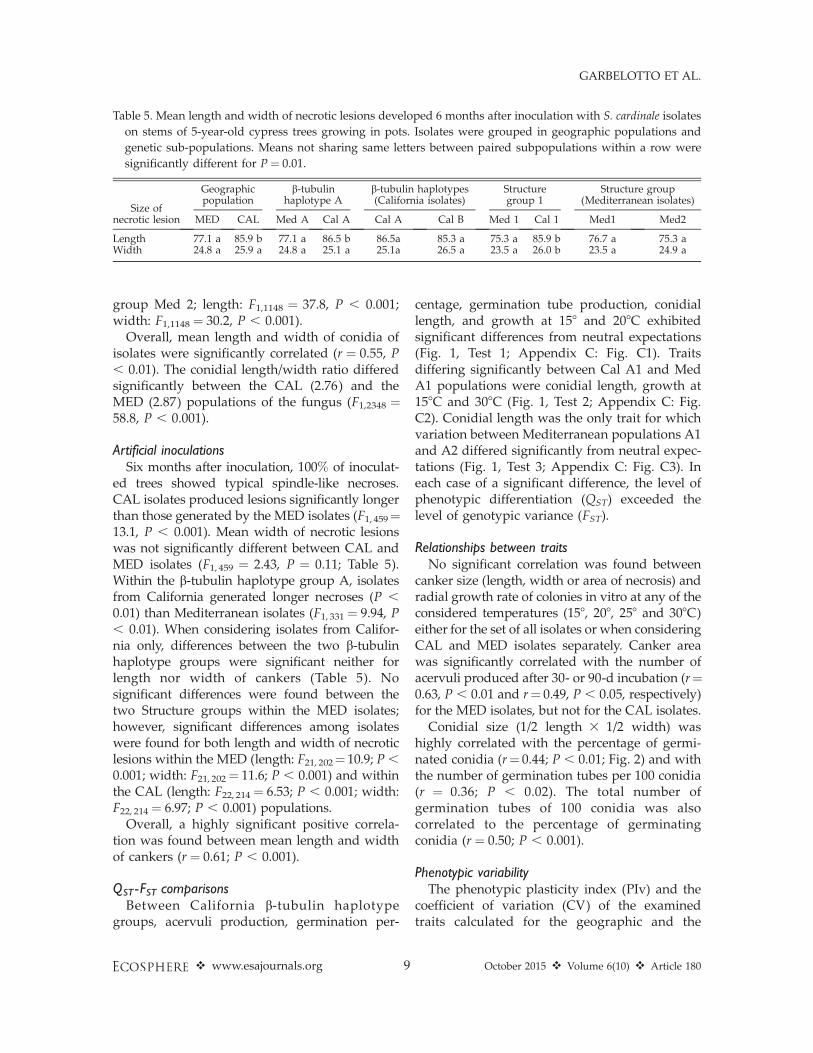
Artificial inoculationsSix months after inoculation, 100% of inoculat-
ed trees showed typical spindle-like necroses.CAL isolates produced lesions significantly longerthan those generated by the MED isolates (F1, 459¼13.1, P , 0.001). Mean width of necrotic lesionswas not significantly different between CAL andMED isolates (F1, 459 ¼ 2.43, P ¼ 0.11; Table 5).Within the b-tubulin haplotype group A, isolatesfrom California generated longer necroses (P ,
0.01) than Mediterranean isolates (F1, 331¼ 9.94, P, 0.01). When considering isolates from Califor-nia only, differences between the two b-tubulinhaplotype groups were significant neither forlength nor width of cankers (Table 5). Nosignificant differences were found between thetwo Structure groups within the MED isolates;however, significant differences among isolateswere found for both length and width of necroticlesions within the MED (length: F21, 202¼ 10.9; P ,
0.001; width: F21, 202¼ 11.6; P , 0.001) and withinthe CAL (length: F22, 214¼ 6.53; P , 0.001; width:F22, 214¼ 6.97; P , 0.001) populations.
Overall, a highly significant positive correla-tion was found between mean length and widthof cankers (r ¼ 0.61; P , 0.001).
QST -FST comparisonsBetween California b-tubulin haplotype
groups, acervuli production, germination per-
centage, germination tube production, conidiallength, and growth at 158 and 208C exhibitedsignificant differences from neutral expectations(Fig. 1, Test 1; Appendix C: Fig. C1). Traitsdiffering significantly between Cal A1 and MedA1 populations were conidial length, growth at158C and 308C (Fig. 1, Test 2; Appendix C: Fig.C2). Conidial length was the only trait for whichvariation between Mediterranean populations A1and A2 differed significantly from neutral expec-tations (Fig. 1, Test 3; Appendix C: Fig. C3). Ineach case of a significant difference, the level ofphenotypic differentiation (QST) exceeded thelevel of genotypic variance (FST).
Relationships between traitsNo significant correlation was found between
canker size (length, width or area of necrosis) andradial growth rate of colonies in vitro at any of theconsidered temperatures (158, 208, 258 and 308C)either for the set of all isolates or when consideringCAL and MED isolates separately. Canker areawas significantly correlated with the number ofacervuli produced after 30- or 90-d incubation (r¼0.63, P , 0.01 and r¼ 0.49, P , 0.05, respectively)for the MED isolates, but not for the CAL isolates.
Conidial size (1/2 length 3 1/2 width) washighly correlated with the percentage of germi-nated conidia (r¼ 0.44; P , 0.01; Fig. 2) and withthe number of germination tubes per 100 conidia(r ¼ 0.36; P , 0.02). The total number ofgermination tubes of 100 conidia was alsocorrelated to the percentage of germinatingconidia (r ¼ 0.50; P , 0.001).
Phenotypic variabilityThe phenotypic plasticity index (PIv) and the
coefficient of variation (CV) of the examinedtraits calculated for the geographic and the
Table 5. Mean length and width of necrotic lesions developed 6 months after inoculation with S. cardinale isolates
on stems of 5-year-old cypress trees growing in pots. Isolates were grouped in geographic populations and
genetic sub-populations. Means not sharing same letters between paired subpopulations within a row were
significantly different for P ¼ 0.01.
Size ofnecrotic lesion
Geographicpopulation
b-tubulinhaplotype A
b-tubulin haplotypes(California isolates)
Structuregroup 1
Structure group(Mediterranean isolates)
MED CAL Med A Cal A Cal A Cal B Med 1 Cal 1 Med1 Med2
Length 77.1 a 85.9 b 77.1 a 86.5 b 86.5a 85.3 a 75.3 a 85.9 b 76.7 a 75.3 aWidth 24.8 a 25.9 a 24.8 a 25.1 a 25.1a 26.5 a 23.5 a 26.0 b 23.5 a 24.9 a
v www.esajournals.org 9 October 2015 v Volume 6(10) v Article 180
GARBELOTTO ET AL.
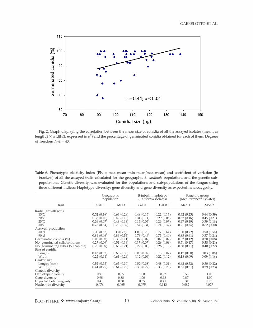
Fig. 2. Graph displaying the correlation between the mean size of conidia of all the assayed isolates (meant as
length/23width/2, expressed in l2) and the percentage of germinated conidia obtained for each of them. Degrees
of freedom N-2 ¼ 43.
Table 6. Phenotypic plasticity index (PIv ¼ max mean�min mean/max mean) and coefficient of variation (in
brackets) of all the assayed traits calculated for the geographic S. cardinale populations and the genetic sub-
populations. Genetic diversity was evaluated for the populations and sub-populations of the fungus using
three different indices: Haplotype diversity; gene diversity and gene diversity as expected heterozygosity.
Trait
Geographicpopulation
b-tubulin haplotype(California isolates)
Structure group(Mediterranean isolates)
CAL MED Cal A Cal B Med 1 Med 2
Radial growth (cm)158C 0.52 (0.16) 0.66 (0.29) 0.49 (0.15) 0.22 (0.16) 0.62 (0.23) 0.64 (0.39)208C 0.36 (0.10) 0.49 (0.18) 0.31 (0.11) 0.29 (0.08) 0.37 (0.16) 0.45 (0.21)258C 0.26 (0.07) 0.48 (0.18) 0.15 (0.05) 0.26 (0.07) 0.47 (0.19) 0.39 (0.16)308C 0.75 (0.34) 0.70 (0.32) 0.54 (0.31) 0.74 (0.37) 0.71 (0.34) 0.62 (0.30)
Acervuli production30 d 1.00 (0.67) 1 (0.72) 1.00 (0.70) 0.77 (0.64) 1.00 (0.72) 0.50 (0.56)90 d 0.81 (0.46) 0.86 (0.55) 0.79 (0.49) 0.73 (0.44) 0.85 (0.61) 0.37 (0.24)
Germinated conidia (%) 0.08 (0.02) 0.30 (0.11) 0.07 (0.02) 0.07 (0.02) 0.32 (0.12) 0.20 (0.08)No. germinated cells/conidium 0.27 (0.09) 0.51 (0.19) 0.17 (0.07) 0.26 (0.09) 0.51 (0.17) 0.38 (0.21)No. germinating tubes (50 conidia) 0.28 (0.09) 0.63 (0.21) 0.22 (0.08) 0.26 (0.10) 0.58 (0.21) 0.40 (0.22)Size of conidia
Length 0.13 (0.07) 0.63 (0.30) 0.08 (0.07) 0.13 (0.07) 0.17 (0.08) 0.03 (0.06)Width 0.22 (0.11) 0.61 (0.29) 0.12 (0.09) 0.22 (0.12) 0.18 (0.09) 0.09 (0.16)
Canker sizeLength (mm) 0.52 (0.33) 0.63 (0.30) 0.52 (0.38) 0.48 (0.31) 0.62 (0.32) 0.30 (0.22)Width (mm) 0.44 (0.25) 0.61 (0.29) 0.35 (0.27) 0.35 (0.25) 0.61 (0.31) 0.29 (0.23)
Genetic diversityHaplotype diversity 0.91 0.65 1.00 0.92 0.58 1.00Gene diversity 0.98 0.88 1.00 0.98 0.87 1.00Expected heterozygosity 0.41 0.30 0.35 0.41 0.31 0.20Nucleotide diversity 0.076 0.065 0.075 0.113 0.082 0.027
v www.esajournals.org 10 October 2015 v Volume 6(10) v Article 180
GARBELOTTO ET AL.
genetic sub-populations of the fungus are report-ed in Table 6. Generally, both PIv and CV of allevaluated traits were higher for MED isolatesthan for CAL isolates. Within the MED isolates,those belonging to Structure group Med 1showed higher PIv values than isolates ofStructure group Med 2 for all of the evaluatedtraits except radial growth of colonies at 158 and208C. Values of CV followed the same trend asPIv except for the number of germinating tubesper 100 conidia and width of conidia, which werehigher for the Structure group Med 2 isolates.Within California, b-tubulin haplotype A isolates(Cal A) showed higher PIv and CV values thanhaplotype B isolates (Cal B) for most of theevaluated traits except for radial growth ofcolonies at 258 and 308C, number of germinatingtubes per 100 conidia, and the length and widthof conidia, which were higher for the b-tubulinhaplotype B isolates. All three calculated indicesof genetic diversity were higher for the CALcompared to the MED isolates, for the Cal Acompared to Cal B, and for Med 2 compared to
Med 1 within the Mediterranean (Table 6).
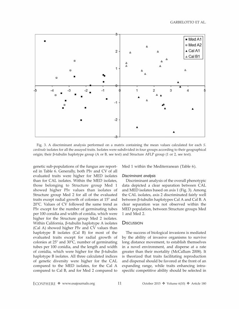
Discriminant analysisDiscriminant analysis of the overall phenotypic
data depicted a clear separation between CALand MED isolates based on axis 1 (Fig. 3). Amongthe CAL isolates, axis 2 discriminated fairly wellbetween b-tubulin haplotypes Cal A and Cal B. Aclear separation was not observed within theMED population, between Structure groups Med1 and Med 2.
DISCUSSION
The success of biological invasions is mediatedby the ability of invasive organisms to survivelong distance movement, to establish themselvesin a novel environment, and disperse at a rategreater than their mortality (McCallum 2008). Itis theorized that traits facilitating reproductionand dispersal should be favored at the front of anexpanding range, while traits enhancing intra-specific competitive ability should be selected in
Fig. 3. A discriminant analysis performed on a matrix containing the mean values calculated for each S.
cardinale isolates for all the assayed traits. Isolates were subdivided in four groups according to their geographical
origin; their b-tubulin haplotype group (A or B, see text) and Structure AFLP group (1 or 2, see text).
v www.esajournals.org 11 October 2015 v Volume 6(10) v Article 180
GARBELOTTO ET AL.

the area behind the expanding range wheredensities of invasive populations may increase(Burton et al. 2010).
In the case of pathogens, a further complica-tion is that their dispersal is linked to successfulinfection, a process mediated by responses andtraits of both pathogen and host populations.Until recently, literature on the subject ofinvasions by plant pathogens has mostly focusedon so-called qualitative aspects of successfulinfection, and in particular on the effects thatpresence/absence of compatible resistance andvirulence genes in hosts and pathogens, respec-tively, may have on success of the invasion. Thishas resulted in an excessive use of the ‘‘lack of co-evolution’’ hypothesis to interpret invasions byplant pathogens, while more inclusive studies onfactors such as infection efficiency, latent period,spore production rate, spore size, infectiousperiod, lesion size, and toxin production arescant (Lannou 2012). The roles of these quanti-tative traits and their trade-offs have been wellstudied in several pathosystems (Roff 2002,Lannou 2012), but this knowledge has rarelybeen transferred to the study of invasive patho-gens (Brasier 2001, Cobb et al. 2010, Robin et al.2010, Giordano et al. 2014).
With regard to our first hypothesis, resultsshow traits associated with enhanced transmis-sion to be favored over those associated withincreased virulence and greater competitiveability. Our results confirm that invasion by S.cardinale is associated with decreasing spore sizeduring both stages of its range expansion. Thistrait is known to be strongly and positivelycorrelated with increased dispersal range evenfor microscopical propagules (Norros et al. 2014),and in our study it appears to confer anadvantage, in spite of the documented trade-offdue to the reduced germinability of smallerspores (Fig. 2). On the other hand, lesion sizeon cypress, a quantitative trait that is the result ofhost-pathogen interactions and that can be usedas a proxy of the aggressiveness of the invasiveorganism, was significantly lower in invasivethan in native populations. Because lesions areresponsible for the girdling and death of theinfected portion of the plant, leading to acomplete arrest of sporulation, it is reasonableto assume that intermediate virulence may beselected for in an epidemic mode such as that
recorded in the Mediterranean. In fact, highlyvirulent genotypes may actually lead to overalllower sporulation loads (Violle et al. 2007), whilewithin the intermediate range, higher levels ofvirulence may be beneficial to transmission. Inour study, this benefit was confirmed by thepositive correlation observed in the Mediterra-nean populations between lesion size on cypressand production of acervuli.
Acervuli production at 90 days was unchangedin the founder Med 1 population compared toCalifornia populations, but it increased signifi-cantly in the derived Med 2 population, suggest-ing that an overall higher spore production maybe favored. The ‘‘delay’’ in the change of this traitmay indicate its evolvability may be lower, e.g., itmay require a longer period of time. Interesting-ly, acervuli production in the short term was notincreased. Likewise, higher spore germinabilitywas not favored either; to the contrary, lowergerminability was observed in invasive popula-tions. These two observations suggest that S.cardinale is not encountering significant interspe-cific competition requiring quicker establish-ment; this finding is in agreement with theobservation that other pathogens causing asimilar disease are rather infrequent (Graniti1998, Danti et al. 2013a). In summary, our resultssuggest that traits affecting transmission andlong-range dispersal, such as overall sporeproduction and smaller spore size, are favoredduring the invasion process, while traits favoringvirulence and competitive establishment, such asrapid growth in planta, quick production ofspores, and high spore germinability, are not.
With regard to our second hypothesis, our dataindicated demographic plasticity of most traits tobe greatly increased in the first stages of invasion.Notably, despite the reduced genetic variabilityin the invasive population Med 1, the amplitudeof responses of all traits as indicated by theindices PIv and CV is greater than in the sourceCAL population. On the contrary, when compar-ing the two Mediterranean populations, in spiteof an increase in genetic variability in Med 2(likely to be due to an increase in populationsize), we recorded a significant decrease in theamplitude of responses of all traits measured.The only exceptions were growth rates at coldand hot temperatures, 158 and 308C respectively,in which cases lack of adaptation justifies the
v www.esajournals.org 12 October 2015 v Volume 6(10) v Article 180
GARBELOTTO ET AL.

maintenance of variable responses at the popu-lation level. We suggest that the observedpatterns imply a significant role played byplasticity in the first stages of the invasionprocess (plasticity Med 1 . CAL) followed by adecrease of such a role as invasive populationsfind their most suitable niche in the novelgeographic range (plasticity Med 2 , Med 1).
The expectations of our third hypothesis werealso met; i.e., only some of the measured traitsappeared to be under selection. It should,however, be pointed out that lack of supportfor selection in QST-FST analyses does notnecessarily mean selection is not at play, butrather that its role cannot be discerned given thelevel of neutral variation for the genetic markersemployed. The genetic markers employed in thisand in previous studies are not necessarily linkedto the phenotypic traits here analyzed; nonethe-less, some generalized conclusions can be drawnfrom a comparative analysis of all traits. Thediscriminant analysis based on phenotypic traitsshows clear distinction of all four populations,with overall statistically significant differencesbetween CAL and MED populations. Amongtraits under adaptive selection, decreasing sporesize is the trait that appears to be continuouslyfavored in both stages of the invasion, whilegrowth at low and high temperatures (158 and308C, respectively) appears to rapidly decreaseduring the first stages of the invasion, possiblysuggesting a maladaptation of introduced popu-lations to temperatures extremes. When compar-ing the two native populations Cal A and Cal B,evidence for significant selection was found onceagain for conidial size, but also for germinability(which may confer an intraspecific competitiveadvantage) and for growth at low and interme-diate temperatures (158 and 208C, respectively),suggesting the two populations are competitivelyinteracting as suggested by spore germinabilitydata, but may be differently adapted to coastaland interior habitats, favored by higher growthat low/medium or high temperatures, respective-ly.
Finally, our last hypothesis stated that someindividuals in native populations may havetraits—or, more properly, may have responsesin various traits—that differ significantly fromresponses measured for individuals in invasivepopulations. The discriminant analysis presented
in this study indicates that California populationsare overall phenotypically distinct from theMediterranean populations. Our analyses identi-fy spore size, growth rates, growth rates 3
temperature, and pathogenicity as traits clearlydistinct among populations from the two re-gions. We believe some of these traits may bedetrimental to naıve hosts in the zone of invasion(ZOI), if acquired by invasive populations. Forinstance, while our study points to virulence as atrait under stabilizing selection in the ZOI(Leonard and Czochor 1980), it also indicatesthat additional introductions of S. cardinale fromCalifornia into the Mediterranean basin couldhave serious consequences on the survival ofnative host populations, resulting in highermortality rates. Furthermore, a further expansionin areas both warmer and colder than thosecurrently occupied by the invasive pathogencould be an obvious outcome of the introductionof genotypes with different thermal optima.Because control regulations are relaxed whenthe same species is reported in two differentcountries, this finding underlines a considerableweakness of current international policies aimedat limiting introduction of invasive species.
To our knowledge, this is one of the firststudies presenting a comparative analysis be-tween source and invasive fungal populationsfocusing on a broad array of phenotypic traitsand including a comparison with genetic data.Our findings indicate the presence of a measur-able phenotypic differentiation between nativeand invasive populations belonging to the samespecies. Furthermore, it appears from QST-FSTcomparisons that selection on traits related todispersal is stronger than that for traits related tovirulence, at least when comparing invasive tosource populations. Our data further suggest thatin the absence of significant competition, traitsenhancing competitive fitness—such as rapidsporulation—may not be selected for. We alsoshow that plasticity increases in the first phasesof the invasion, possibly assisting the invasivepopulation in surviving in novel habitats, andthen it decreases, possibly as the invasivepopulation adapts and expands in the mostsuitable niches. Finally, we show that quantita-tive pathogenicity does not increase betweensource and invasive populations, either becauseit is not easily evolvable, as it may be determined
v www.esajournals.org 13 October 2015 v Volume 6(10) v Article 180
GARBELOTTO ET AL.

by multiple loci of both host and pathogen, and/or because intermediate pathogenicity levels maybe ideal to maximize transmission. Nonetheless,native source populations include genotypes thatare genetically distinct and display higher path-ogenicity than genotypes already introduced:their introduction could lead to increased mor-tality rates of hosts in the exotic range, even ofcultivars currently deemed resistant (Danti et al.2006, 2013b). Based on these results, we concludethat regulations should prevent any furtherintroductions of S. cardinale into the Mediterra-nean region: this conclusion is also supported bythe fact that introductions of a compatible matingtype postulated to exist in California (Della Roccaet al. 2011) could lead to sexual reproductionamong individuals from different provenances, amechanism known to increase the adaptivepotential of invasive species (Facon et al. 2008).
ACKNOWLEDGMENTS
The work reported in this paper was partiallysupported by the Short-Term Mobility Program ofCNR of Italy, which financed a 1-month residency ofDr. Della Rocca at the U.C. Berkeley Forest Pathologyand Mycology Laboratory. We thank Dr. KatherineHayden for much discussion on plasticity and its rolein adaptation.
LITERATURE CITED
Anastassiades, B. 1963. A new for Greece disease of thecypress. Annals of the Institute of PhytopathologyBenaki 5:164–166.
Barnes, I., J. Roux, and M. J. Wingfield. 2001.Characterization of Seiridium spp. associated withcypress canker based on b-tubulin and histonesequences. Plant Disease 85:317–321.
Barthelet, J., and M. Vinot. 1944. Notes sur les maladiesdes cultures meridionales. Annales des Epiphyties10:18–20.
Birch, T. T. C. 1933. Gummosis diseases of Cupressusmacrocarpa. New Zealand Journal of Forestry 3:108–113.
Brasier, C. M. 2001. Rapid evolution of introducedplant pathogens via interspecific hybridization.BioScience 51:123.
Burton, O. J., B. L. Phillips, and J. M. J. Travis. 2010.Trade-offs and the evolution of life-histories duringrange expansion. Ecology Letters 13:1210–1220.
Caetano, M. F. F. 1980. Uma grave doenca dasCupressaceas em Portugal. Agros 63:5–10.
Chou, C. K. S. 1989. Morphological and cultural
variation of Seiridium spp. from cankered Cupressa-ceae hosts in New Zealand. European Journal ofForest Pathology 19:435–445.
Cobb, R. C., R. K. Meentemeyer, and D. M. Rizzo.2010. Apparent competition in canopy trees deter-mined by pathogen transmission rather thansusceptibility. Ecology 91:327–333.
Danti, R., G. Della Rocca, and F. El Wahidi. 2009.Seiridium cardinale newly reported on Cupressussempervirens in Morocco. Plant Pathology 58:1174.
Danti, R., G. Della Rocca, and A. Panconesi. 2013a.Cypress canker. Pages 359–375 in G. Nicolotti andP. Gonthier, editors. Infectious forest disease. CABIPress, Oxfordshire, UK.
Danti, R., V. Di Lonardo, A. Pecchioli, and G. DellaRocca. 2013b. ‘Le Crete 1’ and ‘Le Crete 2’: twonewly patented Seiridium cardinale canker-resistantcultivars of Cupressus sempervirens. Forest Pathol-ogy 43:204–210.
Danti, R., A. Panconesi, V. Di Lonardo, G. Della Rocca,and P. Raddi. 2006. ‘Italico’ and ‘Mediterraneo’: twoSeiridium cardinale canker-resistant cypress culti-vars of Cupressus sempervirens. HortScience41:1357–1359.
Della Rocca, G., C. A. Eyre, R. Danti, and M.Garbelotto. 2011. Sequence and SSR analyses ofthe fungal pathogen Seiridium cardinale indicateCalifornia is the most likely source of the cypresscanker epidemic for the Mediterranean region.Phytopathology 101:1408–1417.
Della Rocca, G., T. Osmundson, R. Danti, A. Pecchioli,F. Donnarumma, E. Casalone, and M. Garbelotto.2013. AFLP analysis of California and Mediterra-nean populations of Seiridium cardinale provideinsights on its origin, biology and spread path-ways. Forest Pathology 43:211–221.
Facon, B., J. P. Pointier, P. Jarne, V. Sarda, and P. David.2008. High genetic variance in life-history strategieswithin invasive populations by way of multipleintroductions. Current Biology 18:363–367.
Fisher, R. A. 1930. The genetical theory of naturalselection. Oxford University Press, Oxford, UK.
Funk, A. 1974. Microfungi associated with dieback ofnative Cupressaceae in British Columbia. CanadianPlant Disease Survey 54:166–168.
Garbelotto, M., R. Linzer, G. Nicolotti, and P. Gonthier.2010. Comparing the influences of ecological andevolutionary factors on the successful invasion of afungal forest pathogen. Biological Invasions12:943–957.
Giordano, L., P. Gonthier, G. Lione, P. Capretti, and M.Garbelotto. 2014. The saprobic and fruiting abilitiesof the exotic forest pathogen Heterobasidion irregu-lare may explain its invasiveness. Biological Inva-sions 16:803–814.
Gonthier, P., and M. Garbelotto. 2013. Reducing thethreat of emerging infectious diseases of forest
v www.esajournals.org 14 October 2015 v Volume 6(10) v Article 180
GARBELOTTO ET AL.

trees: mini review. CAB Reviews 8:025.Goudet, J. 2005. Hierfstat, a package for R to compute
and test hierarchical F-statistics. Molecular EcologyNotes 5:184–186.
Graniti, A. 1998. Cypress canker: a pandemic inprogress. Annual Review of Phytopathology36:91–114.
Grasso, V. 1951. Un nuovo agente patogeno delCupressus macrocarpa Hartw. in Italia. Italia Fore-stale e Montana 6:63–65.
Keller, S. R., and D. R. Taylor. 2008. History, chanceand adaptation during biological invasion: sepa-rating stochastic phenotypic evolution from re-sponse to selection. Ecology Letters 11:852–866.
Krokene, P., I. Barnes, B. D. Wingfield, and M. J.Wingfield. 2004. A PCR-RFLP based diagnostictechnique to rapidly identify Seiridium speciescausing cypress canker. Mycologia 96:1352–1354.
Lannou, C. 2012. Variation and selection of quantita-tive traits in plant pathogens. Annual Review ofPhytopathology 50:319–338.
Leinonen, T., R. J. S. McCairns, R. B. O’Hara, and J.Merila. 2013. QST-FST comparisons: evolutionaryand ecological insights from genomic heterogene-ity. Nature Reviews Genetics 14:179–190.
Leonard, K. J., and R. J. Czochor. 1980. Theory ofgenetic interactions among populations of plantsand their pathogens. Annual Review of Phytopa-thology 18:237–258.
Lind, M. I., P. K. Ingvarsson, H. Johansson, D. Hall,and F. Johansson. 2011. Gene flow and selection onphenotypic plasticity in an island system of Ranatemporaria. Evolution 65:684–697.
McCallum, H. 2008. Landscape structure, disturbance,and disease dynamics. Pages 100–122 in R. S.Ostfeld, F. Keesing, and V. T. Eviner, editors.Infectious disease ecology. Princeton UniversityPress, Princeton, New Jersey, USA.
Mordue, J. E. M. 1976. CMI descriptions of pathogenicfungi and bacteria. Number 514. CommonwealthMycological Institute, Kew, Surrey, UK.
Moricca, S., I. Børja, G. G. Vendramin, and P. Raddi.2000. Differentiation of Seiridium species associatedwith virulent canker on cypress in the Mediterra-nean region by PCR-SSCP. Plant Pathology 49:774–781.
Muller, H. J. 1932. Some genetic aspects of sex.American Naturalist 66:118–138.
Norros, V., U. Rannik, T. Hussein, T. Petaja, T. Vesala,and O. Ovaskainen. 2014. Do small spores dispersefurther than larger spores? Ecology 95:1612–1621.
Panconesi, A. 1990. Pathological disorders in theMediterraneran basin. Pages 54–81 in J. Ponchet,editor. Agriculture—AGRIMED research pro-gramme: progress in EEC research on cypressdiseases. Report EUR 12493 EN. Commission of theEuropean Communities, Brussels, Luxemburg.
Pedron, L., G. Piva, and N. La Porta. 2007. The geneticstructure of Cypress canker fungus in Italy usingRAPD and minisatellite Markers. Acta SilvaticaLignaria Hungarica, Special Edition:159–168.
Pigliucci, M. 2001. Phenotypic plasticity: beyondnature and nurture. Johns Hopkins UniversityPress, Baltimore, Maryland, USA.
Pritchard, J. K., M. Stephens, and P. Donnelly. 2000.Inference of population structure using multilocusgenotype data. Genetics 155:945–959.
Prout, T., and J. S. F. Barker. 1993. F statistics inDrosophila buzzatii: selection, population size andinbreeding. Genetics 134:369–375.
Richards, C. L., O. Bossdorf, N. Z. Muth, J. Gurevitch,and M. Pigliucci. 2006. Jack of all trades, master ofsome? On the role of phenotypic plasticity in plantinvasions. Ecology Letters 9:981–993.
Robin, C., S. Lanz, A. Soutrenon, and D. Rigling. 2010.Dominance of natural over released biologicalcontrol agents of the chestnut blight fungusCryphonectria parasitica in south-eastern France isassociated with fitness-related traits. BiologicalControl 53:55–61.
Roff, D. A. 2002. Life history evolution. SinauerAssociates, Sunderland, Massachusetts, USA.
Roques, A., and A. Battisti. 1999. Cypress pests. Pages75–95 in E. Teissier Du Cros, editor. Cypress, apractical handbook. EU FAIR 3 Concerted Action,Florence, Italy.
Sarav�ı Cisneros, R. 1953. Cancrosis de los Cipresesprovocada por Coryneum cardinale Wag. En laprovincia de Buenos Aires (Argentina). Revista dela Facultad de Agronomia de La Plata 29:107–119.
Solel, Z., R. Messinger, Y. Golan, and Z. Madar. 1983.Coryneum canker of cypress in Israel. Plant Disease67:550–551.
Spitze, K. 1993. Population structure in Daphnia obtusa:quantitative genetic and allozymic variation. Ge-netics 135:367–374.
Strouts, R. G. 1970. Coryneun canker of Cupressus. PlantPathology 19:149–150.
Suarez, A. V., and N. D. Tsutui. 2008. The evolutionaryconsequences of biological invasions. MolecularEcology 17:351–360.
Sutton, B. C., and I. A. S. Gibson. 1972. Seiridiumcardinale. Commonwealth Mycological Institutedescription of pathogenic fungi and bacteria.Number 326. CMI, Kew, Surrey, UK.
Torres, J. 1969. Grave enfermedad de los cipreses enEspana. Boletin del Servicio de Plagas Forestales12:97–99.
Valladares, F., D. Sanchez-Gomez, and M. A. Zavala.2006. Quantitative estimation of phenotypic plas-ticity: bridging the gap between the evolutionaryconcept and its ecological applications. Journal ofEcology 94:1103–1116.
van Kleunen, M., W. Dawson, D. Schlaepfer, J. M.
v www.esajournals.org 15 October 2015 v Volume 6(10) v Article 180
GARBELOTTO ET AL.
Jeschke, and M. Fischer. 2010. Are invadersdifferent? A conceptual framework of comparativeapproaches for assessing determinants of invasive-ness. Ecology Letters 13:947–958.
Viljoen, C. D., B. D. Wingfield, and M. J. Wingfield.1993. Comparison of Seiridium isolates associatedwith cypress canker using sequence data. Experi-mental Mycology 17:323–328.
Violle, C., M. L. Navas, D. Vile, E. Kazakou, C.Fortunel, I. Hummel, and E. Garnier. 2007. Let theconcept of trait be functional! Oikos 116:882–892.
Wagener, W. W. 1928. Coryneum canker of cypress.
Science 67:584.Wagener, W. W. 1939. The canker of Cupressus induced
by Coryneum cardinale n. sp. Journal of AgriculturalResearch 58:1–46.
Whitlock, M. C., and F. Guillaume. 2009. Testing forspatially divergent selection: comparing QST to FST.Genetics 183:1055–1063.
Wingfield, M. J., and W. J. Swart. 1988. Cypress cankerin South Africa. Page 361. in Abstracts of 5thInternational Congress of Plant Pathology, Kyoto,Japan, 1988 August 20–27. MAFTech, New Zea-land.
SUPPLEMENTAL MATERIAL
ECOLOGICAL ARCHIVES
Appendices A–C are available online: http://dx.doi.org/10.1890/ES14-00426.1.sm
v www.esajournals.org 16 October 2015 v Volume 6(10) v Article 180
GARBELOTTO ET AL.