an experimental approach to the dynamics of nuclear polarisation
TRANSCRIPT
ARTICLE IN PRESS
Nuclear Instruments and Methods in Physics Research A 526 (2004) 81–90
*Corresp
E-mail a
0168-9002/$
doi:10.1016
An experimental approach to the dynamics of nuclearpolarisation
B. van den Brandta, H. Gl.attlib, I. Grilloc, P. Hautlea,*, H. Jouved, J. Kohlbrechera,J.A. Kontera, E. Leymarieb, S. Mangoa, R. Mayc, A. Michelsa, H.B. Stuhrmannd,e,
O. Zimmerf
aPaul Scherrer Institute, CH-5232 Villigen PSI, SwitzerlandbCommissariat "a l’Energie Atomique, CE Saclay, DSM/DRECAM/SPEC and LLB, F-91191 Gif-sur Yvette cedex, France
c Institut Laue Langevin, BP 156, F-38042 Grenoble cedex 9, Franced Institut de Biologie Structurale Jean-Pierre Ebel, F-38027 Grenoble cedex 1, France
eGKSS Forschungszentrum, D-21502 Geesthacht, GermanyfTechnische Universit .at M .unchen, James-Franck Strasse, D-85748 Garching, Germany
Abstract
In the past 3 years a series of experiments have shed new light on the mechanism of dynamic nuclear polarisation
(DNP). Time-resolved polarised small-angle neutron scattering and nuclear magnetic resonance have been used
simultaneously to study the nuclear polarisation build-up around paramagnetic centres during DNP. This approach,
which aims at visualising the nuclear polarisation process on a microscopic scale, shall be exemplified and the
experimental techniques used described in some detail.
r 2004 Elsevier B.V. All rights reserved.
PACS: 76.70.Fz; 61.12.Ex; 29.25.Pj
Keywords: Dynamic nuclear polarization; Neutron scattering techniques; Small angle neutron scattering
1. Introduction
In the frame of the spin temperature theory themacroscopic aspects of dynamic nuclear polarisa-tion (DNP) are well understood [1]. The thermo-dynamic model assigns heat reservoirs to thevarious degrees of freedom of the electronic andnuclear spin systems that are coupled via mutualor external interactions. The DNP mechanism isthen described as a two step process: the cooling of
onding author.
ddress: [email protected] (P. Hautle).
- see front matter r 2004 Elsevier B.V. All rights reserve
/j.nima.2004.03.155
the electron non-Zeeman reservoir by a non-saturating microwave field and the subsequenttransfer of entropy from the nuclear Zeemansystem via thermal mixing. It is normally assumedthat this transfer is efficient so that the tempera-tures of the two systems will equalise. An upperlimit for the maximum achievable nuclear polar-isation can then be given [2]. However, even arefined model which takes into account a non-idealcooling process considering an electron spinsystem with hyperfine interactions and g-factoranisotropy [3] yields much too optimistic values. Inpractice largely different maximum bulk nuclear
d.
ARTICLE IN PRESS
B. van den Brandt et al. / Nuclear Instruments and Methods in Physics Research A 526 (2004) 81–9082
polarisations can be observed for materials be-longing to the same class, hinting at the impor-tance of the second step in DNP, the thermalmixing. As it has been laid out [4,5], the magnitudeof the mixing is largely dependent on the micro-scopic structure of the material and in particularon the nuclear spins close to the unpaired electronspins. In a simple microscopic picture of DNP thenuclear polarisation emerges from suitable para-magnetic centres (concentration B1019 cm�3). Asthe coupling is provided by the electron–nucleardipolar interaction that falls off with the thirdpower of the distance between electron andnuclear moments, far away (bulk) nuclei rely onspin diffusion to reach equilibrium. The samemechanism, i.e. dipolar interaction of nuclei close
to paramagnetic impurities combined with spindiffusion, is responsible for nuclear relaxation inmost insulating solids, but of course without theselectivity of microwave pumping in DNP. Despiteconsiderable theoretical and experimental work,mainly based on a one-centre model, this mechan-ism is still not fully understood. The problem iscomplicated by the fact that the local field createdby a paramagnetic moment displaces the Larmorfrequency of close nuclei far enough from that ofthe bulk nuclei to render them ‘‘invisible’’ byNMR and to suppress spin diffusion between thetwo. Furthermore, interactions between the para-magnetic centres (the electron non-Zeeman reser-voir in the thermodynamic model) play animportant role in the establishment of equilibrium(‘‘common spin temperature’’) between close andbulk nuclei and between different nuclear speciesin a sample. A simple one-centre model is theninadequate. To elucidate the DNP mechanism onthe microscopic level we have developed experi-mental techniques sensitive to the proton polarisa-tion and relaxation dynamics on time and lengthscales encountered in substances frequently usedfor polarised targets. They combine stroboscopicsmall-angle neutron scattering (SANS) with cw-NMR synchronised to cyclic microwave or RFirradiation and can provide complementary in-formations. Whereas NMR senses the polarisationof protons in the bulk, SANS in combination withspin contrast variation is a sensitive means todetect the shape of the proton polarisation
inhomogeneities which appear around unpairedelectrons under DNP. They have been successfullyused in a series of experiments [6,7], mostly onEHBA-CrV doped organic compounds [8], andshall be described in some detail in the following.The basic observational techniques will be brieflyreviewed and a description of the main parts of theexperimental equipment, the sample materials andtheir preparation will be given. Special emphasiswill be then put on the discussion of differentpossibilities to create non-equilibrium protonpolarisation situations and to observe their timeevolution. The power of the method will beillustrated by an example.
2. Small-angle neutron scattering (SANS)
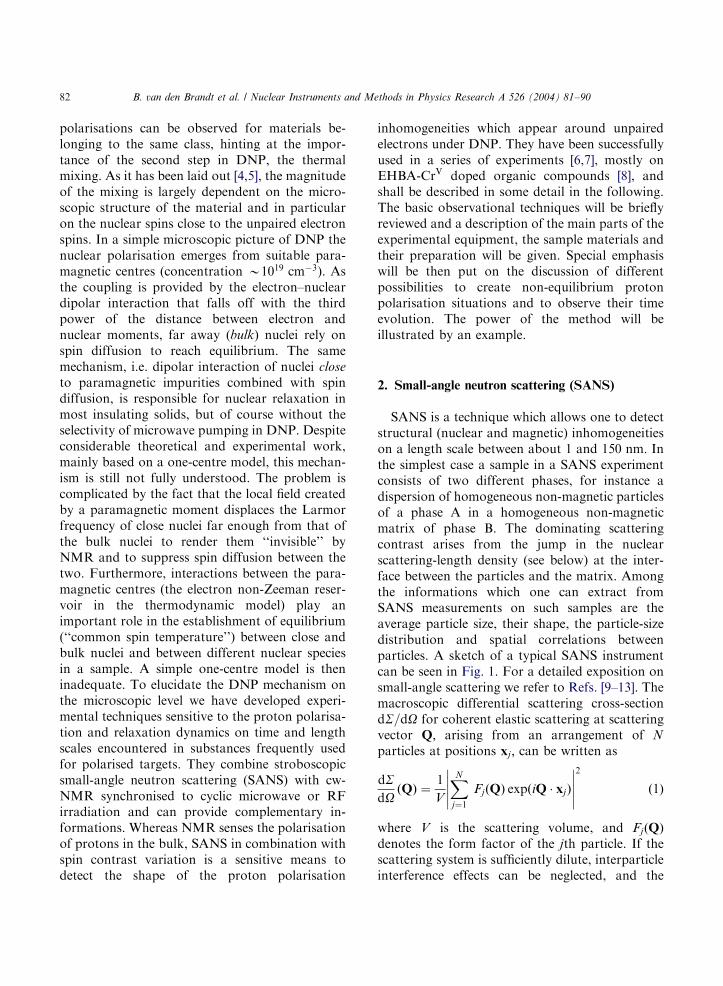
SANS is a technique which allows one to detectstructural (nuclear and magnetic) inhomogeneitieson a length scale between about 1 and 150 nm: Inthe simplest case a sample in a SANS experimentconsists of two different phases, for instance adispersion of homogeneous non-magnetic particlesof a phase A in a homogeneous non-magneticmatrix of phase B. The dominating scatteringcontrast arises from the jump in the nuclearscattering-length density (see below) at the inter-face between the particles and the matrix. Amongthe informations which one can extract fromSANS measurements on such samples are theaverage particle size, their shape, the particle-sizedistribution and spatial correlations betweenparticles. A sketch of a typical SANS instrumentcan be seen in Fig. 1. For a detailed exposition onsmall-angle scattering we refer to Refs. [9–13]. Themacroscopic differential scattering cross-sectiondS=dO for coherent elastic scattering at scatteringvector Q; arising from an arrangement of N
particles at positions xj ; can be written as
dSdO
ðQÞ ¼1
V
XN
j¼1
FjðQÞ expðiQ � xjÞ
����������2
ð1Þ
where V is the scattering volume, and FjðQÞdenotes the form factor of the jth particle. If thescattering system is sufficiently dilute, interparticleinterference effects can be neglected, and the
ARTICLE IN PRESS
Fig. 1. Sketch of a small-angle neutron scattering instrument.
B. van den Brandt et al. / Nuclear Instruments and Methods in Physics Research A 526 (2004) 81–90 83
SANS cross-sections of the individual particles areadditive. As a result, Eq. (1) can be simplified to
dSdO
ðQÞ ¼1
V
XN
j¼1
jFjðQÞj2 ¼1
V
XN
j¼1
Dr2h2j ðQÞ: ð2Þ
The quantity Dr2 in Eq. (2) denotes the squareddifference in the (nuclear) scattering-length den-sities between particle j and the matrix, andh2
j ðQÞ ¼ jR
Vp; jexpðiQ � xÞ d3xj2 depends on the size
and the shape of particle j with volume Vp;j : If wefurther consider the scattering from N particleswith identical size or, alternatively, scattering onlyby the particles of one size class (which is to a goodapproximation the case with the present samples),then Eq. (2) reduces to
dSdO
ðQÞ ¼N
VDr2h2ðQÞ: ð3Þ
The function hðQÞ can be calculated analyticallyfor many simple particle shapes. Relevant for ourSANS data analysis is the scattering function of asphere with radius R; for which
hðQRÞ ¼ 3Vp
sinðQRÞ � QR cosðQRÞ
ðQRÞ3: ð4Þ
An important integral characteristic of the scatter-ing cross-section, which will be used later on, is thePorod invariant K ; which (for a two-phase system)is obtained by integrating dS=dO over the whole q-space, according to
K ¼Z
N
0
dSdO
ðQÞQ2 dQ ¼ 2p2fð1� fÞDr2; ð5Þ
where f is the volume fraction of the scatteringobjects. Apart from the homogeneity of both theparticles and the matrix phase, Eq. (5) makes nofurther assumptions on the microstructure of thescattering system. As we shall see below, analysisof the SANS data in terms of the Porod invariantprovides a possibility to analyse the relaxationdynamics of polarised protons domains aroundparamagnetic centres. Note that, since dS=dO isnot available in absolute units ðcm�1 sr�1Þ; theresulting Porod invariants (see below) will also begiven in arbitrary units. The basic idea why SANSand DNP may be combined originates from thespin-dependence of the coherent scattering ampli-tudes of atoms possessing a non-zero nuclear spin.As mentioned above, the relevant quantity forsmall-angle scattering is the scattering-lengthdensity r; which is defined as the sum of all theindividual atomic scattering lengths of the scatter-ing object divided by its volume. For the EHBA-CrV-molecule the scattering-length density is
rðtÞ ¼ ½8:35752:26PðtÞ� 10�7 (A�2
ð6Þ
with the þð�Þ sign for neutron spin antiparallel(parallel) to the direction of nuclear polarisationand �1pPp1: Since DrpPðtÞ; the quantityffiffiffiffiffiffiffiffiffi
KðtÞp
is expected to reflect the time dependenceof the close protons as well as of the bulk protons.Since DNP allows to polarise either positively ornegatively, it becomes clear that the ensuingcoherent SANS cross-section can be varied sig-nificantly by means of DNP.
ARTICLE IN PRESS
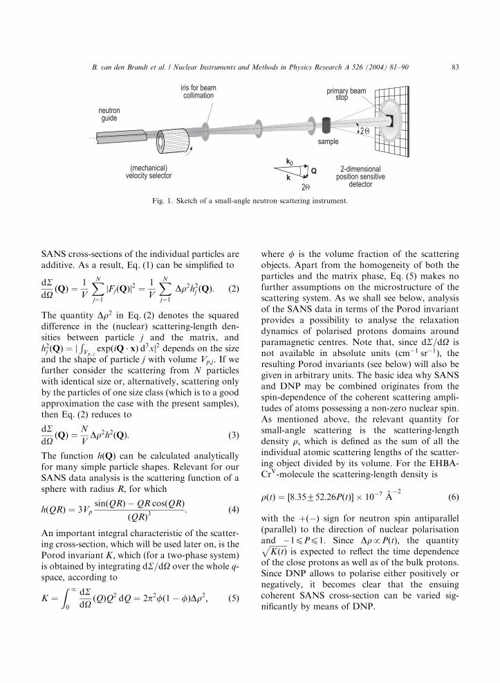
Fig. 2. The PSI 3:5 T/1 K polarised target system in operation
at the instrument D22 at ILL (slightly turned toward the
observer to show the exit window).
B. van den Brandt et al. / Nuclear Instruments and Methods in Physics Research A 526 (2004) 81–9084
2.1. Time-resolved SANS with polarised neutrons
Small-angle neutron scattering experiments havebeen performed at the instruments SANS-I (PSI,Switzerland), D22 (ILL, Grenoble) and Papol(LLB, Saclay). The instruments at PSI and LLBare equipped with a neutron polariser. At D22 aSch.arpf-type bender polariser has been installedabout 1:5 m in front of the sample. The incidentpolarised-neutron beam is produced by a super-mirror, which may be considered as a device withzero reflectivity for the neutron-spin state antipar-allel to the magnetisation ð�Þ and total reflectivityfor the parallel neutron spin state ðþÞ: At theSANS-I instrument at PSI we are using thetransmitted ð�Þ polarised beam so that thegeometry of the instrument remains unchanged,i.e. its direction is the same as the unpolarisedbeam. The polariser is placed far away from thesample in order to suppress parasitic SANS fromthe polariser itself. On the flight path betweenpolariser and sample ð15 mÞ a small magnetic fieldof about 10–20 Gauss must be applied to preservethe neutron polarisation. At all SANS instrumentsthe number of time bins and the time width of eachsingle bin can be chosen. The data acquisitioncycle is triggered by an external signal.
3. The polarised target system
3.1. Cryostat and 4He refrigerator insert
A PSI polarised target system [14] has beenmodified for experiments on a cold-neutron beam(see Fig. 2) [15]. A 3:5 T split coil, wound on analuminium former, is attached to the bottom of aliquid 4He vessel. A | 49 mm stainless steel tubewith an aluminium end cap runs axially throughthe helium vessel and then in vacuum down to thecentre of the magnet. It accommodates a contin-uous flow 4He refrigerator insert with a toploading sample holder device. Cooling down fromroom temperature to 77 K takes about 3 h; from77 to 4 K about 2 h: Liquid helium is transferredfrom the main bath to a gas–liquid separator, via aU-shaped transfer line. Regulating the pumpingrate in the separator and adjusting the helium
expansion via a needle valve allows to control theoperating conditions. Care has been taken toemploy materials with a high-neutron transmissionand a low SANS cross-section. The vacuum vesselwindows are 0:3 mm thick aluminium sheets andthe central stainless-steel tube ends in a cylindrical+ 49 mm ‘‘cup’’ with windows of 240 mm AlMg3,glued with Stycast 2850FT. The system features acooling power of 10 mW at 1 K using pumps witha nominal pumping speed of 1000 m3=h and runswith excellent stability over extended periods oftime. The temperature is measured with a ruthe-nium oxide thermometer placed in the liquidhelium around the sample. In the course of aseries of 1 week experiments the samples have beenchanged routinely every day. A total heliumconsumption of 30 l per day has been measured.
ARTICLE IN PRESS
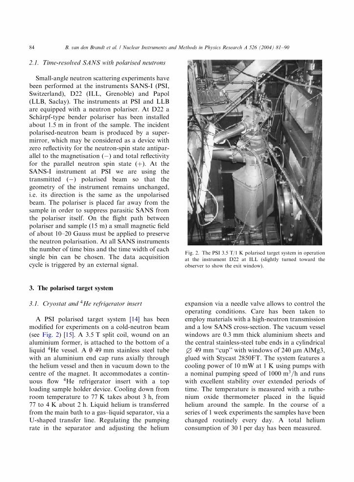
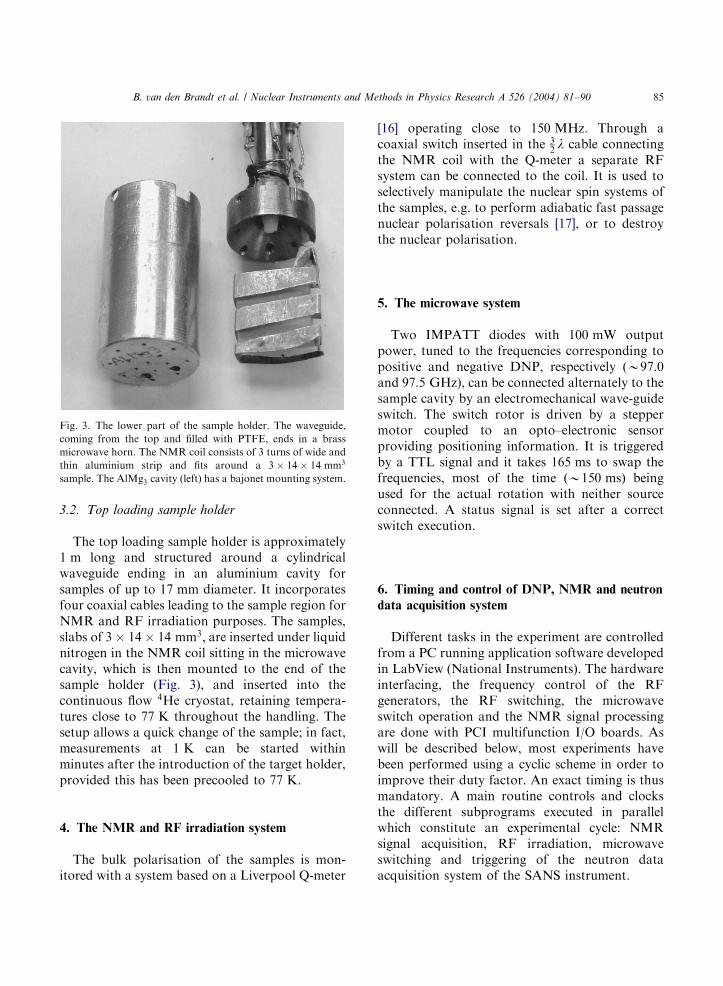
Fig. 3. The lower part of the sample holder. The waveguide,
coming from the top and filled with PTFE, ends in a brass
microwave horn. The NMR coil consists of 3 turns of wide and
thin aluminium strip and fits around a 3 14 14 mm3
sample. The AlMg3 cavity (left) has a bajonet mounting system.
B. van den Brandt et al. / Nuclear Instruments and Methods in Physics Research A 526 (2004) 81–90 85
3.2. Top loading sample holder
The top loading sample holder is approximately1 m long and structured around a cylindricalwaveguide ending in an aluminium cavity forsamples of up to 17 mm diameter. It incorporatesfour coaxial cables leading to the sample region forNMR and RF irradiation purposes. The samples,slabs of 3 14 14 mm3; are inserted under liquidnitrogen in the NMR coil sitting in the microwavecavity, which is then mounted to the end of thesample holder (Fig. 3), and inserted into thecontinuous flow 4He cryostat, retaining tempera-tures close to 77 K throughout the handling. Thesetup allows a quick change of the sample; in fact,measurements at 1 K can be started withinminutes after the introduction of the target holder,provided this has been precooled to 77 K:
4. The NMR and RF irradiation system
The bulk polarisation of the samples is mon-itored with a system based on a Liverpool Q-meter
[16] operating close to 150 MHz: Through acoaxial switch inserted in the 3
2l cable connecting
the NMR coil with the Q-meter a separate RFsystem can be connected to the coil. It is used toselectively manipulate the nuclear spin systems ofthe samples, e.g. to perform adiabatic fast passagenuclear polarisation reversals [17], or to destroythe nuclear polarisation.
5. The microwave system
Two IMPATT diodes with 100 mW outputpower, tuned to the frequencies corresponding topositive and negative DNP, respectively (B97.0and 97:5 GHz), can be connected alternately to thesample cavity by an electromechanical wave-guideswitch. The switch rotor is driven by a steppermotor coupled to an opto–electronic sensorproviding positioning information. It is triggeredby a TTL signal and it takes 165 ms to swap thefrequencies, most of the time ðB150 msÞ beingused for the actual rotation with neither sourceconnected. A status signal is set after a correctswitch execution.
6. Timing and control of DNP, NMR and neutron
data acquisition system
Different tasks in the experiment are controlledfrom a PC running application software developedin LabView (National Instruments). The hardwareinterfacing, the frequency control of the RFgenerators, the RF switching, the microwaveswitch operation and the NMR signal processingare done with PCI multifunction I/O boards. Aswill be described below, most experiments havebeen performed using a cyclic scheme in order toimprove their duty factor. An exact timing is thusmandatory. A main routine controls and clocksthe different subprograms executed in parallelwhich constitute an experimental cycle: NMRsignal acquisition, RF irradiation, microwaveswitching and triggering of the neutron dataacquisition system of the SANS instrument.
ARTICLE IN PRESS
B. van den Brandt et al. / Nuclear Instruments and Methods in Physics Research A 526 (2004) 81–9086
7. Samples and sample preparation
The proton polarisation build-up during DNPhas been studied in a set of model systems withunpaired electrons, each surrounded by a numberof hydrogen atoms (their nuclei being the so-called‘‘close protons’’), embedded in a matrix with achosen concentration of hydrogen atoms (provid-ing the ‘‘bulk protons’’). Free radicals with a singleunpaired electron per molecule have been used.The mean distance between the unpaired electronshas been varied by varying the concentration ofthe paramagnetic molecules, the number of pro-tons around each unpaired electron being deter-mined by the number of hydrogen atoms in theparamagnetic molecule. Deuterated organic sub-stances have served as a matrix, the degree ofdeuteration determining the concentration of thebulk protons. Thus, the distance between theunpaired electrons, the number of close protonssurrounding each of them and the number of bulkprotons could be chosen within the limits given bythe solubility. As model systems were used
1. Solutions of NaðC12H20O7CrVÞH2O (EHBA-
CrV) in glycerol–water mixtures. The degree ofdeuteration of these mixtures was chosen to be0%, 80%, 84%, 88%, 92%, 95% and 98%, theconcentration of the free radical moleculesbetween 1:25 1019 and 5 1019=cm3:
2. A solution of NaðC20H32O7CrVÞH2O (EHBA-
CrV) in a 98% deuterated glycerol–water mixture.3. Solutions of C34H44N5O6 (waxy-DPPH) in
polystyrene–p-terphenyl mixtures of 92%,95% and 98% deuteration.
Mostly solutions of EHBA-CrV; the well knownparamagnetic centres commonly used in polarisedtargets, in glycerol–D2O have been investigated.Glassy slabs of 3 mm thickness were obtained byinjecting the solution into a copper mould cooledto 77 K:
8. Creation and observation of polarisation
gradients
In a microscopic description of DNP, polarisa-tion gradients can be expected at least for times
short compared to spin diffusion at the onset ofmicrowave irradiation. Visually speaking, a po-larised proton domain then ‘‘dresses’’ the unpairedelectron and locally changes the scattering lengthdensity which is observed as a contrast betweenpolarised close protons and yet unpolarised bulkprotons in the SANS spectra. Several experimentalschemes can be envisaged for the study of thepropagation of nuclear polarisation near para-magnetic centres (see below). They differ in theway how nuclear polarisation gradients are createdbut have in common that this gradients are short-lived (order of seconds). Thus a time resolved dataacquisition scheme involving a large number ofrepetitions with efficient recycling becomes neces-sary to achieve the desired precision in the neutronscattering patterns within a reasonable time. Forstroboscopic data collection reproducible time-dependent non-equilibrium polarisations must becreated. Several schemes with different dutyfactors yielding different contrasts can be envi-saged, e.g.
a.
Starting from a high nuclear polarisation, apolarisation reversal by adiabatic fast passageof either the bulk or the close protonsestablishes the desired non-equilibrium.b.
Starting from zero-polarisation, obtained by asaturating RF pulse, microwave irradiationstarts the polarisation build-up.c.
Periodic polarisation reversal achieved byfrequency switching of the microwaves.All methods have been explored but (c), being themost efficient, has been adopted as the standardscheme of our experiments. Typically, the protonpolarisation is reversed every 10 s and a synchro-nous acquisition of neutron scattering intensityspectra of 0:1 s each is started. In one cycle of 20 s(positive DNP in the first 10 s; negative DNP inthe next 10 s) 200 SANS spectra are collected in200 so-called time frames. In parallel, the NMRsystem records the evolution of the bulk protonpolarisation. The SANS spectra taken duringseveral hundreds of these cycles are averaged toyield the scattering intensity for each time frame.Fig. 4 illustrates the evolution of the neutronscattering contrast observed in one cycle. Theperiodicity and the length of the time frames have

ARTICLE IN PRESS
Fig. 4. Schematic evolution of the neutron scattering length
density contrast as expected to be observed during cyclic DNP.
Fig. 5. Time-dependent neutron scattering intensity from a
sample of 98% deuterated glycerol–water solution containing
5 1019=cm3 EHBA-CrV:
B. van den Brandt et al. / Nuclear Instruments and Methods in Physics Research A 526 (2004) 81–90 87
been adapted to the characteristic time scales ofDNP in different samples.
Fig. 6. Data from Fig. 5 evaluated in a model independent way
after Eq. (5), the Porod invariant.
9. An example: EHBA-CrV
The approach shall be exemplified with experi-ments performed on a sample of 98% deuteratedglycerol–water solution containing 5 1019=cm3
EHBA-CrV complexes. Fig. 5 shows the time-dependent neutron scattering intensity of thesample subjected to the cyclic DNP processdescribed above. The strong time dependence forsmall Q values reflects the change in the scatteringcontrast of a small object, the EHBA-CrV mole-cule. The neutron scattering intensity IðQÞ is,according to Eq. (3), proportional to the productof the polarisation and thus time dependentscattering length density and the form factor ofthe scatterer. The latter can be derived from theknown molecular structure of the dissolvedEHBA-CrV molecules and can be simply approxi-mated by two homogeneous regions—a shellcontaining four C2H5 residues surrounding the½CrO7C4�� core. As was shown in Ref. [6], only fivefree parameters are then sufficient to fit thescattering data of 200 time frames simultaneouslyin order to deduce the time dependence of thepolarisation PðtÞ of the protons in the EHBA, i.e.the close protons. An alternative and model-independent SANS data analysis can be performedin terms of the so-called Porod invariant [18]. As
can be seen from Eq. (5), the Porod invariantreflects the time dependence of the protonpolarisation PðtÞ; which is contained in thescattering-length density contrast Dr2: Fig. 6shows the results for the temporal evolution ofthe square-root of the Porod invariant, which wasestimated by numerical integration of Eq. (5). Thefunction K1=2ðtÞ reflects the symmetry of thepolarisation evolution and the time-dependence

ARTICLE IN PRESS
Fig. 7. Time constants deduced from fits of the core-shell
model to the measured time-dependent neutron scattering
intensities for various degrees of solvent deuteration. Solid
lines: prediction from the thermal reservoir model.
B. van den Brandt et al. / Nuclear Instruments and Methods in Physics Research A 526 (2004) 81–9088
of the polarisation PðtÞ can be obtained from a fitof a simple model to the data.
9.1. The evolution of the polarisation
In general, a suitable model for the space- andtime-dependent polarisation Pðr; tÞ has to befound. In the example case the derived functionPðtÞ can be well described as a sum of a constantand two exponentials with corresponding timeconstants t1 and t2: Within the error bars, thevalues determined by the general approach usingPorod’s invariant (t1 ¼ 1:070:3 s andt2 ¼ 7:274:0 s) agree with the ones determinedwith the core-shell model (t1 ¼ 1:170:1 s andt2 ¼ 5:570:6 s) and support the conclusionsdrawn in Ref. [6]. This behaviour suggests aninterpretation in terms of rate equations describinga flow of polarisation between three thermalreservoirs coupled in series. The reservoirs areidentified as follows: the electronic spin–spininteraction reservoir, R0; is cooled by the micro-waves and acts as a ‘‘source’’ of polarisation. The20 protons of the EHBA-CrV molecule constitute areservoir R1 coupled to this source, and the bulkprotons form a reservoir R2 coupled to R1: Tworate equations then govern the dynamics of theprocess (see Ref. [19]). The model well describesthe tendency of the experimentally determinedtime constants for increasing bulk proton concen-trations ðcbulkÞ; as illustrated in Fig. 7. The longtime constant t2 essentially describes the build-upof the bulk polarisation. It is slowed down withincreasing bulk proton concentration, the heatcapacity of R2 being proportional to cbulk: Thedependence of the short time constant t1 on cbulkcan be partly understood as being due to a‘‘loading’’ effect of R2 on R1 induced by a bettercoupling between these reservoirs. With increasingsolvent protonation, the neutron scattering signalsget increasingly difficult to observe due to a largerincoherent background and a decreasing contrast.The simple step function used to describe both thestructure of the molecule and the spatial depen-dence of nuclear polarisation may become inap-propriate. A refined model should further spatiallyresolve the polarisation evolution by identifyinggroups of hydrogens in the sample, e.g. in terms of
several step functions or even by continuousdescription of the polarisation Pðr; tÞ: The Porodinvariant analysis should in a similar way beextended to multiple phases. When consideringlarger molecules, e.g. waxy-DPPH, the analysisshould explicitly take into account the knownmolecular structure. Very suitable is a multipoleexpansion of the scattering intensity [20] which cantake account of individual scattering amplitudesfor different parts of the molecule. Eventually athermal reservoir might be assigned to eachscattering amplitude. However, in the case ofEHBA-CrV; dispersed in not too largely proto-nated matrices, the sketched model can be hardlyimproved.
9.2. Remarks on thermal mixing
In the spin temperature theory formulation ofDNP via thermal mixing, an ideal couplingbetween electron non-Zeeman and nuclear Zee-man systems is commonly assumed. The mechan-ism is then often somewhat misleadingly called the
ARTICLE IN PRESS
B. van den Brandt et al. / Nuclear Instruments and Methods in Physics Research A 526 (2004) 81–90 89
‘‘EST (equal spin temperature theory)-model’’ ofDNP. A non-ideal coupling, stemming mostlyfrom microscopic features of the sample, is usuallytreated by the introduction of a leak factor. Morecan be learned from the expression for thecoupling a written phenomenologically as [5,21]
apX
k
Xj
/jHkjSI j
2So2
n
: ð7Þ
The electron–nucleus interaction HSI is dipolar,thus mainly those nuclei ð jÞ contribute to themagnitude of the mixing that are close to anelectron spin ðkÞ: To determine which nuclei haveto be taken in the summation has to be answeredfor every specific sample. Goldman and co-work-ers elucidate the coupling mechanism [4], making aclear distinction between ‘‘abnormal’’ spins lyinginside a diffusion barrier and normal spins outside,a separation similar to ours in close and bulkprotons. Assuming the abnormal spins to be partof an extended non-Zeeman reservoir they arriveat an expression for the mixing rate between thisreservoir and the nuclear Zeeman interactions
WpA2FðoÞ ð8Þ
where FðoÞ is related to a correlation function/SzðtÞSzð0ÞS and is thus completely distinctfrom the EPR line shape, that corresponds to/SxðtÞSxð0ÞS: FðoÞ has a truncated Lorentzianform with truncation points determined by theminimum distance between two paramagneticcentres. To fulfill the criterion for the thermalcontact the resonance frequency of the nuclei mustlie within the wings of the spectrum of FðoÞ;which is narrower than the EPR spectrum. Acontains the small contribution to the mixing ratefrom the dipolar coupling of the nuclei outside thediffusion barrier to the centre. Eq. (8) expresses sothe importance of the ’’abnormal spins’’, i.e. theclose protons, in the DNP process and stressestheir role in mediating the nuclear polarisation tothe bulk. Probing these protons indirectly by off-centre RF irradiation in samples comparable toours, the authors of Ref. [22] arrive at a similarconclusion.
10. Conclusions
Time resolved SANS measurements combinedwith NMR at the onset of the DNP process aresensitive to the nuclear polarisation build-uparound paramagnetic centres on a time scalepreviously not accessible. They can provide spatialinformation which in the past could be inferredonly much less directly. In fact, for the first timethe polarisation evolution of protons close to aparamagnetic centre could be directly observed insubstances, where thermal mixing is the mainmechanism of DNP. For the samples investigated,this polarisation evolution can be satisfactoryexplained by a thermodynamic model with heatreservoirs corresponding to two groups of protons.
References
[1] A. Abragam, M. Goldman, Rep. Prog. Phys. 41 (1978)
395.
[2] St. Goertz, W. Meyer, G. Reicherz, Prog. Part. Nucl. Phys.
49 (2002) 403.
[3] M. Borghini, Phys. Rev. Lett. 20 (1968) 419.
[4] M. Goldman, S.F.J. Cox, V. Bouffard, J. Phys. C 7 (1974)
2940.
[5] W.Th. Wenckebach, Proceedings of the 2nd Workshop on
Polarised Target Materials, Rutherford and Appleton
Labs, RL-80-080, 1980.
[6] B. van den Brandt, H. Gl.attli, I. Grillo, P. Hautle, H.
Jouve, J. Kohlbrecher, J.A. Konter, E. Leymarie, S.
Mango, R.P. May, H.B. Stuhrmann, O. Zimmer, Euro-
phys. Lett. 59 (2002) 62.
[7] B. van den Brandt, H. Gl.attli, I. Grillo, P. Hautle, H.
Jouve, J. Kohlbrecher, J.A. Konter, E. Leymarie, S.
Mango, R.P. May, H.B. Stuhrmann, O. Zimmer, Physica
B 335 (2003) 193.
[8] M. Krumpolc, J. RoWek, J. Am. Chem. Soc. 101 (1979)
3206.
[9] A. Guinier, G. Fournet, Small-Angle Scattering of
X-Rays, Wiley, New York, 1955.
[10] O. Glatter, O. Kratky (Eds.), Small-Angle X-Ray Scatter-
ing, Academic Press, London, 1982.
[11] S.-H. Chen, T.-L. Lin, in: K. Sk .old, D.L. Price (Eds.),
Neutron Scattering, Vol. 23, Part B, Academic Press, San
Diego, 1987, pp. 489–543.
[12] L.A. Feigin, D.I. Svergun, Structure Analysis by Small-
Angle X-Ray and Neutron Scattering, Plenum, New York,
1987.
[13] P. Lindner, T. Zemb, Neutron, X-Ray and Light Scatter-
ing: Introduction to an Investigative Tool for Colloidal

ARTICLE IN PRESS
B. van den Brandt et al. / Nuclear Instruments and Methods in Physics Research A 526 (2004) 81–9090
and Polymeric Systems, North-Holland, Amsterdam,
1991.
[14] B. van den Brandt, et al., AIP Proc. 187 (1989) 1251.
[15] B. van den Brandt, P. Hautle, J.A. Konter, S. Mango, PSI
Scientific Report 2000, Vol. III, 2001, p. 78.
[16] G.R. Court, et al., Nucl. Instr. and Meth. A 324 (1992)
433.
[17] P. Hautle, et al., Phys. Rev. B 46 (1992) 6596.
[18] G. Porod, in: O. Glatter, O. Kratky (Eds.), Small-angle X-
ray Scattering, Academic Press, London, 1982, pp. 17–51.
[19] B. van den Brandt, et al., in Proc. 3rd European
Conference on Neutron Scattering, Montpellier, 3–6
September 2003, Physica B, to be published.
[20] H.B. Stuhrmann, Acta Crystallogr. A 26 (1970) 297.
[21] W.Th. Wenckebach, T.S.B. Swanenburg, N.J. Poulis,
Phys. Rep. 14 (1974) 181.
[22] S.F.J. Cox, S.F.J. Read, W.Th. Wenckebach, J. Phys. C 10
(1977) 2917.