an exafs, and preliminary x-ray crystallographic, investigation of an iron-containing product from...
TRANSCRIPT

Biochimica et Biophysica Acta, 1036 (1990) 71-77 71 Elsevier
BBAGEN 23352
An EXAFS, and preliminary X-ray crystallographic, investigation of an iron-containing product from the lichen Cladonia deformis
Lucilla Alagna 1, Tommaso Prosperi 1 and Anthony A.G. Tomlinson 1, Helge Kjosen 2 and Frode Mo 3
1 I.T.S.E., Area della R icerca di Roma, C N.R., Rome (Italy), 2 Institutt for organisk kjemi and ~ Institutt for fysikk, Universitetet i Trondheim-NTH, Trondheim (Norway)
(Received 12 March 1990)
Key words: X-ray crystallography; Iron; Spectroscopy; ( C deformis)
An iron-containing product in the acetone extract from the lichen Cladonia deformis has been investigated using chemical, spectroscopic and X-ray crystallographic methods. Visible-near UV, EPR and IR spectra indicate that the iron is present as high-spin Fe(Ill) and coordinates in an oxygen-containing environment arising from graciliformin (or graciliformin-like) ligands. This has been confirmed by an XAS (X-ray absorption) study using synchrotron radiation. Comparison of the EXAFS and XANES results with those obtained from a model, tris(pent-2,4-dionato)Fe(lll), and detailed fitting using the single-scattering, curved-wave formalism for the EXAFS strongly supports the presence of a Fe(lll) coordinated to five oxygen atoms from the graciliformin.
Introduction
Lichens contain several low-molecular-weight organ- ic compounds which are capable of absorbing a wide variety of elements from the environment. So much so that their avid efficacy in the uptake and retention of metals and non-metals, including sulphur, has been utilized to monitor air and substrate pollution [1,2].
Several compounds have been separated and identi- fied [3,4], yet little information is available on their complex chemistry. We report the purification, and the chemical and preliminary crystallographic characterisa- tion, together with spectroscopic identification - includ- ing a detailed EXAFS study - of the chelate centre of a natural iron-containing product extracted from the lichen Cladonia deformis.
Experimental procedures
The lichen Cladonia deformis was collected in N. Trondelag, Norway, and a dark red crystalline material was isolated by Dr. T. Bruun (NTH; deceased 1977), from a sample of about 4.4 kg, using extraction with chloroform. This extract was then fractionally crystal-
Abbreviations: XAS, X-ray absorption; XANES, X-ray absorption near-edge structure; IR, infrared; UV, ultraviolet.
Correspondence: A.A.G. Tomlinson, I.T.S.E., Area della Ricerca di Roma, C.N.R., C.P. 10 Monterotondo Staz., 00016 Rome, Italy.
lised several times from acetone and the acetone ex- tracts finally separated by chromatography on an acidic (0.25 M oxalic acid) Kieselgel G column, using 95:5 benzene/methanol as eluant.
The fraction containing a dark red pigment was retained and further purified by crystallisation from acetone (or chloroform) to yield thin, dark red rhombic plates with boundary faces (110) and (001), the latter defining the base plane of the rhomboid. The crystals closely resemble some of the crystal forms described by Zopf [5] many years ago for bellidiflorin, which was first isolated from Cladonia bellidiflora (Ach.) var. coc- cocephala(Ach.). Bellidiflorin has also been found in other Cladonia species [4].
Elemental analyses of this iron-containing product showed that carbon, hydrogen, oxygen and iron were present. The average composition from four different analyses was: C, 62.43; H, 4.04; O, 30.05; and Fe, 2.75%. No methoxy groups are present, as deduced from standard chemical methods and IR spectra.
The crystals showed a strong tendency to aggregate on the (001) faces. They did not diffract well, and film exposures at ambient and low temperatures suggested that disorder was present.
A small, single crystal of dimensions approx. 0.2 × 0.2 × 0.02 mm was used for measuring cell parameters and intensity data. All intensities were recorded photo- graphically with an oscillating camera and Fe-filtered Co-K~ radiation ( h = 1.7903 ,A). Exposures for time periods of 16 h were required for oscillation ranges of
0304-4165/90/$03.50 © 1990 Elsevier Science Publishers B.V. (Biomedical Division)

72
6 ° . Total angular ranges of 116 ° about [100] and 21 ° about [010] were explored by successive 6 ° exposures with 1 ° overlap.
EPR spectra were registered on a Varian E9 instru- ment equipped with standard liquid nitrogen attach- ment. Visible-near UV absorption spectra were mea- sured on a Perkin-Elmer 303 instrument using matched quartz ceils and IR spectra (KBr discs) on a Bruker 113 FT- IR instrument.
XAS spectra were obtained at room temperature from finely powdered samples held in kapton-covered holders at the PULS facility, INFN, Frascati. Data points for the XANES were collected at 0.035 eV inter- vals over the Fe-K-edge (to 125 eV beyond the onset of absorbance) and the EXAFS at 2 eV intervals over a range of 1200 eV.
R e s u l t s
Crystals of the iron-containing extract from Cladonia deformis are orthorhombic, space group P212121 from systematic extinctions: a = 37.0 (3), b = 25.1 (3), c = 21.2 (3) ,~; V = 19 688(400) ,~; D m = 1.415(2)Mg. m-3. These values lead to a molecular weight of 2100(45) with Z = 8, or 4200 (90) with Z = 4. The former case corresponds to two crystallographically distinct molecu- lar units in the asymmetric unit. An independent esti- mate of the molecular weight obtained from the elemen- tal analysis is approx. 2030 - or 4060 - assuming one - or two - Fe atoms per molecule, respectively. The two sets of experimental values are in agreement within the 20 limit of the data. Diffraction goes out to approx. 40 ° in 28 with Co-K~ radiation, corresponding to a resolution ( d m i n ) of approx. 2.6 ,~. This fact, and the poor crystal quality constitute a major obstacle to a full crystallographic structure analysis.
The powder EPR spectrum of this Fe-containing product gives a three-line signal with apparent g-values of 8.68, 4.1 and 1.69, which are diagnostic for the presence of a high-spin Fe(III) in a rhombic environ-
I I I I I I I I
I I I I I I I I 0 0 2 0 4 0 6
B / Tesla
Fig. l . EPR spectrum (X-band, 77 K) of a powdered sample of Fe product extracted from the lichen Cladonia deformis.
ment [6,7]. There are no signals ascribable to Fe(II) (Fig. 1). The weak lines observed at 0.32 mT are prob- ably due to trace amounts of Cu II [8,9].
The visible-near-UV spectrum (acetone solution) shows no absorbance between 1000 and 500 nm, again in agreement with the presence of a high-spin [FeO6]- or [FeOs]-containing complex. The spectrum may be com- pared with that of Fe(pd)3(pd = pent-2,4-dionato ion), which gives only very weak (c < 2) spin-forbidden d-d transitions, at 650-750 nm [10], whereas Fe(II) (pd)3 shows a relatively intense broad d-d band at 819.5 nm [11]. Turning to the non-d -d bands, there is a major absorbance peak at 388 nm, together with shoulders at 402, 435 and 460 nm (Fig. 2). Both the main band and major shoulder lie at energies in close agreement with those found for the bellidiflorin (387 and 465 nm) reported by Ejiri et al. [12]. The IR spectrum (in KBr disc) is very rich in detail in the region below 1200 cm-1. Carbonyl frequencies at 1749 and 1704 cm 1 and further maxima at 1618, 1585, 1229 and 1045 cm -1 are in fair agreement with the data reported for bellidiflorin (in HCCI3) [12]. The bellidiflorin of Ref. 12 was ex- tracted from Cladonia graciliformis and it was demon- strated that removal of the iron present gave rise to graciliformin:
O 0
Scheme I ON
In sum, the presence of a,T-ketoenolate rings sug-
C . . . . . C I,," "',J
gests that coordination of the iron occurs via o e o moieties to give six-membered rings analogous not only as regards coordinating atoms but also in second-shell atoms (C~atoms) to those present in tris(pent-2,4-di-
E i
, I . . . . I , , , , I , , , , I , I , , 350 400 450 500 550
r ~ / n m
Fig. 2. Visible-near ultraviolet absorbance spectrum (25 °C, acetone solution) of the Fe-product extracted from Cladonia deformis.

onato)Fe(III) mentioned above, the structure of which is known to contain a high-spin [FeO6] unit [13]. The latter (prepared as in the Ref. 12 and the references therein) was used as the EXAFS 'model ' for extracting the parameters necessary for analysing the data ob- tained for the Fe-containing product, using both stan- dard, plane-wave formalism [14,15] and the spherical- wave formulation (see below).
Discussion
Fig. 3 shows the experimental Fe-K-edge spectra of the two compounds. The energy positions of the main peak (measured from derivative spectra) are very simi- lar, confirming the presence of Fe(III) in the Fe-lichen product. Further, the relative energies and intensities of the other features in the spectra beyond the edge (up to approx. 60 eV) are also very similar, the minor dif- ferences - in peaks b and c - being ascribable to the influence of differences in the backbone structure of the complex ligand present in the Fe-lichen product [16]. Conversely, the spectra of both compounds differ from those of Fe(III) complexes with non-axial structures [17]. Roe et al. [18] have described the pre-edge features of some 28 synthetic iron compounds and found an inverse correlation between the intensity of this band and the coordination number of the Fe. This feature (a in Fig. 3) which may be assigned to the Fe ls ~ 3d transition [18], is of slightly higher intensity in the Fe-lichen product than in Fe(pd)3, indicating that the former may contain the Fe(III) in a five-coordinate, rather than six-coordinate, environment.
The experimental extracted EXAFS of both com- pounds are shown in Fig. 4. They are almost superim-
! ~ 03
O2
c
02 ~ 0.1 w ~ - O
O1
o,-I" , J I , , , , I , 710 715 720
E / keV Fig. 3. Experimental Fe-K-edge spectrum of Fe(pd)3 (A) and the
Fe-product extracted from Cladonia deformis (B).
73
posable, except for details in the regions 8-9 and 10-12 ,~-1. The first step in the analysis consisted of perfor- ming a series of firstshell fits to the Fourier-filtered R = 1.5-2.5 ,~ region of the EXAFS of Fe(pd) 3. This standard procedure [14] gave the results of Table I. A good fit was obtained for a F e - O distance of 1.99(1) ,~, in good agreement with the crystallographic mean dis- tance of 1.99 A (range 2.004-1.986 A, from visually estimated intensities [13]). Spherical-wave fits were then performed over an R-range for Fourier-filtering which covered the entire molecule, i.e., such as to include the C~, C a and Cv atoms, and maintaining the F e - O bond length fixed at 1.99 A. The ab initio phase shifts and single-scattering spherical wave formalism of Lee and Pendry [19] was used, together with the fast, curved-wave algorithm, the parameterisation of which is described in detail by Gurman et al. [20]. The refined fit obtained is shown in Fig. 5C. As shown by the intermediate stages, it was not possible to obtain good-quality simulation between 4.2-5.0 and 8.0-9.0 .~-1 by including the molecular distances alone. However, when a further shell, of intermolecular F e . . . C contacts longer than 4.5 ,~, was included there was a significant improvement in the fit index. Of particular interest is the fact that the F e . . . C/~ and Fe . . . Cy distances are significantly shorter in the EXAFS, by 0.042 and 0.368 A, respectively. (Inserting the crystallographic Fe...C~,B, ~ distances in the simulations gave very unsatisfactory fits.) Such EX- AFS distance contractions are characteristic of the pres- ence of multiple scattering effects, as found in im- idazole-containing complexes and proteins [21]. Accord- ing to Teo [22,23], multiple scattering effects become more important whenever the central interatomic angle (fl) in a three-atom system is larger than approx. 140 ° and reaches a maximum (highest enhanced forward scattering through the central atom, oxygen in this case) when the three atoms involved are in a line (i.e., fl = 180°). This agrees with the crystallographic results: Fe-O-C a angles lie in the range 128-130 °, too low for multiple scattering effects to come into play, whereas the Fe-O-C# angles lie in the range 163-166 °. This is the first observation of multiple scattering effects in such six-membered rings and further work on 'model ' compounds is under way to clarify the procedures for applying corrections.
The EXAFS spectrum of the Fe-lichen product was then simulated using exactly the same procedure, i.e., progressive inclusion of outer shells in the Fourier-filter- ing windows, keeping the parameters (E 0, etc.) ex- tracted from the analysis of the Fe(pd)3 fixed. Again, the major F e - O peak in the FT was fitted, varying N (number of neighbours), Ao 2 (differences in Debye- Waller factors) and R (distance). The results are sum- marised in Table II. Regular 5, 6 or 7 coordinations gave unsatisfactory fits. Fits improved when two F e - O distances were inserted. The best fits were obtained for

74
" A I I | . . . . I I I l , j I ] 0 3
- 0 3
~ l l I I I l 0,5 B
o
v - - 1 I I 1 I i J I 1
4 6 8 10 k /~ -1
Fig. 4. Experimental extracted EXAFS spectra of the Fe product extracted from Cladonia deformis (A) and Fe(pd)3 (B).
both a rhombic six-coordinate structure and five-coordi- nation with four equal in-plane F e - O at 1.97 ,~ and one longer F e - O at 2.28 A.
The Fe-lichen EXAFS was then simulated over the
TABLE I
Results of simulations for Fe(pd) 3
N R(.~) 202(A 2)
A FeO 6 , 6
B Fe(pd)3 F e - O 6 Fe. . .C, , 6 Fe . . .Ca 3 Fe. . .Cv 6
C Fe(pd)3+ intermolecular carbon atoms
F e - O 6 Fe...C,~ 6 Fe. . .C~ 3 Fe . . . Cv 6
( i n t . ) F e . . . C 3
X-ray crystallography
F e - O 6 Fe. . .C,, 6 Fe . . . O 3 Fe . , . C a 3 Fe. . .Cy 3 Fe . , . C 3 Fe . , . C 3
1.99 0.015
1.98 0.014 2.99 0.022 3.28 0.005 3.93 0.035
1.98 0.014 2.99 0.022 3.28 0.005 3.93 0.035 4.53 0.001
o2(A 2)
1.989 0.014 2.992 0.012 3.000 0.072 3.330 0.012 3.900 0.024 4.010 0.039 4.531 0.002
Table II
Single-shell simulations for C deformis Fe product
F.I., fit index, defined as:
(Rk k') F.1. = 2 - , N ~ n t s x 100.
R, residual points at point i, (X~xp(k) - Xtheor(k )) k, wavenumber at point i. kT, weighting (3).
N R (A) 202 (A 2) F.I.
Four-coordinate 4 1.969 0.014 0.024
Tetragonal 4 1.968 0.013 0.011 Five-coordinate 1 2.279 0.038
Five-coordinate 5 1.972 0.019 0.062
Seven-coordinate 5 1.972 0.018 0.032 2 2.25 0.035
Six-coordinate 6 1.973 0.025 0.121
Tetragonal 4 1.964 0.013 0.014 Six-coordinate 2 2.242 0.062
Rhombic 2 1.961 0.006 Six-coordinate 2 2.054 0.021 0.013
2 2.113 0.048
R-range up to 5 .~. As for Fe(III)(pd)3, simulations were not satisfactory when only second shell C~ atoms were included; it was necessary to introduce C atoms as far as approx. 4.5 A, especially to obtain satisfactory agreement in the range 4 -5 ~,-~ in the back-transform. However, the 'goodness of fit' was little influenced by the number of C atoms introduced at these long dis- tances.
Thus, the first four oxygen atoms about the Fe in the
TABLE II1
Full-shell simulations for C. deformis Fe product
N R(A) 202(,~ 2 ) F.I.
FeO 4 + 0 F e - O 4 1.972 0.014 F e - O 1 2.282 0.042 Fe . . . C,, 3 2.993 0.020
0.011 Fe . . . Cp 3 3.295 0.025 Fe . . . Cv 3 3.887 0.036 Fe . . . C 3 4.477 0.011
FeO 4 + 20 F e - O 4 1.977 0.044 F e - O 2 2.291 0.071 Fe . . . C,, 3 2.984 0.020 Fe . . . C a 3 3.287 0.027 0.25 Fe . . . Cv 3 3.888 0.034 F e . . . C 3 4.477 0.012
FeO 6 F e - O 6 1.977 0.025 Fe. . .C~ 2 3.00 s 0.013 Fe . . . C a 4 3.305 0.033
0.3 Fe. . . C~, 3 3.838 0.041 F e . . . C 3 4.484 0.012

Fe-containing product appear to be in a square-based array of a square-based pyramidal structure. Although a further (i.e., sixth) O atom along the z-axis of the molecule cannot be completely excluded, the measura-
75
ble shortening of the four Fe-O bonds with respect to those in Fe(pd)3 are in agreement with this conclusion. (A preliminary crystal structure analysis of (Fe(III) (Pd)2C1) showed it to be square-based pyramidal with
2 0
m
~ 0 a~ v
- 2 0
A I I I I I f I
I I I I I I I I 4 6 8 10
k / ,& ~
! I I 1 I I I I I 1 2 3 4 5
~x051 B
e
- 0 5
I I I I I 1 I !
I I I I I I I I 4 6 8 10
k / J~ "~
0 1 2
0 5 I C I I I I I I t I
ot £ A - 0 5
1 i I I i I I I 4 6 8 10
k /,&-I
I I I I I i o I I 3 4 5 3 4 5
i
/
w I ~ I 0 1 2
F i g . 5 . Fits of the EXAFS spectrum of Fe(pd) 3. Fit of FeO 6 shell alone (A); the entire molecule of F e ( p d ) 3 ( B ) ; and the entire molecule of F e ( p d ) 3
plus the intermolecular C atoms of Table I ( C ) .

76 m .
20
10
0
-10
- 2 0
2 0
% 0
- lo
-2C
2 0
10
0
10
- 2 0
30
2O
10
. 0
-10 ;,<
-2Q
- 3 0
I I I I I I 4 +11 I I " I I
IAA I V,I
I I
I I I I I I I I
I i I I I I ! I
I I I I I I U I I
I I I i I I I I I 4 6 8 10
k l,&-1
I ' I ' I ~ t ' I ' I ' I ' I - - E
Vv i I , I , I , I i I i I i I
4 6 8 1 0 k / , & -~
,'l
I I I 1 2
1 l I 1 I I 3 4 5 6
R / ~ "~
3C
2C
1(2
O
;"< -1C
-2C
- 3 0
, !
" D
V I , I I I
4 6
I i I ' I i l ' I ' I ' I
i , I , I , I I I 8 10
k / , & "1
il I\ I i I ' I I ' I r 1 2 3 4 5
R / ~ - 1
2O
I0
0
-10
- 2 0
F' I ' I ' I I ' I I I I
.,.,,,.,.,,,,,,i'1 4 6 8 10
kl~, "I
0 1 2 3 4 5
i ~ / / t
!
6
Fig. 6. Fits of the EXAFS spectrum of the Fe product extracted from Cladonia deformis. A - C , the FeO 6 shell alone and D - F , with progres-
sive inclusion of backbone C atoms, as in Table II.
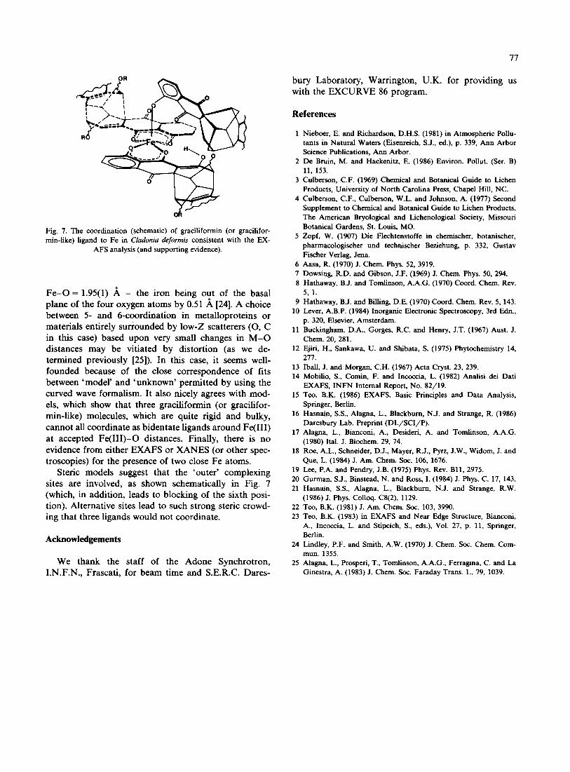
Fig. 7. The coordination (schematic) of graciliformin (or gracilifor- rain-like) ligand to Fe in Cladonia deformis consistent with the EX-
AFS analysis (and supporting evidence).
F e - O = 1.95(1) A - the iron being out of the basal plane of the four oxygen atoms by 0.51 .~ [24]. A choice between 5- and 6-coordination in metalloproteins or materials entirely surrounded by low-Z scatterers (O, C in this case) based upon very small changes in M - O distances may be vitiated by distortion (as we de- termined previously [25]). In this case, it seems well- founded because of the close correspondence of fits between 'model' and 'unknown' permitted by using the curved wave formalism. It also nicely agrees with mod- els, which show that three graciliformin (or gracilifor- rain-like) molecules, which are quite rigid and bulky, cannot all coordinate as bidentate ligands around Fe(III) at accepted Fe(III)-O distances. Finally, there is no evidence from either EXAFS or XANES (or other spec- troscopies) for the presence of two close Fe atoms.
Steric models suggest that the 'outer' complexing sites are involved, as shown schematically in Fig. 7 (which, in addition, leads to blocking of the sixth posi- tion). Alternative sites lead to such strong steric crowd- ing that three ligands would not coordinate.
Acknowledgements
We thank the staff of the Adone Synchrotron, I.N.F.N., Frascati, for beam time and S.E.R.C. Dares-
77
bury Laboratory, Warrington, U.K. for providing us with the EXCURVE 86 program.
References
1 Nieboer, E. and Richardson, D.H.S. (1981) in Atmospheric Poilu- tams in Natural Waters (Eisenrcich, S.J., ed.), p. 339, Ann Arbor Science Publications, Ann Arbor.
2 De Bruin, M. and Hackenitz, E. (1986) Environ. Pollut. (SCr. B) 11, 153.
3 Culberson, C.F. (1969) Chemical and Botanical Guide to Lichen Products, University of North Carolina Press, Chapel Hill, NC.
4 Culberson, C.F., Culberson, W.L. and Johnson, A. (1977) Second Supplement to Chemical and Botanical Guide to Lichen Products, The American Bryological and Lichenological Society, Missouri Botanical Gardens, St. Louis, MO.
5 Zopf, W. (1907) Die Flechtenstoffe in ehemischer, botanischer, pharmacologiseher und technischer Beziehung, p. 332, Gustav Fischer Verlag, Jena.
6 Aasa, R. (1970) J. Chem. Phys. 52, 3919. 7 Dowsing, R.D. and Gibson, J.F. (1969) J. Chem. Phys. 50, 294. 8 Hathaway, B.J. and Tomlinson, A.A.G. (1970) Coord, Chem. Rev.
5,1. 9 Hathaway, B.J. and Billing, D.E. (1970) Coord. Chem. Rev. 5, 143.
10 Lever, A.B.P. (1984) Inorganic Electronic Spectroscopy, 3rd Edn., p. 320, Elsevier, Amsterdam.
11 Buckingham, D.A., Gorges, R.C. and Henry, J.T. (1967) Aust. J. Chem. 20, 281.
12 Ejiri, H., Sankawa, U. and Shibata, S. (1975) Phytochemistry 14, 277.
13 IbaU, J. and Morgan, C.H. (1967) Acta Cryst. 23, 239. 14 Mobilio, S., Comin, F. and Incoecia, L. (1982) Analisi dei Dati
EXAFS, INFN Internal Report, No. 82/19. 15 Too, B.K. (1986) EXAFS. Basic Principles and Data Analysis,
Springer, Berlin. 16 Hasnain, S.S., Alagna, L., Blackburn, N.J. and Strange, R. (1986)
Daresbury Lab. Preprint (DL/SCI/P) . 17 Alagna, L., Bianconi, A., Desideri, A. and Tomlinson, A.A.G.
(1980) Ital. J. Biochem. 29, 74. 18 Roe, A.L., Schneider, D.J., Mayer, R.J., Pyrz, J.W., Widom, J. and
Que, L. (1984) J. Am. Chem. Soc. 106, 1676. 19 Lee, P.A. and Pendry, J.B. (1975) Phys. Rev. Bl l , 2975. 20 Gurman, S.J., Binstead, N. and Ross, I. (1984) J. Phys. C. 17, 143. 21 Hasnain, S.S., Alagna, L., Blackburn, N.J. and Strange, R.W.
(1986) J. Phys. Colloq. C8(2), 1129. 22 Teo, B.K. (1981) J. Am. Chem. Soc. 103, 3990. 23 Teo, B.K. (1983) in EXAFS and Near Edge Structure, Bianconi,
A., Incoccia, L. and Stipcich, S., eds.), Vol. 27, p. 11, Springer, Berlin.
24 Lindley, P.F. and Smith, A.W. (1970) J. Chem. Soc. Chem. Com- mun. 1355.
25 Alagna, L., Prosperi, T., Tomlinson, A.A.G., Ferragina, C. and La Ginestra, A. (1983) J. Chem. Soc. Faraday Trans. 1., 79, 1039.