an essential receptor for adeno-associated virus infection · an essential receptor for...
TRANSCRIPT

0 0 M O N T H 2 0 1 6 | V O L 0 0 0 | N A T U R E | 1
LETTERdoi:10.1038/nature16465
An essential receptor for adeno-associated virus infectionS. Pillay1*, N. L. Meyer2*, A. S. Puschnik1, O. Davulcu2, J. Diep1, Y. Ishikawa2,3, L. T. Jae4, J. E. Wosen1, C. M. Nagamine5, M. S. Chapman2 & J. E. Carette1
Adeno-associated virus (AAV) vectors are currently the leading candidates for virus-based gene therapies because of their broad tissue tropism, non-pathogenic nature and low immunogenicity1. They have been successfully used in clinical trials to treat hereditary diseases such as haemophilia B (ref. 2), and have been approved for treatment of lipoprotein lipase deficiency in Europe3. Considerable efforts have been made to engineer AAV variants with novel and biomedically valuable cell tropisms to allow efficacious systemic administration1,4, yet basic aspects of AAV cellular entry are still poorly understood. In particular, the protein receptor(s) required for AAV entry after cell attachment remains unknown. Here we use an unbiased genetic screen to identify proteins essential for AAV serotype 2 (AAV2) infection in a haploid human cell line. The most significantly enriched gene of the screen encodes a previously uncharacterized type I transmembrane protein, KIAA0319L (denoted hereafter as AAV receptor (AAVR)). We characterize AAVR as a protein capable of rapid endocytosis from the plasma membrane and trafficking to the trans-Golgi network. We show that AAVR directly binds to AAV2 particles, and that anti-AAVR antibodies efficiently block AAV2 infection. Moreover, genetic ablation of AAVR renders a wide range of mammalian cell types highly resistant to AAV2 infection. Notably, AAVR serves as a critical host factor for all tested AAV serotypes. The importance of AAVR for in vivo gene delivery is further highlighted by the robust resistance of Aavr−/− (also known as Au040320−/− and Kiaa0319l−/−) mice to AAV infection. Collectively, our data indicate that AAVR is a universal receptor involved in AAV infection.
AAV2, the most commonly studied AAV serotype, attaches to cells using heparan sulfate proteoglycan5. For several other non-enveloped viruses, initial attachment is followed by engagement of a protein receptor, which dictates entry into the cytoplasm. Whether AAV also requires such a protein receptor is unclear. Surface proteins including human fibroblast growth factor receptor-1 (FGFR1) and hepatocyte growth factor receptor (MET) have been reported as putative AAV2 co-receptors6,7. However, using isogenic knockout cell lines (Extended Data Fig. 1a, b), we observed no significant effect on AAV2 infection in cells lacking FGFR1, and only minimal effect as a result of MET loss (Extended Data Fig. 1c), suggesting a modest role in AAV2 infection for these proteins. To identify host factors critical for AAV2 infec-tion, we used an unbiased genome-wide screening approach based on insertional mutagenesis in haploid human cells (HAP1)8. We infected a library of mutagenized cells, carrying knockouts in virtually all non-essential genes, with an AAV2 vector that expresses red fluores-cent protein (RFP) (Extended Data Fig. 2a). Mutant cells refractory to AAV2 infection were isolated through iterative cycles of fluores-cence-activated cell sorting (FACS) (Extended Data Fig. 2b). The screen yielded 46 significant hits (Fig. 1a, Supplementary Table 1), many of
which were implicated in heparan sulfate proteoglycan biosynthesis (depicted in blue). AAV2 hijacks endosomal pathways to travel from the cell surface to the nucleus, and several endosomal trafficking genes (depicted in green) were prominently identified in the screen, specif-ically those encoding members of the retromer (VPS29, VPS35) and Golgi-associated retrograde protein complexes (VPS51, VPS52, VPS53, VPS54). These proteins are involved in retrograde transport from the endosomes to the Golgi9,10, but have not been specifically associated with AAV2 infection before now. The most significantly enriched gene of the screen was KIAA0319L (denoted hereafter as AAVR), with 570 independent mutations identified. This gene encodes a poorly char-acterized transmembrane protein. Little is known about the cellular function of AAVR, but it has been linked to dyslexia, with a potential role in neuronal migration11.
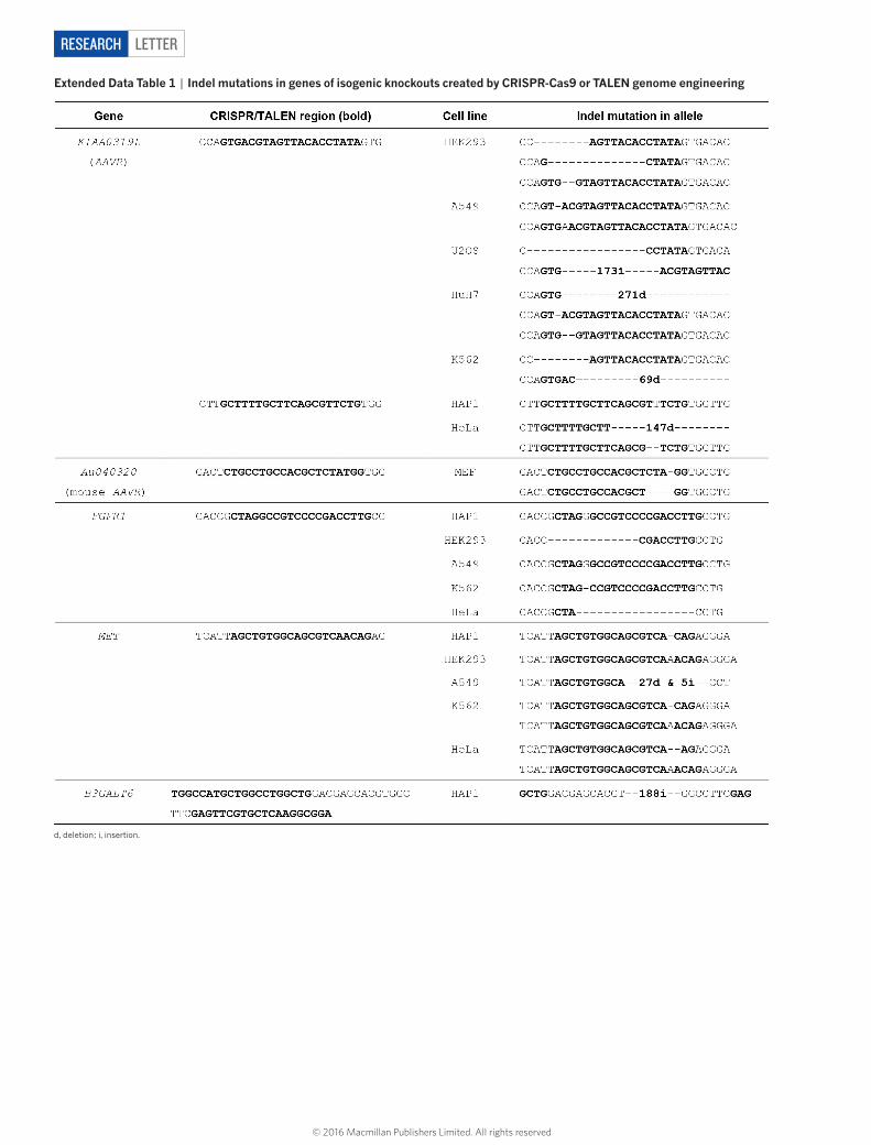
To validate the role of AAVR in AAV2 infection, we used CRISPR-Cas9 genome engineering to generate isogenic AAVR-knockout (AAVRKO) cell lines in a panel of cell types representing various human and mouse tissues (Extended Data Table 1). In all eight cell types, AAVR knockout rendered cells highly resistant to AAV2 infection (20,000 viral genomes (vg) per cell) (Fig. 1b). At a multiplicity of infection as high as 100,000 vg per cell, AAVRKO cells still remained poorly sus-ceptible to infection by an AAV2–luciferase vector (Extended Data Fig. 3a). This also held true for wild-type AAV2 infection, where AAV2 replication was negligible in AAVRKO cells (Extended Data Fig. 3b). Notably, MET and FGFR1 knockouts demonstrated no resistance to infection in multiple cell types (Extended Data Fig. 3e). Genetic complementation of AAVR in AAVRKO cells (Extended Data Fig. 3c) restored susceptibility to AAV2 in all cell types assessed, confirming that the resistance phenotype observed in AAVRKO cells was caused by loss of AAVR expression (Extended Data Fig. 3d). To examine further if AAVR expression can limit AAV2 infection, we overexpressed AAVR in four cell lines previously identified as poorly permissive to AAV2 (refs 12, 13). We observed an increase in susceptibility to AAV2 in all AAVR-overexpressing cell lines compared to wild-type cells, emphasiz-ing the important role of AAVR in AAV2 infection (Fig. 1c).
AAVR is a predicted type I transmembrane protein with five immu-noglobulin-like (Ig-like) domains in its ectodomain, referred to as polycystic kidney disease (PKD) domains14. Ig-like domains mediate cell–cell adhesion and are present in various well-characterized virus receptors15, including those for poliovirus, measles virus and reovirus. On the basis of their similarity to other receptors and the heavy dependence of AAV2 infection on AAVR, we hypothesized that AAVR acts as an AAV2 receptor. We first determined whether AAVR PKD domains are responsible for mediating AAV2 infection by creating a series of AAVR deletion mutants and expressing each in AAVRKO cells (Fig. 2a). Simultaneous deletion of AAVR PKD domains 1 and 2, or 2 and 3, abrogated its role in AAV2 infection, whereas deletions
1Department of Microbiology and Immunology, Stanford University School of Medicine, 299 Campus Drive, Stanford, California 94305, USA. 2Department of Biochemistry and Molecular Biology, School of Medicine, Oregon Health & Science University, 3181 Sam Jackson Park Road, Portland, Oregon 97239-3098, USA. 3Shriners Hospital for Children, 3101 Sam Jackson Park Road, Portland, Oregon 97239, USA. 4Netherlands Cancer Institute, Plesmanlaan 121, 1066 CX, Amsterdam, Netherlands. 5Department of Comparative Medicine, Stanford University School of Medicine, 287 Campus Drive, Stanford, California 94305, USA.*These authors contributed equally to this work.
© 2016 Macmillan Publishers Limited. All rights reserved

2 | N A T U R E | V O L 0 0 0 | 0 0 M O N T H 2 0 1 6
LETTERRESEARCH
1 2 3 4 5AAVR
ΔMANEC
ΔPKD3–4
ΔPKD2–3
ΔPKD1–2
MiniAAVR
ΔPKD4–5
AAV2 infection (%)
a b
AAVR
ΔMANEC
ΔPKD3–4
ΔPKD2–3
ΔPKD1–2
MiniAAVR
ΔPKD4–5
AAVRKO
0 20 40 60 80
0.1 1 10 100 1,000 10,000
0
1
2
3
4
c
[Soluble AAVR] (nM)
AAVR PKD1–5
Soluble AAVR
MBP
Bin
din
g (A
450)
0 0.08 0.01 0.02 0.08 0.2
[Soluble AAVR][MBP]
AA
V2
infe
ctio
n (%
)
0
20
40
60
80
100d e
Antibody concentration (μg ml–1)
AA
V2
infe
ctio
n (R
LU)
103
104
105
0 0.5 5 50
IgG isotypeAnti-AAVR
μM
TMIg-like PKD domainsSP MANEC
C-tailEctodomain
1 2 3 4 5
Figure 2 | AAVR binds specifically to AAV2 via Ig-like PKD domains. a, Schematic of AAVR domains and deletion mutants; dotted lines represent deletions. b, AAV2–RFP infection of HAP1 AAVRKO cells expressing AAVR deletion mutants (MOI: 20,000 vg per cell). c, ELISA showing binding of soluble AAVR (fusion protein between MBP and AAVR PKD1–5) to AAV2 particles. d, AAV2 neutralization assay of cells incubated with soluble AAVR or MBP during AAV2–GFP infection
(MOI: 7,500 vg per cell). e, Antibody inhibition assay of wild-type HeLa cells incubated with anti-AAVR or IgG isotype control antibodies (at indicated concentrations) at 4 °C before AAV2–luciferase infection (MOI: 1,000 vg per cell). Data depict mean with s.d. for triplicate infections; transgene expression measured after 24 h. MANEC, motif at amino terminus with eight cysteines; RLU, relative light units; SP, signal peptide; TM, transmembrane.
a
Sig
ni�c
ance
(–lo
g P
)
1,000
100
10
1
0.1Other hitsHeparan sulfate
biosynthesisPoorly characterized transmembrane proteins
Endosome/Golgi-related
b cWT AAVRKO
HAP1 HEK293 HeLa U2-OS A549 K562 HuH7 MEF HT29 Caco-2 Raji NIH3T3
Cell lines Cell lines
100
0
20
40
60
80
WT AAVR overexpression
**
***
***
*
AA
V2
infe
ctio
n (%
)
AA
V2
infe
ctio
n (%
)
0
20
40
60
80
100*** ***
***
*** ***
***
*** ***
C16orf62
RGP1
KIAA0319L (AAVR)
GPR108
TM9SF2
VPS29
VPS54 VPS52
VPS53 SWIP
OSBPL11
RIC1COG8
OSBPL9VPS35
RAB6A
B3GAT3
SLC35B2EXT1XYLT2
B3GALT6 B4GALT7
ATP2C1
EXT2ATP6V0A2
RNF121
JTBARPC2NDST1
NAA38
COMMD3ARPC4
HTTDMXL1
VPS51
CCDC22TRAPPC13
MALAT1
ACP2
GPR107
Figure 1 | An unbiased haploid genetic screen identifies KIAA0319L (AAVR), an essential host factor for AAV2 infection. a, Bubble plot illustrating the significance of enrichment of gene-trap insertions within identified genes (relative to unselected control population). Bubbles represent genes with width proportional to number of independent gene-trap insertions. Top 40 significant genes (P ≤ 0.001) are coloured and grouped by function. b, AAV2–RFP infection in wild-type (WT) cells and
AAVR knockout (AAVRKO) cells, evaluated in AAV2-susceptible human and mouse cell lines. c, AAV2–RFP infection of poorly permissive human and mouse cell lines with and without AAVR overexpression. Data depict mean with s.d. for triplicate infections. Infections were performed using a multiplicity of infection (MOI) of 20,000 vg per cell for 24 h. Significance was determined using unpaired parametric two-sided Student’s t-test, with a Welch post-correction; *P < 0.05; **P < 0.01; ***P < 0.001.
© 2016 Macmillan Publishers Limited. All rights reserved

0 0 M O N T H 2 0 1 6 | V O L 0 0 0 | N A T U R E | 3
LETTER RESEARCH
in other regions were tolerated (Fig. 2b). An AAVR minimal mutant (miniAAVR) consisting of PKD domains 1–3 in the ectodomain effi-ciently rescued AAV2 infection, highlighting the importance of the first three PKD domains for infection. Notably, soluble AAVR (recombinant protein comprising a fusion between maltose-binding protein (MBP) and AAVR PKD domains 1–5 expressed in Escherichia coli), but not MBP alone, bound directly to AAV2 particles (Fig. 2c and Extended Data Fig. 4a) with a dissociation constant (Kd) of ~150 nM (measured using surface plasmon resonance, see Extended Data Fig. 4b). We next investigated whether AAV2 infection could be neutralized in the pres-ence of soluble AAVR. Indeed, infection efficiency was inhibited in a concentration-dependent manner when soluble AAVR was included during infection (Fig. 2d and Extended Data Fig. 4c). Consistent with this inhibition assay, antibodies directed against AAVR were capable of potently blocking AAV2 infection by more than tenfold when incu-bated with cells before infection, in contrast to control IgG antibodies (Fig. 2e). This suggests that blocking viral access to AAVR on the cell surface substantially limits infection.
Characterization of the subcellular localization of AAVR revealed distinct perinuclear localization, demonstrating a strong association with the cis-medial Golgi marker (giantin), and complete co-localization with the trans-Golgi network (TGN) marker (TGN46) (Fig. 3a). Many TGN proteins are dynamically recycled from the plasma membrane through motifs in their carboxy terminal cytoplasmic tail (C-tail) that direct endocytosis and intracellular trafficking (reviewed in refs 16, 17). To determine whether AAVR is such a recycling receptor, we spe-cifically labelled the cell surface pool of AAVR by incubating live
AAVR-complement cells with anti-AAVR antibodies under cold condi-tions. Cells were warmed to initiate endocytosis and fixed at defined time points. Labelled AAVR gradually moved from the surface into the cell, and concentrated in a perinuclear location associated with the Golgi marker (Fig. 3b). This rapid endocytosis may explain why we did not observe AAVR at the cell surface in steady state (Fig. 3a). As a con-trol, AAVRKO cells were labelled similarly to AAVR-expressing cells, but no AAVR was detected on these cells (Extended Data Fig. 5a). Interestingly, the intracellular trafficking route of AAVR mapped here is remarkably similar to that of AAV particles, trafficking from the plasma membrane to the Golgi18. To determine whether AAVR endocytosis contributes to mediation of AAV2 infection, we removed the C-tail, which encodes endocytic motifs of AAVR. C-tail deletion (ΔC-tail) led to increased cell surface expression of AAVR (Fig. 3c and Extended Data Fig. 5b) and prevented endocytosis (Extended Data Fig. 5c). Importantly, the ΔC-tail was incapable of mediating AAV2 infection upon complementation in AAVRKO cells (Fig. 3d), suggesting that AAVR endocytosis is required for AAV2 infection. We further investigated whether AAVR requires intracellular trafficking to the TGN to mediate infection by replacing the C-tail of miniAAVR with those of cellular receptors with well-characterized endocytic motifs (Extended Data Fig. 6a). These included the cation-independent mannose 6-phosphate receptor (Ci-MPR), the prototypical receptor that mediates trafficking from the plasma membrane to the TGN19. We also included the low density lipoprotein receptor (LDLR)20 and polio-virus receptor (PVR)21, both of which endocytose and traffic between the plasma membrane and endosomes but are not reported to travel to
Figure 3 | AAVR traffics from the plasma membrane to the trans-Golgi network and AAVR endocytosis is necessary for AAV2 infection. a, Endogenous AAVR localization in wild-type HeLa cells shown with markers for cis-medial Golgi (giantin) and trans-Golgi network (TGN46). b, Tracking AAVR endocytosis using anti-AAVR antibodies. AAVR-complement cells were incubated with anti-AAVR antibodies for 1 h at 4 °C, washed and then incubated at 37 °C. At respective time points,
cells were fixed and anti-AAVR antibodies were visualized to track the trafficking of surface AAVR. c, AAVR surface expression in AAVRKO cells with and without overexpression of full-length AAVR and ΔC-tail (depicted in schematic). d, AAV2–RFP infection (MOI: 20,000 vg per cell; measured after 24 h) in AAVRKO cells stably expressing constructs depicted in c. Data depict the mean with s.d. for triplicate infections. Scale bars, 10 μm.
aAAVR Giantin Merge
MergeAAVR TGN46
2 min 10 min 30 min 60 min
cAAVR ΔC-tail
d
AAVR surface expression
0
20
40
60
80
100
0
50
0 103 104 105 0 103 104 105
100
150
200
0
50
100
150
200
HeLa W
T
+ΔC-ta
ilNone
+AAVR
AAVRKO
AA
V2
infe
ctio
n (%
)
b
AAVRKO AAVR ΔC-tail
Cou
nts
Ectodomain Ectodomain
AAVR
Giantin
DAPI
AAVR
Giantin
DAPI
Phalloidin
TMSP TMSP
C-tail
© 2016 Macmillan Publishers Limited. All rights reserved

4 | N A T U R E | V O L 0 0 0 | 0 0 M O N T H 2 0 1 6
LETTERRESEARCH
the TGN. Each of the fusion constructs displayed cellular localization patterns comparable to their parent receptors, as PVR-tail and LDLR-tail were detectable on the cell surface, and Ci-MPR-tail co-localized with a TGN marker and displayed a dispersed pattern in the cytoplasm (Extended Data Fig. 6b). Remarkably, all fusion constructs rescued AAV2 infection, albeit to different degrees (Extended Data Fig. 6c). Compared to the restored infection in miniAAVR-expressing cells, the LDLR and PVR fusion constructs yielded twofold and fourfold reduced infectivity, respectively. Conversely, routeing AAVR to the TGN using Ci-MPR endocytosis signals resulted in infection rates simi-lar to miniAAVR-expressing cells. Thus, trafficking to the TGN appears to increase AAV infection efficiency, but varying rates of endocytosis between the constructs may also contribute to infectivity differences. Nonetheless, all fusion constructs rescued infectivity to an extent, even those that are not reported to traffic to the TGN, indicating that traf-ficking to the TGN may not be a strict requirement. Collectively, these data suggest a model where AAVR interacts with AAV at the cell surface and facilitates trafficking to the TGN, but do not exclude the possi-bility that the interaction is initiated in early/late endosomes and/or the TGN.
To test whether other naturally occurring AAV serotypes are also dependent on AAVR, we infected AAVRKO cells with a panel of AAV serotypes including AAV1, 2, 3B, 5, 6, 8 and 9 (expressing green flu-orescent protein (GFP) or RFP). We also infected cells with a recom-binant adenovirus 5 vector expressing RFP (Ad5–RFP). AAVRKO cells displayed a robust resistance to all AAV serotypes (Fig. 4a), irrespec-tive of the different glycan attachment factors used by each serotype. AAV susceptibility was also restored in AAVR-complemented cells, as previously observed with AAV2. Moreover, there was no significant difference in Ad5–RFP infection among the three cell lines tested. The role of AAVR in infection for the tested viruses is therefore specific to AAV and is ubiquitously required for a variety of human- and simian- derived AAV serotypes.
Finally, we tested the contribution of AAVR to in vivo gene deliv-ery. We generated Aavr-knockout mice (Aavr−/−) using transcription activator-like effector nuclease (TALEN)-mediated gene targeting. Aavr−/− mice did not display any apparent developmental or phys-ical phenotype. Wild-type (Aavr+/+), heterozygous (Aavr+/−) and Aavr−/− FVB mice (genotypes depicted in Extended Data Fig. 7a)
were injected intraperitoneally with AAV9–luciferase, chosen because of its high transduction efficiency in vivo compared to AAV2 (ref. 22). Bioluminescence (a measure of luciferase expression) was strongest in the lower abdomen of Aavr+/+ mice, intensifying over 14 days (Fig. 4b, c and Extended Data Fig. 7b). Aavr heterozygosity did not significantly reduce AAV9 infection in vivo; however, Aavr−/− mice displayed a pronounced reduction in bioluminescence, comparable to background levels obtained in uninfected wild-type mice (Fig. 4d and Extended Data Fig. 7c).
Overall, this study identifies AAVR as a key host receptor for AAV infection in vitro and in vivo, using an unbiased and comprehensive genetic screening approach. AAV vector usage for gene therapy is rapidly growing, and recent advances in genome editing23 and vec-tored immunoprophylaxis24 are expected to further expand its utility. Exploiting AAVR as a tool to improve AAV-based applications may enhance its efficacy in basic research and clinical settings. An under-standing of AAVR tissue distribution will be important in determining the contribution of AAVR to AAV tropism, although additional factors25 including glycan usage, AAV nuclear import and other genes identified in our screen could also influence tropism. Lastly, AAV vectors are com-monly used in experimental mouse models; hence expression of AAVR under specific promoters (for example, for cells in the substantia nigra) in an Aavr−/− background may aid in developing better mouse models for human diseases such as those for neurological disorders.
Online Content Methods, along with any additional Extended Data display items and Source Data, are available in the online version of the paper; references unique to these sections appear only in the online paper.
Received 29 May; accepted 18 November 2015.
Published online 27 January 2016.
1. Kotterman, M. A. & Schaffer, D. V. Engineering adeno-associated viruses for clinical gene therapy. Nature Rev. Genet. 15, 445–451 (2014).
2. Nathwani, A. C. et al. Adenovirus-associated virus vector-mediated gene transfer in hemophilia B. N. Engl. J. Med. 365, 2357–2365 (2011).
3. Gaudet, D., Methot, J. & Kastelein, J. Gene therapy for lipoprotein lipase deficiency. Curr. Opin. Lipidol. 23, 310–320 (2012).
4. Lisowski, L. et al. Selection and evaluation of clinically relevant AAV variants in a xenograft liver model. Nature 506, 382–386 (2014).
5. Summerford, C. & Samulski, R. J. Membrane-associated heparan sulfate proteoglycan is a receptor for adeno-associated virus type 2 virions. J. Virol. 72, 1438–1445 (1998).
AA
V in
fect
ion
(%)
a bWT AAVRKO AAVR complement
Day 3
Day 7
Day 10
2.0
1.0
0.5
1.5
×107Luminescence
Radiance (photons s–1
cm–2 sr–1)
Day 14
d
Aavr+/+
Aavr+/–
Aavr–/–
Day 7**
NS
AAV1 AAV2 AAV3B AAV5 AAV6 AAV8 AAV9 Ad5
Uninfe
cted
0
20
40
60
80
100
c
103
104
105
106
103
104
105
106
107
Days after infection
Aavr+/+ Aavr+/– Aavr–/–
3 7 10 14
AA
V9
infe
ctio
n (p
hoto
ns s
–1 c
m–2
sr–1
)
AA
V9
infe
ctio
n (p
hoto
ns s
–1 c
m–2
sr–1
)
Uninfe
cted
Aavr+/+
Aavr+/–
Aavr–/–
Figure 4 | AAVR is a critical host factor for the infection of naturally occurring AAV serotypes and essential for AAV infection in vivo. a, Infection of wild-type HeLa cells, AAVRKO cells and AAVRKO cells overexpressing AAVR (AAVR complement), using AAV vectors of different serotypes (MOI: 105 vg per cell; RFP/GFP expression measured at 24 h). b, Bioluminescence of AAV9-infected wild-type (Aavr+/+), heterozygous (Aavr+/−) and AavrKO (Aavr−/−) FVB mice over 14 days;
representative images of mice from each group are shown with a radiance range of 5 × 10⁵ to 1 × 10⁷ photons s−1 cm−2 sr−1. c, AAV9–luciferase infection for Aavr+/+, Aavr+/− and Aavr−/− groups (measured as average radiance) at the respective days after infection. d, AAV9–luciferase infection of mice at day 7. Data depicts the mean (with s.d. in a and c). Significance was determined using unpaired two-sided Mann–Whitney t-test; **P < 0.01; NS, not significant.
© 2016 Macmillan Publishers Limited. All rights reserved

0 0 M O N T H 2 0 1 6 | V O L 0 0 0 | N A T U R E | 5
LETTER RESEARCH
6. Kashiwakura, Y. et al. Hepatocyte growth factor receptor is a coreceptor for adeno-associated virus type 2 infection. J. Virol. 79, 609–614 (2005).
7. Qing, K. et al. Human fibroblast growth factor receptor 1 is a co-receptor for infection by adeno-associated virus 2. Nature Med. 5, 71–77 (1999).
8. Carette, J. E. et al. Ebola virus entry requires the cholesterol transporter Niemann-Pick C1. Nature 477, 340–343 (2011).
9. Bonifacino, J. S. & Hierro, A. Transport according to GARP: receiving retrograde cargo at the trans-Golgi network. Trends Cell Biol. 21, 159–167 (2011).
10. McGough, I. J. & Cullen, P. J. Recent advances in retromer biology. Traffic 12, 963–971 (2011).
11. Poelmans, G., Buitelaar, J. K., Pauls, D. L. & Franke, B. A theoretical molecular network for dyslexia: integrating available genetic findings. Mol. Psychiatry 16, 365–382 (2011).
12. Ellis, B. L. et al. A survey of ex vivo/in vitro transduction efficiency of mammalian primary cells and cell lines with nine natural adeno-associated virus (AAV1–9) and one engineered adeno-associated virus serotype. Virol. J. 10, 74 (2013).
13. Hansen, J., Qing, K., Kwon, H. J., Mah, C. & Srivastava, A. Impaired intracellular trafficking of adeno-associated virus type 2 vectors limits efficient transduction of murine fibroblasts. J. Virol. 74, 992–996 (2000).
14. Ibraghimov-Beskrovnaya, O. et al. Strong homophilic interactions of the Ig-like domains of polycystin-1, the protein product of an autosomal dominant polycystic kidney disease gene, PKD1. Hum. Mol. Genet. 9, 1641–1649 (2000).
15. Bhella, D. The role of cellular adhesion molecules in virus attachment and entry. Phil. Trans. R. Soc. Lond. B 370, 20140035 (2015).
16. Maxfield, F. R. & McGraw, T. E. Endocytic recycling. Nature Rev. Mol. Cell Biol. 5, 121–132 (2004).
17. Kelly, B. T. & Owen, D. J. Endocytic sorting of transmembrane protein cargo. Curr. Opin. Cell Biol. 23, 404–412 (2011).
18. Nonnenmacher, M. & Weber, T. Adeno-associated virus 2 infection requires endocytosis through the CLIC/GEEC pathway. Cell Host Microbe 10, 563–576 (2011).
19. Ghosh, P., Dahms, N. M. & Kornfeld, S. Mannose 6-phosphate receptors: new twists in the tale. Nature Rev. Mol. Cell Biol. 4, 202–213 (2003).
20. Beglova, N. & Blacklow, S. C. The LDL receptor: how acid pulls the trigger. Trends Biochem. Sci. 30, 309–317 (2005).
21. Ohka, S. et al. Receptor (CD155)-dependent endocytosis of poliovirus and retrograde axonal transport of the endosome. J. Virol. 78, 7186–7198 (2004).
22. Zincarelli, C., Soltys, S., Rengo, G. & Rabinowitz, J. E. Analysis of AAV serotypes 1–9 mediated gene expression and tropism in mice after systemic injection. Mol. Ther. 16, 1073–1080, (2008).
Supplementary Information is available in the online version of the paper.
Acknowledgements The authors thank K. Kirkegaard, M. Kay and T. Brummelkamp for critical reading of the manuscript and valuable advice; T. Lerch, J. Tyner, D. Kabat and H. Nakai for assistance with preliminary experiments; H. P. Bächinger for advice and assistance with surface plasmon resonance experiments; Stanford Shared FACS facility and its staff; X. Ji (Stanford Functional Genomics Facility); Stanford mouse facility; T. Doyle for small animal imaging training; L. Popov for guidance in generating immunofluorescent images; G. Fuchs for technical assistance; and members of the Carette and Chapman laboratories for intellectual discussions and support. The work was funded in part by NIH R01 GM066875 (M.S.C.), DP2 AI104557 (J.E.C.) and U19 AI109662 (J.E.C.). J.E.C. is a David and Lucile Packard Foundation fellow.
Author Contributions S.P., M.S.C. and J.E.C. were responsible for overall design of the study. S.P. performed the haploid genetic screen, generated isogenic knockout and AAVR complement cell lines, and performed antibody inhibition and AAVR tracking studies. N.L.M. designed soluble AAVR construct and performed binding and soluble AAVR inhibition studies. J.E.C. designed AAVR-generated deletion mutant constructs. A.S.P. performed the wild-type AAV2 infection assay and all in vivo studies, under the technical expertise of C.M.N. O.D. was responsible for heterologous overexpression and purification of soluble AAVR, J.D. assisted in the production of FGFR1KO and METKO cell lines, Y.I. performed surface plasmon resonance measurements, L.T.J. generated the B3GALT6KO cell line, and J.E.W. created the ΔC-tail construct. S.P., M.S.C. and J.E.C. wrote the manuscript.
Author Information DNA sequencing data have been deposited in the NCBI sequencing read archive under NCBI BioProject PRJNA284536 with BioSample SAMN03703230 (gene-trap control data set) and SAMN04244346 (AAV screen). Reprints and permissions information is available at www.nature.com/reprints. The authors declare competing financial interests: details are available in the online version of the paper. Readers are welcome to comment on the online version of the paper. Correspondence and requests for materials should be addressed to J.E.C. ([email protected]) or M.S.C. ([email protected]).
23. Ran, F. A. et al. In vivo genome editing using Staphylococcus aureus Cas9. Nature 520, 186–191 (2015).
24. Balazs, A. B. et al. Antibody-based protection against HIV infection by vectored immunoprophylaxis. Nature 481, 81–84 (2012).
25. Nonnenmacher, M. & Weber, T. Intracellular transport of recombinant adeno-associated virus vectors. Gene Ther. 19, 649–658 (2012).
© 2016 Macmillan Publishers Limited. All rights reserved
LETTERRESEARCH
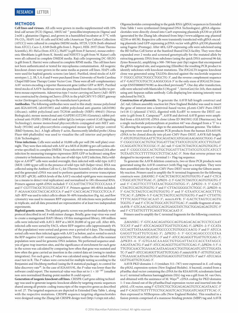
METHODSCell lines and viruses. All cells were grown in media supplemented with 10% fetal calf serum (FCS) (Sigma), 100 IU ml−1 penicillin/streptomycin (Sigma) and 2 mM l-glutamine (Sigma), and grown in a humidified incubator at 37 °C with 5% CO2. HAP1 (ref. 8) cells and K562 cells (American Type Culture Collection (ATCC)) were cultured in complete IMDM media. HT29, U2OS (both obtained from ATCC), Caco-2, A549 (both gifts from L. Popov), HEK-293T (from Thermo Scientific), H1-HeLa (from ATCC), HuH7 (a gift from P. Sarnow), mouse embry-onic fibroblasts (a gift from K. Storek) and NIH3T3 (a gift from W. Kaiser) cells were all cultured in complete DMEM media. Raji cells (expressing DC-SIGN) (a gift from E. Harris) were cultured in complete RPMI media. The cell lines have not been authenticated or tested for mycoplasma contamination. All isogenic knockout clones were grown in the same media as parent cell lines. HAP1 cells were used for haploid genetic screens (see later). Purified, titred stocks of AAV serotypes 1, 2, 3B, 5, 6, 8 and 9 were purchased from University of North Carolina Chapel Hill Gene Therapy Center Vector Core. These were all self-complementary AAV vectors encoding a reporter fluorescent gene (either GFP or RFP). Purified titred stocks of AAV9–luciferase were also purchased from this core facility to per-form mouse experiments. Adenovirus type 5 vector carrying mCherry (Ad5–RFP) was constructed by cloning mCherry cDNA in the pAd/CMV/V5-DEST gateway vector (Invitrogen) according to the manufacturer’s protocol.Antibodies. The following antibodies were used in this study: mouse polyclonal anti-KIAA0319L (ab105385) and rabbit polyclonal anti-giantin (ab24586) (Abcam); rabbit polyclonal anti-TGN46 antibody (NBP1-49643) (Novus Biologicals); mouse monoclonal anti-GAPDH (GT239) (Genetex); rabbit pol-yclonal anti-FGFR1 (D8E4) and rabbit IgG2a isotype control (Cell Signaling Technology); mouse monoclonal phycoerythrin-conjugated anti-MET anti-body (95106) and phycoerythrin-conjugated mouse IgG1 isotype control (R&D Systems, Inc). A high-affinity F-actin, fluorescently labelled probe (Alexa Fluor-660 phalloidin) was used to visualize the cell interior and periphery (Life Technologies).Virus infections. Cells were seeded at 10,000 cells per well (96-well plate) over-night. They were then infected with AAV at a MOI of 20,000 vg per cell (unless oth-erwise specified) in complete DMEM. Virus infectivity was determined 24 h after infection by measuring transgene expression (RFP, GFP or luciferase) using flow cytometry or bioluminescence. In the case of wild-type AAV2 infection, HeLa wild-type or AAVRKO cells were seeded overnight, then infected with wild-type AAV2 (MOI: 1,000 vg per cell) in the presence of wild-type Ad5 (helper virus). RNA was collected using the Ambion Cell-to-CT kit (Thermo Scientific) 24 h after infection, and the generated cDNA was used to perform quantitative reverse-transcriptase PCR (RT–qPCR). mRNA levels of the AAV2-encoded rep68 gene were measured (as a means to detect viral replication) and normalized to 18S ribosomal RNA. Primers against rep68 cDNA included: 5′-CCAATTACTTGCTCCCCAAA-3′ and 5′-CGTTTACGCTCCGTGAGATT-3′. Primers against 18S rRNA included: 5′-AGAAACGGCTACCACATCCA-3′ and 5′-CACCAGACTTGCCCTCCA-3′. Ad5–RFP was used to infect cells to obtain 50–60% transduction (Fig. 4a), and flow cytometry was used to measure RFP expression. All infections were performed in triplicate, and all data presented are representative of at least two independent experiments.Haploid genetic screen. The haploid genetic screen was performed similarly to the protocol described in ref. 8 with minor changes. Briefly, gene-trap virus was used to create a mutagenized HAP1 library. Of this mutagenized library, 100 million cells were infected with AAV2–RFP at a MOI 20,000 of vg per cell. After 48 h, infected cells were sorted by FACS, where RFP-negative cells (approximately 4% of the population) were sorted and grown over a period of 4 days. The resulting sorted cells were then infected again with AAV2 as before, and re-sorted to enrich the RFP-negative (AAV-resistant) population. Thirty-million cells of the resistant population were used for genomic DNA isolation. We performed sequence anal-ysis of gene-trap insertion sites, and the significance of enrichment for each gene in the screen was calculated by comparing how often that gene was mutated and how often the gene carried an insertion in the control data set (owing to random integration). For each gene, a P value was calculated using the one-sided Fisher exact test in R. The P values were corrected for multiple testing according to the Benjamini and Hochberg method (using the R statistical package), to control for false discovery rate26. In the case of KIAA0319L, the P value was lower than the software could report. The numerical value was thus set to 1 × 10−307 (smallest non-zero normalized floating-point number R could report).Generation of isogenic knockout cell lines. CRISPR-Cas9 gene editing technol-ogy was used to generate isogenic knockout alleles by targeting exonic sequences shared among all protein-coding transcripts of the respective genes as described in ref. 27. The targeted sequences are depicted in Extended Data Table 1, along with the respective mutations. CRISPR sequence targeting oligonucleotides were designed using the Zhang lab CRISPR design tool (http://crispr.mit.edu).
Oligonucleotides corresponding to the guide RNA (gRNA) sequences in Extended Data Table 1 were synthesized (Integrated DNA Technologies). gRNA oligonu-cleotides were directly cloned into Cas9-expressing plasmids pX330 or pX458 (generated by the Zhang lab; obtained from http://www.addgene.org; plasmid 42230 or 48138). Respective cells were transiently transfected with gRNA-encod-ing plasmids (and GFP-expressing pcDNA vector with gRNA-pX330 plasmids) using Fugene (Promega). After 48 h, GFP-expressing cells were subcloned using the BD InFlux Cell Sorter at the Stanford Shared FACS facility. They were then expanded over 2 weeks and screened genotypically for the mutated allele by extracting genomic DNA from subclones (using the quick DNA universal 96-kit; Zymo Research), amplifying a 500–700 base-pair (bp) region that encompassed the gRNA-targeted site, and sequencing (ElimBio) the resulting PCR product to identify subclones with knockout mutations. The B3GALT6 isogenic knockout clone was generated using TALENs directed against the nucleotide sequence 5′-TGGCCATGCTGGCCTGGCTG-3′, and the reverse complement sequence of 5′-GAGTTCGTGCTCAAGGCGGA-3′ in the only exon of B3GALT6 (tran-script ENST00000379198) as described previously28. One day after transfection, cells were selected with blasticidin S (30 μg ml−1, InvivoGen) for 24 h, then stained using anti-heparan sulfate antibody. Cells displaying low staining intensity were subcloned by FACS.Construction of plasmids. To generate the AAVR full-length construct and ΔC-tail, Gibson assembly reaction kit (New England Biolabs) was used to insert the gene of interest into a lentiviral-based vector, pLenti-CMV-Puro-DEST (w118-1) (plasmid 17452), digested with EcoRV to remove the DEST cas-sette (a gift from E. Campeau)29. AAVR and derived AAVR genes were ampli-fied from a KIAA0319L cDNA clone (clone ID 3843301) (GE Dharmacon), but a single nucleotide polymorphism at position 447 was changed from a ‘T’ to a ‘G’, allowing the sequence to align to the annotated human genome. The follow-ing primers were used to generate PCR products from the human KIAA0319L cDNA to be cloned directly into pLent-CMV-Puro-DEST. AAVR full-length: 5′-ATGTGTGGTGGAATTCTGCAGATACCATGGAGAAGAGGCTGGG-3′ and 5′- CG GC CG CC AC TG TG CT GG AT TT AC TT AT CG TC GT CA TC CT TG TA ATCCAGGATCTCCTCCCGC-3′; ΔC-tail: 5′-GACTCTAGTCCAGTGTGGTG-3′ and 5′-CGGCCGCCACTGTGCTGGATTTACTTATCGTCGTCATCCT TGTAATCTCCTTTTTGCCTCTTACAAC-3′. Note that reverse primer was designed to incorporate a C-terminal 1× Flag-tag sequence.
To generate the AAVR deletion constructs, two or three PCR products were generated using the AAVR construct (with Flag-tag) as a template. They were then assembled into the pLenti-CMV-Puro-DEST vector using the Gibson assem-bly reaction. Primers used to amplify the N-terminal fragments for the following constructs were: ΔMANEC: 5′-GACTCTAGTCCAGTGTGGTG-3′ and 5′-CTCA CTGGCATCTGTTGAC-3′; ΔPKD1–2: 5′-GACTCTAGTCCAGTGTGGTG-3′ and 5′-CAGTTCCTTTATAACTGGGTATGG-3′; ΔPKD2–3: 5′-GACT CTAGTCCAGTGTGGTG-3′ and 5′-CTTACGGGGCTCTGGC-3′; ΔPKD3–4: 5′-GACTCTAGTCCAGTGTGGTG-3′ and 5′-GTAATCCACAGCTTTG TTCAC-3′; ΔPKD4–5: 5′-GACTCTAGTCCAGTGTGGTG-3′ and 5′-CTTATTG TTTTCAGGTTGCACAAT-3′; miniAAVR: 5′-GACTCTAGTCCAGTG TGGTG-3′ and 5′-CTCACTGGCATCTGTTGAC-3′; middle fragment of min-iAAVR: 5′-GTCAACAGATGCCAGTGAGGTATCTGCTGGAGAGAGTGTC-3′ and 5′-CTTATTGTTTTCAGGTTGCACAAT-3′.
Primers used to amplify the C-terminal fragments for the following constructs were:
ΔMANEC: 5′-GTCAACAGATGCCAGTGAGACACACTCCTCCAAT TCCAT-3′ and 5′-ATCCAGAGGTTGATTGTCGAG-3′; ΔPKD1–2: 5′-CCATA CCCAGTTATAAAGGAACTGCCCCCTGTGGCCAACG-3′ and 5′-ATCCA GAGGTTGATTGTCGAG-3′; ΔPKD2–3: 5′-GCCAGAGCCCCGTAA GCCTCCTCAGGCAGATGC-3′ and 5′-ATCCAGAGGTTGATTGTCGAG-3′; ΔPKD3–4: 5′-GTGAACAAAGCTGTGGATTACCCACCTATAGCC AAGATAACTG-3′ and 5′-ATCCAGAGGTTGATTGTCGAG-3′; ΔPKD4–5: 5′- ATTGTGCAACCTGAAAACAATAAGAACCTGGTGGAGATCATCTTGGATA TC-3′ and 5′-ATCCAGAGGTTGATTGTCGAG-3′; miniAAVR: 5′-ATTGTGCAAC CTGAAAACAATAAGTGTGAGTGGAGCGTGTTATATG-3′ and 5′-ATCCAGA GGTTGATTGTCGAG-3′.
AAVR PKD domains 1–5 (residues 311–787) were expressed in E. coli using the pMAL expression system (New England Biolabs). A bacmid, created from a pFastBac dual vector containing the cDNA for the KIAA0319L ectodomain fused to a C-terminal influenza haemagglutinin (HA)-tag was a gift from M. van Oers, and obtained with the assistance of M. Waye30. cDNA coding for PKD domains 1–5 was cloned out of the pFastBacDual expression vector and inserted into the pMAL-c5X vector, using 5′-GTATCTGCTGGAGAGAGTGTCCAGATAACC-3′ and 5′-CAGGTTGTTTTTCCTGCAGGTCACCTGGGATCAGGTTTCAC-3′, then expressed in NEBexpress cells (New England Biolabs). This resulted in a fusion protein comprised of a mannose-binding protein (MBP) tag and AAVR
© 2016 Macmillan Publishers Limited. All rights reserved

LETTER RESEARCH
PKD domains 1–5 (referred to as: soluble AAVR). MBP was specifically used as an affinity tag for ease of purification.
To create AAVR fusion constructs, Ci-MPR-tail, LDLR-tail and PVR-tail, the Gibson assembly reaction was used to fuse amplified miniAAVR with-out its C-terminal to the C-terminal of the respective proteins, and insert it into the pLenti-CMV-Puro-DEST vector. Primers used for amplification and insertion included: miniAAVR without C-terminal and transmem-brane domain for Ci-MPR-tail: 5′-GACTCTAGTCCAGTGTGGTG-3′ and 5′-CTTATTGTTTTCAGGTTGCACAAT-3′; MPR C-terminal and transmem-brane: 5′-ATTGTGCAACCTGAAAACAATAAGGCTGTGGGAGCTGTGC-3′ and 5′-CGGCCGCCACTGTGC-3′; miniAAVR without C-terminal and transmem-brane domain for LDLR-tail or PVR-tail: 5′-GACTCTAGTCCAGTGTGGTG-3′ and 5′-CTTATTGTTTTCAGGTTGCACAAT-3′; LDLR or PVR C-terminal and transmembrane: 5′-ATTGTGCAACCTGAAAACAATAAG-3′ and 5′-TAAATCCAGCACAGTGGCGGCCG-3′.Generation of stable cell lines. Lentiviral transduction was used to create stable cell lines expressing a selected gene of interest under a CMV promoter. Using Gibson assembly reaction, the respective genes of interest (see ‘construction of plasmids’ section) were inserted into the pLenti-CMV-Puro-DEST vector, and used as described previously29. Lentivirus was produced using HEK293 cells and used to transduce the respective cell lines overnight. Cells stably expressing the gene of interest were selected by treatment with 1–3 μg ml−1 puromycin over 2 days (InvivoGen). A lentivirus carrying the mCherry (RFP) gene was used as a control for AAVR complementation in AAVRKO cells.Flow cytometry. All flow cytometry was performed at the Stanford Shared FACS facility. To perform the haploid genetic screen, FACS was carried out on a FACS Aria flow cytometer (BD). To measure virus transgene expression (RFP/GFP) in all other experiments, cells were trypsinized 24 h after infection and a LSRII-UV flow cytometer (BD) was used to detect fluorescent cells. For cell surface staining, cells were trypsinized and washed using FACS buffer (PBS supplemented with 2% FCS, 1 mM EDTA and 0.1% sodium azide). They were subsequently incubated for 40 min at 4 °C with the respective primary antibodies at a 1:50 dilution (see ‘Antibodies’ section), washed, and incubated for a further 40 min at 4 °C with Alexa488- or Alexa594-conjugated secondary antibodies (1:500 dilution; if the primary was not conjugated) (Life Technologies). This was followed by a final wash and resuspension of cells in FACS buffer before reading fluorescence. All data presented are representative of at least two independent experiments. Data were analysed and assembled using FlowJo software (TreeStar Inc).Immunoblot analysis. Cell pellets of 2 × 10⁶ cells were lysed with Laemmli SDS sample buffer containing 5% β-mercaptoethanol and boiled for 10 min at 96 °C. Lysates were separated by SDS–PAGE using the Mini-Protean system (Bio-Rad) on 4–15% polyacrylamide gradient gels (Bio-Rad). Proteins were transferred onto polyvinylidene fluoride membranes (Bio-Rad) using the Bio-Rad Transblot protein transfer system in a semi-wet preparation. Membranes were blocked by incubating with PBS containing 5% non-fat milk for 1 h at room temperature. Membranes were subsequently incubated overnight at 4 °C with primary antibodies at a dilu-tion of 1:1000 (anti-KIAA0319L antibody) or 1:2,000 (anti-GAPDH antibody) in blocking buffer. Membranes were washed 3 times for 5 min using wash buffer (PBS with 0.1% Tween-20), and further incubated in horseradish peroxidase (HRP)-conjugated secondary antibodies (anti-mouse and anti-rabbit 1:5,000 in blocking buffer) (GeneTex) for 1 h at room temperature. After another set of three washes, antibody-bound proteins were visualized on film using the West Pico and Extended Duration chemiluminescence peroxide solutions (Thermo Scientific).Immunofluorescence. Cells were seeded overnight at 40,000 cells per well onto LabTekII glass chamber slides (Thermo Scientific). They were washed once with PBS, and either treated or fixed immediately with 4% paraformaldehyde for 15 min. They were washed three times with PBS before being incubated for 1 h at room temperature with primary antibodies against the respective proteins at a dilution of 1:100 (anti-KIAA0319L and anti-TGN46) or 1:200 (anti-giantin) in immunofluorescence blocking buffer (PBS with 3% BSA, 1% saponin and 1% Triton X-100). Cells were then washed three times in PBS, and incubated for a further hour in DAPI stain (1:500) and fluorescently tagged secondary antibodies (Alexa488 anti-mouse and Alexa594 anti-rabbit; Life Technologies) at a dilution of 1:300. Cells were washed a final three times in PBS, and 5 μl of Vectashield (Vector Laboratories Inc) was applied to each slide chamber before a glass cover slip (VWR International) was placed over slide to mount samples. Cells were visualized directly with a Zeiss LSM 700 confocal microscope.ELISA. Purification of the soluble AAVR was achieved through amylose-based MBP affinity chromatography (GE Healthcare). ELISA plates (Corning Costar) were coated overnight at 4 °C with 50 μl AAV2 virus-like particles at 2.5 μg ml−1 in 100 mM NaHCO3 (pH 9.6). Plates were then washed with TBST buffer (0.05% Tween-20 in TBS) and blocked with 3% BSA in TBST for 1 h at room tempera-ture. Subsequent washing was followed by incubation with soluble AAVR or MBP
control at the indicated concentrations for 2 h at room temperature. Anti-MBP–HRP (1:500, 1 h incubation at room temperature) was used to detect soluble AAVR and MBP controls, requiring no secondary antibody. Samples were developed with 1-Step Ultra TMB-ELISA substrate as per the manufacturer’s instructions (Thermo Scientific) and optical density assayed by microplate reader (Molecular Devices SpectraMax M2e) at 450 nm. Curve fitting was performed in SigmaPlot v12.5 (Systat Software, Inc). All data presented are representative of at least three independent experiments.Surface plasmon resonance analysis of binding. Surface plasmon resonance analy-sis was carried out using a BIAcore X instrument (GE Healthcare) and a flow rate of 10 μl min−1 at 20 °C in HBS-P buffer (10 mM HEPES (pH 7.5), 150 nM NaCl and 0.005% surfactant P20). His-tagged soluble AAVR (His-tagged MBP fusion with AAVR PKD domains 1–5) at various concentrations was mixed with His-tagged MBP to a total concentration of 0.2 μM in 10 mM sodium acetate buffer (pH 4.0) and immobilized on a CM5 sensor chip by amide coupling. MBP at 0.2 μM was sufficient to block nonspecific binding to the dextran. For the analysis of binding affinity, all curves were measured in triplicate and were fitted with a Langmuir 1:1 binding model (BIAevaluation software, GE Healthcare).Antibody inhibition assay. Wild-type HeLa cells were seeded in 96-well plates at 10,000 cells per well overnight. Anti-AAVR antibody (ab105385) or IgG isotype control (both from Abcam) were incubated with cells (at concentrations ranging from 0.5 to 50 μg ml−1 in DMEM media) for 1 h at 4 °C. Cells were then infected with AAV2–luciferase at a MOI of 1,000 vg per cell, and left for 24 h at 37 °C. A luciferase assay kit (E1500, Promega) was used to detect bioluminescence, with measurements being taken on the Promega GloMax luminometer. Notably, the storage buffers of both antibodies did not contain preservatives such as azide that could interfere with the assay. All data presented are representative of two inde-pendent experiments.Competitive inhibition assay. HeLa cells were seeded in 96-well plates at 10,000 cells per well overnight. Purified soluble AAVR or MBP control was then intro-duced to the medium at the specified concentrations. Cells were transduced with AAV2–GFP at a MOI of 7,500 vg per cell and incubated for 24 h at 37 °C. This was followed by trypsinization and measuring transgene expression by flow cytometry. For immunofluorescence imaging, the concentration of soluble AAVR and MBP controls was 0.1 μM, and transduction was performed using 7,000 vg per cell. At 24 h post-transduction, cells were incubated with 1 μg ml−1 Hoechst stain (Thermo Scientific) in PBS for 10 min at 37 °C, before washing with PBS and subsequent fluorescent imaging (Nikon Eclipse Ti-E). All data presented are representative of two independent experiments.Tracking surface-bound AAVR using anti-AAVR antibodies. These experi-ments were performed similarly to Ci-MPR tracking assays, as described in ref. 31. AAVRKO cells with or without overexpression of AAVR or ΔC-tail were incubated at 4 °C with anti-AAVR antibodies (approximately 25 μg ml−1) for 1 h. Cells were then washed three times with PBS and transferred to 37 °C for specific time points (2, 10, 30 and 60 min), at which time they were fixed with 4% paraformaldehyde for 15 min. Following fixation, immunofluorescence staining (as described earlier) was performed to visualize AAVR endocytosis. All data presented are representative of two independent experiments.Ethics statement and animal studies. All the experiments involving animals were conducted in strict accordance with the Institutional Animal Care and Use Committee of Stanford University. Mice were housed in a Stanford University vivarium, accredited by the Association for Assessment and Accreditation of Laboratory Animal Care International. Mice were housed in irradiated dispos-able caging (Innovive Inc) with bi-weekly cage changes. Mice were provided with irradiated food and ultraviolet-irradiated acidified water. Health surveillance was performed via trimester testing of dirty bedding CD1 sentinels (Charles River Laboratories). Sentinels were consistently negative for mouse parvovirus, minute virus of mice, mouse hepatitis virus, rotavirus, mouse encephalomyelitis virus, Sendai virus, mouse adenovirus 1 and 2, ectromelia virus, lymphocytic chori-omeningitis virus, pneumonia virus of mice, reovirus 3, Mycoplasma pulmonis, and endo- and ectoparasites. No statistical methods were used to predetermine sample size. In our animal study protocol, we state that the number of animals in each experimental group varies, and is based on similar previous study32. Randomization was not used to allocate animals to experimental groups and the investigators were not blinded to allocation during experiments and outcome assessment.AAV infection in mice. TALEN technology was used to create AAVR isogenic knockout FVB mice (purchased from Cyagen Biosciences). TALEN-targeted sequences were 5′-TGGGAGTCAAGCCAAGTC-3′ and 5′-GCCAGGATATTGTTGGCAGA-3′. Two founder males were mated to FVB/NCrl (Charles River Laboratories) females. After three rounds of breeding, wild-type (Aavr+/+), heterozygous (Aavr+/−) and homozygous AAVRKO (Aavr−/−) mice were generated, determined by genotyping. All genotypes (wild-type, heterozygous
© 2016 Macmillan Publishers Limited. All rights reserved

LETTERRESEARCH
and knockout) were obtained in the expected Mendelian ratios after breeding. At 5 weeks of age, 10 female and 9 male animals were used to examine the effect of Aavr KO on AAV infection in vivo. Animals from each group (Aavr+/+, n = 7 (2 litter mates and 5 purchased FVB mice); Aavr+/−, n = 4; Aavr−/−, n = 4 and uninfected mice, n = 4) were injected intraperitoneally with 1 × 10¹¹ viral genomes of AAV9–luciferase in 200 μl of PBS. All of the mice recovered from the injection quickly without loss of mobility or interruption of grooming activity. Aavr+/+ and Aavr−/− mice were found to be significantly different in two independent experiments.In vivo bioluminescence imaging. The mice were anaesthetized with 2% iso-fluorane and oxygen. The d-luciferin substrate (Biotium) was injected intraperito-neally (3.3 μg per mouse). After 10 min, the mice were then placed in a light-tight chamber, and images were generated using a cryogenically cooled charge-coupling device camera IVIS 100 (Xenogen), recording bioluminescence at 1, 10, 60 and 100 s. The visual output represents the average radiance as the number of photons emitted per second per cm2 as a false colour image where the maximum is red and the minimum is dark blue. All animals were imaged on a schedule of 3, 7, 10 and 14 days after AAV vector injection. At each time-point a ‘region of inter-est’ was designated surrounding each animal in order to quantify the radiance (photons s−1 cm−2 sr−1) being released by luciferase activity. This region was kept the same for each mouse and at each time point. The mean and standard deviation of radiance measurements were determined for each mouse group at each time point.
Statistics. The unpaired parametric two-sided Student’s t-test was used for sta-tistical calculations involving two group comparisons in all tissue-culture-based experiments (*P < 0.05, **P < 0.01, ***P < 0.001), with a Welch post-correction accounting for different standard deviations. An unpaired two-sided Mann–Whitney t-test was used for statistical calculations involving two group com-parisons in in vivo experiments. GraphPad Prism was used for statistical calculations.
26. Benjamini, Y. & Hochberg, Y. Controlling the false discovery rate: a practical and powerful approach to multiple testing. J. R. Stat. Soc. Ser. A Stat. Soc. 57, 289–300 (1995).
27. Ran, F. A. et al. Genome engineering using the CRISPR-Cas9 system. Nature Protocols 8, 2281–2308 (2013).
28. Sanjana, N. E. et al. A transcription activator-like effector toolbox for genome engineering. Nature Protocols 7, 171–192 (2012).
29. Campeau, E. et al. A versatile viral system for expression and depletion of proteins in mammalian cells. PLoS ONE 4, e6529 (2009).
30. Holster, S. et al. Expression of the dyslexia candidate gene Kiaa0319-like in insect cells J. Biochem. Mol. Biol. Post Gen. Era 2, 45–52 (2013).
31. Seaman, M. N. Cargo-selective endosomal sorting for retrieval to the Golgi requires retromer. J. Cell Biol. 165, 111–122 (2004).
32. Jae, L. T. et al. Virus entry. Lassa virus entry requires a trigger-induced receptor switch. Science 344, 1506–1510 (2014).
© 2016 Macmillan Publishers Limited. All rights reserved
LETTER RESEARCH
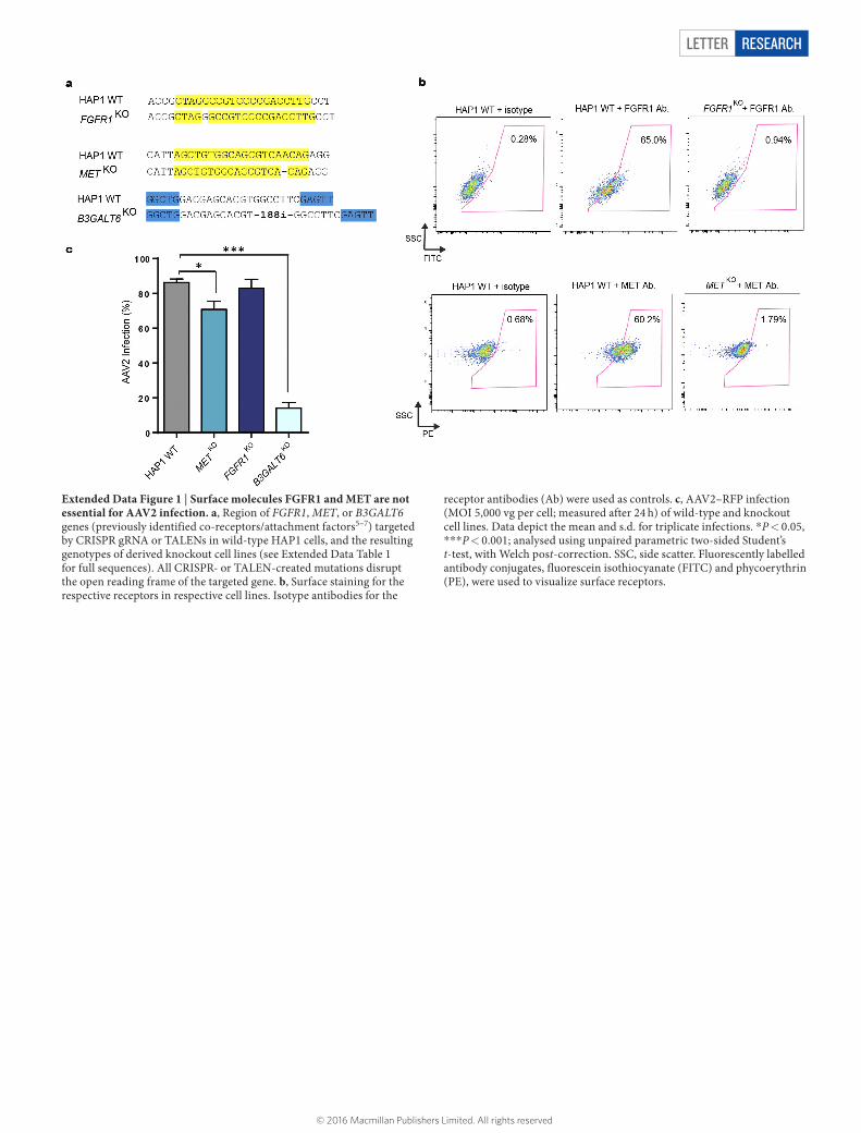
Extended Data Figure 1 | Surface molecules FGFR1 and MET are not essential for AAV2 infection. a, Region of FGFR1, MET, or B3GALT6 genes (previously identified co-receptors/attachment factors5–7) targeted by CRISPR gRNA or TALENs in wild-type HAP1 cells, and the resulting genotypes of derived knockout cell lines (see Extended Data Table 1 for full sequences). All CRISPR- or TALEN-created mutations disrupt the open reading frame of the targeted gene. b, Surface staining for the respective receptors in respective cell lines. Isotype antibodies for the
receptor antibodies (Ab) were used as controls. c, AAV2–RFP infection (MOI 5,000 vg per cell; measured after 24 h) of wild-type and knockout cell lines. Data depict the mean and s.d. for triplicate infections. *P < 0.05, ***P < 0.001; analysed using unpaired parametric two-sided Student’s t-test, with Welch post-correction. SSC, side scatter. Fluorescently labelled antibody conjugates, fluorescein isothiocyanate (FITC) and phycoerythrin (PE), were used to visualize surface receptors.
© 2016 Macmillan Publishers Limited. All rights reserved

LETTERRESEARCH
Extended Data Figure 2 | Haploid unbiased genetic screen evaluating host factors important for AAV2 infection. a, A schematic depicting the strategy for the AAV2 genetic screen. A library of mutagenized haploid HAP1 cells was created with a retroviral gene-trap vector, and subsequently infected with AAV2–RFP (MOI: 20,000 vg per cell) for 24 h. RFP-negative cells were sorted using FACS to isolate cells with mutations
in genes essential for AAV2 infection. These cells were re-infected for a second iteration of selection. DNA was then extracted from this enriched population and sequenced to map specifically where the gene-trap insertions occurred that resulted in the mutation. b, The gating strategy for the FACS-based AAV2 screen.
© 2016 Macmillan Publishers Limited. All rights reserved

LETTER RESEARCH
Extended Data Figure 3 | AAVR is a critical host factor for AAV2 infection. a, Effect of AAVR isogenic knockout (AAVRKO) upon AAV2–luciferase infection, evaluated in HAP1 and HeLa cell background from a MOI of 100 to 100,000 vg per cell. b, RT–qPCR to detect wild-type AAV2 infection in wild-type HeLa or AAVRKO cells. Cells were infected with wild-type AAV2 and adenovirus (helper virus required for AAV2 replication), and AAV2 Rep68 mRNA levels were measured to assess AAV2 infection. c, Immunoblot analysis evaluating AAVR expression in wild-type, AAVRKO and AAVRKO overexpressing AAVR (AAVR Comp.) cell
lines of HAP1 and HeLa origin. GAPDH was immunoblotted as a control. AAVR (predicted 115 kilodaltons (kDa)) appears at 150 kDa owing to six glycosylation sites. d, AAV2–luciferase infection (MOI 20,000 vg per cell; measured after 24 h) in AAVRKO cells stably complemented with AAVR or control lentiviral vector, evaluated in several AAV2-susceptible human and mouse cell lines. e, Comparison of AAV2–RFP infection (MOI: 20,000 vg per cell; measured after 24 h) in wild-type, AAVRKO, METKO and FGFR1KO cells, evaluated in several AAV2-susceptible human cell lines. Data depict the mean and s.d. for triplicate infections.
© 2016 Macmillan Publishers Limited. All rights reserved
LETTERRESEARCH
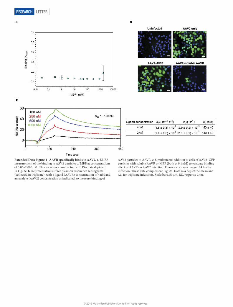
Extended Data Figure 4 | AAVR specifically binds to AAV2. a, ELISA measurement of the binding to AAV2 particles of MBP at concentrations of 0.05–2,000 nM. This serves as a control to the ELISA data depicted in Fig. 2c. b, Representative surface plasmon resonance sensograms (collected in triplicate), with a ligand (AAVR) concentration of 4 nM and an analyte (AAV2) concentration as indicated, to measure binding of
AAV2 particles to AAVR. c, Simultaneous addition to cells of AAV2–GFP particles with soluble AAVR or MBP (both at 0.1 μM) to evaluate binding effect of AAVR on AAV2 infection. Fluorescence was imaged 24 h after infection. These data complement Fig. 2d. Data in a depict the mean and s.d. for triplicate infections. Scale bars, 50 μm. RU, response units.
© 2016 Macmillan Publishers Limited. All rights reserved

LETTER RESEARCH
Extended Data Figure 5 | AAVR ΔC-tail is detected at the cell surface and does not endocytose to the TGN. AAVRKO cells (a) or ΔC-tail-expressing cells (c) were incubated with anti-AAVR antibodies for 1 h at 4 °C, washed and then transferred to 37 °C. At respective time points, cells were fixed and
antibody-bound AAVR was visualized. These data complement Fig. 3b. b, Permeabilized and unpermeabilized immunostaining of full-length AAVR and ΔC-tail when expressed in AAVRKO cells. These data complements Fig. 3c. Scale bars, 10 μm.
© 2016 Macmillan Publishers Limited. All rights reserved

LETTERRESEARCH
Extended Data Figure 6 | AAVR endocytosis is crucial for AAV2 infection. a, Schematic of the miniAAVR and domain-swapped derivatives probing the localization of AAVR through the swapping of the AAVR C-tail with that of well-characterized recycling receptors: Ci-MPR (traffics from plasma membrane through endosomes to the TGN), LDLR and PVR (both traffic from plasma membrane to endosomal compartments but
are not reported to traffic to TGN). b, Corresponding permeabilized and unpermeabilized immunofluorescence images of constructs depicted in a when expressed in AAVRKO cells. c, AAV2–RFP infection (MOI: 20,000 vg per cell; measured after 24 h) in AAVRKO cells stably expressing constructs depicted in a. Data depict the mean and s.d. for triplicate infections. Scale bars, 10 μm.
© 2016 Macmillan Publishers Limited. All rights reserved
LETTER RESEARCH
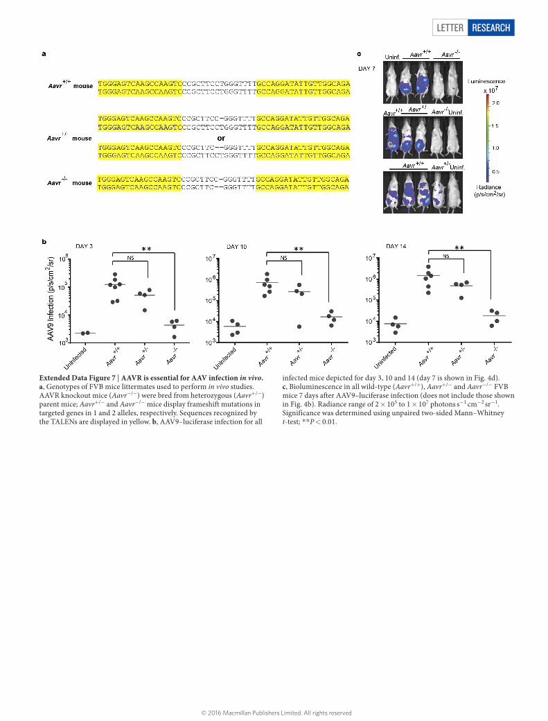
Extended Data Figure 7 | AAVR is essential for AAV infection in vivo. a, Genotypes of FVB mice littermates used to perform in vivo studies. AAVR knockout mice (Aavr−/−) were bred from heterozygous (Aavr+/−) parent mice; Aavr+/− and Aavr−/− mice display frameshift mutations in targeted genes in 1 and 2 alleles, respectively. Sequences recognized by the TALENs are displayed in yellow. b, AAV9–luciferase infection for all
infected mice depicted for day 3, 10 and 14 (day 7 is shown in Fig. 4d). c, Bioluminescence in all wild-type (Aavr+/+), Aavr+/− and Aavr−/− FVB mice 7 days after AAV9–luciferase infection (does not include those shown in Fig. 4b). Radiance range of 2 × 10⁵ to 1 × 10⁷ photons s−1 cm−2 sr−1. Significance was determined using unpaired two-sided Mann–Whitney t-test; **P < 0.01.
© 2016 Macmillan Publishers Limited. All rights reserved
LETTERRESEARCH
Extended Data Table 1 | Indel mutations in genes of isogenic knockouts created by CRISPR-Cas9 or TALEN genome engineering
d, deletion; i, insertion.
© 2016 Macmillan Publishers Limited. All rights reserved