an approach to coma - ministry of healthmohs.gov.mm/ckfinder/connector?command=proxy&lang... ·...
TRANSCRIPT

An approach to comaTint Tint Kyi
Consciousness• dependent on the function of two separate anatomical and
physiological syetems:
1. The ascending reticular activationg sys( ARAS) projecting from
brainstem to thalamus.{determines arousal}( the level of
consciousness )
2. The cerebral cortex: determines the content of consciousness
Impaired functioning of either anatomical system may cause coma
Disturbed consciousness: def
• Coma- a state of unrousable unresponsiveness.
• Level of consciousness represents a continuum between being
alert and deeply conmatose.
• It may be qualified using the GCS
• Coma GCS < 8
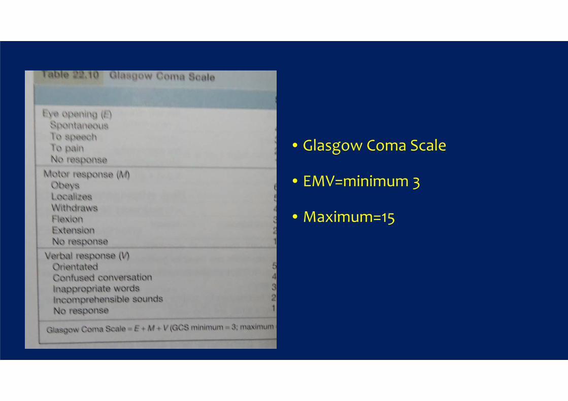
• Glasgow Coma Scale
• EMV=minimum 3
• Maximum=15
delirium
• The term used to describe a confusional state in which reduced
attention is a cardinal feature, usually with altered behavior,
coginition , orientation and a fluctuating level of consciousness
from agitation to hypoarousal
Stupor and obtundation
• No longer use
Principle causes
• Diffuse brain dysfunction
• Direct effect within brainstem
• Pressure effect on brainstem

Diffuse brain dysfunction
• Drug overdose
• Encephalitis, meningitis, cerebral malaria
• SAH
• CO poisoning
• Trauma to brain
• Hypo,hyperglycemia
• Organ failure- severe uraemia,hepatic encephalopathy,respira

Continue:
• Hypercalcaemia, hypoCa
• Hypoadrenalism, hypopit and hypothyroidism
• Hyponatraemia, hypernatraemia
• Metabolic acidosis
• Hypothermia, hyperpyrexia
• Seizures-post epileptic state, non-convulsive state

Continue;
• Metabolic rarities eg porphyria
• Extensive cortical damage
• Hypoxic ischaemic brain injury eg cardiac arrest
Direct effect within brain stem
• Brainstem haemorrhage, infarction or demyelination
• Brainstem neoplasm eg glioma
• Wernicke-korsakoff syndrome

Pressure effect on brainstem
• Tumor, massive hemisphere infarction with edema
• Haematoma,
• Abscess
• Cerebellar mass

mechanism
• Altered consciousness is produced by four mechanisms affecting
the ARAS in the brainstem or thalamus, and / or widespread
impairment of cortical function

• Brain stem lesion- a discrete brainstem or thalamic lesion, eg stroke
may damage the ARAS
• Brainstem compression: a supratentorial mass lesion within the
brain compresses the brainstem, inhibiting the ARAS, ge coning
from a brain tumour or haemorrhage. Mass lesion within the post
fossa prone to cause and hydrocephalous

Diffuse brain dysfunction
• Diffuse brain dysfunction: generalized severe metabolic or toxic
disorders( eg alcohol, sedatives, uraemia, hypercapnia,depress
cortical and ARAS function)
Massive cortical damage
• Unlike brainstem lesion, extensive damage of the cerebral cortex and
cortical connections is required to cause coma, eg meningitis
or hypoxic-ischaemic damage after cardiac arrest
• A single focal hemisphere or cerebellar lesion does not reduce coma
unless it compresses the brain stem.
• Cerebral edema frequently surrounds masses, increasing their pressure
effects.

Commonest causes of coma are:
• Metabolic disorder 35%
• Drug and toxin- 25%
• Mass lesion 20%
• Others- including trauma, stroke and CNS infectiona


Immediate assessment and management
• Check the airway, breathing and circulation
• Stix for blood glucose: if hypo- give glucose ( 25ml 50%)
• Treat seizures with buccal midazolam and if not terminated,
intravenous phenytoin
• If there is fever and meninism: give IV antibiotics
check ICT malaria or blood film
If alcohol
• Thiamine- dose IV parbrinex 1 pair 250mg 8hrly for W-K Syn
• Naloxone
• flumazenil
Obtain as much history as possible
• Limited history is one of the problems faced in assessing the
unconscious patient.
• What were the circumstances?
• Ask paramedics, police, and witnesses
• Contact the patient’s relatives, friends and GP and
• Obtain past hospital notes, drug details, bottles, identifications data

General and neurological examination
Fundi plus pupilsBrain stem reflexNeck stiffness/ sign of traumaNeuro limbs – lateralizationGeneral system examination

General and neurological examination(2)
• Temp, check for meningism
• Sniff the patient’s breath for ketones, alcohol and hepatic fetor
• Survey the skin for signs of trauma or spinal injury,
• rash ( meningococcal sepsis) jaundice or stigmata of chronic liver
diseases, cyanosis, injection marks
• Respiratory pattern-
Respiratory pattern
• Cheyne-stroke ( alternating hyperpnoea and periods of apnoea
indicating bilateral cerebral or upper brainstem dysfunction or
acidotic ( Kussmaul respiration(deep,sighing hyperventilation seen
in diabetic ketoacidosis and uraemia)
neuro
• Aim: GCS, Brainstem function, lateralization of pathology
• GCS: repeated regularly progressively declining
• Use painful stimulus nail bed pressure or central area, record
best response
• Shout commands

fundi
• Look for papilloedema and subhyaloid retinal haemorrhage ( SAH)
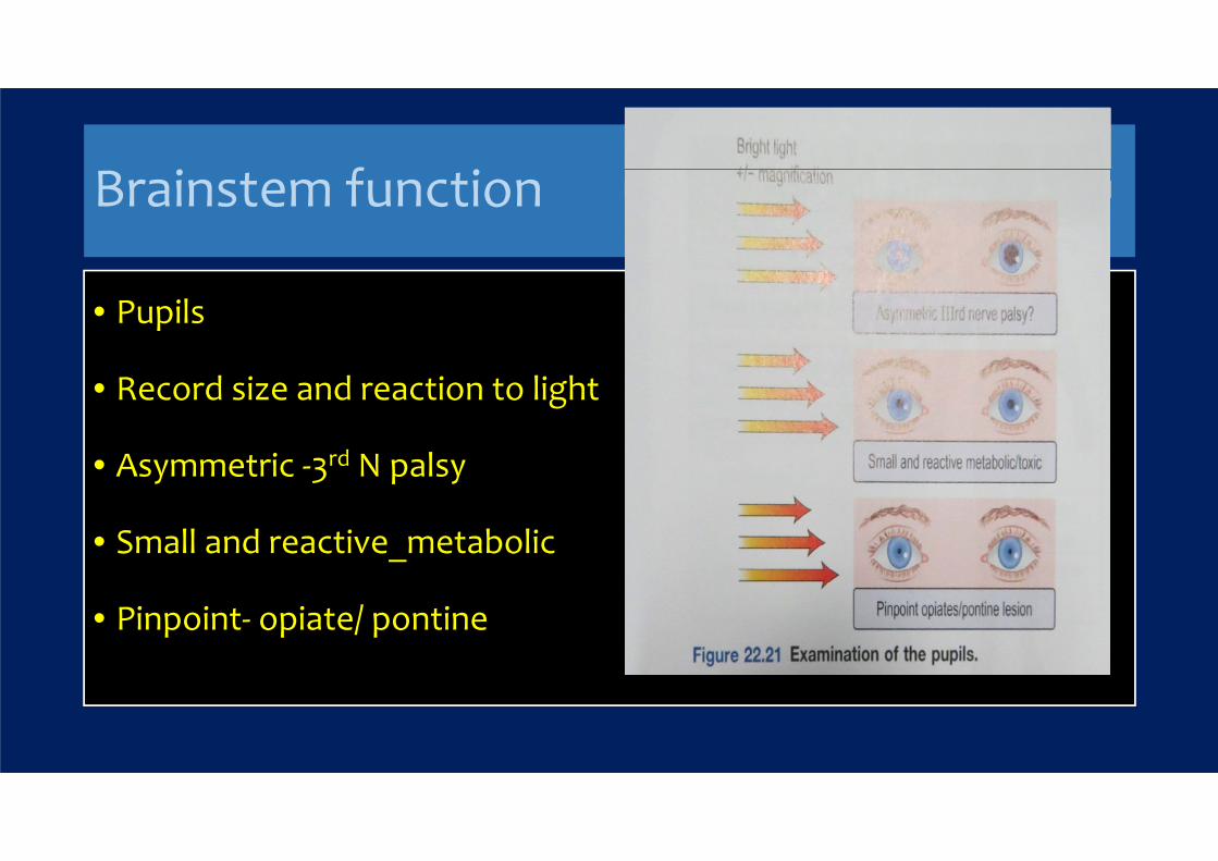
Brainstem function
• Pupils
• Record size and reaction to light
• Asymmetric -3rd N palsy
• Small and reactive_metabolic
• Pinpoint- opiate/ pontine

• 3rd nerve –potential neurosurgical emergency
• Bilateral mid point Reactive pupils – normal pupils – sedative
except opiate
• Bilateral light-fixed, dilated- cardinal sign of brain death, it can
occur in deep coma of any cause esp barbiturate intoxication,
hypothermia
• Bilateral pin point light-fixed pupils – pontine lesions(
haemorrhage) and opiate
• Mydriatic drugs and previous pupillary surgery can cause diagnostic
difficulty
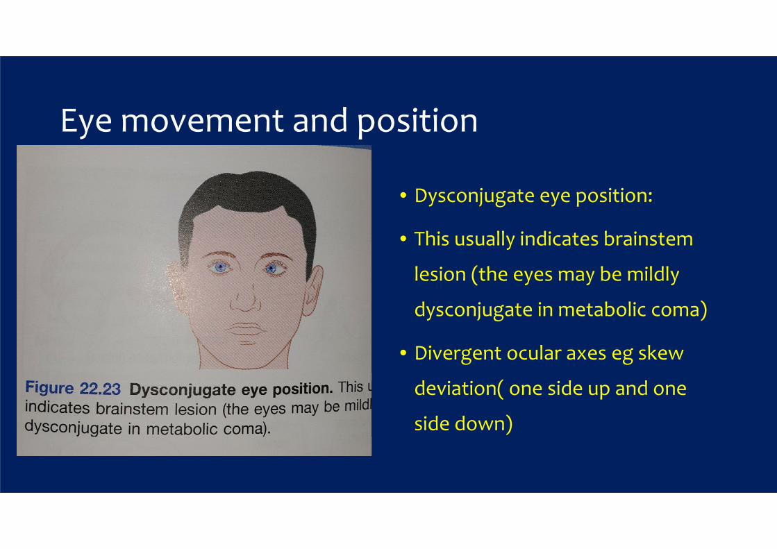
Eye movement and position
• Dysconjugate eye position:
• This usually indicates brainstem
lesion (the eyes may be mildly
dysconjugate in metabolic coma)
• Divergent ocular axes eg skew
deviation( one side up and one
side down)

Conjugate gaze deviation
• Towards the lesion in the
frontal lobe and the normal
limbs ( unopposed activity of
the intact frontal eye fields
drives eyes to the opposite
side)
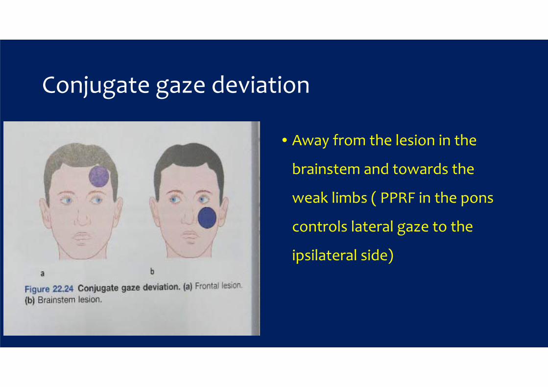
Conjugate gaze deviation
• Away from the lesion in the
brainstem and towards the
weak limbs ( PPRF in the pons
controls lateral gaze to the
ipsilateral side)

• Paramedium pontine reticular
formation (PPRF)

Impulses from PPRF pass via ipslateral VthCN to lateral rectus muscle( Abduction)
A lesion of th MLF (X) causes failure of or slow ADDuction in the right eye and nystagmus in the left eye with left lateral gazes

Doll’s eye movement
Passive head turning produces
conjugate ocular deviation away
from the direction of rotation
(doll’s head reflex)
Disappear in deep coma,in
brainstem lesions and brain
death

Windscreen wiper eyes(ping pong eyes) slow side to side movements demonstrate diffuse cortical dysfunctionCommon in light comaIt indicates extensive cortical damaged
Other brain stem reflexes
• Corneal reflex
• Gag/ cough reflex( via ET tube if incubated)
• Respiratory derive
Lateralizing sign
• Coma makes it difficult to recognize lateralizing sign
• Helpful points:
• Asymmetry of response to visual threat in a stuporose patient
suggest hemianopia
• Asymmetry of face drooping or dribbling on one side blowing in
and out of mouth when the paralysed cheek does not move

• Asymmetry of tone => unilateral flaccidity or spasticity may be the
only sign of hemiparesis
• Asymmetry of decerebrate and decorticate posturing
• Asymmetrical response to painful stimuli
• Asymmetry of tendon reflexes and plantar responses- both planter
are often extensor in deep coma
Coma look-alikes
• Psychogenic coma
• Loaked-in syndrome: complete paralysis except vertical eye
movements/blinking in verntral pontine infarction
• Patients have a functioning cerebral cortex and are fully aware but
unable to communicate except through eye movements
• Severe paralysis, eg myaesthenic crisis or severe Guillain-barre syndrome

Features distinguishing between coma and related states
Diagnosis and Investigation in coma
• Often the cause is evident- trauma, metabolic, overdose
• Lateralizing signs or brainstem pathologyMass lesion or nfarct/
haemorrhage ( careful : hypoglycemia may cause focal sign)
• No cause further Ix are essential

Blood and urine
• Drug screen
• Biochemistry ( U&E, glucose, calcium,LFT)
• Metabolic and endocrine- hypothyroid,corticol
• Arterial blood gases- acidosis, CO2 increase
• Other- cerebral malaria, porphyria
Brain imaging
• CT
• MRI useful if CT normal
• CT is quick and effective ( mass and haemorrhage) ( infarct may be
missed in early stages and where brainstem only affected)

CSF examination
• LP- only after careful assessment
• If mass suspect- CI (CT choice)
• Role_ meningoencephalitis, other infections and SAH( CT normal)

EEG
• Metabolic coma
• Encephalitis
• Non convulsive status epilepticus

General management
• Comatose patients need careful nursing, meticulous attention to
the airway, and frequent monitoring of vital functions.
• Longer-term essentials are-
• Skin care,eye care, fluids, feeding, sphinceters

Management of coma
• Skin care- turning to avoid pressure sores and pressure palsies
• Oral hygiene- mouthwashes, suction
• Eye care- prevention of corneal damage ( lid taping, irrigation)
• Fluids- nasogastric or IV
• Feeding- via a fine bore nasogastric tube or via peg
• Sphincters- catheterization when essential ( use penile urinary sheath if
possible in men) rectal evacuation
prognosis
• Depend on the cause of coma and extent of brain damage
sustained
• Metabolic and toxic – best Px
• Following hypoxic ischaemic brain injury only 11% make a good
recovery and following stroke the prognosis is worse still with only
7% recovering

• Of those patients who do not recover consciousness, a substantial
proportion will remain in a vegetative or minimally responsive
state.

Vegetative state
• Is usually a consequence of extensive cortical damage. Brainstem
function is intact so breathing is normal without the need for
mechanical ventilation and the patient appears awake with eye
opening and sleep-wake cycles.
However there is no sign of awareness or response to
environmental stimuli except reflex movements. Patients may
remain in this state for years.
Permanent (PVS)
• There is no recovery after 12 months where trauma is the cause and
after 6 months for all other causes
• Prolonged support of patients after this time presents a number of
ethical issues – families may apply to the courts for withdrawal of
feeding in PVS

Minimally conscious state (MCS)
• It describes patients with some limited awareness eg apparent,
vague pain perception. A patient may emerge from VS into MCS
• Distinguishing VS from MCS requires careful specialist assessment
over a long period. Functional brain imaging has recently been used
for this purpose.
Brain stem death
• Brain death means the irreversible loss of the capacity for consciousness
combined with the irreversible loss of the capacity to breathe.
• Both of these are essentially functions of the brainstem.
• Death if thought of in this way, can arise either from causes outside the brain(
respiratory and cardiac arrest ) or from causes within the cranial cavity.
• With the evident of mechanical ventilation it became possible to support such
a dead patient temporarily although in all cases cardiovascular failure
eventually supervenes and progresses to asystole

• Before deciding on a diagnosis of brainstem death, it is essential
that certain preconditions and exclusions are fulfilled
preconditions
• The patient must be in apneic coma ( ie unresponsive and on a
ventilator, with no spontaneous respiratory efforts)
• Irremediable structural brain damage due to a disorder that can
cause brainstem death must have been diagnosed with certainty
eg head injury, ICH

exclusion
• Possibility of apnoea due to sedatives or neuromuscular block
• Hypothermia
• Significant metabolic or endocrine disturbances
• Profound abn plasma E, AB abn, glucose

Confirmation test for BS death
• Oculo-cephalic reflexes should be absent. In a comatose patient
whose brainstem is intact the eyes will rotate relative to the orbit (
doll’s eye) in brainstem dead, the eyes remain stationary relative to
the orbit
• Fixed and unresponsive to bright light ( DLR, CLR)
• Corneal reflexes- absent

BS death (continued)
• No vestibule ocular reflexes on caloric testing
• No motor response within the cranial nerve territory to painful stimuli
applied centrally or peripherally
• Spinal reflex movements may be present
• No gag or cough reflex in response to Ph, Lar, or tracheal stimaulation
• No spontaneous respiration

• Thank you