an analysis of the disease risks, other than … · in this import risk analysis report ... peq...
TRANSCRIPT

Attachment B
AN ANALYSIS OF THE DISEASE RISKS, OTHER THANSCRAPIE, ASSOCIATED WITH THE IMPORTATION OF
OVINE AND CAPRINE SEMEN AND EMBRYOSFROM CANADA, THE UNITED STATES OF AMERICA AND
MEMBER STATES OF THE EUROPEAN UNION
FINAL REPORT
August 2000
Australian Quarantine and Inspection ServiceGPO Box 858
Canberra ACT 2601

2
AUSTRALIA

3
TABLE OF CONTENTS
EXECUTIVE SUMMARY 4
ABBREVIATIONS AND ACRONYMS 5
GLOSSARY 6
1. INTRODUCTION 8
1.1 Scope of risk analysis 8
1.2 Current quarantine policy and practice 8
2. HAZARD IDENTIFICATION 8
3. EXPOSURE PATHWAYS 9
4. RISK ASSESSMENT 15
4.1 Foot and mouth disease virus 16
4.2 Bluetongue virus 17
4.3 Capripoxvirus (sheep and goat pox virus ) 19
4.4 Mycobacterium paratuberculosis (Johne’s disease/paratuberculosis) 20
4.5 Brucella ovis 21
4.6 Brucella melitensis 22
4.7 Mycoplasma agalactiae and related mycoplasmas) (Contagious agalactia) 22
4.8 Maedi-visna virus 24
4.9 Caprine arthritis-encephalitis virus 25
4.10 Mycoplasma capricolum subsp. capripneumoniae (contagious caprine pleuropneumonia) 26
4.11 Chlamydia psittaci (enzootic abortion of ewes) 28
4.12 Jaagsiekte virus 29
4.13 Louping-ill and related viruses 30
4.14 Leptospira spp 31
5. RISK MANAGEMENT OPTIONS 33
5.1 Disease freedom of animals in country, zone or flock 33
5.2 Washing embryos 33
5.3 Testing and examination 34
5.4 Vaccination 34
5.5 Treatment 34
5.6 Pre-export quarantine (PEQ ) 34
5.7 Post-arrival quarantine (PAQ) 34
6 RISK MANAGEMENT FOR EACH PATHOGEN 35
6.1 Foot and mouth disease virus 35
6.2 Bluetongue virus 36
6.3 Capripoxvirus 37
6.4 Mycobacteria paratuberculosis 38
6.5 Brucella ovis 40
6.6 Brucella melitensis 41
6.7 Mycoplasma agalactiae (Contagious agalactia) 42
6.8 Maedi-visna virus 42
6.9 Caprine arthritis-encephalitis virus 45
6.10 Mycoplasma capricolum subsp. capripneumoniae (contagious caprine pleuropneumonia) 46
6.11 Chlamydia psittaci (enzootic abortion of ewes) 47
6.12 Jaagsiekte virus 48
APPENDIX 1 51
APPENDIX 2 52
REFERENCES 53

4
EXECUTIVE SUMMARYIn this import risk analysis report (IRA) AQIS has assessed the risk of importing disease agents(other than scrapie infective agent) with ovine and caprine genetic material from the United States ofAmerica (USA), Canada and the Member States of the European Union (EU). Australia currentlyprohibits the importation of ovine and caprine semen from all countries except New Zealand (NZ)and ovine and caprine embryos from all countries except NZ and the Republic of South Africa(RSA). Australia accepts that both NZ and RSA are free from scrapie. The risks of scrapie entrywere assessed separately and the final report released with this document. The final conditions forimportation of ovine and caprine genetic material from Canada, the USA and EU are theamalgamation of the risk management options identified by these two risk analyses.
The hazards identified in this import risk analysis (IRA) are ovine and caprine disease agents whichcould be introduced with ovine and caprine embryos and/or semen and adversely affect Australianlivestock industries, other animal based industries and/or native species.
The risks are qualitatively assessed. The assessment includes:• consideration of the epidemiological features affecting the likelihood of pathogens
infecting or contaminating ovine and caprine semen and embryos;• the likelihood of pathogens remaining after the semen is prepared and embryos washed;
and• the likelihood of infected or contaminated semen or embryos causing disease in
recipients or offspring and this disease then spreading to other susceptible hosts.
The following pathogens were identified as requiring risk management:• foot and mouth disease virus,• bluetongue virus,• capripoxvirus (sheep and goat poxvirus),• Mycobacterium (avium subsp) paratuberculosis,• Brucella melitensis,• Brucella ovis,• Mycoplasma capricolum subsp. capripneumoniae,• Mycoplasma agalactiae,• Chlamydia psittaci (enzootic abortion of ewes),• maedi-visna virus,• caprine arthritis encephalitis virus and• jaagsiekte virus.
Risk management options for the importation of genetic material were evaluated in terms of theirefficacy at reducing the assessed risk and also the level of restriction that their adoption would placeon trade. Initially, OIE recommendations were considered and are included in the final conditions ifconsidered suitable. Where it was considered that necessary risk management was not adequatelyaddressed by OIE recommendations alternative measures are required.
Quarantine measures include country, zone or flock freedom, testing of donor animals, treatment ofsemen and washing embryos.
Final conditions for importation are attached.

5
ABBREVIATIONS AND ACRONYMS
AGID agar gel immunodiffusion (test)AI artificial inseminationAPHIS Animal and Plant Health Inspection Service of the USDAAQIS Australian Quarantine and Inspection ServiceAQPM Animal Quarantine Policy MemorandumAUSVETPLAN Australian Veterinary Emergency PlanBTV bluetongue virusCA contagious agalactiaCAE caprine arthritis/encephalitisCCPP contagious caprine pleuropneumoniaCEE central European encephalitisCFT complement fixation testCPV capripox virusELISA enzyme-linked immunosorbent assayET embryo transferEU Member States of the European UnionFMD foot and mouth diseaseHAI haemagglutination inhibition (test)IETS International Embryo Transfer SocietyIRA import risk analysisLI louping illLIV louping ill virusLIRV louping ill and related virusesOJD ovine Johne’s diseaseJV jaagsiekte virusMV maedi-visnaOIE Office International des EpizootiesPAQ post-arrival quarantinePBMC peripheral blood mononuclear cellsPCR polymerase chain reactionPEQ pre-export quarantineRSSE Russian spring-summer encephalitisSSEV Spanish sheep encephalitis virusTBE tick-borne encephalitis (caused by members of the flavivirus group)TSEV Turkish sheep encephalitis virusUSA United States of AmericaUSDA United States Department of AgricultureVNT virus neutralisation testZP zona pellucida
6
GLOSSARYAccredited artificial insemination (AI) centreA facility for the collection, handling and storage of semen accredited by the VeterinaryAdministration and used exclusively for donor animals which meet the conditions set out in Code(Article 4.2.2.2.).
Centre veterinarianThe centre veterinarian directly supervises an accredited AI centre and is approved by theVeterinary Administration.
CodeOffice International des Epizooties (OIE) International Animal Health Code.
Consequence assessmentThe relationship between specified exposures to a biological agent and the consequences of thoseexposures. A causal process must exist by which exposures produce adverse health orenvironmental consequences, which may in turn lead to socio-economic consequences. Theconsequence assessment describes the potential consequences of a given exposure and estimates theprobability of them occurring.
EmbryoFor the purposes of this risk analysis the embryo is regarded as the conceptus from fertilised singlecell to unhatched blastocyst stages (zona pellucida intact). (Technically, the period of the embryo isthe stage during which the main tissues, organs and systems are formed).
Exposure assessmentThe biological pathway(s) necessary for exposure of animals and humans in the importing countryto the hazards (in this case the pathogenic agents) released from a given risk source, and estimatingthe probability of the exposure(s) occurring.
Genetic materialEmbryos, ova and/or semen.
Incubation periodThe longest period which elapses between the introduction of the pathogen into the animal and theoccurrence of the first clinical signs of the disease.
Intrauterine transmissionTransmission of infection from infected dam to embryo or foetus in the uterus.
IETS ManualManual of the International Embryo Transfer Society (IETS), 3rd Edition, April 1998. Edited byStringfellow DA and Seidel SM. Published by: IETS, 1111 North Dunlap Ave, Savoy, Illinois,61874 USA.
in vitroA process or procedure performed outside the body in a test tube or other laboratory apparatus.

7
in vivoA process occurring in a living organism or under natural circumstances.
ManualOIE Manual of Standards for Diagnostic Tests and Vaccines.
National control programA program that is nationally consistent and uses flock accreditation based on monitoring and testing.The program is usually supported by a government/industry compensation scheme.
Pre-collection periodThe pre-collection period is 30 days immediately prior to the first collection of semen or embryos.
Release assessmentThe biological pathway(s) necessary for an importation activity to “release” (that is, introduce)pathogenic agents into a particular environment, and estimating the probability of that completeprocess occurring, either qualitatively (in words) or quantitatively (as a numerical estimate). Therelease assessment describes the probability of the “release” of each of the potential hazards (thepathogenic agents) under each specified set of conditions with respect to amounts and timing, andhow these might change as a result of various actions, events or measures.
Risk estimationConsists of integrating the results from the release assessment, exposure assessment, andconsequence assessment to produce overall measures of risks associated with the hazards identifiedat the outset. Thus risk estimation takes into account the whole of the risk pathway from hazardidentified to unwanted outcome.
Stamping-out policyCarrying out under the authority of the Veterinary Administration, on confirmation of a disease, thekilling of the animals which are affected and those suspected of being affected in the herd and,where appropriate, those in other herds which have been exposed to infection by direct animal toanimal contact, or by indirect contact of a kind likely to cause the transmission of the causalpathogen. All susceptible animals, vaccinated or unvaccinated, on an infected premises should bekilled and their carcasses destroyed by burning or burial, or by any other method which will eliminatethe spread of infection through the carcasses or products of the animals killed.
Team veterinarianThe team veterinarian supervises the embryo collection team, and is responsible for all teamprocedures and should be specifically approved for this purpose by the Official Veterinarian.
Washing embryosThe washing of in-vivo derived embryos with intact zona pellucida as described in Chapter 6 of theIETS Manual where embryos are washed ten times to remove pathogens.
Veterinary Administration

8
The National Veterinary Service having authority in the whole country for implementing andsupervising or auditing the carrying out of the animal health measures and certification process whichthe Code recommends.
1. INTRODUCTION
1.1 Scope of risk analysisThis document analyses the risks, other than scrapie, associated with importing in vivo derived ovineand caprine embryos and ovine and caprine semen from the United States of America (USA),Canada and Member States of the European Union1 (EU) into Australia. The analysis is confined toa consideration of quarantine risks, ie. the probability of entry and establishment of exotic diseaseagents. A number of ovine and caprine diseases that occur in USA, Canada and EU do not occur inAustralia. Imported semen and embryos may transmit some of these disease agents to susceptiblerecipients or their offspring.
The IRA:• identifies the disease hazards other than scrapie which may be found in semen and in
vivo derived embryos and which have the potential to cause harm,• assesses the probability of transmission of these disease agents by semen and embryos
to other susceptible animals resulting in establishment and spread of disease,• assesses the probability of adverse consequences of establishment of these disease
agents,• identifies the risk management options for minimising the risks of introducing diseases
other than scrapie into Australia with ovine and caprine semen and embryos,• recommends risk management measures which could be applied to each disease agent
before importation, and• provides final quarantine conditions for importation.
1.2 Current quarantine policy and practiceCurrent quarantine policy relating to ovine and caprine embryos and semen is described in the IRAfor scrapie.
Australia permits the importation of in vivo derived ovine and caprine embryos from New Zealand(NZ) and the Republic of South Africa (RSA) and ovine and caprine semen from NZ. As the animalhealth status of the USA, Canada and the EU differs from NZ and RSA the development ofconditions for importation from these countries required an IRA.
2. HAZARD IDENTIFICATION
AQIS has used a process of categorisation to identify disease agents requiring further considerationin this IRA. A disease agent is identified as a hazard and the risk of entry via embryos or semen isassessed in this paper if it is :
• exotic to Australia,2 or present in Australia but subject to a National control programand
1 The Member States of the EU are Austria, Belgium, Denmark, Finland, France, Germany, Greece, Ireland, Italy,Luxembourg, the Netherlands, Portugal, Sweden, Spain and the UK.2 More virulent strains or serotypes may be reported overseas.

9
• the cause of significant disease and• potentially transmitted by ovine and/or caprine genetic material and• reported in at least one of the countries which are the subject of this IRA.
Disease agents that have been identified as requiring risk assessment are grouped according to theCode listing in Table 1. Disease agents associated with disease in sheep and goats and not identifiedas hazards for the purposes of this IRA are listed in appendix 1.
3. EXPOSURE PATHWAYSExposure pathways trace the potential route/s by which animals and humans in Australia may beexposed to pathogens harboured by imported genetic material. For an exotic disease agent tobecome established and spread in sheep or goats (or other susceptible species) in Australia throughthe importation of genetic material the pathogen must:
• be associated with ovine and/or caprine genetic material,• survive exposure to environmental stressors, eg. washing, exposure to diluents,
antibiotics, freezing, etc. and remain infectious after importation and transfer orinsemination,
• infect the recipient of the genetic material or offspring, and• spread from the index case(s) to a sufficient number of susceptible hosts to become
established.
Association of the disease agent with imported genetic material depends on several factors whichmay differ for embryos and semen. Infection of embryos depends on the susceptibility of the sheepor goat to the disease agent and whether the tissue tropism includes the ovaries and other parts of thegenital tract. Transmission by embryo transfer (ET) may also arise if the disease agent is acontaminant of the embryo storage medium or is present on contaminated personnel, instruments orequipment.
Few studies have been made of the interaction between embryos and pathogens in small ruminants incomparison with those conducted on bovine embryos. As a consequence, few disease agentsaffecting sheep and goats have been categorised by the International Embryo Transfer SocietyImport/Export Committee (IETS) Research subcommittee for their capacity to be transmitted viaET. The role of the zona pellucida (ZP) in preventing penetration of infectious agents into the embryoand also as a medium for the carriage of pathogens is discussed in the draft risk analysis for bovinesemen and embryos from Argentina and Brazil (AQPM 1999/34). However, characteristics ofembryos and their interactions with pathogens cannot be generalised. Embryos of different speciesdiffer in the glycoprotein composition of the ZP. This structure in sheep and goats differs from that incattle (Chen and Wrathall 1989; Dunbar et al 1991). It has been suggested that ovine ZP is‘stickier’ than that of bovine embryos, and less likely to resist penetration and adherence ofpathogens (Singh et al 1997). This may explain the higher probability of binding between the ZP andvarious pathogens in these species. In the absence of relevant information, infection patterns forsheep and goat embryos and semen are based on studies of infection of bovine genetic material.Nevertheless, the uncertainty of this extrapolation is acknowledged.
Semen is a complex association of cells and fluids from several organs. Even though some diseaseagents have been reported in the semen of rams, most are found in the seminal fluid or leucocytesrather than within or attached to the spermatozoon. Some pathogens, eg. certain RNA viruses such

10
as bluetongue virus (BTV), have been implicated in gamete infections (Eaglesome et al 1980), but itis generally accepted that this mode of infection is not significant. Increased white cell numbers dueto accompanying infection can increase the likelihood of the presence of some pathogens in semen(de la Concha-Bernejillo et al 1996). Similarly blood may be occasionally present depending on thecollection technique used or if concurrent infections cause damage to capillaries resulting in infectedblood cells in semen, eg. BTV.
Without the imposition of risk management measures, the practice of collection, handling, transportand transfer of embryos or semen to recipients represents a very direct exposure pathway for anydisease agents present in imported material. In Australia sheep and goats are usually kept inextensively managed flocks providing an opportunity for the direct spread of disease from infectedanimals to other stock or other animals in the surrounding environment.
11
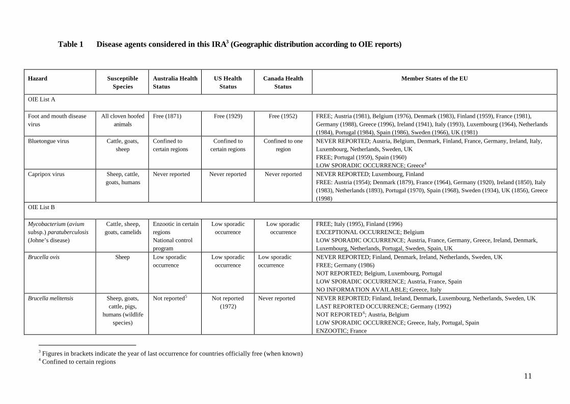
Table 1 Disease agents considered in this IRA3 (Geographic distribution according to OIE reports)
Hazard SusceptibleSpecies
Australia HealthStatus
US HealthStatus
Canada HealthStatus
Member States of the EU
OIE List A
Foot and mouth diseasevirus
All cloven hoofedanimals
Free (1871) Free (1929) Free (1952) FREE; Austria (1981), Belgium (1976), Denmark (1983), Finland (1959), France (1981),Germany (1988), Greece (1996), Ireland (1941), Italy (1993), Luxembourg (1964), Netherlands(1984), Portugal (1984), Spain (1986), Sweden (1966), UK (1981)
Bluetongue virus Cattle, goats,sheep
Confined tocertain regions
Confined tocertain regions
Confined to oneregion
NEVER REPORTED; Austria, Belgium, Denmark, Finland, France, Germany, Ireland, Italy,Luxembourg, Netherlands, Sweden, UKFREE; Portugal (1959), Spain (1960)LOW SPORADIC OCCURRENCE; Greece4
Capripox virus Sheep, cattle,goats, humans
Never reported Never reported Never reported NEVER REPORTED; Luxembourg, FinlandFREE: Austria (1954); Denmark (1879), France (1964), Germany (1920), Ireland (1850), Italy(1983), Netherlands (1893), Portugal (1970), Spain (1968), Sweden (1934), UK (1856), Greece(1998)
OIE List B
Mycobacterium (aviumsubsp.) paratuberculosis(Johne’s disease)
Cattle, sheep,goats, camelids
Enzootic in certainregionsNational controlprogram
Low sporadicoccurrence
Low sporadicoccurrence
FREE; Italy (1995), Finland (1996)EXCEPTIONAL OCCURRENCE; BelgiumLOW SPORADIC OCCURRENCE; Austria, France, Germany, Greece, Ireland, Denmark,Luxembourg, Netherlands, Portugal, Sweden, Spain, UK
Brucella ovis Sheep Low sporadicoccurrence
Low sporadicoccurrence
Low sporadicoccurrence
NEVER REPORTED; Finland, Denmark, Ireland, Netherlands, Sweden, UKFREE; Germany (1986)NOT REPORTED; Belgium, Luxembourg, PortugalLOW SPORADIC OCCURRENCE; Austria, France, SpainNO INFORMATION AVAILABLE; Greece, Italy
Brucella melitensis Sheep, goats,cattle, pigs,
humans (wildlifespecies)
Not reported5 Not reported(1972)
Never reported NEVER REPORTED; Finland, Ireland, Denmark, Luxembourg, Netherlands, Sweden, UKLAST REPORTED OCCURRENCE; Germany (1992)NOT REPORTED6; Austria, BelgiumLOW SPORADIC OCCURRENCE; Greece, Italy, Portugal, SpainENZOOTIC; France
3 Figures in brackets indicate the year of last occurrence for countries officially free (when known)4 Confined to certain regions

12
Hazard SusceptibleSpecies
Australia HealthStatus
US HealthStatus
Canada HealthStatus
Member States of the EU
Mycoplasma capricolumsubsp. capripneumoniae(Contagious caprinepleuropneumonia)
Goats Never reported Never reported Never reported LAST REPORTED OCCURRENCE; Sweden (1983)NEVER REPORTED; Belgium, Finland, France, Germany, UK Ireland, Denmark, Luxembourg,NetherlandsNOT REPORTED; Austria, Italy, Portugal, SpainLOW SPORADIC OCCURRENCE; Greece
Maedi-visna virus Sheep Never reported Never reported7 Low sporadicoccurrence
LAST REPORTED OCCURRENCE; Ireland (1986)NOT REPORTED; Austria, Italy8
EXCEPTIONAL OCCURRENCE; FinlandLOW SPORADIC OCCURRENCE; Belgium, Germany, Greece, Denmark, Luxembourg,Netherlands, Portugal, Spain, Sweden, UK
Caprinearthritis/encephalitis virus
Goats Low sporadicoccurrence9
Low sporadicoccurrence
Low sporadicoccurrence
NEVER REPORTED; Luxembourg, Finland, Netherlands, Portugal, Spain10
NOT REPORTED; Austria, BelgiumSUSPECTED BUT NOT CONFIRMED; Denmark, Ireland,LOW SPORADIC OCCURRENCE; Germany, Sweden, UK, Greece, ItalyENZOOTIC; France
Mycoplasma agalactiae(Contagious agalactia)
Sheep, goat Not reported11 Never reported12 Never reported NEVER REPORTED; Finland, Germany, Ireland, Luxembourg, Netherlands, SwedenNOT REPORTED; Belgium, Denmark, UKEXCEPTIONAL OCCURRENCE; SpainLOW SPORADIC OCCURRENCE; France, Greece, Italy, Portugal,SUSPECTED BUT NOT CONFIRMED; Austria
Leptospira spp. All mammals(includinghumans)
Present 13 Present Present PRESENT; Sweden, Austria, Italy, Spain, France, Germany, Greece, Ireland, Netherlands,Portugal, UKLAST REPORTED OCCURRENCE; Finland (1997)NOT REPORTED; Denmark, Luxembourg, Belgium
5 Brucella melitensis is reported sporadically in humans in Australia. The disease is described as an uncommon but serious infection, affecting people who have aquired the infectionoverseas (Chan and Hardiman 1993). This is probably also true of the USA, Canada and Member States of the EU.6 Not reported in this table means that the disease is probably present in the country but has not been reported to the OIE or described in the scientific literature7 There are numerous reports of MVV in USA as ovine progressive pneumonia (OPP), Montana sheep disease or ovine lentivirus infection (Brodie et al 1994) and Snowder et al (1990)writes that it is well established that OPP is wide-spread in sheep in North America.8 Infection appears to be widespread in France and Italy (Lujan et al 1993).9 CAEV is included as a hazard in this IRA even though the virus is present in Australia. There is evidence that strains of CAE which can cause disease in sheep are not present inAustralia but are apparently present overseas (Smith et al 1985).10 Contreras et al (1998) report CAE in goats in Spain11 Mycoplasma agalactiae has been isolated in Australia, but the Australian strains do not produce contagious agalactia in sheep.12 M agalactiae capable of causing mastitis in goats is now considered to be present in the USA (Kinde et al 1994)

13
13 Pathogenic leptospire serovars present in sheep or goats in the EU, Canada and the USA may not be present in Australia.
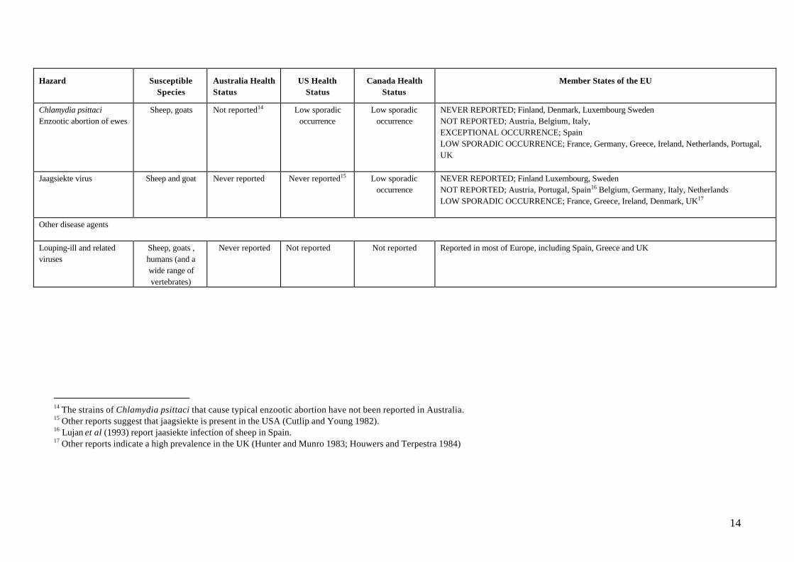
14
Hazard SusceptibleSpecies
Australia HealthStatus
US HealthStatus
Canada HealthStatus
Member States of the EU
Chlamydia psittaciEnzootic abortion of ewes
Sheep, goats Not reported14 Low sporadicoccurrence
Low sporadicoccurrence
NEVER REPORTED; Finland, Denmark, Luxembourg SwedenNOT REPORTED; Austria, Belgium, Italy,EXCEPTIONAL OCCURRENCE; SpainLOW SPORADIC OCCURRENCE; France, Germany, Greece, Ireland, Netherlands, Portugal,UK
Jaagsiekte virus Sheep and goat Never reported Never reported15 Low sporadicoccurrence
NEVER REPORTED; Finland Luxembourg, SwedenNOT REPORTED; Austria, Portugal, Spain16 Belgium, Germany, Italy, NetherlandsLOW SPORADIC OCCURRENCE; France, Greece, Ireland, Denmark, UK17
Other disease agents
Louping-ill and relatedviruses
Sheep, goats ,humans (and awide range ofvertebrates)
Never reported Not reported Not reported Reported in most of Europe, including Spain, Greece and UK
14 The strains of Chlamydia psittaci that cause typical enzootic abortion have not been reported in Australia.15 Other reports suggest that jaagsiekte is present in the USA (Cutlip and Young 1982).16 Lujan et al (1993) report jaasiekte infection of sheep in Spain.17 Other reports indicate a high prevalence in the UK (Hunter and Munro 1983; Houwers and Terpestra 1984)
15
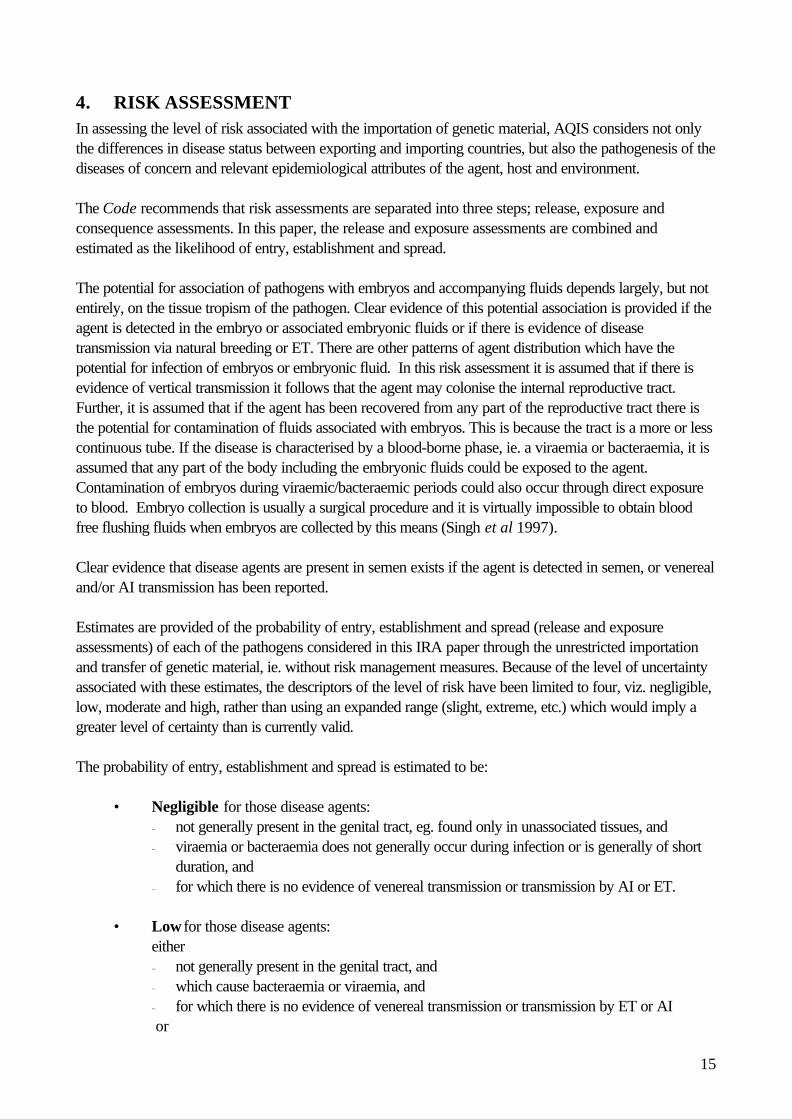
4. RISK ASSESSMENTIn assessing the level of risk associated with the importation of genetic material, AQIS considers not onlythe differences in disease status between exporting and importing countries, but also the pathogenesis of thediseases of concern and relevant epidemiological attributes of the agent, host and environment.
The Code recommends that risk assessments are separated into three steps; release, exposure andconsequence assessments. In this paper, the release and exposure assessments are combined andestimated as the likelihood of entry, establishment and spread.
The potential for association of pathogens with embryos and accompanying fluids depends largely, but notentirely, on the tissue tropism of the pathogen. Clear evidence of this potential association is provided if theagent is detected in the embryo or associated embryonic fluids or if there is evidence of diseasetransmission via natural breeding or ET. There are other patterns of agent distribution which have thepotential for infection of embryos or embryonic fluid. In this risk assessment it is assumed that if there isevidence of vertical transmission it follows that the agent may colonise the internal reproductive tract.Further, it is assumed that if the agent has been recovered from any part of the reproductive tract there isthe potential for contamination of fluids associated with embryos. This is because the tract is a more or lesscontinuous tube. If the disease is characterised by a blood-borne phase, ie. a viraemia or bacteraemia, it isassumed that any part of the body including the embryonic fluids could be exposed to the agent.Contamination of embryos during viraemic/bacteraemic periods could also occur through direct exposureto blood. Embryo collection is usually a surgical procedure and it is virtually impossible to obtain bloodfree flushing fluids when embryos are collected by this means (Singh et al 1997).
Clear evidence that disease agents are present in semen exists if the agent is detected in semen, or venerealand/or AI transmission has been reported.
Estimates are provided of the probability of entry, establishment and spread (release and exposureassessments) of each of the pathogens considered in this IRA paper through the unrestricted importationand transfer of genetic material, ie. without risk management measures. Because of the level of uncertaintyassociated with these estimates, the descriptors of the level of risk have been limited to four, viz. negligible,low, moderate and high, rather than using an expanded range (slight, extreme, etc.) which would imply agreater level of certainty than is currently valid.
The probability of entry, establishment and spread is estimated to be:
• Negligible for those disease agents:− not generally present in the genital tract, eg. found only in unassociated tissues, and− viraemia or bacteraemia does not generally occur during infection or is generally of short
duration, and− for which there is no evidence of venereal transmission or transmission by AI or ET.
• Low for those disease agents:either− not generally present in the genital tract, and− which cause bacteraemia or viraemia, and− for which there is no evidence of venereal transmission or transmission by ET or AI or

16
− which are transmitted by vector.
• Moderate for those disease agents :either− not generally associated with the genital tract, and− which have been isolated from embryos/semen, and− for which there is no evidence of venereal transmission, and− which spread readily via direct contact.or− generally present in the genital tract, and− for which there is no evidence of transmission by ET or AI, and− which spread readily via direct contact.
• High for those disease agents− for which there is evidence of transmission by ET or AI, and− which spread readily via direct contact.
The assessment of the consequences of disease establishment in Australia combines an economic andenvironmental assessment. These separate assessments are combined according to the following matrix.
HIGH low high high highMODERATE low moderate moderate highLOW low low moderate highNEGLIGIBLE negligible low low low
NEGLIGIBLE LOW MODERATE HIGH
Probability ofadverseenvironmentalconsequences
Probability of adverse economic consequences
A risk estimate (estimate of the integrated risk) for each disease agent was obtained by combining the riskratings for the probability of agent entry, establishment and spread with the probability of adverseconsequences (release assessment, exposure assessment and consequence assessment). This is anestimated probability that a disease agent establishes in susceptible animals in this country and results insignificant harm in human, economic and environmental terms. The integrated risk estimate for each diseaseagent is derived from the following matrix:
NEGLIGIBLE low negligible negligible negligibleLOW moderate low low negligible
MODERATE high moderate low negligibleHIGH high high moderate low
HIGH MODERATE LOW NEGLIGIBLE
Probability ofagent, entry,establishmentand spread
Probability of adverse consequences
In this risk analysis, risk management measures are considered unnecessary for disease agents with anegligible rating.
4.1 Foot and mouth disease virusSheep are highly susceptible to infection with foot and mouth disease virus (FMDV). Both sheep and goatshave been considered to play an important role in the epidemiology of foot and mouth disease in someEuropean and Turkish outbreaks (Pay 1988). In sheep and goats the disease is usually mild or inapparent

17
but expression depends on the strain of virus, the breed of the animal and environmental conditions.Infected animals usually remain asymptomatic while excreting foot and mouth disease virus (FMDV) andmay carry the virus for up to over 7 months (Sharma 1978). The incubation period after natural exposure isfrom 2 to 8 days (Mann and Sellers 1990).
Evidence that FMDV infects ovine and caprine embryos and semenThe presence of FMDV has been reported in bovine genetic materials but not in that of sheep or goats.Even so, FMDV is widely distributed in the tissues of infected animals, including vaginal tissues (Srinivasand Rajasekhar 1992) and blood of infected sheep (Sharma and Murty 1981). FMDV has beenrecovered from embryo collection fluid taken from experimentally infected donor cows.FMDV can spread via semen (Mann and Sellers 1990) but transmission of infection via semen has notbeen reported in sheep or goats. Infected bovine semen has been shown to transmit virus to other animalswhich suggests that this route of transmission may be possible in sheep and goats.
Probability of FMDV entry, establishment and spreadFMDV is not typically associated with the genital tract but has been detected in semen and can survivefreezing (Bane 1981). Sheep and goats excrete FMDV for long periods even when asymptomatic (Sharmaand Murty 1981). In addition, interspecies transmission has occurred during several outbreaks. In theSikkim outbreak goats transferred the disease to cattle and in the UK, FMD spread rapidly from sheep tocattle (Srinivas and Rajasekhar 1992). FMD is very contagious, and infected sheep and goats are likely tobe involved in the spread to other cloven hooved animals. Consequently, the probability that geneticmaterial collected from infected sheep or goats results in the entry, establishment and spread is estimated tobe moderate for embryos and semen.
Probability of adverse consequences arising through FMDV entry, establishment and spreadRuminants and pigs are susceptible to FMDV. Losses would result due to deaths, morbidity, cost oferadication and/or control measures and the effect on trade. The social and economic effects of the spreadof FMD in this country are detailed in the AUSVETPLAN (1996). The probability of adverse economicconsequences is estimated to be high. The effect of FMDV on native Australian animals is not fully knownbut birds, reptiles and amphibians are not susceptible. Snowdon (1968) failed to infect various Australianmammals including 3 species of kangaroo, a wombat, a wallaby, a potoroo, a bandicoot, a possum, anechidna, a dasyurid and a water rat. Consequently the probability of adverse environmental consequencesis estimated to be low. The overall estimate for probability of adverse consequences is high.
Risk estimate for FMDVThe risk estimate for FMDV without risk management measures is moderate for embryos and high forsemen.
4.2 Bluetongue virusBTV is transmitted by a range of Culicoides species. Serotypes 4 and 10 have caused epidemics in partsof Europe and serotypes 2,10,11,13 and 17 are endemic in North America (Gibbs and Greiner 1994).These serotypes are exotic and not amongst the eight serotypes of BTV which infect livestock in parts ofAustralia. The virulence of BTV serotypes varies considerably and Australian serotypes are considered tobe less virulent than some overseas serotypes (Geering et al 1995).
Although BTV may infect many species of ruminant, sheep are usually the most severely affected. Viraemiain sheep and goats commences from 3 days post infection and may last up to 54 days (Koumbati et al1999). The Code recognises an infective period for BT of 60 days. Sheep breeds from temperate

18
countries are more susceptible to BT than tropical breeds and the disease is now considered to begenerally a disease of temperate areas. For example, in temperate areas such as California BT outbreaksoften occur annually causing significant economic impact (Gibbs and Greiner 1994).
Evidence that BTV infects ovine and caprine embryos and semenReproductive disorders including early embryonic deaths, abortions, malformed foetal lambs, transientinfertility in rams, and shedding of virus in semen have been described as BT related (Osburn 1994). BTVmay be transmitted transplacentally but this occurs infrequently (Mellor and Boorman 1995). Thetransmission of BTV with incompletely washed embryos from infected to susceptible sheep has beendescribed (Gilbert et al 1987). Ovine embryos exposed in vitro to BTV remained infected after 10washes, however the integrity of embryos and duration of exposure to BTV during this study was unclear(Singh et al 1997).
BTV is shed in the semen of infected rams (Hare et al 1988). However, Hare et al (1988) found thatewes bred to these rams did not become infected.
Probability of BTV entry, establishment and spreadBTV has been transmitted by infected ovine embryos. The virus is shed in ovine semen but there is no clearevidence that infection is transmitted by naturally infected ovine semen. Whilst studies in cattle suggest thatthis form of transmission can occur transmission of BTV by ovine genetic material is considered infrequent(Mellor and Boorman 1999) and unimportant in the epidemiology of the disease (Geering et al 1995).Consequently, the probability that BTV entry could occur through infection of recipients of infectedembryos or semen is estimated to be low.
The influence of vectors on the viruses they transmit is poorly understood and Tabachnick et al (1991)suggest that vector/serotype specificity has not been demonstrated. However, Australian studies haveshown that the capacity of Culicoides species feeding on sheep to transmit BTV (vector competence)varies with serotype (Standfast et al 1985). Also, when exotic serotypes are introduced into differentecosystems they appear to die out if efficient vectors are absent (Gibbs and Greiner 1994). The vectorcompetence of Australian Culicoides species for exotic strains of BTV is unknown, but based on overseasexperience, it is estimated that even if recipients or progeny became infected via imported embryos orsemen, the probability of spread to other ruminants is unlikely. Consequently, the probability of BTV entry,establishment and spread in the Australian zone of possible BT transmission is estimated to be low in theabsence of risk management measures.
Probability of adverse consequences arising through BTV entry, establishment and spreadAll domestic ruminants are susceptible to BTV but only sheep and deer show clinical symptoms. A detailedassessment of the social and economic effects of the spread of exotic serotypes of BTV is presented inAUSVETPLAN (1996). The probability of adverse economic consequences is estimated to be moderate.The effect of BTV on native Australian animals is unknown but there are no reports of BT in mammalsother than ruminants, birds, reptiles or amphibians. Consequently the probability of adverse environmentalconsequences is estimated to be low. The overall estimate for probability of adverse consequences ismoderate.
Risk estimate for BTVThe risk estimate for BTV without risk management measures is low for embryos and semen.

19
4.3 Capripoxvirus (sheep and goat pox virus )Apart from strain differences, expressed as host preferences for either sheep or goats, pox virusesrecovered from infected sheep and goats are considered to be indistinguishable (Kitching and Taylor 1985;Munz and Dumbell 1994) and are referred to by the Code as capripoxvirus (CPV).
Capripox is endemic in most of Africa, the Middle East and Asia. The disease was present in Greece until1996. In 1998 Greece reported to the OIE that the disease was eradicated by a stamping out/nonvaccination policy and previously affected areas were screened serologically.
Mortality rates may reach 10% in endemic areas and 100% in imported animals. Morbidity rates can reach90% in sheep, but less in goats (Code; Munz and Dumbell 1994). Incubation periods range from 2 dayswith experimental infection (Merza and Mushi 1990) to 3 weeks in natural infections but 12 days isaccepted as the average (Geering et al 1995; Code). Susceptibility to disease is breed dependent in sheepand infected resistant breeds show few signs of disease. The disease lasts from 3 to 4 weeks but does notbecome chronic (Geering et al 1995).
Evidence that CPV infects ovine and caprine embryos and semenSpecific information is not available on which to base an evaluation of the potential for transmission ofcapripoxviruses by ovine and caprine embryos. Viraemia occurs and may result in exposure of embryos toinfection. Intrauterine transmission can occur in the course of generalised cowpox infection (Mayr andCzerny 1990).
No information is available on the transmission of capripoxvirus in semen. Orchitis is reported in goats(Merza and Mushi 1990) and in both sheep and goats pox lesions may occur on the skin and mucousmembrane surfaces, including the prepuce. These symptoms may result in viral contamination of semenbefore or during collection from infected animals. Transmission via semen is considered likely(AUSVETPLAN 1996).
Probability of CPV entry, establishment and spreadIn the absence of information to the contrary, and extrapolating from transmission of other poxviruses, it isassumed that there is a risk that infected genetic material may be collected from infected animals. Capripoxis a contagious disease and rapid spread can occur within large flocks (Kitching and Taylor 1985) and viafomites and insect vectors (Merza and Mushi 1990). Because of the high mortality rate in susceptiblepopulations, ease of diagnosis and lack of a carrier state, it is highly likely that CPV would be quicklyeradicated if introduced into Australia. Based on this limited information, it is assumed that the probabilityof entry, establishment and spread is low.
Probability of adverse consequences arising through CPV entry, establishment and spreadSheep and goats are susceptible to CPV and Merinos and British breeds are very susceptible to disease(AUSVETPLAN 1996). Losses would result from death of sheep, morbidity, the cost of eradicationand/or control measures and some effect on trade, especially the potential loss of wool markets(AUSVETPLAN 1996). Based on this the probability of adverse economic consequences is estimated tobe high. The effect of CPV on native Australian animals is unknown but there are no reports of CP in othermammals, birds, reptiles, amphibians, marsupials or monotremes. Consequently the probability of adverseenvironmental consequences is estimated to be low. The overall estimate for probability of adverseconsequences is moderate.

20
Risk estimate for CPVThe risk estimate for CPV without risk management measures is low for embryos and semen.
4.4 Mycobacterium paratuberculosis (Johne’s disease/paratuberculosis)Isolates of M paratuberculosis (M avium subsp. paratuberculosis) appear to demonstrate hostpreferences rather than host specificities (Gunnarson and Fodstad 1979; Huchzermeyer et al 1994). Sheepstrains usually only infect sheep, causing ovine Johne’s disease (OJD) and characteristically can beextremely difficult to culture (Whittington et al 1999). Nevertheless, different strains can infect a range ofspecies. Molecular and epidemiological studies have shown that genetically similar isolates can berecovered from sheep, goats and cattle (Collins et al 1990; Feizabadi et al 1997). Such studies suggestbut do not prove that inter-species transmission occurs.
Ovine JD is a chronic disease and infected animals are usually asymptomatic. OJD has a prolongedincubation period, usually 3 to 4 years and animals remain infected for life (Huchzermeyer et al 1994).Sheep shed large numbers of bacteria in their faeces, especially in the terminal stages of disease,contaminating pastures. Transmission to susceptible sheep occurs by contact with contaminated pasture(Whittington et al 1999).
OJD is endemic in the eastern states of Australia and now subject to a nationally coordinated controlprogram supported by State legislation.
Evidence that M paratuberculosis infects ovine and caprine embryos and semenIn the terminal stage of infection M paratuberculosis is distributed widely throughout the body(Huchzermeyer et al 1994) and embryos could be exposed to infection. Infection can be acquired inutero if the disease in the ewe is advanced (Gilmore and Angus 1991). It is unlikely that collected embryoswould be infected as collections would only be made from animals which did not show clinical signs.
M paratuberculosis can be isolated from bull semen but there is no specific evidence of Mparatuberculosis shedding in sheep or goat semen or of transmission by semen.
Probability of M paratuberculosis entry, establishment and spreadM paratuberculosis is not typically associated with the genital tract, but intrauterine transmission has beenreported. However, the collection of embryos from infected donors showing clinical signs is unlikely.Consequently, the probability of transmission of M paratuberculosis to recipients via genetic material isestimated to be negligible for embryos and low for semen.
Probability of adverse consequences arising through M paratuberculosis entry, establishmentand spreadSheep and goats are susceptible to M paratuberculosis. Losses would result from ill-thrift, cost oferadication and/or control measures. In Australia M paratuberculosis has been detected in NSW,Victoria, Kangaroo Island (SA) and Flinders Island (Tasmania) but has not been reported fromQueensland, the Northern Territory or Western Australia. The disease is notifiable and subject tocompulsory government controls, including quarantine and movement restrictions.
The probability of adverse economic consequences in M paratuberculosis free regions in Australia isestimated to be moderate. M paratuberculosis was recently cultured from the intestinal tissues of twoTammar wallabies from Kangaroo Island (Primary Industries and Resources South Australia) but thesusceptibility of other native Australian animals is unknown and there are no reports of M paratuberculosis

21
in other Australian mammals, birds, reptiles or amphibians. Consequently the probability of adverseenvironmental consequences is estimated to be low. The overall estimate for probability of adverseconsequences is moderate.
Risk estimate for M paratuberculosisThe risk estimate for M paratuberculosis without risk management measures is negligible for embryos andlow for semen.
4.5 Brucella ovisDisease caused by B ovis affects sheep in Australia and is notifiable. The incubation period is typicallyprolonged, ranging from 50 to 250 days (van Tonder et al 1996).
Male goats have been infected experimentally (Burgess et al 1985) but disease in goats is unusual. B ovisis not considered a hazard of caprine genetic material in this risk assessment.
Evidence that B ovis infects ovine embryos and semenEwes are relatively resistant to infection with B ovis and infections are transient and generally short-lived.Bacteraemia occurs but rarely results in abortion. When abortion does occur bacteria may be present inuterine tissues and discharges (van Tonder et al 1996). Recent experimental and field studies found that theuterus was one of the main sites of infection (Grillo et al 1999; Marco et al 1994) and that persistentinfection did occur. Even so, latent infection and infection of progeny was unusual (Grillo et al 1999).
It is not known whether embryos may become infected naturally but in vitro exposed embryos transmittedinfection to recipient ewes (Riddell et al 1990).
B. ovis is shed in semen of infected rams for up to four years (van Tonder et al 1996) and inseminatedinfected semen may cause infection in recipients.
Probability of B ovis entry, establishment and spreadThe probability that embryos infected with B ovis will infect recipients is estimated to be low. B ovis maybe present in semen collected from asymptomatic infected donors and transmit infection. In the event thatanimals become infected through transfer of imported genetic material they can be detected and culled thuspreventing establishment and spread. Consequently, the probability that B ovis gains entry, establishes andspreads is estimated to be low for embryos and high for semen.
Probability of adverse consequences arising through B ovis entry, establishment and spreadB ovis is endemic in many sheep flocks in Australia and causes losses in sheep flocks mainly through poorreproductive performance in rams and their subsequent culling. Losses would also be incurred if eradicationand/or control measures were adopted. The introduction of B ovis via embryos or semen into regionswhere B ovis free flocks are present, for example stud sheep raising areas, would have a localised impact.However, the impact of introduction into most other areas where the disease is endemic would be minimal.Overall it is estimated that the probability of adverse economic consequences would be negligible. B ovisinfects mice under laboratory conditions (Jimenez et al 1994). There are no reports of B ovis infection innative Australian animals, even though the agent is endemic in many regions and the probability of adverseenvironmental consequences is estimated to be negligible. The overall estimate for probability of adverseconsequences is negligible.

22
Risk estimate for B ovisThe risk estimate for B ovis in sheep without risk management measures is negligible for embryos and lowfor semen.
4.6 Brucella melitensisThe usual mode of transmission of Brucella melitensis infection is through direct contact with the placenta,foetal fluids or vaginal discharges expelled by infected ewes after abortion or full-term parturition (Garin-Bastuji et al 1998). Young animals may recover from infection with Brucella melitensis but adults seldomrecover, becoming inapparent carriers. Flocks may remain infected for years with a high prevalence ofinfection even in the absence of obvious disease (Herr 1994).
All caprine breeds appear to be equally susceptible, but susceptibility to B melitensis infection variesbetween breeds of sheep (Herr 1994).
Evidence that B melitensis infects ovine and caprine embryos and semenB melitensis can localise in uterine tissue of sheep (Grillo et al 1997) and cause abortion in goats andsheep. Birth or abortion may be followed by a copious purulent discharge from the genital tract of infectedanimals for up to 3 months (Herr 1994). Transmission of infection from sheep to lambs occurs. Infectioncan be acquired in utero but the majority of transmission is thought to occur via colostrum or milk (Grilloet al 1997).
B melitensis is commonly shed in semen (Garin-Bastuji et al 1998) but there is no specific evidence of Bmelitensis transmission via ovine or caprine semen.
Probability of B melitensis entry, establishment and spreadThere is some evidence to suggest that B melitensis may be associated with embryos or semen collectedfrom asymptomatic infected donors. Consequently, the probability that B melitensis will gain entry,establish and spread if present in imported genetic material is estimated to be moderate.
Probability of adverse consequences arising through B melitensis entry, establishment andspreadLosses arising from the spread of B melitensis in sheep and goats result from ‘abortion storms’, ill thrift,the cost of control and eradication measures. Brucellosis is an important zoonosis. Dogs, cattle and rodentsare also susceptible to infection even though less important hosts in epidemiological terms. Consequently,the cost of control and eradication would extend beyond measures applicable to sheep and goat propertiesand the probability of adverse (socio-)economic consequences due to B melitensis spread is estimated tobe high. The susceptibility of native Australian animals is unknown. B melitensis, in common with otherBrucella species, has a fairly broad host range and has the potential to cause disease in these species. Theprobability of adverse environmental consequences is estimated to be high. The overall estimate forprobability of adverse consequences is high.
Risk estimate for B melitensisThe risk estimate for B melitensis without risk management measures is high for embryos and semen.
4.7 Mycoplasma agalactiae and related mycoplasmas (Contagious agalactia)The principal causal agent of contagious agalactia (CA) in sheep is Mycoplasma agalactiae. In goats Magalactiae, M capricolum subsp. capricolum (Mcc), M putrefasciens, and M mycoides subsp.mycoides LC (MmmLC) produce a similar clinical picture and some authors consider these mycoplasmas,

23
separately or in combination, as causal agents (Bergonier et al 1997). In a recent report of CA in Spain Magalactiae was isolated from 79% of outbreaks and accounted for 82.7% of all isolates (Gil et al 1999).M capricolum, M putrefaciens and MmmLC have been isolated from goats in Australia (Cottew andYeats 1982). For the purposes of this risk analysis only M agalactiae is considered further as the cause ofCA.
M agalactiae has been detected in the external ear canal of goats in Australia (Cottew and Yeats 1982),but the Australian strains do not appear to produce contagious agalactia in goats or sheep. Avirulent strainshave also been reported overseas (Bergonier et al 1997).Goats are more commonly affected than sheep and disease in goats is usually more acute. Long termchronic infections occur in both sheep and goats (Bergonier et al 1997). Bacteraemia occurs commonlyand M agalactiae can be isolated from the blood of infected sheep (Ak et al 1995). Clinical signs of CAwere evident 20 days after experimental infection of sheep with M agalactiae and antibody titres were firstdetected from 28 to 35 days after infection (Buonavoglia et al 1999)
Evidence that M agalactiae infects ovine and caprine embryos and semenInfection involves the genital organs of the female and in goats may cause granular vulvo-vaginitis(Bergonier et al 1997; Singh et al 1974). Abortion occurs infrequently. Vertical transmission has beensuspected following isolation of M agalactiae from swollen joints of neonate kids and lambs (Bergonier etal 1997).
M agalactiae was isolated from the semen of experimentally infected sheep (Ak et al 1995).
Probability of M agalactiae entry, establishment and spreadGenital tract involvement, abortion, semen infection and suspected vertical transmission have been reportedin association with M agalactiae infection. The disease spreads quickly once established in a flock (Tola etal 1996) and sheep and goats may harbour infection asymptomatically. Consequently the probability thatentry establishment and spread of M agalactiae would occur via genetic material from infected sheep orgoats is estimated to be moderate for embryos and semen.
Probability of adverse consequences arising through M agalactiae entry, establishment andspreadCA is considered to be one of the most serious diseases affecting small ruminants (Gil et al 1999)characteristically causing localised outbreaks. It is a major obstacle to sheep and goat production in bothtraditional and intensive forms of stock management (Bergonier et al 1997). For example, Tola et al(1996) claim that CA has caused major economic loss since introduction into Sardinia in 1980. Spread ofthe causative agent in sheep and goat flocks in Australia would cause losses due to deaths, loss ofproduction and the cost of control and eradication measures. The probability of adverse economicconsequences due to CA is estimated to be moderate. The susceptibility of native Australian animals isunknown. There are no reports of CA in mammals other than ruminants. Consequently the probability ofadverse environmental consequences is estimated to be low. The overall estimate for probability of adverseconsequences is moderate.
Risk estimate for M agalactiaeThe risk estimate for M agalactiae without risk management measures is moderate for embryos andsemen.

24
4.8 Maedi-visna virusThe ovine lentiviruses, maedi-visna virus (MVV) and South African ovine maedi-visna virus (SA-MVV)can infect sheep causing maedi-visna disease (Banks et al 1983). This disease is also called ovineprogressive pneumonia, Montana sheep disease, zwoegersiekte, la bouhite, lungers, Marsh’s progressivepneumonia and Graaff-Reinet disease. The ovine lentiviruses are closely related to, but genetically andserologically distinct from, caprine arthritis encephalitis virus (CAEV) (Verwoerd and Tustin 1994; Pasick1998).
MV has been reported from the US (as ovine progressive pneumonia), Canada and most of the countriesin the EU. The disease has never been recorded in sheep or goats in Australia and there is no evidence tosuggest that the disease is present.
MVV infections are characterised by a long and variable incubation period and life-long viral persistence(Cutlip et al 1988). The antibody response confers no resistance to disease and the clinical course ofdisease is generally progressive (Carey and Dalziel 1993; Verwoerd and Tustin 1994). Natural infectionfollowing introduction of MV infected sheep into susceptible flock can occur after 11 months (Houwers etal 1987) but clinical signs are rarely seen in sheep less than 2 years old (Constable et al 1996).
Differences in breed susceptibility to MVV have been reported (Houwers et al 1989). Icelandic breedsappear to be more susceptible than British breeds and Texels and Border Leicester are more susceptible todisease than Columbia sheep (Cutlip et al 1986; Joag et al 1996). Also, Snowder et al (1990)determined significant differences in the seroprevalence of MV between the 6 breed types comprising aflock of 2,976 sheep. Nevertheless, complete breed-associated resistance has not been demonstrated(Houwers 1990). Houwers et al (1989) suggest that apparent susceptibility may also depend on the strainof MVV.
MVV infection often co-exists with jaagsiekte and concurrent infection can lead to increasedseroprevalence of MV and increased lateral transmission of MVV. Carey and Dalziel (1993) suggest thatthis may be due to increased alveolar macrophages in the lungs of sheep affected by jaagsiekte providingextra sites for MVV replication.
Evidence that MVV infect embryos and semenThe evidence for transplacental transmission of MVV is equivocal. Preventing colostral transfer and earlycontact with infected dams has been regarded as an effective means of obtaining MV free progeny (DeBoer et al 1979; Cutlip et al 1988; Sihvonen 1980). Long-term absence of MVV infection wasdemonstrated in a group of approximately 40 lambs separated from infected ewes immediately after birthand reared in isolation (De Boer et al 1979). Similar results were reported by Light et al (1979) andHouwers et al (1987). Other studies suggest that the potential for transplacental infection cannot be entirelydismissed. Cutlip et al (1981) reported prenatal transmission based on the detection of MVV from 1foetus and 2 newborn lambs out of 70 progeny. Cross et al (1975) reported infection in a small proportionof hysterectomy derived lambs from infected dams. More recently, Brodie et al (1994) detected MVVDNA in the peripheral blood mononuclear cells (PBMC) of 11% of lambs removed from their infecteddams immediately after birth.
Of perhaps greater relevance to an assessment of the potential for infection of ova or embryos with MVVis that viraemia develops shortly after infection and plays a major role in distribution of monocyteassociated virus throughout the body (Georgsson 1990). This might provide an opportunity for exposure ofembryos to virus. Using PCR techniques, Woodall et al (1993) failed to detect MVV in either uterine

25
washes or washed embryos collected from 10 infected ewes. This suggests that exposure of embryos toMVV during infection does not occur.
Ovine lentivirus was detected in the semen of rams concurrently infected with Brucella ovis (de la Concha-Bermejillo et al 1996). These authors suggest that inflammatory lesions of the genital tract causingleucocytospermia, as caused by B ovis, predispose infected rams to shed ovine lentivirus in their semen.Moreover semen may contain blood or plasma and MVV capsid antigen has been detected in plasma ofinfected sheep (Brodie et al 1994). The target cells for MVV replication are mononuclear cells andtransmission of virus occurs via these cells (Joag et al 1996). Nevertheless these studies do not provideclear evidence that the MVV components detected are infectious or that infection is transmitted to recipientewes or offspring via infected semen.
Probability of MVV entry, establishment and spreadBecause of the long incubation period, persistent infection, lack of overt clinical signs and genital tractinvolvement (in males), MVV has the potential to infect genetic material collected from infected donors.Susceptible species are present in this country and it is estimated that the probability of MVV introductionand spread in this country is low for embryos and moderate for semen.
Probability of adverse consequences arising through MVV entry, establishment and spreadMVV infection causes ill-thrift, chronic respiratory disease and indurative mastitis in adult sheep andreduced growth rates in lambs. In flocks with high prevalence the disease is economically significant andmeasures have been adopted overseas to eradicate MVV. Spread of MVV in sheep in this country wouldcause losses due to deaths, ‘ill thrift’ and the cost of control and eradication measures. Snowder et al(1990) studied reproductive performance, ewe weight and wool production in 6 breed types in the USAand concluded that subclinical infection did not appear to have an adverse economic effect. Even so, theprobability of adverse economic consequences due to MVV is estimated to be moderate. Thesusceptibility of native Australian animals is unknown and there are no reports of MVV in animals otherthan small ruminants. Consequently the probability of adverse environmental consequences is estimated tobe low. The overall estimate for probability of adverse consequences is moderate.
Risk estimate for MVVThe risk estimate for MVV without risk management measures is low for embryos and moderate forsemen.
4.9 Caprine arthritis-encephalitis virusCaprine arthritis encephalitis (CAE) is also called chronic arthritis-synovitis, big-knee, viralleukoencephalomyelitis, progressive interstitial pneumonia and caprine retrovirus disease. Goats infectedwith CAE virus are usually asymptomatic. The disease is usually expressed in adults as a chronic arthritis,and occasionally as progressive interstitial pneumonia or chronic mastitis. The disease is enzootic in goats inAustralia (Greenwood et al 1995) but voluntary control programs are in place in some parts of thecountry.
CAEV and MVV are generally regarded as closely related but separate viruses. Strong evidence thatAustralasian strains of CAEV do not transmit from goats to sheep is provided by separate studies inAustralia and New Zealand. Smith et al (1985) failed to demonstrate the development of any significantpathological lesions in Merino lambs exposed to a Western Australian strain of CAEV by inoculation andthrough close contact with infected goats. 1394 sheep in New Zealand exposed to goat flocks with amoderate to high seroprevalence of CAE tested negative for CAEV infection (McKenzie 1991). However

26
some other studies suggest that overseas strains of CAEV may infect sheep and some authors refer broadlyto small ruminant lentiviruses, regarding MV viruses and CAE virus as separate viral ‘quasispecies’ (Pasick1998). Experimental infections of sheep with CAEV have been reported (Banks et al 1983; Castro et al1999) and disease signs typical of CAE have been observed in sheep (Oliver 1981). Based onphylogenetic studies of nucleic acid sequences of overseas isolates of CAEV and MVV, Zanoni (1998)suggests that there are clear indications for cross-species transmission and advises that eradicationprograms should be aware of this risk. Consequently, the potential for strains of CAEV not present in thiscountry to infect sheep cannot be entirely dismissed.
CAE is a progressively debilitating disease with life-long viral persistence (Verwoerd and Tustin 1994).The arthritic form of CAE predominates and is usually expressed in adult goats when they are 2 to 9 yearsold (Narayan and Cork 1990).
Evidence that CAEV infects embryos and semenColostrum and milk from infected goats usually contain infected cells (Narayan and Cork 1990) anderadication efforts are usually based on colostrum deprivation (Nord et al 1998). However, there is noevidence of infection of the female reproductive tract or transplacental transmission.
CAEV has been detected by PCR in seminal fluid and non-spermatic cells in semen from experimentallyinfected bucks (Travassos et al 1998).
Probability of CAEV entry, establishment and spreadCAEV has been detected in caprine semen. A monocyte associated viraemia is typical of CAE and couldexpose ova or embryos to virus during infection. However, this appears unlikely as in one experiment,CAEV was not isolated from uterine flushings from seropositive does and transferred embryos did notseroconvert recipients or progeny (Wolfe et al 1987). Susceptible species are present in this country andcould be exposed to introduced exotic strains of the virus. The probability of CAEV introduction andspread in this country is estimated to be low for embryos and moderate for semen.
Probability of adverse consequences arising through CAEV entry, establishment and spreadCAEV infection causes neurological problems in young goats and ill-thrift and chronic lameness in adultgoats. The disease is considered to be economically significant such that control measures have beenadopted overseas and in Australia. Introduction and spread of strains of CAEV which cause disease insheep could cause losses due to deaths, ‘ill thrift’ and the cost of control and eradication measures. Theprobability of adverse economic consequences due to the introduction of overseas strains of CAEV isestimated to be moderate. Except for goats and sheep, there is no other known host for CAEV (Narayanand Cork 1990). Consequently the probability of adverse environmental consequences is estimated to below. The overall estimate for probability of adverse consequences is moderate.
Risk estimate for CAEVThe risk estimate for CAEV without risk management measures is low for embryos and moderate forsemen.
4.10 Mycoplasma capricolum subsp. capripneumoniae (contagious caprinepleuropneumonia)The causative agent of contagious caprine pleuropneumonia (CCPP), Mycoplasma capricolum subsp.capripneumoniae (Mccp) was formerly known as the F38 biotype (Leach et al 1993). The diagnosis ofCCPP is complicated as disease resembling CCPP in goats may be caused by M mycoides subsp. capri
27
(Mmc) and caprine variants of M mycoides subsp. mycoides (MmmLC). Mccp, Mmm and Mmc crossreact in serological tests and are grouped together with bovine serogroup 7 and M capricolum as the “Mmycoides cluster’ (Nicolet 1994; Taylor et al 1992). MmmLC, M capricolum and Mmc have beenrecorded in Australia (Beveridge 1983; Taylor et al 1992).
CCPP is transmitted by the respiratory route and spreads rapidly within a herd by close direct contact(Geering et al 1995; MacOwan and Minette 1977). The incubation period for the disease, ranges from 3to 9 days after experimental infection and 12 to 35 days after natural contact (MacOwan and Minette1977). A much longer incubation period, from 24 to 50 days, was reported in animals previously exposedto high passage culture of Mccp (MacOwan and Minette 1978). Chronic infections can occur andapparently healthy carriers can excrete mycoplasmas (Geering et al 1995; Nicolet 1994). Anotherexperimental study showed that goats recovered from acute CCPP may have lesions for a long timethereafter but provided no evidence of a carrier state among long-term survivors (Wesonga et al 1998).Mccp has been isolated from asymptomatic sheep held in affected goats herds but sheep are considered tobe resistant to disease (Harbi et al 1983; Mare 1994). However, Bolske et al (1995) reported theisolation of Mccp from sheep with typical signs of CCPP.
Evidence that Mccp infects caprine embryos and semenMccp localises in the respiratory tract and does not cause septicaemia or pathology in the genital tract.There is no evidence to suggest that embryos are exposed to Mccp in naturally infected animals.
Untyped mycoplasmas have been recovered from semen and preputial swabs from bucks (Kapoor et al1983). There is no other specific information which can be used to estimate the potential for transmission ofMccp via caprine semen.
Probability of Mccp entry, establishment and spreadMccp is typically a respiratory disease so the probability of infection of embryos is estimated to be low.Based on very limited information it is estimated that the likelihood Mccp is present in semen from infectedanimals is low. CCPP spreads rapidly and efficiently among susceptible animals by direct contact. Shouldinfected genetic material cause infection in recipients or progeny the likelihood that the disease wouldspread is high. In summary, the probability that Mccp will gain entry, establish and spread is estimated tobe low for semen and embryos.
Probability of adverse consequences arising through Mccp entry, establishment and spreadCCPP is a disease of major economic importance, posing a significant restraint to goat production due tohigh mortalities and rapid spread. Large scale treatment has proven ineffective in eradicating the disease inendemic areas (Bolske et al 1995). Feral goats (Capra hircus) occur in all states and territories ofAustralia except the Northern Territory. The main populations are in western New South Wales, the semi-arid pastoral areas of Western Australia and southern Queensland. Spread of Mccp in goats in this countrywould cause losses due to deaths, ‘ill thrift’, the cost of control and eradication measures. The economicvalue of the goat industry is small in comparison with other livestock industries. Therefore, the probabilityof adverse economic consequences due to Mccp on a national level is estimated to be low. Thesusceptibility of native Australian animals is unknown but there are no reports of Mccp in mammals otherthan goats. Consequently the probability of adverse environmental consequences is estimated to be low.The overall estimate for probability of adverse consequences is low.
Risk estimate for MccpThe risk estimate for Mccp without risk management measures is low for embryos and semen.

28
4.11 Chlamydia psittaci (enzootic abortion of ewes)Chlamydia psittaci isolates from ruminants have been split into 2 groups based on their virulence in mice(Salinas et al 1995). Strains virulent for mice form a well defined group and have been designated asserotype 1. Ovine abortion strains causing enzootic abortion of ewes (EAE) are placed within this serotype(Kaltenboeck et al 1997; Boumedine and Rodolakis 1998). The taxonomy of the family Chlamydiaceaehas been recently reviewed and Everett et al (1999) propose that ovine abortion strains are included in thenewly constructed species Chlamydophila abortus. Goats as well as sheep can be affected by ovineabortion strains of C psittaci (Salinas et al 1995; Liao et al 1997; Donn et al 1997).
No evidence of chlamydial infection of aborting ewes or dead lambs was found in a large scale study ofperinatal mortality and ovine abortion in NSW (Plant et al 1972). The recovery of isolates indistinguishablefrom abortifacient C psittaci in sheep flocks free of abortion in the UK and in Greece suggests theexistence of avirulent forms of ovine C psittaci (Jones 1997). Naturally occurring sporadic abortionepisodes due to chlamydial infections have been recorded in sheep and goats in Australia (Brown et al1988; Seaman 1985). Even so, the strains of C psittaci that cause typical enzootic abortion have not beenreported in Australia.
EAE appears to have a minimum incubation period of 56 days (Williams et al 1998). There is no evidenceof breed predisposition to EAE.
Evidence that C psittaci infects embryos and semenC psittaci is a naturally occurring reproductive infection in sheep that persists following primary infection. Cpsittaci antigen or DNA was detected in the vagina, uterus and oviduct of experimentally infected sheep(Papp and Shewen 1996). C psittaci infection involves the female genital tract and neonatally-infected ewelambs may develop placental infection during their first pregnancy and some may abort (Aitkin 1991). Cpsittaci has also been isolated from abortion outbreaks in goats (Liao et al 1997; Vretou et al 1996),cattle (Griffiths et al 1995) and humans. The disease in sheep is characterised by a long-term persistentphase (Entrican et al 1999).
Chronic infection of the male reproductive tract and seminal excretion can occur in ruminants (Storz et al1976). C psittaci has been isolated from semen of young rams born to experimentally infected ewes(Appleyard et al 1985). Venereal transmission under experimental conditions has been reported. In oneexperimental study, nine out of ten ewes inseminated with semen to which C psittaci was addedseroconverted and the organism was recovered from 3 of these. Fourteen ewes served by four ramsintravenously infected with C psittaci seroconverted but the agent was not recovered at any stage fromserved ewes. These authors concluded that venereal transmission of C psittaci is biologically feasible insheep (Appleyard et al 1985) but is not a major route of spread.
Probability of C psittaci entry, establishment and spreadC psittaci infection involves the female genital tract and vertical transmission of infection from ewes tolambs can occur. EAE spreads easily from infected aborting ewes to unaffected ewes under intensivelambing conditions. Transmission by ET has not been reported. The probability of entry, establishment andspread of C psittaci via infected embryos is rated as moderate.
There is no evidence to suggest that rams or bucks play a significant role in the transmission of EAE(Appleyard et al 1985) or that transmission via AI has occurred. However seminal excretion and venerealtransmission in ruminants have been reported and the probability of entry, establishment and spread of Cpsittaci via infected semen is rated as moderate.
29
Probability of adverse consequences arising through C psittaci entry, establishment and spreadEAE is a major cause of economic loss to sheep farmers in the UK and several other European countries,especially when intensive sheep management is practiced (Aitken 1991). Other ruminant species aresusceptible to chlamydial abortion (Aitkin 1991) but the taxonomic relationship of causative chlamydia inother ruminants to that causing EAE is uncertain. All breeds of sheep would be expected to be susceptibleto C psittaci but expression of disease is dependent on the intensity of management at lambing. In Australiamost lambing takes place in paddocks and pregnant ewes are not confined together. Therefore theeconomic effect of EAE spread in Australia is estimated to be low. Under natural conditions chlamydialspecies are considered to have a relatively limited host range and relatively limited disease range (Everettand Anderson 1999). Nevertheless, the ovine abortion strain of C psittaci has apparently caused abortionin humans and cattle. C psittaci strains infect Australian birds and cause disease in koalas (Girjes et al1993). However, the relationship between these strains and that causing EAE is unclear. It is concludedthat the susceptibility of native Australian animals to EAE is unknown and the probability of adverseenvironmental consequences estimated to be low. The overall estimate for probability of adverseconsequences is low.
Risk estimate for C psittaciThe risk estimate for ovine abortion strains of C psittaci without risk management measures is low forembryos and semen.
4.12 Jaagsiekte virusA type D retrovirus, called jaagsiekte sheep retrovirus (JV) is the aetiological agent of a contagious lungtumour of sheep. The disease is known as jaagsiekte, ovine pulmonary adenomatosis, ovine pulmonarycarcinoma, sheep pulmonary adenocarcinoma and sheep pulmonary adenomatosis. There is still somecontroversy about the pathological classification of the lung tumour (Verwoerd 1996) and the originaldescriptive name jaagsiekte is often used. The disease has been experimentally reproduced in lambs byinfection with a jaagsiekte virus (JV) cloned construct (Palmarini et al 1999). Endogenous and exogenousforms of the virus exist with differing nucleotide sequences. Three distinct groups have been identified;
i) exogenous JV sequence from the UK,ii) exogenous JV sequences from Southern Europe andiii) the exogenous South African strain plus all the endogenous sequences analysed and collected
from sheep in Australia, Italy, UK and South Africa (Rosati et al 2000). The endogenousretroviral sequences are present within the ovine genome and distinct from JV (Bai et al1999).
The prevalence of jaagsiekte in sheep can be high in some countries, for example, UK (Scotland) (Hunterand Munro 1983) and the Netherlands (Houwers and Terpesta 1984). Even though the disease has beenexperimentally transmitted from sheep to goats the disease is uncommon in goats and the risk oftransmission is regarded as low (Sharp et al 1986).
The influence of genetics on resistance to jaagsiekte is poorly understood. Some breeds are apparentlymore susceptible to disease than others, eg. English breeds, Texel, Merinos and Awassi (Parker et al1998; Hod et al 1972; Hunter and Munro 1983). The presence of endogenous viruses in sheep genomesmay influence the susceptibility of sheep to the disease (Hecht et al 1996).
The incubation period following experimental infection ranges from 3 weeks in lambs to several years inolder animals. Several studies determined the peak incidence of jaagsiekte in sheep to be 3 to 4 years
30
(Hunter and Munro 1983; Parker et al 1998). The average duration of clinical signs is 2 months, with arange of a few days to 6 months (Moulton 1990).
Evidence that JV infects ovine and caprine embryos and semenJV has not been detected in embryos or uterine fluids. However, during the later stages of disease retroviraltranscripts are widely distributed in the tissues of affected animals, including peripheral blood mononuclearcells (PBMC). Based on these findings, Parker et al (1998) suggested that exposure of embryos, ova andsemen to virus was possible.
The distribution of retroviral transcripts could include genital tract tissues and semen could be infected. Theonly study conducted to investigate this found that the transfer of infection from one diseased ram toprogeny and recipients did not occur (Parker et al 1998). JSV has been detected in lymphoid tissues,including PBMC and dissemination of virus by these cells is thought to precede tumour formation (Hollandet al 1999). Any concurrent disease process in a JV infected ram which increases the mononuclear contentof semen may lead to the production of JV infected semen.
Probability of JV entry, establishment and spreadBecause of the long period before overt clinical signs are detectable, JV may be present in genetic materialcollected from asymptomatic infected donors. It is estimated that the probability of JV introduction andspread via embryos is low and via semen moderate.
Probability of adverse consequences arising through JV entry, establishment and spreadJaagsiekte causes significant problems in sheep in countries in which it is enzootic. It is naturally highlytransmissible among sheep causing respiratory disease followed by fatality in over 95% of cases. Becauseof the long preclinical incubation period and contagious nature of jaagsiekte as well as the short life span ofmost sheep under modern management systems, JV may enter and disseminate widely in a flock before thedisease is recognised. Economic losses can arise from mortalities, the cost of control and eradicationmeasures. The probability of adverse economic consequences due to jaagsiekte is estimated to be high.The susceptibility of native Australian animals is unknown and there are no reports of jaagsiekte inmammals other than small ruminants. Consequently the probability of adverse environmental consequencesis estimated to be low. The overall estimate for probability of adverse consequences is moderate.
Risk estimate for JVThe risk estimate for JV without risk management measures is low for embryos and moderate for semen.
4.13 Louping-ill and related virusesViruses of the tick-borne encephalitis (TBE) complex within the flavivirus genus occur across Europe toNorth-East Asia and can cause disease in humans (Kaiser et al 1997) and significant losses in smallruminant production (Davies 1997). Closely related viruses within this group cause louping ill (LI), Russianspring-summer encephalitis (RSSE) and central European encephalitis (CEE) in some European countries(Monath and Heinz 1996). Hubalek et al (1995) propose that these viruses represent one virus speciesnamed Eurasian TBE virus (Flavivirus ixodes) and group LI and CEE together as a Western subtype.Relationships between LI and other members of the TBE complex, including Turkish sheep encephalitis(TSE) and Spanish sheep encephalitis (SSE), are unclear. For the purposes of this risk assessment thelouping ill virus (LIV) and louping-ill like viruses which affect sheep and goats (TSEV and SSEV) areregarded as forming a separate sub-group or complex of genetically distinct but closely related sheepencephalitis viruses. This grouping is largely based on clinical signs, serology and host range (Marin et al1995; Hubalek et al 1995) and referred to here as louping ill and related viruses (LIRV). These viruses are

31
reported from the UK, Spain, Bulgaria, Turkey, Norway and Greece (Marin et al 1995; Gao et al 1993,1997; Papadopoulos et al 1971).
LI is predominantly a disease of sheep but can affect goats. The incubation period following experimentalinfection, taken as the onset of viraemia, varies from one to eleven days in sheep (Swanepoel 1994; Reidand Doherty 1971) and from one to six days in goats (Reid et al 1984). Following the primary viraemicphase, LIV replicates in both neural and extraneural tissue but is strongly neurotropic and can sequester innervous tissue (Monath and Heinz 1996).
TBE viruses are naturally transmitted by ticks within genera Ixodes, Dermacentor and Haemaphysalis.Geering et al (1995) suggest that the viruses are unlikely to become established in this country unlesssuitable vectors are first introduced. However, Swanepoel (1994) suggests that louping ill virus, in commonwith other tick-borne viruses, may be capable of transmission by a wide range of ticks.
Evidence that LIRV infects ovine and caprine embryos and semenA TBE complex arbovirus was isolated from a flock of goats with abortions (Papadopoulos et al 1971).However, there is no evidence that LIV can infect ruminant embryos or embryonic fluids.
No information is available on the presence of LIV in ovine or caprine semen.
Probability of LIRV entry, establishment and spreadLIV causes a viraemia and has the potential to infect genetic material but the likelihood that infected geneticmaterial may cause disease in recipients or offspring is unknown. The likelihood that disease may spreadfrom index cases depends on the capacity of biological vectors in this country to transmit LIRV. There isvery little evidence of the movement of strains of LIV from different areas of UK despite substantial tradeof livestock. Other viruses within the TBE complex tend to remain geographically restricted despitemovement of humans and animals. This suggests that the geographical distribution of LIRVs is determinedby the distribution of ticks, that the viruses are vector specific and that spread of viruses from specificlocalities is unusual. Overall, the probability that LIRV establish and spread via infected genetic material isestimated to be negligible.
Probability of adverse consequences arising through LIV entry, establishment and spreadLIV is locally important in some areas of Europe but is not generally regarded as a significant diseaseaffecting sheep. However, LIV and probably the other LIRVs pose a risk of human infection. It does nottransmit without intervention of a competent vector and the capacity of arthropods in Australia to fill thisrole is uncertain. Consequently the probability of adverse (socio-) economic consequences due to LIV isestimated to be low. The susceptibility of native Australian animals is unknown and LIV has a wide hostrange (Davies 1997). However the uncertainty of transmission is also relevant here. Consequently theprobability of adverse environmental consequences is estimated to be low. The overall estimate forprobability of adverse consequences is low.
Risk estimate for LIRVThe risk estimate for LIRV without risk management measures is negligible for semen and embryos.
4.14 Leptospira sppLeptospirosis is a zoonotic disease with a worldwide distribution caused by pathogenic species ofLeptospira.

32
Leptospirosis has been reported in sheep and/or goats in many European countries and the US. In Portugal3.3% of sheep tested gave positive results. The predominant serogroups involved were Canicola,Pomona, Cynopteri, Sejroe and Icterohaemorrhagiae. From the 1,631 goats examined serologically, 5.0%gave positive results, mainly to serogroups Grippotyphosa, Canicola, Pomona, Icterohaemorrhagiae andPyrogenes (Rocha 1998). Infection of sheep has been reported in Italy (L hardjobovis and Linterrogans sv hardjo) (Farina et al 1996; Cerri et al 1996), the Netherlands (L interrogans sv hardjoand L hardjo bovis) (Gerritsen et al 1995), UK (Sejroe and hebdomadis serogroups, sv bratislava andautumnalis) (Little et al 1987; Hathaway et al 1983), Spain (sv pomona, sejroe, icterohaemorrhagiae andgrippotyphosa) (Leon-Vizcaino et al 1987) and France (Gaumont 1966).
In one study in the US, leptospiral antibody titres were present in 27 of 326 ovine foetal sera tested.However, leptospires were not detected in any of the 326 ovine foetuses tested by FAT (Kirkbride andJohnson 1989). L interrogans sv hardjo and pomona have been reported in sheep in the US (Schmitz et al1981; Davidson and Hirsh 1980).
Sporadic cases of leptospirosis in sheep in Australia are caused by L borgpetersenii sv hardjo andmembers of the hebdomadis serogroup ( McCaughan et al 1980). Serosurveys have indicated thatserovars tarassovi, pomona, australis, autumnalis, copenhageni, grippotyphosa and hardjoprajitno (Linterrogans sv hardjo) may be present in sheep (Gordon 1980).
Infection with pathogenic leptospires may cause a range of clinical signs, from asymptomatic to acute andfatal. Chronic infections can occur and affected animals may remain carriers for long periods with bacterialocalising in the kidneys or reproductive tract.
Evidence that leptospires infect ovine and caprine embryos and semenLeptospires could not be recovered from the reproductive tracts of ewes and rams experimentally infectedwith L interrogans hardjo (Farina et al 1996). In addition, neither venereal transmission nor recoveryfrom semen has been demonstrated in sheep and goats (Anon 1981).
A very small percentage of abortions in sheep and goats were attributed to infection with leptospires inSpain and the UK (Leon-Vizcaino et al 1987; Ellis et al 1983). One more recent, large study in the USfailed to detect leptospires in 326 aborted ovine foetuses in spite of serological evidence of infection(Kirkbride and Johnson 1989). There is no published information on leptospire transmission in ruminantsby ET.
Probability of leptospire entry, establishment and spreadLeptospira can be detected in bovine semen (Heinemann et al 1999). However, transmission via AI inruminants has not been recorded and Philpott (1993) cites evidence that freeze-thawing destroysleptospires in semen. Palit et al (1986) found that glycerol which is used as a cryoprotectant in ruminantsemen was toxic to the majority of Leptospira interrogans serovars tested. This indicates that theprobability leptospires will be present in semen and survive normal processing is negligible. Similarly,normal processing of embryos should preclude contamination with leptospires. Overall, the probability thatexotic pathogenic serovars of Leptospira establish and spread via infected genetic material is estimated tobe negligible.
Probability of adverse consequences arising through leptospire entry, establishment and spreadOf the serovars reported in sheep and/or goats overseas, several have not been reported in Australia. Eventhough leptospirosis is endemic in Australia, introduction of these exotic strains may pose a public health

33
risk and could cause loss of production in naive animals. However, the pathogenicity of the exotic serovarsunder Australian conditions is unknown and the significance of leptospirosis in this country mainly derivesfrom the zoonotic potential. Consequently, the probability of adverse consequences is estimated to be low.
A wide range of leptospire serovars has been recovered from native Australian animals without symptoms(Glazebrook et al 1978) indicating that the probability of adverse environmental consequences is negligible.The overall estimate for probability of adverse consequences is low.
Risk estimate for leptospiresThe risk estimate for pathogenic serovars of leptospires not present in Australia without risk managementmeasures is negligible for semen and embryos and risk management measures are not required.
5. RISK MANAGEMENT OPTIONSRisk management options considered in this section are additional to the recommendations of IETS andOIE (appendices 4.2.2.2. and 4.2.3.3.) for the collection and processing of semen and embryos. The OIEappendices refer to artificial insemination (AI) centres, embryo collection teams and processinglaboratories. Measures recommended for collection include donor flock/herd freedom, animal testing andembryo washing as described in the IETS Manual (Stringfellow 1998).
5.1 Disease freedom of animals in country, zone or flockReliance on country freedom to ensure minimal risk of disease transmission via implantation or inseminationof genetic material assumes that adequate disease surveillance and veterinary controls are in place in thatcountry. The length of time that the flock of origin has been in existence, the frequency and source ofrecruitment to the flock and the period of residence of the donor in the flock of origin are also relevant.
The geographical distribution of disease agents for the purposes of this risk analysis, is obtained from themost recent OIE annual reports and other scientific reports.
The OIE Code definition of flock/herd freedom for each disease agent is used when available. Flock/herdfreedom is relied on as a risk reduction option when individual animal testing is not useful. This arises whenthere are no reliable ante-mortem methods for detecting infected animals and where available serologicaltests are of low sensitivity.
5.2 Washing embryosSpecial sanitary measures required for in vivo production of embryos include the washing of embryos.The washing procedure, as described in the IETS Manual (Stringfellow 1998), usually removes all traces ofpathogens associated with embryos or picked up by the embryo during uterine flushes. The addition oftrypsin assists in the removal of pathogens, such as certain enveloped viruses, from the surface of the ZPduring the washing process. Appropriate antibiotics can be used in the medium to reduce populations ofsome bacteria and mycoplasmas. Certain viruses and bacteria have been found to adhere so firmly to theZP that even standard washing procedures may fail to remove them. This appears to be true for Brucellaovis in sheep and MmmLC in goats (Guerin et al 1997). Once hatched from the ZP the embryo couldbecome infected by these pathogens.
The Research Subcommittee of the IETS import/export committee has categorised some of the pathogensof sheep and goats in terms of the risk of transmission via embryo transfer. An explanation of each of thefour categories and sheep and goat diseases grouped within each of these categories is provided at

34
appendix 2. This information was not provided in the previous section as risk assessments were madeassuming that no risk management measures were imposed, ie. that the embryos were not necessarilyhandled between collection and transfer according to IETS recommendations.
5.3 Testing and examinationDonors and/or the donor’s flock/herd of origin may be clinically examined for signs of disease or testedusing serological or pathogen detection methods. Semen and embryos or embryo collection fluids can alsobe tested for the presence of pathogens.
Determination of the infection status of individual donor animals pre-collection, at the time of collection orpost-collection may be carried out. Sampling times are based, wherever possible, on a knowledge ofincubation periods, the duration of infection and host antibody responses following infection.
Generally, standard culture or virus isolation methods as well as PCR techniques can be applied to semensamples and embryo collection and washing fluids samples.
5.4 VaccinationThree factors are considered before accepting the use of vaccines in donor animals as risk managementoptions, viz, whether the vaccine:• protects animals from infection or disease• is live or killed• is manufactured according to the methodology in the OIE Manual.
5.5 TreatmentAppropriate antibiotics can be used to treat infected animals prior to collection or added to the medium inwhich the genetic material is suspended to reduce populations of some bacteria and mycoplasmas. Pre-collection treatment is only considered as a useful quarantine risk management option if the treatment cancompletely eliminate the infection. This option is not included as a risk management measure for thepathogens considered in this IRA as the efficacy of available treatments cannot be guaranteed.
5.6 Pre-export quarantine (PEQ)Holding animals in a defined flock or herd of origin according to the requirements in Appendix 4.2.3.3.during the pre-collection period or in an officially controlled premises, eg. an AI centre, is a form of pre-export quarantine. The major value of this measure is to provide an opportunity for expression of disease insusceptible animals, the prevention of exposure to new infections and the collection of samples for testing.
5.7 Post-arrival quarantine (PAQ)Recipients of imported genetic material and their progeny may be subjected to a period of quarantine afterarrival of the genetic material in Australia. Testing and clinical assessment of recipients and/or progeny canbe carried out during this period. This option has not been included for any of the pathogens considered inthis IRA. Other risk reduction measures are considered to be more efficacious and practical.

35
6 RISK MANAGEMENT FOR EACH PATHOGEN
6.1 Foot and mouth disease virus
Disease freedom of animals in country, zone or flockAll countries considered in this IRA are recognised by the OIE as FMD free countries without vaccination.The last reported occurrence of FMD in Greece was in 1996 prior to eradication by stamping out with novaccination.
Washing embryosThibier and Guerin (1999) report on recent unpublished research which indicates that washing embryosmay reduce the risk of FMDV transfer with infected embryos. In one study embryos obtained from 31naturally infected ewes mated to infected rams were washed and 50 were assayed and found to benegative to FMDV. The remaining 25 embryos were cryopreserved and transferred to recipients. Allrecipients remained uninfected as did the 4 progeny. Another study found that 35% of sheep embryostested were found positive after in vitro exposure to FMDV, but there is no information on whether theseembryos were washed. These same workers reported that 185 embryos collected from 19 FMD infectedsheep and washed 10 times were negative on FMDV testing.
There is clear evidence that washing removes FMDV from bovine embryos (Mebus and Singh 1988). Thestructure of the ZP differs in sheep and goats embryos and the value of embryo washing in small ruminantsis less certain. This uncertainty is reflected in the IETS ranking of FMD as a Category 1 disease for cattlebut Category 3 for sheep and goats.
Testing and examinationTesting is not included for the risk management of FMD.
VaccinationVaccination against FMD is not used as frequently in sheep and goats as it is in cattle and the reaction ofsheep and goats to vaccination is unclear. Vaccinated cattle may become infected with the virus, despitehaving protection against the disease, and may spread the virus (Hutber et al 1999). Antibodies to FMDVcan be detected in vaccinated animals for up to three years after vaccination. These disadvantages wouldbe expected to apply to vaccination in sheep and goats and reduce the value of this risk managementmeasure.
Pre-export quarantinePre-export quarantine is not included as a risk management option for FMD.
Measures for embryosDuring the 12 months immediately prior to collection the donors lived only in countries or zones recognisedby the OIE as being free from FMD. The embryos were collected, washed, processed and stored inaccordance with OIE Animal Health Code (Appendix 4.2.3.3.).
Measures for semenDuring the 12 months immediately prior to collection the donors lived only in a countries or zonesrecognised by the OIE as being free from FMD.

36
6.2 Bluetongue virus
Disease freedom of animals in country, zone or flockCountry or territory freedom is defined in Article 2.1.9.2. of the Code. Of the countries considered in thisrisk analysis, bluetongue (BT) has been reported recently in Greece, Canada and the USA. Australiarecognises seasonal zone freedom for the bluetongue low incidence States in the USA and Canada.
Washing embryosThe transfer of BTV to recipient sheep via in vitro exposed embryos and also embryos from infected eweshas been described (Gilbert et al 1987). Other studies have demonstrated that washing embryos accordingto IETS recommendations prevents the transmission of BTV via the transfer of embryos from viraemicsheep donors. Hare et al (1988) failed to transfer infection to seronegative ewes with 39 embryos frominfected donors bred by infected rams. Singh et al (1997) demonstrated that in vitro and naturallyexposed ovine embryos did not remain infected after washing nor transmit disease to seronegative ewes ortheir lambs.
Transfer of washed embryos from a group of does in which 47% tested positive to the virus by serologyfailed to transmit BTV to susceptible goats and their offspring (Chemineau et al 1986).
Published research indicates that the washing of embryos prevents ET transmission of BTV in sheep andgoats, however until further research has confirmed this, additional risk management measures areconsidered warranted. IETS regards BT in sheep as Category 2, and Category 4 in goats.
Testing and examinationBTV infections may be diagnosed by direct virus isolation or by using serological techniques.
BT virus can be recovered from blood samples taken from febrile animals by culture on embryonated chickeggs. PCR techniques can be used to detect virus in blood samples and can be used to determine theserotype of orbiviruses if this information is required (Wade-Evans 1990).
Antibodies are usually first detected from 1 to 2 weeks after infection (Manual). The AGID and indirectELISA test lack specificity in that both tests detect antibodies to other orbiviruses, particularly those in theEHD serogroup. The use of monoclonal anti-BTV antibodies in a competitive ELISA (cELISA) avoidsthese non-specific reactions (Jeggo et al 1992) and this test is now the recommended prescribed test forinternational trade (Code; Reddington et al 1991).
Roberts et al (1993) noted that a serological response may be absent in some infected animals.
VaccinationAttenuated live vaccines are currently used in several countries and provide some protection from diseasein sheep. It is unclear whether or not the attenuated viruses in vaccines are transferred via semen orembryos.
Pre-export quarantineSheep or goats in quarantine should be protected from Culicoides sp. for at least 60 days before collectionof embryos or semen. This can be achieved by holding animals in insect-proof premises in a BTV free orseasonally free country/zone. The OIE Code recommendations for artificial insemination centres for smallruminants (Article 4.2.2.2.) include insect proofing or spatial or temporal isolation from vector activity.

37
Measures for embryosDonorsmet the requirements laid down in Code Article 2.1.9.11., 2.1.9.12. or Article 2.1.9.13. as appropriatewhich require certification of embryo collection and processing and that the donor female was eithersourced from a BTV free country/zone/quarantine premise or tested negativeandthe embryos were collected, washed, processed and stored in accordance with OIE Animal Health Code(Appendix 4.2.3.3.).
Measures for semenDonorsmet the requirements laid down in Code Article 2.1.9.7., 2.1.9.8 or 2.1.9.9. as appropriate which requirecertification of semen collection and processing and that the donor male was either sourced from a BTVfree country/zone/quarantine premise or tested negative.
6.3 Capripoxvirus
Disease freedom of animals in country, zone or flockThe Code defines country freedom from capripox (sheep and goat pox) when the disease has not beenreported for at least the past 3 years. This reduces to 6 months after the slaughter of the last affected animalfor countries in which a stamping-out policy is practised with or without vaccination against capripox.Greece is the only country of those considered in this IRA in which recent outbreaks of capripox haveoccurred. In 1997, 58 outbreaks of sheep pox occurred. Two outbreaks occurred in July 1998. Stampingout of affected and in-contact sheep and goats was used in all cases as a control measure.
Washing embryosThere is no information on the effect of washing on the transfer of capripoxvirus via embryos.Capripoxvirus has not been classified by IETS.
Testing and examinationThe virus neutralisation test (VNT) is described in the OIE Manual for the detection of animals infectedwith capripoxvirus. However the immune reaction is predominantly cell mediated and serological tests maynot detect infected animals with low levels of antibody. Western blotting of serum is apparently moresensitive and specific than the VNT (Chand et al 1994). However this test is of limited practical value as itis expensive and difficult to perform. An immunocapture ELISA and PCR techniques have been used todetect capripoxvirus in tissues, but application of these tests to semen or embryos has not been reported(Ireland and Binepal 1998; Mangana-Vougiouka et al 1999; Rao et al 1997).
VaccinationLive attenuated vaccines using a single strain of capripoxvirus protect both sheep and goats from infectionwith all field strains of virus. Inactivated vaccines are less effective than live virus in stimulating the cell-mediated immune response which is the predominant protective response to poxvirus infection. There areproblems associated with the use of live vaccines, eg. genetic material from vaccinates may harbour livevirus. A sub-unit vaccine has been developed which avoids these difficulties but unfortunately it does notcompletely protect animals from infection (Carn et al 1994).

38
Pre-export quarantineThe incubation period of capripox is 8 to 12 days (Munz and Dumbell 1994) but given as 21 days in theCode. Infected animals may be detected if they express disease while held in PEQ during the pre-collectionperiod. Therefore PEQ may be of some value as a risk reduction measure for capripox.
Measures for embryosDuring the 6 months immediately prior to collection donors lived only in countries free from sheep and goatpox in accordance with Code Article 2.1.10.2.andthe embryos were collected, washed, processed and stored in accordance with OIE Animal Health Code(Appendix 4.2.3.3.).
Measures for semenDuring the 6 months immediately prior to collection donors lived only in countries free from sheep and goatpox in accordance with Code Article 2.1.10.2.
6.4 Mycobacteria paratuberculosis
Disease freedom of animals in country, zone or flockThere are no Code recommendations for trade in semen or embryos in respect of paratuberculosis. TheCode provides recommendations for trade in domestic ruminants, including requirements on which “flockfreedom” may be based (Article 3.1.6.1.) but does not include definitions of country freedom.
Washing embryosThe risk of transmission of M paratuberculosis has not been determined by the Research Subcommitteeof IETS Import/Export Committee.
M paratuberculosis is not consistently removed from bovine embryos by washing (Rhode et al 1990) andthis measure cannot be relied on to remove these bacteria from ovine and caprine embryos. Howeverembryos from infected animals are unlikely to be infected.
The Australian National Johne’s Disease Program accepts that the risk of transmission of ovine Johne’sdisease in semen or washed embryos is negligible. In the Australian Sheep Johne’s Disease MarketAssurance Program (May 1997) semen and washed embryos are listed as acceptable means for theintroduction of stock into an assessed flock. This is also the recommendation in the Australian Johne’sDisease Market Assurance Program for Goats (April 1999).
Testing and examinationDetection of infection in asymptomatic sheep is difficult. Faecal culture is specific, but is not reliable in smallruminants (Perez et al 1997). The CF test, ELISA and AGID are commonly used for serological diagnosisof infected animals. Of these, the absorbed ELISA is considered to be the most sensitive serological test.The sensitivity of the ELISA test in sheep ranges from 48.4% to 83% (Perez et al 1997; Gwordz et al1997).
VaccinationEffective treatment regimes for Johne’s disease in small ruminants have not been published. Vaccinationcan be effective in reducing disease incidence but does not eliminate or prevent infection.

39
Pre-export quarantineThe extended duration of the incubation period for Johne’s disease precludes any benefit which might arisefrom holding animals in quarantine for prolonged periods.
Measures for embryosNo risk management measures are required for embryos.

40
Measures for semenDonorsEITHER showed no clinical symptoms of Johne’s disease during the pre-collection period and duringcollection and gave a negative result to an approved absorbed enzyme-linked immunosorbent assay(ELISA) or an agar gel immunodiffusion test (AGID) for JD between 90 days before the first collection ofsemen and exportORhave been kept in a flock or herd in which no clinical sign of paratuberculosis was officially reported duringthe 5 years immediately prior to collection.
6.5 Brucella ovis
Disease freedom of animals in country, zone or flockThe recommended requirements for country or zone freedom from B ovis are described in Code Article3.3.2.1.
Washing embryosWashing embryos may not remove B ovis. Wolfe et al (1988) failed to remove bacteria from ovineblastocysts after exposing them in vitro to B ovis then washing them ten times. Ovine embryos exposed invitro to B ovis and washed ten times without antibiotics, transmitted infection to recipient ewes (Riddell etal 1990). B ovis is an IETS category 4 pathogen in sheep.
Testing and examinationThe AGID, CF or ELISA using soluble surface antigens obtained from B ovis are the most widely usedserological tests. A combination of AGID and ELISA is reported to give the highest sensitivity but theprescribed test for international trade is the CFT.
Measures for ovine embryosNo risk management measures are required for embryos.
Measures for ovine semenDonors :EITHERhave lived only in countries in which B ovis infection has not been reportedORhave lived only in flocks/herds accredited officially free by the Veterinary Administration of theexporting countryORgave a negative result to a complement fixation test (CFT) or an approved absorbed enzyme-linkedimmunosorbent assay (ELISA) for B ovis between 90 days before the first collection of semen and export.There are no measures for goats.
41
6.6 Brucella melitensis
Disease freedom of animals in country, zone or flockThe recommended requirements for country or zone freedom from caprine and ovine brucellosis (excludingB ovis) are described in Code Article 3.3.2.1. Brucellosis caused by B melitensis is reported in France,Spain, Greece, Portugal and Italy.
Washing embryosThere is no information on the removal of B melitensis from the surface of embryos by washing orexposure to trypsin or antibiotics. B ovis appears to attach closely to the ZP of ovine blastocysts andembryos. Under experimental conditions B ovis was not removed after ten washings and treatment withpenicillin and streptomycin (Wolfe et al 1988; Riddell et al 1990). B abortus, by contrast, was removedby washing and antibiotic treatment of infected ovine (and bovine) embryos (Riddell et al 1989b). Theseinconsistent results of embryo washing may be due to differences in the nature of bacterial cell surfaces. Ifso, B melitensis, a smooth form like B abortus, should be readily washed from ovine and caprineembryos.
The risk of transmission of B melitensis by embryo transfer has not been determined by the ResearchSubcommittee of IETS.
Testing and examinationThe CFT and Rose Bengal plate agglutination tests using B abortus biovar 1 as antigen are prescribed testsfor international trade. Both tests are considered to be too insensitive to detect individual animals infectedwith B melitensis (Blasco et al 1994). Latent or carrier infections of sheep occur and infected animals maybe seronegative with these tests (Grillo et al 1997; Alton 1984). ELISA tests under development promiseincreased sensitivity (Bercovich et al 1998). Until these tests become commercially available, the use ofboth the CFT and Rose Bengal plate agglutination tests to screen flocks is recommended in the OIEManual.
VaccinationAn attenuated live vaccine is available for use in endemic countries to protect animals from disease (Scharpet al 1999). However, the Code does not consider animals from herds containing vaccinated animalssuitable for international trade into countries free from B melitensis.
Measures for embryosDonors :EITHERhave lived only in countries or zones which meet Code requirements for country freedom (Article 3.3.2.1.)ORimmediately prior to the pre-collection period, was part of a flock/herd officially free from Br melitensisinfection (Article 3.3.2.2.) and gave a negative result to a complement fixation test (CFT) and a RoseBengal plate agglutination test for Br melitensis infection on the same blood sample taken during the pre-collection periodANDthe embryos were collected, washed, processed and stored in accordance with OIE Animal Health Code(Appendix 4.2.3.3.).
42
Measures for semenDonors:EITHERhave lived only in countries or zones which meet Code requirements for country freedom (Article 3.3.2.1.)ORimmediately prior to the pre-collection period, was part of a flock/herd officially free from Br melitensisinfection (Article 3.3.2.2.) and gave a negative result to a complement fixation test (CFT) and a RoseBengal plate agglutination test for Br melitensis infection on the same blood sample taken during the pre-collection period.
6.7 Mycoplasma agalactiae (Contagious agalactia)
Disease freedom of animals in country, zone or flockCA is widely distributed in the Mediterranean basin (Bergonier et al 1997). The Code does not providerecommendations for country or flock freedom for CA.
Washing embryosThe risk of transmission of Mycoplasma agalactiae has not been determined by the ResearchSubcommittee of IETS Import/Export Committee.
Testing and examinationThe Code describes a CFT which is in current use for the serodiagnosis of contagious agalactia (Perreau1976). An ELISA has been recently described that gives results which correlate well with those of theCFT, is specific for M agalactiae and is more sensitive than the CFT (Levisohn et al 1991). Several PCRassays for M agalactiae have been reported (Dedieu et al 1995; Tola et al 1997).
VaccinationKilled vaccines are considered to lack efficacy in the field (Manual).
Measures for embryosDonors must be free of clinical signs and have lived on premises in which contagious agalactia had not beendiagnosed during the 6 months immediately prior to the pre-collection period.ANDthe embryos were collected, washed, processed and stored in accordance with OIE Animal Health Code(Appendix 4.2.3.3.).
Measures for semenDonors must be free of clinical signs and have lived on premises in which contagious agalactia had not beendiagnosed during the 6 months immediately prior to the pre-collection period.
6.8 Maedi-visna virus
Disease freedom of animals in country, region or flockThe Code does not provide requirements for country/territory freedom. Houwers (1990) recommendscertification of freedom from MVV of flocks of origin based on recent serological examination of the wholeflock with negative results. The reporting of country status with respect to MV is unreliable (Brodie et al1994; Constable et al 1996), so a requirement for country freedom is not considered to be a useful riskmanagement option.

43
Studies have shown the average flock seroprevalence of MV ranged from 19% to 97% in Canada, theUSA and some EU countries (Constable et al 1996; Lujan et al 1993; Houwers et al 1987).
MV accreditation schemes operate in the UK, Denmark and the Netherlands. Isolation and testing withremoval of reactors in these flocks over prolonged periods provides assurance that animals are free fromMV.
WashingMVV did not transmit from infected sheep and goats through transfer of washed embryo (Dawson andWilmot 1988; Young 1993). In spite of these findings, IETS (1998) regard this disease agent as Category4 in sheep and unclassified in goats.
Testing and examinationClinically normal infected animals may be detected by serology or virus isolation.
The period between exposure to virus and the detection of antibodies varies with the route of infection,form of exposure and breed of sheep. Seroconversion occurs from 4 to 6 weeks following experimentalinfection and antibody levels tend to stay relatively constant (Petursson 1990). The first appearance ofantibodies following natural infection can range from 11 months to over 5 years (Houwers et al 1987).Persistent high antibody titres are usual in infected animals but disease in the absence of positive serologyhas been described (Houwers et al. 1987). A complicating factor is that significant viral antigenic variationcan occur in MVV infected animal over time (Narayan et al 1977). Also, the serological response toMVV varies with age and breed of sheep (Constable et al 1996).
The AGID and ELISA are most commonly used serological tests (Simard and Briscoe 1990). The AGIDis the prescribed test for international trade. The test is specific, reproducible and simple to set up, butinterpretation of results requires experience and it is less sensitive than the ELISA (Rosati et al 1994;Saman et al 1999). ELISAs using both recombinant protein antigens and whole virus are of comparablespecificity. While both the sensitivity and specificity of the ELISA depend on the quality of the antigen, testsusing whole virus are generally regarded to be more sensitive than tests based on recombinant antigens(Pasick 1998; Rosati et al 1994). Recombinant MVV proteins can be used in ELISA tests for theserodiagnosis of both CAEV and MVV infections (Pasick 1998b; Saman et al 1999).
Seropositivity increases with age of sheep in all breed types. Snowder et al (1990) determined the averageseroprevalence to be 11% at one year of age and 93% in sheep 7 years or older. Cutlip et al (1992) foundthat prevalence increased from 4% at less than 1 year to 34 % at 4 years, with variability associated withbreed type.
In summary, the time required for seroconversion following infection can be relatively prolonged andunpredictable. An infected animal may give a negative result to single antibody test so more than one testover a period would be expected to increase the likelihood of detecting infected animals.
MV virus can be detected by virus isolation or nucleic acid detection methods. Even though virus cannot berecovered directly from tissue homogenates (Carey and Dalziel 1993), virus isolation can be attempted byco-cultivating monocytes from blood or milk or other tissues with ovine cell cultures (Manual). A PCRassay can detect gag DNA (provirus) in monocytes of sheep, especially during the early stages of infection.
44
This PCR has been used to detect infected sheep in the Dutch National MVV/CAEV control program(Wagter et al 1998).
VaccinationThere are no vaccines available.
Pre-export quarantineAnimals infected with MVV typically show a very long incubation period so that PEQ is impractical.
Measures for ovine embryosDonors :EITHERimmediately prior to embryo collection were part of an accredited MV free flock recognised by theVeterinary Administration of the exporting countryORimmediately prior to embryo collection were part of a flock in which MV had not been diagnosed duringthe previous 3 years and during this 3 year period no contact with goats occurred and no animals wereintroduced from flocks with a lesser disease statusAND
gave a negative result toeitheran approved ELISA for MV antibodies on two blood samples collected 30 days apart during thepre-collection period, at collection or at autopsyoran approved virus isolation technique using white blood cells (mononuclear cells) or embryocollection fluids collected at embryo collection or at autopsyoran approved nucleic acid recognition test, eg. a PCR test on white blood cells (mononuclear cells) orembryo collection fluids collected at embryo collection or at autopsy
ANDthe embryos were collected, washed, processed and stored in accordance with OIE Animal HealthCode (Appendix 4.2.3.3.).
Measures for ovine semenDonors :EITHERimmediately prior to entry into the AI centre were part of an accredited MV free flock recognised by theVeterinary Administration of the exporting country and during this 3 year period no contact with goatsoccurred and no animals were introduced from flocks with a lesser disease statusORimmediately prior to entry into the AI centre were part of a flock in which MV had not been diagnosedduring the previous 3 years and during this 3 year period no contact with goats occurred and no animalswere introduced from flocks with a lesser disease statusAND
gave a negative result toeitheran approved ELISA for MV antibodies on two blood samples collected 30 days apart during thepre-collection period, at the time of collection or at autopsy

45
oran approved virus isolation technique using white blood cells (mononuclear cells) or semen collectedduring semen collection after the pre-collection period or autopsyoran approved nucleic acid recognition test, eg. a PCR test on white blood cells (mononuclear cells) orsemen collected during semen collection after the pre-collection period or autopsy.
6.9 Caprine arthritis-encephalitis virus
Disease freedom of animals in country, region or flockThe Code does not provide recommendations for country/territory freedom for CAE. OIE country statusreports can be inconsistent with reports of this disease in the literature (Contreras et al 1998) and countryfreedom is not included as a risk management measure.
WashingIETS (1998) classifies CAEV as Category 4.
Testing and examinationClinically normal infected animals may be detected by serology or virus detection.
Ovine and caprine lentivirus infections are persistent, so that antibody detection is a valuable serologicaltool for identifying virus carriers. Recombinant MVV proteins can be used in ELISA tests for theserodiagnosis of both CAEV and MVV infections (Pasick 1998; Saman et al 1999).
Seroconversion following infection can be relatively prolonged and an infected animal may give a negativeresult to a single antibody test. More than one test over a period would be expected to increase thelikelihood of detecting infected animals.
CAE virus can be detected by virus isolation or nucleic acid detection methods. Virus isolation can beattempted by co-cultivating monocytes from blood or milk or other tissues with ovine cell cultures(Manual).
VaccinationThere are no vaccines available.
Pre-export quarantineAnimals infected with CAEV typically show a very long incubation period so that PEQ is impractical.
Measures for caprine embryosDonors :EITHERimmediately prior to embryo collection were part of an accredited CAE free herd recognised by theVeterinary Administration of the exporting countryORimmediately prior to embryo collection were part of a herd in which CAE had not been diagnosed duringthe previous 3 years and during this 3 year period no contact with goats or sheep occurred and no animalswere introduced from herds or flocks with a lesser disease status

46
ANDgave a negative result toeitheran approved ELISA for CAE antibodies on two blood samples collected 30 days apart during thepre-collection period, at the time of collection or at autopsyoran approved virus isolation technique using white blood cells (mononuclear cells) or embryocollection fluids collected at embryo collection or at autopsyoran approved nucleic acid recognition test, eg. a PCR on white blood cells (mononuclear cells) orembryo collection fluids collected at embryo collection or autopsy
ANDthe embryos were collected, washed, processed and stored in accordance with OIE Animal HealthCode (Appendix 4.2.3.3.).
Measures for caprine semenDonors :EITHERimmediately prior to entry into the AI centre were part of an accredited CAE free herd recognised by theVeterinary Administration of the exporting countryORimmediately prior to entry into the AI centre were part of a herd in which CAE had not been diagnosedduring the previous 3 years and during this 3 year period no contact with sheep occurred and no animalswere introduced from herds with a lesser disease statusAND
gave a negative result toeitheran approved ELISA for CAE antibodies on two blood samples collected 30 days apart during thethe pre-collection periodoran approved virus isolation technique using white blood cells (mononuclear cells) or semen collectedduring semen collection after the pre-collection period or autopsyoran approved nucleic acid recognition test, eg. a PCR test on white blood cells (mononuclear cells) orsemen collected during semen collection after the pre-collection period or autopsy.
6.10 Mycoplasma capricolum subsp. capripneumoniae (contagious caprinepleuropneumonia)
Disease freedom of animals in country, zone or flockThe Code recommends recognition of country freedom one year following a stamping-out policy. TheCode also acknowledges that chronic infections can occur and Nicolet (1994) further states thatasymptomatic carriers can excrete mycoplasmas. A stamping out policy without serological screening offlocks may not ensure freedom of infection with Mycoplasma capricolum subsp. capripneumoniae.

47
WashingIt appears highly likely that the washing procedures recommended by IETS will not clear caprine embryosof mycoplasmas (Philpott 1993). Bovine embryos exposed in vitro to Mycoplasma bovis and Mbovigenitalium remained infected despite washing and exposure to antibiotic and trypsin (Riddell et al.1989a). Similarly Mycoplasma mycoides subsp. mycoides remained attached to the ZP of caprineembryos despite standard washing procedures (Guerin et al 1997).
The risk of transmission of mycoplasmas by the transfer of caprine embryos has not been determined bythe IETS.
Testing and examinationSerological tests are of limited value in detecting infected individuals. Available tests are not highly sensitiveor specific and acute cases caused by Mccp rarely show positive titres to the organism before death(Muthomi and Rurangirwa 1983; MacOwen and Minnette 1977). Also, cross reactions can occur in serataken from goats infected with other members of the “M mycoides cluster”. Wherever possible pairedserum samples collected 7-8 weeks apart should be tested. This allows for the development of antibodieswithin the maximum incubation period recorded (50 days). The CFT and indirect haemagglutination (IHA)tests are recommended in the OIE Manual and it is acknowledged that the CFT is more specific and lesssensitive than the IHA test. A diagnostic method for CCPP based on PCR of the 16S rRNA genes fromMccp and restriction enzyme analysis of the PCR product has been developed (Ros Bascunana et al1994) but this method is not practical for health certification purposes.
VaccinationNumerous vaccines have been developed but their efficacy may be doubtful or imperfectly evaluated(Nicolet 1994). The role of vaccination remains controversial as a way of preventing mycoplasmosis.
Measures for caprine embryosDonors had lived only in countries or zones which met Code requirements for country freedom (Article3.3.6.2.)ANDthe embryos were collected, washed, processed and stored in accordance with OIE Animal Health Code(Appendix 4.2.3.3.).
Measures for caprine semenDonors had lived only in countries or zones which met Code requirements for country freedom (Article3.3.6.2.).
6.11 Chlamydia psittaci (enzootic abortion of ewes)
Disease freedom of animals in country, zone or flockThe Code provides recommendations for freedom from EAE infection for sheep flocks and goat herds.These recommendations include serological testing of a statistically valid sample of herds and flocks. Thismeasure is considered unsuitable given the lack of specificity of available serological tests and the uncertaintaxonomic relationship between chlamydial isolates.
Washing embryosEmbryos collected from EAE infected ewes and washed ten times according the recommendations of theIETS did not transmit infection to recipient ewes or progeny (Williams et al 1998). The Research
48
Subcommittee of IETS Import/Export Committee has placed Chlamydia psittaci into Category 4 forsheep.
Testing and examinationThe CFT is considered the standard test for EAE serodiagnosis. A rise in antibody titre to C. psittaci,detected by complement fixation (CF), is common after abortion or stillbirth, but this does not occur inevery case. C psittaci shares common antigens with C pecorum and some Gram-negative bacteria, so thatthe CF test is not wholly specific, nor does it distinguish between responses to vaccination and to infection.Alternative serological tests have been developed, for example a recently described cELISA (Gut-Zanggeret al 1999), but none has been sufficiently appraised so far for field use.
VaccinationKilled and live vaccines are available that reduce the incidence of abortion, but do not prevent infection.
Measures for embryosDonors have lived only on premises in which enzootic abortion of ewes had not been diagnosed for 2 yearsANDgave a negative result to a CFT test for EAE during the pre-collection periodANDthe embryos were collected, washed, processed and stored in accordance with OIE Animal Health Code(Appendix 4.2.3.3.).
Measures for semenDonors have lived only on premises in which enzootic abortion of ewes had not been diagnosed for 2 yearsANDgave a negative result to a CFT test for EAE during the pre-collection period.
6.12 Jaagsiekte
Disease freedom of animals in country, zone or flockThe Code has no recommendations for reducing the risk of introducing jaagsiekte via embryo transfer orartificial insemination and does not provide recommendations for country/territory freedom for jaagsiekte.OIE country status reports can be inconsistent with reports of this disease in the literature. This appears tobe the case for Spain (Lujan et al 1993) and the USA (Kwang et al 1995). Country freedom is thereforenot included as a risk management measure.
Infected individuals cannot be reliably detected so the presumption that donors do not carry the virusdepends on flock freedom. The disease free period required is greater than the observed outside medianincubation period (4 years). This degree of caution is warranted as a number of situations could maskexpression of disease:• the flock of origin comprises a resistant breed, or breeds,• the flock is managed so that animals older than 3 years are culled, and• sheep with signs of respiratory disease are culled without determination of the cause.
WashingSeveral significant trials conducted in the UK demonstrated that transmission of the virus by the transfer ofwashed embryos did not occur (Parker et al. 1998). In one study, 38 of 51 progeny from jaagsiektepositive donors survived for at least 5 years without evidence of jaagsiekte in recipients or progeny. A

49
range of British breeds were represented in both the donor and recipient ewes. Recipients were obtainedfrom separate flocks which had a long history of freedom from jaagsiekte. In a separate study, 4 of 5progeny from uninfected donors mated to an infected ram survived for at least 5 years and did not developjaagsiekte. The recipients and their progeny were kept in a closed, isolated jaagsiekte-free flock. Theseresults have not, as yet, led to changes to the IETS (1998) categorisation and JV is still regarded asCategory 3.
Testing and examinationDetection of subclinical infection is difficult and diagnosis is dependent on identifying animals in a flock withtypical clinical and post mortem signs. Disease expression may not occur in flocks which do not containanimals older than 3 years. If few sheep from these flocks are necropsied and subjected to thehistopathological examination required to detect early jaagsiekte the presence of the disease may not berecognised. Early characteristic histopathological changes due to JV were detected in the lungs as multiple,small, well circumscribed nodules of mainly type II secretory cells lining the alveoli (Hunter and Munroe1983; OIE Manual). These changes were described in detail by Moulton (1990). Nodules were detectedwithin 10-20 days following experimental inoculation of newborn lambs (Hecht et al 1996).
The value of serological testing for the detection of infected animals is unclear. An ELISA has beendescribed using a recombinant protein component of the putative virus as the antigen (Kwang et al 1995).However, most other workers have concluded that a specific immune response against JSV in sheep andgoats naturally affected by jaagsiekte does not occur (York et al 1992; Ortin et al 1998).
A number of other detection methods for JV have been recently reported. A competitionradioimmunoassay (RIA) was used to detect JV antigen in tumor cell homogenates, lung fluid, and cellculture supernatant fluids in sheep with naturally occurring and experimentally-induced infection (Kajikawaet al 1990). Recent research has shown that JV DNA can be detected in tumours in lung tissue, lungsecretions of JV infected sheep, lymphoid tissues and PBMC by PCR (Palmarini et al 1996, 1999). Anexogenous virus-specific hemi-nested PCR was developed using primers in the 3’ unique sequence (U3)region of JV, where major differences between endogenous and exogenous sequences exist. This testconsistently detected JV in mediastinal lymph nodes draining the lungs. JV transcripts were also detected inspleen, thymus , bone marrow and peripheral blood mononuclear cells (Palmarini et al 1996). A kineticstudy of JV infection in the mediastinal lymphocyte population of newborn lambs inoculated with JV foundthat JV proviral DNA could be detected as early as 7 days post inoculation although the proviral burdenwas much less than that detected in natural cases in adult animals (Holland et al 1999).
VaccinationVaccines are not available.
Pre-export quarantineAnimals with jaagsiekte typically show a very long incubation period so that PEQ is impractical.
Measures for ovine embryosDonors :EITHER
have only lived in flocks which include animals older than 5 years, and in which, as far as can bedetermined, after due enquiry and examination of official records, all animals remained free fromjaagsiekte, based on the absence of clinical signs, for at least 5 years immediately prior to collectionof embryos during which no animals were introduced from flocks with a lesser jaagsiekte status

50
ORgave a negative result to a pathological examination or immune or nucleic acid test for jaagsiektevirus/viral components in lung and associated lymphoid tissues in accordance with proceduresapproved by the Veterinary Administration of the exporting country for the detection of jaagsiekte.
ANDthe embryos were collected, washed, processed and stored in accordance with OIE Animal Health Code(Appendix 4.2.3.3.).
Measures for ovine semenDonorsEITHER
have only lived in flocks which include animals older than 5 years, and in which, as far as can bedetermined, after due enquiry and examination of official records, all animals remained free fromjaagsiekte, based on the absence of clinical signs, for at least 5 years immediately prior to collectionof semen during which no animals were introduced from flocks with a lesser jaagsiekte statusANDgave a negative result to a pathological examination or immune or nucleic acid test for jaagsiektevirus/viral components in lung and associated lymphoid tissues in accordance with proceduresapproved by the Veterinary Administration of the exporting country for the detection of jaagsiekte.
51
Appendix 1The following pathogens associated with disease in sheep and goats are not given detailed consideration inthis risk analysis. Pathogens are grouped under headings which provide an explanation for their exclusion.
Group 1 The following diseases/ pathogens are not considered in this risk analysis asthey are present in Australia and not subject to a National control program.ActinomycosisBacillus anthracis (anthrax)Clostridial diseasesBotulismCampylobacter spp.Caseous lymphadenitisCoccidiosisContagious opthalmiaContagious pustular dermatitisCoxiella burnetii (Q Fever)Cryptosporidia spp.Dermatophilus congolensisEchinococcus granulosus (hydatidosis)
ListeriosisLiver flukeMelioidosisPasteurellosisPestivirus (Border disease virus)Salmonella spp.Sheep mangeToxoplasmosisAkabane disease
Group 2 The following exotic18 diseases/pathogens are not considered in this riskanalysis as transmission via transfer of ovine and caprine embryos or semen is regarded ashighly unlikely.ScrewwormTrichinella spp.Borna diseaseCoenurosisFoot rotSheep scabVesicular stomatitis 19
Group 3 The following diseases/pathogens are not reported in the countries which arethe subject of this IRA.RinderpestPestes de petits ruminantsRift Valley feverHeartwaterWesselsbron disease
18 Diseases/ pathogens not present in Australia, or present but subject to a National control program19 Vesicular stomatitis is a disease of horses, cattle and pigs. Even though sheep, goats and many other wild species aswell as humans can be infected, the virus is not considered a hazard of sheep and goats in this IRA.

52
Appendix 2 Explanation of IETS categories
Category 1Diseases or disease agents for which sufficient evidence has accrued to show that the risk of transmission isnegligible provided that the embryos are properly handled20 between collection and transfer.
Category 2Diseases for which substantial evidence has accrued to show that the risk of transmission is negligibleprovided that the embryos are properly handled19 between collection and transfer, but for which additionaltransfers are required to verify existing data.
Category 3Diseases or disease agents for which preliminary evidence indicates that the risk of transmission is negligibleprovided that the embryos are properly handled19 between collection and transfer, but for which additionalin vitro and in vivo experimental data are required to substantiate the preliminary findings.
Category 4Diseases or disease agents on which preliminary work has been conducted or is in progress.
Sheep and Goat diseases categorised by IETS (1998)Category 1Nil
Category 2Bluetongue (sheep)
Category 3Campylobacter fetus abortion (sheep)FMD (sheep and goats)Scrapie (sheep)Ovine pulmonary adenomatosis (Jaagsiekte)
Category 4Ureaplasma/Mycoplasma spp. infection (goats)Maedi visnaScrapie (goats)Bluetongue (goats)CAEChlamydia psittaci (sheep)Brucella ovis infection (sheep)Border disease (sheep)Bovine spongiform encephalopathy (goats)
20 IETS Manual, 2nd edition (Recommendations for the sanitary handling of embryos).

53
ReferencesAitkin ID (1991) Enzootic (Chlamydial) abortion in Diseases of sheep ed. WB Martin and ID Aitken 2nd
ed Blackwell Scientific Publications pp 43-49
Ak K, Ak S, Gurel A, Hasoksuz M, Baran A, Ozturkler Y, Ileri IK, Minbay A (1995) Experimentalstudies on the effects of Mycoplasma agalactia on the spermatozoa and genital organs of rams PendikVeterinar Mikrobiyoloji Dergisis 26: 139-155
Alton GG (1984) The epidemiology of Brucella melitensis infection in sheep and goats in Brucellamelitensis. Current topics in veterinary medicine and animal science vol. 32 ed. JM Verger and MPlommet pp.187-196
Anon (1981) Disease control in semen and embryos – report of the expert consultation on animal diseasecontrol in international movement of semen and embryos, Food and Agriculture Organization of the Unitednations Rome 1981
Appleyard WT, Aitken ID, Anderson IE (1985) Attempted venereal transmission of Chlamydia psittaciin sheep. Vet Rec 116:535-8
AUSVETPLAN 1996 Australian veterinary emergency plan Edition 2 ARMCANZ Commonwealth ofAustralia
Bai J, Bishop JV, Carlson JO, DeMartini JC (1999) Sequence comparison of JSRV with endogenousproviruses: envelope genotypes and a novel ORF with similarity to a G-protein-coupled receptor Virology258:333-43
Bane A (1981) Diseases of sheep and goats in relation to artificial insemination with frozen semen inDisease control in semen and embryos – report of the expert consultation on animal disease control ininternational movement of semen and embryos, Food and Agriculture Organization of the United nationsRome 1981
Banks KL, Adams DS, McGuire TC, Carlson J (1983) Experimental infection of sheep by caprine-arthritisencephalitis virus and goats by progressive ovine pneumonia virus Am J Vet Res 44: 2307-11
Bercovich Z, Guler L, Baysal T, Schreuder BEC, van Zijderveld FG (1998) Evaluation of the currentlyused diagnostic procedures for the detection of Brucella melitensis in sheep. Small Ruminant Research31: 1-6
Bergonier D, Berthelot X, Poumarat F (1997) Contagious agalactia of small ruminants: current knowledgeconcerning epidemiology, diagnosis and control. Rev Sci Tech 16:848-73
Beveridge WIB (1983) Animal health in Australia Vol 4. Bacterial diseases of cattle, sheep and goatsAustralian Govt Publishing Service, Canberra
Blasco JM, Garin-Bastuji B, Marin CM, Gerbier G, Fanlo J, Jimenez de Bagues MP, Cau C (1994)Efficacy of different Rose Bengal and complement fixation antigens for the diagnosis of Brucella melitensisinfection in sheep and goats. Vet Rec 134: 415-420

54
Bolske G, Johansson KE, Heinonen R, Panvuga PA, Twinamasiko E (1995) Contagious caprinepleuropneumonia in Uganda and isolation of Mycoplasma capricolum subspecies capripneumoniae fromsheep and goats. Vet Rec 137: 594
Boumedine KS, Rodolakis A (1998) AFLP allows the identification of genomic markers of ruminantChlamydia psittaci strains useful for typing and epidemiological studies. Res Microbiol 149:735-44
Brodie SJ, de la Concha-Bermajillo, Koenig G, Snowder GD, DeMartini JC (1994) Maternal factorsassociated with prenatal transmission of ovine lentivirus. J Infect Dis 169;653-657
Buonavoglia D, Fasanella A, Greco G, Pratelli A (1999) A study of an experimental infection of sheep withMycoplasma agalactiae. New Microbiol 22:27-30
Brown AS, Amos ML, Lavin MF, Girjes AA, Timms P, Woolcock JB (1988) Isolation and typing of astrain of Chlamydia psittaci from Angora goats Aust Vet J 65: 288-289
Burgess GW, Spencer TL, Norris MJ (1985) Experimental infection of goats with Brucella ovis. Aust VetJ 62: 262-264
Carey N and Dalziel RG (1993) The biology of maedi-visna- an overview. Br Vet J 149: 437-453
Carn VM, Timms CP, Chand P, Black DN, Kitching RP (1994) Protection of goats against capripox usinga subunit vaccine. Vet Rec 135: 434-436
Castro RS, Greenland T, Leite RC, Gouveia A, Mornex JF, Cordier G (1999) Conserved sequencemotifs involving the tat reading frame of Brazilian caprine lentiviruses indicate affiliations to both caprinearthritis-encephalitis virus and visna-maedi virus.J Gen Virol 80:1583-9
Cerri D, Nuvoloni R, Ebani V, Pedrini A, Mani P, Andreani E, Farina R (1996) Leptospira interrogansserovar hardjo in the kidneys and genital tracts of naturally infected sheep. New Microbiol 19:175-8
Chan R, Hardiman RP (1993) Endocarditis caused by Brucella melitensis. Med J Aust 158:631-2
Chand P, Kitching RP and Black DN (1994) Western blot analysis of virus-specific antibody responses tocapripox and contagious pustular dermatitis infections in sheep. Epid Infect 113: 377-385
Chemineau P, Procureur R, Cognie Y, Lefevre PC, Locatelli A, Chupin D (1986) Production, freezing andtransfer of embryos from a bluetongue-infected goat herd without bluetongue transmission.Theriogenology 26: 279-290
Chen SS and Wrathall AE (1989) The importance of the zona pellucida for disease control in livestock byembryo transfer. Brit Vet J 145: 129-140
Collins DM, Gabric DM, and de Lisle GW (1990) Identification of two groups of M paratuberculosisstrains by restriction endonuclease analysis and DNA hybridization J Clin Microbiol 28: 1591-1596
55
Constable PD, Meier WA, Foley GL, Morin D, Cutlip RC, Zachary JF (1996) Visna-like disease in a ramwith chronic demyelinating encephalomyelitis. JAVMA 208: 117-120
Contreras A, Corrales JC, Sanchez A, Aduriz JJ, Gonzalez L, Marco J (1998) Caprine arthritis-encephalitis in an indigenous Spanish breed of dairy goat.Vet Rec 142:140-2
Cottew GS and Yeats FR (1982) Mycoplasmas and mites in the ears of clinically normal goats. Aust Vet J59: 77-81
Cross RF, Smith CK, Moorhead PD (1975) Vertical transmission of progressive pneumonia (maedi) insheep. Am J Veter Res 36:465-468
Cutlip RC, Lehmkuhl HD, Jackson TA (1981) Intrauterine transmission of ovine progressive pneumoniavirus Am J Vet Res 42:1795-1797
Cutlip RC, Young S (1982) Sheep pulmonary adenomatosis (Jaagsiekte) in the United States Am J VetRes 43: 2108-2113
Cutlip RC, Lehmkuhl HD, Brogden KA, Sacks JM (1986) Breed susceptibility to ovine progressivepneumonia (maedi/visna) virus. Vet Microbiol 12: 283-8
Cutlip RC, Lehmkuhl HD, Schmerr MJ, Brogden KA (1988) Ovine progressive pneumonia (maedi-visna)in sheep. Vet Microbiol 17: 237-50
Cutlip RC (1992) Seroprevalence of ovine progressive pneumonia virus in sheep in the United States asassessed by analyses of voluntarily submitted samples. Am J Vet Res 53: 976-9.
Davidson JN, Hirsh DC (1980) Leptospirosis in lambs. J Am Vet Med Assoc 176:124
Davies FG (1997) Tick virus diseases of sheep and goats Parasitologia. 39: 91-4
Dawson M, Wilmot PL (1988) quoted in Parsonson I (1989) Scientific review of Australian quarantineprotocols for importation of sheep and goat embryos. Working paper No.5/89, Bureau of RuralResources, Canberra
De Boer GF, Terpestra C, Hendrinks J ,Houwers DJ (1979) Studies in epidemiology of maedi-visna insheep. Res Vet Sc 26: 202-208
Dedieu L, Mady V, Lefevre PC (1995) Development of two PCR assays for the identification ofmycoplasmas causing contagious agalactia. FEMS Microbiol Lett 129:243-9
De la Concha-Bermejillo A, Magnus-Corral S, Brodie SJ, DeMartini JC (1996) Venereal shedding ofovine lentivirus in infected rams. AVJR 57: 684-688
Donn A, Jones GE, Ruiu A, Ladu M, Machell J, Stancanelli A (1997) Serological diagnosis of chlamydialabortion in sheep and goats: comparison of the complement fixation test and an enzyme-linkedimmunosorbent assay employing solubilised proteins as antigen.Vet Microbiol 59:27-36

56
Dunbar BS, Prasad SV, Timmons TM (1991) Comparative structure and function of mammalian zonaepellucidae in A comparative overview of mammalian fertilization ed. BS Dunbar and MG O’Rand NYPlenum press pp. 97-116Eaglesome MD, Hare WCD and Singh EL (1980) Canadian Vet J 21: 106
Ellis WA (1994) Leptospirosis as a cause of reproductive failure Vet Clin North Am Food Anim Pract10:463-78
Entrican G, Wilkie R, McWaters P, Scheerlinck J, Wood PR, Brown (1999) Cytokine release by ovinemacrophages following infection with Chlamydia psittaci. J Clin Exp Immunol117:309-15
Everett KD, Andersen AA (1999) Identification of nine species of the Chlamydiaceae using PCR-RFLP.Int J Syst Bacteriol 49:803-13
Everett KD, Bush RM, Andersen AA (1999) Emended description of the order Chlamydiales, proposalof Parachlamydiaceae fam. nov. and Simkaniaceae fam. nov., each containing one monotypic genus,revised taxonomy of the family Chlamydiaceae, including a new genus and five new species, andstandards for the identification of organisms. Int J Syst Bacteriol 49:415-40
Farina R, Cerri D, Renzoni G, Andreani E, Mani P, Ebani V, Pedrini A, Nuvoloni R (1996) Leptospirainterrogans in the genital tract of sheep. Research on ewes and rams experimentally infected with serovarhardjo (hardjobovis). New Microbiol 19:235-42
Feizabadi MM, Robertson ID, Hope A, Cousins DV, Hampson DJ (1997) Differentiation of Australianisolates of Mycobacterium paratuberculosis using pulsed-field gel electrophoresis Aust Vet J 75;887-889
Garin-Bastuji B, Blasco JM, Grayon M, Verger JM (1998) Brucella melitensis infection in sheep: presentand future Vet Res 29: 255-74
Gao GF, Jiang WR, Hussain MH, Venugopal K, Gritsun TS, Reid HW, Gould EA (1993) Sequencing andantigenic studies of a Norwegian virus isolated from encephalomyelitic sheep confirm the existence oflouping ill virus outside Great Britain and Ireland. Journal of General Virology 74: 109-114
Gao G.F, Hussain MH, Reid HW, Gould E.A (1994) Identification of naturally occurring monoclonalantibody escape variants of louping ill virus. J gen virol 75: 609-614.
Gao GF, Zanotto PM, de A, Holmes EC, Reid HW, Gould EA (1997) Molecular variation, evolution andgeographical distribution of louping ill virus. Acta-Virologica. 41: 259-268
Gaumont R (1966) [Study of the presence of ant-leptospira agglutinins in domestic animals in France]. BullOff Int Epizoot 66:833-48
Geering WA, Forman AJ and Nunn MJ (1995) Exotic diseases of animals-a field guide for Australianveterinarians Australian Government Publishing Service, Canberra
Georgsson G (1990) Maedi-visna pathology and pathogenesis in Maedi-visna and related diseases ed. GPetursson and R Hoff-Jorgensen Kluwer Academic Publishers pp.19-54

57
Gerritsen MA, Smits MA, Olyhoek T (1995) Random amplified polymorphic DNA fingerprinting for rapididentification of leptospiras of serogroup Sejroe. J Med Microbiol 42:336-9
Gibbs EPJ,Greiner EC (1994) The epidemiology of bluetongue Comp Immunol Microbiol Infect Dis 17:207-220
Gilbert, RO, Coubrough RI, Weiss KE (1987) The transmission of bluetongue virus by embryo transfer insheep. Theriogenology 27: 527-540.
Gil, MC, Hermoso de Mendoza M, Rey J, Alonso JM, Poveda JB, Hermoso de Mendoza J (1999)Aretiology of caprine agalactia syndrome in Extramadura, Spain Vet Rec 144: 24-25
Gilmore and Angus (1991) Johne’s disease in Sheep Diseases WB Martin and ID Aitken Blackwell
Girjes AA, Hugall A, Graham DM, McCaul TF, Lavin MF (1993) Comparison of type I and type IIChlamydia psittaci strains infecting koalas (Phascolarctos cinereus). Vet Microbiol 37:65-83
Glazebrook JS, Campbell RS, Hutchinson GW, Stallman ND (1978) Rodent zoonoses in NorthQueensland: the occurrence and distribution of zoonotic infections in North Queensland rodents. Aust JExp Biol Med Sci Apr;56(2):147-56
Gordon LM (1980) Isolation of Leptospira interrogans serovar Hardjo from sheep Aust Vet J. 56:348-9
Greenwood PL, North RN, Kirkland PD (1995) Prevalence, spread and control of caprine arthritis-encephalitis virus in dairy goat herds in New South Wales AVJ 72:341-345
Griffiths PC, Plater JM, Martin TC, Hughes SL, Hughes KJ, Hewinson RG, Dawson M (1995) Epizooticbovine abortion in a dairy herd: characterization of a Chlamydia psittaci isolate and antibody response. BrVet J 151:683-93
Grillo MJ, Marin CM, Barberan M and Blasco JM (1997) Transmission of Brucella melitensis fromsheep to lambs. Vet Rec 140: 602-605
Grillo MJ, Barberan M and Blasco JM (1999) Experimental Brucella ovis infection in pregnant ewes. VetRec 144: 555-558
Guerin B, Nibart M, Marquant-Le Guienne B, Humblot P (1997) Sanitary risks related to embryo transferin domestic species. Theriogenology 47: 33-42
Gunnersson E and Fodstad FH (1979) Cultural and biochemical characteristics of Mycobacteriumparatuberculosis isolated from goats in Norway Acta Veterinaria Scand 20: 122-134
Gut-Zangger P, Vretou E, Psarrou E, Pospischil A, Thoma R (1999) [No title available].Schweiz ArchTierheilkd;141:361-6

58
Gwozdz JM, Reichel MP, Murray A, Manktelow W, West DM, Thompson KG (1997) Detection ofMycobacterium avium subsp. paratuberculosis in ovine tissues and blood by the polymerase chainreaction. Vet Microbiol 57: 233-244
Harbi MSMA, El-Tahir MS, Salim MO, Nayil AA, Mageed IA (1983) Experimental contagious caprinepleuropneumonia Goat diseases, Mycoplasma mycoides. Trop Anim Health Prod. Edinburgh : Longman.15: 51-52.
Hare WCD, Luedke AJ, Thomas FC, Bowen RA, Singh EL, Eaglesome MD, Randall GCB, Bielanski A(1988) Non-transmission of bluetongue virus by embryos from bluetongue virus-infected sheep. Amer. JVet Res. 49: 468-472
Hathaway SC, Little TW, Stevens AE, Ellis WA, Morgan J (1983) Serovar identification of leptospires ofthe Australis serogroup isolated from free-living and domestic species in the United Kingdom. Res Vet Sci35:64-8
Hecht SJ, Sharp JM, Demartini JC (1996) Retroviral aetiopathogenesis of ovine pulmonary carcinoma: acritical appraisal. Br Vet J 152:395-409
Heinemann MB, Garcia JF, Nunes CM, Morais ZM, Gregori F, Cortez A, Vasconcellos SA, Visintin JA,Richtzenhain LJ (1999) Detection of leptospires in bovine semen by polymerase chain reaction. Aust Vet J77:32-4
Herr S (1994) Brucella melitensis infection in Infectious diseases of livestock with special reference toSouth Africa JAW Coetzer, GR Thomson, RC Tustin (eds.) Oxford pp 1073-1075
Hod I, Perk K, Nobel TA, Klopfer U (1972) Lung carcinoma of sheep (Jaagsiekte), III Lymph node,blood, and immunoglobulin J Natl Cancer Instit 48: 487-507
Holland MJ, Palmarini M, Garcia-Goti M, Gonzalez L, McKendrick I, de las Heras M, Sharp JM (1999)Jaagsiekte retrovirus is widely distributed both in T and B lymphocytes and in mononuclear phagocytes ofsheep with naturally and experimentally acquired pulmonary adenomatosis. J Virol 73:4004-8
Houwers DJ (1990) Economic importance, epidemiology and control in Maedi-Visna and relateddiseases ed. G Petursson and R Hoff-Jorgensen Kluwer Academic Publishers pp. 83-117
Houwers DJ and Terpestra C (1984) Sheep pulmonary adenomatosis Vet Rec 114: 23
Houwers DJ, van der Molen EJ (1987) A five-year serological study of natural transmission of maedi-visnavirus in a flock of sheep, completed with post mortem examination. J Vet Med. B 34: 421-431
Houwers DJ, Konig CD, Bakker J, de Boer MJ, Pekelder JJ, Sol J, Vellema P, de Vries G (1987)Maedi-visna control in sheep. III: Results and evaluation of a voluntary control program in The Netherlandsover a period of four years. Vet Q Nov;9 Suppl 1:29S-36S
Houwers DJ, Visscher AH and Defize PR (1989) Importance of ewe/lamb relationship and breed in theepidemiology of maedi-visna virus infections. Res Vet Science 46:5-8

59
Hubalek Z; Pow I; Reid HW; Hussain MH (1995) Antigenic similarity of Central European encephalitisand louping-ill viruses. Acta-Virologica. 39: 251-256.
Huchzermeyer HFAK, Bruckner GK, Bastianello SS (1994) Paratuberculosis in Infectious diseases oflivestock with special reference to South Africa JAW Coetzer, GR Thomson, RC Tustin (eds.) Oxfordpp 1445-1457
Hunter AR and Munro R (1983) The diagnosis, occurrence and distribution of sheep pulmonaryadenomatosis in Scotland. Brit Vet J 139: 153-164
Hutber AM, Kitching RP, Conway DA (1999) Predicting the level of herd infection for outbreaks of foot-and-mouth disease in vaccinated herds. Epidemiol Infect 122: 539-44
Ireland DC and Binepal YS (1998) Improved detection of capripoxvirus in biopsy samples by PCR. JVirol Methods 74: 1-7
Joag SV, Stephens EB, Narayan O (1996) Lentiviruses in Fields virology, third edition ed. BN Fields,DM Kapie, PM Howley et al. Lippincott- Raven Publishers, Philadelphia, pp. 1977-1999
Jones GE (1997) Guest editorial; Chlamydial disease-More than just abortion Vet J 153 249-251
Jeggo M, Wright P, Anderson J, Eaton B, Afshar A, Pearson J, Kirkland P, Ozawa Y (1992) Review ofthe IAEA meeting in Vienna on standardisation of the competitive ELISA test and reagents for thediagnosis of bluetongue in Bluetongue, African horse sickness and related orbiviruses Proceedings ofthe second international symposium eds. Walton TE and Osburn BI CRC Press 547- 560
Jimenez de bagues MP (1994) Vaccination with Brucella abortus rough mutant RB51 protects BALB/cmice against virulent strains of Brucella abortus, Brucella melitensis, and Brucella ovis.Infect Immun. 62:4990-6.
Kaltenboeck B, Heard D, DeGraves FJ, Schmeer N J (1997) Use of synthetic antigens improvesdetection by enzyme-linked immunosorbent assay of antibodies against abortigenic Chlamydia psittaci inruminants. Clin Microbiol 35:2293-8
Kaiser R, Kern A, Kampa D, Neumann-Haefelin D (1997) Prevalence of antibodies to Borreliaburgdorferi and tick-borne encephalitis virus in an endemic region in southern Germany. ZentralblBakteriol 286:534-41
Kajikawa O, Dahlberg JE, Rosadio RH, De Martini JC (1990) Detection and quantitation of a type Dretrovirus gag protein in ovine pulmonary carcinoma (sheep pulmonary adenomatosis) by means of acompetition radioimmunoassay. Vet Microbiol 25:17-28
Kapoor SG, Singh PP, Pathak RC (1983) Prevalence of Mycoplasma/Acholeplsma in the genital tract ofgoats Indian J Comp Microbiol, Immunol and Infectious dis. 4:102-106
Kennedy DJ (1999) Progress in National Control and Assurance programs for Bovine Johne’s Disease inAustralia. Sixth International Colloquium on Paratuberculosis Melbourne, Abstract pp. 24.

60
Kinde H, DaMassa Alj, Wakenell PS, Petty R (1994) Mycoplasma infection in a commercial goat dairycaused by Mycoplasma agalactia and Mycoplasma mycoides subsp. mycoides (caprine biotype) J VetDiagn Invest 6: 423-427
Kirkbride CA, Johnson MW (1989) Serologic examination of aborted ovine and bovine fetal fluids for thediagnosis of border disease, bluetongue, bovine viral diarrhea, and leptospiral infections. J Vet DiagnInvest 1:132-8
Kitching RP, Taylor WP (1985) Transmission of capripoxvirus. Research in Veterinary-Science. 39:196-199
Koumbati M, Mangana O, Nomikou K, Mellor PS, Papadopoulos O (1999) Duration of bluetongueviraemia and serological responses in experimentally infected European breeds of sheep and goats VetMicrobiol 64:277-85
Kwang J, Keen J, Roast S and Toiler F (1995) Development and application of an antibody ELISA forthe marker protein of ovine pulmonary carcinoma. Vet Immunol and Immunoparasitol 47: 323-331
Leach RH, Erna H, MacOwan KJ (1993) Proposal for designation of F38-type caprine mycoplasmas asMycoplasma capricolum subsp. capripneumoniae subsp. nov. and consequent obligatory relegation ofstrains currently classified as M. capricolum (Tully, Barile, Edward, Theodore, and Erno 1974) to anadditional new subspecies, Mycoplasma capricolum subsp. capricolum subsp. nov Int J Syst Bacteriol43: 603-605
Leon-Vizcaino L, Hermoso de Mendoza M, Garrido F (1987) Incidence of abortions caused byleptospirosis in sheep and goats in Spain. Comp Immunol Microbiol Infect Dis;10:149-53Levisohn S., Davidson I., Caro Vergara M.-R. & Rapaport E. (1991). Use of an ELISA for differentialdiagnosis of Mycoplasma agalactiae and M. mycoides subsp. mycoides LC in naturally infected goatherds. Res. Vet. Sci. 51, 66-71.
Liao YK, Chain CY, Lu YS, Li NJ, Tsai HJ, Liou PP (1997) Epizootic of Chlamydia psittaci infection ingoats in Taiwan. J Basic Microbiol; 37:327-33
Light MR, Schipper IA, Molitor TW, Tilton JE and Slanger WD (1979) Progressive pneumonia in sheep:incidence of natural infection and establishment of clean flocks J Anim Sc 49: 1157-1160
Little TW, Stevens AE, Hathaway SC (1987) Serological studies of British leptospiral isolates of theSejroe serogroup. III. The distribution of leptospires of the Sejroe serogroup in the British Isles EpidemiolInfect 99:117-26
Lujan L, Badiola JJ, Garcia-Marin JF, Moreno B, Vargas MA, Fernandez de Luco D, Perez V (1993)Seroprevalence of maedi-visna infection in sheep in the north-east of Spain. Preventive VeterinaryMedicine. 15: 181-190
MacCaughan CJ, Gordon LM and Rahaley RS (1980) Evidence for infection of sheep in Victoria withleptospires of the hebdomadis group Aust Vet J 56:201-202

61
MacOwan KJ and Minette JE (1977) Contact transmission of experimental contagious pleuropneumonia(CCPP). Trop Anim Health Prod 9:185-188
MacOwan KJ and Minette JE (1978) The effect of high passage mycoplasma strain F38 on the course ofcontagious caprine pleuropneumonia (CCPP). Trop Anim Health Prod 10: 31-35
Mangana-Vougiouka O, Markoulatos P, Koptopoulos G, Nomikou K, Bakandritsos N, Papadopoulos OJ (1999) Sheep poxvirus identification by PCR in cell cultures Virol Methods 77: 75-9
Mann JA, Seller RF (1990) Foot and mouth disease in Virus infections of ruminant ed. Z Dinter and BMorein, Elsevier pp.503-512
Marco J, Gonzalez L, Cuervo LA, Beltran de Heredia F, Barberan M, Marin C, Blasco JM (1994)Brucella ovis infection in two flocks of sheep. Vet Rec 10:254-6
Mare CJ (1994) Contagious caprine pleuropneumonia in Infectious diseases of livestock with specialreference to South Africa JAW Coetzer, GR Thomson, RC Tustin (eds.) Oxford pp 1495-1497
Marin MS, McKenzie J, Gao GF, Reid HW, Antoniadis A, Gould EA (1995) The virus causingencephalomyelitis in sheep in Spain: a new member of the tick-borne encephalitis group. Res Vet Sci58:11-13
Mayr A and Czerny CP (1990) Cowpox virus in Virus infections of ruminants (ed.) Z Dinter and BMorein Elsevier Science pp. 9-16
McKenzie J (1991) Survey for caprine arthritis encephalitis antibodies in sheep Surveillance 18:19-20
Mebus CA, Singh EL (1988) Failure to transmit foot-and-mouth disease via bovine embryo transfer. Proc.U.S. Animal Health Assoc 92: 183-185.
Mellor PS, Boorman J (1995) The transmission and geographical spread of African horse sickness andbluetongue viruses. Ann Trop Med Parasitol 89:1-15
Merza M and Mushi EZ (1990) Sheep pox virus in Virus infections of ruminants (ed.) Z Dinter and BMorein Elsevier Science pp. 43-48
Moulton JE (1990) Tumours of the respiratory system in Tumours in domestic animals 3rd edition ed. JEMoulton. University of California Press pp.308-346
Monath TP and Heinz FX (1996) Flaviviruses in Fields Virology Third Edition, ed. BN Fields, DMKnipe, PM Howley et al. Lippincott-Raven Publishers, Philadelphia
Marin MS, McKenzie J, Gao GF, Reid HW, Antoiadis A, Gould EA (1995) The virus causingencephalomyelitis in sheep in Spain: a new member of the tick-borne encephalitis group. Res Vet Science58: 11-13
Munz E and Dumbell K (1994) Sheeppox and goatpox in Infectious diseases of livestock with specialreference to South Africa JAW Coetzer, GR Thomson, RC Tustin (eds.) Oxford pp.613-615

62
Muthomi EK, Rurangirwa FR (1983). Passive haemagglutination and complement fixation as diagnostictests for contagious caprine pleuropneumonia caused by the F38 strain mycoplasma. Res. Vet. Sci. 35: 1-4.
Narayan O, Cork LC (1990) Caprine arthritis-encephalitis virus in Virus infections of ruminants ed. ZDinter and B Morein, Elsevier, Amsterdam, pp. 441-452
Narayan O, Griffin DE, Chase J (1977) Antigenic shift of visna virus in a persistently infected sheep.Science 197: 376-378
Nicolet J (1994). Mycoplasma infections in cattle, sheep and goats: methods for diagnosis andprophylaxis.Comprehensive-reports-on-technical-items-presented-to-the-International-Committee-or-to-Regional-Commissions-1994, 31-66; Office International des Epizooties; Paris; FranceNord K, Holstad G, Eik LO, Gronstol H (1998) Control of caprine arthritis-encephalitis virus andCorynebacterium pseudotuberculosis infection in a Norwegian goat herd Acta Vet Scand 39:109-17
Oliver RE, Gorham JR, Parish SF, Hadlow WT, Narayan O (1981) Ovine progressive pneumonia;pathologic and virologic studies on the naturally occurring disease Am J Vet Res 42: 1554- 1559
Osburn BI (1994) The impact of bluetongue virus on reproduction. Comp Immunol Microbiol Infect Dis17:189-96
Ortin A, Minguijon E, Dewar P, Garcia M, Ferrer LM, Palmarini M, Gonzalez L, Sharp JM, De las HerasM (1998) Lack of a specific immune response against a recombinant capsid protein of Jaagsiekte sheepretrovirus in sheep and goats naturally affected by enzootic nasal tumour or sheep pulmonaryadenomatosis. Vet Immunol Immunopathol 61:229-37
Palit A, Haylock LM, Cox JC (1986) Storage of pathogenic leptospires in liquid nitrogen J Appl Bacteriol61:407-11
Palmarini M, Holland MJ, Cousens C, Dalziel RG, Sharp JM (1996) Jaagsiekte retrovirus establishes adisseminated infection of the lymphoid tissues of sheep affected by pulmonary adenomatosis J gen virol77: 2991- 2998
Palmarini M, Sharp JM, de las Heras M, Fan H (1999) Jaagsiekte sheep retrovirus is necessary andsufficient to induce a contagious lung cancer in sheep.J Virol 73:6964-72
Papadopoulos O, Paschaleri-Papadopoulos E, Deligaris N, Doukas G (1971) [Isolation of tick-borneencephalitis virus from a flock of goats with abortions and fatal disease (a preliminary report)].Kteniatrika-Nea. 3: 112-114.
Papp JR, Shewen PE (1996) Localization of chronic Chlamydia psittaci infection in the reproductive tractof sheep.J Infect Dis 174:1296-302
Parker BNJ, Wrathall AE, Saunders, RW, Dawson M, Done SH, Francis PG, Dexter I, Bradley R (1998)Prevention of transmission of sheep pulmonary adenomatosis by embryo transfer. Vet Rec 142:687-689
63
Pasick J (1998) Use of a recombinant maedi-visna virus protein ELISA for the serologic diagnosis oflentivirus infections in small ruminants. Can J Vet Res 62: 307-310
Pay TWF (1988) Foot-and mouth disease in sheep and goats: A review Foot and Mouth DiseaseBulletin 26: 2-13
Perez V, Tellechea J, Badiola JJ, Gutierrez M, Garcia Marin JF (1997) Relation between serologicresponse and pathologic findings in sheep with naturally acquired paratuberculosis. Am J Vet Res 58: 799-803
Perreau P Le Goff C. & Giauffret A. (1976). Le diagnostic sérologique de l'agalactie contagieuse despetits ruminants : un test de fixation du complément. Bull. Acad. Vét. Fr., 49, 185-192.Petursson G (1990) Maedi-visna. Etiology and immune response in Maedi-visna and related diseases ed.G Petursson and R Hoff-Jorgensen Kluwer Academic Publishers pp.55-71
Philpott M (1993) The dangers of disease transmission by artificial insemination and embryo transfer. Britvet J 149: 339-369
Plant JW, Beh KJ, Acland HM (1972) Laboratory findings from ovine abortion and perinatal mortalityAust Vet J 48: 558-561
Rao TV, Malik P, Nandi S, Negi BS (1997) Evaluation of immunocapture ELISA for diagnosis of goatpox. Acta Virol 1: 345-8
Reddington JJ, Reddington GM, MacLachlan NJ (1991) A competitive ELISA for detection of antibodiesto the group antigen of bluetongue virus. J Vet Diagn Inves, 3, 144-147
Reid HW and Doherty PC (1971) Louping ill encephalomyelitis in the sheep 1. The relationship of viraemiaand the antibody response to susceptibility. J Comp Pathol 81: 521-530
Reid HW; Buxton D; Pow I; Finlayson J (1984) Transmission of louping-ill virus in goat milk. Vet Rec.114: 163-165
Riddell KP, Stringfellow DA, Wolfe DF and Galik PK (1989a) In vitro exposure of ovine ova to Brucellaabortus. Theriogenology 31: 895-901
Riddell KP, Stringfellow DA and Panaangala VS (1989b) Interaction of Mycoplasma bovis andMycoplasma bovigenitalium with preimplantation bovine embryos. Theriogenology 32: 633-641
Riddell KP, Stringfellow DA, Wolfe DF and Galik PK (1990) Seroconversion of recipient ewes aftertransfer of embryos exposed to Brucella ovis in vitro. Theriogenology 34: 965-973.
Roberts DH, Lucas MH, Bell RA (1993) Animal and animal product importation and the assessment ofrisk from bluetongue and other ruminant orbiviruses. Br Vet J 149: 87-99;
Rocha T (1998) A review of leptospirosis in farm animals in Portugal. Rev Sci Tech Dec;17:699-712

64
Rohde RF; Shulaw WP; Hueston WD; Bech Nielsen S; Haibel GK; Hoffsis GF (1990) Isolation ofMycobacterium paratuberculosis from washed bovine ova after in vitro exposure. Am J Vet Res51:708-710.
Rosati S, Kwang J, Tolari F, Keen J (1994) A comparison of whole virus and recombinant transmembraneELISA and immunodiffusion for detection of ovine lentivirus antibodies in Italian sheep flocks. Veterinary-Research-Communications. 18: 73-80.
Rosati S, Pittau M, Alberti A, Pozzi S, York DF, Sharp JM, Palmarini M (2000) An accessory openreading frame (orf-x) of jaagsiekte sheep retrovirus is conserved between different virus isolates. Virus Res66:109-16
Ros Bascunana C, Mattson JG, Bolske G, Johansson KE (1994) Characterisation of the 16S rRNA genesfrom Mycoplasma sp. strain F38 and development of an identification system based on PCR. J Bacteriol176: 2577-2586
Salinas J, Souriau A, Cuello F, Rodolakis A (1995) Antigenic diversity of ruminant Chlamydia psittacistrains demonstrated by the indirect microimmunofluorescence test with monoclonal antibodies VetMicrobiol 43: 219-226
Saman E, Van Eynde G, Lujan L, Extramiana B, Harkiss G, Tolari F, Gonzalez L, Amorena B, Watt N,Badiola (1999) A new sensitive serological assay for detection of lentivirus infections in small ruminants.JClin Diagn Lab Immunol 6:734-40
Seaman JT (1985) Chlamydia isolated from abortion in sheep AustVet J 62:436
Scharp DW, al Khalaf SA, al Muhanna MW, Cheema RA, Godana W (1999) Use of mass vaccinationwith a reduced dose of REV 1 vaccine for Brucella melitensis control in a population of small ruminants.Trop Anim Health Prod 31:135-41
Sharma SK (1978) Studies on foot-and-mouth disease in sheep with special reference to distribution of thevirus and carrier state. Vet Res Bull 1: 156-157.
Sharma SK, Murty DK (1981) Foot-and-mouth disease in sheep: pattern of virus excretion anddistribution in the experimentally infected animals. Indian Journal of Animal Sciences. 51: 61-66.
Sharp JM, Angus KW, Jassim FA, Scott FMM (1986) Experimental transmission of sheep pulmonaryadenomatosis to a goat. Vet Rec 119: 245
Sihvonen L (1980) Studies on transmission of maedi virus to lambs. Acta Vet Scandinavica 21: 689-698.
Simard CL, Briscoe MR (1990) An enzyme-linked immunosorbent assay for detection of antibodies tomaedi-visna virus in sheep. A simple technique for production of antigen using sodium dodecyl sulfatetreatment. Can. J. Vet. Res. 54, 446-450.
Singh EL, Dulac GC, Henderson JM (1997) Embryo transfer as a means of controlling the transmission ofviral infections. XV. Failure to transmit bluetongue virus through the transfer of embryos from viraemicsheep donors. Theriogenology. 47: 1205-1214
65
Singh N, Rajya BS, Mohanty GC (1974) Granular vulvo-vaginitis (GVV) in goats associated withMycoplasma agalactia. Cornell Vet 64: 435-442
Schmitz JA, Coles BM, Shires GM (1981) Fatal hemolytic disease in sheep attributed to Leptospirainterrogans serotype hardjo infection. Cornell Vet 71:175-82
Smith VW, Dickson J, Coackley W, Carman H (1985) Response of merino sheep to inoculation with acaprine retrovirus. Vet Rec 117: 61-3
Snowder GD, Gates NL, Glimp HA, Gorham JR (1990) Prevalence and effect of subclinical ovineprogressive pneumonia virus infection on ewe wool and lamb production. J Am Vet Med Assoc 197:475-9
Snowdon WA (1968) The susceptibility of some Australian fauna to infection with foot-and-mouth diseasevirus. Aust Jnl Exp Med Sci 46: 667
Srinivas RP, Rajasekhar M (1992) Foot and mouth disease in sheep and goats Livestock adviser 17: 3-9
Standfast HA, Dyce AL, Muller MJ (1985) Vectors of bluetongue virus in Australia in Bluetongue andrelated orbiviruses Alan R.Liss inc. pp 177-186
Storz J, Carroll EJ, Stephenson EH, Ball L, Eugster AK (1976) Urogenital infection and seminal excretionafter inoculation of bulls and rams with chlamydiae Am J Vet Res 37: 517-520
Stringfellow DA (1998) Recommendations for the sanitary handling of in-vivo-derived embryos in Manualof the International Embryo Transfer Society DA Stringfellow and SM Seidel (eds.) 3rd edition,Internationa Embryo Transfer Society, Illinois, USA, pp. 79-84
Swanepoel R (1994) Louping ill in Infectious diseases of livestock with special reference to SouthernAfrica ed. JAW Coetzer, GR Thomson, RC Tustin Oxford university press Vol 1 pp671-677
Tabachnick WJ, Mellor PS, Standfast HA (1991) Working team report on vectors: recommendations forresearch on Culicoides vector biology in Bluetongue, African horse sickness and related orboviruses,Proceedings of the second international symposium ed. TE Walton and BI Osburn CRC Press pp977-989
Taylor K, Bashiruddin JB, Gould AR (1992) Relationships between members of the Mycoplasmamycoides cluster as shown by DNA probes and sequence analysis. Int J.Systematic Bact 42: 593-
Thibier M, Guerin B (1999) Embryo transfer in small ruminants: the method of choice for health control ingermplasm exchanges in Livestock Production Science, Special issue “Reproductive Biotechnologiesand disease control” (In press).
Tola S, Idini G, Manunta D, Galleri G, Angioi A, Rocchigiani AM, Leori G (1996) Rapid and specificdetection of Mycoplasma agalactiae by polymerase chain reaction. Vet Microbiol 51:77-84

66
Tola S, Angioi A, Rocchigiani AM, Idini G, Manunta D, Galleri G, Leori G (1997) Detection ofMycoplasma agalactiae in sheep milk samples by polymerase chain reaction. Vet Microbiol Jan:54:17-22
Travassos C, Benoit C, Valas S, da Silva A, Perrin G (1998) [Detection of caprine arthritis encephalitisvirus in sperm of experimentally infected bucks].[Article in French] Vet Res 29:579-84
Van Tonder EM, Herr S, Bishop GC, Bosman PP (1996) Brucella ovis infection in Infectious diseasesof livestock with special reference to Southern Africa JAW Coetzer, GR Thomson, RC Tustin (eds.)Oxford University Press, Southern Africa, Capetown, Volume 2, pp 1065-1072
Verwoerd DW (1996) Guest editorial - Ovine pulmonary adenomatosis (jaagsiekte) Brit Vet J 152: 369-372
Verwoerd DW, Tustin RC (1994) Caprine arthritis-encephalitis in Infectious Diseases of Livestock withspecial reference to Southern Africa. Coetzer JAW, Thomson GR, Tustin (eds.) Oxford UniversityPress, Southern Africa, Capetown. Volume 2, pp. 797-799
Vretou E, Loutrari H, Mariani L, Costelidou K, Eliades P, Conidou G, Karamanou S, Mangana O,Siarkou V, Papadopoulos O (1996) Diversity among abortion strains of Chlamydia psittaci demonstratedby inclusion morphology, polypeptide profiles and monoclonal antibodies. Vet Microbiol 51:275-89
Wade-Evans AM, Mertens PPC, Bostock CJ (1990) Development of the polymerase chain reaction forthe detection of bluetongue virus in tissue samples J Virol Methods, 30, 15-24
Wagter LH, Jansen A, Bleumink-Pluym NM, Lenstra JA, Houwers DJ (1998) PCR detection of lentiviralGAG segment DNA in the white blood cells of sheep and goats. Vet Res Commun 22:355-62
Wesonga HO, Lindberg R, Litamoi JK, Bolske G (1998) Late lesions of experimental contagious caprinepleuropneumonia caused by Mycoplasma capricolum ssp. capripneumoniae. Zentralbl Veterinarmed45:105-14
Whittington RJ, Marsh I, McAllister S, Turner MJ, Marshall DJ, Fraser CA (1999) Evaluation of modifiedBACTEC 12B radiometric medium and solid media for culture of Mycobacterium avium subsp.paratuberculosis from sheep. J Clin Microbiol 37:1077-83
Williams AF, Beck NF, Williams SP (1998) The production of EAE-free lambs from infected dams usingmultiple ovulation and embryo transfer. Vet J 155:79-84
Wolfe DF, Nusbaume KE, Lauerman LH, Mysinger PW, Riddell MG, Putnam MR, Shumway LS, PoweTA (1987) Embryo transfer from goats seropositive for caprine arthritis-encephalitis virus Theriogeniology28:307-316
Wolfe DF, Stringfellow DA, Riddell MG, Lauerman LH, Galik PK (1988) Adherence of Brucella ovis topreimplantation ovine ova Theriogeniology 30:387-393

67
Woodall CJ, Mylne MJA, McKelvey WAC, Watt NJ (1993) Polymerase chain reaction (PCR) as a novelmethod for investigating the transmission of maedi-visna virus (MVV) by pre-implantation embryos Proc3rd international sheep veterinary congress. Edinburgh June 1993 p 232.
York DF, Vigne R, Verwoerd DW, Querat G (1992) Nucleotide sequence of the jaagsiekte retrovirus, anexogenous and endogenous type D and B retrovirus of sheep and goats J Virol 66:4930-9
Young S (1993) Progress towards eradication of ovine progressive pneumonia from the Iowa StateUniversity teaching flocks, Proc 11th annual meeting of Iowa veterinary medical association Jan 22-241993
Zanoni RG (1998) Phylogenetic analysis of small ruminant lentiviruses J Gen Virol 79: 1951-1961
J:\ANIMAL_Q\PROJECTS\SH_GOAT\DOTSFinal1.doc