an ∼400 kda membrane-associated complex that contains one molecule of the resistance protein cf-4
TRANSCRIPT
An ~400 kDa membrane-associated complex that containsone molecule of the resistance protein Cf-4
Susana Rivas1, Tatiana Mucyn1, Harrold A. van den Burg2, Jacques Vervoort2 and Jonathan D. G. Jones1,*1The Sainsbury Laboratory, John Innes Centre, Norwich Research Park, Colney Lane, Norwich NR4 7UH, UK, and2Wageningen University, Laboratory of Biochemistry, Dreyenlaan 3, 6703 HA, Wageningen, The Netherlands
Received 28 August 2001; revised 18 December 2001; accepted 21 December 2001*For correspondence (fax: +44 1603 450011; email: [email protected])
Summary
Despite sharing more than 91% sequence identity, the tomato Cf-4 and Cf-9 proteins discriminate
between two Cladosporium-encoded avirulence determinants, Avr4 and Avr9. Comparative studies
between Cf-4 and Cf-9 are thus of particular interest. To investigate Cf-4 protein function in initiating
defence signalling, we established transgenic tobacco lines and derived cell suspension cultures
expressing c-myc-tagged Cf-4. Cf-4:myc encodes a membrane-localized glycoprotein of approximately
145 kDa, which confers recognition of Avr4. Elicitation of Cf-4:myc and Cf-9:myc tobacco cell cultures
with Avr4 and Avr9, respectively, triggered the synthesis of active oxygen species and MAP kinase
activation. Additionally, an Agrobacterium-mediated transient assay was used to express Cf-4:myc and a
newly engineered fusion protein Cf-4:TAP. Both transiently expressed proteins were found to be
functional in an in vivo assay, conferring a hypersensitive response (HR) to Avr4. Consistent with
previous observations that Cf-9 is present in a protein complex, gel ®ltration analysis of microsomal
fractions solubilized with octylglucoside revealed that epitope-tagged Cf-4 proteins migrated at a
molecular mass of 350±475 kDa. Using blue native gel electrophoresis, the molecular size was con®rmed
to be approximately 400 kDa. Signi®cantly, this complex appeared to contain only one Cf-4 molecule,
supporting the idea that, as previously described for Cf-9, additional glycoprotein partners participate
with Cf-4 in the perception of the Avr4 protein. Intriguingly, Cf proteins and Clavata2 (CLV2) of
Arabidopsis are highly similar in structure, and the molecular mass of Cf-4 and CLV complexes is also
very similar (400 and 450 kDa, respectively). However, extensive characterization of the Cf-4 complex
revealed essentially identical characteristics to the Cf-9 complex and signi®cant differences from the
CLV2 complex.
Keywords: resistance gene, protein complex, glycoprotein, Cladosporium fulvum, tomato, plant defence
Introduction
In plants, speci®c recognition of invading pathogens is
frequently mediated by resistance (R) genes. Surprisingly,
R genes that confer resistance to different types of
pathogens, with distinct colonization strategies, encode
very similar proteins with a limited number of motifs,
suggesting that the mechanisms controlling pathogen
recognition and initiation of signal transduction are
broadly conserved. The different combinations of these
motifs de®ne ®ve major structural classes of R proteins
(Dangl and Jones, 2001). The vast majority of R genes
encode cytoplasmic proteins containing a nucleotide-
binding site (NB) and leucine-rich repeats (LRR).
Members of this NB±LRR class have been cloned from
various plant species and confer resistance against bac-
terial, fungal, aphid, oomycete, nematode and viral patho-
gens (Dangl and Jones, 2001).
There are four other classes of R genes. Pto from tomato
confers resistance to Pseudomonas syringae strains carry-
ing avrPto, and encodes a functional cytoplasmic serine/
threonine protein kinase with no LRRs (Martin et al., 1993),
which may trigger a phosphorylation cascade upon inter-
action with AvrPto (Sco®eld et al., 1996; Tang et al., 1996).
An NB±LRR protein, Prf, is required for Pto function
(Salmeron et al., 1996). Xa21 from rice, which confers
resistance to a range of Xanthomonas oryzae pv oryzae
strains, represents a third class of R genes and encodes a
The Plant Journal (2002) 29(6), 783±796
ã 2002 Blackwell Science Ltd 783
transmembrane protein, with predicted extra-cytoplasmic
LRRs and a functional cytoplasmic serine/threonine
protein kinase domain (Song et al., 1995). RPW8 from
Arabidopsis encodes a small, probable transmembrane
protein with a coiled-coil domain and essentially no
homology to known proteins (Xiao et al., 2001). Finally,
the tomato Cf genes confer resistance to infection by the
biotrophic leaf mould pathogen Cladosporium fulvum
expressing the matching avirulence determinants (Avr),
and encode type I transmembrane proteins with extra-
cellular LRRs and a short cytoplasmic region with no
similarity to known signalling domains.
When expressed in tomato or tobacco, Cf-4 and Cf-9
induce a hypersensitive response (HR), dependent on the
presence of the cognate Avr protein (Hammond-Kosack
and Jones, 1997). A comparative analysis between Cf-9
and Cf-4 is thus of particular interest because they
discriminate between two Cladosporium-encoded aviru-
lence determinants. Both Cf-4 and Cf-9 share more than
91% sequence identity and are distinguished by sequences
in their N-terminal domains A and B, their N-terminal LRRs
in domain C1 and their LRR copy number (25 and 27 LRRs,
respectively). In contrast, the Cf-9 and Cf-4 C-termini are
highly conserved, with the last 351 amino acids being
identical (Thomas et al., 1997). Sequence variation within
the central LRRs of domain C1 and variation in LRR copy
number play a major role in determining recognition
speci®city in Cf-9 and Cf-4 (Van der Hoorn et al., 2001a;
Wulff et al., 2001).
In contrast to the overall similarities among isolated R
genes, sequence analysis of bacterial and fungal Avr genes
revealed few similarities and few clues to their functions
for the pathogen. In particular, Avr4 shows no sequence
homology to Avr9 or to other known proteins (Joosten
et al., 1994; Van Kan et al., 1991). Both genes encode
cysteine-rich pre-pro-proteins that are processed by fungal
and plant proteases to yield cystine knot peptides of 86±88
amino acids (Avr4; Joosten et al., 1997), and 28 amino
acids (Avr9; Van der Ackerveken et al., 1993; Van der
Hooven et al., 2001).
At present, elucidation of the role of LRR-containing R
proteins in pathogen recognition and initiation of defence
signalling is the subject of active research. Annotation has
revealed approximately 170 LRR receptor-like kinases in
the Arabidopsis genome. Some, such as FLS2, are implic-
ated in defence (Go mez-Go mez and Boller, 2000). Others
play a role in various plant developmental processes. For
instance, ERECTA is required for proper organ elongation,
Clavata1 (CLV1) determines cell fate in shoot and ¯oral
meristems, and BRI1 encodes a putative brassinosteroid
receptor (Clark et al., 1997; Li and Chory, 1997; Torii et al.,
1996). Intriguingly, the CLV2 protein, which associates with
CLV1, shares high structural similarity with Cf gene
products, in that it carries extracellular LRRs and a
transmembrane region, but no obvious signalling domain
(Jeong et al., 1999; Rivas et al., 2002; Thomas et al., 1997).
Additionally, small extracellular peptide ligands (CLV3 and
Avr9, respectively) are required in both cases to trigger the
appropriate responses, and it has been shown that both
CLV1/CLV2 and Cf-9 participate in membrane-associated
protein complexes of similar size (approximately 450 and
420 kDa, respectively) (Rivas et al., 2002; Trotochaud et al.,
1999). However, extensive analysis of the Cf-9 complex
revealed signi®cant differences between the CLV1/CLV2
and the Cf-9 complexes (Rivas et al., 2002). Therefore, Cf-9-
dependent defence signalling and CLV1/CLV2-dependent
regulation of meristem development seem to be accom-
plished via distinct mechanisms, despite the structural
similarity of their key components Cf-9 and CLV2.
We set out to investigate whether the type of protein
complex previously described for Cf-9 constitutes an
example of a more general case for other members of
the Cf family of R proteins, in particular Cf-4. Despite the
identi®cation of a high-af®nity binding site (HABS) for Avr9
in plasma membranes of solanaceous plants (Kooman-
Gersmann et al., 1996), the molecular mechanism of Avr
perception remains unclear (Dixon et al., 2000). Various
models have been proposed to address this question
(Joosten and De Wit, 1999). Among them, the `guard'
hypothesis (Dangl and Jones, 2001; Dixon et al., 2000)
predicts that the HABS is the pathogenicity target of Avr9.
In this model, Cf-9 would `guard' the Avr9 HABS, which is
also found in plants that do not express Cf-9, and detect
the association between the HABS and Avr9. According to
this model, Cf-9 and Cf-4 might `guard' different pathogen
targets and Avr4 might thus interact with different
`guarded' proteins. Therefore, one might expect that Cf-9
and Cf-4 could participate in different types of complexes.
We set out to investigate this possibility.
A better understanding of the molecular basis of
recognitional speci®city in Cf genes requires the
generation of additional protein biochemistry tools. In
this paper, we describe the generation of Cf-4:myc tobacco
plants and cell cultures that retain recognition speci®city
and functionality towards Avr4. This enables studies on
Cf-4 protein function in Avr4 perception and signal trans-
mission, complementing the SLJ9161 and SLJ9171 lines
that express Cf-9:myc (Piedras et al., 2000). In addition, we
describe a new TAP-tagged Cf-4 construct. In an earlier
study, the TAP sequence (Tandem Af®nity Puri®cation)
was used to epitope-tag the Cf-9 gene, and, using an
Agrobacterium-mediated transient assay for expression of
functional tagged Cf-9 proteins in Nicotiana benthamiana
leaves, facilitated additional characterization of the Cf-9
complex (Rivas et al., 2002). Using the c-myc-tagged Cf-4
plants and cell cultures and the transiently expressed Cf-
4:myc and Cf-4:TAP, we showed, by both gel ®ltration and
blue native non-denaturing gel electrophoresis, that the
784 Susana Rivas et al.
ã Blackwell Science Ltd, The Plant Journal, (2002), 29, 783±796
Cf-4 protein is part of a protein complex with a molecular
mass of approximately 400 kDa. Signi®cantly, this com-
plex appeared to contain only one Cf-4 molecule, support-
ing the idea that, as previously described for Cf-9,
additional protein partners participate with Cf-4 in the
perception of the Avr4 protein. Finally, we showed that the
Cf-4 complex, being essentially identical to the Cf-9
complex, appears to have different characteristics com-
pared to that proposed for CLV function in Arabidopsis
development. In conclusion, taken together, these data
suggest that a common mechanism of pathogen recogni-
tion and signalling initiation may be used by Cf-9 and Cf-4
to confer disease resistance to Cladosporium fulvum.
Results
Characterization of functional c-myc-tagged Cf-4 tobacco
lines
Transgenic tobacco plants and cell cultures were pre-
viously established that expressed triple c-myc-tagged
Cf-9, in which a triple c-myc sequence was inserted in-
frame either behind the putative signal peptide cleavage
site or in the putative cytoplasmic tail of the Cf-9 protein
(Cf-9:mycB and Cf-9:mycG, respectively) (Piedras et al.,
2000). We engineered Cf-4:mycB and Cf-4:mycG inserting
a triple c-myc sequence (EQKLISEEDL) at positions analo-
gous to Cf-9:mycB and Cf-9:mycG. These constructs
were fused to the 35S promoter and cloned into an
Agrobacterium binary plasmid. Tobacco plants were
transformed and 10 independent kanamycin-resistant
plants were analysed for each construct. The analysis of
the Cf-4:mycG lines is described below. Similar method-
ology was followed for Cf-4:mycB.
Primary transformants were analysed for the expression
of Cf-4:myc by immunoblot with anti-c-myc antibodies.
After fractionation of the total extracts, a strong cross-
reacting band of approximately 145 kDa was detected in
the microsomal fraction of three lines (G981, G986 and
G990), whereas no band of that size was detected in the
soluble fraction in any case (not shown). No signal was
observed in the protein samples obtained from control
untransformed plants, con®rming the speci®city of the
anti-c-myc antibody.
In order to determine whether Cf-4:myc-expressing
plants responded to Avr4, two different in vivo assays
based on the induction of a Cf-4/Avr4-dependent HR were
conducted. Firstly, all Cf-4:myc tobacco primary transfor-
mants were in®ltrated with A. tumefaciens expressing
either PVX:Avr4 (Thomas et al., 1997) or PVX:Avr9
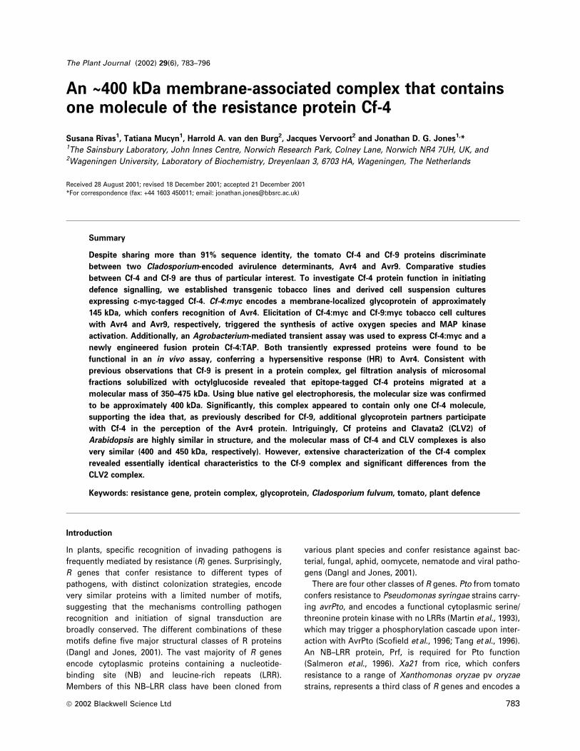
(Hammond-Kosack et al., 1995). As in the wild-type Cf-4
transgenic tobacco plants (Figure 1a, middle panel), strong
necrosis was observed in the Cf-4:myc leaf panels 5 days
after in®ltration with PVX:Avr4 but not PVX:Avr9
(Figure 1a, right panel). Importantly, this phenotype was
only observed in the three lines expressing Cf-4:myc
(G981, G986 and G990), while the other seven primary
transformants showed very slight or no necrosis (not
shown). In addition, the Petit Havana control line showed
no symptoms after in®ltration with either PVX:Avr4 or
PVX:Avr9 (Figure 1a, left panel). Secondly, Cf-4:myc trans-
genic tobacco plants were crossed to homozygous
35S:Avr4 transgenic tobacco lines. A stunted seedling
phenotype (Thomas et al., 2000) was observed in 50% of
the progeny from the G981, G986 and G990 lines (not
shown), consistent with a T-DNA insertion in a single
locus. The speci®city of this phenotype was demonstrated
by the normal growth shown by seedlings obtained from
crosses to Avr9 control plants. Therefore, c-myc-tagged
Cf-4 heterologously expressed in tobacco speci®cally
responds to the Avr4 peptide and is functional in vivo.
Tobacco lines G981, G986 and G990 were selected for
further analysis and six independent T2 lines were pro-
duced from each primary transformant. Based on Cf-4:myc
protein expression levels and the induction of the Avr4-
dependent necrotic response, one homozygous (G990F)
and three heterozygous (G986A, G986F and G990A) T2
lines were selected for generation of stable transformants,
and transgenic tobacco cell suspension cultures were
produced. In contrast to the Cf-9:mycG line, which
exhibited slightly weaker Avr9 responsiveness (Piedras
et al., 2000) compared to Cf-9:mycB and wild-type Cf-9
tobacco, Cf-4:mycB and Cf-4:mycG showed a similar
response to Avr4 challenge compared with each other
and wild-type Cf-4. This is consistent with the observation
that Cf-4/Avr4 interaction elicits a stronger response com-
pared to Cf-9/Avr9.
Cf-4:myc protein could also be detected in the cell
suspension cultures (Figure 1b). Although the predicted
molecular mass of Cf-4 is approximately 88 kDa, the
protein cross-reacted with the anti-c-myc antibodies as a
band of approximately 145 kDa, slightly smaller than
Cf-9:myc. The insertion of the triple c-myc sequence
(approximately 5 kDa) cannot account for the difference
between the observed and the estimated molecular
masses. Because the Cf-4 protein contains 20 putative
glycosylation sites, we investigated whether, as in Cf-9,
glycosylation of Cf-4 protein occurred. Solubilized micro-
somal fractions obtained from c-myc-tagged Cf-4 tobacco
plants were treated with PNGase F, a glycoamidase that
liberates nearly all N-linked oligosaccharides from glyco-
proteins (Maley et al., 1989). Incubation of solubilized
microsomes with PNGase F induced a shift in the electro-
phoretic mobility of the Cf-4:myc protein (Figure 1c).
Within 5 min, two smaller bands, of approximately 137
and 97 kDa, were detected. As the endoglycosidase reac-
tion proceeded, the intensity of the 97 kDa band, which is
close to the predicted molecular mass of the Cf-4 amino
Cf-4 resistance protein complex 785
ã Blackwell Science Ltd, The Plant Journal, (2002), 29, 783±796
acid sequence, increased, while the 137 kDa band became
fainter. This indicates that Cf-4, like Cf-9, is highly
glycosylated.
The in vivo functionality of Cf-4:myc- and Cf-9:myc-
expressing cell cultures was studied by analysing the
induction of Avr-dependent early defence responses.
Upon speci®c elicitation, a rapid production of active
oxygen species (AOS) occurred, with the highest H2O2
levels being detected in Cf-4:myc lines, particularly in the
homozygous G990F line (Figure 2a). No effect was
observed after treatment of the Cf-9:myc lines with Avr4,
or of Cf-4:myc lines with Avr9 (not shown). Untransformed
tobacco cultures did not respond to Avr treatment, under-
lining the speci®city of the observed response. In addition,
Figure 1. Functional analysis of c-myc-tagged Cf-4 tobacco primarytransformants, and expression analysis and characterization of c-myc-tagged Cf-4 cell suspension cultures.(a) Tobacco lines Petit Havana (wild-type), transgenic for Cf-4 or Cf-4:myc(G990) were in®ltrated with Agrobacteria expressing PVX:Avr4 (left leafhalf; Thomas et al., 1997) and PVX:Avr9 (right leaf half; Hammond-Kosack, 1995). After 5 days, the Cf-4/Avr4-dependent hypersensitive celldeath reaction was observed in both Cf-4-expressing lines (middle andright panel). (b) Microsomal fractions were prepared from tobacco cellcultures that are heterozygous (G986A, G986F, G990A) and homozygous(G990F) for Cf-4:myc, or homozygous for Cf-9:myc (9161, carrying a triplec-myc tag in the G domain; Piedras et al., 2000). Proteins (50 mg) wereseparated in an SDS±PAGE gel and analysed by immunoblot using ananti-c-myc antibody. The positions of Cf-4:myc and Cf-9:myc areindicated by arrowheads. (c) Deglycosylation of Cf-4:myc protein.Aliquots (50 mg) of Cf-4:myc microsomes from homozygous G990Ftobacco plants were incubated with 5 units of glycosidase PNGase F forthe times indicated. Reactions were subsequently analysed by SDS geland immunoblot. The positions of the molecular mass markers in kDaare indicated on the left.
Figure 3. Function and detection of TAP and c-myc-tagged Cf proteinstransiently expressed in N. benthamiana leaves.(a) Leaves of N. benthamiana transgenic for Avr4 (left) or Avr9 (right)were in®ltrated with Agrobacterium carrying different Cf constructs, asindicated: (1) 35S:Cf-4:TAP; (2) Gen:Cf-4:TAP; (3) 35S:Cf-4:mycB; (4)35S:Cf-4:mycG; (5) 35S:Cf-4; (6) 35S:Cf-9; (7) 35S:Cf-9:TAP; (8)Gen:Cf-9:TAP; (9) 35S:Cf-9:mycB; (10) 35S:Cf-9:mycG. After 5 days, theCf/Avr-dependent hypersensitive cell death reaction was observed. (b) N.benthamiana leaves were in®ltrated as in (a). Microsomal fractions wereprepared 2 days after in®ltration. Proteins (50 mg) were separated in anSDS±PAGE gel and analysed by immunoblot using a PAP or anti-c-mycantibody for detection of TAP- and c-myc-tagged Cf-4 and Cf-9,respectively. The numbers above the columns indicate the constructused for agro-in®ltration, see (a). Sizes of molecular mass markers areshown on the right.
786 Susana Rivas et al.
ã Blackwell Science Ltd, The Plant Journal, (2002), 29, 783±796
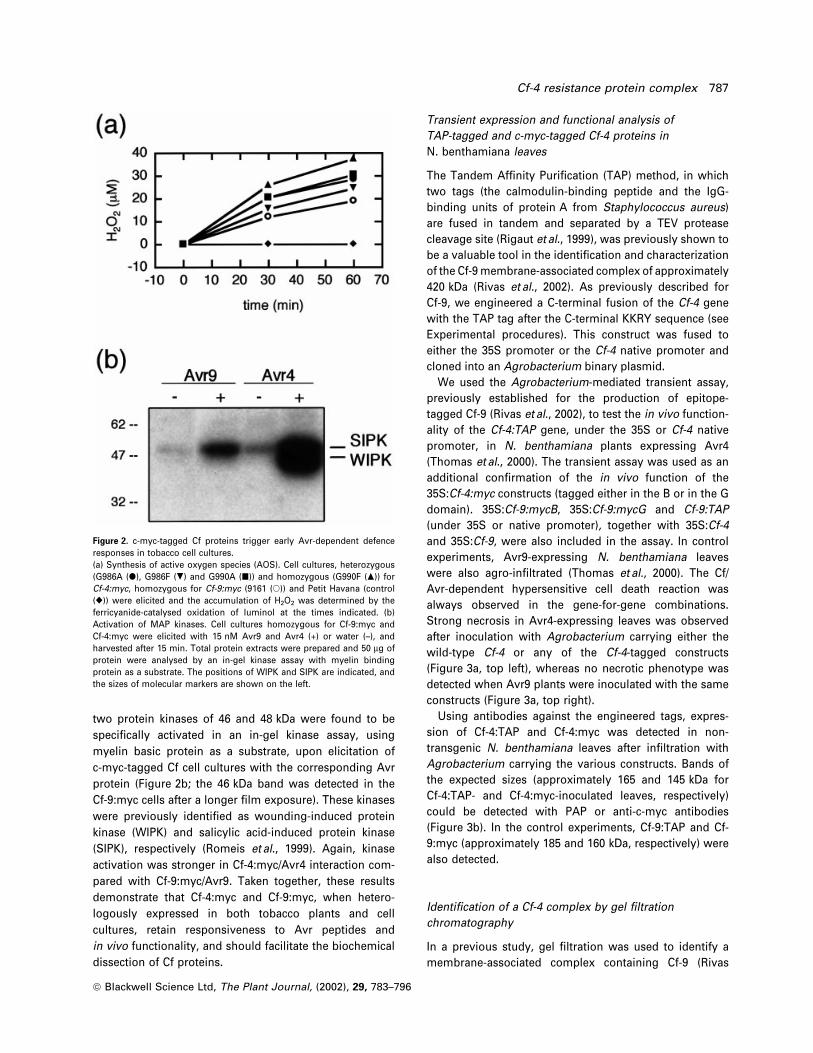
two protein kinases of 46 and 48 kDa were found to be
speci®cally activated in an in-gel kinase assay, using
myelin basic protein as a substrate, upon elicitation of
c-myc-tagged Cf cell cultures with the corresponding Avr
protein (Figure 2b; the 46 kDa band was detected in the
Cf-9:myc cells after a longer ®lm exposure). These kinases
were previously identi®ed as wounding-induced protein
kinase (WIPK) and salicylic acid-induced protein kinase
(SIPK), respectively (Romeis et al., 1999). Again, kinase
activation was stronger in Cf-4:myc/Avr4 interaction com-
pared with Cf-9:myc/Avr9. Taken together, these results
demonstrate that Cf-4:myc and Cf-9:myc, when hetero-
logously expressed in both tobacco plants and cell
cultures, retain responsiveness to Avr peptides and
in vivo functionality, and should facilitate the biochemical
dissection of Cf proteins.
Transient expression and functional analysis of
TAP-tagged and c-myc-tagged Cf-4 proteins in
N. benthamiana leaves
The Tandem Af®nity Puri®cation (TAP) method, in which
two tags (the calmodulin-binding peptide and the IgG-
binding units of protein A from Staphylococcus aureus)
are fused in tandem and separated by a TEV protease
cleavage site (Rigaut et al., 1999), was previously shown to
be a valuable tool in the identi®cation and characterization
of the Cf-9 membrane-associated complex of approximately
420 kDa (Rivas et al., 2002). As previously described for
Cf-9, we engineered a C-terminal fusion of the Cf-4 gene
with the TAP tag after the C-terminal KKRY sequence (see
Experimental procedures). This construct was fused to
either the 35S promoter or the Cf-4 native promoter and
cloned into an Agrobacterium binary plasmid.
We used the Agrobacterium-mediated transient assay,
previously established for the production of epitope-
tagged Cf-9 (Rivas et al., 2002), to test the in vivo function-
ality of the Cf-4:TAP gene, under the 35S or Cf-4 native
promoter, in N. benthamiana plants expressing Avr4
(Thomas et al., 2000). The transient assay was used as an
additional con®rmation of the in vivo function of the
35S:Cf-4:myc constructs (tagged either in the B or in the G
domain). 35S:Cf-9:mycB, 35S:Cf-9:mycG and Cf-9:TAP
(under 35S or native promoter), together with 35S:Cf-4
and 35S:Cf-9, were also included in the assay. In control
experiments, Avr9-expressing N. benthamiana leaves
were also agro-in®ltrated (Thomas et al., 2000). The Cf/
Avr-dependent hypersensitive cell death reaction was
always observed in the gene-for-gene combinations.
Strong necrosis in Avr4-expressing leaves was observed
after inoculation with Agrobacterium carrying either the
wild-type Cf-4 or any of the Cf-4-tagged constructs
(Figure 3a, top left), whereas no necrotic phenotype was
detected when Avr9 plants were inoculated with the same
constructs (Figure 3a, top right).
Using antibodies against the engineered tags, expres-
sion of Cf-4:TAP and Cf-4:myc was detected in non-
transgenic N. benthamiana leaves after in®ltration with
Agrobacterium carrying the various constructs. Bands of
the expected sizes (approximately 165 and 145 kDa for
Cf-4:TAP- and Cf-4:myc-inoculated leaves, respectively)
could be detected with PAP or anti-c-myc antibodies
(Figure 3b). In the control experiments, Cf-9:TAP and Cf-
9:myc (approximately 185 and 160 kDa, respectively) were
also detected.
Identi®cation of a Cf-4 complex by gel ®ltration
chromatography
In a previous study, gel ®ltration was used to identify a
membrane-associated complex containing Cf-9 (Rivas
Figure 2. c-myc-tagged Cf proteins trigger early Avr-dependent defenceresponses in tobacco cell cultures.(a) Synthesis of active oxygen species (AOS). Cell cultures, heterozygous(G986A (d), G986F (.) and G990A (j)) and homozygous (G990F (m)) forCf-4:myc, homozygous for Cf-9:myc (9161 (s)) and Petit Havana (control(r)) were elicited and the accumulation of H2O2 was determined by theferricyanide-catalysed oxidation of luminol at the times indicated. (b)Activation of MAP kinases. Cell cultures homozygous for Cf-9:myc andCf-4:myc were elicited with 15 nM Avr9 and Avr4 (+) or water (±), andharvested after 15 min. Total protein extracts were prepared and 50 mg ofprotein were analysed by an in-gel kinase assay with myelin bindingprotein as a substrate. The positions of WIPK and SIPK are indicated, andthe sizes of molecular markers are shown on the left.
Cf-4 resistance protein complex 787
ã Blackwell Science Ltd, The Plant Journal, (2002), 29, 783±796
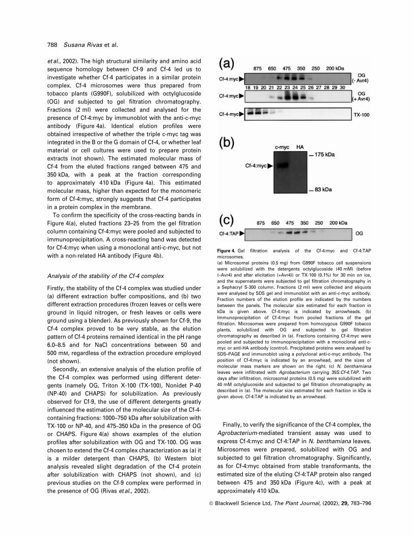
et al., 2002). The high structural similarity and amino acid
sequence homology between Cf-9 and Cf-4 led us to
investigate whether Cf-4 participates in a similar protein
complex. Cf-4 microsomes were thus prepared from
tobacco plants (G990F), solubilized with octylglucoside
(OG) and subjected to gel ®ltration chromatography.
Fractions (2 ml) were collected and analysed for the
presence of Cf-4:myc by immunoblot with the anti-c-myc
antibody (Figure 4a). Identical elution pro®les were
obtained irrespective of whether the triple c-myc tag was
integrated in the B or the G domain of Cf-4, or whether leaf
material or cell cultures were used to prepare protein
extracts (not shown). The estimated molecular mass of
Cf-4 from the eluted fractions ranged between 475 and
350 kDa, with a peak at the fraction corresponding
to approximately 410 kDa (Figure 4a). This estimated
molecular mass, higher than expected for the monomeric
form of Cf-4:myc, strongly suggests that Cf-4 participates
in a protein complex in the membrane.
To con®rm the speci®city of the cross-reacting bands in
Figure 4(a), eluted fractions 23±25 from the gel ®ltration
column containing Cf-4:myc were pooled and subjected to
immunoprecipitation. A cross-reacting band was detected
for Cf-4:myc when using a monoclonal anti-c-myc, but not
with a non-related HA antibody (Figure 4b).
Analysis of the stability of the Cf-4 complex
Firstly, the stability of the Cf-4 complex was studied under
(a) different extraction buffer compositions, and (b) two
different extraction procedures (frozen leaves or cells were
ground in liquid nitrogen, or fresh leaves or cells were
ground using a blender). As previously shown for Cf-9, the
Cf-4 complex proved to be very stable, as the elution
pattern of Cf-4 proteins remained identical in the pH range
6.0±8.5 and for NaCl concentrations between 50 and
500 mM, regardless of the extraction procedure employed
(not shown).
Secondly, an extensive analysis of the elution pro®le of
the Cf-4 complex was performed using different deter-
gents (namely OG, Triton X-100 (TX-100), Nonidet P-40
(NP-40) and CHAPS) for solubilization. As previously
observed for Cf-9, the use of different detergents greatly
in¯uenced the estimation of the molecular size of the Cf-4-
containing fractions: 1000±750 kDa after solubilization with
TX-100 or NP-40, and 475±350 kDa in the presence of OG
or CHAPS. Figure 4(a) shows examples of the elution
pro®les after solubilization with OG and TX-100. OG was
chosen to extend the Cf-4 complex characterization as (a) it
is a milder detergent than CHAPS, (b) Western blot
analysis revealed slight degradation of the Cf-4 protein
after solubilization with CHAPS (not shown), and (c)
previous studies on the Cf-9 complex were performed in
the presence of OG (Rivas et al., 2002).
Finally, to verify the signi®cance of the Cf-4 complex, the
Agrobacterium-mediated transient assay was used to
express Cf-4:myc and Cf-4:TAP in N. benthamiana leaves.
Microsomes were prepared, solubilized with OG and
subjected to gel ®ltration chromatography. Signi®cantly,
as for Cf-4:myc obtained from stable transformants, the
estimated size of the eluting Cf-4:TAP protein also ranged
between 475 and 350 kDa (Figure 4c), with a peak at
approximately 410 kDa.
Figure 4. Gel ®ltration analysis of the Cf-4:myc and Cf-4:TAPmicrosomes.(a) Microsomal proteins (0.5 mg) from G990F tobacco cell suspensionswere solubilized with the detergents octylglucoside (40 mM) (before(±Avr4) and after elicitation (+Avr4)) or TX-100 (0.1%) for 30 min on ice,and the supernatants were subjected to gel ®ltration chromatography ina Sephacryl S-300 column. Fractions (2 ml) were collected and aliquotswere analysed by SDS gel and immunoblot with an anti-c-myc antibody.Fraction numbers of the elution pro®le are indicated by the numbersbetween the panels. The molecular size estimated for each fraction inkDa is given above. Cf-4:myc is indicated by arrowheads. (b)Immunoprecipitation of Cf-4:myc from pooled fractions of the gel®ltration. Microsomes were prepared from homozygous G990F tobaccoplants, solubilized with OG and subjected to gel ®ltrationchromatography as described in (a). Fractions containing Cf-4:myc werepooled and subjected to immunoprecipitation with a monoclonal anti-c-myc or anti-HA antibody (control). Precipitated proteins were analysed bySDS±PAGE and immunoblot using a polyclonal anti-c-myc antibody. Theposition of Cf-4:myc is indicated by an arrowhead, and the sizes ofmolecular mass markers are shown on the right. (c) N. benthamianaleaves were in®ltrated with Agrobacterium carrying 35S:Cf-4:TAP. Twodays after in®ltration, microsomal proteins (0.5 mg) were solubilized with40 mM octylglucoside and subjected to gel ®ltration chromatography asdescribed in (a). The molecular size estimated for each fraction in kDa isgiven above. Cf-4:TAP is indicated by an arrowhead.
788 Susana Rivas et al.
ã Blackwell Science Ltd, The Plant Journal, (2002), 29, 783±796
In conclusion, we have shown that Cf-4 is part of a 475±
350 kDa membrane-associated complex, regardless of (a)
the tissue used as protein source (stably transformed
tobacco plants, cell cultures or transiently transformed
N. benthamiana), (b) the tag used (triple c-myc or TAP), (c)
the position of the tag within the protein sequence (B or G
domain of Cf-4), and (d) the extraction conditions (different
buffer compositions or extraction procedures).
Blue native PAGE analysis of the Cf-4 complex
The most important problem in native electrophoresis of
membrane proteins is that membrane protein aggregation
should be minimized. Blue native polyacrylamide gel
electrophoresis (BN±PAGE) has been developed for the
isolation of membrane-associated protein complexes in
enzymatically active form (Arnold et al., 1999; Caliebe et al.,
1997; SchaÈgger and von Jagow, 1991). In this technique,
Coomassie dyes and aminocaproic acid are introduced to
induce a charge shift and improve the solubilization of
membrane proteins, thereby avoiding the problem of
detergent interference observed in gel ®ltration analysis
(see below). In an earlier study, use of this methodology
allowed a more accurate estimation of the molecular size
of the Cf-9 complex (Rivas et al., 2002). To detect Cf-4:myc
within potential protein complexes, OG-solubilized
Cf4:TAP microsomes from agro-in®ltrated N. benthamiana
leaves were subjected to BN±PAGE (Figure 5a). Similarly,
Cf-9:TAP microsomes were used in a control experiment
(Figure 5b). Single lanes of the BN gel were cut, mounted
on a denaturing SDS gel in the second dimension, and
subjected to immunoblot analysis using a PAP antibody
(see Experimental procedures). Strong cross-reacting
bands, which, by comparison with protein standards
from the BN±PAGE, corresponded to a molecular mass
of approximately 400 and 420 kDa were detected for the
Cf-4:TAP and the Cf-9:TAP microsomes, respectively.
(Figure 5a,b, top panel). The same result was obtained
when TX-100 was used for solubilization of microsomal
preparations (not shown). No cross-reacting signal was
detected at this position when microsomes from non-
in®ltrated N. benthamiana leaves were subjected to the
same experimental procedure (Figure 5a, bottom panel).
To con®rm that the detected signal corresponded to a Cf-4-
containing complex, microsomes were incubated with
PNGase F prior to loading in BN±PAGE. This treatment
resulted in a shift of the cross-reacting band from 400 kDa
Figure 5. Blue native polyacrylamide gel electrophoresis of solubilizedCf-4:TAP microsomes.N. benthamiana leaves were in®ltrated or not with Agrobacteriumcarrying 35S:Cf-4:TAP (a) or 35S:Cf-9:TAP (b). After two days, microsomalproteins (0.5 mg) of in®ltrated leaves (upper and middle panel) and non-injected leaves (lower panel) were solubilized with 40 mm octylglucosideand 750 mM aminocaproic acid in 50 mM Bis-Tris for 30 min on ice.Aliquots of supernatants (80 mg of protein in 40 ml) were either treatedwith PNGase F (middle panel; see legend to Figure 1c) or directlyincubated with 5% Coomassie brilliant blue G (upper panel). Proteinswere separated on a 4.5±10.5% BN±polyacrylamide gradient gel. Cfcomplexes were resolved by second dimension 7.5% SDS gel and blottedonto nitrocellulose. Cf-4:TAP and Cf-9:TAP were ®nally detected byimmunoblot of the SDS gel using the PAP antibody. The sizes of themolecular mass markers are indicated on top (BN±PAGE) and on theright (SDS-PAGE). The positions of Cf-4:TAP and Cf-9:TAP are indicatedby arrowheads.
Cf-4 resistance protein complex 789
ã Blackwell Science Ltd, The Plant Journal, (2002), 29, 783±796
to approximately 230 kDa in the BN±PAGE (and from
145 kDa to about 97 kDa in the SDS±PAGE, see Figure 1c)
(Figure 5a, middle panel). Assuming that approximately
48 kDa of sugar groups are released from Cf-4 after
glycosidase treatment, the extra 122 kDa due to glycosyl-
ation might be derived from other components of the
complex.
Finally, Cf-4:myc from either tobacco plants or cell
cultures was also subjected to BN±PAGE analysis, using
an anti-c-myc antibody for the immunoblot analysis.
Identical results were obtained for Cf-4:myc (not shown),
con®rming that Cf-4 is part of a membrane complex of
approximately 400 kDa.
The Cf-4 complex does not contain homo-multimers of
Cf-4
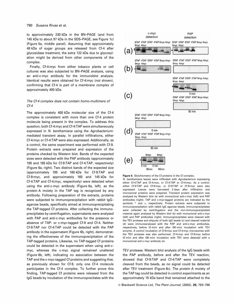
The approximately 400 kDa molecular size of the Cf-4
complex is consistent with more than one Cf-4 protein
molecule being present in the complex. To address this
question, both Cf-4:myc and Cf-4:TAP were simultaneously
expressed in N. benthamiana using the Agrobacterium-
mediated transient assay. In parallel in®ltrations, either
Cf-4:myc or Cf-4:TAP were also expressed. Additionally, as
a control, the same experiment was performed with Cf-9.
Protein extracts were prepared and expression of the
proteins checked by Western blot. Bands of the expected
sizes were detected with the PAP antibody (approximately
185 and 165 kDa for Cf-9:TAP and Cf-4:TAP, respectively)
(Figure 6a, right). Two distinct bands of the expected size
(approximately 185 and 160 kDa for Cf-9:TAP and
Cf-9:myc, and approximately 165 and 145 kDa for
Cf-4:TAP and Cf-4:myc, respectively) were detected when
using the anti-c-myc antibody (Figure 6a, left), as the
protein A moiety in the TAP tag is recognized by any
antibody. Following preparation of the extracts, proteins
were subjected to immunoprecipitation with rabbit IgG±
agarose beads, speci®cally aimed at immunoprecipitating
the TAP-tagged Cf proteins. After collecting the immuno-
precipitates by centrifugation, supernatants were analysed
with PAP and anti-c-myc antibodies for the presence or
absence of TAP- or c-myc-tagged Cf-9 and Cf-4. Neither
Cf-9:TAP nor Cf-4:TAP could be detected with the PAP
antibody in the supernatant (Figure 6b, right), demonstrat-
ing the effectiveness of the immunoprecipitation of the
TAP-tagged proteins. Likewise, no TAP-tagged Cf proteins
could be detected in the supernatant when using anti-c-
myc, whereas the c-myc signal remained unaltered
(Figure 6b, left), indicating no association between the
TAP and the c-myc-tagged Cf proteins and suggesting that,
as previously shown for Cf-9, only one Cf-4 molecule
participates in the Cf-4 complex. To further prove this
®nding, TAP-tagged Cf proteins were released from the
IgG beads by incubation of the immunoprecitates with the
TEV protease. Western blot analysis of the IgG beads with
the PAP antibody, before and after the TEV reaction,
showed that Cf-9:TAP and Cf-4:TAP were completely
cleaved from the beads, as no signal could be detected
after TEV treatment (Figure 6c). The protein A moiety of
the TAP tag could be detected in control experiments as an
approximately 15 kDa band that remained attached to the
Figure 6. Stoichiometry of the Cf proteins in the Cf complex.N. benthamiana leaves were in®ltrated with Agrobacterium expressingeither Cf-4:TAP and Cf-4:myc, or Cf-4:TAP or Cf-4:myc. As a control,either Cf-9:TAP and Cf-9:myc, or Cf-9:TAP or Cf-9:myc were alsoexpressed. Leaves were harvested 2 days after in®ltration andmicrosomal proteins were prepared. Transient protein expression wasanalysed by Western blot (a) with monoclonal anti-c-myc (left) and PAPantibodies (right). TAP and c-myc-tagged proteins are indicated by thesymbols * and >, respectively. Protein extracts were subjected toimmunoprecipitation with rabbit IgG agarose beads, immunoprecipitateswere collected by centrifugation and the non-immunoprecipitatedmaterial again analysed by Western blot (b) with monoclonal anti-c-myc(left) and PAP antibodies (right). Immunoprecipitates were cleaved withthe TEV protease and aliquots of both IgG beads (c) and cleaved material(d) were immunoanalysed with the PAP and anti-c-myc antibodies,respectively, before (0 min) and after (90 min) incubation with TEVenzyme. A control incubation of Cf-9:myc and Cf-4:myc microsomes withthe TEV protease was also performed. Cf-4:myc and Cf-9:myc before(0 min) and after (90 min) incubation with TEV were detected with amonoclonal anti-c-myc antibody (e).
790 Susana Rivas et al.
ã Blackwell Science Ltd, The Plant Journal, (2002), 29, 783±796
beads after TEV proteolysis (not shown). Therefore, no
signal corresponding to Cf protein was detected when
analysing the cleaved material with the PAP antibody, as
the protein A sequence was cleaved off from the TAP tag
(not shown). Signi®cantly, no Cf-4:myc signal was
detected in the released material either (Figure 6d). The
possibility of protein degradation by the TEV enzyme was
ruled out in a parallel incubation of Cf-9:myc and Cf-4:myc
with TEV in which no effect of the protease on c-myc-
tagged Cf proteins could be detected after treatment
(Figure 6e). Taken together, these results con®rm that
Cf-4:TAP and Cf-4:myc do not associate with each other,
and suggest that, as previously shown for Cf-9, there is
only one Cf-4 protein molecule in the Cf-4 complex.
Cf-4 is not disulphide-linked to another protein and the
Cf-4 complex does not change in size or recruit GTP-
binding proteins on elicitation
In an attempt to further characterize the Cf-4 complex, we
investigated whether the Cf-4 complex shares further
similarities with the CLV or the Cf-9 complex, beyond the
similarity in protein structure and complex sizes (Rivas
et al., 2002; Trotochaud et al., 1999). In particular, we were
interested in testing three major properties of the CLV
complex, previously tested for the Cf-9 complex (Rivas
et al., 2002). Conserved cysteine pairs, immediately before
and after the LRRs (Jones and Jones, 1997) were proposed
to be involved in the formation of a disulphide-linked
heterodimer between CLV1 and CLV2 (Jeong et al., 1999).
In contrast, we showed that Cf-9 is not associated with
other proteins in the membrane by the formation of
covalent disulphide bonds between cysteine residues.
Thus, solubilized extracts from either cell cultures or
leaves expressing Cf-4:myc were boiled in SDS±PAGE
loading buffer in the presence or absence of the reducing
agent b-mercaptoethanol. Samples were then analysed by
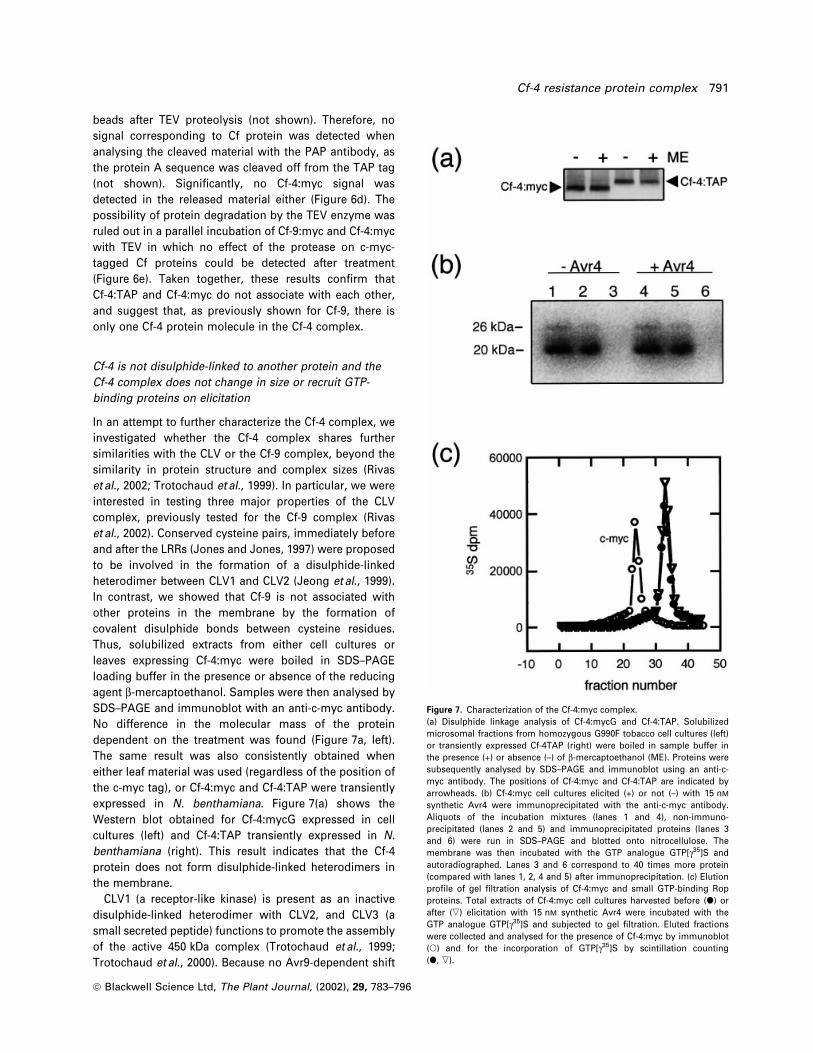
SDS±PAGE and immunoblot with an anti-c-myc antibody.
No difference in the molecular mass of the protein
dependent on the treatment was found (Figure 7a, left).
The same result was also consistently obtained when
either leaf material was used (regardless of the position of
the c-myc tag), or Cf-4:myc and Cf-4:TAP were transiently
expressed in N. benthamiana. Figure 7(a) shows the
Western blot obtained for Cf-4:mycG expressed in cell
cultures (left) and Cf-4:TAP transiently expressed in N.
benthamiana (right). This result indicates that the Cf-4
protein does not form disulphide-linked heterodimers in
the membrane.
CLV1 (a receptor-like kinase) is present as an inactive
disulphide-linked heterodimer with CLV2, and CLV3 (a
small secreted peptide) functions to promote the assembly
of the active 450 kDa complex (Trotochaud et al., 1999;
Trotochaud et al., 2000). Because no Avr9-dependent shift
Figure 7. Characterization of the Cf-4:myc complex.(a) Disulphide linkage analysis of Cf-4:mycG and Cf-4:TAP. Solubilizedmicrosomal fractions from homozygous G990F tobacco cell cultures (left)or transiently expressed Cf-4TAP (right) were boiled in sample buffer inthe presence (+) or absence (±) of b-mercaptoethanol (ME). Proteins weresubsequently analysed by SDS±PAGE and immunoblot using an anti-c-myc antibody. The positions of Cf-4:myc and Cf-4:TAP are indicated byarrowheads. (b) Cf-4:myc cell cultures elicited (+) or not (±) with 15 nM
synthetic Avr4 were immunoprecipitated with the anti-c-myc antibody.Aliquots of the incubation mixtures (lanes 1 and 4), non-immuno-precipitated (lanes 2 and 5) and immunoprecipitated proteins (lanes 3and 6) were run in SDS±PAGE and blotted onto nitrocellulose. Themembrane was then incubated with the GTP analogue GTP[g35]S andautoradiographed. Lanes 3 and 6 correspond to 40 times more protein(compared with lanes 1, 2, 4 and 5) after immunoprecipitation. (c) Elutionpro®le of gel ®ltration analysis of Cf-4:myc and small GTP-binding Ropproteins. Total extracts of Cf-4:myc cell cultures harvested before (d) orafter (,) elicitation with 15 nM synthetic Avr4 were incubated with theGTP analogue GTP[g35]S and subjected to gel ®ltration. Eluted fractionswere collected and analysed for the presence of Cf-4:myc by immunoblot(s) and for the incorporation of GTP[g35]S by scintillation counting(d, ,).
Cf-4 resistance protein complex 791
ã Blackwell Science Ltd, The Plant Journal, (2002), 29, 783±796
in the gel ®ltration pro®le of the Cf-9 complex was
previously found, we investigated whether treatment
with Avr4 induces changes in the elution of the Cf-4-
containing complexes. Cf-4:myc tobacco cell cultures were
therefore treated with the Avr4 peptide and harvested
20 min after elicitation. Microsomes were prepared, solu-
bilized with OG and subjected to gel ®ltration analysis.
However, no change in the elution pro®le of the Cf-4
protein after Avr4 treatment was observed (Figure 4a).
Rop-like proteins identi®ed in plants belong to the Rho
protein subfamily of small GTPases (Li et al., 1998). Rop
proteins were found to be recruited by the active CLV
complex (Trotochaud et al., 1999), but not by the Cf-9
complex either before or after elicitation (Rivas et al.,
2002). We therefore tested for the presence of small
GTPases in the Cf-4 complex using the GTP analogue
GTP[g-35S]. Microsomes were prepared from cell cultures,
before and after elicitation with Avr4, solubilized and
immunoprecipitated with an anti-c-myc antibody. The
presence of Rho-GTPase-related proteins of approximately
20 and 26 kDa was detected in total extracts by SDS±PAGE
analysis and autoradiography (Figure 7b, lanes 1 and 4),
but no radioactivity was found in the immunoprecipitated
material (Figure 7b, lanes 3 and 6). This observation
suggests that small GTP-binding proteins and Cf-4 are
not likely to be associated, even after elicitation. In
addition, solubilized Cf-4:myc microsomes were incubated
with GTP[g-35S] and subjected to gel ®ltration chromato-
graphy. Fractions were collected and incorporated radio-
activity was determined. No radioactivity co-eluted with
Cf-4 either in the unelicited or the elicited state (Figure 7c).
Identical results were obtained using wild-type Cf-4-
expressing plants and cell cultures before and after
elicitation with Avr4 (not shown). This suggests that the
approximately 400 kDa Cf-4 complex does not contain
small GTP-binding proteins, either before or after elicita-
tion.
Taken together, our data lead us to conclude that, as for
Cf-9, and despite the structural similarities between Cf type
proteins and CLV2, different molecular mechanisms are
involved in CLV2 function in Arabidopsis and Cf-mediated
responses to Avr proteins in tomato and tobacco.
Discussion
Using epitope-tagged Cf-9, expressed transiently or stably,
we previously identi®ed and characterized a membrane-
associated complex that contains Cf-9 (Rivas et al., 2002).
Here we show that c-myc:Cf-4 is a membrane-associated
glycoprotein that confers speci®c recognition of Avr4.
Interestingly, the Avr-dependent responses were stronger
in the c-myc-tagged Cf-4 lines than in the c-myc-Cf-9 lines
(Figure 2), consistent with the observation that Cf-4-
expressing tobacco plants exhibit a faster and stronger
necrotic response to Avr treatment compared to Cf-9
plants (Thomas et al., 2000; Van der Hoorn et al., 2001a;
Wulff et al., 2001). Alternatively, these results could re¯ect
higher protein expression in the Cf-4 transgenic lines
compared to the Cf-9 transformants (Figure 1b). In sum-
mary, these functional c-myc-tagged Cf-4 tobacco lines
constitute a valuable new tool to study the earliest events
in Avr-dependent Cf-mediated defence responses.
In addition, we engineered a Cf-4:TAP construct (Rigaut
et al., 1999), tagged after the conserved C-terminal KKRY
domain which was previously reported to confer ER
localization of Cf-9 (Bengezhal et al., 2000). However, data
presented in other studies argued against the ER location
of functional Cf-9 (Piedras et al., 2000; Rivas et al.,
2002; Van der Hoorn et al., 2001b). Here, using an
Agrobacterium-mediated transient assay (Rivas et al.,
2002), we show that the TAP-tagged Cf-4 protein is
functional in vivo (Figure 3a), indicating that a free con-
served C-terminal di-lysine motif, required for retrieval of
type I membrane proteins from the Golgi apparatus to the
ER (Cosson and Letourneur, 1994; Cosson et al., 1996;
Jackson et al., 1990), is not required for Cf-4 function.
Based on these and other results, it is tempting to
speculate about a similar localization of Cf-9 and Cf-4,
although no data regarding the cellular localization of Cf-4
have been reported to date and the Cf-9 location remains
controversial (Bengezhal et al., 2000; Piedras et al., 2000).
Gel ®ltration chromatography in the presence of OG was
previously used to identify the Cf-9 complex (Rivas et al.,
2002). Likewise, we showed that the Cf-4 protein
solubilized with OG participates in an essentially identical
kind of complex ranging in size between 475 and 350 kDa,
with a peak at approximately 410 kDa (Figure 4). Extensive
analysis of the Cf-4 complex in the presence of different
detergents revealed very similar characteristics to the
previously described Cf-9 complex (Rivas et al., 2002).
The high molecular size observed with TX-100 and NP-40
(1000±750 kDa), very close to the exclusion volume of the
column, could re¯ect artefactual protein aggregation
(Simons et al., 1973). As Cf proteins are type I membrane
proteins with just a short stretch of the protein structure
forming a single transmembrane domain, it is very
unlikely that interaction with OG makes a large contribu-
tion to the size of the Cf complexes (Mùller and le Marie,
1993). Therefore, the 475±350 kDa complex observed using
CHAPS or OG most likely re¯ects Cf-4 association with
other protein(s) and/or with itself.
Independent con®rmation of the presence of the Cf-4
protein in an approximately 400 kDa protein complex was
provided by means of BN±PAGE (Figure 5a). The speci®-
city of the detected signal was demonstrated by both the
shift of the cross-reacting signal after endoglucanase
treatment and the lack of signal when using microsomes
from non-in®ltrated N. benthamiana leaves.
792 Susana Rivas et al.
ã Blackwell Science Ltd, The Plant Journal, (2002), 29, 783±796

The estimated molecular size of the Cf-4-containing
fractions is higher than that expected for the monomeric
form of Cf-4. This suggests that Cf-4 associates with
additional protein partner(s) and/or with itself. In order to
establish the stoichiometry of the Cf-4 protein in the
complex, co-immunoprecipitation experiments with Cf-4
using two independent tags, namely c-myc and TAP, were
performed. Interestingly, our data are inconsistent with the
presence of more than one Cf-4 protein molecule in the
Cf-4 complex (Figure 6), indicating that additional protein
partners participate with Cf-4 in initiating defence signall-
ing. As glycosidase treatment shifts the size of the
complex from approximately 400 to 230 kDa, these addi-
tional partners might include at least one glycoprotein and
thus at least one other protein with an extracellular
domain.
Here we show that, despite the high structural similarity
between CLV2 and Cf proteins, the nature of the Cf-4
complex appears to be different to that of the CLV
complex, consistent with the signi®cant differences pre-
viously found between the Cf-9 and CLV complexes (Rivas
et al., 2002). Indeed, unlike CLV2 and similarly to Cf-9, the
Cf-4 protein did not form disulphide-linked heterodimers,
no ligand (Avr4)-dependent shift in the molecular mass of
the Cf-4 complex was detected, and no GTP-binding
proteins were found to be associated with Cf-4 under the
conditions tested. Therefore, the data presented in this
study further validate our inference that different
molecular mechanisms probably govern regulation of
meristem development in Arabidopsis and resistance to
Cladosporium fulvum in tomato. Furthermore, except for
the minor difference in the molecular weight of the
complex, consistent with the different sizes of Cf-4 and
Cf-9, both Cf complexes appeared to be indistinguishable,
containing only one Cf protein molecule per complex.
To what extent do our data on the Cf-9 and Cf-4
complexes test the idea that Cf proteins `guard' host
targets for the Avr9 and Avr4 presumed pathogenicity
factors? The guard model is consistent with at least two
possible molecular mechanisms. The association of R
proteins with their `guarded' protein might be induced
upon Avr binding to a pathogen target. Alternatively, R
proteins might be constitutively associated with the
`guarded' protein, and, upon Avr binding, a conform-
ational change is produced that activates downstream
signalling components (Dangl and Jones, 2001). If the
`guard' model is valid, data from this study would not be
consistent with the ®rst alternative, as we did not observe
an Avr-dependent shift in the size of the Cf complex.
Furthermore, if Cf-9 and Cf-4 function by detecting a
conformational change in host proteins that are targets for
pathogenicity factors, they must thus either `guard' two
molecules of about the same size or the same molecule.
Alternatively, Cf-4 and Cf-9 proteins might provide distinct
recognition speci®cities to a 400/420 kDa receptor com-
plex. Although no evidence for direct binding between Cf-9
and Avr9 was found in previous studies (Luderer et al.,
2001), the possibility of Cf-9 being part of an Avr9-binding
complex was not ruled out, as such a complex might have
been unstable under the conditions tested.
Future studies thus need to focus on the identi®cation of
protein partner(s) of the Cf gene products in the membrane
and their role in Avr perception. It is also crucial to
investigate whether the similarities between Cf-9 and Cf-4
complexes extend further, and, importantly, to other
members of the Cf family such as Cf-2 and Cf-5. The
c-myc-tagged Cf tobacco lines, and transiently expressed
Cf9:TAP and Cf-4:TAP, have proved to be excellent tools to
facilitate the investigation of the similarities/differences
between the protein(s) associated with Cf-9 and/or Cf-4 to
achieve disease resistance.
Experimental procedures
Generation of c-myc-tagged Cf-4:myc transgenic tobacco
lines.
Two constructs, SLJ11932 and SLJ133902, were engineered forCf-4:mycG and Cf-4:mycB expression, respectively. To generateCf-4mycG, a 2 kb XhoI±BglII fragment from p129P6A-4 (Wulffet al., 2001) was isolated and subcloned into XhoI±BglII-digestedSLJ8951, which contained a triple c-myc sequence coding for a C-terminal tag fused in-frame to Cf-9 (Piedras et al., 2000), yieldingSLJ11902. Both ClaI±NdeI and NdeI±BamHI fragments fromSLJ11902 were cloned into ClaI±BamHI-digested SLJ4K1 (Joneset al., 1992) to produce plasmid SLJ11922, which codes for theCf-4 gene with a triple c-myc tag in the G domain of the protein,under the control of the 35S promoter. The EcoRI±XhoI fragmentfrom SLJ4K1, containing the 35S promoter, and the XhoI±BamHIfragment of SLJ11922, containing the Cf-4:mycG gene, wereligated into the EcoRI±BamHI-digested SLJ7292 binary plasmid toproduce SLJ11932.
To generate Cf-4mycB, EcoRI and HindIII restriction sites werecreated in the B domain of Cf-4 by oligomutagenesis. Twodifferent DNA fragments were ampli®ed by PCR using 35SCf4(Thomas et al., 1997) as a template. 35S1 primer (Wulff et al., 2001)with 5¢-CAA ATG GAA TTC AGG TAA GGA TGA CGA GGA AAC-3¢and 5¢-TTA CCT GAA TTC ATG AAG CTT CAT TTG TGC CCC GAAGAT CAA 3¢ with CTom7 primer (Wulff et al., 2001) were used toinsert the underlined EcoRI and HindIII sites, respectively. Theresulting PCR fragments were digested with ClaI±EcoRI andEcoRI±BglII, respectively, and puri®ed. A 3.2 kb ClaI±HindIII frag-ment from Cf4DS (Wulff et al., 2001) plasmid was isolated andself-ligated after blunt-end reaction with T4 DNA polymerase. Theresulting plasmid was digested with ClaI±BglII and ligated withthe digested PCR fragments to produce SLJ133898. The triplec-myc coding sequence was isolated from a KS pBluescript vectorafter EcoRI±HindIII digestion (Piedras et al., 2000) and insertedwithin the created EcoRI and HindIII sites from SLJ133898generating SLJ133899. The ClaI±BglII fragment from this latterplasmid was inserted in ClaI±BglII-digested Cf4DS to produceSLJ133900. Finally, the 35S promoter (as an EcoRI±ClaI fragmentfrom SLJ4K1) and the ®nal Cf-4 with the triple c-myc epitope inthe B domain (as a ClaI±BamHI fragment from SLJ133900) were
Cf-4 resistance protein complex 793
ã Blackwell Science Ltd, The Plant Journal, (2002), 29, 783±796
cloned in the binary vector SLJ7291 digested with EcoRI andBamHI to generate the vector SLJ133902.
Generation of the Cf-4:TAP construct
The TAP sequence was inserted at the 3¢ end of the Cf-4 gene asan overlapping PCR product of 1.3 kb which was puri®ed anddigested with PvuII and BamHI (Rivas et al., 2002). The 5¢ end ofCf-4 was obtained from ClaI±PvuII digestion of SLJ11922 (seeabove) and ligated with the 3¢ end containing the TAP sequenceinto a ClaI±BamHI-digested pBluescript vector, generatingSLJ13920. The ClaI±BamHI fragment from SLJ13920 was ligatedwith either the EcoRI±ClaI fragment from SLJ4K1, containing the35S promoter (Jones et al., 1992), or the XbaI±ClaI fragment fromp129P6A-6, containing the native Cf-4 promoter (Thomas et al.,1997), into a pBin19 vector (Frisch et al., 1995). Therefore, theresulting plasmids SLJ14040 and SLJ14180 contained the TAP-tagged Cf-4 gene under the control of the 35S or the native Cf-4promoter, respectively.
Plant transformation and growth conditions
The binary clones SLJ11932 and SLJ133902 were mobilized intoAgrobacterium tumefaciens LBA4404 and introduced into tobaccocultivar Petit Havana (Nicotiana tabaccum) as described byPiedras et al. (2000). Plants were grown as described byHammond-Kosack et al. (1998).
Generation of suspension cultures and elicitation
Cultures were generated from leaf pieces taken from 11932 and133902 tobacco plants, as described by Piedras et al. (1998). Forelicitation, cells were challenged with either Avr9 or Avr4, asindicated. Synthetic Avr9 (Piedras et al., 1998) was used at aconcentration of 15 nM. Avr4 was produced using the methylo-trophic yeast Pichia pastoris in a fermentor. The protein accumu-lated to up to 125 mg l±1 in a 300 ml fermentor, which is at least 24times greater than in a shaken ¯ask culture. The methanol-utilizing strain (Mut+) was used, with methanol as the only carbonsource. Puri®cation of Avr4 from the cell-free culture ®ltrate wasachieved using a phenyl-Sepharose fast-¯ow column followed bya Q-Sepharose fast-¯ow column. The puri®ed protein waschecked on SDS±PAGE as well as with MALDI±TOF MS (matrixassisted laser Desorption/Ionization-Time of ¯ight spectrometry)and LC±MS (liquid chromatography±Mass spectrometry). Avr4was used at a concentration of 15 nM. At the time indicated, cellswere harvested by ®ltration, immediately frozen in liquid nitrogenand stored at ±70°C.
Agrobacterium in®ltration
Stationary-phase bacterial cultures of A. tumefaciens GV3101strain expressing either PVX:Avr4 (Thomas et al., 1997) orPVX:Avr9 (Hammond-Kosack et al., 1995) were in®ltrated inNicotiana tabacum leaves following the protocol described byWulff et al. (2001).
Transient expression of proteins in Nicotiana benthamiana
Overnight bacterial cultures of A. tumefaciens GV3101 strainexpressing the protein of interest were harvested by centrifuga-
tion (1000 g). Cells were resuspended in induction buffer (10 mM
MgCl2, 10 mM MES, pH 5.6, 150 mM acetosyringone) to an OD of0.1, unless otherwise indicated. After 2 h at 22°C, cells werein®ltrated into leaves of 4-week-old N. benthamiana plants. At theindicated times after Agrobacterium-in®ltration, leaf discs usedfor experiments were harvested, immediately frozen in liquidnitrogen and stored at ±70°C.
Determination of AOS
The production of AOS was measured by chemiluminiscencefrom the ferricyanide-catalysed oxidation of luminol as previouslydescribed (Piedras et al., 1998).
MAP kinase assays.
Preparation of protein extracts and in-gel kinase assays withmyelin basic protein (MBP) as a kinase substrate were performedas previously described (Romeis et al., 1999).
Protein techniques
All manipulations of Cf proteins (preparation of protein extracts,deglycosylation assays, Cf homo-multimerization assays, disul-phide bond detection, gel ®ltration, blue native gel electrophoresis,immunoprecipitations, SDS±PAGE and immunoblotting, GTP-[g-35S] overlays, and detection of Rop-like proteins by gel ®ltration)were performed as described previously (Rivas et al., 2002).
Acknowledgements
The Sainsbury Laboratory is funded by the Gatsby CharitableFoundation. We thank Matthew Smoker for the propagation of thecell cultures and Sara Perkins for horticultural assistance. S.R.was supported by the Federation of European BiochemicalSocieties and the United Kingdom Biotechnology and BiologicalSciences Research Council (Grant 83/P13272). H.A.vdB. wassupported by the Dutch Foundation of Chemical Research (SON)and Life Sciences Foundation (SLW) with ®nancial aid from theDutch Organization for Scienti®c Research (NWO).
References
Arnold, I., Pfeiffer, K., Neupert, W., Stuart, R.A. and SchaÈgger, H.(1999) ATP synthase of yeast mytochondria. J. Biol. Chem. 274,36±40.
Bengezhal, M., Wasteneys, G. and Jones, D.A. (2000) The C-terminal dilysine motif confers endoplasmic reticulumlocalization to type I membrane proteins in plants. Plant Cell,12, 1179±1202.
Caliebe, A., Grimm, R., Kaiser, G., LuÈ beck, J., Soll, J. and Heins, L.(1997) The chloroplastic protein import machinery contains aRieske-type iron±sulfur cluster and a mononuclear iron-bindingprotein. EMBO J. 16, 7342±7350.
Clark, S.E., Williams, R.W. and Meyerowitz, E.M. (1997) TheClavata1 gene encodes a putative receptor kinase that controlsshoot and ¯oral meristem size in Arabidopsis. Cell, 89, 575±585.
Cosson, P. and Letourneur, F. (1994) Coatomer interaction with di-lysine endoplasmic reticulum retention motifs. Science, 263,1629±1631.
Cosson, P., DeÂmollieÁ re, C., Hennecke, S., Duden, R. and
794 Susana Rivas et al.
ã Blackwell Science Ltd, The Plant Journal, (2002), 29, 783±796
Letourneur, F. (1996) d and x COPI, two coatomer subunitshomologous to clathrin-associated proteins, are involved in ERretrieval. EMBO J. 15, 1792±1798.
Dangl, J.L. and Jones, J.D.G. (2001) Plant pathogens andintegrated defence responses to infection. Nature, 411, 826±833.
Dixon, M.S., Golstein, C., Thomas, C.M., Van der Biezen, E.A. andJones, J.D.G. (2000) Genetic complexity of pathogenperception by plants: the example of Rcr3, a tomato generequired speci®cally by Cf-2. Proc. Natl Acad. Sci. USA, 97,8807±8814.
Frisch, D.A., Harris-Haller, L.W., Yokubatis, N.T., Thomas, T.L.,Hardin, S.H. and Hall, T.C. (1995) Complete sequence of thebinary vector pBin19. Plant Mol. Biol. 27, 405±409.
Go mez-Go mez, L. and Boller, T. (2000) FLS2: an LRR receptor-likekinase involved in the perception of the bacterial elicitor¯agellin in Arabidopsis. Mol. Cell, 5, 1003±1011.
Hammond-Kosack, K.E. and Jones, J.D.G. (1997) Plant diseaseresistance genes. Annu. Rev. Plant Physiol. Plant Mol. Biol. 48,575±607.
Hammond-Kosack, K.E., Staskawicz, B.J., Jones, J.D.G. andBaulcombe, D.C. (1995) Functional expression of a fungalavirulence gene from a modi®ed potato virus X. Mol. Plant±Microbe Interact. 8, 181±185.
Hammond-Kosack, K.E., Tang, S.J., Harrison, K. and Jones, J.D.G.(1998) The tomato Cf-9 disease resistance gene functions intomato and potato to confer responsiveness to the fungalavirulence gene product Avr9. Plant Cell, 10, 1251±1266.
Jackson, M.R., Nilsson, T. and Peterson, P.A. (1990) Identi®cationof a consensus motif for retention of transmembrane proteinsin the endoplasmic reticulum. EMBO J. 9, 3153±3162.
Jeong, S., Trotochaud, A.E. and Clark, S.E. (1999) The ArabidopsisClavata2 gene encodes a receptor-like protein required for thestability of the Clavata1 receptor-like kinase. Plant Cell, 11,1925±1933.
Jones, D.A. and Jones, J.D.G. (1997) The role of leucine-richrepeat proteins in plant defences. Adv. Bot. Res. Inc. Adv. PlantPathol. 24, 89±167.
Jones, J.D.G., Shlumukov, L., Carland, F.J., Sco®eld, S., Bishop,G. and Harrison, K. (1992) Effective vectors for transformation,expression of heterologous genes, and assaying transposonexcision in transgenic plants. Transgenic Res. 1, 285±297.
Joosten, M.H.A.J. and De Wit, P.J.G.M. (1999) The tomato±Cladosporium fulvum interaction: a versatile experimentalsystem to study plant±pathogen interactions. Annu. Rev.Phytopathol. 37, 335±367.
Joosten, M.H.A.J., Cozijnsen, A.J. and De Wit, P.J.G.M. (1994)Host resistance to a fungal tomato pathogen lost by asingle base-pair change in an avirulence gene. Nature, 367,384±387.
Joosten, M.H.A.J., Vogelsang, R., Cozijnsen, A.J., Verberme, M.C.and De Wit, P.J.G.M. (1997) The biotrophic fungusCladosporium fulvum circumvents Cf-4-mediated resistanceby producing Avr4 unstable elicitors. Plant Cell, 9, 1±13.
Kooman-Gersmann, M., HoneÂe, G., Bonnema, G. and De Wit,P.J.G.M. (1996) A high-af®nity binding site for the Avr9 peptideelicitor of Cladosporium fulvum is present on plasmamembranes of tomato and other solanaceous plants. PlantCell, 8, 929±938.
Li, H., Wu. G., Ware, D., Davis, K.R. and Yang, Z. (1998)Arabidopsis Rho-related GTPases: differential gene expressionin pollen and polar localization in ®ssion yeast. Plant Physiol.118, 407±417.
Li, J. and Chory, J. (1997) A putative leucine-rich repeat receptor
kinase involved in brassinosteroid signal transduction. Cell, 90,929±938.
Luderer, R., Rivas, S., NuÈ rnberger, T., et al. (2001) No evidence forbinding between resistance gene product Cf-9 of tomato andavirulence gene product AVR9 of Cladosporium fulvum. Mol.Plant±Microbe Interact. 14, 867±876.
Maley, F., Trimble, R.B., Tarentino, A.L. et al. (1989)Characterization of glycoproteins and their associatedoligosaccharides through the use of endoglycosidases. Anal.Biochem. 180, 195±204.
Martin, G.B., Brommonschenkel, S.H., Chunwongse, J., Frary, A.,Ganal, M.W., Spivey, R., Wu, T.Y., Earle, E.D. and Tanksley, S.D.(1993) Map-based cloning of a protein-kinase gene conferringdisease resistance in tomato. Science, 262, 1432±1436.
Mùller, J. and le Marie, M. (1993) Detergent binding as a measureof hydrophobic surface area of integral membrane proteins. J.Biol. Chem. 268, 18659±18672.
Piedras, P., Hammond-Kosack, K.E., Harrison, K. and Jones,J.D.G. (1998) Rapid, Cf-9 and Avr9-dependent, production ofactive oxygen species in tobacco suspension cultures. Mol.Plant±Microbe Interact. 11, 1155±1166.
Piedras, P., Rivas, S., Droge, S., Hillmer, S. and Jones, J.D.G.(2000) Functional, c-myc-tagged Cf-9 resistance gene productsare plasma-membrane localized and glycosylated. Plant J. 21,529±536.
Rigaut, G., Shevchenko, A., Rutz, B., Wilm, M., Mann, M. andSe raphin, B. (1999) A generic puri®cation method for proteincomplex characterization and proteome exploration. NatureBiotechnol. 17, 1030±1032.
Rivas, S., Romeis, T. and Jones, J.D.G. (2002) The Cf-9 diseaseresistance protein is present in a ~ 420 kDa heteromultimericmembrane-associated complex at one molecule per complex.Plant Cell, 14, 1±15.
Romeis, T., Piedras, P., Zhang, S., Klessig, D.F., Hirt, H. and Jones,J.D.G. (1999) Rapid Avr9- and Cf-9-dependent activation of MAPkinases in tobacco cell cultures and leaves: convergence ofresistance gene, elicitor, wound, and salicylate responses. PlantCell, 11, 273±287.
Salmeron, J.M., Oldroyd, G.E.D., Rommens, C.M.T., Sco®eld,S.R., Kim, H.S., Lavelle, D.T., Dahlbeck, D. and Staskawicz, B.J.(1996) Tomato Prf is a member of the leucine-rich repeat classof plant disease resistance genes and lies embedded within thePto kinase gene cluster. Cell, 86, 123±133.
SchaÈgger, H. and von Jagow, G. (1991) Blue nativeelectrophoresis for isolation of membrane protein complexesin enzymatically active form. Anal. Biochem. 199, 223±231.
Sco®eld, S.R., Tobias, C.M., Rathjen, J.P., Chang, J.H., Lavalle,D.T., Michelmore, R.W. and Staskawicz, B.J. (1996) Molecularbasis of gene-for-gene speci®city in bacterial speck disease oftomato. Science, 274, 2063±2065.
Simons, K., Helenius, A. and Garoff, H. (1973) Solubilization of themembrane proteins of Semliki Forest Virus with Triton X-100.J. Mol. Biol. 80, 119.
Song, W.Y., Wang, G.L., Chen, L.L., et al. (1995) Receptor kinase-like protein encoded by the rice disease resistance gene, Xa21.Science, 270, 1804±1806.
Tang, X., Frederick, R.D., Zhov, J., Halterman, D.A., Jia, Y. andMartin, G.B. (1996) Initiation of plant disease resistance byphysical interaction of AvrPto and Pto kinase. Science, 274,2060±2063.
Thomas, C.M., Jones, D.A., Parniske, M., Harrison, K., Balint-Kurti, P.J., Hatzixanthis, K. and Jones, J.D.G. (1997)Characterization of the tomato Cf-4 gene for resistance toCladosporium fulvum identi®es sequences that determine
Cf-4 resistance protein complex 795
ã Blackwell Science Ltd, The Plant Journal, (2002), 29, 783±796

recognitional speci®city in Cf-4 and Cf-9. Plant Cell, 9, 2209±2224.
Thomas, C.M., Tang, S., Hammond-Kosack, K. and Jones, J.D.G.(2000) Comparison of the hypersensitive response induced bytomato Cf-4 and Cf-9 genes in Nicotiana spp. Mol. Plant±Microbe Interact. 13, 465±469.
Torii, K.U., Mitsukawa, N., Oosumi, T., Matsuura, Y., Yokoyama,R., Whitiier, R.F. and Komeda, Y. (1996) The ArabidopsisERECTA gene encodes a putative receptor protein kinase withextracellular leucine-rich repeats. Plant Cell, 8, 735±746.
Trotochaud, A.E., Hao, T., Wu, G., Yang, Z. and Clark, S.E. (1999)The Clavata1 receptor-like kinase requires Clavata3 for itsassembly into a signalling complex that includes KAPP and aRho-related protein. Plant Cell, 11, 393±405.
Trotochaud, A.E., Jeong, S. and Clark, S.E. (2000) Clavata3, amultimeric ligand for the Clavata1 receptor-like kinase. Science,289, 613±617.
Van der Ackerveken, Van Kan, J.A.L., Joosten, M.H.A.J., Muisers,J.M., Verbakel, H.M. and De Wit, P.J.G.M. (1993)Characterization of two putative pathogenicity genes of thefungal tomato pathogen Cladosporium fulvum. Mol. Plant±Microbe Interact. 6, 210±215.
Van der Hoorn, R.A.L., Roth, R. and Joosten, M.H.A.J. (2001a)Identi®cation of distinct speci®city determinants in resistance
protein Cf-4 allows construction of a Cf-9 mutant that confersrecognition of avirulence protein Avr4. Plant Cell, 13, 273±285.
Van der Hoorn, R.A.L., Van der Ploeg, A., De Wit, P.J.G.M. andJoosten, M.H.A.J. (2001b) The C-terminal dilysine motif fortargeting to the endoplasmic reticulum is not required for Cf-9function. Mol. Plant±Microbe Interact. 14, 412±415.
Van der Hooven, H.W., van den Burg, H.A., Vossen, P., Boeren, S.,De Wit, P.J.G.M. and Vervoort, J. (2001) Disul®de bondstructure of the AVR9 elicitor of the fungal tomato pathogenCladosporium fulvum; evidence for a cystine knot.Biochemistry, 40, 3458±3466.
Van Kan, J.A.L., Van den Ackerveken, G.F.J.M. and De Wit,P.J.G.M. (1991) Cloning and characterization of cDNA ofavirulence gene avr9 of the fungal tomato pathogenCladosporium fulvum, causal agent of tomato leaf mould.Mol. Plant±Microbe Interact. 4, 52±59.
Wulff, B.B.H., Thomas, C.M., Smoker, M., Grant, M. and Jones,J.D.G. (2001) Domain swapping and gene shuf¯ing identifysequences required for induction of an Avr-dependenthypersensitive response by the tomato Cf-4 and Cf-9 proteins.Plant Cell, 13, 255±272.
Xiao, S.Y., Ellwood, S., Calis, O., Patrick, E., Li, T.X., Coleman, M.and Turner, J.G. (2001) Broad-spectrum mildew resistance inArabidopsis thaliana mediated by RPW8. Science, 291, 118±120.
796 Susana Rivas et al.
ã Blackwell Science Ltd, The Plant Journal, (2002), 29, 783±796