amylolytic strains of lactobacillus plantarum isolated
TRANSCRIPT
Vol. 14(4), pp. 310-318, 28 January, 2015
DOI: 10.5897/AJB2014.14149
Article Number: 1B124F949962
ISSN 1684-5315
Copyright © 2015
Author(s) retain the copyright of this article http://www.academicjournals.org/AJB
African Journal of Biotechnology
Full Length Research Paper
Amylolytic strains of Lactobacillus plantarum isolated from barley
Hattingh, M. 1, Alexander, A.2, Meijering, I.2, Van Reenen, C. A.1 and Dicks, L. M. T.1*
1Department of Microbiology, University of Stellenbosch, Stellenbosch 7600, South Africa.
2Southern Associated Maltsters (Pty) LTD, P.O. Box 27, Caledon 7230, South Africa.
Received 3 September, 2014; Accepted 15 January, 2015
Two α-amylase-producing strains of Lactobacillus plantarum were isolated from South African barley. The extracellular α-amylase activity produced by strain A.S1.2 coincided with cell growth, while strain B.S1.6 produced α-amylase mainly during stationary growth. Cell wall α-amylases in both strains were approximately five times higher than recorded for extracellular α-amylases. Both strains demonstrated highest extracellular α-amylase activity in 2% (w/v) maltose, followed by 2% (w/v) malt extract and 2% (w/v) starch, respectively. The α-amylase produced by the two strains functioned optimally at 50°C and under alkaline conditions. The two strains of L. plantarum fermented carbohydrates naturally present in barley, and produced cell-bound and cell-free α-amylase at alkaline conditions. The two strains may be developed into starter cultures to facilitate the germination of barley and produce malt with a higher fermentable sugar content. Key words: Lactobacillus plantarum, starch hydrolysis, barley, malting.
INTRODUCTION Lactic acid bacteria (LAB) are fastidious and usually only grow in environments rich in monosaccharides, disac-charides, amino acids, peptides, nucleotide bases, vita-mins, minerals and fatty acids (Hammes and Hertel, 2009). Complex carbohydrates are seldom fermented, especially in environments rich in glucose or disac-charides such as sucrose (Hammes and Hertel, 2009; Reddy et al., 2008). However, a few strains degrade starch in the presence of easier fermentable carbo-hydrates. Amylolytic (starch hydrolysing) strains of Lactobacillus plantarum have been isolated from fermented cassava, maize and sorghum (Guyot, 2010; Haydersah et al., 2012; Nwankwo et al., 1989; Sanni et
al., 2002; Songré-Ouattara et al., 2008), from burong isda prepared from fermented fish and rice (Olympia et al., 1995) and fermented rice noodles (Kanpiengjai et al., 2014). Amylolytic strains of L. fermentum were isolated from ogi and mawè, a Benin maize sourdough product (Agati et al., 1998; Tchekessi et al., 2014), from traditionally fermented Nigerian foods (Sanni et al., 2002) and a starchy soil waste in the Cameroon (Fossi and Tavea, 2013). Amylolytic strains of L. manihotivorans were isolated from fermented cassava (Morlon-Guyot et al., 1998) and sorghum silage (Chahrour et al., 2013), and L. amylolyticus from beer malt (Bohak et al., 1998) and swine waste (Nakamura and Crowell, 1979). A
*Corresponding author. Email address: [email protected]. Author(s) agree that this article remains permanently open access under the terms of the Creative Commons Attribution License 4.0 International License
starch-hydrolysing strain of Streptococcus macedonicus was isolated from pozol, a Mexican fermented maize beverage (Díaz-Ruiz et al., 2003).
Amylolytic LAB (ALAB) have also been isolated from raw starch, corn and potato and play an important role in cereal-based fermented foods such as European sour rye bread, Asian salt bread, sour porridges, dumplings and non-alcoholic beverage production (Chatterjee et al., 1997; Nakamura, 1981; Rodriguez-Sanoja et al., 2000).
Knowledge regarding ALAB in barley is poor. However, lactic acid bacteria proliferate extensively on barley when fermentable sugars become available during malting (Booysen et al., 2002; O`Sullivan et al., 1999; Petters et al., 1988). In the brewing industry, malting is essential to supply yeast with fermentable substrates, and the optimization of this process demands sufficient starch degradation (Laitila et al., 2007). We are the first to report on L. plantarum, isolated from barley, with extracellular and cell wall α-amylases. The strains may be exploited as starter cultures during malting to contribute to barley germination, and hence malt with a higher fermentable sugar content. MATERIALS AND METHODS Screening for amylolytic lactic acid bacteria and differentiation of isolates Barley (10 g) from eight different samples, collected in the Western Cape, South Africa, was suspended in 100 ml sterile distilled water and incubated at 30°C for 48 h. The suspensions were serially diluted in sterile distilled water, plated onto modified MRS agar and incubated at 30°C for 48 h. The composition of the medium was as described by De Man et al. (1960), except that glucose was
replaced with 2% (w/v) soluble starch (Merck) (MRS-starch medium) and 200 µg/ml
Delvocid (Gist Brocades NV, Delft, The
Netherlands) was added to prevent the growth of yeasts and molds. Plates were flooded with a potassium iodide solution (0.6%, w/v, K and 0.3%, w/v, I crystals, dissolved in distilled water). Colonies surrounded by the largest clear zones were selected and streaked onto MRS-starch plates to obtain pure cultures. Starch hydrolysis was confirmed by inoculating the isolates (10%, v/v) into MRS-starch broth. After 48 h of incubation at 30°C, the starch hydrolysed was calculated from the residual, thus unfermented starch and expressed as a percentage of the original concentration. The spectrophotometric method of Nakamura (1981) was used.
The isolates were Gram-stained and visualised with a phase contrast microscope. Catalase activity was determined by covering colonies on MRS agar plates with 5% (v/v) hydrogen peroxide. Phenotypic and genetic relatedness of the isolates were determined by carbohydrate sugar fermentation reactions and RAPD-PCR,
respectively. The API 50 CHL system (BioMerieux, Maray 1’Etoile, France) was used, according to instructions of the manufacturer. DNA was extracted from each isolate using the ZR Fungal/Bacterial DNA kit (Zymo Research Corporation, Orange, California, USA). The PCR method described by Van Reenen and Dicks (1993) was used, but with a few modifications. Each reaction of 25 µl contained 40 ng genomic DNA, 1.25 mmol/l of each dNTP, 1 µmol/l of a single 10 base primer and 2.5 units Taq Polymerase. Three single primers [5’-CCAGCAGCTT-3’ (OPL-03), 5’-GGGCGGTACT-3’ (OPL-12), 5’-AGGTTGCAGG-3’ (OPL-16)] were used. The cycling program included an initial cycle of 94°C for 4 min, followed by 45 cycles of
Hattingh et al. 311 94°C (1 min), 36°C (1 min), and 72°C (1 min). Final incubation was at 72°C for 5 min and ended by cooling to 4°C. PCR were performed in triplicate. DNA banding patterns were visualized by electrophoresis in 1.4% (w/v) agarose gels. Lambda DNA, digested with HindIII and EcoR1 (Boehringer Mannheim, Darmstadt, Germany) was used as molecular marker. Identification of strains
Strains with unique phenotypic and genetic characteristics were identified to species level by sequencing the 16S rRNA and recA genes. The 16S rDNA from each isolate was amplified using
primers 8F (5’-CACGGATCCAGACTTTGATYMTGGCTCAG-3’) and 1512R (5’-GTGAAGCTTACGGYTAGCTTGTTACGACTT-3’), as described by Felske et al. (1997). The recA gene was amplified using primers AmpF (5’-GCCCTAAAAAARATYGAAAAGAAHTTYGGTAAAGG-3’) and AmpR (5’-AATGGT GGCGCYACYTTGTTTTTHACAACTTT-3’), according to the method of Endo and Okada (2008). PCR products of the 16S rRNA gene and recA were purified using the QIAquick PCR purification kit (Qiagen Inc., Valencia, California, USA). The
DNA fragments were cloned into pGEM-T Easy vector (Promega Corporation, Madison, Wisconsin, USA) and transformed into E. coli DH5α. Competent E. coli DH5α cells were prepared as described by Neveling et al. (2012). Plasmids were isolated using the Qiagen plasmid miniprep kit (Qiagen). The 16S rRNA and recA genes were sequenced using the BigDye Terminator V3.1 sequencing kit (Applied Biosystems, Foster City, California) as prescribed by the manufacturer. Blast analysis was conducted on the 16S rDNA and recA sequences and aligned with ClustalW. Phylogenetic trees
were constructed with sequences of approximately 1500 bp 16S rDNA and 600 bp recA. Phylogenetic analysis was done using the neighbour-joining method, maximum-likelihood (Cavalli-Sforza and Edwards, 1967) and maximum parsimony algorithms (Kluge and Farris, 1969). Bootstrapping was performed as described by Felsenstein (1985). Determination of α-amylase activity
The isolates were grown in MRS broth (Biolab) to mid-exponential phase, inoculated (4%, v/v) into 200 ml MRS-starch broth (pH 7.0) and incubated at 30°C for 40 h, without pH control, on an orbital shaker (120 rpm). Samples of 1 ml were collected every 2 h from the 200 ml culture. The cells were harvested (10 000 g, 15 min) and the cell-free supernatant tested for extracellular α-amylase activity, using the Cerelpha method (Megazyme International, Ireland, Ltd, County Wicklow, Ireland). Results were expressed in Cerelpha Units (CU)/ml, with one unit defined as the amount of enzyme required to release one micromole p-nitrophenol from a blocked p-nitrophenyl maltoheptaoside oligosaccharide substrate (BPNPG7) per min. Cell wall α-amylase activity was measured after 24 h of incubation at 30°C. The cells were harvested as before, washed twice in sterile saline (0.85 %, w/v) and resuspended in extraction buffer (1 mol/l Na-malate, 1 mol/l
NaCl, 40 mmol/l
CaCl, pH 5.4).
Malt flour was used as positive control and an isolate of L.
plantarum with no α-amylase activity on plate assays, was used as negative control. All experiments were performed in triplicate.
Effect of carbon source, temperature and pH on α-amylase activity
Production of α-amylase from different carbon sources was
determined by inoculating active-growing cells (4%, v/v) of each isolate into MRS broth and MRS broth with glucose replaced by 2%
312 Afr. J. Biotechnol.
Figure 1. A, Starch hydrolysis on MRS-starch agar plates, showing clear zones. Staining was with KI. From left to right: Isolates A.S1.2, A.S4.10, A.S8.10 and B.S1.6. B, Residual starch content after fermentation in MRS-starch broth, as determined using the method of Nakamura (1981). Significant levels in the paired t-test: *p < 0.05.
(w/v) starch (Merck, Darmstadt, Germany), 2% (w/v) maltose (Sigma-Aldrich, St. Louis, Missouri), 2% (w/v) cellobiose (Sigma), and 2, 4 and 6% (w/v) malt extract (Muntons, Suffolk, England), respectively. In another experiment, 2% (w/v) maltose was added to MRS broth. An additional experiment was performed by inoculating cells into MRS broth supplemented with a combination of 2% (w/v) malt extract and 2% (w/v) maltose. All cultures were incubated at 30°C for 24 h. Viable cell numbers were determined by plating onto MRS agar. Extracellular α-amylase activity was measured in the cell-free supernatants, as described before. All experiments were performed in triplicate.
The stability of extracellular α-amylase at different temperatures and pH conditions was determined by using cell-free supernatants collected from 24 h-old cultures grown in MRS-starch broth at 30°C. Temperature effects were determined by incubating cell-free
supernatants at 20 to 70°C (with 10°C intervals) for 10 min. The effect of pH was determined by incubating cell-free supernatants at pH 3-9, with increases of one pH unit, for 10 min. Citrate phosphate buffer (0.1 mol/l) was used for pH 3-6, phosphate buffer (0.1 mol/l) for pH 7 and 8 and Tris buffer (0.1 mol/l) for pH 9. All experiments were performed in triplicate. Statistical analysis
Statistical analysis was done with Statistica (v. 10, StatSoft, Inc.). The students’-test was performed at 95 % confidence levels.
RESULTS Selection of amylolytic strains
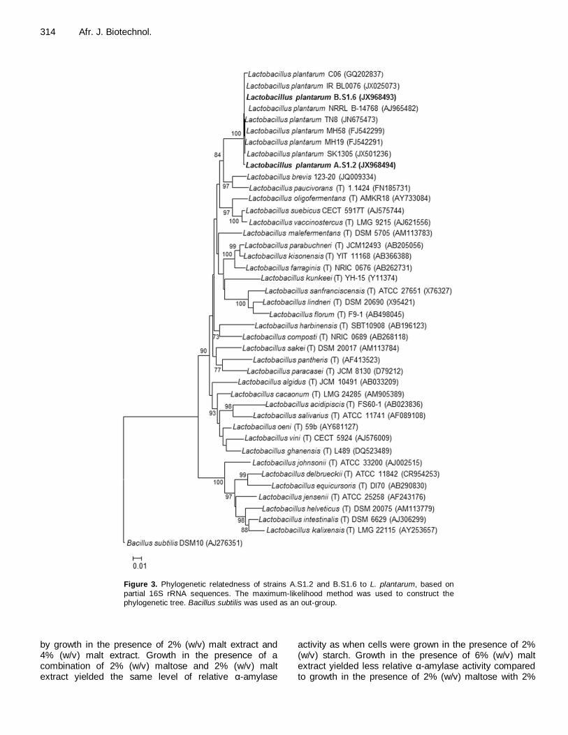
From the 185 colonies on the MRS-starch plates, 90 were surrounded by clearing zones, suggesting that they hydrolysed starch. Four of the 90 isolates (A.S1.2, A.S4.10, A.S8.10 and B.S1.6) produced the largest clearing zones and hydrolysed at least 75% of the starch in MRS-starch medium (Figure 1). All four isolates were Gram-positive and catalase negative. Isolates A.S1.2, A.S4.10 and A.S8.10 hydrolysed approximately the same amount of starch within 48 h (Figure 1), had an identical sugar fermentation profile (not shown) and displayed similar RAPD-PCR banding patterns (Figure 2). Isolate B.S1.6, on the other hand, degraded less starch (Figure 1) and had a different DNA profile (Figure 2). Further-more, strain B.S1.6 differed from the other three strains by fermenting L-arabinose, rhamnose and lactose, but not α-methyl-D-mannoside and inuline. Strains A.S1.2 and B.S1.6 formed a tight cluster with other strains of L. plantarum when 16S rRNA (Figure 3) and recA sequences (not shown) were compared. All four strains
Hattingh et al. 313
Figure 2. DNA banding patterns obtained with RAPD-PCR. Three primer sets were
used (OPL 3, OPL 12 and OPL 16). Order of isolates per primer set: Lane 1 = A.S1.2, lane 2 = A.S4.10, lane 3 = A.S8.10 and lane 4 = B.S1.6. M =lambda DNA digested with HindIII and EcoRI.
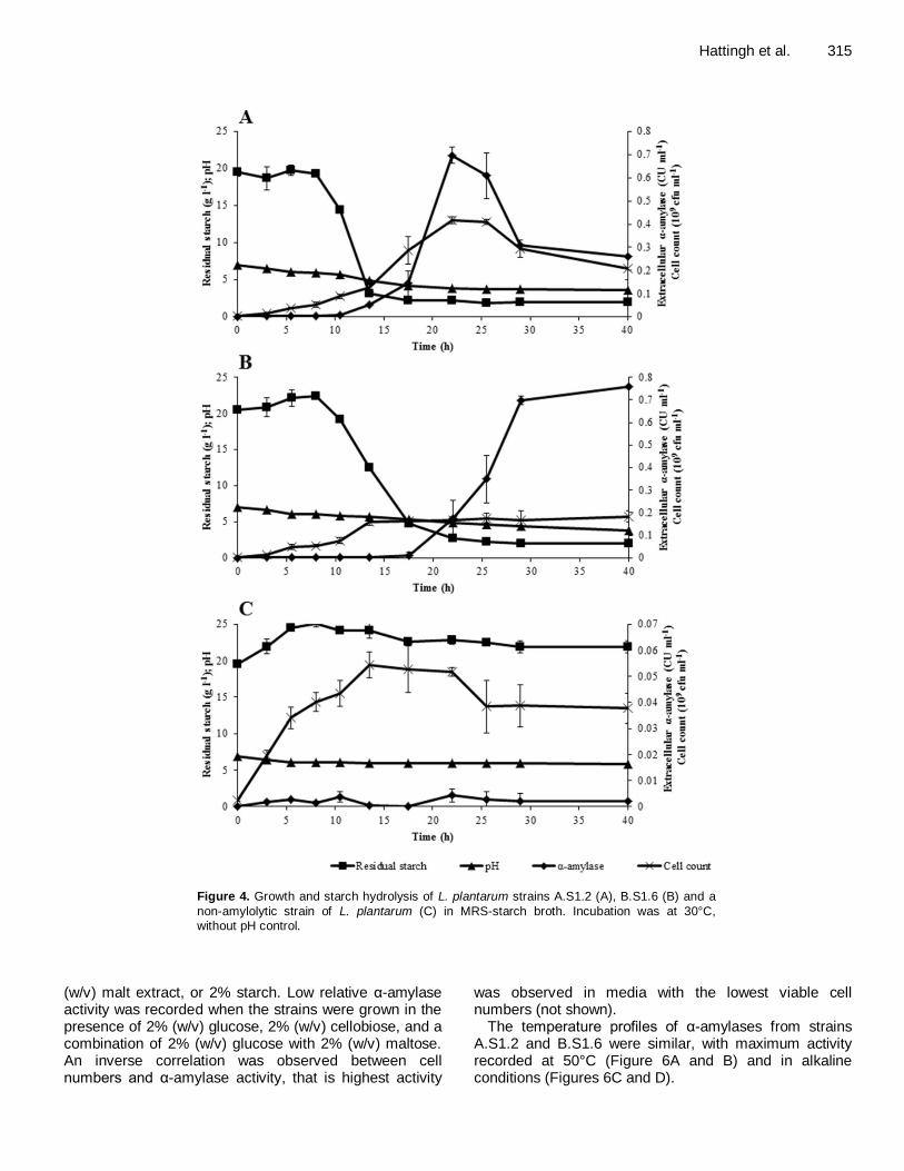
were classified as L. plantarum based on RAPD-PCR profiles (Figure 2) and comparison of 16S rRNA sequences (Figure 3). Strain A.S1.2 was selected as the representative of three strains with the same RAPD-PCR profile (Figure 2). The RAPD-PCR profile of strain B.S1.6 was, however, different (Figure 2). Strains A.S1.2 and B.S1.6 were thus selected for further studies. α-Amylase activity Extracellular α-amylase production of strain A.S1.2 coincided with cell growth (Figure 4A). Highest cell numbers of strain A.S1.2 (4 × 10
8 CFU/ml) were recorded
after 22 h of inoculation. Starch hydrolysis started 8 h after inoculation, approximately 2 h before the first extracellular α-amylase activity was recorded. α-Amylase activity increased to ~ 0.1 CU/ml after 18 h of inoculation and increased rapidly to 0.7 CU/ml over the next 4 h. After 22 h of fermentation, α-amylase activity decreased to 0.3 CU/ml within 8 h and remained close to this level for the duration of the 40-h fermentation. The pH decreased from an initial 7.0 to approximately 3.8 towards the end of fermentation.
Extracellular α-amylase production of strain B.S1.6 did not coincide with cell growth (Figure 4B). Cell numbers increased to 2 × 10
8 CFU/ml after 14 h of inoculation and
remained close to this level throughout the experiment.
As recorded for strain A.S1.2, starch hydrolysis started 8 h after inoculation, and approximately 10 h before the first extracellular α-amylase activity was recorded. α-Amylase activity increased to ~ 0.7 CU/ml after 28 h of incubation and slowly increased to 0.75 CU/ml towards the end of the 40-h fermentation. In contrast to strain A.S1.2, α-amylase activity increased as the culture pH decreased. No starch was degraded by the negative control, a non-amylolytic strain of L. plantarum, despite some limited growth (Figure 4C).
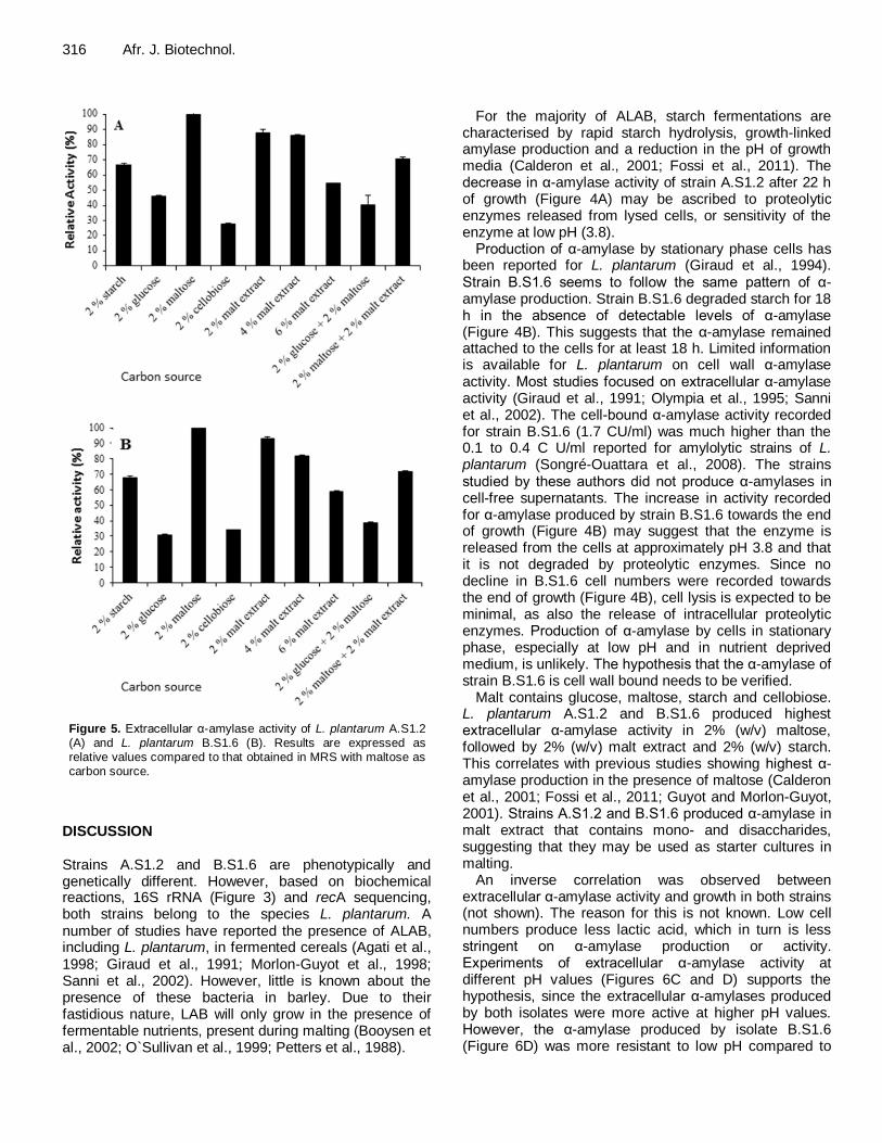
Cell-bound α-amylase activities recorded for strains A.S1.2 and B.S1.6 after 24 h of fermentation were 3.2 and 1.7 CU/ml, respectively (not shown), which is approximately five times higher than the extracellular α-amylase activities recorded (Figure 4A and B). Effect of carbon source, temperature and pH on α-amylase activity For both strains A.S1.2 and B.S1.6 highest α-amylase activity was recorded in the presence of 2% (w/v) maltose (Figure 5A and 5B, respectively). Activity of α-amylase recorded from growth in the presence of other carbohydrates is expressed as activity relative to that recorded for growth in the presence of maltose. In the case of both strains, highest α-amylase activity was recorded in the presence of 2% (w/v) maltose, followed
314 Afr. J. Biotechnol.
Figure 3. Phylogenetic relatedness of strains A.S1.2 and B.S1.6 to L. plantarum, based on
partial 16S rRNA sequences. The maximum-likelihood method was used to construct the phylogenetic tree. Bacillus subtilis was used as an out-group.
by growth in the presence of 2% (w/v) malt extract and 4% (w/v) malt extract. Growth in the presence of a combination of 2% (w/v) maltose and 2% (w/v) malt extract yielded the same level of relative α-amylase
activity as when cells were grown in the presence of 2% (w/v) starch. Growth in the presence of 6% (w/v) malt extract yielded less relative α-amylase activity compared to growth in the presence of 2% (w/v) maltose with 2%
Hattingh et al. 315
Figure 4. Growth and starch hydrolysis of L. plantarum strains A.S1.2 (A), B.S1.6 (B) and a
non-amylolytic strain of L. plantarum (C) in MRS-starch broth. Incubation was at 30°C,
without pH control.
(w/v) malt extract, or 2% starch. Low relative α-amylase activity was recorded when the strains were grown in the presence of 2% (w/v) glucose, 2% (w/v) cellobiose, and a combination of 2% (w/v) glucose with 2% (w/v) maltose. An inverse correlation was observed between cell numbers and α-amylase activity, that is highest activity
was observed in media with the lowest viable cell numbers (not shown).
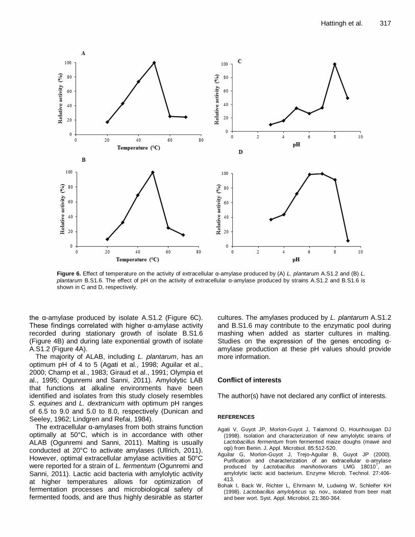
The temperature profiles of α-amylases from strains A.S1.2 and B.S1.6 were similar, with maximum activity recorded at 50°C (Figure 6A and B) and in alkaline conditions (Figures 6C and D).
316 Afr. J. Biotechnol.
Figure 5. Extracellular α-amylase activity of L. plantarum A.S1.2 (A) and L. plantarum B.S1.6 (B). Results are expressed as relative values compared to that obtained in MRS with maltose as carbon source.
DISCUSSION Strains A.S1.2 and B.S1.6 are phenotypically and genetically different. However, based on biochemical reactions, 16S rRNA (Figure 3) and recA sequencing, both strains belong to the species L. plantarum. A number of studies have reported the presence of ALAB, including L. plantarum, in fermented cereals (Agati et al., 1998; Giraud et al., 1991; Morlon-Guyot et al., 1998; Sanni et al., 2002). However, little is known about the presence of these bacteria in barley. Due to their fastidious nature, LAB will only grow in the presence of fermentable nutrients, present during malting (Booysen et al., 2002; O`Sullivan et al., 1999; Petters et al., 1988).
For the majority of ALAB, starch fermentations are characterised by rapid starch hydrolysis, growth-linked amylase production and a reduction in the pH of growth media (Calderon et al., 2001; Fossi et al., 2011). The decrease in α-amylase activity of strain A.S1.2 after 22 h of growth (Figure 4A) may be ascribed to proteolytic enzymes released from lysed cells, or sensitivity of the enzyme at low pH (3.8).
Production of α-amylase by stationary phase cells has been reported for L. plantarum (Giraud et al., 1994). Strain B.S1.6 seems to follow the same pattern of α-amylase production. Strain B.S1.6 degraded starch for 18 h in the absence of detectable levels of α-amylase (Figure 4B). This suggests that the α-amylase remained attached to the cells for at least 18 h. Limited information is available for L. plantarum on cell wall α-amylase activity. Most studies focused on extracellular α-amylase activity (Giraud et al., 1991; Olympia et al., 1995; Sanni et al., 2002). The cell-bound α-amylase activity recorded for strain B.S1.6 (1.7 CU/ml) was much higher than the 0.1 to 0.4 C U/ml reported for amylolytic strains of L. plantarum (Songré-Ouattara et al., 2008). The strains studied by these authors did not produce α-amylases in cell-free supernatants. The increase in activity recorded for α-amylase produced by strain B.S1.6 towards the end of growth (Figure 4B) may suggest that the enzyme is released from the cells at approximately pH 3.8 and that it is not degraded by proteolytic enzymes. Since no decline in B.S1.6 cell numbers were recorded towards the end of growth (Figure 4B), cell lysis is expected to be minimal, as also the release of intracellular proteolytic enzymes. Production of α-amylase by cells in stationary phase, especially at low pH and in nutrient deprived medium, is unlikely. The hypothesis that the α-amylase of strain B.S1.6 is cell wall bound needs to be verified.
Malt contains glucose, maltose, starch and cellobiose. L. plantarum A.S1.2 and B.S1.6 produced highest extracellular α-amylase activity in 2% (w/v) maltose, followed by 2% (w/v) malt extract and 2% (w/v) starch. This correlates with previous studies showing highest α-amylase production in the presence of maltose (Calderon et al., 2001; Fossi et al., 2011; Guyot and Morlon-Guyot, 2001). Strains A.S1.2 and B.S1.6 produced α-amylase in malt extract that contains mono- and disaccharides, suggesting that they may be used as starter cultures in malting.
An inverse correlation was observed between extracellular α-amylase activity and growth in both strains (not shown). The reason for this is not known. Low cell numbers produce less lactic acid, which in turn is less stringent on α-amylase production or activity. Experiments of extracellular α-amylase activity at different pH values (Figures 6C and D) supports the hypothesis, since the extracellular α-amylases produced by both isolates were more active at higher pH values. However, the α-amylase produced by isolate B.S1.6 (Figure 6D) was more resistant to low pH compared to
Hattingh et al. 317
Figure 6. Effect of temperature on the activity of extracellular α-amylase produced by (A) L. plantarum A.S1.2 and (B) L.
plantarum B.S1.6. The effect of pH on the activity of extracellular α-amylase produced by strains A.S1.2 and B.S1.6 is shown in C and D, respectively.
the α-amylase produced by isolate A.S1.2 (Figure 6C). These findings correlated with higher α-amylase activity recorded during stationary growth of isolate B.S1.6 (Figure 4B) and during late exponential growth of isolate A.S1.2 (Figure 4A).
The majority of ALAB, including L. plantarum, has an optimum pH of 4 to 5 (Agati et al., 1998; Aguilar et al., 2000; Champ et al., 1983; Giraud et al., 1991; Olympia et al., 1995; Ogunremi and Sanni, 2011). Amylolytic LAB that functions at alkaline environments have been identified and isolates from this study closely resembles S. equines and L. dextranicum with optimum pH ranges of 6.5 to 9.0 and 5.0 to 8.0, respectively (Dunican and Seeley, 1962; Lindgren and Refai, 1984).
The extracellular α-amylases from both strains function optimally at 50°C, which is in accordance with other ALAB (Ogunremi and Sanni, 2011). Malting is usually conducted at 20°C to activate amylases (Ullrich, 2011). However, optimal extracellular amylase activities at 50°C were reported for a strain of L. fermentum (Ogunremi and Sanni, 2011). Lactic acid bacteria with amylolytic activity at higher temperatures allows for optimization of fermentation processes and microbiological safety of fermented foods, and are thus highly desirable as starter
cultures. The amylases produced by L. plantarum A.S1.2 and B.S1.6 may contribute to the enzymatic pool during mashing when added as starter cultures in malting. Studies on the expression of the genes encoding α-amylase production at these pH values should provide more information. Conflict of interests The author(s) have not declared any conflict of interests. REFERENCES Agati V, Guyot JP, Morlon-Guyot J, Talamond O, Hounhouigan DJ
(1998). Isolation and characterization of new amylolytic strains of Lactobacillus fermentum from fermented maize doughs (mawé and
ogi) from Benin. J. Appl. Microbiol. 85:512-520.
Aguilar G, Morlon-Guyot J, Trejo-Aguilar B, Guyot JP (2000). Purification and characterization of an extracellular α-amylase produced by Lactobacillus manihotivorans LMG 18010
T, an
amylolytic lactic acid bacterium. Enzyme Microb. Technol. 27:406-413.
Bohak I, Back W, Richter L, Ehrmann M, Ludwing W, Schleifer KH (1998). Lactobacillus amylolyticus sp. nov., isolated from beer malt
and beer wort. Syst. Appl. Microbiol. 21:360-364.
318 Afr. J. Biotechnol. Booysen C, Dicks LMT, Meijering I, Ackermann A (2002). Isolation,
identification and changes in the composition of lactic acid bacteria during the malting of two different barley cultivars. Int. J. Food
Microbiol. 76:63-73. Calderon M, Loiseau G, Guyot JP (2001). Nutritional requirements and
simplified cultivation medium to study growth and energetic of a sourdough lactic acid bacterium Lactobacillus fermentum Ogi E1
during heterolactic fermentation of starch. J. Appl. Microbiol. 90:508-516.
Cavalli-Sforza LL, Edwards AWF (1967). Phylogenetic analysis models and estimation procedures. Am. J. Hum. Genet. 19:233-257.
Chahrour W, Merzouk Y, Henni JE, Haddaji M, Kihal M (2013).
Screening and identification of lactic acid bacteria isolated from sorghum silage processes in west Algeria. Afr. J. Biotechnol. 12:1703-1709.
Champ M, Szylit O, Raibaud P, Ait-Abdelkader N (1983). Amylase production by three Lactobacillus strains isolated from chicken crop.
J. Appl. Bacteriol. 55:487-493.
Chatterjee M, Chakrabarty SL, Chattopadhyay BD, Mandal RK (1997). Production of lactic acid by direct fermentation of starchy wastes by an amylase-producing Lactobacillus. Biotechnol. Lett. 19: 873-874.
De Man JC, Rogosa M, Sharpe EM (1960). A medium for the cultivation of Lactobacilli. J. Appl. Bacteriol. 23:130-135.
Díaz-Ruiz G, Guyot JP, Ruiz-Teran F, Morlon-Guyot J, Wacher C
(2003). Microbial and physiological characterization of weak amylolytic but fast growing lactic acid bacteria: a functional role in supporting microbial diversity in pozol, a Mexican maize sourdough.
Appl. Environ. Microbiol. 69: 4367-4374. Dunican LK, Seeley HW (1962). Starch hydrolysis by Streptococcus
equines. J. Bacteriol. 83: 264-269.
Endo A, Okada S (2008). Reclassification of the genus Leuconostoc and the proposal of Fructobacillus fructosus gen. nov., comb. nov., Fructobacillus durionis comb. nov., Fructobacillus ficulneus comb.
nov. and Fructobacillus pseudoficulneus comb. nov. Int. J. Syst. Evol.
Microbiol. 58: 2195-2205. Felsenstein J (1985). Confidence limits on phylogenetics: an approach
using the bootstrap. J. Evol. Biol. 39:783-791. Felske A, Rheims H, Wolterink A, Stackebrandt E, Akkermans ADL
(1997). Ribosome analysis reveals prominent activity of an
uncultured member of the class Actinobacteria in grassland soils. Microbiology 143:2983-2989.
Fossi BT, Tavea F (2013). Application of amylolytic Lactobacillus
fermentum 04BBA19 in fermentation for simultaneous production of
thermostable α-amylase and lactic acid. In: Kongo JM, editor. Lactic Acid Bacteria - R & D for Food, Health and Livestock Purposes.
InTech, DOI: 10.5772/50456, pp. 633-658. Fossi BT, Tavea J, Ndouenkeu C (2011). Simultaneous production of
raw starch degrading highly thermostable α-amylase and lactic acid by Lactobacillus fermentum 04BBA19. Afr. J. Biotechnol. 32:6565-
6574. Giraud E, Brauman A, Keleke S, Lelong B, Raimbault M (1991).
Isolation and physiological study of an amylolytic strain of Lactobacillus plantarum. Appl. Microbiol. Biotechnol. 36:379-383.
Giraud E, Champailler A, Raimbault M (1994). Degradation of raw starch by a wild amylolytic strain of Lactobacillus plantarum. Appl.
Environ. Microbiol. 12:4319-4323. Guyot JP (2010). Fermented cereal products. In: Tamang JP, editor.
Fermented foods and beverages of the world. London: CRC Press
(Taylor and Francis Group), pp. 247-261. Guyot JP, Morlon-Guyot J (2001). Effect of different cultivation
conditions on Lactobacillus manihotivorans OND32T, an amylolytic
Lactobacillus isolated from sour starch cassava fermentation. Int. J.
Food Microbiol. 67:217-225. Hammes WP, Hertel C (2009). Genus Lactobacillus. In: De Vos P,
Garrity GM, Jones D, Krieg NR, Ludwig W, Rainey FA, Schleifer K-H, Whitman WB (eds) Bergey’s Manual of Systematic Bacteriology, 2nd edn, vol 3, Springer, New York, pp. 465-511.
Haydersah J, Chevallier I, Rochette I, Mouquet-Rivier C, Picq C, Marianne-Pepin T, Icard-Verniere C, Guyot JP (2012). Fermentation by amylolytic lactic acid bacteria and consequences for starch
digestibility of plantain, breadfruit, and sweet potato flours. J. Food Sci. 77:466-472.
Kanpiengjai A, Rieantrakoonchai W, Pratanaphon R, Pathom-aree W,
Lumyong S, Khanongnuch C (2014). High efficacy bioconversion of starch to lactic acid using an amylolytic lactic acid bacterium isolated
from Thai indigenous fermented rice noodles. Food Sci. Biotechnol. 23:1541-1550.
Kluge AG, Farris JS (1969). Quantitative phyletics and the evolution of
the anurans. Syst. Zool. 18:1-32. Laitila A, Kotaviita E, Peltola P, Home S, Wilhelmson A (2007).
Indigenous microbial community of barley greatly influences grain
germination and malt quality. J. Inst. Brew. 113:9-20. Lindgren S, Refai O (1984). Amylolytic lactic acid bacteria in fish silage.
J. Appl. Bacteriol. 57:221-228.
Morlon-Guyot J, Guyot JP, Pot B, De Haut JI, Raimbault M (1998). Lactobacillus manihotivorans sp. nov., a new starch-hydrolyzing lactic
acid bacterium isolated from cassava sour starch fermentation. Int. J.
Syst. Bacteriol. 48:1101-1109. Nakamura LK (1981). Lactobacillus amylovorus, a new starch-
hydrolyzing species from cattle waste-corn fermentations. Int. J. Syst.
Bacteriol. 31: 56-63. Nakamura LK, Crowell CD (1979). Lactobacillus amylolyticus. A new
starch hydrolyzing species from swine waste corn fermentation. Dev.
Ind. Microbiol. 20: 531–540. Neveling D, Endo A, Dicks LMT (2012). Fructophilic Lactobacillus
kunkeei and Lactobacillus brevis isolated from fresh flowers, bees
and bee-hives. Curr. Microbiol. 5: 507-515. Nwankwo D, Anadu E, Usoro R (1989). Cassava fermenting organisms.
MIRCEN J. Appl. Microbiol. Biotechnol. 5:169-179.
O`Sullivan T, Walsh Y, O`Mahony A, Fitzgerald G, Van Sinderen D (1999). A comparative study of malthouse and brewhouse microflora. J. Inst. Brew. 105:55-61.
Ogunremi OR, Sanni AI (2011). Occurrence of amylolytic and/or bacteriocin-producing lactic acid bacteria in Ogi and Fufu. Ann. Food Sci. Technol. 12:71-77.
Olympia M, Fukudu H, Ono H, Kaneko H, Takano M (1995). Characterization of starch-hydrolyzing lactic acid bacteria isolated from a fermented fish and rice food, “Burong Isda” and its amylolytic
enzyme. J. Ferment. Bioeng. 2:124-130. Petters HI, Flannigan B, Austin B (1988). Quantitative and qualitative
studies of the microflora of barley malt production. J. Appl. Bacteriol.
65:279. Reddy G, Altaf M, Naveena BJ, Venkateshwar M, Vijay Kumar E
(2008). Amylolytic bacterial lactic acid fermentation – A review.
Biotechnol. Adv. 26: 22-34. Rodriguez-Sanoja R, Morlon-Guyot J, Jore J, Pintado J, Juge J, Guyot
JP (2000). Comparative characterization of complete and truncated forms of Lactobacillus amylovorus α-amylase and the role of the C-
terminal direct repeats in raw starch binding. Appl. Environ. Microbiol. 66:3350-3356.
Sanni A, Morlon-Guyot J, Guyot JP (2002). New efficient amylase-producing strains of Lactobacillus plantarum and L. fermentum
isolated from different Nigerian traditional fermented foods. Int. J. Food. Microbiol. 72:53-62.
Songré-Ouattra LT, Mouquet-Rivier C, Icard-Verniere C, Humblot C, Diawara B, Guyot JP (2008). Enzyme activities of lactic acid bacteria from a pearl millet fermented gruel (ben-saalga) of functional interest
in nutrition. Int. J. Food Microbiol. 128:395-400. Tchekessi CKC, Bokossa1 IY, Azokpota P, Agbangla C, Daube G,
Scippo ML, Korsak N, Gotcheva V, Blagoeva G, Angelov A (2014).
Isolation and quantification of lactic acid bacteria from traditional fermented products in Benin. Int. J. Curr. Microbiol. Appl. Sci. 3: 1-8.
Ullrich SE (2011). Barley: Production, improvement, and uses. Wiley-
Blackwell, USA. Van Reenen CA, Dicks LMT (1993). Evaluation of numerical analysis of
random amplified polymorphic DNA (RAPD)-PCR as a method to differentiate Lactobacillus plantarum and Lactobacillus pentosus.
Curr. Microbiol. 32:183-187.