aminoglycoside interactions and impacts on the eukaryotic ... · aminoglycoside interactions and...
TRANSCRIPT

Aminoglycoside interactions and impacts on theeukaryotic ribosomeIrina Prokhorovaa,1, Roger B. Altmanb,1, Muminjon Djumagulova, Jaya P. Shresthac, Alexandre Urzhumtseva,d,Angelica Fergusonb,e, Cheng-Wei Tom Changc, Marat Yusupova,2, Scott C. Blanchardb,e,2, and Gulnara Yusupovaa,2
aDepartment of Integrated Structural Biology, Institut de Génétique et de Biologie Moléculaire et Cellulaire, INSERM U964, CNRS UMR7104, Université deStrasbourg, 67404 Illkirch, France; bDepartment of Physiology and Biophysics, Weill Cornell Medicine, New York, NY 10065; cDepartment of Chemistry andBiochemistry, Utah State University, Logan, UT 84322; dDépartement de Physique, Faculté des Sciences et des Technologies, Université de Lorraine, 54506Vandoeuvre-lès-Nancy, France; and eTri-Institutional PhD Training Program in Chemical Biology, Weill Cornell Medicine, Rockefeller University, MemorialSloan-Kettering Cancer Center, New York, NY 10065
Edited by Joseph D. Puglisi, Stanford University School of Medicine, Stanford, CA, and approved November 8, 2017 (received for review September 1, 2017)
Aminoglycosides are chemically diverse, broad-spectrum antibi-otics that target functional centers within the bacterial ribosometo impact all four principle stages (initiation, elongation, termina-tion, and recycling) of the translation mechanism. The propensityof aminoglycosides to induce miscoding errors that suppress thetermination of protein synthesis supports their potential astherapeutic interventions in human diseases associated with pre-mature termination codons (PTCs). However, the sites of interac-tion of aminoglycosides with the eukaryotic ribosome and theirmodes of action in eukaryotic translation remain largely un-explored. Here, we use the combination of X-ray crystallographyand single-molecule FRET analysis to reveal the interactions ofdistinct classes of aminoglycosides with the 80S eukaryoticribosome. Crystal structures of the 80S ribosome in complex withparomomycin, geneticin (G418), gentamicin, and TC007, solved at3.3- to 3.7-Å resolution, reveal multiple aminoglycoside-bindingsites within the large and small subunits, wherein the 6′-hydroxylsubstituent in ring I serves as a key determinant of binding to thecanonical eukaryotic ribosomal decoding center. Multivalent bind-ing interactions with the human ribosome are also evidencedthrough their capacity to affect large-scale conformational dynam-ics within the pretranslocation complex that contribute to multipleaspects of the translation mechanism. The distinct impacts of theaminoglycosides examined suggest that their chemical composi-tion and distinct modes of interaction with the ribosome influencePTC read-through efficiency. These findings provide structural andfunctional insights into aminoglycoside-induced impacts on theeukaryotic ribosome and implicate pleiotropic mechanisms of ac-tion beyond decoding.
ribosome | aminoglycosides | PTC read-through | translation |protein synthesis
Aminoglycosides are broad-spectrum, bactericidal antibioticsof critical importance to the treatment of life-threatening
infections. Despite their proven clinical utility, these therapeuticscan lead to potential toxic side effects, including ototoxicity andnephrotoxicity, and an increased prevalence of resistance (1, 2).The most heavily employed and extensively investigated amino-glycosides contain a central 2-deoxystreptamine (2-DOS) ring.This class is comprised of both natural products (such as neo-mycin and paromomycin) and semisynthetic derivatives (such asdibekacin and amikacin).The 2-DOS aminoglycosides effectively inhibit protein syn-
thesis in bacteria by targeting the mechanisms of translationelongation, termination, and recycling (3–6). These activitieshave, in part, been distilled to the capacity of the 2-DOS rings toengage chemical features within the deep and narrow majorgroove of the 16S rRNA secondary structure (7). Structural in-sights into the mechanisms of 2-DOS aminoglycoside actionwere first obtained through chemical footprinting methods (8)and later using RNA fragments of the bacterial ribosome (7, 9)and isolated Thermus thermophilus 30S ribosomal subunits (10–
12). These investigations revealed that aminoglycosides interactwithin the major groove of a conserved, asymmetric internal loopwithin the helix 44 (h44) decoding center of 16S rRNA within thesmall ribosomal subunit to affect the decoding mechanism.Structural investigations using isolated 30S ribosome subunits
led to the hypothesis that the universally conserved A1492, A1493,and G530 residues within the h44 decoding center actively “mon-itor” the interaction between the tRNA anticodon and the mRNAcodon (12). To do so, A1492/A1493 must extrude from the helicalaxis of h44 to “recognize” the codon–anticodon helix through A-minor groove interactions. This local conformational change thencouples to global conformational changes in the ribosome (domainclosure) that enable tRNA accommodation. It was also suggestedthat the inability of mismatched near-cognate tRNA to formproper A-minor groove interactions prevents domain closure,thereby favoring tRNA rejection (11). The binding of paromomy-cin and neomycin to helix 44 in the crystals of isolated 30S subunitsalso extrudes both decoding nucleotides A1492 and A1493, leadingto the hypothesis that stabilization of extrahelical A1492/A1493
Significance
Aminoglycosides are well known as antibiotics that target thebacterial ribosome. However, they also impact the eukaryotictranslation mechanism to promote read-through of prematuretermination codons (PTCs) in mRNA. Aminoglycosides aretherefore considered as potential therapies for PTC-associatedhuman diseases. Here, we performed a comprehensive study ofthe mechanism of action of aminoglycosides in eukaryotes byapplying a combination of structural and functional approaches.Our findings reveal complex interactions of aminoglycosideswith eukaryotic 80S ribosome caused by their multiple bindingsites, which lead to inhibition of intersubunit movement withinthe human ribosome that impact nearly every aspect ofprotein synthesis.
Author contributions: M.Y., S.C.B., and G.Y. designed research; I.P., R.B.A., M.D., and A.F.performed research; J.P.S. and C.-W.T.C. contributed new reagents/analytic tools; I.P.,R.B.A., A.U., and S.C.B. analyzed data; and I.P., R.B.A., M.D., J.P.S., A.U., A.F., C.-W.T.C.,M.Y., S.C.B., and G.Y. wrote the paper.
The authors declare no conflict of interest.
This article is a PNAS Direct Submission.
This open access article is distributed under Creative Commons Attribution-NonCommercial-NoDerivatives License 4.0 (CC BY-NC-ND).
Data deposition: Atomic coordinated and structure factors for crystal structures havebeen deposited in the Protein Data Bank, https://www.rcsb.org/pdb/home/home.do{PDB ID codes 5NDV (80S–paromomycin), 5OBM (80S–gentamicin), 5NDW (80S–TC007),5NDG [80S–geneticin (G418)], 5NDK (70S–tRNA–mRNA–TC007 cocrystallized), and5NDJ (70–tRNA–mRNA–TC007 soaked)}.1I.P. and R.B.A. contributed equally to this work.2To whom correspondence may be addressed. Email: [email protected], [email protected], or [email protected].
This article contains supporting information online at www.pnas.org/lookup/suppl/doi:10.1073/pnas.1715501114/-/DCSupplemental.
www.pnas.org/cgi/doi/10.1073/pnas.1715501114 PNAS | Published online December 5, 2017 | E10899–E10908
BIOCH
EMISTR
YPN
ASPL
US
Dow
nloa
ded
by g
uest
on
Mar
ch 2
4, 2
020

positions is directly related to the misincorporation of near- andnoncognate tRNAs into the ribosome during translation (10, 12).However, later studies of the fused-ring 2-DOS aminoglycosideapramycin demonstrated that extrahelical A1492/A1493 positionsalone are insufficient to induce miscoding (13). The non-DOSaminoglycoside streptomycin, which also promotes translationerrors, exerts distinct conformational changes in the decoding sitesuch that residues A1492 and A1493 remain intercalated withinthe h44 helical axis (14).Recent structural studies of the functional 70S ribosome in
complex with mRNA and tRNAs in the P- and E-sites (peptidyl-and exit-tRNA–binding sites, respectively) show that theA1493 nucleotide adopts an extrahelical positon in the absenceof tRNA within the decoding site (15). By contrast, the decoding-specific changes in positions of nucleotides A1492 and G530 anddomain closure require the binding of either cognate or mis-incorporated near-cognate tRNAs (16, 17). In the context of the70S ribosome, paromomycin binding to the decoding center wasalso shown to elicit moderate structural rearrangements in theA-site tRNA-binding pocket, which may influence translationaccuracy (16, 18, 19).Binding of 2-DOS aminoglycosides to the ribosome has also been
documented within the major groove of Helix 69 (H69) of the largeribosomal subunit, which forms a critical intersubunit bridge (B2a)that directly contacts the h44 decoding site of the small subunit (3).Paromomycin or neomycin binding to H69 alters the conformationof bridge B2 and the process of small subunit rotation with respectto the large subunit that accompanies nearly every aspect of thetranslation mechanism (4, 20). These impacts also hinge on inter-actions of the 6′-OH group of h44-bound aminoglycosides with theuniversally conserved A1913 residue located within the apical tip ofthe H69 stem loop (21).The basis of 2-DOS aminoglycoside-class antibiotic selectivity is
understood to arise from structural differences in the h44 decodingsites of bacterial and eukaryotic ribosomes. In eukaryotes, thepresence of A1408G and G1491A base substitutions (bacterialnumeration) within h44 (Fig. 1A) alter key binding interactions
mediated by aminoglycoside rings I and II (22, 23). Nonetheless,specific 2-DOS aminoglycosides such as geneticin (G418) retainthe capacity to bind eukaryotic ribosomes. G418 belongs to the 4,6-linked aminoglycoside class that contains a ring I 6′-OH group(Fig. S1). Its interactions with the eukaryotic h44 decoding regionare accommodated by conformational plasticity within both thetarget and drug that enables a network of specific, stabilizing in-teractions (24).Investigations of aminoglycoside activity in both human cells
and the wheat embryo system revealed that aminoglycosidessuch as paromomycin and G418, which both possess a 6′-OHgroup in ring I, are efficient in promoting missense errorsduring protein synthesis (25, 26). Paromomycin and, with muchless efficiency, neomycin were also recognized as being effec-tive suppressors of nonsense mutations (27). Since that time,gentamicin, G418, tobramycin, and amikacin, which possesseither a 6′-OH or 6′-NH2 moiety in ring I, have all been shownto induce suppression of premature termination codons (PTCs)(28). Mutations that introduce PTCs are understood to becausative in ∼11% of the >5,000 human genetic diseases iden-tified to date, including sporadic cancers arising from mutationsin tumor-suppressor genes such as TP53 (29, 30). Consequently,aminoglycosides are regarded as potential therapies for thetreatment of human disease.The application of aminoglycosides for suppression therapies
has been limited in practice by their toxicities and their low ef-ficiencies of stop-codon read-through (31, 32). Despite theseshortcomings, aminoglycosides have been enrolled in clinicaltrials for the treatment of cystic fibrosis (33) and Duchennemuscular dystrophy (34) and have shown therapeutic potentialfor the treatment of dystrophic epidermolysis bullosa (35) andWerner syndrome (36) as well as specific cancers (37). Theneomycin derivative TC007 has also been tested in the context ofspinal muscular atrophy (SMA) in both human fibroblasts (32)and mouse models of disease (38).As the molecular basis of aminoglycoside action against
eukaryotic ribosomes is currently lacking, we have examined the
A
PAR-1PAR-1
A1756
A1755
eS30eS30A1754h44h44
H69H69
PAR (80S)PAR (80S)PAR (70S)PAR (70S)
A1754 (80S)A1754 (80S)G1491 (70S)G1491 (70S)
C1646 (80S)C1646 (80S)C1409 (70S)C1409 (70S)
III
ring IV
A1754
G1645
A1756
A1755
C1641
G1642
GENT-1GENT-1
III
II
IVIII
I
III
B
C D
E. coliS. cerevisiaeH. sapiens
CGUCA
CAC
GCUGA
GUG
A
CGUCG
CUA
GCUGA
AAU
A
5’
5’
5’
5’
3’ 3’
3’3’
1408
1491
1645
1754
5’’-OH
h44h44
U1758
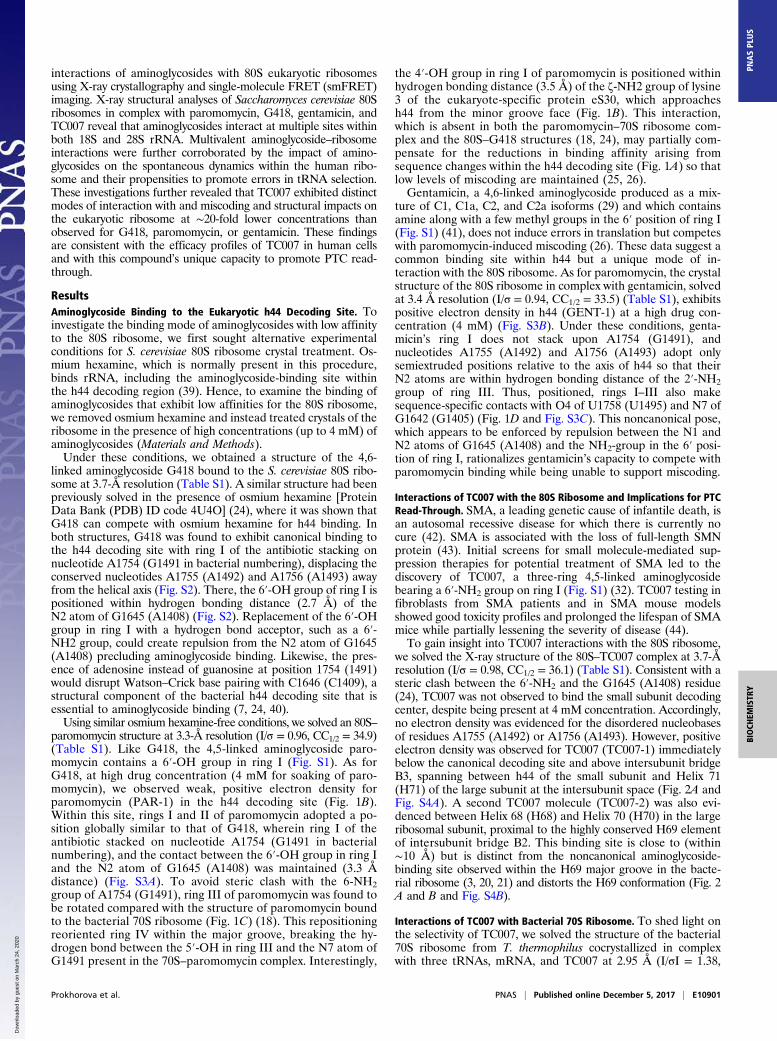
Fig. 1. Aminoglycosides target the decoding center ofthe 80S ribosome in a different ways. (A) Secondarystructure of h44 of the small ribosomal subunit frombacteria (E. coli) and eukaryotes (identical in S. cerevisiaeand Homo sapiens). Substituted nucleotides implicatedin the selectivity of aminoglycosides are marked in red.(B) Binding of paromomycin (PAR-1) to h44 in the 80Sribosome from S. cerevisiae. Paromomycin is coloredviolet, and rings I, II, III, and IV of paromomycin aremarked. Ring I is in stacking with A1754. ResiduesA1754, and G1645 are colored pink. h44 is shown inorange; H69 of the large ribosomal subunit is shownin light blue; and the eukaryote-specific proteineS30 is shown in green. Oxygen atoms are coloredred, and nitrogen atoms are colored blue. (C) Com-parison of PAR-1 binding to h44 in the 70S ribosomefrom T. thermophilus (PDB ID code 5EL6) and the 80Sribosome from S. cerevisiae. Paromomycin in complexwith 70S is shown in yellow; residues of 16S rRNA of70S are in green; other color-coding is as in B. Theshift in the position of A1754 and the movement of5′-OH group in ring III of paromomycin are markedwith arrows. (D) Binding of gentamicin (GENT-1) toh44 in the 80S ribosome from S. cerevisiae. Genta-micin is shown in green; other color-coding is as inB. Rings I, II, and III of gentamicin are marked, andatoms located at a hydrogen bonding distance areconnected by dashed lines.
E10900 | www.pnas.org/cgi/doi/10.1073/pnas.1715501114 Prokhorova et al.
Dow
nloa
ded
by g
uest
on
Mar
ch 2
4, 2
020
interactions of aminoglycosides with 80S eukaryotic ribosomesusing X-ray crystallography and single-molecule FRET (smFRET)imaging. X-ray structural analyses of Saccharomyces cerevisiae 80Sribosomes in complex with paromomycin, G418, gentamicin, andTC007 reveal that aminoglycosides interact at multiple sites withinboth 18S and 28S rRNA. Multivalent aminoglycoside–ribosomeinteractions were further corroborated by the impact of amino-glycosides on the spontaneous dynamics within the human ribo-some and their propensities to promote errors in tRNA selection.These investigations further revealed that TC007 exhibited distinctmodes of interaction with and miscoding and structural impacts onthe eukaryotic ribosome at ∼20-fold lower concentrations thanobserved for G418, paromomycin, or gentamicin. These findingsare consistent with the efficacy profiles of TC007 in human cellsand with this compound’s unique capacity to promote PTC read-through.
ResultsAminoglycoside Binding to the Eukaryotic h44 Decoding Site. Toinvestigate the binding mode of aminoglycosides with low affinityto the 80S ribosome, we first sought alternative experimentalconditions for S. cerevisiae 80S ribosome crystal treatment. Os-mium hexamine, which is normally present in this procedure,binds rRNA, including the aminoglycoside-binding site withinthe h44 decoding region (39). Hence, to examine the binding ofaminoglycosides that exhibit low affinities for the 80S ribosome,we removed osmium hexamine and instead treated crystals of theribosome in the presence of high concentrations (up to 4 mM) ofaminoglycosides (Materials and Methods).Under these conditions, we obtained a structure of the 4,6-
linked aminoglycoside G418 bound to the S. cerevisiae 80S ribo-some at 3.7-Å resolution (Table S1). A similar structure had beenpreviously solved in the presence of osmium hexamine [ProteinData Bank (PDB) ID code 4U4O] (24), where it was shown thatG418 can compete with osmium hexamine for h44 binding. Inboth structures, G418 was found to exhibit canonical binding tothe h44 decoding site with ring I of the antibiotic stacking onnucleotide A1754 (G1491 in bacterial numbering), displacing theconserved nucleotides A1755 (A1492) and A1756 (A1493) awayfrom the helical axis (Fig. S2). There, the 6′-OH group of ring I ispositioned within hydrogen bonding distance (2.7 Å) of theN2 atom of G1645 (A1408) (Fig. S2). Replacement of the 6′-OHgroup in ring I with a hydrogen bond acceptor, such as a 6′-NH2 group, could create repulsion from the N2 atom of G1645(A1408) precluding aminoglycoside binding. Likewise, the pres-ence of adenosine instead of guanosine at position 1754 (1491)would disrupt Watson–Crick base pairing with C1646 (C1409), astructural component of the bacterial h44 decoding site that isessential to aminoglycoside binding (7, 24, 40).Using similar osmium hexamine-free conditions, we solved an 80S–
paromomycin structure at 3.3-Å resolution (I/σ = 0.96, CC1/2 = 34.9)(Table S1). Like G418, the 4,5-linked aminoglycoside paro-momycin contains a 6′-OH group in ring I (Fig. S1). As forG418, at high drug concentration (4 mM for soaking of paro-momycin), we observed weak, positive electron density forparomomycin (PAR-1) in the h44 decoding site (Fig. 1B).Within this site, rings I and II of paromomycin adopted a po-sition globally similar to that of G418, wherein ring I of theantibiotic stacked on nucleotide A1754 (G1491 in bacterialnumbering), and the contact between the 6′-OH group in ring Iand the N2 atom of G1645 (A1408) was maintained (3.3 Ådistance) (Fig. S3A). To avoid steric clash with the 6-NH2group of A1754 (G1491), ring III of paromomycin was found tobe rotated compared with the structure of paromomycin boundto the bacterial 70S ribosome (Fig. 1C) (18). This repositioningreoriented ring IV within the major groove, breaking the hy-drogen bond between the 5′-OH in ring III and the N7 atom ofG1491 present in the 70S–paromomycin complex. Interestingly,
the 4′-OH group in ring I of paromomycin is positioned withinhydrogen bonding distance (3.5 Å) of the ζ-NH2 group of lysine3 of the eukaryote-specific protein eS30, which approachesh44 from the minor groove face (Fig. 1B). This interaction,which is absent in both the paromomycin–70S ribosome com-plex and the 80S–G418 structures (18, 24), may partially com-pensate for the reductions in binding affinity arising fromsequence changes within the h44 decoding site (Fig. 1A) so thatlow levels of miscoding are maintained (25, 26).Gentamicin, a 4,6-linked aminoglycoside produced as a mix-
ture of C1, C1a, C2, and C2a isoforms (29) and which containsamine along with a few methyl groups in the 6′ position of ring I(Fig. S1) (41), does not induce errors in translation but competeswith paromomycin-induced miscoding (26). These data suggest acommon binding site within h44 but a unique mode of in-teraction with the 80S ribosome. As for paromomycin, the crystalstructure of the 80S ribosome in complex with gentamicin, solvedat 3.4 Å resolution (I/σ = 0.94, CC1/2 = 33.5) (Table S1), exhibitspositive electron density in h44 (GENT-1) at a high drug con-centration (4 mM) (Fig. S3B). Under these conditions, genta-micin’s ring I does not stack upon A1754 (G1491), andnucleotides A1755 (A1492) and A1756 (A1493) adopt onlysemiextruded positions relative to the axis of h44 so that theirN2 atoms are within hydrogen bonding distance of the 2′-NH2group of ring III. Thus, positioned, rings I–III also makesequence-specific contacts with O4 of U1758 (U1495) and N7 ofG1642 (G1405) (Fig. 1D and Fig. S3C). This noncanonical pose,which appears to be enforced by repulsion between the N1 andN2 atoms of G1645 (A1408) and the NH2-group in the 6′ posi-tion of ring I, rationalizes gentamicin’s capacity to compete withparomomycin binding while being unable to support miscoding.
Interactions of TC007 with the 80S Ribosome and Implications for PTCRead-Through. SMA, a leading genetic cause of infantile death, isan autosomal recessive disease for which there is currently nocure (42). SMA is associated with the loss of full-length SMNprotein (43). Initial screens for small molecule-mediated sup-pression therapies for potential treatment of SMA led to thediscovery of TC007, a three-ring 4,5-linked aminoglycosidebearing a 6′-NH2 group on ring I (Fig. S1) (32). TC007 testing infibroblasts from SMA patients and in SMA mouse modelsshowed good toxicity profiles and prolonged the lifespan of SMAmice while partially lessening the severity of disease (44).To gain insight into TC007 interactions with the 80S ribosome,
we solved the X-ray structure of the 80S–TC007 complex at 3.7-Åresolution (I/σ = 0.98, CC1/2 = 36.1) (Table S1). Consistent with asteric clash between the 6′-NH2 and the G1645 (A1408) residue(24), TC007 was not observed to bind the small subunit decodingcenter, despite being present at 4 mM concentration. Accordingly,no electron density was evidenced for the disordered nucleobasesof residues A1755 (A1492) or A1756 (A1493). However, positiveelectron density was observed for TC007 (TC007-1) immediatelybelow the canonical decoding site and above intersubunit bridgeB3, spanning between h44 of the small subunit and Helix 71(H71) of the large subunit at the intersubunit space (Fig. 2A andFig. S4A). A second TC007 molecule (TC007-2) was also evi-denced between Helix 68 (H68) and Helix 70 (H70) in the largeribosomal subunit, proximal to the highly conserved H69 elementof intersubunit bridge B2. This binding site is close to (within∼10 Å) but is distinct from the noncanonical aminoglycoside-binding site observed within the H69 major groove in the bacte-rial ribosome (3, 20, 21) and distorts the H69 conformation (Fig. 2A and B and Fig. S4B).
Interactions of TC007 with Bacterial 70S Ribosome. To shed light onthe selectivity of TC007, we solved the structure of the bacterial70S ribosome from T. thermophilus cocrystallized in complexwith three tRNAs, mRNA, and TC007 at 2.95 Å (I/σI = 1.38,
Prokhorova et al. PNAS | Published online December 5, 2017 | E10901
BIOCH
EMISTR
YPN
ASPL
US
Dow
nloa
ded
by g
uest
on
Mar
ch 2
4, 2
020

CC1/2 = 47.1) (Table S1), where the drug was at 50-fold excessover the ribosome (Materials and Methods). As expected, weobserved strong positive electron density in the h44 decodingsite, where rings I and II were observed to overlap with those ofparomomycin (Fig. S4C). In contrast to ring III of paromomycin,which interacts with the Hoogsteen face of G1491 (Escherichiacoli numbering), the O7 atom of ring III of TC007 is positionedwithin hydrogen bonding distance of N4 of C1407. When TC007was soaked into preformed crystals of the same 70S–tRNA–
mRNA complex (500 μM for 24 h), we were able to obtain astructure at 3.05 Å resolution (I/σI = 1.25, CC1/2 = 39.9) (TableS1) in which TC007 was bound in an identical position within h44(TC007–h44) (Fig. S4D). As for neomycin, gentamicin, andparomomycin binding to H69 within the Escherichia coli 70S ri-bosome (3, 20, 21), TC007 was observed to make an array ofhydrogen bonding contacts with bases lining the H69 majorgroove, including sequence-specific contacts with residuesG1921 and G1922 (Fig. S4 B and E). Remarkably, the orienta-tion of TC007 is different from that in a previously reported70S–neomycin structure (21): The position of ring II is similarin the two antibiotics, but the positions of rings I and III areswapped. As for gentamicin, intersubunit bridging contactspresent in both paromomycin and neomycin structures were notobserved due to the absence of ring IV. As H69 is stericallyaccessible within the eukaryotic 80S ribosome, we attribute theobserved differences in TC007 binding to Ψ2264 (G1921) andC2265 (G1922) substitutions present in the 80S ribosome (Fig.S4 B and F), which likely disrupt the drug’s capacity to packtightly against the floor of the major groove. These findingsconfirm that the TC007-binding sites in the T. thermophilus 70Sribosome are distinct from those found in the 80S ribosome ofS. cerevisiae (Fig. S4B).
Aminoglycosides Inhibit Intersubunit Rotation of the EukaryoticRibosome. To assess the impact of aminoglycosides on the dy-namics of subunit rotation within functional 80S ribosomecomplexes, we performed smFRET imaging on surface-immobilized human 80S pretranslocation (PRE) complexes inthe absence and presence of paromomycin, G418, gentamicin,and TC007 (45). As previously reported (45), in the absence ofdrug, human PRE complexes predominantly exhibited a mixtureof lower-FRET (H2: ∼0.2 and H1: ∼0.4) hybrid state configu-
rations in which the 3′-CCA end of deacylated tRNA occupiesthe large subunit E-site and the 3′-CCA end of peptidyl-tRNAoccupies either the aminoacyl (A)- or P-site, respectively (A/Aand P/E; A/P and P/E) (Fig. 3 A and B, Left). The addition ofparomomycin to the 80S PRE complex first lowered the averageFRET value of the hybrid-state tRNA configurations, suggestinga relative stabilization of the H2 hybrid state in which peptidyl-tRNA returns to its classic (A/A) position while deacylatedtRNA remains in its hybrid (P/E) state (46). This impact wasmaximized at a drug concentration of ∼10 μM. Above this con-centration, we observed the appearance of an intermediate-FRET (∼0.55) state, followed by stabilization of a high-FRET,classic (C) PRE complex conformation (Fig. 3B). The estimatedEC50 of this effect was ∼35 μM. These data are consistent with amultivalent impact of paromomycin binding to the 80S PREcomplex, which first stabilizes peptidyl-tRNA in its classic posi-tion within the A-site, followed by a shift of deacylated P-sitetRNA from its hybrid to its classic position. By contrast, G418,which also bears a ring I 6′-OH substituent, only stabilized at anintermediate-FRET state (∼0.55) (Fig. 3C). Notably, consistentwith the concentration range used to inhibit mammalian cellculture growth (47), the EC50 of this impact was ∼2 mM. Byanalogy to bacterial systems, the intermediate-FRET state mayreflect a reversal of subunit rotation from the P/E hybrid state,which promotes a chimeric, intersubunit hybrid configuration ofdeacylated P-site tRNA associated with the global inhibition oftranslation factor binding (20, 21).Analogous investigations of gentamicin, which contains a 6′-
NH2 group on ring I, also led to intermediate-FRET (∼0.55)state stabilization, but the EC50 of its impact was approximatelyan order of magnitude lower (∼100 μM) than that of G418 (Fig.3D). TC007, which also has a 6′-NH2 group on ring I, exhibitedimpacts on the 80S PRE complex that shared characteristics ofG418 and gentamicin as well as paromomycin, in which drugbinding was bimodal in nature. At concentrations below 2 μM,TC007 predominantly promoted a lower-FRET PRE complexconformation, in line with H2 hybrid state stabilization (A/A;P/E). Higher TC007 concentrations increasingly stabilized anintermediate-FRET (∼0.55) state, similar to that evidenced inthe presence of subsaturating paromomycin and saturating G418and gentamicin concentrations (Fig. 3E). At 10 μM, both H2 andintermediate-FRET PRE complex conformations persisted. Above
G1645
A B
C2248
G2249
G2250
A2309
U2310G2311
H69 (60S-TC007)H69 (60S-TC007)H69 (60S-GENT)H69 (60S-GENT)
H70H70
H68H68
TC007-2TC007-2
h44h44h44h44
TC007-1TC007-1TC007-1TC007-1
H71H71
H69H69
H70H70
h44h44h44h44
h69h69h69h69 TC007-2TC007-2TC007-2TC007-2
TC007-1TC007-1TC007-1TC007-1
G2305
C2304
A2303
A1749
G1748
G1747 B3 bridgeB3 bridgeB3 bridgeB3 bridge
TC007-2TC007-2TC007-2TC007-2
Fig. 2. Interactions of the aminoglycoside derivative TC007 with the 80S ribosome. (A, Left) The view from the A-site of the ribosome is indicated. (Right)Enlarged view showing binding sites of TC007-1 and TC007-2 in close proximity to h44 of the small ribosomal subunit and H69 of the large ribosomal subunit,respectively. The regions of h44 of the small ribosomal subunit and H71 of the large ribosomal subunit, which form intersubunit bridge B3, are colored violet.The 40S subunit is shown in orange. Contacts of TC007 and rRNA residues are depicted by dashed lines. The 60S subunit is shown in blue, and TC007 is inmagenta. Oxygen atoms are colored red, and nitrogen atoms are colored blue. (B) Close-up view of the second binding site of TC007-2 located betweenhelices H68, H69, and H70 of the large ribosomal subunit. Local alignment demonstrates that TC007-2 would clash with the phosphate group of C2248 ifH69 adopted the conformation characterized by the different rotational state of the ribosomal subunits as observed in the 80S–gentamicin structure. The 60Ssubunit from the 80S–gentamicin structure is colored gray; residue C2248 is colored lime and is marked with spheres; other color-coding is as in A.
E10902 | www.pnas.org/cgi/doi/10.1073/pnas.1715501114 Prokhorova et al.
Dow
nloa
ded
by g
uest
on
Mar
ch 2
4, 2
020
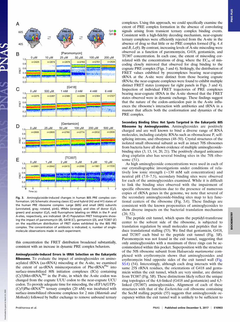
this concentration the FRET distribution broadened substantially,consistent with an increase in dynamic PRE complex behaviors.
Aminoglycoside-Induced Errors in tRNA Selection on the EukaryoticRibosome. To evaluate the impact of aminoglycosides on amino-acylated tRNA (aa-tRNA) miscoding at the A-site, we examinedthe extent of aa-tRNA misincorporation of Phe-tRNAPhe intosurface-immobilized 80S initiation complexes (ICs) containing(Cy3)Met-tRNAi
Met in the P-site, in which the A-site codon waschanged from the cognate UUU codon to the near-cognate UCUcodon. To provide adequate time for miscoding, the eEF1A(GTP)-(Cy5)Phe-tRNAPhe ternary complex (20 nM) was incubated withsurface-immobilized ribosome complexes for 2 min (Materials andMethods) followed by buffer exchange to remove unbound ternary
complexes. Using this approach, we could specifically examine theextent of PRE complex formation in the absence of convolutingsignals arising from transient ternary complex binding events.Consistent with a high-fidelity decoding mechanism, near-cognateternary complexes were efficiently rejected from the A-site in theabsence of drug so that little or no PRE complex formed (Fig. 4 Aand B, Left). By contrast, increasing levels of A-site miscoding wereobserved as a function of paromomycin, G418, gentamicin, andTC007 concentration. In each case, the extent of miscoding cor-related with the concentrations of drug, where the EC50 of mis-coding closely mirrored that observed for drug binding to thecognate PRE complex (Figs. 3 and 4). Strikingly, the distribution ofFRET values exhibited by precomplexes bearing near-cognatetRNA at the A-site were distinct from those bearing cognatetRNAs; the near-cognate complexes were found to exhibit multipledistinct FRET states (compare far right panels in Figs. 3 and 4).Inspection of individual FRET trajectories of PRE complexesbearing near-cognate tRNA in the A-site showed that the FRETstates observed were in dynamic exchange. These findings suggestthat the nature of the codon–anticodon pair in the A-site influ-ences the ribosome’s interaction with antibiotics and tRNA in amanner that affects both the conformation and dynamics of thePRE complex.
Secondary Binding Sites: Hot Spots Targeted in the Eukaryotic 80SRibosome by Aminoglycosides. Aminoglycosides are positivelycharged and are well known to bind a diverse range of RNAmolecules, including catalytic RNAs such as ribonuclease P, self-splicing introns, and ribozymes (48–50). Crystal structures of theisolated small ribosomal subunit as well as intact 70S ribosomesfrom bacteria have all shown evidence of multiple aminoglycoside-binding sites (3, 13, 14, 20, 21). The positively charged anticancerdrug cisplatin also has several binding sites in the 70S ribo-some (51).As high aminoglycoside concentrations were used in each of
our crystallographic investigations under conditions of rela-tively low ionic strength (∼130 mM salt concentration) andneutral pH (7.0–7.5), secondary binding sites were observedfor each of the aminoglycosides examined. While it is difficultto link the binding sites observed with the impairment ofspecific ribosome functions due to the presence of numerouscopies of rRNA genes in the genome, we note that several ofthe secondary aminoglycoside-binding sites map to key func-tional centers of the ribosome (Fig. 5A). These findings areconsistent with the known propensities of aminoglycosides toshow diverse impacts on the bacterial translation mechanism(20, 52).The peptide exit tunnel, which spans the peptidyl-transferase
center to the solvent side of the ribosome, is subjected totranslation regulation by small molecules and peptides that in-duce translational stalling (53). We find that gentamicin, G418,and TC007 each bind to the peptide exit tunnel (Fig. 5B).Paromomycin was not found in the exit tunnel, suggesting thatonly aminoglycosides with a maximum of three rings can be ac-commodated within this pocket. Superposition with the structureof the 50S ribosome subunit from Haloarcula marismortui com-plexed with erythromycin shows that aminoglycosides anderythromycin bind opposite sides of the exit tunnel wall (Fig.S5A) (54). Interestingly, although each drug interacts with thesame 25S rRNA residues, the orientations of G418 and genta-micin within the exit tunnel, which are very similar, are distinctfrom TC007 (Fig. 5B). These distinctions likely reflect the uniquering topologies of the 4,6-linked (G418 and gentamicin) and 4,5-linked (TC007) aminoglycosides. Alignment of each of thesestructures with that of the Escherichia coli ribosome containingthe SecM stalling peptide (55) suggests that aminoglycoside oc-cupancy within the exit tunnel wall is unlikely to be sufficient to
0.0
0.2
0.4
0.6
0.8
1.0
FRE
T
[Paromomycin]
n=2783 n=2425 n=2874 n=2878 n=2761 n=2626
25 µM 100 µM 0 µM 50 µM 10 µM 200 µM
0.0
0.2
0.4
0.6
0.8
1.0
[G418] 1 mM 4 mM 0 µM 2 mM 500 µM 6 mM
n=2295 n=2275 n=2287 n=2380 n=2364 n=2327
FRE
T
0.0
0.2
0.4
0.6
0.8
1.0
[Gentamicin] 50 µM 200 µM 0 µM 100 µM 25 µM 500 µM
n=2774 n=2908 n=2805 n=2780 n=2632 n=2606
FRE
T
0 10 10.0
0.2
0.4
0.6
0.8
1.0
FRE
T
[TC007] 2 µM 5 µM 0 µM 3 µM 1 µM 10 µM
n=2090 n=2681 n=2642 n=2415 n=1861 n=1530
1 2
1
111 1Time (s)
PRE-H2 Cy3Cy5
PRE-H1PRE-C60S
40S
A
B
C
D
E
Fig. 3. Aminoglycoside-induced changes in human 80S PRE complex con-formation. (A) Schematic showing classic (C) and hybrid (H2 and H1) states ofthe human PRE ribosome complex. Large (60S) and small (40S) subunits(unrotated, gray; rotated, pink), tRNAs (orange), and sites of donor (Cy3,green) and acceptor (Cy5, red) fluorophore labeling on tRNA in the P- andA-sites, respectively, are indicated. (B–E) Population FRET histograms show-ing the impact of paromomycin (B), G418 (C), gentamicin (D), and TC007 (E)on the equilibrium distribution of FRET states exhibited by the 80S PREcomplex. The concentration of antibiotic is indicated; n, number of single-molecule observations made in each experiment.
Prokhorova et al. PNAS | Published online December 5, 2017 | E10903
BIOCH
EMISTR
YPN
ASPL
US
Dow
nloa
ded
by g
uest
on
Mar
ch 2
4, 2
020

block the path of nascent peptides lacking bulky amino acid sidechains (Fig. S5B) (56, 57).The E-site of the small ribosomal subunit is a potential point
of regulation of eukaryotic protein synthesis (45, 58, 59). Weobserve that paromomycin (PAR-3), geneticin (G418-3), andgentamicin (GENT-2) bind in the space normally occupied bythe E-site mRNA codon and exhibit similar ring I and II posi-tions (Fig. 5C). Aminoglycoside binding in this region may haveimportant impacts on tRNA occupation within the translatingribosome (45) and/or alter the mRNA-binding and -scanningmechanism required for translation initiation (60). Similar to thebinding mode of streptomycin to the bacterial 30S subunit (14),gentamicin (GENT-3) was also found within <4 Å of the mRNAbackbone wedged between helices 1, 44, 18, and 27 within the A-site, where it interacts with the phosphate groups of residuesU9 and A11 (Fig. S6 A–C). In contrast to streptomycin, however,gentamicin does not appear to induce substantial conformationalchanges in h44 and h45. This same pocket was also occupied bygeneticin (G418-4) (Fig. S6C). In addition to potential impactson the initiation mechanism, binding in such proximity to thedecoding region may influence tRNA selection and translocationof the mRNA–tRNA module during the elongation phase ofprotein synthesis.Aminoglycosides interact extensively with the intersubunit
region of the ribosome. Paromomycin (PAR-3) provides addi-tional contacts between the subunits in the vicinity of bridge B2cformed by h24 of the small subunit and H66 of the large subunit(Fig. 5D). Gentamicin (GENT-4, 5) is also observed to bindbridge B2c (Fig. 5E), and GENT-6 binds bridge B4, a protein–RNA bridge comprised of H34 of the large subunit and proteinuS15 of the small subunit (Fig. 5F). The interactions of theGENT-5 molecule with bridge B2c are expected to precludebridge B2c rearrangements relative to h24 in the small subunit(Fig. 5E). These findings, together with the observation thatTC007 binds close to bridge B2c (Fig. S6D) and B3 (Fig. 2A), areconsistent with aminoglycosides interfering with intersubunitrotation in distinct ways to affect the translation mechanism.Such distinctions may impact the mechanism of translocation inparticular, which requires dramatic remodeling events within thecentral bridge B2 domain (20, 21), as well as rearrangements inB4 (61, 62).Additional impacts on the elongation mechanism may also
arise from aminoglycoside binding in proximity to the peptidyl-transferase center. Paromomycin (PAR-4) is located just 3 Åaway from the phosphate groups of the catalytic residues A2820(A2450) and C2821 (C2451) and 3.4 Å away from C75 of the A-site tRNA (Fig. S7A). Aminoglycosides are also observed to bindthe base of the P-stalk and sarcin–ricin loop (H95), elements ofthe large ribosomal subunit that interact with translation elon-gation factors (Fig. 5G and Fig. S7B).
DiscussionThe structural and functional insights into aminoglycoside in-teractions with the 80S eukaryotic ribosome obtained throughthe present investigations serve as a foundation for exploring themolecular mechanisms of aminoglycoside action in eukaryotes.Although the impairment of mitochondrial translation is con-sidered one of the main causes of side effects produced byaminoglycosides in eukaryotic cells (63, 64), emerging evidence
0.0
0.2
0.4
0.6
0.8
1.0
0.0
0.2
0.4
0.6
0.8
1.0
0.0
0.2
0.4
0.6
0.8
1.0
0.0
0.2
0.4
0.6
0.8
1.0
FRE
T
[Paromomycin]
n=2225 n=304 n=1117 n=1630 100 µM 0 µM 200 µM 50 µM
[G418] 2 mM 0 µM 4 mM 1 mM
n=797 n=304 n=597 n=690
FRE
T
[Gentamicin] 200 µM 0 µM 500 µM 100 µM
FRE
T
0 1
FRE
T
[TC007] 5 µM 0 µM 10 µM 2 µM
n=2318 n=304 n=817 n=2606
111Time (s)
2
A/T
GTPHYDROLYSIS
Cy3Cy5
PRE-HPRE-CPBF
60S
40S
n=304 n=1418 n=2261n=1161
A
B
Fig. 4. Aminoglycoside-induced miscoding during tRNA selection on thehuman ribosome. (A) Schematic showing the process of tRNA selection, inwhich the ternary complex of eEF1A (blue), GTP, and aa-tRNA (orange) en-ters the A-site of the 80S ribosome. The process of tRNA selection proceedsthrough the A/T state in which codon–anticodon pairing on the small sub-unit occurs while the 3′-aminoacylated CCA-end of tRNA remains bound toeEF1A. GTP hydrolysis by eEF1A facilitates the aa-tRNA release from eEF1Aand the accommodation of its ~3′CCA end into the large subunit A-site on theclassically (C) configured (unrotated) PRE complex. Peptidyl- and aminoacyl-tRNAs in the P- and A-sites, respectively, then undergo peptide bond for-mation (PBF), enabling deacylated P-site tRNA and peptidyl-tRNA to achievehybrid (H2 and H1) states. Large (60S) and small (40S) subunits (unrotated,gray; rotated, pink), tRNAs (orange), and sites of donor (Cy3, green) and
acceptor (Cy5, red) fluorophore labeling on tRNA in the P- and A-sites, re-spectively, are indicated. (B) Population FRET histograms showing thataminoglycoside-induced errors in tRNA selection lead to the accumulation ofPRE ribosome complexes bearing near-cognate tRNA in the A-site. Theconcentration of antibiotic is indicated; n, number of single-molecule ob-servations made in each experiment.
E10904 | www.pnas.org/cgi/doi/10.1073/pnas.1715501114 Prokhorova et al.
Dow
nloa
ded
by g
uest
on
Mar
ch 2
4, 2
020

suggests that aminoglycosides also exert effects on cytosolic ri-bosomes to alter translation elongation and termination in amanner that induces read-through of PTCs (27, 65). Thesefindings have led to their consideration as potent drugs to treathuman diseases caused by PTCs (28). Aminoglycosides alsooperate against eukaryotic human pathogens, including Leish-mania and Trypanosoma families (66, 67), due to sequence var-iations in their canonical h44 decoding sites (Fig. S8).The concentrations of aminoglycosides required for eukaryotic
cell growth inhibition and the EC50 values measured for the in-hibition of eukaryotic translation by distinct aminoglycosides invitro generally correlate with their affinities for the canonicalh44 decoding region within the small subunit A-site (23, 68). Asillustrated by crystal structures of the 80S ribosome with genta-micin and TC007, which adopt noncanonical poses in the vicinityof the h44 decoding site, the eukaryotic-specific G1645 residue
(equivalent to A1408 in bacteria) within h44 tends to precludethe binding of aminoglycosides containing a 6′-NH2 substituentin the ring I (Figs. 1D and 2A). Paromomycin and G418, whichcontain a 6′-OH substituent, can achieve canonical interactionswith the h44 decoding site, but the absence of Watson–Crickbase pairing between C1646 and A1754 impedes drug-bindinginteractions that hinge on ring I interactions with the floor of theaminoglycoside-binding site (Fig. 1B and Fig. S1). Human mi-tochondrial ribosomes contain an adenosine at the 1408 position(bacterial numbering), making it a good target for aminoglyco-sides with both 6′-OH and 6′-NH2 substituents in ring I (63).However, aminoglycoside affinity to mitochondrial ribosomes islikely to be attenuated by two consecutive noncanonical basepairs [C1494–A1555 (A1410–U1490) and C1493–C1556 (C1409–G1491)] that are likely to strongly disrupt ring I interactions(Fig. S8). Consistent with this notion, the reestablishment of
A
B C
D
U2140
eL37eL37
U2955
U2404
U2811C923
A917
A2138
A2139
G418-2G418-2GENT-7GENT-7TC007-3TC007-3
G418-3G418-3PAR-2PAR-2GENT-2GENT-2
E-site mRNA codonE-site mRNA codonh24h24h24h24
uS11uS11
C1006
G1768
A1005A1003
G1002
G1150
H42H42H42H42
uL6uL6Arg62
H97H97H97H97
Leu137
uS15uS15(40S)(40S)
Glu142
GENT-6GENT-6
C840
A841
G842A843
H34H34(60S)(60S)
E
GENT-4GENT-4
uL2 (60S)uL2 (60S)
G985
G986
C2179
Asp176
U2190
U981
U982
U983
FH66H66
uL2 uL2
h24 h24
60S subunit60S subunit
40S subunit40S subunit
Asp176
PAR-3PAR-3
A2152
G984
G
40S40S 60S60S
PARPARG418G418TC007TC007GENTGENT
h44h44h44h44 uS15uS15uS15uS15
h24h24h24h24h69h69h69h69
h34h34h34h34
h66h66h66h66
SRLSRLSRLSRL
P-stalkP-stalkP-stalkP-stalk
GENT-8GENT-8TC007-4TC007-4
GENT-5GENT-5
PAR-5PAR-5PAR-6PAR-6
TC007-4TC007-4GENT-8GENT-8
G418-2G418-2GENT-7GENT-7TC007-3TC007-3
PAR-3PAR-3GENT-4,5GENT-4,5TC007-5TC007-5
GENT-6GENT-6
TC007-2TC007-2
PAR-2PAR-2GENT-2GENT-2G418-4G418-4
PAR-3PAR-3GENT-4,5GENT-4,5TC007-5TC007-5
PAR-1PAR-1G418-1G418-1GENT-1GENT-1
TC007-1TC007-1 GENT-6GENT-6
G418-3G418-3GENT-3GENT-3 PAR-4PAR-4
P-stalk (GENT, TC007)P-stalk (GENT, TC007)P-stalk apo (4V88)P-stalk apo (4V88)
H66 (60S-GENT,H66 (60S-GENT,60S-PAR)60S-PAR)
h24 (40S-GENT)h24 (40S-GENT)h24 (40S-PAR)h24 (40S-PAR)
Fig. 5. Overview of the secondary binding sites ofaminoglycosides in 80S ribosome. (A) Binding sites ofgentamicin (GENT), G418, TC007, and paromomycin(PAR) in the 80S ribosome from S. cerevisiae. Allstructures were aligned either on 18S rRNA or on 28SrRNA, for small and large subunits, respectively, inthe 80S–gentamicin structure. The ribosome is col-ored light gray; elements of the binding pockets ofaminoglycosides are colored light orange; gentami-cin is colored green; G418 is colored yellow; TC007 iscolored magenta; and paromomycin is colored violet.(B) Binding of GENT-7, G418-2, and TC007-3 to thepeptide exit tunnel of the 80S ribosome. G418 iscolored yellow; gentamicin is colored light green;TC007 is colored magenta; the large ribosomal sub-unit is colored blue; and the eukaryote-specific pro-tein eL37 is colored green. Similar poses are observedfor G418-2 and GENT-7, but the orientation ofTC007-3 is different. Oxygen atoms are colored red,and nitrogen atoms are colored blue. (C) Bindingsites of PAR-2, GENT-2, and G418-3 in the E-site ofthe small ribosomal subunit overlapping the positionof the mRNA. Structures of the 80S ribosome incomplex with paromomycin, gentamicin, and G418were locally aligned on the structure of the 70S ri-bosome from T. thermophilus in complex with tRNAsand mRNA (PDB ID code 5EL6). mRNA is colored red;elements of the 70S ribosome are omitted for clarity.The 40S subunit is colored orange; paromomycin iscolored violet; the universal protein uS11 is coloredgreen; and other color-coding is as in A. (D) Interac-tions of PAR-3 with the elements of the intersubunitbridge B2c. Contacts made between paromomycinand G984 of the 40S subunit, A2152 of the 60S sub-unit, and Asp176 of uL2 are marked with dashedlines. The 40S subunit is colored light pink; paromo-mycin is colored violet; the 60S subunit is coloredblue; and the universal protein L2 (uL2) is coloredblue. Oxygen atoms are colored red, and nitrogenatoms are colored blue. (E) GENT-4 and GENT-5 sta-bilize particular conformations of bridge B2c. The80S–paromomycin structure was aligned on the 80S–gentamicin structure based on the 28S rRNA. Thealignment demonstrates that rearrangement of thebridge B2c would be blocked by gentamicin due to aclash with h24 of the 40S subunit in the 80S–paro-momycin structure (colored in pink). 80S–gentamicincontacts are marked with dashed lines. The 40Ssubunit from 80S–gentamicin structure is coloredlime; other color-coding is as in A–C. (F) GENT-6 tar-gets bridge B4 formed by the universal proteinuS15 protein and H34 of the large subunit. uS15 is shown in yellow; other color-coding is as in D. Glutamine 142, which interacts with GENT-6, is depicted asspheres. (G) Interactions of GENT-8 and TC007-4 with the elements of the ribosomal P-stalk. Different conformation of the helices 42 and 97 of the P-stalk areshown in blue for 80S-GENT and 80S-TC007 structures and in cyan for the 80S-apo structure (PDB ID code 4V88). H42 in the apo conformation would clash withGENT-8. Arg62 in the uL6 protein approaching TC007 is shown as spheres. Color-coding is as in A–E.
Prokhorova et al. PNAS | Published online December 5, 2017 | E10905
BIOCH
EMISTR
YPN
ASPL
US
Dow
nloa
ded
by g
uest
on
Mar
ch 2
4, 2
020

Watson–Crick interactions in the floor of the aminoglycoside-binding site by the naturally occurring mutations A1555G orC1494T leads to aminoglycoside hypersusceptibility in humans(69, 70).Our smFRET experiments indicate that each of the amino-
glycosides tested increases the error rate of A-site decoding (Fig.4). However, the behaviors of the miscoded 80S precomplexesbearing near-cognate tRNA in the A-site are unique for eachdrug. These findings suggest that aminoglycosides may promotePTC read-through by distinct mechanisms. Aminoglycosidescontaining 6′-OH substituent in ring I likely induce miscodingand PTC read-through due to residual binding to the canonicalbinding site in h44 of the eukaryotic ribosome. In this case, near-cognate or noncognate tRNA may efficiently accommodate atthe stop codon-programmed A-site to compete with the termi-nation factors. Aminoglycosides containing a 6′-NH2 constituentin ring I, including gentamicin and TC007, do not bind h44 in acanonical fashion. Their impacts on PTC read-through may en-tail alternative mechanisms, including intersubunit rotation ef-fects that are anticipated to hamper RF1 interactions with theclassically configured ribosome (Fig. S9) (71).The propensity of eukaryotic ribosomes to adopt rotated states
and the impact of aminoglycosides in enforcing closer interac-tions (higher FRET) between deacylated and peptidyl-tRNAwithin the 80S human PRE complex suggest that aminoglycosidebinding to intersubunit regions of the ribosome (bridges B2c andB4 for gentamicin and bridge B3 for TC007) facilitate confor-mational changes in the PRE 80S–ribosome complexes that shiftpeptidyl tRNA toward the P-site. Such impacts may relate to thestabilization of partially rotated ribosome configurations thatmove deacylated tRNA toward the A-site (20, 21).The nature of the observed aminoglycoside interactions with
the eukaryotic ribosome hint at potentially multiple modes ofaction on the translation mechanism. These insights also providean important framework for understanding the diversity ofaminoglycoside interaction sites and drug-binding modes withthe 80S ribosome. The combined perspectives afforded by X-raycrystallography and direct imaging of aminoglycoside impacts onfunctional ribosome complexes using smFRET has the potentialto facilitate the design of new antibiotic derivatives and may beparticularly suited for the identification of compounds capable ofmediating efficient PTC read-through. Such efforts will begreatly aided by in-depth functional investigations of a diversityof functional ribosome complexes relevant to termination. In thisregard, the present findings suggest that nonspecific impacts ondecoding may be reduced by avoiding aminoglycoside scaffoldsbearing a ring I 6′-OH moiety, which exhibit generally higheraffinity for the h44 decoding site, and instead focusing on ami-noglycosides, other compounds, or mixtures of compounds thatgive rise to stop codon-specific miscoding in the absence ofdominant-negative downstream impacts.
Materials and MethodsYeast and Bacterial Ribosome Purification, Crystallization, and CrystalTreatment. Ribosomes from S. cerevisiae were purified and crystallized asdescribed (39). The crystal treatment procedure was modified based on theprocedure described previously (39). Briefly, crystals were transferred to thesolution containing 80 mM Tris·acetate (pH 7.0), 70 mM KSCN, 10 mMMg(OAc)2, 20% (vol/vol) glycerol, 5% (wt/vol) PEG 20,000, 6.5 mMspermidine, 7.5 mM NH4OAc, 1.4 mM N,N’-bis-(3-D-gluconamidopropyl)deoxycholamide (Deoxy Big Chap), 2 mM DTT, and stepwise increasing con-centrations of PEG 4000, PEG 3350, or PEG 2000 MME up to 20%. The crystalswere incubated for 1 h or 4 h and were flash-frozen in a stream of liquid ni-trogen. All manipulations were performed at 4 °C. Aminoglycosides G418,paromomycin, and gentamicin were ordered from Sigma. TC007 was obtainedas described in ref. 32. High-concentration stocks of aminoglycosides wereprepared and introduced during the last steps of treatment. We observed thatsoaking in high concentrations of paromomycin or gentamicin improves thediffraction of the crystal. For example, crystals prepared in the same
conditions and soaked in 2 mM of paromomycin diffracted up to 3.7-Åresolution. Ribosomes from T. thermophilus were purified and crystal-lized as described in ref. 72. TC007 was added for cocrystallization in 50-fold excess over the ribosome concentration (70S = 1.25 μM; TC007 =62.5 μM). The crystal treatment was performed as described. If neces-sary, TC007 was added for soaking during all steps of the crystal treat-ment procedure at a concentration of 500 μM.
Purification of 40S and 60S Ribosomal Subunits from Human Cells. Preparationof small (40S) and large (60S) human ribosomal subunits was adapted fromrefs. 45 and 73. Specific deviations implemented for the purification ofpolysome fractions from human tissue culture are described here. Cell pelletswere resuspended in lysis buffer [20 mM Tris HCl (pH 7.5), 2.5 mM MgCl2,10 mM KCl, and 1 mM freshly prepared DTT] with the RNase inhibitor RNaseOut (Invitrogen), EDTA-free Halt Protease Inhibitor (Thermo Scientific), andcycloheximide (Sigma) at 100 μg/mL (∼350 μM). The solution was incubatedon ice for 10 min before centrifugation in a Microfuge 22R RefrigeratedCentrifuge (Beckman Coulter) at 14,000 rpm for 10 min at 4 °C to pellet celldebris. The supernatant was loaded onto precooled 10–50% sucrose densitygradients and spun at 35,000 rpm for 3 h at 4 °C in an Optima L-100 XP ul-tracentrifuge (Beckman Coulter). The gradients were then fractionated us-ing a BR-186-1 Fractionator and a UA-6 UV/Vis detector (Teledyne Isco).Fractions corresponding to polysomes were collected and subsequentlypelleted and dissociated into subunits according to ref. 73. Pelleted subunitswere resuspended with storage buffer [30 mM Hepes (pH 7.5), 15 mMMgCl2, 50 mM NH4Cl, 2 mM spermidine, 5 mM putrescine, 1 mM DTT, and6% sucrose] for stable, long-term storage in liquid nitrogen.
Data Collection, Crystal Structure Determination, and Analysis. Diffractiondata were collected at 90 K using 0.05° oscillation on beamline PROXIMA I atthe Soleil synchrotron (Saint-Aubin, France) equipped with a Pilatus 6Mdetector (Dectris) or on the PXI beamline at the Swiss Lightsource synchro-tron (Villigren, Switzerland) equipped with an Eiger 16M detector (Dectris).Two to eight crystals were used for each dataset. Data were reduced andscaled using the XDS suite (74). Coordinates of vacant 80S ribosome fromS. cerevisiae from PDB 4V88 were used to determine structures of 80S–paromomycin, 80S–gentamicin, and 80S–TC007. Phenix software was usedfor structure refinement, starting with several rounds of a rigid body re-finement, and validation (75, 76). P-stalk elements were disordered andwere removed from the structures except for P-stalk rRNA in the 80S–paromomycin structure and P-stalk rRNA and protein L12 in the 80S–gen-tamicin structure. Due to weak electron density, protein S31 was removedfrom the 80S–paromomycin structure, and a few structural elements wereremodeled, in particular, amino acids 103–113 of protein uL16 and residues80–87 of 5.8S rRNA. An unbiased difference electron density map (Fobs −Fcalc) was used to locate the binding sites of aminoglycosides. Ligand fittingwas performed in Coot (77). Geometry restraints for antibiotics were gen-erated with the help of Grade web server (Global Phasing, grade.global-phasing.org/cgi-bin/grade/server.cgi). Peaks of positive electron densitymaps were inspected manually to add magnesium ions, with coordinatedwater molecules often replacing osmium hexamine molecules. Manual cor-rections were followed by several iterations of reciprocal space refinementof atomic coordinates, B-factors (one isotropic B-factor per residue), andoccupancies (one occupancy value per ligand and individual occupancies formagnesium ions). Real-space refinement in Phenix was applied to fitrotamer outliers. Finally, translation–libration–screw-rotation (TLS) re-finement was performed with two TLS groups. For structure determinationof 70S–tRNA–mRNA–TC007 complexes, coordinates of the 70S ribosomefrom PDB 4WSM and tRNAs and mRNAs coordinates from PDB 4V6F wereused for two rounds of rigid-body refinements. The electron density mapswere inspected manually, and the molecules of TC007 were localized in thepeaks of positive electron density. Secondary binding sites of TC007 (four intotal) were located on the periphery of the ribosome far from the functionalcenters. Additionally, one binding site of TC007 to the intersubunit regionwas detected. Here TC007 interacts with the low part of h44 of the 30Ssubunit and the junction of helices 62 and 64 in the 25S rRNA of the 50Ssubunit between intersubunit bridges B5 and B6. Geometry restraints forTC007 were generated with the help of Grade web server (Global Phasing,grade.globalphasing.org). Manual modeling was done in Coot and wasfollowed by several rounds of reciprocal space refinement of atomic coor-dinates and B-factors. Crystallographic statistics are reported in Table S1. Allfigures were generated using PyMOL 1.5 ( https://pymol.org/2/) (Schrödinger).Local structure alignments were performed in Coot (77). Ribosomal proteinsare named throughout the paper according to the newly established no-menclature (78). Atomic coordinated and structure factors for structures
E10906 | www.pnas.org/cgi/doi/10.1073/pnas.1715501114 Prokhorova et al.
Dow
nloa
ded
by g
uest
on
Mar
ch 2
4, 2
020

of 80S–paromomycin, 80S–gentamicin, 80S–TC007, 80S–geneticin (G418), 70S-tRNAs-mRNA–TC007 (cocrystallization), and 70S–tRNAs–mRNA–TC007 (soak-ing) have been deposited in the Protein Data Bank (https://www.rcsb.org/pdb/home/home.do) under ID codes 5NDV (80S–paromomycin), 5OBM (80S–gen-tamicin), 5NDW (80S–TC007), 5NDG [80S–geneticin (G418)], 5NDK (70S–tRNA–mRNA–TC007 cocrystallized), and 5NDJ (70–tRNA–mRNA–TC007 soaked).
Preparation of Native and Fluorescently Labeled tRNAs. E. coli tRNAfMet andtRNAPhe were purified as previously described (79, 80). Aminoacylation andfluorescent labeling of tRNAs (tRNAi
Met at 4sU8 and tRNAPhe at acp3 U47positions) were performed following established protocols.
In Vitro Reconstitution of 80S ICs. As previously described (45), 80S ICs wereassembled following a procedure that bypasses the need for exogenousinitiation factors (81). Purified 40S subunits were mixed with an equal vol-ume of 80S association buffer [30 mM Hepes (pH 7.5), 5 mM MgCl2, 50 mMNH4Cl, 2 mM spermidine, 5 mM putrescine, 1 mM DTT] and then were heatactivated at 42 °C for 5 min. Fourfold excess of mRNA with the sequence 5′-CAA CCU AAA ACU UAC ACA CCC UUA GAG GGA CAA UCG AUG UUU UUUUUU UUU UUU UUU UUU-3′ (Dharmacon) (henceforth referred to as “MFFmRNA”) or 5′-CAA CCU AAA ACU UAC ACA CCC UUA GAG GGA CAA UCGAUG UCU UUC UUC UUC UUC UUC UUC-3′ (henceforth referred to as “MFFnear-cognate mRNA”) was added, heated to 37 °C for 10 min and sub-sequently cooled on ice. To this mixture, a twofold excess of fluorescentlylabeled Met-tRNAi
Met (prepared as described in ref. 82) was added, and thereaction was heated and cooled as above. At this time, equimolar amountsof 60S subunits were heat activated at 42 °C for 5 min. The 60S subunits werethen added to the mixture of 40S/tRNA/mRNA. After an additional heatingand cooling cycle, the MgCl2 concentration of the reaction was raised to15 mM, and the mixture remained on ice for 5 min. It was then loaded on a10–30% sucrose gradient in 80S association buffer and was ultracentrifugedin a Beckman SW41 rotor at 35,000 rpm for 1.5 h at 4 °C before fraction-ation. The peak corresponding to 80S complexes was collected and ali-quoted before storage in liquid nitrogen. To achieve surface immobilization,the mRNA was hybridized to a double-stranded, biotinylated DNA oligo-nucleotide (sequence 1: 5′-GTA AGT TTT AGG TTG CCC CCC TTT TTT TTT TTTTTT TTT TTT TTT TTT TTT-3′; sequence 2: 5′-AAA AAA AAA AAA AAA AAAAAA AAA AAA AAA-3′) before its mixture with the 40S subunit.
Formation of the eEF1A(GTP)–aa-tRNA Ternary Complex. aa-tRNAs (tRNAPhe,tRNAMet) were first generated as previously described (82) and were mixedwith 1 mM GTP, 6 mM phosphoenolpyruvate, 12 units/mL pyruvate kinase,and 12 units/mL myokinase. A twofold excess of eEF1A isolated from rabbitreticulocyte lysate (45), which bears 100% sequence identity with humaneEF1A, was then added, and the mixture was incubated at 37 °C for 5 min toform the ternary complex.
Single-Molecule Fluorescence Imaging. Complexes were surface-immobilizedvia the biotin–streptavidin interaction in PEG-passivated quartz chambers.All imaging experiments were performed in Hepes (KOH)-Polymix buffer(pH 7.5) containing 5 mMMgCl2, 50 mM NH4Cl, 2 mM spermidine, and 5 mMputrescine, as well as an oxygen scavenging system (2 mM protocatechuicacid, 50 nM protocatechuate 3,4-dioxygenase) together with a mixture ofsolution additives (1 mM Trolox, 1 mM cyclooctatetraene, 1 mM nitrobenzyl-
alcohol) (83) to reduce photobleaching. As previously described (82), single-molecule fluorescence imaging was performed using a custom prism-basedtotal internal reflection fluorescence microscope. Cy3 fluorophores were il-luminated with an Opus 532-nm solid-state laser (Laser Quantum), and fluo-rescence emissions from Cy3 and Cy5 fluorophores were collected using a 60×,1.27 NA Plan-Apo water immersion objective (Nikon) and were spectrallyseparated using a MultiCam-LS device (Cairn) equipped with a T635lpxr-UF2 dichroic mirror (Chroma) and imaged onto ORCA-Flash 4.0 v2 sCMOScameras (Hamamatsu). Data were acquired at 40-ms time resolution usingcustom software implemented in LabVIEW (National Instruments).
Single-Molecule tRNA Selection Assay. As previously described (45), the pro-cess of tRNA selection on the ribosome was performed by stopped-flowinjection of a 20-nM solution of ternary complex [eEF1A(GTP)–aa-tRNA]containing (Cy5)Phe-tRNAPhe into surface-immobilized ribosome complexescontaining (Cy3)tRNAi
Met in the P-site. Here, 80S ICs were formed with thenear-cognate UCU mRNA codon in the A-site, and the period of incubationwith the ternary complex was extended from 30 s to 2 min. To prevent spu-rious, nonenzymatic binding of deacylated tRNAPhe to the E-site, tRNA selec-tion experiments were performed in the presence of 500 μM cyclohexamide.Subsequent steady-state imaging of PRE complexes was performed followingbuffer exchange into a solution lacking the ternary complex.
Analysis of smFRET Data. Analysis of single-molecule fluorescence data wasperformed using the SPARTAN analysis software package MATLAB (84).Single-molecule fluorescence traces were extracted from wide-field moviesand were corrected for background, spectral crosstalk, and unequal appar-ent brightness (85). FRET trajectories were calculated as EFRET = IA/(IA + ID),where IA and ID are the acceptor and donor fluorescence intensities at eachframe, respectively. Traces were selected for further analysis according tothe following criteria: (i) single-step photobleaching; (ii) signal-to-background noise ratio >8; (iii) fewer than four donor blinking events;and (iv) >0.12 FRET efficiency for at least 50 frames (2 s). FRET histogramswere calculated from the first 50 frames of all individual molecules passingthe aforementioned criteria from each dataset with bin sizes of 0.03.
ACKNOWLEDGMENTS. We thank Daniel S. Terry for helpful discussions andsupport with smFRET data analysis; the French Infrastructure for IntegratedStructural Biology ANR-10-INSB-05-01 and Instruct, which is part of theEuropean Strategy Forum on Research Infrastructures; all staff members ofSOLEIL synchrotron PROXIMA-1 beamline; and all staff members of SwissLightsource synchrotron PX-1 beamline. This work was supported by FrenchNational Research Agency (ANR) Grant ANR-15-CE11-0021-01 (to G.Y.);Fondation ARC pour la Recherché sur le Cancer (G.Y.); La Fondation pourla Recherche Médicale Grant DBF20160635745, France (to G.Y.); EuropeanResearch Council Advanced Grant 294312 (to M.Y.); the Russian GovernmentProgram of Competitive Growth of Kazan Federal University (M.Y.); an As-sociation Française Contre les Myopathies Telethon Postdoctoral Fellowship(I.P.); and Grant ANR-10-LABX-0030-INRT from a French State Fund managedby the ANR under the frame program Investissements d’Avenir ANR-10-IDEX-0002-02 (to M.D.). M.D. is an International PhD Program fellow ofthe Institute of Genetics and Molecular and Cellular Biology supported byLabEx Integrative Biology: Nuclear Dynamics, Regenerative and Transla-tional Medicine funds. Funding support for S.C.B., R.B.A., and A.F. was pro-vided by NIH Grant GM079238 and the Tri-Institutional Stem Cell Initiativesupported by the Starr Foundation.
1. Fosso MY, Li Y, Garneau-Tsodikova S (2014) New trends in aminoglycosides use.MedChemComm 5:1075–1091.
2. Davies J (1994) Inactivation of antibiotics and the dissemination of resistance genes.Science 264:375–382.
3. Borovinskaya MA, et al. (2007) Structural basis for aminoglycoside inhibition of bac-terial ribosome recycling. Nat Struct Mol Biol 14:727–732.
4. Feldman MB, Terry DS, Altman RB, Blanchard SC (2010) Aminoglycoside activity ob-served on single pre-translocation ribosome complexes. Nat Chem Biol 6:244.
5. Youngman EM, He SL, Nikstad LJ, Green R (2007) Stop codon recognition by releasefactors induces structural rearrangement of the ribosomal decoding center that isproductive for peptide release. Mol Cell 28:533–543.
6. Davies J, Gorini L, Davis BD (1965) Misreading of RNA codewords induced by ami-noglycoside antibiotics. Mol Pharmacol 1:93–106.
7. Fourmy D, Recht MI, Blanchard SC, Puglisi JD (1996) Structure of the A site of Escherichiacoli 16S ribosomal RNA complexed with an aminoglycoside antibiotic. Science 274:1367–1371.
8. Moazed D, Noller HF (1987) Interaction of antibiotics with functional sites in 16S ri-bosomal RNA. Nature 327:389–394.
9. Recht MI, Fourmy D, Blanchard SC, Dahlquist KD, Puglisi JD (1996) RNA sequencedeterminants for aminoglycoside binding to an A-site rRNA model oligonucleotide.J Mol Biol 262:421–436.
10. Carter AP, et al. (2000) Functional insights from the structure of the 30S ribosomal
subunit and its interactions with antibiotics. Nature 407:340–348.11. Ogle JM, Murphy FV, Tarry MJ, Ramakrishnan V (2002) Selection of tRNA by the ri-
bosome requires a transition from an open to a closed form. Cell 111:721–732.12. Ogle JM, et al. (2001) Recognition of cognate transfer RNA by the 30S ribosomal
subunit. Science 292:897–902.13. Matt T, et al. (2012) Dissociation of antibacterial activity and aminoglycoside ototoxicity in the
4-monosubstituted 2-deoxystreptamine apramycin. Proc Natl Acad Sci USA 109:10984–10989.14. Demirci H, et al. (2013) A structural basis for streptomycin-induced misreading of the
genetic code. Nat Commun 4:1355.15. Jenner L, Demeshkina N, Yusupova G, Yusupov M (2010) Structural rearrangements
of the ribosome at the tRNA proofreading step. Nat Struct Mol Biol 17:1072–1078.16. Demeshkina N, Jenner L, Westhof E, Yusupov M, Yusupova G (2012) A new un-
derstanding of the decoding principle on the ribosome. Nature 484:256–259.17. Rozov A, Demeshkina N, Westhof E, Yusupov M, Yusupova G (2016) New structural
insights into translational miscoding. Trends Biochem Sci 41:798–814.18. Rozov A, et al. (2016) Novel base-pairing interactions at the tRNA wobble position
crucial for accurate reading of the genetic code. Nat Commun 7:10457.19. Rozov A, Westhof E, Yusupov M, Yusupova G (2016) The ribosome prohibits the G•U
wobble geometry at the first position of the codon-anticodon helix. Nucleic Acids Res44:6434–6441.
Prokhorova et al. PNAS | Published online December 5, 2017 | E10907
BIOCH
EMISTR
YPN
ASPL
US
Dow
nloa
ded
by g
uest
on
Mar
ch 2
4, 2
020

20. Wang L, et al. (2012) Allosteric control of the ribosome by small-molecule antibiotics.Nat Struct Mol Biol 19:957–963.
21. Wasserman MR, et al. (2015) Chemically related 4,5-linked aminoglycoside antibioticsdrive subunit rotation in opposite directions. Nat Commun 6:7896.
22. Recht MI, Douthwaite S, Puglisi JD (1999) Basis for prokaryotic specificity of action ofaminoglycoside antibiotics. EMBO J 18:3133–3138.
23. Fan-Minogue H, Bedwell DM (2008) Eukaryotic ribosomal RNA determinants ofaminoglycoside resistance and their role in translational fidelity. RNA 14:148–157.
24. Garreau de Loubresse N, et al. (2014) Structural basis for the inhibition of the eu-karyotic ribosome. Nature 513:517–522.
25. Wilhelm JM, Jessop JJ, Pettitt SE (1978) Aminoglycoside antibiotics and eukaryoticprotein synthesis: Stimulation of errors in the translation of natural messengers inextracts of cultured human cells. Biochemistry 17:1149–1153.
26. Wilhelm JM, Pettitt SE, Jessop JJ (1978) Aminoglycoside antibiotics and eukaryoticprotein synthesis: Structure—function relationships in the stimulation of misreadingwith a wheat embryo system. Biochemistry 17:1143–1149.
27. Palmer E, Wilhelm JM, Sherman F (1979) Phenotypic suppression of nonsense mutantsin yeast by aminoglycoside antibiotics. Nature 277:148–150.
28. Lee HL, Dougherty JP (2012) Pharmaceutical therapies to recode nonsense mutationsin inherited diseases. Pharmacol Ther 136:227–266.
29. Baradaran-Heravi A, et al. (2017) Gentamicin B1 is a minor gentamicin componentwith major nonsense mutation suppression activity. Proc Natl Acad Sci USA 114:3479–3484.
30. Mort M, Ivanov D, Cooper DN, Chuzhanova NA (2008) A meta-analysis of nonsensemutations causing human genetic disease. Hum Mutat 29:1037–1047.
31. Xue X, et al. (2014) Synthetic aminoglycosides efficiently suppress cystic fibrosistransmembrane conductance regulator nonsense mutations and are enhanced byivacaftor. Am J Respir Cell Mol Biol 50:805–816.
32. Mattis VB, et al. (2006) Novel aminoglycosides increase SMN levels in spinal muscularatrophy fibroblasts. Hum Genet 120:589–601.
33. Keeling KM, Wang D, Conard SE, Bedwell DM (2012) Suppression of premature ter-mination codons as a therapeutic approach. Crit Rev Biochem Mol Biol 47:444–463.
34. Wagner KR, et al. (2001) Gentamicin treatment of Duchenne and Becker musculardystrophy due to nonsense mutations. Ann Neurol 49:706–711.
35. Cogan J, et al. (2014) Aminoglycosides restore full-length type VII collagen by over-coming premature termination codons: Therapeutic implications for dystrophic epi-dermolysis bullosa. Mol Ther 22:1741–1752.
36. Agrelo R, et al. (2015) A novel Werner syndrome mutation: Pharmacological treat-ment by read-through of nonsense mutations and epigenetic therapies. Epigenetics10:329–341.
37. Bordeira-Carriço R, Pêgo AP, Santos M, Oliveira C (2012) Cancer syndromes andtherapy by stop-codon readthrough. Trends Mol Med 18:667–678.
38. Mattis VB, Tom Chang CW, Lorson CL (2012) Analysis of a read-through promotingcompound in a severe mouse model of spinal muscular atrophy. Neurosci Lett 525:72–75.
39. Ben-Shem A, et al. (2011) The structure of the eukaryotic ribosome at 3.0 Å resolution.Science 334:1524–1529.
40. Pfister P, et al. (2005) Mutagenesis of 16S rRNA C1409-G1491 base-pair differentiatesbetween 6‘OH and 6’NH3+ aminoglycosides. J Mol Biol 346:467–475.
41. Byrne KM, Kershner AS, Maehr H, Marquez JA, Schaffner CP (1977) Separation ofgentamicin C-complex into five components by Craig distribution. J Chromatogr A131:191–203.
42. Iannaccone ST, Smith SA, Simard LR (2004) Spinal muscular atrophy. Curr NeurolNeurosci Rep 4:74–80.
43. Lefebvre S, et al. (1995) Identification and characterization of a spinal muscularatrophy-determining gene. Cell 80:155–165.
44. Mattis VB, Ebert AD, Fosso MY, Chang CW, Lorson CL (2009) Delivery of a read-through inducing compound, TC007, lessens the severity of a spinal muscular atro-phy animal model. Hum Mol Genet 18:3906–3913.
45. Ferguson A, et al. (2015) Functional dynamics within the human ribosome regulatethe rate of active protein synthesis. Mol Cell 60:475–486.
46. Munro JB, Altman RB, O’Connor N, Blanchard SC (2007) Identification of two distincthybrid state intermediates on the ribosome. Mol Cell 25:505–517.
47. Bar-Nun S, Shneyour Y, Beckmann JS (1983) G-418, an elongation inhibitor of 80 Sribosomes. Biochim Biophys Acta 741:123–127.
48. Stage TK, Hertel KJ, Uhlenbeck OC (1995) Inhibition of the hammerhead ribozyme byneomycin. RNA 1:95–101.
49. von Ahsen U, Davies J, Schroeder R (1991) Antibiotic inhibition of group I ribozymefunction. Nature 353:368–370.
50. Mikkelsen NE, Brännvall M, Virtanen A, Kirsebom LA (1999) Inhibition of RNase P RNAcleavage by aminoglycosides. Proc Natl Acad Sci USA 96:6155–6160.
51. Melnikov SV, Söll D, Steitz TA, Polikanov YS (2016) Insights into RNA binding by theanticancer drug cisplatin from the crystal structure of cisplatin-modified ribosome.Nucleic Acids Res 44:4978–4987.
52. Weisblum B, Davies J (1968) Antibiotic inhibitors of the bacterial ribosome. BacteriolRev 32:493–528.
53. Gupta P, et al. (2016) Nascent peptide assists the ribosome in recognizing chemicallydistinct small molecules. Nat Chem Biol 12:153–158.
54. Tu D, Blaha G, Moore PB, Steitz TA (2005) Structures of MLSBK antibiotics bound tomutated large ribosomal subunits provide a structural explanation for resistance. Cell121:257–270.
55. Zhang J, et al. (2015) Mechanisms of ribosome stalling by SecM at multiple elongationsteps. Elife 4:09684.
56. Arenz S, et al. (2016) A combined cryo-EM and molecular dynamics approach revealsthe mechanism of ErmBL-mediated translation arrest. Nat Commun 7:12026.
57. Tsui WH, et al. (2004) Dual effects of MLS antibiotics: Transcriptional modulation andinteractions on the ribosome. Chem Biol 11:1307–1316.
58. Budkevich T, et al. (2011) Structure and dynamics of the mammalian ribosomal pre-translocation complex. Mol Cell 44:214–224.
59. Wilson DN, Nierhaus KH (2006) The E-site story: The importance of maintaining twotRNAs on the ribosome during protein synthesis. Cell Mol Life Sci 63:2725–2737.
60. Kozak M (1978) How do eucaryotic ribosomes select initiation regions in messengerRNA? Cell 15:1109–1123.
61. Bock LV, Blau C, Vaiana AC, Grubmüller H (2015) Dynamic contact network betweenribosomal subunits enables rapid large-scale rotation during spontaneous trans-location. Nucleic Acids Res 43:6747–6760.
62. Liu Q, Fredrick K (2013) Contribution of intersubunit bridges to the energy barrier ofribosomal translocation. Nucleic Acids Res 41:565–574.
63. Hobbie SN, et al. (2008) Genetic analysis of interactions with eukaryotic rRNA identifythe mitoribosome as target in aminoglycoside ototoxicity. Proc Natl Acad Sci USA 105:20888–20893.
64. Böttger EC, Springer B, Prammananan T, Kidan Y, Sander P (2001) Structural basis forselectivity and toxicity of ribosomal antibiotics. EMBO Rep 2:318–323.
65. Manuvakhova M, Keeling K, Bedwell DM (2000) Aminoglycoside antibiotics mediatecontext-dependent suppression of termination codons in a mammalian translationsystem. RNA 6:1044–1055.
66. El-On J, Bazarsky E, Sneir R (2007) Leishmania major: In vitro and in vivo anti-leishmanial activity of paromomycin ointment (Leshcutan) combined with the im-munomodulator Imiquimod. Exp Parasitol 116:156–162.
67. Hobbie SN, et al. (2011) Genetic reconstruction of protozoan rRNA decoding sitesprovides a rationale for paromomycin activity against Leishmania and Trypanosoma.PLoS Negl Trop Dis 5:e1161.
68. Hobbie SN, et al. (2007) Engineering the rRNA decoding site of eukaryotic cytosolicribosomes in bacteria. Nucleic Acids Res 35:6086–6093.
69. Zhao H, et al. (2004) Maternally inherited aminoglycoside-induced and nonsyndromicdeafness is associated with the novel C1494T mutation in the mitochondrial 12S rRNAgene in a large Chinese family. Am J Hum Genet 74:139–152.
70. Prezant TR, et al. (1993) Mitochondrial ribosomal RNA mutation associated with bothantibiotic-induced and non-syndromic deafness. Nat Genet 4:289–294.
71. Brown A, Shao S, Murray J, Hegde RS, Ramakrishnan V (2015) Structural basis for stopcodon recognition in eukaryotes. Nature 524:493–496.
72. Rozov A, Demeshkina N, Westhof E, Yusupov M, Yusupova G (2015) Structural in-sights into the translational infidelity mechanism. Nat Commun 6:7251.
73. Bommer UA, et al. (1996) Ribosomes and Polysomes, eds Graham JM, Rickwood D (IRLat Oxford Univ Press, Oxford), pp 271–300.
74. Kabsch W (2010) Xds. Acta Crystallogr D Biol Crystallogr 66:125–132.75. Adams PD, et al. (2010) PHENIX: A comprehensive Python-based system for macro-
molecular structure solution. Acta Crystallogr D Biol Crystallogr 66:213–221.76. Afonine PV, et al. (2012) Towards automated crystallographic structure refinement
with phenix.refine. Acta Crystallogr D Biol Crystallogr 68:352–367.77. Emsley P, Lohkamp B, Scott WG, Cowtan K (2010) Features and development of Coot.
Acta Crystallogr D Biol Crystallogr 66:486–501.78. Ban N, et al. (2014) A new system for naming ribosomal proteins. Curr Opin Struct Biol
24:165–169.79. Dunkle JA, et al. (2011) Structures of the bacterial ribosome in classical and hybrid
states of tRNA binding. Science 332:981–984.80. Wang L, Altman RB, Blanchard SC (2011) Insights into the molecular determinants of
EF-G catalyzed translocation. RNA 17:2189–2200.81. Burgess AB, Mach B (1971) Formation of an initiation complex with purified mam-
malian ribosomal subunits. Nature 233:209–210.82. Blanchard SC, Kim HD, Gonzalez RL, Jr, Puglisi JD, Chu S (2004) tRNA dynamics on the
ribosome during translation. Proc Natl Acad Sci USA 101:12893–12898.83. Dave R, Terry DS, Munro JB, Blanchard SC (2009) Mitigating unwanted photophysical
processes for improved single-molecule fluorescence imaging. Biophys J 96:2371–2381.
84. Juette MF, et al. (2016) Single-molecule imaging of non-equilibrium molecular en-sembles on the millisecond timescale. Nat Methods 13:341–344.
85. Roy R, Hohng S, Ha T (2008) A practical guide to single-molecule FRET. Nat Methods 5:507–516.
E10908 | www.pnas.org/cgi/doi/10.1073/pnas.1715501114 Prokhorova et al.
Dow
nloa
ded
by g
uest
on
Mar
ch 2
4, 2
020