amine incorporation into insulin as catalyzed by
TRANSCRIPT

Masthead LogoFordham University
DigitalResearch@Fordham
Chemistry Faculty Publications Chemistry
1959
Amine incorporation into insulin as catalyzed bytransglutaminase / M. J. Mycek, D. D. Clarke, A.Neidle and H. Waelsch From the Department ofBiochemistry, College of Physicians and Surgeons,Columbia University, New York, and the New YorkState Psychiatric Institute, New York, New YorkM. J. MycekNew York State Psychiatric Institute
Donald Dudley Clarke PhDFordham University, [email protected]
A. Neidle
Heinrich WaelschNew York State Psychiatric Institute
Follow this and additional works at: https://fordham.bepress.com/chem_facultypubs
Part of the Biochemistry Commons
This Article is brought to you for free and open access by the Chemistry at DigitalResearch@Fordham. It has been accepted for inclusion in ChemistryFaculty Publications by an authorized administrator of DigitalResearch@Fordham. For more information, please contact [email protected].
Recommended CitationMycek, M. J.; Clarke, Donald Dudley PhD; Neidle, A.; and Waelsch, Heinrich, "Amine incorporation into insulin as catalyzed bytransglutaminase / M. J. Mycek, D. D. Clarke, A. Neidle and H. Waelsch From the Department of Biochemistry, College of Physiciansand Surgeons, Columbia University, New York, and the New York State Psychiatric Institute, New York, New York" (1959). ChemistryFaculty Publications. 39.https://fordham.bepress.com/chem_facultypubs/39

Reprinted from ARCHIVES OF BIOCHEMISTRY AND BIOPHYSICS, Volume 84, No.2, October, 1959 Copyright © 1959 by Academic Press Inc. Printed in U.S.A .
Amine Incorporation into Insulin as Catalyzed by Transglutaminase
M. J. Mycek, D. D. Clarke, A. Neidle and H. Waelsch

ARCHIVES OF BIOCHEMISTRY AND BIOPHYSICS 84, 528-540 (1959)
Amine Incorporation into Insulin as Catalyzed by Transglutaminase1
• 2
M. J. Mycek, D. D. Clarke, A. Neidle and H. Waelsch
From the Department of Biochemistry, College of Physicians and Surgeons, Columbia University, New York, and the New York State Psychiatric Institute, New York, New York
(Received June 5, 1959)
Dedicated to F. F. Nord on the Occasion of His 70th Birthday
INTRODUCTION
An enzyme system present in a variety of mammalian organs catalyzes the binding of amines to proteins (2, 3). According to our present knowledge, the -reaction may be expressed as follows:
ca++ protein + amine -----4 amine-protein + NHa .
enzyme
As shown previously, the amine is bound in covalent linkage to the acceptor protein. A variety of primary amines, among them lysine and glycinamide as well as hydroxylamine and ammonia, are incorporated into the acceptor protein, in this Ca ++-activated enzymic reaction.
Not only proteins occurring in tissue extracts but also a number of purified proteins, such as casein, pepsin, or ,8-lactoglobulin, may serve as amine acceptors; whereas others, such as bovine serum albumin or ')'-globulin incorporate only insignificant quantities of amine. It is of considerable interest to determine why amine acceptor activity is confined to only some proteins. Since strong indication had been obtained that the enzyme catalyzes the exchange of the amide group of protein-bound glutamine with the amine ( 4), the properties of the protein as amine acceptor may be determined by the amino acid sequence around an active glutamine residue. It has already been reported that zinc-free insulin is one of the most active
1 This work was supported by grants from the National Institute of Neurological Diseases and Blindness (Grant B-557) and from the Ford Foundation and by a contract between the Office of Naval Research and the Psychiatric Institute. Presented in part before the 43rd Annual Meeting of the American Society of Biological Chemists in Atlantic City, New Jersey, April13- 17, 1958 (1).
2 We are indebted to Dr. Otto K. Behrens of Eli Lilly and Co. for the generous gifts of crystalline bovine insulin employed in this investigation.
528
•

•
•
•
TRANSGLUTAMINASE ACTION ON INSULIN 529
amine-accepting substrates (4). This protein, with its known amino acid sequence (5) containing six amide groups (three glutamine and three asparagine residues), appeared to be an ideal substrate for a more detailed study of the mechanism of the amine incorporation reaction. Such an investigation could serve to generalize the results previously obtained with casein that protein-bound glutamine is the active site of amine incorporation. An answer as to whether or not all three glutamine residues participate equally in the enzymic reaction would pave the way to our understanding of the significance of the amino acid sequence around an active glutamine residue and ultimately to the synthesis of suitable substrates.
The investigation of the amine-incorporating reaction has been carried out with the aid of C14-labeled amines or N15-labeled ammonia. In the present study, hydroxylamine also was used as the replacing amine since the hydroxamate formed from an acid-soluble substrate could be measured directly as the ferric chloride complex. When acid-insoluble substrates were used, the bound hydroxylamine could be determined after hydrolysis of the protein.
It had been reported previously that ammonia is evolved during the amine incorporation reaction, a fact which first directed our attention to the possibility of the protein-bound amides as active sites. The quantity of ammonia evolved was usually in excess of the quantity of amine incorporated. Furthermore, even in the absence of an added amine, ammonia is evolved under the conditions necessary for amine incorporation. This finding led to the suggestion, supported by observation on casein as substrate, that, in the absence of added amine, the €-amino group of proteinbound lysine can act as the replacing agent ( 4). Experiments to determine the extent of the participation of lysine in cross-linking reactions with insulin as substrate are also included in this paper.
METHODS
Enzyme. The enzyme employed in these studies was the guinea pig liver preparation described previously (3).
Zinc -Free Insulin. Crystalline beef zinc- insulin (1 g.) was suspended in 20 ml. of cold water. Sodium hydroxide (6 N) was added dropwise until solution was effected. The solution was then adjusted to pH 8 and dialyzed in the cold room overnight against two changes of 7 I. each of 0.001 M sodium ethylenediamine tetracetate (pH ca. 7.5).
Separation of the A and B Chains of Insulin
After performic acid oxidation of 1 g. insulin (6), the reaction mixture was evaporated to a thin sirup in a rotary vacuum evaporator. The sirup was dissolved in 10 ml. of 50% formic acid and placed on a column (30 X 0.9 em.) of Dowex 50 X 4 (100 mesh) in the H+ form which had been washed with 20 ml. of 50% formic acid. The A

530 MYCEK, CLARKE, NEIDLE AND WAELSCH
chain was then eluted with water. It emerged with the solvent front as detected by measurement of the absorption at 270 m,u.
Following the elution of the A chain, the column was washed with water until the effluent had a pH near neutrality. Ammonium hydroxide (1 N) was then used to displace the B chain.
The fractions containing A and B chain, respectively, were concentrated to a small volume in vacuo, precipitated with acetone, and washed. Yield (A chain): 360 mg. (81 %) . Yield (B chain): 450 mg. (74%).
The basic amino acids, present in insulin only in the B chain, were not detected on two-dimensional paper chromatography (7) of the 6 N HCl hydrolyzate of these A chain preparations.
Determination of the Incorporation of Labeled A mines into Insulin
The incubation conditions are given in the legends to the tables and figures. At the end of the incubation period, insulin was precipitated by adding 50% trichloroacetic acid (TCA) to a final concentration of 5% . The precipitated protein was washed repeatedly with 5% TCA and then dissolved in a minimal amount of absolute ethanol and reprecipitated with ether. The methods for the determination of radioactivity and amide N 15 content of the recovered protein have been described (3). The quantity of amine incorporated (,umoles/ ,umole insulin) was calculated from the specific activities of the protein and the entering amine both measured at infinite thickness.
Hydroxylamine Incorporation
The extent of hydroxylamine incorporation into the A chain of insulin can be measured directly by the color reaction of the hydroxamate formed (8). An A chain sample of known hydroxamic acid content was employed as a standard. In order to obtain this material, insulin A chain following incubation with hydroxylamine and enzyme was conveniently freed of the unreacted base by passage of the reaction mixture through a short column of Dowex 50 X 4 in the H+ form. The absolute hydroxylamine content of this A chain was then determined after acid hydrolysis by the method of Csaky (9). By comparing this value with that obtained from the FeCl 3
procedure, it was found that A chain-bound glutamohydroxamic acid had a color equivalent 1.2 times that of free glutamohydroxamic acid.
Determination of N 15 Distribution
Insulin, containing 8.45 atom% excess N 15 in the amide nitrogen, was prepared enzymically by utilizing ammonia-N16 (61 atom% excess) as the replacing amine. The labeled insulin (350 mg.) was incubated with 40 mg. each of Pan-protease and Peptidase (Worthington Biochemical Corp.), in 45 ml. of 0.02 M phosphate buffer (pH 8.0) with 1 mmole sodium thioglycolate at 38°0. for 48 hr. in the presence of a few drops of toluene as a preservative. After incubation, amino nitrogen determination (10) indicated about 50% hydrolysis. The incubation mixture was dialyzed in the cold room against two changes of 300 mi. water. The dialyzate was concentrated in vacuo to 5 mi. and mixed with an equal volume of 0.2 N sodium acetate buffer pH 3.45 and placed on a 100 X 4 em. column of Dowex 50 X 8 which had been equilibrated with the same buffer. Aliquots of the effluent fractions were analyzed by t he ninhydrin procedure of Moore and Stein (11), and by treatment with half-saturated KOH in the Conway microdiffusion apparatus to locate glutamine and asparagine (12). The fractions yielding ammonia were combined and subjected to precipitation with

•
•
TRANSGLUTAMINASE ACTION ON INSULIN 531
mercuric nitrate (13). The mercury complex was resuspended in water and decomposed with H 2S. The mercuric sulfide was removed by centrifugation and washed with several portions of water. The supernatant fluid and washings were combined, concentrated, streaked on Whatman No. 1 filter paper, and chromatographed in a solvent system of butanol- formic acid- water (8: 1: 2) (7). The bands corresponding to glutamine and asparagine were eluted and concentrated. Treatment of the two concentrates with half-saturated KOH in Conway microdiffusion vessels released the amide ammonia which could then be analyzed for N 15 content in the mass spectrometer.
A minor modification of this method in which the glutamine and asparagine amide N were recovered by differential hydrolysis of the unresolved amide fraction was applied previously to localization of the ammonia-N15 incorporated into casein (4).
Determination of Lysine Bound by Its e-Amino Group
The protein (40- 50 mg.) obtained after enzymic incubation and washed in the usual manner was dissolved in 1 ml. of 90% acetic acid and 2 ml. of 50% potassium nitrite solution added dropwise with periodic stirring during the course of 2 hr. The reaction mixture was diluted to 5 ml., and 5 ml. of 10% TCA was added. The precipitated protein was separated by centrifugation and washed with 5% TCA until the washings were free of nitrite (starch- iodide test). The protein was then washed with ethanol-ether (1:5) and then with ether, and finally dried in a vacuum desiccator. The dried material was redissolved in 1 ml. of 90% acetic acid, and the deamination procedure was repeated.
Weighed samples of the protein which had been subjected twice to deamination and 2 ml. of 6 N HCl were placed in sealed tubes and heated at 100°C. for 16 hr. The hydrolyzates were taken to dryness, dissolved in water, neutralized, decolorized with activated charcoal, and made up to 5 ml. Aliquots were taken for determination of lysine by the microbiological procedure of Henderson and Snell (14).
c:-Hydroxy-a -amino caproic acid neither replaced lysine nor acted as an antagonist of the amino acid in the microbiological assay. When the deamination procedure was performed on insulin which had been incubated with enzyme in the absence of Ca++, about 3% of the total lysine content was recovered in the microbiological assay. The experimental values were corrected accordingly. The determination by this method of the extent of lysine cross-linking using casein as a substrate has been reported (4).
RESULTS AND DISCUSSION
Substrate Properties of Insulin
The incorporation of a variety of primary amines into zinc-free insulin, in the presence of calcium, is catalyzed by the same protein fraction which has been shown to incorporate amines into other proteins (3) (Table I). Insulin is unique among the proteins tested as substrates in having an absolute requirement for reducing agents (Table I), while amine incorporation into other protein substrates proceeds to an appreciable extent even in the absence of added reducing agents. The effective reducing agents tested include thioglycolate, thioethanol, glutathione, and sodium borohydride (Fig. 1).

532 MYCEK, CLARKE, NEIDLE AND WAELSCH
TABLE I
The Incorporation of A mines into Insulin
Complete system contained: insulin, 15 mg.; amine-C14 (specific activity approx. 0.05 ,uc./,umole or ammonia-N15 (32 N 15 atom % excess), 10 ,umoles; GSH, 100 ,umoles; CaCh, 5,umoles; Tris buffer, 40,umoles, pH 8.0; enzyme, 1 mg. protein. Total volume: 2.0 ml. Incubated at 38°C. for 1 hr.
Amine
Cadaverine, complete system Cadaverine, minus Ca++ Cadaverine, minus GSH Cadaverine, minus enzyme Putrescine, complete system Methylamine, complete system Histamine, complete system Ammonia, complete system
c: 1.2 ;:) II)
c:
'+- 1.0 0
Q)
0 E ::t. 0.8 .... Q)
a. Q)
c: u 0.6 II)
Q) .... +-;:)
a. '+- 0.4 0
II) Q)
0 E 0.2 ::t.
10 20 30
Glutathione
40 p.moles
ML.
Amine Insulin
1-'moles / 1-'mole
50
0.98 0.03 0.01 0.00 1.10 1.10 1.20 1.20
FrG. 1. The dependence of amine incorporation into insulin on glutathione. The conditions were the same as those in Table I except that the amine employed was putrescine-C 14 (0.015 ,uc. / ,umole) and the glutathione concentration was varied.

•
•
TRANSGLUTAMINASE ACTION ON INSULIN 533
The Site of A mine Incorporation
Previous evidence suggests that the incorporation of ammonia into casein is the result of the displacement of the amide groups of proteinbound glutamine (4). If a similar mechanism is operative for the incorporation of ammonia-N15 into insulin, then glutamine-N15 should be recoverable from an enzymic hydrolyzate of the labeled insulin (amide nitrogen =
8.45 N 15 atom % excess). When both glutamine and asparagine were recovered from such an experiment, the label was found to be present exclusively in the glutamine amide nitrogen (10.85 N 15 atom % excess; asparagine amide nitrogen = 0.05 N 15 atom %excess). It should be noted that the value for the atom % excess N 15(10.85) was lower than that expected (2 X 8.45) on the basis of the atom % excess N 15 in the total amide nitrogen of insulin. This result is probably explained by the fact that there is an unequal release of the three glutamine residues in the course of the proteolytic digestion. However, these considerations do not affect the results, namely, that only the isolated glutamine is labeled. Thus, for both insulin and casein, the incorporation of amines leads to proteins with modified glutaminyl amide groups.
Of the three glutamine residues present in insulin, two are in the A chain and one is in the B chain. Insulin, enzymically labeled with ammonia-N15
or methylamine-C14 was subjected to performic acid oxidation. The distribution of label in the isolated A and B chains was determined: 97 and 98% of the respective bases were found in the A chain (Table II).
After oxidation of insulin, the isolated A and B chains obtained were individually tested for substrate activity with hydroxylamine as the replacing amine (Table III). The A chain incorporated hydroxylamine equivalent to 1.3 .Mmoles/ J..tmole of A chain; however, B chain showed hardly any substrate activity. It should be noted that, under the experimental conditions used, A chain did not require the presence of a sulfhydryl compound for full activity as a substrate.
TABLE II
Distribution of Label after Amine Incorporation into Insulin
Conditions of incubation were the same as those in Table I.
Insulin A chain B chain
Amine incorporated
Methylamine-C14 Ammonia-N1 5
p.molesj p.mole p.molesj p.mole
0.96 0.92 0.02
0.70 0.70 0.02
534 MYCEK, CLARKE, NEIDLE AND WAELSCH
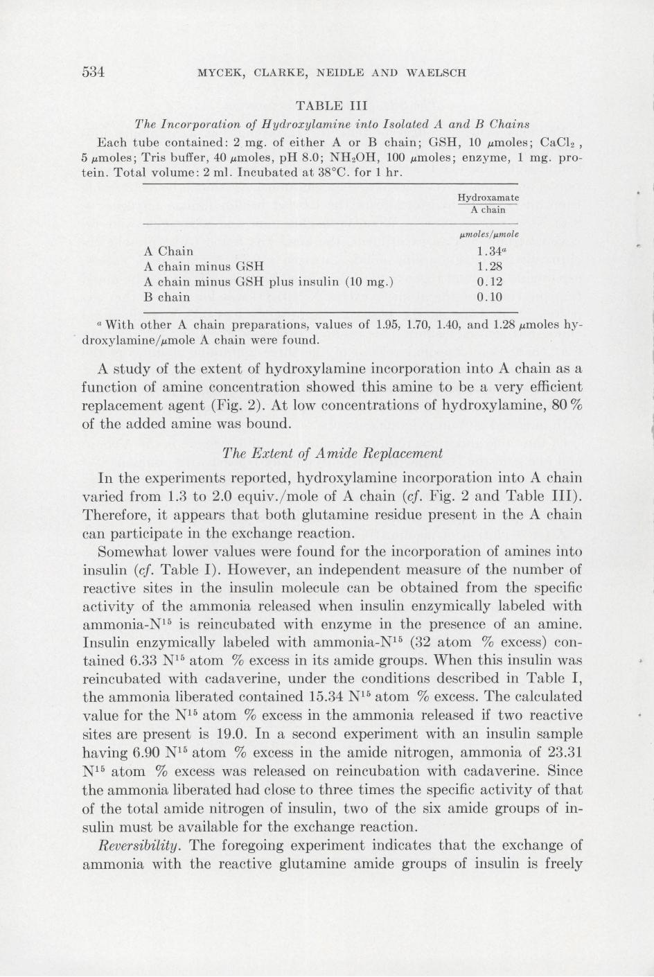
TABLE III
The Incorporation of Hydroxylamine into Isolated A and B Chains
Each tube contained: 2 mg. of either A or B chain; GSH, 10 J.Lmoles; CaCl2, 5 J.Lmoles; Tris buffer, 40 J.Lmoles, pH 8.0; NH20H, 100 J.Lmoles; enzyme, 1 mg. protein. Total volume: 2 ml. Incubated at 38°C. for 1 hr.
A Chain A chain minus GSH A chain minus GSH plus insulin (10 mg.) B chain
Hydroxama te A chain
p.molesfl.~,mole
1.34a 1.28 0 . 12 0.10
a With other A chain preparations, values of 1.95, 1.70, 1.40, and 1.28 J.Lmoles hydroxylamine/ J.Lmole A chain were found.
A study of the extent of hydroxylamine incorporation into A chain as a function of amine concentration showed this amine to be a very efficient replacement agent (Fig. 2). At low concentrations of hydroxylamine, 80% of the added amine was bound.
The Extent of Amide Replacement
In the experiments reported, hydroxylamine incorporation into A chain varied from 1.3 to 2.0 equiv. / mole of A chain (cf. Fig. 2 and Table III). Therefore, it appears that both glutamine residue present in the A chain can participate in the exchange reaction.
Somewhat lower values were found for the incorporation of amines into insulin (cf. Table I). However, an independent measure of the number of reactive sites in the insulin molecule can be obtained from the specific activity of the ammonia released when insulin enzymically labeled with ammonia-N15 is reincubated with enzyme in the presence of an amine. Insulin enzymically labeled with ammonia-N15 (32 atom % excess) contained 6.33 N 15 atom %excess in its amide groups. When this insulin was reincubated with cadaverine, under the conditions described in Table I, the ammonia liberated contained 15.34 N 15 atom % excess. The calculated value for the N 15 atom % excess in the ammonia released if two reactive sites are present is 19.0. In a second experiment with an insulin sample having 6.90 N 15 atom % excess in the amide nitrogen, ammonia of 23.31 N 15 atom % excess was released on reincubation with cadaverine. Since the ammonia liberated had close to three times the specific activity of that of the total amide nitrogen of insulin, two of the six amide groups of insulin must be available for the exchange reaction.
Reversibility. The foregoing experiment indicates that the exchange of ammonia with the reactive glutamine amide groups of insulin is freely

•
..
TRANSGLUTAMINASE ACTION ON INSULIN 535
1.4 -o
c: 0 1.2 ~ .r:. 0 I
<l cu 0 1.0 E :t ... cu a. cu 0.8 c: E 0
>-)(
0 0.6 ..... "0 >-
.r:. (/)
cu 0 0.4 E :t
0.2
I I I I I
5 10 15 20 25
. J.Lmoles Hydroxylamine ML.
FIG. 2. The incorporation of hydroxylamine into A chain. The conditions of incubation were those of Table III except that the hydroxylamine concentration was varied.
reversible. In addition, putrescine bound to insulin could be displaced by ammonia, hydroxylamine, histamine, and cadaverine.
Ammonia Release and A mine Incorporation
In Fig. 3, the quantities of ammonia released from insulin in the presence and absence of cadaverine are shown as a function of pH. At pH values of 8 and above, since all the ammonia formed during the reaction can be accounted for by the cadaverine incorporated, the amine-independent ammonia-liberating reaction is suppressed, thus suggesting that both reactions compete for common sites on the insulin molecule.
For casein, it has been shown that much of the amine-independent ammonia release results from a cross-linking reaction involving the €-amino groups of protein-bound lysine (4). However, with insulin as substrate, at most 25% of the amine-independent reaction can be accounted for in this way (Fig. 4). It is of interest that amine-independent ammonia liberation can be observed also with A chain, a substrate which contains no lysine.

536
c
:l If)
c
...... 0
C1)
0 E :t '-C1)
n If) C1)
0 E :t
MYCEK, CLARKE, NEIDLE AND WAELSCH
1.0
0.9
0.8
0.7
0.6
0.5
0.4
0.3
0.2
0.1
5 6 7
pH
+
8 9
FIG. 3. Ammonia liberation and cadaverine incorporation as a function of pH Conditions as in Table I. At end of incubation, TCA was added to each tube, and the precipitated insulin was counted at infinite thickness, the supernatant fluids were neutralized, and aliquots were taken for ammonia determination (15). Ammonia liberation in the absence of added amine ( +); ammonia release in the presence of added amine ( 0); cadaverine incorporated (e ).
GENERAL CoNSIDERATIONs
The experiments presented in this report establish that the A chain acts as the amine-accepting moiety of insulin in the enzymically catalyzed amine incorporation reaction. Not only is no amine incorporated into B chain with whole insulin as substrate, but also when employed separately the B chain proves to be inert. The necessity of adding sulfhydryl compounds to experiments with whole insulin as substrate suggests that some of the disulfide linkages between the two chains have been reduced and that the substrate was not the intact protein. Without added sulfhydryl compounds, insulin appears to inactivate the enzyme which has been shown previously to be inhibited by reagents which react with sulfhydryl groups.
•

•
•
c :::1 1/)
c
.... 0
cv 0 E :1.. '-cv a. 1/)
cv 0 E :1..
TRANSGLUTAMINASE ACTION ON INSULIN
0.7
0.6
0.5
0.3
0.2
0.1
5 6
+
+~ +
7
pH
+
8 9
537
FrG. 4. The ammonia liberation and cross-linking involving the «:-amino group o protein-bound lysine as a function of pH. The conditions are the same as those in Fig. 3, except that an amine was not present. Ammonia release in the absence of added amine ( +); micromoles of lysine recovered after deamination of the protein ( 0). (See Methods.)
In keeping with these observations, the A chain can serve as a substrate without the addition of a reducing agent. It is of interest that the presence of weakly acidic sulfhydryl groups or strongly acidic sulfonic acid groups does not interfere with the ability of insulin or the A chain, respectively, to serve as substrate.
Before the characteristics of insulin as substrate were known in detail, insulin samples, after incubation with various amines, were tested for physiological activity through the courtesy of Dr. Otto Behrens in the laboratories of Eli Lilly and Co. The various insulin samples had lost most of their activity, an observation understandable if a splitting of its disulfide groups by added reducing agents were to precede or accompany incorporation of the amine (16).
In insulin as well as in casein, N15H 3 was found to replace the amide groups of the glutamine residues but not those of asparagine. No direct evidence has been obtained that amines other than ammonia also replace
538 MYCEK, CLARKE, NEIDLE AND WAELSCH
the amide groups of glutamine. However, that this is the case is strongly suggested by the fact that these amines displace ammonia-N15 from enzymically labeled insulin upon reincubation with enzyme, calcium, and the respective amine. The incorporation of amine approaching 2 equiv./ mole of A chain indicates that the amide groups of both glutamine residues are replaceable.
In order to clarify the question as to whether or not the amino acid sequence around a given glutamine residue is necessary as a whole or in part for substrate activity, a synthesis of the corresponding peptides is under way. In preliminary experiments, oxytocin, vasopressin, corticotropin, and ribonuclease and a number of glutamine peptides3 were tested-all with negative results. On the other hand, enzymic casein hydrolyzates showed considerable substrate activity.
With insulin as substrate, the ammonia release in the absence of added amine can only be accounted for partially by cross-linking with the €-amino groups of lysine. This may be due to the fact that the lysine content of insulin (2.5 %) is lower than that of casein for which most of the ammonia liberation under these conditions could be attributed to cross-linking with lysine. The particular significance of these results is that it underlines the need for additional mechanisms to explain ammonia release in the absence of amine. Studies are in progress to determine whether a hydrolytic reaction or a displacement involving the amino groups of N-terminal amino acids accounts for this lack of stoichiometry.
There is now evidence that the amine-incorporating enzyme catalyzes an exchange reaction between a wide variety of amines and the amide ammonia of certain glutamine residues in peptide linkage. The conditions for the activity of this enzyme are obviously different from those for the glutamotransferase found in mammalian tissues (17). However, the reaction does have some resemblance to that catalyzed by the bacterial glutamotransferase enzyme (18); that is, the )'-amide of protein-bound glutamine in the former instance and free glutamine in the latter is replaced by a nucleophilic agent to form a new amide bond and ammonia. Though the specificity for the )'-amide group of glutamine appears to be the same, the bacterial glutamotransferase is not capable of incorporating amines into proteins, nor can the amine-incorporating enzyme catalyze the formation of glutamohydroxamic acid from glutamine and hydroxylamine (3). In addition, calcium is an obligatory factor for the amine-incorporating activity, but it has no influence on the activity of the bacterial transferase
3 We wish to acknowledge the gifts of vasopressin and oxytocin from Dr. D. A. McGinty of Parke, Davis and Co., of corticotropin A from Dr. W. F. White of Armour and Co., of .a-corticotropin from Dr. C. E. Graham of The Wilson Laboratories, of ribonuclease from Dr. W. H. Stein of The Rockefeller Institute, and of a variety of glutamine peptides from Dr. Klaus Hoffman of the University of Pittsburgh.
,

•
•
TRANSGLUTAMINASE ACTION ON INSULIN 539
(18). Reducing agents or sulfhydryl inhibitors have no apparent effect on the bacterial enzyme (18), in contrast to the amine-incorporating enzyme.
Since it would be convenient to refer to the amine-incorporating system by name, we propose to call this enzyme activity, transglutaminase. Thus, transglutaminase describes the activity of an enzyme system which catalyzes the exchange of amide groups of peptide-bound glutamine with a variety of compounds containing a primary amino group to yield new 'Y-amide bonds, and ammonia .
AcKNOWLEDGMENTS
The authors wish to acknowledge the capable technical assistance of Miss Gloria Catalano and Mrs. Lydia Hersh.
SUMMARY
Since insulin serves as an excellent substrate in the enzymically catalyzed amine incorporation reaction, a detailed study of its substrate properties was undertaken. A method for the separation of the A and B chains of oxidized insulin employing Dowex 50 resin is described.
Native zinc-free insulin inhibited the enzyme, an inhibition which could be overcome by the addition of glutathione or other reducing agents.
The mechanism of the reaction with insulin as substrate involves a replacement of the amide groups of peptide-bound glutamine but not of asparagine by the amine. The incorporated amines (N15H 3 , methylamine, hydroxylamine) are localized solely in the A chain of insulin. Isolated A chain but not B chain can serve as an amine acceptor.
The use of hydroxylamine as a replacing amine permits the measurement of the reactivity of acid-soluble peptides and proteins readily by determining the extent of hydroxylamine incorporation calorimetrically.
In the presence of added amine, the ammonia equivalents released correspond to those of amine incorporated at the pH optimum. In the absence of added amine, cross-linking of the protein-bound lysine accounted only for a small portion of the liberated ammonia.
The name transglutaminase is sugge ted for the enzymic activity catalyzing amine incorporation .
REFERENCES
1. NEIDLE, A., MYCEK, M. J., CLARKE, D. D., AND WAELSCH, H., Federation Proc. 18, 293 (1959).
2. SARKAR, N. K., CLARKE, D. D., AND WAELSCH, H., Biochim. et Biophys. Acta 25, 451 (1957).
3. CLARKE, D. D., MYCEK, M. J., NEIDLE, A., AND WAELSCH, H., Arch. Biochem. Biophys. 79, 338 (1959).
4. NEIDLE, A., MYCEK, M. J., CLARKE, D. D., AND WkELSCH, H., Arch. Biochem. Biophys. 77, 227 ( 1958) .

540 MYCEK, CLARKE, NEIDLE AND WAELSCH
5. SANGER, F., AND TuPPY, H., Biochem. J. 49, 463, 481 (1951); SANGER, F., AND THOMPSON, E. 0. P., Biochem. J. 53,366 (1953); RYLE, A. P., SANGER, F., SMITH, L. F., AND KIT AI, R., Biochem. J. 60, 541 (1955).
6. PIERCE, J. G., J. Am. Chem. Soc. 77, 184 (1955). 7. RoBERTs, R. B., ABELSON, P. H., CowiE, D. B., BoLTON, E. T., AND BRITTEN,
R. J., Carnegie Inst. Wash. Publ. No. 607,31 (1955). 8. LIPMANN, F., AND TuTTLE, L. C., J. Biol. Chem. 159, 21 (1954). 9. CsAKY, T. Z., Acta Chem. Scand. 2, 450 (1948).
10. VANSLYKE, D. D., J. Biol. Chem. 83, 425 (1929). 11. MooRE, S., AND STEIN, W. H., J. Biol. Chem. 211, 908 (1954). 12. MESSER, M., Biochim. et Biophys. Acta 17, 151 (1955). 13. VICKERY, H. B., AND PucHER, G. W., Biochem. Preparations 1, 44 (1949). 14. HENDERSON, L., AND SNELL, E. E., J. Biol. Chem. 172, 15 (1948). 15. CoNWAY, E. J., "Microdiffusion Analysis and Volumetric Error," 4th ed., p. 98.
The Macmillan Co., New York, 1958. 16. DU VIGNEAUD, V., FITCH, A., PEKAREK, E., AND LocKwooD, W. W., J. Biol. Chem.
94, 233 (1931). 17. LAJTHA, A., MELA, P., AND WAELSCH, H., J. Biol. Chem. 205, 553 (1953). 18. GRossowrcz, N., WAINFAN, E., BoREK, E., AND WAELSCH, H., J. Biol. Chem. 187,
111 (1950).
•