american society of neuroimaging 34th annual meeting annual meeting/handouts/razumovsky tcd... ·...
TRANSCRIPT

1142013
1
Alex Razumovsky PhD FAHA Director
Sentient NeuroCare Services Inc
Specific TCD applications for
vasospasm diagnosis and
monitoring after SAH TBI and
tumor resection
American Society of Neuroimaging
36th Annual Meeting
American Society of Neuroimaging 35th Annual Meeting
DISCLOSURES
Alexander Razumovsky PhD FAHA
Full-Time employee for Private Practice Sentient NeuroCare Services Inc
UnlabeledUnapproved Uses Disclosure None
HISTORY OF ANEURYSMS
1142013
2
History of Aneurysm Identification
bull The aneurysm originated from the Greek aneurysma where ana meant across and eurys meant broad
bull The Alexandrian surgeon Rufus of Ephesus in 177 BC studied aneurysms
bull Galen was probably first to define and describe the entity of aneurysm Antyllos in the second century AD distinguish between true and false aneurysms
bull Arabian surgeon Al-Zahrawi (Abulcasis) (913-1013 AD) performed surgical treatment of aneurysms
Galen of Pergamum (AD 129 ndash 217)
Giovanni Battista Morgagni (1682-1771)
bull Giovanni Morgagni of Padua reported an autopsy case and suggested that aneurysms could be the cause of intracranial hemorrhage
bull 1594
Tradition says the Anatomy Theatre of
Padua University is the oldest in the world
It was built and paid for by Girolamus
Fabricius ab Acquapendente who at the
time was Professor of Anatomy and
Surgery The structure was designed by
Fra Paolo Sarpi Dissection was carried
out in secret as it contravened both the civil
and religious Laws of the period
1142013
3
History of Aneurysms Clinical Diagnosis
bull The first reports of possible connection between intracranial aneurysms and SAH were by Bonet (1679) and Wiseman (1696)
bull John Blackwhall of Oxford (1813) the first to describe a ruptured intracranial aneurysm confirmed at autopsy
History of Aneurysms Clinical Diagnosis
bull Lumbar puncture In 1891 Quinke showed blood in the subarachnoid space as a result of SAH
bull Radiography Heuer and Dandy in 1916 showed intracranial aneurysm calcification on plain skull x-rays
bull Angiography In 1926 Egaz Moniz carried out the first cerebral angiogram in dogs and in 1927 in human In 1933 Moniz reported visualization of cerebral aneurysm
Quinke needles
1842-1922
1142013
4
Dr Heinrich Quinke
bull Born in Germany Quincke was educated in this country under mentors such as von Koumllliker Helmholtz and Virchow
bull Early on he held a chair in medicine but after 30 years he retired to Frankfort-am-main continue his neurologic pursuits
bull Amongst his contributions to the literature were his classic description of angioneurotic edema the studies of the mechanism of body temperatures He recognized the syndrome of meningitis serosa and wrote on anosmia traumatic brain lesions and on hyperthermia in cord lesions His introduction of the spinal puncture and procedures earned him a place in the history of medicine
Dr Egas Moniz (1874-1955)
bull In 1926 Egas Moniz carried out the first cerebral angiogram in dogs and in 1927 in human In 1933 Moniz reported visualization of cerebral aneurysm
bull In 1949 he received the Nobel Prize for his discovery of the therapeutic value of leucotomy in certain psychoses
M Heuer and W Dandy
bull In 1916 Heuer and Dandy at Johns Hopkins reported a case of a 26-year-old telegraph operator with a history of sudden violent frontal headache nausea and vomiting 4 years before admission in whom complete blindness in the left eye subsequently developed followed by partial visual disturbance in the right eye
bull The x-ray demonstrated ldquoa series of shadows consisting of broad curved lines and plaquesrdquo The patient refused an extensive operation Eighteen months later he experienced an explosive headache vomiting and loss of consciousness and died At autopsy two giant aneurysms were found
1142013
5
History of Aneurysms Surgical amp Endovascular Management
bull Ligation of carotids
bull In 1911 H Cushing described his vascular clips
bull Walter Dandy of Baltimore MD the first to successfully clip an intracranial aneurysm on March 23 1937
bull Fedor Serbinenko introduce balloon occlusion in 1971
bull Guglielmi G et al Electrothrombosis of saccular aneurysms via endovascular approach in1991
Fedor Serbinenko (1928-2002)
Guglielmi G Aneurysm coiling
1142013
6
AN OVERVIEW OF ANEURYSMS
Aneurysm of the terminal ICA segment
BA giant aneurysm
1142013
7
Aneurysms
Types of cerebral aneurysms
bull Cerebral aneurysms are classified both by size and shape
bull Small aneurysms have a diameter of less than 15 mm
bull Larger aneurysms include those classified as large (15 to 25 mm) giant (25 to 50 mm) and super giant (over 50 mm)
bull Saccular aneurysm refers to any aneurysm with a saccular outpouching including berry aneurysms Saccular aneurysms are the most common form of cerebral aneurysm
bull Berry aneurysm is a type of saccular aneurysm with a neck or stem resembling a berry
bull A fusiform aneurysm describes an aneurysm without a stem
Aneurysms Classification
bull Congenital Berry-shaped or saccular
bull Arterioscleroticfusiform
bull Dissecting as in fibromuscular dysplasia
bull Inflammatory and infectious (mycotic genesis)
bull Traumatic
bull Neoplastic
bull Micro (Charcot-Bouchard)
1142013
8
Most common location of aneurysms
bull ACommA (30 - 35)
bull Bifurcation of the ICA and PCommA (30 - 35)
bull Bifurcation of MCA (20)
bull BA bifurcation (5)
bull Remaining posterior circulation arteries (5)
Aneurysms Association with particular diseases
bull Systemic lupus erythematosus
bull Ehler-Danlos syndrome
bull Coarctation of aorta
bull Polycystic kidney disease
bull Fibromuscular dysplasia
bull Moya-Moya disease
bull Hereditary connective tissue disorder
bull Bacterial meningitis
Aneurysms Risk factors
bull More common among tobacco users
bull Alcohol use or binge drinking
bull Hypertension may be a risk factor
bull Increased rates in women using oral contraceptives
bull Drug use particularly stimulants or cocaine
1142013
9
Aneurysms Risk factors
bull Gender
In most series of aneurysmal SAH there is a striking female preponderance Adult women are affected by aneurysmal SAH more than men by ratio of 60 to 40
bull Genetics Family history (up to 11)
bull Geographic factors The incidence of SAH higher in Finland Japan and low in New Zealand and Middle East It varies significantly from 22-23100000 in Finland and Japan to 8-12100000 in other regions
Aneurysms Incidence
bull Incidence of brain aneurysms in general population is 1 - 5
bull Frequency of brain aneurysms ranged from 02 - 99
bull As many as 400000 people in the USA may have a brain aneurysm
SAH
1142013
10
SAH Etiology
bull Aneurysm 51
bull Hypertension 15
bull AVM 6
bull Other 28
Aneurysmal SAH Epidemiology
bull Approximately 5 - 10 of all strokes
bull Incidence averages 11 per 100000 (range 6-16
per 100000)
bull Adults of all ages
bull Incidence not declining
Aneurysmal SAH Epidemiology
bull 30000 Americans suffer non-traumatic SAH
each year
bull Overall mortality rates are 25
bull Morbidity among survivors is 50
1142013
11
Aneurysmal SAH ndash Presentation
bull Sudden severe headache
bull The hallmark of aneurysmal SAH-headache is that it develops within seconds
bull It is critical to inquire on how quickly the headache developed
bull Even a careful history cannot distinguish between headache secondary to aneurysm rupture and other more common and benign forms of headache
Congenital aneurysms
bull The circle of Willis has been dissected and three berry aneurysms are seen Multiple aneurysms are seen in about 20-30 of cases of berry aneurysm Such aneurysms are congenital in the sense that the defect in the arterial wall is present from birth but the actual aneurysm takes years to develop so that rupture is most likely to occur in young to middle age adults
Ruptured berry aneurysm in the circle of Willis
1142013
12
SAH Diagnosis
bull Non-contrast CT scan confirms subarachnoid blood in 98 of patients if performed within the first 12 hours after onset
bull Distribution of blood on CT provides information on the origin of the SAH
The Fisher Scale
bull The Fischer scale is a way of grouping subarachnoid
haemorrhage CT scans into four goups according to the amount of blood and is useful in predicting cerebral vasospasm
bull Group 1 No blood detected
bull Group 2 Diffuse thin (lt1mm) SAH with no clots
bull Group 3 Localised clots and or layers of blood gt1 mm in thickness
bull Group 4 Intracerebral or intraventricular blood (+- SAH)
SAH CT-Scan
bull Fisher Grade 3
1142013
13
Hunt amp Hess Scale describes the severity of subarachnoid
haemorrhage and is used as a predictor of survival bull Grade 1
ndash asymptomatic or minimal headache and slight neck stiffness
ndash 70 survival
bull Grade 2
ndash moderate to severe headache neck stiffness no neurologic deficit except cranial nerve palsy
ndash 60 survival
bull Grade 3
ndash drowsy minimal neurologic deficit
ndash 50 survival
bull Grade 4
ndash stuporous moderate to severe hemiparesis possibly early decerebrate rigidity and vegetative disturbances
ndash 20 survival
bull Grade 5
ndash deep coma decerebrate rigidity moribund
ndash 10 survival
The WFNS grading system uses the Glasgow Coma Scale and presence of focal neurological deficits
bull GCS 15 No deficit
bull GCS 13-14 No deficit
bull GCS 13-14 No deficit
bull GCS 7-12 may be a neurological deficit
bull GCS lt7 may be a neurological deficit
SAH Diagnosis - MRI
bull In the acute phase MRI with FLAIR demonstrates SAH as reliably as CT
bull MRI is superior to CT in evaluating extravasated blood after a few days (up to 6 weeks) and it is a useful tool in patients referred in a delayed fashion if the original diagnosis is in question
1142013
14
SAH Diagnosis ndash Lumbar puncture
bull Necessary in patients with suggestive history but negative CT
bull Preferably done after 6 or better 12 hours to allow for xanthochromia
bull Xanthochromia persists for at least 2 weeks after SAH
CIRCULATORY PATHOPHYSIOLOGY
Pathophysiological Alterations following Aneurysmal SAH
bull Intracerebral amp Intraventricular Hemorrhage
bull Hydrocephalus
bull Cerebral edema
bull Seizures amp Seizure-like Activity
bull Alteration in Respiratory Function
bull Effect on the Heart
bull Fluid and Electrolyte Disturbance
bull Cerebral ischemia
1142013
15
Vasospasm and Cerebral Ischemia
bull Although there are medical therapies for vasospasm after SAH early detection of vasospasm and initiation of aggressive medical therapy is of utmost importance to avoid delayed neurological ischemia morbidity and mortality
CEREBRAL VASOSPASM AND ITS CLINICAL SIGNIFICANCE
Cerebral Vasospasm
bull Some substances (numerous neurotransmitters blood constituents or breakdown products and autocoids) released at the time of SAH acts on smooth muscle wall to cause vasoconstriction
bull Morphological changes of the arterial wall consistent with vasonecrosis or vasculopathy
bull Mechanical compression may also result in vessels constriction
1142013
16
Vasospasm mechanisms Leung Stroke 2002
Macdonald NeurosurgRev 2006
Blood breakdown products
bull ApolipoproteinE genotype Immunomodulatory neurotoxic oxidative effects APOE4 less effective than APOE3 in suppressing
neurotoxicity
bull Endothelin1 release from CSF leukocytes Potent vasoconsrictor Synergistic effect in vasoconstriction between
bull APOE and Endothelin1
Double-hit model of delayed ischemic neurological deficits after SAH based on Dreier et al The two hits on the brain parenchyma consist of acutely triggered microvascular spasm in response to spreading depolarizations
superimposed on chronic vasospasm
Early Brain Injury Sehba F 2011
1142013
17
Early Brain Injury and Potential Therapy Sehba F 2011
Clinical Features of Symptomatic Vasospasm
bull Variable clinical course
bull Usually peaks at 7-10 days following SAH
bull Usually gradually evolves with waxing and waning symptoms
bull New HA seizures or decreased alertness
bull New focal neurological signs - MCAACAborder zone
Cerebral Vasospasm Clinical Significance
bull Cerebral vasospasm constitutes a major complication of SAH
bull The presence of vasospasm has been correlated with a 15 to 3-fold increase in mortality in the first 2 weeks after SAH
bull DID occur during a period ranging from 4 to 12 days but early (3d) or late manifestations (le 3 wk) may be observed
1142013
18
SAH Outcome
bull 30-day mortality rate still high
bull Many patients die before or shortly after reaching medical attention
bull Large percentage of survivors suffers from cognitive sequelae even after ldquosuccessfulrdquo treatment
Cerebral Vasospasm and Delayed Ischemic Deficit Dorsch et al 1994
bull Literature review of more than 30 000 cases
bull Angiographic vasospasm occurred in 433 (range 19 - 97)
bull DID occurred in 323 (range 5-90)
bull Outcome of DID - Death in 303 - Permanent deficit in 34 - Good outcome in 357
A clinical review of cerebral vasospasm and delayed ischaemia following aneurysm
rupture Dorsch N
2011
bull Online and physical searches have been made of the relevant literature
bull The incidence of delayed ischemic deficit (DID) or symptomatic vasospasm reported in 1994 was 325 in over 30000 reported cases In recent years 1994-2009 it was 677523806 or 285
bull Many of the recent reports did not specify whether a calcium antagonist was used routinely and when this was stated (usually nimodipine or nicardipine) DID was noted in 220 of 10739 reported patients
bull The outcome of DID in the earlier survey was a death rate of 316 with favorable outcomes in 362 In recent reports though with fewer than 1000 patients the outcome is possibly better with death in 256 and good outcome in 541
bull It thus appears likely that delayed vasospasm is still common but less so and that the overall outcome has improved This may be due to the more widespread use of calcium antagonists and more effective fluid management
1142013
19
SAH Management Macdonald Neurosurg Rev 2006
Anderson et al Am J Neuroradiol 2000
bull Vasospasm Diagnosis Change in clinical status 4-vessel angiography bull CTA Sensitivity 100 Specificity 92 Proximal arteries Internal carotid basilar and first
segments of ACA and MCA Less useful for distal vessels and for differentiating mild and
moderate spasm bull MRA Sensitivity 46 Specificity 70 Related to movement and time required for study bull TCD Sensitivity 50 ndash60 Specificity 90 () bull PbtO2 CBF microdialysis bull Glutamate glycerol lactate pyruvate bull CBF lt 15 mL 100 gm min
Management of comlications
bull Angiographic VSP is more common (occurring in about two thirds of patients) than is symptomatic vasospasm (with clinical evidence of cerebral ischemia)
bull TCD is performed daily to monitor for VSP which is defined as a mean CBFV of more than 100 cmsec in a major vessel TCD has a sensitivity that is similar to that of cerebral angiography for the detection of narrowed vessels particularly in the MCA and ICA
bull Once symptomatic VSP is evident (with focal neurologic signs) patients are treated with hypervolemia and induced hypertension
bull Patients whose condition does not improve with medical therapy undergo emergency cerebral angiography and transluminal angioplasty or vasodilator infusion when focal vessel narrowing is evident
Prevention of Vasospasm and DID Naval CCM 2006
HHH Therapy 1 Hypertensive ndashMAP = 100 to 120 mm Hg
2 Hypervolemic ndashCVP = 8 to 12 mm Hg
3 Hemodilution ndashHct = 30
bull Prevention is controversialhelliphelliphelliphelliphellip
bull Statins-protection
bull Mg++ -need further studies
1142013
20
VasoSpasm 2011 11th International Conference
bull Dr L MacDonald presented (CONSCIOUSS 1 2 and 3) C-2 led to a non-significant reduction in mortalityVSP related morbidity but was not associated with an improvement in GOSE (Glasgow Outcome Score Extended) C-3 trial was stopped due to the lack of efficacy data from the Phase 3 clinical study CONSCIOUS-2
bull Effect of intracisternal magnesium therapy Dr Mori et al from Japan Experimental work in dogs the reversible effect of intracisternal magnesium MgSO4 therapy required CSF Mg2+ concentration of more than 3 mEql effect was evident for 3 to 6 hrs therefore continuous or intermittent infusion probably needed to ameliorate VSP
bull Prolonged release of nicardipine from pellets (NP) that are placed around vessels during surgical clipping presented by Dr Kasuya (Japan) initially treated 100 pts provided very impressive results later 136 multicenter trial in Japan was similar to first results Now the randomized double-blind trial of 32 pts with severe SAH done in Germany and the incidence of angiographic vasospasm was significantly reduced 73 control vs 7 in patients with NPs
VasoSpasm 2011 11th International Conference
bull Current medical therapy (nimodipinenicardipine) or aggressive 3H-therapy will not prevent patients after aSAH to have vasospasm
bull Close to 100 of patients after aSAH would have vasospasm demonstrated by cerebral angiography andor TCD
bull No clear predictive value for patients who will have symptomatic vasospasm based on angio or TCD data developed
bull Early brain injury after a-SAH emerges as a new recent concept with emphasis on complex pathophysiological mechanisms that are linked to initial bleed However it remains unknown whether global ischemia itself or subsequent events are responsible for the detected cell death and neurodegeneration
bull Numerous experimental work going on trying to identify therapies for vasospasm preventiontreatment
Effects of a Single Dose of Dantrolene in Patients With Cerebral Vasospasm After Subarachnoid Hemorrhage
A Prospective Pilot Study Muechschlegel et al Stroke 2011
bull Dantrolene is a known ryanodine receptor inhibitor and is already approved by the
US FDA for other indications There is evidence that dantrolene is neuroprotective Furthermore in an ex vivo rat model dantrolene has been shown to inhibit cerebral vasoconstriction alone as well as in combination with nimodipine
bull In a prospective open-label single-dose ascending safety trial 5 patients received iv dantrolene 125 mgkg and the next 5 patients received 25 mgkg over the course of 60 minutes TCD was performed at 0 45 90 and 135 minutes relative to infusion start
bull Peak systolic CBFV decreased significantly (minus26 cms) with a borderline change in mean CBFV in the low-dose group (minus16 cmsec) and peak systolic CBFV in the high-dose group (minus26 cms)
bull In this pilot study a single dose of iv dantrolene in cerebral vasospasm after SAH appears feasible while inhibiting vasoconstriction in the low-dose group but it may lower MAP
1142013
21
TCD DIAGNOSIS OF VASOSPASM
CBF Measurements
Xe 133 SPECT
CBF vs CBFV
bull 133Xe
bull Stable xenon-enhanced CT
bull MRI
bull PET
bull SPECT
bull The time expense and complexity of these techniques still limits its use in routine clinical practice
1142013
22
TCD Diagnosis of Vasospasm Newell et al 1993
Diagnosis and Monitoring of Vasospasm ANGIOGRAPHY
bull Degree of angiographic vasospasm does not always correlate with the clinical condition Some patients remain asymptomatic with severe vasospasm demonstrated by angiography
bull Incidence of angiographic vasospasm is nearly twice that of DID
Diagnosis and Monitoring of Vasospasm TCD
bull High CBFV can identify patients at higher risk for developing DID but also may occur in asymptomatic patients
bull NeurologistNeurointensivist must determine whether the severity and location of the vessel narrowinghigh CBFV are appropriate to cause the clinical deficit
1142013
23
Is routine TCD monitoring useful in the management of SAH
Warlaw et al Neurosurgery 1998
bull 186 patients after SAH
bull Routine TCD examinations made an important positive contribution to the diagnosis of DID in 72 and led to altered management for the benefit of the patient in 43
bull In 9 of pts with recent SAH it was believed that outcome might have better if the TCD results had been acted upon appropriately
Is routine TCD monitoring useful in the management of SAH
Warlaw et al Neurosurgery 1998
bull The TCD results appeared to alter management beneficially in 37 of emergency and 48 of elective patients
bull In the patients admitted on an emergency basis this was often because TCD monitoring showed elevated CBFVrsquos and influenced (delaying or advancing) the timing of surgery or discharge or even led to simple actions such as continuing bet rest or administration of IV fluids for an additional 24 hrs
Transcranial Doppler in cerebral vasospasm Newell et al 1990
MCA CBFV ge 120 cms 25 narrowing
MCA CBFV ge 140 cms 25-50 narrowing
MCA CBFV ge 200 cms 50 narrowing
1142013
24
TCD Criteria for diagnosis of MCA vasospasm
Mean CBFV MCAICA ratio Interpretation (cms) (Lindegaard Ratio)
lt100 lt 3 Nonspecific
100-140 3-6 Mild
140-200 3-6 Moderate
gt200 gt6 Severe
TCD and anterior circulation vasospasm
TCD amp Angiography
bull MCA Sensitivity 94 Specificity 90 PP value 100
bull ACA Sensitivity 71 Specificity 100 PP value 100
TCD and posterior circulation vasospasm TCD and angiography
Sloan et al 1994
bull VA Sensitivity 44 Specificity 88 PP Value 54
bull BA Sensitivity 77 Specificity 79 PP Value 62
1142013
25
Basilar artery vasospasm and delayed posterior circulation ischemia after aneurysmal subarachnoid hemorrhage
Sviri et al Stroke 2004
bull Patients with very high BA CBFV (gt115 cms) had a 50 chance of developing delayed BS ischemia BA-VS was found at a higher rate in patients who experienced reduced rCBF in the cerebellum (563) thalamic nuclei (684) and occipital lobe (818) Although patients with delayed BS hypoperfusion did not present with a higher clinical grade their clinical outcome was significantly worse (Glasgow Outcome Score after 30 days 248+-116 versus 33+-127 P=0001)
bull These findings suggest for the first time that BA-VS after aneurysmal SAH is associated with hypoperfusion to BS and other posterior circulation territories
bull The risk for delayed BS ischemia increased significantly when TCD BA-FVs were gt115 cms
TCD Grading Criteria for BA vasospasm
Sviri et al 2006
bull 123 pts SAH angiography BA and extracranial VA TCD within 4 hrs before the DSA
bull CBFV ratio between the BA and extracranial VA strongly correlated with the degree of BA narrowing (r=086 plt00001)
bull A ratio higher than 2 associated with 73 sensitivity and 80 sensitivity for BA vasospasm
bull A ratio higher than 25 with BA CBFV 85 cmsec was associated with 86 sensitivity and 96 specificity for BA narrowing more than 25
bull A ratio higher than 30 with BA CBFV higher than 85 cmsec was associated with 92 sentivity and 97 specificity for BA narrowing more than 50
Prediction of symptomatic vasospasm after SAH with TCD
bull An early CBFV increase (Seiler et al 1988)
bull A rapid CBFV increase in the first 6 days (Grote et al 1988)
bull A CBFV increase of at least 50 cmsec during 24 hours (Grosset et al 1993)
bull A CBFV increase of 50 cmsec during 48 hours (Wardlow et al 1998)
bull Relative changes in CBFVrsquos (two or threefold CBFV increase) in patients with aneurysmal SAH correlated better with clinically significant vasospasm than absolute CBFVrsquos (Naval et al 2005)
1142013
26
TCD for diagnosis of cerebral vasospasm after SAH mean blood flow velocity ratio of the ipsilateral and contralateral MCA
Nakae et al
2011
bull Retrospective study 142 pts 1262 TCD tests DID defined as neurological deficit or CT evidence of infarct cause by vasospasm ()
bull The threshold value that best discriminated between pts with and wo DID was IC mCBFV of 15
bull IC mCBFV ratio demonstrated a more significant correlation to DID than the absolute m CBFV
Low Pulsatility Index on TCD Predicts Symptomatic Large-Vessel Vasospasm After Aneurysmal SAH
Rajajee et al Neurosurgery 2012
bull Medical records of patients admitted with aSAH between January 2007
and April 2009 were reviewed TCCD sonography was performed daily between days 2 and 14 Patients with unexplained acute neurological decline underwent catheter- or CT-angiography The lowest recorded PI and the highest mCBFV on TCCD were correlated to the occurrence of symptomatic large vessel vasospasm angiographic vasospasm and delayed cerebral infraction in multivariate analysis by use of logistic regression
bull Low PI on TCCD is an independent predictor of symptomatic vasospasm after aSAH whereas mCBFV is a better predictor of angiographic vasospasm
TCD changes after vasospasm
200
140
100
200
140
100
Day 1 Day 2 Day 4 Day 6 Day 7 Day 8
Day 10 Day 12 Day 14 Day 16 Day 18
RIGHTLEFT
RIGHTLEFT
1142013
27
Early Onset Abnormal TCD changes after vasospasm
200
140
100
200
140
100
Day 1 Day 2 Day 4 Day 6 Day 7 Day 8
Day 10 Day 12 Day 14 Day 16 Day 18
RIGHTLEFT
RIGHTLEFT
Unilateral TCD changes after vasospasm
200
140
100
200
140
100
Day 1 Day 3 Day 5 Day 6 Day 7 Day 8
Day 1 Day 3 Day 5 Day 6 Day 7 Day 8
RIGHT
LEFT
High ICP
Unilateral TCD changes after vasospasm
200
140
100
200
140
100
Day 1 Day 3 Day 5 Day 6 Day 7 Day 8
Day 1 Day 3 Day 5 Day 6 Day 7 Day 8
RIGHT
LEFT
More than 50 cms
1142013
28
TCD Wave-Form
Low-Resistant High-Resistant
High ICP ICP treatmentnormalization
1142013
29
Cerebral vasospasm evaluated by TCD at different intracranial pressures
Klingelhofer et al 1991
bull 76 patients with SAH
bull MAP
bull ICP
bull PaCO2
bull TCD
- CBFV - RI
bull Evaluation of the interdependence of the patientrsquos clinical grade VSP ICP and TCD parameters
Cerebral vasospasm evaluated by TCD at different
intracranial pressures
Klingehofer et al 1991
Cerebral vasospasm evaluated by TCD at different intracranial pressures
Klingelhofer et al 1991
bull There was no case in which both high ICP and high CBFV were observed simultaneously
bull During the time course of vasospasm an increase in the resistance index above values of 06 with a simultaneous CBFV decrease indicates a rise in ICP rather than a reduction in VSP
bull With a pronounced increase in ICP evaluation of severity and time course of VSP by TCD based solely upon the mean CBFV can lead to a false-negative results
1142013
30
SAH Pt 1
Day 1
Rt MCA (M1 segm) 60 cms Lt MCA (M1 segm) 64 cms
Day 3
Rt MCA 100 cms Lt MCA 110 cms
SAH Pt 1
Day 8
Rt MCA (M1 segm) 212 cms Lt MCA (M1 segm) 230 cms
Day 15
Rt MCA 308 cms Lt MCA 121 cms
SAH Pt 2
Day 5
Lt MCA 126 cms Rt MCA 88 cms
Day 6
Lt MCA 38 cms Rt MCA 118 cms
1142013
31
SAH Pt 2
Day 7
Lt MCA 139 cms Rt MCA 148 cms
SAH Pt 3
Day 3
Rt MCA 126 cms Lt MCA 88 cms
Day 4
Rt MCA 38 cms Lt MCA 43 cms
SAH Pt 3
Day 5
Rt MCA 131 cms Lt MCA 158 cms
1142013
32
SAH amp ICP
bull We are judging qualitative TCD wave form morphology changes
bull These changes usually will be obvious after ICP will be more 30 mm Hg
bull However one condition must be full filled if you would be using TCD wave from changes to predict intracranial hypertension MAP cardiac output and PaCO2 are normal and not different significantly compared to the previous day
Effect of age on MCA and ICA CBFV Torbey at el Stroke 2011
lt6 8(n=4 7 )
68
(n=3 4 )
p-v a lue
Ma x MC A CBFVMedian (c ms)Range
11435-254
7633-190
0002
Ti me to Max MC A C BFVMedian (days)Range
60-18
72-15
07
MC A pul satil ity i ndexMedianRange
088029-278
109031-283
lt00001
Ma x ICA C BFVMedian (c ms)Range
12640-211
76530-202
0003
Ti me to Max IC A CBFVMedian (days)Range
72-16
553-15
04
IC A pul sati l ity indexMedianRange
088035-55
11804-371
lt00001
lt68 (n=47)
68 (n=34)
p-value
Symptomatic Vasospasm n()
31(66) 15(44) 005
Time to symptomatic vasospasm Median (cms) Range
7
(2-16)
5
(2-10)
006
MCA CBFV 1 day at VSP Median (cms) Range
92
52-243
55
24-190
0009
ICA CBFV 1 day at VSP Median (cms) Range
92
33-196
605
30-171
0079
TCD VSP 42 23 lt00001 TCD confirmed VSP n() 10(53) 5(33) 02 Radiological VSP n() 14(74) 8(47) 01
Effect of age on MCA and ICA CBFV Torbey at el Stroke
2011
1142013
33
Kaplan-Meier Curve for VSP onset time Torbey at al Stroke 2001
Cumulative VSP Probability curve
Time to VSP symptoms
30 20 10 0
Cu
mu
lative
V
SP
11
10
9
8
7
6
5
4
3
AGE GROUP
Age lt68
Age 68
Role of TCD SAH
bull Elevated CBFVrsquos in asymptomatic patients warrant meticulous observation in some closely supervised setting until CBFVrsquos begin trend downward
bull Elevated CBFVrsquos in a particular vascular territory can focus subsequent neurologic examinations to detect subtle changes earlier in their clinical course
Role of TCD SAH
bull In symptomatic patients elevated CBFVrsquos most likely represent significant vessel narrowing and may obviate the need for cerebral angiography At this point triple-H therapy can be initiated or advanced
bull Asymptomatic patients without elevated CBFVrsquos probably can avoid additional angiography However we need to consider patientrsquos age because elderly patientrsquos could develop vasospasm in normal or slightly abnormal CBFV range
1142013
34
Factors influencing interpretation
bull Patient age
bull The presence of moderate to severe anemia (Hct lt27)
bull Impaired CBF autoregulation (passive CBFV variation with MAP changes)
bull Hyperemia induced by triple-H therapy
Guidelines for the Management of Aneurysmal SAH Stroke Council AHA 1994
bull Summary and Recommendations 1 SAH is a medical emergencyhellip 2 CT scanning for suspected SAH is strongly recommendedhellip 3 Selective cerebral angiography to document 4 TCD is recommended for the diagnosis and monitoring of vasospasm although the cerebral angiography may be required for definitive diagnosis
TCD MONITORING OF VASOSPASM
1142013
35
Transcranial Doppler monitoring and clinical decision-making after subarachnoid hemorrhage
McGirt et al 2003
bull Objective was to examine the impact of TCD vasospasm monitoring on clinical decision-making following SAH
bull The records of 50 randomly selected patients undergoing serial TCD monitoring following SAH were reviewed Dates and results of TCDs and cerebral angiograms the use of hypertensive hemodilution (HH) therapy and the development of new neurological deficits were recorded The independent effects of TCD-defined vasospasm and new neurological deficits on patient management were determined with multiple logistical regression Results were validated in a second randomly selected 50 patient cohort
Transcranial Doppler monitoring and clinical decision-making after subarachnoid hemorrhage
McGirt et al 2003
bull Mild or moderate TCD-defined vasospasm developed in 76 of patients 58 +- 05 days after SAH 38 developed severe TCD-defined vasospasm after 79 +- 07 days Focal neurological deficits occurred in 50 after 57 +- 06 days with TCD abnormalities preceding the deficit by 25 +- 07 days in 64
bull TCD-defined vasospasm did not independently influence the use of HH therapy or angiography with both decisions associated with the development of new neurological deficits
bull As TCD-defined vasospasm preceded the neurological deficit in 64 earlier intervention might reduce the incidence of vasospasm-related stroke in institutions with similar practice patterns
MCA and ICA CBFVrsquos Dynamics
1142013
36
Biphasic CBFVrsquos profile after SAH Luft et al Neurocritical Care 2004
bull 18 (99) patients demonstrated biphasic CBFVrsquos profile
bull 1st CBFVrsquos peak (134 + 11 cms) occurred on post-SAH day 6 + 1 2nd CBFVrsquos peak (148 + 12 cms) occurred on post-SAH day 13 + 1
bull Although the 2nd CBFVrsquos peak is usually not associated with a worsening of symptoms these patients were more likely to exhibit clinical symptoms during the 1st CBFV peak
Biphasic CBFVrsquos profile after SAH Luft et al Neurocritical Care 2004
Angiography examination in patient with
ACommA aneurysm before and after aneurysm clipping
1142013
37
Angiography examination after transluminal angioplasty
TCD Monitoring
0
50
100
150
200
250
1 2 3 4 5 6 7 8 9 10 11 12 13 14 15 16 17 18 19 1 2 3 4 5 6 7 8 9 10 11 12 13 14 15 16 17 18 19
MCA M1
MCA M2
ACA A1
ICA C1
VA
BA
MAP
Hct
ICP
Temp
PaCO2
RIGHT LEFT
Days after SAH
1st PEAK
2ND PEAK
Elevated Transcranial Doppler Ultrasound Velocities Following Therapeutic Arterial Dilation
Giller et al 1995
bull Elevated TCD CBFVrsquos seen after cerebral angioplasty are commonly interpreted as evidence of residual or recurrent stenosis but may conceivably arise from hyperemia and require different clinical management
bull Four cases of abnormally elevated mean CBFVrsquos obtained after therapeutic arterial dilation with either balloon angioplasty or intra-arterial administration of papaverine are described In each case cerebral angiography revealed a dilated vessel suggesting that hyperemia and impaired autoregulation were the causes of the high CBFVrsquos
bull These examples suggest that high TCD CBFVrsquos after vessel dilation may be produced by unpredictable amounts of vessel narrowing and flow alteration Although a normalizing CBFV after angioplasty suggests effective vessel dilation high CBFVrsquos may be due partly to hyperemia and cannot be interpreted as arising solely from recurrent stenosis
1142013
38
2011 AHAASA Metrics for Measuring Quality of Care in Comprehensive Stroke Centers
bull Among different measures for Comprehensive Stroke Centers is
Median frequency of noninvasive monitoring for surveillance for vasospasm in patients with aneurysmal SAH during the period between three and 14 days after SAH
POSTTRAUMATIC VASOSPASM
TBI
bull Every 21 seconds one person in the US sustains traumatic brain injury (TBI)
bull An estimated 53 million Americans ndash little more than 2 of the US population ndash currently live with disabilities resulting from brain injury
bull Each year 80000 Americans experience the onset of long-term disability following TBI
bull Okie NEJM 2005 Among surviving soldiers wounded in combat in Iraq and Afghanistan TBI appears to account for a larger proportion of casualties than it has in other recent US wars According to the Joint Theater Trauma Registry 22 had injuries to the head face or neck
Armonda et al Neurosurgery 2006 474 had traumatic cerebral vasospasm Majority were blast related injury
Civilian
Battlefield
1142013
39
Blast TBI
bull Blast injuries historically seen by providers as limited to military concern
Peacetime terrorism over the last decade has reminded us otherwise
bull
Why we need talk about blast TBI
bull Mortality
ndash Oklahoma City 167
ndash US Embassy 223
ndash World Trade Center 2801
ndash Madrid train bombings 191
ndash London 56
ndash Domodedovo 37
ndash Minsk metro 12
Large number of injuredhellip
TBI PATHOPHYSIOLOGY
1142013
40
Balancing Multisystem Interactions
TBI
TBI Pathophysiology
Primary Injury - ContusionsHemorrhages
- Diffuse Axonal Injury (DAI)
Secondary Injury (Intracranial) occurs hours to weeks after injury
- Blood Flow and Metabolic Changes
- Traumatic Hematomas
- Cerebral Edema
- Hydrocephalus
- Increased Intracranial Pressure
Decrease in CBF
BRAIN
EDEMA
More Brain Ischemic Blood Vessels
Edema brain cells Dilate
Increased ICP
Cell Injury
1142013
41
But where is the place for TCD monitoring
Multimodal Monitoring VasospasmIschemiahigh ICP Detection
bull MAP
bull SaO2
bull ECG
bull Et-CO2
bull CVP
bull Urine output
bull ICP
bull CBFVTCD PbO2
cEEG
CTMR Perfusion
The primary goal of
management for
TBI is the
prevention of
secondary damage
due to neuronal
hypoxia and
hypoperfusion
Daily TCD
Continuous EEG
ICP monitoring
Pb02
CBF
1142013
42
Traumatic Brain Injury amp TCD
bull Cerebral ischemia due to the post-traumatic vasospasm
bull Increased Intracranial Pressure
bull Brain Death
Post-Traumatic VSP
bull Ischemic symptoms caused by cerebral arterial spasm following traumatic SAH are comparable to those found following aneurysmal SAH
- appearance of symptoms between Days 4 and 16 after injury with the peak incidence on Days 9 and 10
- close correlation between the main site of the subarachnoid blood and the location of severe vasospasm responsible for the symptoms and a higher incidence of symptoms in patients with massive SAH than those with slight SAH
bull Subarachnoid blood plays an important role in the later development of vasospasm not only following aneurysm rupture but also after head injury Nevertheless there is no general agreement that subarachnoid blood in head injury is an important risk factor in the development of vasospasm and ischemic brain damage
TBI and VSP
bull Cerebral posttraumatic VSP (PTV) was first described by Lorn in 1936
bull The incidence of CT documented traumatic SAH has been identified in 4 to 63 of pts after TBI
bull Study from the University of Mississippi Medical Center indicated that traumatic SAH complicate course of TBI in 69 of the patients due to the presence of PTV
1142013
43
Clinical Significance
bull 41 of patients who died from TBI had PTV (MacPherson et al 1973)
bull 24 with massive tSAH developed ischemic symptoms in contrast to 3 of patients with mild tSAH (Taneda et al 1996)
bull Ischemic symptoms accompanying arterial VSP following tSAH are comparable to those found following aneurysmal SAH
WARTIME TRAUMATIC CEREBRAL VASOSPASM RECENT REVIEW OF COMBAT CASUALTIES
Armonda et al Neurosurgery 2006
bull The first study to analyze the effects of blast-related injury on the cerebral vasculature
bull This study showed that TCV occurred in a substantial number of patients with severe neurotrauma and clinical outcomes were worse for those with this condition
25 yo severe concussion no SAH (Courtesy of Dr Armonda)
CBFV 120 cms CBFV 57 cms
1142013
44
25 yo severe concussion no SAH (Courtesy of Dr Armonda)
CBFV 112 cms CBFV 89 cms
25 yo severe concussion no SAH (Courtesy of Dr Armonda)
CBFV
(cmsec)
Calendar
0
50
100
150
200
250
300
MCA M1
MCA M2
ACA A1
VA
BA
MAP
Hct
ICP
Temp
PaCO2
RIGHT LEFT
Ant circ TLA Post circ TLA Ant circ TLA
SEVERE VSP
MODERATE VSP
MILD VSP
EFFECT OF TLA ON PROLONG (SEVEN DAYS) VASOSPASM INVOLVING ANTERIOR AND POSTERIOR CIRCULATION
1142013
45
The CBFVrsquos and systemic hemodynamic trends TCD signs of severe vasospasm involving posterior and
anterior circulation lasting seven days before TLA
0
50
100
150
200
250
300
MCA M1
ACA A1
ICA C1
VA
BA
MAP
Hct
ICP
PaCO2
RIGHT LEFT
Severe VSP
Moderate VSP
MIld VSP
TLA TLA
Effect of TLA after prolong severe vasospasm
Left MCA CBFV 69 cmsec PI 17 Right MCA CBFV 66 cmsec PI 14
Left MCA 205 cmsec PI 08 Right MCA CBFV 195 cmsec PI 05
CBFVrsquos and PIrsquos Trends (PI x 100)
0
50
100
150
200
250
300
MCA M1
M1 PI
ACA A1
A1 PI
ICA C1
C1 PI
VA
VA PI
BA
BA PI
RIGHT LEFT
1142013
46
The CBFV amp PI Trends
bull MCA (M1 segm)
bull ICA (C1 segm)
0
50
100
150
200
250
300
MCA M1
M1 PI
RIGHT LEFT
Severe VSP
Moderate VSP
Mild VSP
0
50
100
150
200
250
300
ICA C1
C1 PI
RIGHT LEFT
Severe VSP
Moderate VSP
Mild VSP
Clinical Material
bull Ninety patients (2 females) aged 18 to 50 years (mean 259 years) who had suffered wartime TBI injuries (with Glasgow Coma Scale scores ranging from 3 to 15) were investigated with daily TCD studies
bull A total of 567 TCD studies (mean 64 testspatient ranged from 1 to 30) were made after admission
TCD signs of Vasospasm (in ) by type of TBI
7500
3680
6840
2900
3570
520
3150
2350
1430
520
1570 1760
0
10
20
30
40
50
60
70
80
PHI PHIIED CHI CHIIED
Mild ()
Mod ()
Severe ()
1142013
47
Presence of ICH ()
5700
5200
5800
5000
PHI PHIIED CHI CHIIED
TCD signs of VSP amp high ICP
TBI Type Post TBI
VSP
High
ICP
CHI 13144 12133
CHIIED 12133 910
PHI 21233 16177
PHIIED 11122 12133
Total 57633 49544
TCD AND TUMOR RESECTION
1142013
48
Tumor Resection and Vasospasm
bull The occurrence of vasospasm and delayed cerebral ischemia after resection of intracranial tumor has not received extensive attention clinically and is often misdiagnosed and improperly treated as surgical brain damage or brain swelling
bull However DID from vasospasm after tumor resection is a complication that is being reported in increasing numbers
Tumor Resection and Vasospasm Literature review
bull Reports are sparse and mainly are case series
bull Vasospasm was found in 2 to 49 patients No significant difference among age sex surgical approaches pathological diagnosis duration of surgery amount of blood loss and transfusion during surgery were found but significant difference was seen in cisternal hemorrhage on CT scan and the amount of blood in cerebrospinal fluid
Tumor Resection and Vasospasm Probable Mechanism
bull It is suggested that accumulation of blood in the basal cisterns may have been responsible for this unusual condition and it is therefore important to consider vasospasm as a probable etiological cause of clinical deterioration in patients undergoing the surgical removal of a cerebral tumor
bull For this reason whenever any neurological deterioration occurs in such patients it is advisable to perform TCD in order to verify the presence of any vasospasm and promptly commence suitable treatment
1142013
49
Tumor Resection and Vasospasm Management
bull Surgery To decrease the amount of blood in basal cistern by microsurgery
bull ICU TCD utilization in the diagnosis of vasospasm and its monitoring
bull ICU Differentiation of vasospasm from brain swelling are helpful to confirm the coexistent or causal relation based on neurological assessment CT TCD and ICP monitoring both in deciding therapeutic strategy and successfully controlling vasospasm
bull ICU Nimotop played a key role in preventing brain damage from vasospasm and cerebral swelling
Suprasellar tumor resection The CBFVs and Trends of the Systemic
Hemodynamic
RIGHT LEFT
0
20
40
60
80
100
120
140
160
180
103010311111121131141151161171181191110 103010311111121131141151161171181191110
Calendar
CB
FV
s a
nd
Vari
ab
les
0
20
40
60
80
100
120
140
160
180
CB
FV
s a
nd
Vari
ab
les
MCA M1
MCA M2
ACA
ICA C1
VA
BA
MAP
ICP
Hct
PaCO2
Temp
Patient wit tumor
bull 58 yo female sp trans-sphenoidal resection of tuberculum sella meningioma complicated by A-comm complex pseudoaneurysm formation vasospasm and ACA territory infarct
1142013
50
DWI demonstrating ACA territory infarct
DWI demonstrating ACA territory infarct
ICA injections demonstrating vasospasm in A1-A2 bilaterally
A-comm complex pseudoaneurysm and coiling
1142013
51
A-comm complex pseudoaneurysm before and after coiling
TCD Trends
Left Right
Role of TCD Tumor Resection
bull It is useful to perform TCD test after surgery and perform daily TCD studies when patient is in the ICU
bull The frequency with which TCD should be performed may be guided by patient clinical presentation knowledge of risk factors for VSP early clinical course
bull TCD studies should be performed after endovascular treatment to identify patients with recurrent VSP
1142013
52
Role of TCD VSP monitoring
bull It is useful to perform TCD test on admission (or ASAP after surgery) and perform daily TCD studies when patient is in the ICU
bull The frequency with which TCD should be performed may be guided by patient clinical presentation knowledge of risk factors for vasospasm early clinical course
bull TCD studies should be performed after endovascular treatment to identify patients with recurrent vasospasm
Role of TCD VSP Monitoring
bull The presence and temporal profile of CBFVrsquos in all available vessels must be detected and serially monitored
bull The pattern of CBFVrsquos elevation may indicate the need to follow patient carefully for evidence of deficits related to specific vascular territory
bull Waveform appearance either regionally or globally may be clinically significant
TCD and VSP
bull Currently the gold standard for vasospasm diagnosis is cerebral angiography replaceable by CTA only when angiography is not available Obviously it is not feasible to perform such investigation as frequently as bedside clinical assessment
bull Repeated clinical assessments of a patients neurological status carry the problem of detecting the clinical signs and symptoms of vasospasm which occur only after vasospasm has already manifested its deleterious effects on the cerebral parenchyma
bull TCD is a relatively new non-invasive tool allowing for bedside monitoring to determine CBFVrsquos indicative of changes in vascular diameter
1142013
53
TCD and VSP
bull TCD can be useful pre- intra- and post-operatively while helping to recognize the development of cerebral vasospasm before the onset of its clinical effects
bull Vasospasm following SAH is a very important source of morbidity and mortality Too often the first sign is a neurologic deficit which may be too late to reverse
bull TCD assists in the clinical decision-making regarding further diagnostic evaluation and therapeutic interventions When performed in isolation the contribution of TCD to improving patient outcome has not been established Nevertheless TCD has become a regularly employed tool in neurocritical care and perioperative settings
TCD is a Critical Tool in Critical Care
bull The value of TCD in clinical practice is well established especially to measure and grade vasospasm following SAH and TBI
bull Based on AHA Guidelines and many years of clinical practice TCD is a tool employed by the Neurosurgeon Neurointensivist and Neurologist in the management of vasospasm
bull Based on high frequency of posttraumatic vasospasm and intracranial hypertension TCD testing must be utilized for management of patients after SAH (aneurysm rupture wartime or civilian TBI tumor resection)
bull The use of TCD at hospital admission allows identification of patients with brain hypoperfusion due to the vasospasm andor intracranial hypertension In such high-risk patients early TCD goal-directed therapy can restore normal cerebral perfusion and might then potentially help in reducing the extent of secondary brain injury
bull TCD could provide information about abnormally high ICPbrain death
bull In the future incorporation of TCD data may facilitate more injury- and time-specific therapies for patients after SAH (aneurysm rupture wartime or civilian TBI tumor resection)
TCD is a Critical Tool in Critical Care
1142013
54
Do we know everything about TCD and vasospasm
bull TCD criteria for vasospasm for the young (less than 20-30 yo) and old (more than 68-70 yo) patients
bull CBFV calculation formula that will take into account Hct values
bull Cerebral angiography could be negative but TCD could be positive for vasospasm
bull TCD prediction for clinical vasospasm
Future

1142013
3
History of Aneurysms Clinical Diagnosis
bull The first reports of possible connection between intracranial aneurysms and SAH were by Bonet (1679) and Wiseman (1696)
bull John Blackwhall of Oxford (1813) the first to describe a ruptured intracranial aneurysm confirmed at autopsy
History of Aneurysms Clinical Diagnosis
bull Lumbar puncture In 1891 Quinke showed blood in the subarachnoid space as a result of SAH
bull Radiography Heuer and Dandy in 1916 showed intracranial aneurysm calcification on plain skull x-rays
bull Angiography In 1926 Egaz Moniz carried out the first cerebral angiogram in dogs and in 1927 in human In 1933 Moniz reported visualization of cerebral aneurysm
Quinke needles
1842-1922
1142013
4
Dr Heinrich Quinke
bull Born in Germany Quincke was educated in this country under mentors such as von Koumllliker Helmholtz and Virchow
bull Early on he held a chair in medicine but after 30 years he retired to Frankfort-am-main continue his neurologic pursuits
bull Amongst his contributions to the literature were his classic description of angioneurotic edema the studies of the mechanism of body temperatures He recognized the syndrome of meningitis serosa and wrote on anosmia traumatic brain lesions and on hyperthermia in cord lesions His introduction of the spinal puncture and procedures earned him a place in the history of medicine
Dr Egas Moniz (1874-1955)
bull In 1926 Egas Moniz carried out the first cerebral angiogram in dogs and in 1927 in human In 1933 Moniz reported visualization of cerebral aneurysm
bull In 1949 he received the Nobel Prize for his discovery of the therapeutic value of leucotomy in certain psychoses
M Heuer and W Dandy
bull In 1916 Heuer and Dandy at Johns Hopkins reported a case of a 26-year-old telegraph operator with a history of sudden violent frontal headache nausea and vomiting 4 years before admission in whom complete blindness in the left eye subsequently developed followed by partial visual disturbance in the right eye
bull The x-ray demonstrated ldquoa series of shadows consisting of broad curved lines and plaquesrdquo The patient refused an extensive operation Eighteen months later he experienced an explosive headache vomiting and loss of consciousness and died At autopsy two giant aneurysms were found
1142013
5
History of Aneurysms Surgical amp Endovascular Management
bull Ligation of carotids
bull In 1911 H Cushing described his vascular clips
bull Walter Dandy of Baltimore MD the first to successfully clip an intracranial aneurysm on March 23 1937
bull Fedor Serbinenko introduce balloon occlusion in 1971
bull Guglielmi G et al Electrothrombosis of saccular aneurysms via endovascular approach in1991
Fedor Serbinenko (1928-2002)
Guglielmi G Aneurysm coiling
1142013
6
AN OVERVIEW OF ANEURYSMS
Aneurysm of the terminal ICA segment
BA giant aneurysm
1142013
7
Aneurysms
Types of cerebral aneurysms
bull Cerebral aneurysms are classified both by size and shape
bull Small aneurysms have a diameter of less than 15 mm
bull Larger aneurysms include those classified as large (15 to 25 mm) giant (25 to 50 mm) and super giant (over 50 mm)
bull Saccular aneurysm refers to any aneurysm with a saccular outpouching including berry aneurysms Saccular aneurysms are the most common form of cerebral aneurysm
bull Berry aneurysm is a type of saccular aneurysm with a neck or stem resembling a berry
bull A fusiform aneurysm describes an aneurysm without a stem
Aneurysms Classification
bull Congenital Berry-shaped or saccular
bull Arterioscleroticfusiform
bull Dissecting as in fibromuscular dysplasia
bull Inflammatory and infectious (mycotic genesis)
bull Traumatic
bull Neoplastic
bull Micro (Charcot-Bouchard)
1142013
8
Most common location of aneurysms
bull ACommA (30 - 35)
bull Bifurcation of the ICA and PCommA (30 - 35)
bull Bifurcation of MCA (20)
bull BA bifurcation (5)
bull Remaining posterior circulation arteries (5)
Aneurysms Association with particular diseases
bull Systemic lupus erythematosus
bull Ehler-Danlos syndrome
bull Coarctation of aorta
bull Polycystic kidney disease
bull Fibromuscular dysplasia
bull Moya-Moya disease
bull Hereditary connective tissue disorder
bull Bacterial meningitis
Aneurysms Risk factors
bull More common among tobacco users
bull Alcohol use or binge drinking
bull Hypertension may be a risk factor
bull Increased rates in women using oral contraceptives
bull Drug use particularly stimulants or cocaine
1142013
9
Aneurysms Risk factors
bull Gender
In most series of aneurysmal SAH there is a striking female preponderance Adult women are affected by aneurysmal SAH more than men by ratio of 60 to 40
bull Genetics Family history (up to 11)
bull Geographic factors The incidence of SAH higher in Finland Japan and low in New Zealand and Middle East It varies significantly from 22-23100000 in Finland and Japan to 8-12100000 in other regions
Aneurysms Incidence
bull Incidence of brain aneurysms in general population is 1 - 5
bull Frequency of brain aneurysms ranged from 02 - 99
bull As many as 400000 people in the USA may have a brain aneurysm
SAH
1142013
10
SAH Etiology
bull Aneurysm 51
bull Hypertension 15
bull AVM 6
bull Other 28
Aneurysmal SAH Epidemiology
bull Approximately 5 - 10 of all strokes
bull Incidence averages 11 per 100000 (range 6-16
per 100000)
bull Adults of all ages
bull Incidence not declining
Aneurysmal SAH Epidemiology
bull 30000 Americans suffer non-traumatic SAH
each year
bull Overall mortality rates are 25
bull Morbidity among survivors is 50
1142013
11
Aneurysmal SAH ndash Presentation
bull Sudden severe headache
bull The hallmark of aneurysmal SAH-headache is that it develops within seconds
bull It is critical to inquire on how quickly the headache developed
bull Even a careful history cannot distinguish between headache secondary to aneurysm rupture and other more common and benign forms of headache
Congenital aneurysms
bull The circle of Willis has been dissected and three berry aneurysms are seen Multiple aneurysms are seen in about 20-30 of cases of berry aneurysm Such aneurysms are congenital in the sense that the defect in the arterial wall is present from birth but the actual aneurysm takes years to develop so that rupture is most likely to occur in young to middle age adults
Ruptured berry aneurysm in the circle of Willis
1142013
12
SAH Diagnosis
bull Non-contrast CT scan confirms subarachnoid blood in 98 of patients if performed within the first 12 hours after onset
bull Distribution of blood on CT provides information on the origin of the SAH
The Fisher Scale
bull The Fischer scale is a way of grouping subarachnoid
haemorrhage CT scans into four goups according to the amount of blood and is useful in predicting cerebral vasospasm
bull Group 1 No blood detected
bull Group 2 Diffuse thin (lt1mm) SAH with no clots
bull Group 3 Localised clots and or layers of blood gt1 mm in thickness
bull Group 4 Intracerebral or intraventricular blood (+- SAH)
SAH CT-Scan
bull Fisher Grade 3
1142013
13
Hunt amp Hess Scale describes the severity of subarachnoid
haemorrhage and is used as a predictor of survival bull Grade 1
ndash asymptomatic or minimal headache and slight neck stiffness
ndash 70 survival
bull Grade 2
ndash moderate to severe headache neck stiffness no neurologic deficit except cranial nerve palsy
ndash 60 survival
bull Grade 3
ndash drowsy minimal neurologic deficit
ndash 50 survival
bull Grade 4
ndash stuporous moderate to severe hemiparesis possibly early decerebrate rigidity and vegetative disturbances
ndash 20 survival
bull Grade 5
ndash deep coma decerebrate rigidity moribund
ndash 10 survival
The WFNS grading system uses the Glasgow Coma Scale and presence of focal neurological deficits
bull GCS 15 No deficit
bull GCS 13-14 No deficit
bull GCS 13-14 No deficit
bull GCS 7-12 may be a neurological deficit
bull GCS lt7 may be a neurological deficit
SAH Diagnosis - MRI
bull In the acute phase MRI with FLAIR demonstrates SAH as reliably as CT
bull MRI is superior to CT in evaluating extravasated blood after a few days (up to 6 weeks) and it is a useful tool in patients referred in a delayed fashion if the original diagnosis is in question
1142013
14
SAH Diagnosis ndash Lumbar puncture
bull Necessary in patients with suggestive history but negative CT
bull Preferably done after 6 or better 12 hours to allow for xanthochromia
bull Xanthochromia persists for at least 2 weeks after SAH
CIRCULATORY PATHOPHYSIOLOGY
Pathophysiological Alterations following Aneurysmal SAH
bull Intracerebral amp Intraventricular Hemorrhage
bull Hydrocephalus
bull Cerebral edema
bull Seizures amp Seizure-like Activity
bull Alteration in Respiratory Function
bull Effect on the Heart
bull Fluid and Electrolyte Disturbance
bull Cerebral ischemia
1142013
15
Vasospasm and Cerebral Ischemia
bull Although there are medical therapies for vasospasm after SAH early detection of vasospasm and initiation of aggressive medical therapy is of utmost importance to avoid delayed neurological ischemia morbidity and mortality
CEREBRAL VASOSPASM AND ITS CLINICAL SIGNIFICANCE
Cerebral Vasospasm
bull Some substances (numerous neurotransmitters blood constituents or breakdown products and autocoids) released at the time of SAH acts on smooth muscle wall to cause vasoconstriction
bull Morphological changes of the arterial wall consistent with vasonecrosis or vasculopathy
bull Mechanical compression may also result in vessels constriction
1142013
16
Vasospasm mechanisms Leung Stroke 2002
Macdonald NeurosurgRev 2006
Blood breakdown products
bull ApolipoproteinE genotype Immunomodulatory neurotoxic oxidative effects APOE4 less effective than APOE3 in suppressing
neurotoxicity
bull Endothelin1 release from CSF leukocytes Potent vasoconsrictor Synergistic effect in vasoconstriction between
bull APOE and Endothelin1
Double-hit model of delayed ischemic neurological deficits after SAH based on Dreier et al The two hits on the brain parenchyma consist of acutely triggered microvascular spasm in response to spreading depolarizations
superimposed on chronic vasospasm
Early Brain Injury Sehba F 2011
1142013
17
Early Brain Injury and Potential Therapy Sehba F 2011
Clinical Features of Symptomatic Vasospasm
bull Variable clinical course
bull Usually peaks at 7-10 days following SAH
bull Usually gradually evolves with waxing and waning symptoms
bull New HA seizures or decreased alertness
bull New focal neurological signs - MCAACAborder zone
Cerebral Vasospasm Clinical Significance
bull Cerebral vasospasm constitutes a major complication of SAH
bull The presence of vasospasm has been correlated with a 15 to 3-fold increase in mortality in the first 2 weeks after SAH
bull DID occur during a period ranging from 4 to 12 days but early (3d) or late manifestations (le 3 wk) may be observed
1142013
18
SAH Outcome
bull 30-day mortality rate still high
bull Many patients die before or shortly after reaching medical attention
bull Large percentage of survivors suffers from cognitive sequelae even after ldquosuccessfulrdquo treatment
Cerebral Vasospasm and Delayed Ischemic Deficit Dorsch et al 1994
bull Literature review of more than 30 000 cases
bull Angiographic vasospasm occurred in 433 (range 19 - 97)
bull DID occurred in 323 (range 5-90)
bull Outcome of DID - Death in 303 - Permanent deficit in 34 - Good outcome in 357
A clinical review of cerebral vasospasm and delayed ischaemia following aneurysm
rupture Dorsch N
2011
bull Online and physical searches have been made of the relevant literature
bull The incidence of delayed ischemic deficit (DID) or symptomatic vasospasm reported in 1994 was 325 in over 30000 reported cases In recent years 1994-2009 it was 677523806 or 285
bull Many of the recent reports did not specify whether a calcium antagonist was used routinely and when this was stated (usually nimodipine or nicardipine) DID was noted in 220 of 10739 reported patients
bull The outcome of DID in the earlier survey was a death rate of 316 with favorable outcomes in 362 In recent reports though with fewer than 1000 patients the outcome is possibly better with death in 256 and good outcome in 541
bull It thus appears likely that delayed vasospasm is still common but less so and that the overall outcome has improved This may be due to the more widespread use of calcium antagonists and more effective fluid management
1142013
19
SAH Management Macdonald Neurosurg Rev 2006
Anderson et al Am J Neuroradiol 2000
bull Vasospasm Diagnosis Change in clinical status 4-vessel angiography bull CTA Sensitivity 100 Specificity 92 Proximal arteries Internal carotid basilar and first
segments of ACA and MCA Less useful for distal vessels and for differentiating mild and
moderate spasm bull MRA Sensitivity 46 Specificity 70 Related to movement and time required for study bull TCD Sensitivity 50 ndash60 Specificity 90 () bull PbtO2 CBF microdialysis bull Glutamate glycerol lactate pyruvate bull CBF lt 15 mL 100 gm min
Management of comlications
bull Angiographic VSP is more common (occurring in about two thirds of patients) than is symptomatic vasospasm (with clinical evidence of cerebral ischemia)
bull TCD is performed daily to monitor for VSP which is defined as a mean CBFV of more than 100 cmsec in a major vessel TCD has a sensitivity that is similar to that of cerebral angiography for the detection of narrowed vessels particularly in the MCA and ICA
bull Once symptomatic VSP is evident (with focal neurologic signs) patients are treated with hypervolemia and induced hypertension
bull Patients whose condition does not improve with medical therapy undergo emergency cerebral angiography and transluminal angioplasty or vasodilator infusion when focal vessel narrowing is evident
Prevention of Vasospasm and DID Naval CCM 2006
HHH Therapy 1 Hypertensive ndashMAP = 100 to 120 mm Hg
2 Hypervolemic ndashCVP = 8 to 12 mm Hg
3 Hemodilution ndashHct = 30
bull Prevention is controversialhelliphelliphelliphelliphellip
bull Statins-protection
bull Mg++ -need further studies
1142013
20
VasoSpasm 2011 11th International Conference
bull Dr L MacDonald presented (CONSCIOUSS 1 2 and 3) C-2 led to a non-significant reduction in mortalityVSP related morbidity but was not associated with an improvement in GOSE (Glasgow Outcome Score Extended) C-3 trial was stopped due to the lack of efficacy data from the Phase 3 clinical study CONSCIOUS-2
bull Effect of intracisternal magnesium therapy Dr Mori et al from Japan Experimental work in dogs the reversible effect of intracisternal magnesium MgSO4 therapy required CSF Mg2+ concentration of more than 3 mEql effect was evident for 3 to 6 hrs therefore continuous or intermittent infusion probably needed to ameliorate VSP
bull Prolonged release of nicardipine from pellets (NP) that are placed around vessels during surgical clipping presented by Dr Kasuya (Japan) initially treated 100 pts provided very impressive results later 136 multicenter trial in Japan was similar to first results Now the randomized double-blind trial of 32 pts with severe SAH done in Germany and the incidence of angiographic vasospasm was significantly reduced 73 control vs 7 in patients with NPs
VasoSpasm 2011 11th International Conference
bull Current medical therapy (nimodipinenicardipine) or aggressive 3H-therapy will not prevent patients after aSAH to have vasospasm
bull Close to 100 of patients after aSAH would have vasospasm demonstrated by cerebral angiography andor TCD
bull No clear predictive value for patients who will have symptomatic vasospasm based on angio or TCD data developed
bull Early brain injury after a-SAH emerges as a new recent concept with emphasis on complex pathophysiological mechanisms that are linked to initial bleed However it remains unknown whether global ischemia itself or subsequent events are responsible for the detected cell death and neurodegeneration
bull Numerous experimental work going on trying to identify therapies for vasospasm preventiontreatment
Effects of a Single Dose of Dantrolene in Patients With Cerebral Vasospasm After Subarachnoid Hemorrhage
A Prospective Pilot Study Muechschlegel et al Stroke 2011
bull Dantrolene is a known ryanodine receptor inhibitor and is already approved by the
US FDA for other indications There is evidence that dantrolene is neuroprotective Furthermore in an ex vivo rat model dantrolene has been shown to inhibit cerebral vasoconstriction alone as well as in combination with nimodipine
bull In a prospective open-label single-dose ascending safety trial 5 patients received iv dantrolene 125 mgkg and the next 5 patients received 25 mgkg over the course of 60 minutes TCD was performed at 0 45 90 and 135 minutes relative to infusion start
bull Peak systolic CBFV decreased significantly (minus26 cms) with a borderline change in mean CBFV in the low-dose group (minus16 cmsec) and peak systolic CBFV in the high-dose group (minus26 cms)
bull In this pilot study a single dose of iv dantrolene in cerebral vasospasm after SAH appears feasible while inhibiting vasoconstriction in the low-dose group but it may lower MAP
1142013
21
TCD DIAGNOSIS OF VASOSPASM
CBF Measurements
Xe 133 SPECT
CBF vs CBFV
bull 133Xe
bull Stable xenon-enhanced CT
bull MRI
bull PET
bull SPECT
bull The time expense and complexity of these techniques still limits its use in routine clinical practice
1142013
22
TCD Diagnosis of Vasospasm Newell et al 1993
Diagnosis and Monitoring of Vasospasm ANGIOGRAPHY
bull Degree of angiographic vasospasm does not always correlate with the clinical condition Some patients remain asymptomatic with severe vasospasm demonstrated by angiography
bull Incidence of angiographic vasospasm is nearly twice that of DID
Diagnosis and Monitoring of Vasospasm TCD
bull High CBFV can identify patients at higher risk for developing DID but also may occur in asymptomatic patients
bull NeurologistNeurointensivist must determine whether the severity and location of the vessel narrowinghigh CBFV are appropriate to cause the clinical deficit
1142013
23
Is routine TCD monitoring useful in the management of SAH
Warlaw et al Neurosurgery 1998
bull 186 patients after SAH
bull Routine TCD examinations made an important positive contribution to the diagnosis of DID in 72 and led to altered management for the benefit of the patient in 43
bull In 9 of pts with recent SAH it was believed that outcome might have better if the TCD results had been acted upon appropriately
Is routine TCD monitoring useful in the management of SAH
Warlaw et al Neurosurgery 1998
bull The TCD results appeared to alter management beneficially in 37 of emergency and 48 of elective patients
bull In the patients admitted on an emergency basis this was often because TCD monitoring showed elevated CBFVrsquos and influenced (delaying or advancing) the timing of surgery or discharge or even led to simple actions such as continuing bet rest or administration of IV fluids for an additional 24 hrs
Transcranial Doppler in cerebral vasospasm Newell et al 1990
MCA CBFV ge 120 cms 25 narrowing
MCA CBFV ge 140 cms 25-50 narrowing
MCA CBFV ge 200 cms 50 narrowing
1142013
24
TCD Criteria for diagnosis of MCA vasospasm
Mean CBFV MCAICA ratio Interpretation (cms) (Lindegaard Ratio)
lt100 lt 3 Nonspecific
100-140 3-6 Mild
140-200 3-6 Moderate
gt200 gt6 Severe
TCD and anterior circulation vasospasm
TCD amp Angiography
bull MCA Sensitivity 94 Specificity 90 PP value 100
bull ACA Sensitivity 71 Specificity 100 PP value 100
TCD and posterior circulation vasospasm TCD and angiography
Sloan et al 1994
bull VA Sensitivity 44 Specificity 88 PP Value 54
bull BA Sensitivity 77 Specificity 79 PP Value 62
1142013
25
Basilar artery vasospasm and delayed posterior circulation ischemia after aneurysmal subarachnoid hemorrhage
Sviri et al Stroke 2004
bull Patients with very high BA CBFV (gt115 cms) had a 50 chance of developing delayed BS ischemia BA-VS was found at a higher rate in patients who experienced reduced rCBF in the cerebellum (563) thalamic nuclei (684) and occipital lobe (818) Although patients with delayed BS hypoperfusion did not present with a higher clinical grade their clinical outcome was significantly worse (Glasgow Outcome Score after 30 days 248+-116 versus 33+-127 P=0001)
bull These findings suggest for the first time that BA-VS after aneurysmal SAH is associated with hypoperfusion to BS and other posterior circulation territories
bull The risk for delayed BS ischemia increased significantly when TCD BA-FVs were gt115 cms
TCD Grading Criteria for BA vasospasm
Sviri et al 2006
bull 123 pts SAH angiography BA and extracranial VA TCD within 4 hrs before the DSA
bull CBFV ratio between the BA and extracranial VA strongly correlated with the degree of BA narrowing (r=086 plt00001)
bull A ratio higher than 2 associated with 73 sensitivity and 80 sensitivity for BA vasospasm
bull A ratio higher than 25 with BA CBFV 85 cmsec was associated with 86 sensitivity and 96 specificity for BA narrowing more than 25
bull A ratio higher than 30 with BA CBFV higher than 85 cmsec was associated with 92 sentivity and 97 specificity for BA narrowing more than 50
Prediction of symptomatic vasospasm after SAH with TCD
bull An early CBFV increase (Seiler et al 1988)
bull A rapid CBFV increase in the first 6 days (Grote et al 1988)
bull A CBFV increase of at least 50 cmsec during 24 hours (Grosset et al 1993)
bull A CBFV increase of 50 cmsec during 48 hours (Wardlow et al 1998)
bull Relative changes in CBFVrsquos (two or threefold CBFV increase) in patients with aneurysmal SAH correlated better with clinically significant vasospasm than absolute CBFVrsquos (Naval et al 2005)
1142013
26
TCD for diagnosis of cerebral vasospasm after SAH mean blood flow velocity ratio of the ipsilateral and contralateral MCA
Nakae et al
2011
bull Retrospective study 142 pts 1262 TCD tests DID defined as neurological deficit or CT evidence of infarct cause by vasospasm ()
bull The threshold value that best discriminated between pts with and wo DID was IC mCBFV of 15
bull IC mCBFV ratio demonstrated a more significant correlation to DID than the absolute m CBFV
Low Pulsatility Index on TCD Predicts Symptomatic Large-Vessel Vasospasm After Aneurysmal SAH
Rajajee et al Neurosurgery 2012
bull Medical records of patients admitted with aSAH between January 2007
and April 2009 were reviewed TCCD sonography was performed daily between days 2 and 14 Patients with unexplained acute neurological decline underwent catheter- or CT-angiography The lowest recorded PI and the highest mCBFV on TCCD were correlated to the occurrence of symptomatic large vessel vasospasm angiographic vasospasm and delayed cerebral infraction in multivariate analysis by use of logistic regression
bull Low PI on TCCD is an independent predictor of symptomatic vasospasm after aSAH whereas mCBFV is a better predictor of angiographic vasospasm
TCD changes after vasospasm
200
140
100
200
140
100
Day 1 Day 2 Day 4 Day 6 Day 7 Day 8
Day 10 Day 12 Day 14 Day 16 Day 18
RIGHTLEFT
RIGHTLEFT
1142013
27
Early Onset Abnormal TCD changes after vasospasm
200
140
100
200
140
100
Day 1 Day 2 Day 4 Day 6 Day 7 Day 8
Day 10 Day 12 Day 14 Day 16 Day 18
RIGHTLEFT
RIGHTLEFT
Unilateral TCD changes after vasospasm
200
140
100
200
140
100
Day 1 Day 3 Day 5 Day 6 Day 7 Day 8
Day 1 Day 3 Day 5 Day 6 Day 7 Day 8
RIGHT
LEFT
High ICP
Unilateral TCD changes after vasospasm
200
140
100
200
140
100
Day 1 Day 3 Day 5 Day 6 Day 7 Day 8
Day 1 Day 3 Day 5 Day 6 Day 7 Day 8
RIGHT
LEFT
More than 50 cms
1142013
28
TCD Wave-Form
Low-Resistant High-Resistant
High ICP ICP treatmentnormalization
1142013
29
Cerebral vasospasm evaluated by TCD at different intracranial pressures
Klingelhofer et al 1991
bull 76 patients with SAH
bull MAP
bull ICP
bull PaCO2
bull TCD
- CBFV - RI
bull Evaluation of the interdependence of the patientrsquos clinical grade VSP ICP and TCD parameters
Cerebral vasospasm evaluated by TCD at different
intracranial pressures
Klingehofer et al 1991
Cerebral vasospasm evaluated by TCD at different intracranial pressures
Klingelhofer et al 1991
bull There was no case in which both high ICP and high CBFV were observed simultaneously
bull During the time course of vasospasm an increase in the resistance index above values of 06 with a simultaneous CBFV decrease indicates a rise in ICP rather than a reduction in VSP
bull With a pronounced increase in ICP evaluation of severity and time course of VSP by TCD based solely upon the mean CBFV can lead to a false-negative results
1142013
30
SAH Pt 1
Day 1
Rt MCA (M1 segm) 60 cms Lt MCA (M1 segm) 64 cms
Day 3
Rt MCA 100 cms Lt MCA 110 cms
SAH Pt 1
Day 8
Rt MCA (M1 segm) 212 cms Lt MCA (M1 segm) 230 cms
Day 15
Rt MCA 308 cms Lt MCA 121 cms
SAH Pt 2
Day 5
Lt MCA 126 cms Rt MCA 88 cms
Day 6
Lt MCA 38 cms Rt MCA 118 cms
1142013
31
SAH Pt 2
Day 7
Lt MCA 139 cms Rt MCA 148 cms
SAH Pt 3
Day 3
Rt MCA 126 cms Lt MCA 88 cms
Day 4
Rt MCA 38 cms Lt MCA 43 cms
SAH Pt 3
Day 5
Rt MCA 131 cms Lt MCA 158 cms
1142013
32
SAH amp ICP
bull We are judging qualitative TCD wave form morphology changes
bull These changes usually will be obvious after ICP will be more 30 mm Hg
bull However one condition must be full filled if you would be using TCD wave from changes to predict intracranial hypertension MAP cardiac output and PaCO2 are normal and not different significantly compared to the previous day
Effect of age on MCA and ICA CBFV Torbey at el Stroke 2011
lt6 8(n=4 7 )
68
(n=3 4 )
p-v a lue
Ma x MC A CBFVMedian (c ms)Range
11435-254
7633-190
0002
Ti me to Max MC A C BFVMedian (days)Range
60-18
72-15
07
MC A pul satil ity i ndexMedianRange
088029-278
109031-283
lt00001
Ma x ICA C BFVMedian (c ms)Range
12640-211
76530-202
0003
Ti me to Max IC A CBFVMedian (days)Range
72-16
553-15
04
IC A pul sati l ity indexMedianRange
088035-55
11804-371
lt00001
lt68 (n=47)
68 (n=34)
p-value
Symptomatic Vasospasm n()
31(66) 15(44) 005
Time to symptomatic vasospasm Median (cms) Range
7
(2-16)
5
(2-10)
006
MCA CBFV 1 day at VSP Median (cms) Range
92
52-243
55
24-190
0009
ICA CBFV 1 day at VSP Median (cms) Range
92
33-196
605
30-171
0079
TCD VSP 42 23 lt00001 TCD confirmed VSP n() 10(53) 5(33) 02 Radiological VSP n() 14(74) 8(47) 01
Effect of age on MCA and ICA CBFV Torbey at el Stroke
2011
1142013
33
Kaplan-Meier Curve for VSP onset time Torbey at al Stroke 2001
Cumulative VSP Probability curve
Time to VSP symptoms
30 20 10 0
Cu
mu
lative
V
SP
11
10
9
8
7
6
5
4
3
AGE GROUP
Age lt68
Age 68
Role of TCD SAH
bull Elevated CBFVrsquos in asymptomatic patients warrant meticulous observation in some closely supervised setting until CBFVrsquos begin trend downward
bull Elevated CBFVrsquos in a particular vascular territory can focus subsequent neurologic examinations to detect subtle changes earlier in their clinical course
Role of TCD SAH
bull In symptomatic patients elevated CBFVrsquos most likely represent significant vessel narrowing and may obviate the need for cerebral angiography At this point triple-H therapy can be initiated or advanced
bull Asymptomatic patients without elevated CBFVrsquos probably can avoid additional angiography However we need to consider patientrsquos age because elderly patientrsquos could develop vasospasm in normal or slightly abnormal CBFV range
1142013
34
Factors influencing interpretation
bull Patient age
bull The presence of moderate to severe anemia (Hct lt27)
bull Impaired CBF autoregulation (passive CBFV variation with MAP changes)
bull Hyperemia induced by triple-H therapy
Guidelines for the Management of Aneurysmal SAH Stroke Council AHA 1994
bull Summary and Recommendations 1 SAH is a medical emergencyhellip 2 CT scanning for suspected SAH is strongly recommendedhellip 3 Selective cerebral angiography to document 4 TCD is recommended for the diagnosis and monitoring of vasospasm although the cerebral angiography may be required for definitive diagnosis
TCD MONITORING OF VASOSPASM
1142013
35
Transcranial Doppler monitoring and clinical decision-making after subarachnoid hemorrhage
McGirt et al 2003
bull Objective was to examine the impact of TCD vasospasm monitoring on clinical decision-making following SAH
bull The records of 50 randomly selected patients undergoing serial TCD monitoring following SAH were reviewed Dates and results of TCDs and cerebral angiograms the use of hypertensive hemodilution (HH) therapy and the development of new neurological deficits were recorded The independent effects of TCD-defined vasospasm and new neurological deficits on patient management were determined with multiple logistical regression Results were validated in a second randomly selected 50 patient cohort
Transcranial Doppler monitoring and clinical decision-making after subarachnoid hemorrhage
McGirt et al 2003
bull Mild or moderate TCD-defined vasospasm developed in 76 of patients 58 +- 05 days after SAH 38 developed severe TCD-defined vasospasm after 79 +- 07 days Focal neurological deficits occurred in 50 after 57 +- 06 days with TCD abnormalities preceding the deficit by 25 +- 07 days in 64
bull TCD-defined vasospasm did not independently influence the use of HH therapy or angiography with both decisions associated with the development of new neurological deficits
bull As TCD-defined vasospasm preceded the neurological deficit in 64 earlier intervention might reduce the incidence of vasospasm-related stroke in institutions with similar practice patterns
MCA and ICA CBFVrsquos Dynamics
1142013
36
Biphasic CBFVrsquos profile after SAH Luft et al Neurocritical Care 2004
bull 18 (99) patients demonstrated biphasic CBFVrsquos profile
bull 1st CBFVrsquos peak (134 + 11 cms) occurred on post-SAH day 6 + 1 2nd CBFVrsquos peak (148 + 12 cms) occurred on post-SAH day 13 + 1
bull Although the 2nd CBFVrsquos peak is usually not associated with a worsening of symptoms these patients were more likely to exhibit clinical symptoms during the 1st CBFV peak
Biphasic CBFVrsquos profile after SAH Luft et al Neurocritical Care 2004
Angiography examination in patient with
ACommA aneurysm before and after aneurysm clipping
1142013
37
Angiography examination after transluminal angioplasty
TCD Monitoring
0
50
100
150
200
250
1 2 3 4 5 6 7 8 9 10 11 12 13 14 15 16 17 18 19 1 2 3 4 5 6 7 8 9 10 11 12 13 14 15 16 17 18 19
MCA M1
MCA M2
ACA A1
ICA C1
VA
BA
MAP
Hct
ICP
Temp
PaCO2
RIGHT LEFT
Days after SAH
1st PEAK
2ND PEAK
Elevated Transcranial Doppler Ultrasound Velocities Following Therapeutic Arterial Dilation
Giller et al 1995
bull Elevated TCD CBFVrsquos seen after cerebral angioplasty are commonly interpreted as evidence of residual or recurrent stenosis but may conceivably arise from hyperemia and require different clinical management
bull Four cases of abnormally elevated mean CBFVrsquos obtained after therapeutic arterial dilation with either balloon angioplasty or intra-arterial administration of papaverine are described In each case cerebral angiography revealed a dilated vessel suggesting that hyperemia and impaired autoregulation were the causes of the high CBFVrsquos
bull These examples suggest that high TCD CBFVrsquos after vessel dilation may be produced by unpredictable amounts of vessel narrowing and flow alteration Although a normalizing CBFV after angioplasty suggests effective vessel dilation high CBFVrsquos may be due partly to hyperemia and cannot be interpreted as arising solely from recurrent stenosis
1142013
38
2011 AHAASA Metrics for Measuring Quality of Care in Comprehensive Stroke Centers
bull Among different measures for Comprehensive Stroke Centers is
Median frequency of noninvasive monitoring for surveillance for vasospasm in patients with aneurysmal SAH during the period between three and 14 days after SAH
POSTTRAUMATIC VASOSPASM
TBI
bull Every 21 seconds one person in the US sustains traumatic brain injury (TBI)
bull An estimated 53 million Americans ndash little more than 2 of the US population ndash currently live with disabilities resulting from brain injury
bull Each year 80000 Americans experience the onset of long-term disability following TBI
bull Okie NEJM 2005 Among surviving soldiers wounded in combat in Iraq and Afghanistan TBI appears to account for a larger proportion of casualties than it has in other recent US wars According to the Joint Theater Trauma Registry 22 had injuries to the head face or neck
Armonda et al Neurosurgery 2006 474 had traumatic cerebral vasospasm Majority were blast related injury
Civilian
Battlefield
1142013
39
Blast TBI
bull Blast injuries historically seen by providers as limited to military concern
Peacetime terrorism over the last decade has reminded us otherwise
bull
Why we need talk about blast TBI
bull Mortality
ndash Oklahoma City 167
ndash US Embassy 223
ndash World Trade Center 2801
ndash Madrid train bombings 191
ndash London 56
ndash Domodedovo 37
ndash Minsk metro 12
Large number of injuredhellip
TBI PATHOPHYSIOLOGY
1142013
40
Balancing Multisystem Interactions
TBI
TBI Pathophysiology
Primary Injury - ContusionsHemorrhages
- Diffuse Axonal Injury (DAI)
Secondary Injury (Intracranial) occurs hours to weeks after injury
- Blood Flow and Metabolic Changes
- Traumatic Hematomas
- Cerebral Edema
- Hydrocephalus
- Increased Intracranial Pressure
Decrease in CBF
BRAIN
EDEMA
More Brain Ischemic Blood Vessels
Edema brain cells Dilate
Increased ICP
Cell Injury
1142013
41
But where is the place for TCD monitoring
Multimodal Monitoring VasospasmIschemiahigh ICP Detection
bull MAP
bull SaO2
bull ECG
bull Et-CO2
bull CVP
bull Urine output
bull ICP
bull CBFVTCD PbO2
cEEG
CTMR Perfusion
The primary goal of
management for
TBI is the
prevention of
secondary damage
due to neuronal
hypoxia and
hypoperfusion
Daily TCD
Continuous EEG
ICP monitoring
Pb02
CBF
1142013
42
Traumatic Brain Injury amp TCD
bull Cerebral ischemia due to the post-traumatic vasospasm
bull Increased Intracranial Pressure
bull Brain Death
Post-Traumatic VSP
bull Ischemic symptoms caused by cerebral arterial spasm following traumatic SAH are comparable to those found following aneurysmal SAH
- appearance of symptoms between Days 4 and 16 after injury with the peak incidence on Days 9 and 10
- close correlation between the main site of the subarachnoid blood and the location of severe vasospasm responsible for the symptoms and a higher incidence of symptoms in patients with massive SAH than those with slight SAH
bull Subarachnoid blood plays an important role in the later development of vasospasm not only following aneurysm rupture but also after head injury Nevertheless there is no general agreement that subarachnoid blood in head injury is an important risk factor in the development of vasospasm and ischemic brain damage
TBI and VSP
bull Cerebral posttraumatic VSP (PTV) was first described by Lorn in 1936
bull The incidence of CT documented traumatic SAH has been identified in 4 to 63 of pts after TBI
bull Study from the University of Mississippi Medical Center indicated that traumatic SAH complicate course of TBI in 69 of the patients due to the presence of PTV
1142013
43
Clinical Significance
bull 41 of patients who died from TBI had PTV (MacPherson et al 1973)
bull 24 with massive tSAH developed ischemic symptoms in contrast to 3 of patients with mild tSAH (Taneda et al 1996)
bull Ischemic symptoms accompanying arterial VSP following tSAH are comparable to those found following aneurysmal SAH
WARTIME TRAUMATIC CEREBRAL VASOSPASM RECENT REVIEW OF COMBAT CASUALTIES
Armonda et al Neurosurgery 2006
bull The first study to analyze the effects of blast-related injury on the cerebral vasculature
bull This study showed that TCV occurred in a substantial number of patients with severe neurotrauma and clinical outcomes were worse for those with this condition
25 yo severe concussion no SAH (Courtesy of Dr Armonda)
CBFV 120 cms CBFV 57 cms
1142013
44
25 yo severe concussion no SAH (Courtesy of Dr Armonda)
CBFV 112 cms CBFV 89 cms
25 yo severe concussion no SAH (Courtesy of Dr Armonda)
CBFV
(cmsec)
Calendar
0
50
100
150
200
250
300
MCA M1
MCA M2
ACA A1
VA
BA
MAP
Hct
ICP
Temp
PaCO2
RIGHT LEFT
Ant circ TLA Post circ TLA Ant circ TLA
SEVERE VSP
MODERATE VSP
MILD VSP
EFFECT OF TLA ON PROLONG (SEVEN DAYS) VASOSPASM INVOLVING ANTERIOR AND POSTERIOR CIRCULATION
1142013
45
The CBFVrsquos and systemic hemodynamic trends TCD signs of severe vasospasm involving posterior and
anterior circulation lasting seven days before TLA
0
50
100
150
200
250
300
MCA M1
ACA A1
ICA C1
VA
BA
MAP
Hct
ICP
PaCO2
RIGHT LEFT
Severe VSP
Moderate VSP
MIld VSP
TLA TLA
Effect of TLA after prolong severe vasospasm
Left MCA CBFV 69 cmsec PI 17 Right MCA CBFV 66 cmsec PI 14
Left MCA 205 cmsec PI 08 Right MCA CBFV 195 cmsec PI 05
CBFVrsquos and PIrsquos Trends (PI x 100)
0
50
100
150
200
250
300
MCA M1
M1 PI
ACA A1
A1 PI
ICA C1
C1 PI
VA
VA PI
BA
BA PI
RIGHT LEFT
1142013
46
The CBFV amp PI Trends
bull MCA (M1 segm)
bull ICA (C1 segm)
0
50
100
150
200
250
300
MCA M1
M1 PI
RIGHT LEFT
Severe VSP
Moderate VSP
Mild VSP
0
50
100
150
200
250
300
ICA C1
C1 PI
RIGHT LEFT
Severe VSP
Moderate VSP
Mild VSP
Clinical Material
bull Ninety patients (2 females) aged 18 to 50 years (mean 259 years) who had suffered wartime TBI injuries (with Glasgow Coma Scale scores ranging from 3 to 15) were investigated with daily TCD studies
bull A total of 567 TCD studies (mean 64 testspatient ranged from 1 to 30) were made after admission
TCD signs of Vasospasm (in ) by type of TBI
7500
3680
6840
2900
3570
520
3150
2350
1430
520
1570 1760
0
10
20
30
40
50
60
70
80
PHI PHIIED CHI CHIIED
Mild ()
Mod ()
Severe ()
1142013
47
Presence of ICH ()
5700
5200
5800
5000
PHI PHIIED CHI CHIIED
TCD signs of VSP amp high ICP
TBI Type Post TBI
VSP
High
ICP
CHI 13144 12133
CHIIED 12133 910
PHI 21233 16177
PHIIED 11122 12133
Total 57633 49544
TCD AND TUMOR RESECTION
1142013
48
Tumor Resection and Vasospasm
bull The occurrence of vasospasm and delayed cerebral ischemia after resection of intracranial tumor has not received extensive attention clinically and is often misdiagnosed and improperly treated as surgical brain damage or brain swelling
bull However DID from vasospasm after tumor resection is a complication that is being reported in increasing numbers
Tumor Resection and Vasospasm Literature review
bull Reports are sparse and mainly are case series
bull Vasospasm was found in 2 to 49 patients No significant difference among age sex surgical approaches pathological diagnosis duration of surgery amount of blood loss and transfusion during surgery were found but significant difference was seen in cisternal hemorrhage on CT scan and the amount of blood in cerebrospinal fluid
Tumor Resection and Vasospasm Probable Mechanism
bull It is suggested that accumulation of blood in the basal cisterns may have been responsible for this unusual condition and it is therefore important to consider vasospasm as a probable etiological cause of clinical deterioration in patients undergoing the surgical removal of a cerebral tumor
bull For this reason whenever any neurological deterioration occurs in such patients it is advisable to perform TCD in order to verify the presence of any vasospasm and promptly commence suitable treatment
1142013
49
Tumor Resection and Vasospasm Management
bull Surgery To decrease the amount of blood in basal cistern by microsurgery
bull ICU TCD utilization in the diagnosis of vasospasm and its monitoring
bull ICU Differentiation of vasospasm from brain swelling are helpful to confirm the coexistent or causal relation based on neurological assessment CT TCD and ICP monitoring both in deciding therapeutic strategy and successfully controlling vasospasm
bull ICU Nimotop played a key role in preventing brain damage from vasospasm and cerebral swelling
Suprasellar tumor resection The CBFVs and Trends of the Systemic
Hemodynamic
RIGHT LEFT
0
20
40
60
80
100
120
140
160
180
103010311111121131141151161171181191110 103010311111121131141151161171181191110
Calendar
CB
FV
s a
nd
Vari
ab
les
0
20
40
60
80
100
120
140
160
180
CB
FV
s a
nd
Vari
ab
les
MCA M1
MCA M2
ACA
ICA C1
VA
BA
MAP
ICP
Hct
PaCO2
Temp
Patient wit tumor
bull 58 yo female sp trans-sphenoidal resection of tuberculum sella meningioma complicated by A-comm complex pseudoaneurysm formation vasospasm and ACA territory infarct
1142013
50
DWI demonstrating ACA territory infarct
DWI demonstrating ACA territory infarct
ICA injections demonstrating vasospasm in A1-A2 bilaterally
A-comm complex pseudoaneurysm and coiling
1142013
51
A-comm complex pseudoaneurysm before and after coiling
TCD Trends
Left Right
Role of TCD Tumor Resection
bull It is useful to perform TCD test after surgery and perform daily TCD studies when patient is in the ICU
bull The frequency with which TCD should be performed may be guided by patient clinical presentation knowledge of risk factors for VSP early clinical course
bull TCD studies should be performed after endovascular treatment to identify patients with recurrent VSP
1142013
52
Role of TCD VSP monitoring
bull It is useful to perform TCD test on admission (or ASAP after surgery) and perform daily TCD studies when patient is in the ICU
bull The frequency with which TCD should be performed may be guided by patient clinical presentation knowledge of risk factors for vasospasm early clinical course
bull TCD studies should be performed after endovascular treatment to identify patients with recurrent vasospasm
Role of TCD VSP Monitoring
bull The presence and temporal profile of CBFVrsquos in all available vessels must be detected and serially monitored
bull The pattern of CBFVrsquos elevation may indicate the need to follow patient carefully for evidence of deficits related to specific vascular territory
bull Waveform appearance either regionally or globally may be clinically significant
TCD and VSP
bull Currently the gold standard for vasospasm diagnosis is cerebral angiography replaceable by CTA only when angiography is not available Obviously it is not feasible to perform such investigation as frequently as bedside clinical assessment
bull Repeated clinical assessments of a patients neurological status carry the problem of detecting the clinical signs and symptoms of vasospasm which occur only after vasospasm has already manifested its deleterious effects on the cerebral parenchyma
bull TCD is a relatively new non-invasive tool allowing for bedside monitoring to determine CBFVrsquos indicative of changes in vascular diameter
1142013
53
TCD and VSP
bull TCD can be useful pre- intra- and post-operatively while helping to recognize the development of cerebral vasospasm before the onset of its clinical effects
bull Vasospasm following SAH is a very important source of morbidity and mortality Too often the first sign is a neurologic deficit which may be too late to reverse
bull TCD assists in the clinical decision-making regarding further diagnostic evaluation and therapeutic interventions When performed in isolation the contribution of TCD to improving patient outcome has not been established Nevertheless TCD has become a regularly employed tool in neurocritical care and perioperative settings
TCD is a Critical Tool in Critical Care
bull The value of TCD in clinical practice is well established especially to measure and grade vasospasm following SAH and TBI
bull Based on AHA Guidelines and many years of clinical practice TCD is a tool employed by the Neurosurgeon Neurointensivist and Neurologist in the management of vasospasm
bull Based on high frequency of posttraumatic vasospasm and intracranial hypertension TCD testing must be utilized for management of patients after SAH (aneurysm rupture wartime or civilian TBI tumor resection)
bull The use of TCD at hospital admission allows identification of patients with brain hypoperfusion due to the vasospasm andor intracranial hypertension In such high-risk patients early TCD goal-directed therapy can restore normal cerebral perfusion and might then potentially help in reducing the extent of secondary brain injury
bull TCD could provide information about abnormally high ICPbrain death
bull In the future incorporation of TCD data may facilitate more injury- and time-specific therapies for patients after SAH (aneurysm rupture wartime or civilian TBI tumor resection)
TCD is a Critical Tool in Critical Care
1142013
54
Do we know everything about TCD and vasospasm
bull TCD criteria for vasospasm for the young (less than 20-30 yo) and old (more than 68-70 yo) patients
bull CBFV calculation formula that will take into account Hct values
bull Cerebral angiography could be negative but TCD could be positive for vasospasm
bull TCD prediction for clinical vasospasm
Future

1142013
4
Dr Heinrich Quinke
bull Born in Germany Quincke was educated in this country under mentors such as von Koumllliker Helmholtz and Virchow
bull Early on he held a chair in medicine but after 30 years he retired to Frankfort-am-main continue his neurologic pursuits
bull Amongst his contributions to the literature were his classic description of angioneurotic edema the studies of the mechanism of body temperatures He recognized the syndrome of meningitis serosa and wrote on anosmia traumatic brain lesions and on hyperthermia in cord lesions His introduction of the spinal puncture and procedures earned him a place in the history of medicine
Dr Egas Moniz (1874-1955)
bull In 1926 Egas Moniz carried out the first cerebral angiogram in dogs and in 1927 in human In 1933 Moniz reported visualization of cerebral aneurysm
bull In 1949 he received the Nobel Prize for his discovery of the therapeutic value of leucotomy in certain psychoses
M Heuer and W Dandy
bull In 1916 Heuer and Dandy at Johns Hopkins reported a case of a 26-year-old telegraph operator with a history of sudden violent frontal headache nausea and vomiting 4 years before admission in whom complete blindness in the left eye subsequently developed followed by partial visual disturbance in the right eye
bull The x-ray demonstrated ldquoa series of shadows consisting of broad curved lines and plaquesrdquo The patient refused an extensive operation Eighteen months later he experienced an explosive headache vomiting and loss of consciousness and died At autopsy two giant aneurysms were found
1142013
5
History of Aneurysms Surgical amp Endovascular Management
bull Ligation of carotids
bull In 1911 H Cushing described his vascular clips
bull Walter Dandy of Baltimore MD the first to successfully clip an intracranial aneurysm on March 23 1937
bull Fedor Serbinenko introduce balloon occlusion in 1971
bull Guglielmi G et al Electrothrombosis of saccular aneurysms via endovascular approach in1991
Fedor Serbinenko (1928-2002)
Guglielmi G Aneurysm coiling
1142013
6
AN OVERVIEW OF ANEURYSMS
Aneurysm of the terminal ICA segment
BA giant aneurysm
1142013
7
Aneurysms
Types of cerebral aneurysms
bull Cerebral aneurysms are classified both by size and shape
bull Small aneurysms have a diameter of less than 15 mm
bull Larger aneurysms include those classified as large (15 to 25 mm) giant (25 to 50 mm) and super giant (over 50 mm)
bull Saccular aneurysm refers to any aneurysm with a saccular outpouching including berry aneurysms Saccular aneurysms are the most common form of cerebral aneurysm
bull Berry aneurysm is a type of saccular aneurysm with a neck or stem resembling a berry
bull A fusiform aneurysm describes an aneurysm without a stem
Aneurysms Classification
bull Congenital Berry-shaped or saccular
bull Arterioscleroticfusiform
bull Dissecting as in fibromuscular dysplasia
bull Inflammatory and infectious (mycotic genesis)
bull Traumatic
bull Neoplastic
bull Micro (Charcot-Bouchard)
1142013
8
Most common location of aneurysms
bull ACommA (30 - 35)
bull Bifurcation of the ICA and PCommA (30 - 35)
bull Bifurcation of MCA (20)
bull BA bifurcation (5)
bull Remaining posterior circulation arteries (5)
Aneurysms Association with particular diseases
bull Systemic lupus erythematosus
bull Ehler-Danlos syndrome
bull Coarctation of aorta
bull Polycystic kidney disease
bull Fibromuscular dysplasia
bull Moya-Moya disease
bull Hereditary connective tissue disorder
bull Bacterial meningitis
Aneurysms Risk factors
bull More common among tobacco users
bull Alcohol use or binge drinking
bull Hypertension may be a risk factor
bull Increased rates in women using oral contraceptives
bull Drug use particularly stimulants or cocaine
1142013
9
Aneurysms Risk factors
bull Gender
In most series of aneurysmal SAH there is a striking female preponderance Adult women are affected by aneurysmal SAH more than men by ratio of 60 to 40
bull Genetics Family history (up to 11)
bull Geographic factors The incidence of SAH higher in Finland Japan and low in New Zealand and Middle East It varies significantly from 22-23100000 in Finland and Japan to 8-12100000 in other regions
Aneurysms Incidence
bull Incidence of brain aneurysms in general population is 1 - 5
bull Frequency of brain aneurysms ranged from 02 - 99
bull As many as 400000 people in the USA may have a brain aneurysm
SAH
1142013
10
SAH Etiology
bull Aneurysm 51
bull Hypertension 15
bull AVM 6
bull Other 28
Aneurysmal SAH Epidemiology
bull Approximately 5 - 10 of all strokes
bull Incidence averages 11 per 100000 (range 6-16
per 100000)
bull Adults of all ages
bull Incidence not declining
Aneurysmal SAH Epidemiology
bull 30000 Americans suffer non-traumatic SAH
each year
bull Overall mortality rates are 25
bull Morbidity among survivors is 50
1142013
11
Aneurysmal SAH ndash Presentation
bull Sudden severe headache
bull The hallmark of aneurysmal SAH-headache is that it develops within seconds
bull It is critical to inquire on how quickly the headache developed
bull Even a careful history cannot distinguish between headache secondary to aneurysm rupture and other more common and benign forms of headache
Congenital aneurysms
bull The circle of Willis has been dissected and three berry aneurysms are seen Multiple aneurysms are seen in about 20-30 of cases of berry aneurysm Such aneurysms are congenital in the sense that the defect in the arterial wall is present from birth but the actual aneurysm takes years to develop so that rupture is most likely to occur in young to middle age adults
Ruptured berry aneurysm in the circle of Willis
1142013
12
SAH Diagnosis
bull Non-contrast CT scan confirms subarachnoid blood in 98 of patients if performed within the first 12 hours after onset
bull Distribution of blood on CT provides information on the origin of the SAH
The Fisher Scale
bull The Fischer scale is a way of grouping subarachnoid
haemorrhage CT scans into four goups according to the amount of blood and is useful in predicting cerebral vasospasm
bull Group 1 No blood detected
bull Group 2 Diffuse thin (lt1mm) SAH with no clots
bull Group 3 Localised clots and or layers of blood gt1 mm in thickness
bull Group 4 Intracerebral or intraventricular blood (+- SAH)
SAH CT-Scan
bull Fisher Grade 3
1142013
13
Hunt amp Hess Scale describes the severity of subarachnoid
haemorrhage and is used as a predictor of survival bull Grade 1
ndash asymptomatic or minimal headache and slight neck stiffness
ndash 70 survival
bull Grade 2
ndash moderate to severe headache neck stiffness no neurologic deficit except cranial nerve palsy
ndash 60 survival
bull Grade 3
ndash drowsy minimal neurologic deficit
ndash 50 survival
bull Grade 4
ndash stuporous moderate to severe hemiparesis possibly early decerebrate rigidity and vegetative disturbances
ndash 20 survival
bull Grade 5
ndash deep coma decerebrate rigidity moribund
ndash 10 survival
The WFNS grading system uses the Glasgow Coma Scale and presence of focal neurological deficits
bull GCS 15 No deficit
bull GCS 13-14 No deficit
bull GCS 13-14 No deficit
bull GCS 7-12 may be a neurological deficit
bull GCS lt7 may be a neurological deficit
SAH Diagnosis - MRI
bull In the acute phase MRI with FLAIR demonstrates SAH as reliably as CT
bull MRI is superior to CT in evaluating extravasated blood after a few days (up to 6 weeks) and it is a useful tool in patients referred in a delayed fashion if the original diagnosis is in question
1142013
14
SAH Diagnosis ndash Lumbar puncture
bull Necessary in patients with suggestive history but negative CT
bull Preferably done after 6 or better 12 hours to allow for xanthochromia
bull Xanthochromia persists for at least 2 weeks after SAH
CIRCULATORY PATHOPHYSIOLOGY
Pathophysiological Alterations following Aneurysmal SAH
bull Intracerebral amp Intraventricular Hemorrhage
bull Hydrocephalus
bull Cerebral edema
bull Seizures amp Seizure-like Activity
bull Alteration in Respiratory Function
bull Effect on the Heart
bull Fluid and Electrolyte Disturbance
bull Cerebral ischemia
1142013
15
Vasospasm and Cerebral Ischemia
bull Although there are medical therapies for vasospasm after SAH early detection of vasospasm and initiation of aggressive medical therapy is of utmost importance to avoid delayed neurological ischemia morbidity and mortality
CEREBRAL VASOSPASM AND ITS CLINICAL SIGNIFICANCE
Cerebral Vasospasm
bull Some substances (numerous neurotransmitters blood constituents or breakdown products and autocoids) released at the time of SAH acts on smooth muscle wall to cause vasoconstriction
bull Morphological changes of the arterial wall consistent with vasonecrosis or vasculopathy
bull Mechanical compression may also result in vessels constriction
1142013
16
Vasospasm mechanisms Leung Stroke 2002
Macdonald NeurosurgRev 2006
Blood breakdown products
bull ApolipoproteinE genotype Immunomodulatory neurotoxic oxidative effects APOE4 less effective than APOE3 in suppressing
neurotoxicity
bull Endothelin1 release from CSF leukocytes Potent vasoconsrictor Synergistic effect in vasoconstriction between
bull APOE and Endothelin1
Double-hit model of delayed ischemic neurological deficits after SAH based on Dreier et al The two hits on the brain parenchyma consist of acutely triggered microvascular spasm in response to spreading depolarizations
superimposed on chronic vasospasm
Early Brain Injury Sehba F 2011
1142013
17
Early Brain Injury and Potential Therapy Sehba F 2011
Clinical Features of Symptomatic Vasospasm
bull Variable clinical course
bull Usually peaks at 7-10 days following SAH
bull Usually gradually evolves with waxing and waning symptoms
bull New HA seizures or decreased alertness
bull New focal neurological signs - MCAACAborder zone
Cerebral Vasospasm Clinical Significance
bull Cerebral vasospasm constitutes a major complication of SAH
bull The presence of vasospasm has been correlated with a 15 to 3-fold increase in mortality in the first 2 weeks after SAH
bull DID occur during a period ranging from 4 to 12 days but early (3d) or late manifestations (le 3 wk) may be observed
1142013
18
SAH Outcome
bull 30-day mortality rate still high
bull Many patients die before or shortly after reaching medical attention
bull Large percentage of survivors suffers from cognitive sequelae even after ldquosuccessfulrdquo treatment
Cerebral Vasospasm and Delayed Ischemic Deficit Dorsch et al 1994
bull Literature review of more than 30 000 cases
bull Angiographic vasospasm occurred in 433 (range 19 - 97)
bull DID occurred in 323 (range 5-90)
bull Outcome of DID - Death in 303 - Permanent deficit in 34 - Good outcome in 357
A clinical review of cerebral vasospasm and delayed ischaemia following aneurysm
rupture Dorsch N
2011
bull Online and physical searches have been made of the relevant literature
bull The incidence of delayed ischemic deficit (DID) or symptomatic vasospasm reported in 1994 was 325 in over 30000 reported cases In recent years 1994-2009 it was 677523806 or 285
bull Many of the recent reports did not specify whether a calcium antagonist was used routinely and when this was stated (usually nimodipine or nicardipine) DID was noted in 220 of 10739 reported patients
bull The outcome of DID in the earlier survey was a death rate of 316 with favorable outcomes in 362 In recent reports though with fewer than 1000 patients the outcome is possibly better with death in 256 and good outcome in 541
bull It thus appears likely that delayed vasospasm is still common but less so and that the overall outcome has improved This may be due to the more widespread use of calcium antagonists and more effective fluid management
1142013
19
SAH Management Macdonald Neurosurg Rev 2006
Anderson et al Am J Neuroradiol 2000
bull Vasospasm Diagnosis Change in clinical status 4-vessel angiography bull CTA Sensitivity 100 Specificity 92 Proximal arteries Internal carotid basilar and first
segments of ACA and MCA Less useful for distal vessels and for differentiating mild and
moderate spasm bull MRA Sensitivity 46 Specificity 70 Related to movement and time required for study bull TCD Sensitivity 50 ndash60 Specificity 90 () bull PbtO2 CBF microdialysis bull Glutamate glycerol lactate pyruvate bull CBF lt 15 mL 100 gm min
Management of comlications
bull Angiographic VSP is more common (occurring in about two thirds of patients) than is symptomatic vasospasm (with clinical evidence of cerebral ischemia)
bull TCD is performed daily to monitor for VSP which is defined as a mean CBFV of more than 100 cmsec in a major vessel TCD has a sensitivity that is similar to that of cerebral angiography for the detection of narrowed vessels particularly in the MCA and ICA
bull Once symptomatic VSP is evident (with focal neurologic signs) patients are treated with hypervolemia and induced hypertension
bull Patients whose condition does not improve with medical therapy undergo emergency cerebral angiography and transluminal angioplasty or vasodilator infusion when focal vessel narrowing is evident
Prevention of Vasospasm and DID Naval CCM 2006
HHH Therapy 1 Hypertensive ndashMAP = 100 to 120 mm Hg
2 Hypervolemic ndashCVP = 8 to 12 mm Hg
3 Hemodilution ndashHct = 30
bull Prevention is controversialhelliphelliphelliphelliphellip
bull Statins-protection
bull Mg++ -need further studies
1142013
20
VasoSpasm 2011 11th International Conference
bull Dr L MacDonald presented (CONSCIOUSS 1 2 and 3) C-2 led to a non-significant reduction in mortalityVSP related morbidity but was not associated with an improvement in GOSE (Glasgow Outcome Score Extended) C-3 trial was stopped due to the lack of efficacy data from the Phase 3 clinical study CONSCIOUS-2
bull Effect of intracisternal magnesium therapy Dr Mori et al from Japan Experimental work in dogs the reversible effect of intracisternal magnesium MgSO4 therapy required CSF Mg2+ concentration of more than 3 mEql effect was evident for 3 to 6 hrs therefore continuous or intermittent infusion probably needed to ameliorate VSP
bull Prolonged release of nicardipine from pellets (NP) that are placed around vessels during surgical clipping presented by Dr Kasuya (Japan) initially treated 100 pts provided very impressive results later 136 multicenter trial in Japan was similar to first results Now the randomized double-blind trial of 32 pts with severe SAH done in Germany and the incidence of angiographic vasospasm was significantly reduced 73 control vs 7 in patients with NPs
VasoSpasm 2011 11th International Conference
bull Current medical therapy (nimodipinenicardipine) or aggressive 3H-therapy will not prevent patients after aSAH to have vasospasm
bull Close to 100 of patients after aSAH would have vasospasm demonstrated by cerebral angiography andor TCD
bull No clear predictive value for patients who will have symptomatic vasospasm based on angio or TCD data developed
bull Early brain injury after a-SAH emerges as a new recent concept with emphasis on complex pathophysiological mechanisms that are linked to initial bleed However it remains unknown whether global ischemia itself or subsequent events are responsible for the detected cell death and neurodegeneration
bull Numerous experimental work going on trying to identify therapies for vasospasm preventiontreatment
Effects of a Single Dose of Dantrolene in Patients With Cerebral Vasospasm After Subarachnoid Hemorrhage
A Prospective Pilot Study Muechschlegel et al Stroke 2011
bull Dantrolene is a known ryanodine receptor inhibitor and is already approved by the
US FDA for other indications There is evidence that dantrolene is neuroprotective Furthermore in an ex vivo rat model dantrolene has been shown to inhibit cerebral vasoconstriction alone as well as in combination with nimodipine
bull In a prospective open-label single-dose ascending safety trial 5 patients received iv dantrolene 125 mgkg and the next 5 patients received 25 mgkg over the course of 60 minutes TCD was performed at 0 45 90 and 135 minutes relative to infusion start
bull Peak systolic CBFV decreased significantly (minus26 cms) with a borderline change in mean CBFV in the low-dose group (minus16 cmsec) and peak systolic CBFV in the high-dose group (minus26 cms)
bull In this pilot study a single dose of iv dantrolene in cerebral vasospasm after SAH appears feasible while inhibiting vasoconstriction in the low-dose group but it may lower MAP
1142013
21
TCD DIAGNOSIS OF VASOSPASM
CBF Measurements
Xe 133 SPECT
CBF vs CBFV
bull 133Xe
bull Stable xenon-enhanced CT
bull MRI
bull PET
bull SPECT
bull The time expense and complexity of these techniques still limits its use in routine clinical practice
1142013
22
TCD Diagnosis of Vasospasm Newell et al 1993
Diagnosis and Monitoring of Vasospasm ANGIOGRAPHY
bull Degree of angiographic vasospasm does not always correlate with the clinical condition Some patients remain asymptomatic with severe vasospasm demonstrated by angiography
bull Incidence of angiographic vasospasm is nearly twice that of DID
Diagnosis and Monitoring of Vasospasm TCD
bull High CBFV can identify patients at higher risk for developing DID but also may occur in asymptomatic patients
bull NeurologistNeurointensivist must determine whether the severity and location of the vessel narrowinghigh CBFV are appropriate to cause the clinical deficit
1142013
23
Is routine TCD monitoring useful in the management of SAH
Warlaw et al Neurosurgery 1998
bull 186 patients after SAH
bull Routine TCD examinations made an important positive contribution to the diagnosis of DID in 72 and led to altered management for the benefit of the patient in 43
bull In 9 of pts with recent SAH it was believed that outcome might have better if the TCD results had been acted upon appropriately
Is routine TCD monitoring useful in the management of SAH
Warlaw et al Neurosurgery 1998
bull The TCD results appeared to alter management beneficially in 37 of emergency and 48 of elective patients
bull In the patients admitted on an emergency basis this was often because TCD monitoring showed elevated CBFVrsquos and influenced (delaying or advancing) the timing of surgery or discharge or even led to simple actions such as continuing bet rest or administration of IV fluids for an additional 24 hrs
Transcranial Doppler in cerebral vasospasm Newell et al 1990
MCA CBFV ge 120 cms 25 narrowing
MCA CBFV ge 140 cms 25-50 narrowing
MCA CBFV ge 200 cms 50 narrowing
1142013
24
TCD Criteria for diagnosis of MCA vasospasm
Mean CBFV MCAICA ratio Interpretation (cms) (Lindegaard Ratio)
lt100 lt 3 Nonspecific
100-140 3-6 Mild
140-200 3-6 Moderate
gt200 gt6 Severe
TCD and anterior circulation vasospasm
TCD amp Angiography
bull MCA Sensitivity 94 Specificity 90 PP value 100
bull ACA Sensitivity 71 Specificity 100 PP value 100
TCD and posterior circulation vasospasm TCD and angiography
Sloan et al 1994
bull VA Sensitivity 44 Specificity 88 PP Value 54
bull BA Sensitivity 77 Specificity 79 PP Value 62
1142013
25
Basilar artery vasospasm and delayed posterior circulation ischemia after aneurysmal subarachnoid hemorrhage
Sviri et al Stroke 2004
bull Patients with very high BA CBFV (gt115 cms) had a 50 chance of developing delayed BS ischemia BA-VS was found at a higher rate in patients who experienced reduced rCBF in the cerebellum (563) thalamic nuclei (684) and occipital lobe (818) Although patients with delayed BS hypoperfusion did not present with a higher clinical grade their clinical outcome was significantly worse (Glasgow Outcome Score after 30 days 248+-116 versus 33+-127 P=0001)
bull These findings suggest for the first time that BA-VS after aneurysmal SAH is associated with hypoperfusion to BS and other posterior circulation territories
bull The risk for delayed BS ischemia increased significantly when TCD BA-FVs were gt115 cms
TCD Grading Criteria for BA vasospasm
Sviri et al 2006
bull 123 pts SAH angiography BA and extracranial VA TCD within 4 hrs before the DSA
bull CBFV ratio between the BA and extracranial VA strongly correlated with the degree of BA narrowing (r=086 plt00001)
bull A ratio higher than 2 associated with 73 sensitivity and 80 sensitivity for BA vasospasm
bull A ratio higher than 25 with BA CBFV 85 cmsec was associated with 86 sensitivity and 96 specificity for BA narrowing more than 25
bull A ratio higher than 30 with BA CBFV higher than 85 cmsec was associated with 92 sentivity and 97 specificity for BA narrowing more than 50
Prediction of symptomatic vasospasm after SAH with TCD
bull An early CBFV increase (Seiler et al 1988)
bull A rapid CBFV increase in the first 6 days (Grote et al 1988)
bull A CBFV increase of at least 50 cmsec during 24 hours (Grosset et al 1993)
bull A CBFV increase of 50 cmsec during 48 hours (Wardlow et al 1998)
bull Relative changes in CBFVrsquos (two or threefold CBFV increase) in patients with aneurysmal SAH correlated better with clinically significant vasospasm than absolute CBFVrsquos (Naval et al 2005)
1142013
26
TCD for diagnosis of cerebral vasospasm after SAH mean blood flow velocity ratio of the ipsilateral and contralateral MCA
Nakae et al
2011
bull Retrospective study 142 pts 1262 TCD tests DID defined as neurological deficit or CT evidence of infarct cause by vasospasm ()
bull The threshold value that best discriminated between pts with and wo DID was IC mCBFV of 15
bull IC mCBFV ratio demonstrated a more significant correlation to DID than the absolute m CBFV
Low Pulsatility Index on TCD Predicts Symptomatic Large-Vessel Vasospasm After Aneurysmal SAH
Rajajee et al Neurosurgery 2012
bull Medical records of patients admitted with aSAH between January 2007
and April 2009 were reviewed TCCD sonography was performed daily between days 2 and 14 Patients with unexplained acute neurological decline underwent catheter- or CT-angiography The lowest recorded PI and the highest mCBFV on TCCD were correlated to the occurrence of symptomatic large vessel vasospasm angiographic vasospasm and delayed cerebral infraction in multivariate analysis by use of logistic regression
bull Low PI on TCCD is an independent predictor of symptomatic vasospasm after aSAH whereas mCBFV is a better predictor of angiographic vasospasm
TCD changes after vasospasm
200
140
100
200
140
100
Day 1 Day 2 Day 4 Day 6 Day 7 Day 8
Day 10 Day 12 Day 14 Day 16 Day 18
RIGHTLEFT
RIGHTLEFT
1142013
27
Early Onset Abnormal TCD changes after vasospasm
200
140
100
200
140
100
Day 1 Day 2 Day 4 Day 6 Day 7 Day 8
Day 10 Day 12 Day 14 Day 16 Day 18
RIGHTLEFT
RIGHTLEFT
Unilateral TCD changes after vasospasm
200
140
100
200
140
100
Day 1 Day 3 Day 5 Day 6 Day 7 Day 8
Day 1 Day 3 Day 5 Day 6 Day 7 Day 8
RIGHT
LEFT
High ICP
Unilateral TCD changes after vasospasm
200
140
100
200
140
100
Day 1 Day 3 Day 5 Day 6 Day 7 Day 8
Day 1 Day 3 Day 5 Day 6 Day 7 Day 8
RIGHT
LEFT
More than 50 cms
1142013
28
TCD Wave-Form
Low-Resistant High-Resistant
High ICP ICP treatmentnormalization
1142013
29
Cerebral vasospasm evaluated by TCD at different intracranial pressures
Klingelhofer et al 1991
bull 76 patients with SAH
bull MAP
bull ICP
bull PaCO2
bull TCD
- CBFV - RI
bull Evaluation of the interdependence of the patientrsquos clinical grade VSP ICP and TCD parameters
Cerebral vasospasm evaluated by TCD at different
intracranial pressures
Klingehofer et al 1991
Cerebral vasospasm evaluated by TCD at different intracranial pressures
Klingelhofer et al 1991
bull There was no case in which both high ICP and high CBFV were observed simultaneously
bull During the time course of vasospasm an increase in the resistance index above values of 06 with a simultaneous CBFV decrease indicates a rise in ICP rather than a reduction in VSP
bull With a pronounced increase in ICP evaluation of severity and time course of VSP by TCD based solely upon the mean CBFV can lead to a false-negative results
1142013
30
SAH Pt 1
Day 1
Rt MCA (M1 segm) 60 cms Lt MCA (M1 segm) 64 cms
Day 3
Rt MCA 100 cms Lt MCA 110 cms
SAH Pt 1
Day 8
Rt MCA (M1 segm) 212 cms Lt MCA (M1 segm) 230 cms
Day 15
Rt MCA 308 cms Lt MCA 121 cms
SAH Pt 2
Day 5
Lt MCA 126 cms Rt MCA 88 cms
Day 6
Lt MCA 38 cms Rt MCA 118 cms
1142013
31
SAH Pt 2
Day 7
Lt MCA 139 cms Rt MCA 148 cms
SAH Pt 3
Day 3
Rt MCA 126 cms Lt MCA 88 cms
Day 4
Rt MCA 38 cms Lt MCA 43 cms
SAH Pt 3
Day 5
Rt MCA 131 cms Lt MCA 158 cms
1142013
32
SAH amp ICP
bull We are judging qualitative TCD wave form morphology changes
bull These changes usually will be obvious after ICP will be more 30 mm Hg
bull However one condition must be full filled if you would be using TCD wave from changes to predict intracranial hypertension MAP cardiac output and PaCO2 are normal and not different significantly compared to the previous day
Effect of age on MCA and ICA CBFV Torbey at el Stroke 2011
lt6 8(n=4 7 )
68
(n=3 4 )
p-v a lue
Ma x MC A CBFVMedian (c ms)Range
11435-254
7633-190
0002
Ti me to Max MC A C BFVMedian (days)Range
60-18
72-15
07
MC A pul satil ity i ndexMedianRange
088029-278
109031-283
lt00001
Ma x ICA C BFVMedian (c ms)Range
12640-211
76530-202
0003
Ti me to Max IC A CBFVMedian (days)Range
72-16
553-15
04
IC A pul sati l ity indexMedianRange
088035-55
11804-371
lt00001
lt68 (n=47)
68 (n=34)
p-value
Symptomatic Vasospasm n()
31(66) 15(44) 005
Time to symptomatic vasospasm Median (cms) Range
7
(2-16)
5
(2-10)
006
MCA CBFV 1 day at VSP Median (cms) Range
92
52-243
55
24-190
0009
ICA CBFV 1 day at VSP Median (cms) Range
92
33-196
605
30-171
0079
TCD VSP 42 23 lt00001 TCD confirmed VSP n() 10(53) 5(33) 02 Radiological VSP n() 14(74) 8(47) 01
Effect of age on MCA and ICA CBFV Torbey at el Stroke
2011
1142013
33
Kaplan-Meier Curve for VSP onset time Torbey at al Stroke 2001
Cumulative VSP Probability curve
Time to VSP symptoms
30 20 10 0
Cu
mu
lative
V
SP
11
10
9
8
7
6
5
4
3
AGE GROUP
Age lt68
Age 68
Role of TCD SAH
bull Elevated CBFVrsquos in asymptomatic patients warrant meticulous observation in some closely supervised setting until CBFVrsquos begin trend downward
bull Elevated CBFVrsquos in a particular vascular territory can focus subsequent neurologic examinations to detect subtle changes earlier in their clinical course
Role of TCD SAH
bull In symptomatic patients elevated CBFVrsquos most likely represent significant vessel narrowing and may obviate the need for cerebral angiography At this point triple-H therapy can be initiated or advanced
bull Asymptomatic patients without elevated CBFVrsquos probably can avoid additional angiography However we need to consider patientrsquos age because elderly patientrsquos could develop vasospasm in normal or slightly abnormal CBFV range
1142013
34
Factors influencing interpretation
bull Patient age
bull The presence of moderate to severe anemia (Hct lt27)
bull Impaired CBF autoregulation (passive CBFV variation with MAP changes)
bull Hyperemia induced by triple-H therapy
Guidelines for the Management of Aneurysmal SAH Stroke Council AHA 1994
bull Summary and Recommendations 1 SAH is a medical emergencyhellip 2 CT scanning for suspected SAH is strongly recommendedhellip 3 Selective cerebral angiography to document 4 TCD is recommended for the diagnosis and monitoring of vasospasm although the cerebral angiography may be required for definitive diagnosis
TCD MONITORING OF VASOSPASM
1142013
35
Transcranial Doppler monitoring and clinical decision-making after subarachnoid hemorrhage
McGirt et al 2003
bull Objective was to examine the impact of TCD vasospasm monitoring on clinical decision-making following SAH
bull The records of 50 randomly selected patients undergoing serial TCD monitoring following SAH were reviewed Dates and results of TCDs and cerebral angiograms the use of hypertensive hemodilution (HH) therapy and the development of new neurological deficits were recorded The independent effects of TCD-defined vasospasm and new neurological deficits on patient management were determined with multiple logistical regression Results were validated in a second randomly selected 50 patient cohort
Transcranial Doppler monitoring and clinical decision-making after subarachnoid hemorrhage
McGirt et al 2003
bull Mild or moderate TCD-defined vasospasm developed in 76 of patients 58 +- 05 days after SAH 38 developed severe TCD-defined vasospasm after 79 +- 07 days Focal neurological deficits occurred in 50 after 57 +- 06 days with TCD abnormalities preceding the deficit by 25 +- 07 days in 64
bull TCD-defined vasospasm did not independently influence the use of HH therapy or angiography with both decisions associated with the development of new neurological deficits
bull As TCD-defined vasospasm preceded the neurological deficit in 64 earlier intervention might reduce the incidence of vasospasm-related stroke in institutions with similar practice patterns
MCA and ICA CBFVrsquos Dynamics
1142013
36
Biphasic CBFVrsquos profile after SAH Luft et al Neurocritical Care 2004
bull 18 (99) patients demonstrated biphasic CBFVrsquos profile
bull 1st CBFVrsquos peak (134 + 11 cms) occurred on post-SAH day 6 + 1 2nd CBFVrsquos peak (148 + 12 cms) occurred on post-SAH day 13 + 1
bull Although the 2nd CBFVrsquos peak is usually not associated with a worsening of symptoms these patients were more likely to exhibit clinical symptoms during the 1st CBFV peak
Biphasic CBFVrsquos profile after SAH Luft et al Neurocritical Care 2004
Angiography examination in patient with
ACommA aneurysm before and after aneurysm clipping
1142013
37
Angiography examination after transluminal angioplasty
TCD Monitoring
0
50
100
150
200
250
1 2 3 4 5 6 7 8 9 10 11 12 13 14 15 16 17 18 19 1 2 3 4 5 6 7 8 9 10 11 12 13 14 15 16 17 18 19
MCA M1
MCA M2
ACA A1
ICA C1
VA
BA
MAP
Hct
ICP
Temp
PaCO2
RIGHT LEFT
Days after SAH
1st PEAK
2ND PEAK
Elevated Transcranial Doppler Ultrasound Velocities Following Therapeutic Arterial Dilation
Giller et al 1995
bull Elevated TCD CBFVrsquos seen after cerebral angioplasty are commonly interpreted as evidence of residual or recurrent stenosis but may conceivably arise from hyperemia and require different clinical management
bull Four cases of abnormally elevated mean CBFVrsquos obtained after therapeutic arterial dilation with either balloon angioplasty or intra-arterial administration of papaverine are described In each case cerebral angiography revealed a dilated vessel suggesting that hyperemia and impaired autoregulation were the causes of the high CBFVrsquos
bull These examples suggest that high TCD CBFVrsquos after vessel dilation may be produced by unpredictable amounts of vessel narrowing and flow alteration Although a normalizing CBFV after angioplasty suggests effective vessel dilation high CBFVrsquos may be due partly to hyperemia and cannot be interpreted as arising solely from recurrent stenosis
1142013
38
2011 AHAASA Metrics for Measuring Quality of Care in Comprehensive Stroke Centers
bull Among different measures for Comprehensive Stroke Centers is
Median frequency of noninvasive monitoring for surveillance for vasospasm in patients with aneurysmal SAH during the period between three and 14 days after SAH
POSTTRAUMATIC VASOSPASM
TBI
bull Every 21 seconds one person in the US sustains traumatic brain injury (TBI)
bull An estimated 53 million Americans ndash little more than 2 of the US population ndash currently live with disabilities resulting from brain injury
bull Each year 80000 Americans experience the onset of long-term disability following TBI
bull Okie NEJM 2005 Among surviving soldiers wounded in combat in Iraq and Afghanistan TBI appears to account for a larger proportion of casualties than it has in other recent US wars According to the Joint Theater Trauma Registry 22 had injuries to the head face or neck
Armonda et al Neurosurgery 2006 474 had traumatic cerebral vasospasm Majority were blast related injury
Civilian
Battlefield
1142013
39
Blast TBI
bull Blast injuries historically seen by providers as limited to military concern
Peacetime terrorism over the last decade has reminded us otherwise
bull
Why we need talk about blast TBI
bull Mortality
ndash Oklahoma City 167
ndash US Embassy 223
ndash World Trade Center 2801
ndash Madrid train bombings 191
ndash London 56
ndash Domodedovo 37
ndash Minsk metro 12
Large number of injuredhellip
TBI PATHOPHYSIOLOGY
1142013
40
Balancing Multisystem Interactions
TBI
TBI Pathophysiology
Primary Injury - ContusionsHemorrhages
- Diffuse Axonal Injury (DAI)
Secondary Injury (Intracranial) occurs hours to weeks after injury
- Blood Flow and Metabolic Changes
- Traumatic Hematomas
- Cerebral Edema
- Hydrocephalus
- Increased Intracranial Pressure
Decrease in CBF
BRAIN
EDEMA
More Brain Ischemic Blood Vessels
Edema brain cells Dilate
Increased ICP
Cell Injury
1142013
41
But where is the place for TCD monitoring
Multimodal Monitoring VasospasmIschemiahigh ICP Detection
bull MAP
bull SaO2
bull ECG
bull Et-CO2
bull CVP
bull Urine output
bull ICP
bull CBFVTCD PbO2
cEEG
CTMR Perfusion
The primary goal of
management for
TBI is the
prevention of
secondary damage
due to neuronal
hypoxia and
hypoperfusion
Daily TCD
Continuous EEG
ICP monitoring
Pb02
CBF
1142013
42
Traumatic Brain Injury amp TCD
bull Cerebral ischemia due to the post-traumatic vasospasm
bull Increased Intracranial Pressure
bull Brain Death
Post-Traumatic VSP
bull Ischemic symptoms caused by cerebral arterial spasm following traumatic SAH are comparable to those found following aneurysmal SAH
- appearance of symptoms between Days 4 and 16 after injury with the peak incidence on Days 9 and 10
- close correlation between the main site of the subarachnoid blood and the location of severe vasospasm responsible for the symptoms and a higher incidence of symptoms in patients with massive SAH than those with slight SAH
bull Subarachnoid blood plays an important role in the later development of vasospasm not only following aneurysm rupture but also after head injury Nevertheless there is no general agreement that subarachnoid blood in head injury is an important risk factor in the development of vasospasm and ischemic brain damage
TBI and VSP
bull Cerebral posttraumatic VSP (PTV) was first described by Lorn in 1936
bull The incidence of CT documented traumatic SAH has been identified in 4 to 63 of pts after TBI
bull Study from the University of Mississippi Medical Center indicated that traumatic SAH complicate course of TBI in 69 of the patients due to the presence of PTV
1142013
43
Clinical Significance
bull 41 of patients who died from TBI had PTV (MacPherson et al 1973)
bull 24 with massive tSAH developed ischemic symptoms in contrast to 3 of patients with mild tSAH (Taneda et al 1996)
bull Ischemic symptoms accompanying arterial VSP following tSAH are comparable to those found following aneurysmal SAH
WARTIME TRAUMATIC CEREBRAL VASOSPASM RECENT REVIEW OF COMBAT CASUALTIES
Armonda et al Neurosurgery 2006
bull The first study to analyze the effects of blast-related injury on the cerebral vasculature
bull This study showed that TCV occurred in a substantial number of patients with severe neurotrauma and clinical outcomes were worse for those with this condition
25 yo severe concussion no SAH (Courtesy of Dr Armonda)
CBFV 120 cms CBFV 57 cms
1142013
44
25 yo severe concussion no SAH (Courtesy of Dr Armonda)
CBFV 112 cms CBFV 89 cms
25 yo severe concussion no SAH (Courtesy of Dr Armonda)
CBFV
(cmsec)
Calendar
0
50
100
150
200
250
300
MCA M1
MCA M2
ACA A1
VA
BA
MAP
Hct
ICP
Temp
PaCO2
RIGHT LEFT
Ant circ TLA Post circ TLA Ant circ TLA
SEVERE VSP
MODERATE VSP
MILD VSP
EFFECT OF TLA ON PROLONG (SEVEN DAYS) VASOSPASM INVOLVING ANTERIOR AND POSTERIOR CIRCULATION
1142013
45
The CBFVrsquos and systemic hemodynamic trends TCD signs of severe vasospasm involving posterior and
anterior circulation lasting seven days before TLA
0
50
100
150
200
250
300
MCA M1
ACA A1
ICA C1
VA
BA
MAP
Hct
ICP
PaCO2
RIGHT LEFT
Severe VSP
Moderate VSP
MIld VSP
TLA TLA
Effect of TLA after prolong severe vasospasm
Left MCA CBFV 69 cmsec PI 17 Right MCA CBFV 66 cmsec PI 14
Left MCA 205 cmsec PI 08 Right MCA CBFV 195 cmsec PI 05
CBFVrsquos and PIrsquos Trends (PI x 100)
0
50
100
150
200
250
300
MCA M1
M1 PI
ACA A1
A1 PI
ICA C1
C1 PI
VA
VA PI
BA
BA PI
RIGHT LEFT
1142013
46
The CBFV amp PI Trends
bull MCA (M1 segm)
bull ICA (C1 segm)
0
50
100
150
200
250
300
MCA M1
M1 PI
RIGHT LEFT
Severe VSP
Moderate VSP
Mild VSP
0
50
100
150
200
250
300
ICA C1
C1 PI
RIGHT LEFT
Severe VSP
Moderate VSP
Mild VSP
Clinical Material
bull Ninety patients (2 females) aged 18 to 50 years (mean 259 years) who had suffered wartime TBI injuries (with Glasgow Coma Scale scores ranging from 3 to 15) were investigated with daily TCD studies
bull A total of 567 TCD studies (mean 64 testspatient ranged from 1 to 30) were made after admission
TCD signs of Vasospasm (in ) by type of TBI
7500
3680
6840
2900
3570
520
3150
2350
1430
520
1570 1760
0
10
20
30
40
50
60
70
80
PHI PHIIED CHI CHIIED
Mild ()
Mod ()
Severe ()
1142013
47
Presence of ICH ()
5700
5200
5800
5000
PHI PHIIED CHI CHIIED
TCD signs of VSP amp high ICP
TBI Type Post TBI
VSP
High
ICP
CHI 13144 12133
CHIIED 12133 910
PHI 21233 16177
PHIIED 11122 12133
Total 57633 49544
TCD AND TUMOR RESECTION
1142013
48
Tumor Resection and Vasospasm
bull The occurrence of vasospasm and delayed cerebral ischemia after resection of intracranial tumor has not received extensive attention clinically and is often misdiagnosed and improperly treated as surgical brain damage or brain swelling
bull However DID from vasospasm after tumor resection is a complication that is being reported in increasing numbers
Tumor Resection and Vasospasm Literature review
bull Reports are sparse and mainly are case series
bull Vasospasm was found in 2 to 49 patients No significant difference among age sex surgical approaches pathological diagnosis duration of surgery amount of blood loss and transfusion during surgery were found but significant difference was seen in cisternal hemorrhage on CT scan and the amount of blood in cerebrospinal fluid
Tumor Resection and Vasospasm Probable Mechanism
bull It is suggested that accumulation of blood in the basal cisterns may have been responsible for this unusual condition and it is therefore important to consider vasospasm as a probable etiological cause of clinical deterioration in patients undergoing the surgical removal of a cerebral tumor
bull For this reason whenever any neurological deterioration occurs in such patients it is advisable to perform TCD in order to verify the presence of any vasospasm and promptly commence suitable treatment
1142013
49
Tumor Resection and Vasospasm Management
bull Surgery To decrease the amount of blood in basal cistern by microsurgery
bull ICU TCD utilization in the diagnosis of vasospasm and its monitoring
bull ICU Differentiation of vasospasm from brain swelling are helpful to confirm the coexistent or causal relation based on neurological assessment CT TCD and ICP monitoring both in deciding therapeutic strategy and successfully controlling vasospasm
bull ICU Nimotop played a key role in preventing brain damage from vasospasm and cerebral swelling
Suprasellar tumor resection The CBFVs and Trends of the Systemic
Hemodynamic
RIGHT LEFT
0
20
40
60
80
100
120
140
160
180
103010311111121131141151161171181191110 103010311111121131141151161171181191110
Calendar
CB
FV
s a
nd
Vari
ab
les
0
20
40
60
80
100
120
140
160
180
CB
FV
s a
nd
Vari
ab
les
MCA M1
MCA M2
ACA
ICA C1
VA
BA
MAP
ICP
Hct
PaCO2
Temp
Patient wit tumor
bull 58 yo female sp trans-sphenoidal resection of tuberculum sella meningioma complicated by A-comm complex pseudoaneurysm formation vasospasm and ACA territory infarct
1142013
50
DWI demonstrating ACA territory infarct
DWI demonstrating ACA territory infarct
ICA injections demonstrating vasospasm in A1-A2 bilaterally
A-comm complex pseudoaneurysm and coiling
1142013
51
A-comm complex pseudoaneurysm before and after coiling
TCD Trends
Left Right
Role of TCD Tumor Resection
bull It is useful to perform TCD test after surgery and perform daily TCD studies when patient is in the ICU
bull The frequency with which TCD should be performed may be guided by patient clinical presentation knowledge of risk factors for VSP early clinical course
bull TCD studies should be performed after endovascular treatment to identify patients with recurrent VSP
1142013
52
Role of TCD VSP monitoring
bull It is useful to perform TCD test on admission (or ASAP after surgery) and perform daily TCD studies when patient is in the ICU
bull The frequency with which TCD should be performed may be guided by patient clinical presentation knowledge of risk factors for vasospasm early clinical course
bull TCD studies should be performed after endovascular treatment to identify patients with recurrent vasospasm
Role of TCD VSP Monitoring
bull The presence and temporal profile of CBFVrsquos in all available vessels must be detected and serially monitored
bull The pattern of CBFVrsquos elevation may indicate the need to follow patient carefully for evidence of deficits related to specific vascular territory
bull Waveform appearance either regionally or globally may be clinically significant
TCD and VSP
bull Currently the gold standard for vasospasm diagnosis is cerebral angiography replaceable by CTA only when angiography is not available Obviously it is not feasible to perform such investigation as frequently as bedside clinical assessment
bull Repeated clinical assessments of a patients neurological status carry the problem of detecting the clinical signs and symptoms of vasospasm which occur only after vasospasm has already manifested its deleterious effects on the cerebral parenchyma
bull TCD is a relatively new non-invasive tool allowing for bedside monitoring to determine CBFVrsquos indicative of changes in vascular diameter
1142013
53
TCD and VSP
bull TCD can be useful pre- intra- and post-operatively while helping to recognize the development of cerebral vasospasm before the onset of its clinical effects
bull Vasospasm following SAH is a very important source of morbidity and mortality Too often the first sign is a neurologic deficit which may be too late to reverse
bull TCD assists in the clinical decision-making regarding further diagnostic evaluation and therapeutic interventions When performed in isolation the contribution of TCD to improving patient outcome has not been established Nevertheless TCD has become a regularly employed tool in neurocritical care and perioperative settings
TCD is a Critical Tool in Critical Care
bull The value of TCD in clinical practice is well established especially to measure and grade vasospasm following SAH and TBI
bull Based on AHA Guidelines and many years of clinical practice TCD is a tool employed by the Neurosurgeon Neurointensivist and Neurologist in the management of vasospasm
bull Based on high frequency of posttraumatic vasospasm and intracranial hypertension TCD testing must be utilized for management of patients after SAH (aneurysm rupture wartime or civilian TBI tumor resection)
bull The use of TCD at hospital admission allows identification of patients with brain hypoperfusion due to the vasospasm andor intracranial hypertension In such high-risk patients early TCD goal-directed therapy can restore normal cerebral perfusion and might then potentially help in reducing the extent of secondary brain injury
bull TCD could provide information about abnormally high ICPbrain death
bull In the future incorporation of TCD data may facilitate more injury- and time-specific therapies for patients after SAH (aneurysm rupture wartime or civilian TBI tumor resection)
TCD is a Critical Tool in Critical Care
1142013
54
Do we know everything about TCD and vasospasm
bull TCD criteria for vasospasm for the young (less than 20-30 yo) and old (more than 68-70 yo) patients
bull CBFV calculation formula that will take into account Hct values
bull Cerebral angiography could be negative but TCD could be positive for vasospasm
bull TCD prediction for clinical vasospasm
Future
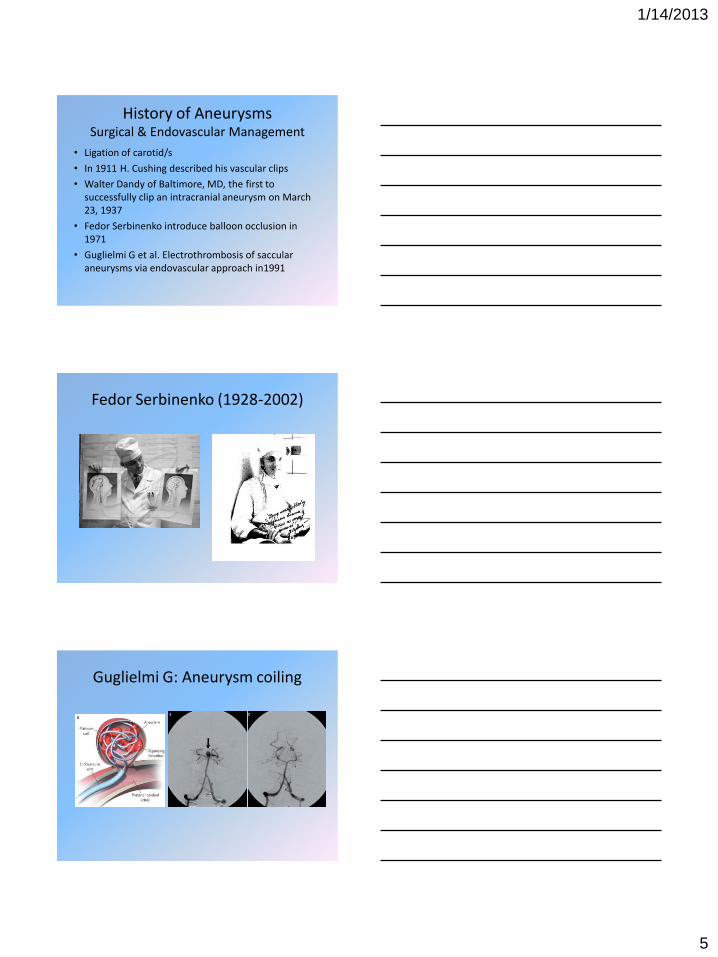
1142013
5
History of Aneurysms Surgical amp Endovascular Management
bull Ligation of carotids
bull In 1911 H Cushing described his vascular clips
bull Walter Dandy of Baltimore MD the first to successfully clip an intracranial aneurysm on March 23 1937
bull Fedor Serbinenko introduce balloon occlusion in 1971
bull Guglielmi G et al Electrothrombosis of saccular aneurysms via endovascular approach in1991
Fedor Serbinenko (1928-2002)
Guglielmi G Aneurysm coiling
1142013
6
AN OVERVIEW OF ANEURYSMS
Aneurysm of the terminal ICA segment
BA giant aneurysm
1142013
7
Aneurysms
Types of cerebral aneurysms
bull Cerebral aneurysms are classified both by size and shape
bull Small aneurysms have a diameter of less than 15 mm
bull Larger aneurysms include those classified as large (15 to 25 mm) giant (25 to 50 mm) and super giant (over 50 mm)
bull Saccular aneurysm refers to any aneurysm with a saccular outpouching including berry aneurysms Saccular aneurysms are the most common form of cerebral aneurysm
bull Berry aneurysm is a type of saccular aneurysm with a neck or stem resembling a berry
bull A fusiform aneurysm describes an aneurysm without a stem
Aneurysms Classification
bull Congenital Berry-shaped or saccular
bull Arterioscleroticfusiform
bull Dissecting as in fibromuscular dysplasia
bull Inflammatory and infectious (mycotic genesis)
bull Traumatic
bull Neoplastic
bull Micro (Charcot-Bouchard)
1142013
8
Most common location of aneurysms
bull ACommA (30 - 35)
bull Bifurcation of the ICA and PCommA (30 - 35)
bull Bifurcation of MCA (20)
bull BA bifurcation (5)
bull Remaining posterior circulation arteries (5)
Aneurysms Association with particular diseases
bull Systemic lupus erythematosus
bull Ehler-Danlos syndrome
bull Coarctation of aorta
bull Polycystic kidney disease
bull Fibromuscular dysplasia
bull Moya-Moya disease
bull Hereditary connective tissue disorder
bull Bacterial meningitis
Aneurysms Risk factors
bull More common among tobacco users
bull Alcohol use or binge drinking
bull Hypertension may be a risk factor
bull Increased rates in women using oral contraceptives
bull Drug use particularly stimulants or cocaine
1142013
9
Aneurysms Risk factors
bull Gender
In most series of aneurysmal SAH there is a striking female preponderance Adult women are affected by aneurysmal SAH more than men by ratio of 60 to 40
bull Genetics Family history (up to 11)
bull Geographic factors The incidence of SAH higher in Finland Japan and low in New Zealand and Middle East It varies significantly from 22-23100000 in Finland and Japan to 8-12100000 in other regions
Aneurysms Incidence
bull Incidence of brain aneurysms in general population is 1 - 5
bull Frequency of brain aneurysms ranged from 02 - 99
bull As many as 400000 people in the USA may have a brain aneurysm
SAH
1142013
10
SAH Etiology
bull Aneurysm 51
bull Hypertension 15
bull AVM 6
bull Other 28
Aneurysmal SAH Epidemiology
bull Approximately 5 - 10 of all strokes
bull Incidence averages 11 per 100000 (range 6-16
per 100000)
bull Adults of all ages
bull Incidence not declining
Aneurysmal SAH Epidemiology
bull 30000 Americans suffer non-traumatic SAH
each year
bull Overall mortality rates are 25
bull Morbidity among survivors is 50
1142013
11
Aneurysmal SAH ndash Presentation
bull Sudden severe headache
bull The hallmark of aneurysmal SAH-headache is that it develops within seconds
bull It is critical to inquire on how quickly the headache developed
bull Even a careful history cannot distinguish between headache secondary to aneurysm rupture and other more common and benign forms of headache
Congenital aneurysms
bull The circle of Willis has been dissected and three berry aneurysms are seen Multiple aneurysms are seen in about 20-30 of cases of berry aneurysm Such aneurysms are congenital in the sense that the defect in the arterial wall is present from birth but the actual aneurysm takes years to develop so that rupture is most likely to occur in young to middle age adults
Ruptured berry aneurysm in the circle of Willis
1142013
12
SAH Diagnosis
bull Non-contrast CT scan confirms subarachnoid blood in 98 of patients if performed within the first 12 hours after onset
bull Distribution of blood on CT provides information on the origin of the SAH
The Fisher Scale
bull The Fischer scale is a way of grouping subarachnoid
haemorrhage CT scans into four goups according to the amount of blood and is useful in predicting cerebral vasospasm
bull Group 1 No blood detected
bull Group 2 Diffuse thin (lt1mm) SAH with no clots
bull Group 3 Localised clots and or layers of blood gt1 mm in thickness
bull Group 4 Intracerebral or intraventricular blood (+- SAH)
SAH CT-Scan
bull Fisher Grade 3
1142013
13
Hunt amp Hess Scale describes the severity of subarachnoid
haemorrhage and is used as a predictor of survival bull Grade 1
ndash asymptomatic or minimal headache and slight neck stiffness
ndash 70 survival
bull Grade 2
ndash moderate to severe headache neck stiffness no neurologic deficit except cranial nerve palsy
ndash 60 survival
bull Grade 3
ndash drowsy minimal neurologic deficit
ndash 50 survival
bull Grade 4
ndash stuporous moderate to severe hemiparesis possibly early decerebrate rigidity and vegetative disturbances
ndash 20 survival
bull Grade 5
ndash deep coma decerebrate rigidity moribund
ndash 10 survival
The WFNS grading system uses the Glasgow Coma Scale and presence of focal neurological deficits
bull GCS 15 No deficit
bull GCS 13-14 No deficit
bull GCS 13-14 No deficit
bull GCS 7-12 may be a neurological deficit
bull GCS lt7 may be a neurological deficit
SAH Diagnosis - MRI
bull In the acute phase MRI with FLAIR demonstrates SAH as reliably as CT
bull MRI is superior to CT in evaluating extravasated blood after a few days (up to 6 weeks) and it is a useful tool in patients referred in a delayed fashion if the original diagnosis is in question
1142013
14
SAH Diagnosis ndash Lumbar puncture
bull Necessary in patients with suggestive history but negative CT
bull Preferably done after 6 or better 12 hours to allow for xanthochromia
bull Xanthochromia persists for at least 2 weeks after SAH
CIRCULATORY PATHOPHYSIOLOGY
Pathophysiological Alterations following Aneurysmal SAH
bull Intracerebral amp Intraventricular Hemorrhage
bull Hydrocephalus
bull Cerebral edema
bull Seizures amp Seizure-like Activity
bull Alteration in Respiratory Function
bull Effect on the Heart
bull Fluid and Electrolyte Disturbance
bull Cerebral ischemia
1142013
15
Vasospasm and Cerebral Ischemia
bull Although there are medical therapies for vasospasm after SAH early detection of vasospasm and initiation of aggressive medical therapy is of utmost importance to avoid delayed neurological ischemia morbidity and mortality
CEREBRAL VASOSPASM AND ITS CLINICAL SIGNIFICANCE
Cerebral Vasospasm
bull Some substances (numerous neurotransmitters blood constituents or breakdown products and autocoids) released at the time of SAH acts on smooth muscle wall to cause vasoconstriction
bull Morphological changes of the arterial wall consistent with vasonecrosis or vasculopathy
bull Mechanical compression may also result in vessels constriction
1142013
16
Vasospasm mechanisms Leung Stroke 2002
Macdonald NeurosurgRev 2006
Blood breakdown products
bull ApolipoproteinE genotype Immunomodulatory neurotoxic oxidative effects APOE4 less effective than APOE3 in suppressing
neurotoxicity
bull Endothelin1 release from CSF leukocytes Potent vasoconsrictor Synergistic effect in vasoconstriction between
bull APOE and Endothelin1
Double-hit model of delayed ischemic neurological deficits after SAH based on Dreier et al The two hits on the brain parenchyma consist of acutely triggered microvascular spasm in response to spreading depolarizations
superimposed on chronic vasospasm
Early Brain Injury Sehba F 2011
1142013
17
Early Brain Injury and Potential Therapy Sehba F 2011
Clinical Features of Symptomatic Vasospasm
bull Variable clinical course
bull Usually peaks at 7-10 days following SAH
bull Usually gradually evolves with waxing and waning symptoms
bull New HA seizures or decreased alertness
bull New focal neurological signs - MCAACAborder zone
Cerebral Vasospasm Clinical Significance
bull Cerebral vasospasm constitutes a major complication of SAH
bull The presence of vasospasm has been correlated with a 15 to 3-fold increase in mortality in the first 2 weeks after SAH
bull DID occur during a period ranging from 4 to 12 days but early (3d) or late manifestations (le 3 wk) may be observed
1142013
18
SAH Outcome
bull 30-day mortality rate still high
bull Many patients die before or shortly after reaching medical attention
bull Large percentage of survivors suffers from cognitive sequelae even after ldquosuccessfulrdquo treatment
Cerebral Vasospasm and Delayed Ischemic Deficit Dorsch et al 1994
bull Literature review of more than 30 000 cases
bull Angiographic vasospasm occurred in 433 (range 19 - 97)
bull DID occurred in 323 (range 5-90)
bull Outcome of DID - Death in 303 - Permanent deficit in 34 - Good outcome in 357
A clinical review of cerebral vasospasm and delayed ischaemia following aneurysm
rupture Dorsch N
2011
bull Online and physical searches have been made of the relevant literature
bull The incidence of delayed ischemic deficit (DID) or symptomatic vasospasm reported in 1994 was 325 in over 30000 reported cases In recent years 1994-2009 it was 677523806 or 285
bull Many of the recent reports did not specify whether a calcium antagonist was used routinely and when this was stated (usually nimodipine or nicardipine) DID was noted in 220 of 10739 reported patients
bull The outcome of DID in the earlier survey was a death rate of 316 with favorable outcomes in 362 In recent reports though with fewer than 1000 patients the outcome is possibly better with death in 256 and good outcome in 541
bull It thus appears likely that delayed vasospasm is still common but less so and that the overall outcome has improved This may be due to the more widespread use of calcium antagonists and more effective fluid management
1142013
19
SAH Management Macdonald Neurosurg Rev 2006
Anderson et al Am J Neuroradiol 2000
bull Vasospasm Diagnosis Change in clinical status 4-vessel angiography bull CTA Sensitivity 100 Specificity 92 Proximal arteries Internal carotid basilar and first
segments of ACA and MCA Less useful for distal vessels and for differentiating mild and
moderate spasm bull MRA Sensitivity 46 Specificity 70 Related to movement and time required for study bull TCD Sensitivity 50 ndash60 Specificity 90 () bull PbtO2 CBF microdialysis bull Glutamate glycerol lactate pyruvate bull CBF lt 15 mL 100 gm min
Management of comlications
bull Angiographic VSP is more common (occurring in about two thirds of patients) than is symptomatic vasospasm (with clinical evidence of cerebral ischemia)
bull TCD is performed daily to monitor for VSP which is defined as a mean CBFV of more than 100 cmsec in a major vessel TCD has a sensitivity that is similar to that of cerebral angiography for the detection of narrowed vessels particularly in the MCA and ICA
bull Once symptomatic VSP is evident (with focal neurologic signs) patients are treated with hypervolemia and induced hypertension
bull Patients whose condition does not improve with medical therapy undergo emergency cerebral angiography and transluminal angioplasty or vasodilator infusion when focal vessel narrowing is evident
Prevention of Vasospasm and DID Naval CCM 2006
HHH Therapy 1 Hypertensive ndashMAP = 100 to 120 mm Hg
2 Hypervolemic ndashCVP = 8 to 12 mm Hg
3 Hemodilution ndashHct = 30
bull Prevention is controversialhelliphelliphelliphelliphellip
bull Statins-protection
bull Mg++ -need further studies
1142013
20
VasoSpasm 2011 11th International Conference
bull Dr L MacDonald presented (CONSCIOUSS 1 2 and 3) C-2 led to a non-significant reduction in mortalityVSP related morbidity but was not associated with an improvement in GOSE (Glasgow Outcome Score Extended) C-3 trial was stopped due to the lack of efficacy data from the Phase 3 clinical study CONSCIOUS-2
bull Effect of intracisternal magnesium therapy Dr Mori et al from Japan Experimental work in dogs the reversible effect of intracisternal magnesium MgSO4 therapy required CSF Mg2+ concentration of more than 3 mEql effect was evident for 3 to 6 hrs therefore continuous or intermittent infusion probably needed to ameliorate VSP
bull Prolonged release of nicardipine from pellets (NP) that are placed around vessels during surgical clipping presented by Dr Kasuya (Japan) initially treated 100 pts provided very impressive results later 136 multicenter trial in Japan was similar to first results Now the randomized double-blind trial of 32 pts with severe SAH done in Germany and the incidence of angiographic vasospasm was significantly reduced 73 control vs 7 in patients with NPs
VasoSpasm 2011 11th International Conference
bull Current medical therapy (nimodipinenicardipine) or aggressive 3H-therapy will not prevent patients after aSAH to have vasospasm
bull Close to 100 of patients after aSAH would have vasospasm demonstrated by cerebral angiography andor TCD
bull No clear predictive value for patients who will have symptomatic vasospasm based on angio or TCD data developed
bull Early brain injury after a-SAH emerges as a new recent concept with emphasis on complex pathophysiological mechanisms that are linked to initial bleed However it remains unknown whether global ischemia itself or subsequent events are responsible for the detected cell death and neurodegeneration
bull Numerous experimental work going on trying to identify therapies for vasospasm preventiontreatment
Effects of a Single Dose of Dantrolene in Patients With Cerebral Vasospasm After Subarachnoid Hemorrhage
A Prospective Pilot Study Muechschlegel et al Stroke 2011
bull Dantrolene is a known ryanodine receptor inhibitor and is already approved by the
US FDA for other indications There is evidence that dantrolene is neuroprotective Furthermore in an ex vivo rat model dantrolene has been shown to inhibit cerebral vasoconstriction alone as well as in combination with nimodipine
bull In a prospective open-label single-dose ascending safety trial 5 patients received iv dantrolene 125 mgkg and the next 5 patients received 25 mgkg over the course of 60 minutes TCD was performed at 0 45 90 and 135 minutes relative to infusion start
bull Peak systolic CBFV decreased significantly (minus26 cms) with a borderline change in mean CBFV in the low-dose group (minus16 cmsec) and peak systolic CBFV in the high-dose group (minus26 cms)
bull In this pilot study a single dose of iv dantrolene in cerebral vasospasm after SAH appears feasible while inhibiting vasoconstriction in the low-dose group but it may lower MAP
1142013
21
TCD DIAGNOSIS OF VASOSPASM
CBF Measurements
Xe 133 SPECT
CBF vs CBFV
bull 133Xe
bull Stable xenon-enhanced CT
bull MRI
bull PET
bull SPECT
bull The time expense and complexity of these techniques still limits its use in routine clinical practice
1142013
22
TCD Diagnosis of Vasospasm Newell et al 1993
Diagnosis and Monitoring of Vasospasm ANGIOGRAPHY
bull Degree of angiographic vasospasm does not always correlate with the clinical condition Some patients remain asymptomatic with severe vasospasm demonstrated by angiography
bull Incidence of angiographic vasospasm is nearly twice that of DID
Diagnosis and Monitoring of Vasospasm TCD
bull High CBFV can identify patients at higher risk for developing DID but also may occur in asymptomatic patients
bull NeurologistNeurointensivist must determine whether the severity and location of the vessel narrowinghigh CBFV are appropriate to cause the clinical deficit
1142013
23
Is routine TCD monitoring useful in the management of SAH
Warlaw et al Neurosurgery 1998
bull 186 patients after SAH
bull Routine TCD examinations made an important positive contribution to the diagnosis of DID in 72 and led to altered management for the benefit of the patient in 43
bull In 9 of pts with recent SAH it was believed that outcome might have better if the TCD results had been acted upon appropriately
Is routine TCD monitoring useful in the management of SAH
Warlaw et al Neurosurgery 1998
bull The TCD results appeared to alter management beneficially in 37 of emergency and 48 of elective patients
bull In the patients admitted on an emergency basis this was often because TCD monitoring showed elevated CBFVrsquos and influenced (delaying or advancing) the timing of surgery or discharge or even led to simple actions such as continuing bet rest or administration of IV fluids for an additional 24 hrs
Transcranial Doppler in cerebral vasospasm Newell et al 1990
MCA CBFV ge 120 cms 25 narrowing
MCA CBFV ge 140 cms 25-50 narrowing
MCA CBFV ge 200 cms 50 narrowing
1142013
24
TCD Criteria for diagnosis of MCA vasospasm
Mean CBFV MCAICA ratio Interpretation (cms) (Lindegaard Ratio)
lt100 lt 3 Nonspecific
100-140 3-6 Mild
140-200 3-6 Moderate
gt200 gt6 Severe
TCD and anterior circulation vasospasm
TCD amp Angiography
bull MCA Sensitivity 94 Specificity 90 PP value 100
bull ACA Sensitivity 71 Specificity 100 PP value 100
TCD and posterior circulation vasospasm TCD and angiography
Sloan et al 1994
bull VA Sensitivity 44 Specificity 88 PP Value 54
bull BA Sensitivity 77 Specificity 79 PP Value 62
1142013
25
Basilar artery vasospasm and delayed posterior circulation ischemia after aneurysmal subarachnoid hemorrhage
Sviri et al Stroke 2004
bull Patients with very high BA CBFV (gt115 cms) had a 50 chance of developing delayed BS ischemia BA-VS was found at a higher rate in patients who experienced reduced rCBF in the cerebellum (563) thalamic nuclei (684) and occipital lobe (818) Although patients with delayed BS hypoperfusion did not present with a higher clinical grade their clinical outcome was significantly worse (Glasgow Outcome Score after 30 days 248+-116 versus 33+-127 P=0001)
bull These findings suggest for the first time that BA-VS after aneurysmal SAH is associated with hypoperfusion to BS and other posterior circulation territories
bull The risk for delayed BS ischemia increased significantly when TCD BA-FVs were gt115 cms
TCD Grading Criteria for BA vasospasm
Sviri et al 2006
bull 123 pts SAH angiography BA and extracranial VA TCD within 4 hrs before the DSA
bull CBFV ratio between the BA and extracranial VA strongly correlated with the degree of BA narrowing (r=086 plt00001)
bull A ratio higher than 2 associated with 73 sensitivity and 80 sensitivity for BA vasospasm
bull A ratio higher than 25 with BA CBFV 85 cmsec was associated with 86 sensitivity and 96 specificity for BA narrowing more than 25
bull A ratio higher than 30 with BA CBFV higher than 85 cmsec was associated with 92 sentivity and 97 specificity for BA narrowing more than 50
Prediction of symptomatic vasospasm after SAH with TCD
bull An early CBFV increase (Seiler et al 1988)
bull A rapid CBFV increase in the first 6 days (Grote et al 1988)
bull A CBFV increase of at least 50 cmsec during 24 hours (Grosset et al 1993)
bull A CBFV increase of 50 cmsec during 48 hours (Wardlow et al 1998)
bull Relative changes in CBFVrsquos (two or threefold CBFV increase) in patients with aneurysmal SAH correlated better with clinically significant vasospasm than absolute CBFVrsquos (Naval et al 2005)
1142013
26
TCD for diagnosis of cerebral vasospasm after SAH mean blood flow velocity ratio of the ipsilateral and contralateral MCA
Nakae et al
2011
bull Retrospective study 142 pts 1262 TCD tests DID defined as neurological deficit or CT evidence of infarct cause by vasospasm ()
bull The threshold value that best discriminated between pts with and wo DID was IC mCBFV of 15
bull IC mCBFV ratio demonstrated a more significant correlation to DID than the absolute m CBFV
Low Pulsatility Index on TCD Predicts Symptomatic Large-Vessel Vasospasm After Aneurysmal SAH
Rajajee et al Neurosurgery 2012
bull Medical records of patients admitted with aSAH between January 2007
and April 2009 were reviewed TCCD sonography was performed daily between days 2 and 14 Patients with unexplained acute neurological decline underwent catheter- or CT-angiography The lowest recorded PI and the highest mCBFV on TCCD were correlated to the occurrence of symptomatic large vessel vasospasm angiographic vasospasm and delayed cerebral infraction in multivariate analysis by use of logistic regression
bull Low PI on TCCD is an independent predictor of symptomatic vasospasm after aSAH whereas mCBFV is a better predictor of angiographic vasospasm
TCD changes after vasospasm
200
140
100
200
140
100
Day 1 Day 2 Day 4 Day 6 Day 7 Day 8
Day 10 Day 12 Day 14 Day 16 Day 18
RIGHTLEFT
RIGHTLEFT
1142013
27
Early Onset Abnormal TCD changes after vasospasm
200
140
100
200
140
100
Day 1 Day 2 Day 4 Day 6 Day 7 Day 8
Day 10 Day 12 Day 14 Day 16 Day 18
RIGHTLEFT
RIGHTLEFT
Unilateral TCD changes after vasospasm
200
140
100
200
140
100
Day 1 Day 3 Day 5 Day 6 Day 7 Day 8
Day 1 Day 3 Day 5 Day 6 Day 7 Day 8
RIGHT
LEFT
High ICP
Unilateral TCD changes after vasospasm
200
140
100
200
140
100
Day 1 Day 3 Day 5 Day 6 Day 7 Day 8
Day 1 Day 3 Day 5 Day 6 Day 7 Day 8
RIGHT
LEFT
More than 50 cms
1142013
28
TCD Wave-Form
Low-Resistant High-Resistant
High ICP ICP treatmentnormalization
1142013
29
Cerebral vasospasm evaluated by TCD at different intracranial pressures
Klingelhofer et al 1991
bull 76 patients with SAH
bull MAP
bull ICP
bull PaCO2
bull TCD
- CBFV - RI
bull Evaluation of the interdependence of the patientrsquos clinical grade VSP ICP and TCD parameters
Cerebral vasospasm evaluated by TCD at different
intracranial pressures
Klingehofer et al 1991
Cerebral vasospasm evaluated by TCD at different intracranial pressures
Klingelhofer et al 1991
bull There was no case in which both high ICP and high CBFV were observed simultaneously
bull During the time course of vasospasm an increase in the resistance index above values of 06 with a simultaneous CBFV decrease indicates a rise in ICP rather than a reduction in VSP
bull With a pronounced increase in ICP evaluation of severity and time course of VSP by TCD based solely upon the mean CBFV can lead to a false-negative results
1142013
30
SAH Pt 1
Day 1
Rt MCA (M1 segm) 60 cms Lt MCA (M1 segm) 64 cms
Day 3
Rt MCA 100 cms Lt MCA 110 cms
SAH Pt 1
Day 8
Rt MCA (M1 segm) 212 cms Lt MCA (M1 segm) 230 cms
Day 15
Rt MCA 308 cms Lt MCA 121 cms
SAH Pt 2
Day 5
Lt MCA 126 cms Rt MCA 88 cms
Day 6
Lt MCA 38 cms Rt MCA 118 cms
1142013
31
SAH Pt 2
Day 7
Lt MCA 139 cms Rt MCA 148 cms
SAH Pt 3
Day 3
Rt MCA 126 cms Lt MCA 88 cms
Day 4
Rt MCA 38 cms Lt MCA 43 cms
SAH Pt 3
Day 5
Rt MCA 131 cms Lt MCA 158 cms
1142013
32
SAH amp ICP
bull We are judging qualitative TCD wave form morphology changes
bull These changes usually will be obvious after ICP will be more 30 mm Hg
bull However one condition must be full filled if you would be using TCD wave from changes to predict intracranial hypertension MAP cardiac output and PaCO2 are normal and not different significantly compared to the previous day
Effect of age on MCA and ICA CBFV Torbey at el Stroke 2011
lt6 8(n=4 7 )
68
(n=3 4 )
p-v a lue
Ma x MC A CBFVMedian (c ms)Range
11435-254
7633-190
0002
Ti me to Max MC A C BFVMedian (days)Range
60-18
72-15
07
MC A pul satil ity i ndexMedianRange
088029-278
109031-283
lt00001
Ma x ICA C BFVMedian (c ms)Range
12640-211
76530-202
0003
Ti me to Max IC A CBFVMedian (days)Range
72-16
553-15
04
IC A pul sati l ity indexMedianRange
088035-55
11804-371
lt00001
lt68 (n=47)
68 (n=34)
p-value
Symptomatic Vasospasm n()
31(66) 15(44) 005
Time to symptomatic vasospasm Median (cms) Range
7
(2-16)
5
(2-10)
006
MCA CBFV 1 day at VSP Median (cms) Range
92
52-243
55
24-190
0009
ICA CBFV 1 day at VSP Median (cms) Range
92
33-196
605
30-171
0079
TCD VSP 42 23 lt00001 TCD confirmed VSP n() 10(53) 5(33) 02 Radiological VSP n() 14(74) 8(47) 01
Effect of age on MCA and ICA CBFV Torbey at el Stroke
2011
1142013
33
Kaplan-Meier Curve for VSP onset time Torbey at al Stroke 2001
Cumulative VSP Probability curve
Time to VSP symptoms
30 20 10 0
Cu
mu
lative
V
SP
11
10
9
8
7
6
5
4
3
AGE GROUP
Age lt68
Age 68
Role of TCD SAH
bull Elevated CBFVrsquos in asymptomatic patients warrant meticulous observation in some closely supervised setting until CBFVrsquos begin trend downward
bull Elevated CBFVrsquos in a particular vascular territory can focus subsequent neurologic examinations to detect subtle changes earlier in their clinical course
Role of TCD SAH
bull In symptomatic patients elevated CBFVrsquos most likely represent significant vessel narrowing and may obviate the need for cerebral angiography At this point triple-H therapy can be initiated or advanced
bull Asymptomatic patients without elevated CBFVrsquos probably can avoid additional angiography However we need to consider patientrsquos age because elderly patientrsquos could develop vasospasm in normal or slightly abnormal CBFV range
1142013
34
Factors influencing interpretation
bull Patient age
bull The presence of moderate to severe anemia (Hct lt27)
bull Impaired CBF autoregulation (passive CBFV variation with MAP changes)
bull Hyperemia induced by triple-H therapy
Guidelines for the Management of Aneurysmal SAH Stroke Council AHA 1994
bull Summary and Recommendations 1 SAH is a medical emergencyhellip 2 CT scanning for suspected SAH is strongly recommendedhellip 3 Selective cerebral angiography to document 4 TCD is recommended for the diagnosis and monitoring of vasospasm although the cerebral angiography may be required for definitive diagnosis
TCD MONITORING OF VASOSPASM
1142013
35
Transcranial Doppler monitoring and clinical decision-making after subarachnoid hemorrhage
McGirt et al 2003
bull Objective was to examine the impact of TCD vasospasm monitoring on clinical decision-making following SAH
bull The records of 50 randomly selected patients undergoing serial TCD monitoring following SAH were reviewed Dates and results of TCDs and cerebral angiograms the use of hypertensive hemodilution (HH) therapy and the development of new neurological deficits were recorded The independent effects of TCD-defined vasospasm and new neurological deficits on patient management were determined with multiple logistical regression Results were validated in a second randomly selected 50 patient cohort
Transcranial Doppler monitoring and clinical decision-making after subarachnoid hemorrhage
McGirt et al 2003
bull Mild or moderate TCD-defined vasospasm developed in 76 of patients 58 +- 05 days after SAH 38 developed severe TCD-defined vasospasm after 79 +- 07 days Focal neurological deficits occurred in 50 after 57 +- 06 days with TCD abnormalities preceding the deficit by 25 +- 07 days in 64
bull TCD-defined vasospasm did not independently influence the use of HH therapy or angiography with both decisions associated with the development of new neurological deficits
bull As TCD-defined vasospasm preceded the neurological deficit in 64 earlier intervention might reduce the incidence of vasospasm-related stroke in institutions with similar practice patterns
MCA and ICA CBFVrsquos Dynamics
1142013
36
Biphasic CBFVrsquos profile after SAH Luft et al Neurocritical Care 2004
bull 18 (99) patients demonstrated biphasic CBFVrsquos profile
bull 1st CBFVrsquos peak (134 + 11 cms) occurred on post-SAH day 6 + 1 2nd CBFVrsquos peak (148 + 12 cms) occurred on post-SAH day 13 + 1
bull Although the 2nd CBFVrsquos peak is usually not associated with a worsening of symptoms these patients were more likely to exhibit clinical symptoms during the 1st CBFV peak
Biphasic CBFVrsquos profile after SAH Luft et al Neurocritical Care 2004
Angiography examination in patient with
ACommA aneurysm before and after aneurysm clipping
1142013
37
Angiography examination after transluminal angioplasty
TCD Monitoring
0
50
100
150
200
250
1 2 3 4 5 6 7 8 9 10 11 12 13 14 15 16 17 18 19 1 2 3 4 5 6 7 8 9 10 11 12 13 14 15 16 17 18 19
MCA M1
MCA M2
ACA A1
ICA C1
VA
BA
MAP
Hct
ICP
Temp
PaCO2
RIGHT LEFT
Days after SAH
1st PEAK
2ND PEAK
Elevated Transcranial Doppler Ultrasound Velocities Following Therapeutic Arterial Dilation
Giller et al 1995
bull Elevated TCD CBFVrsquos seen after cerebral angioplasty are commonly interpreted as evidence of residual or recurrent stenosis but may conceivably arise from hyperemia and require different clinical management
bull Four cases of abnormally elevated mean CBFVrsquos obtained after therapeutic arterial dilation with either balloon angioplasty or intra-arterial administration of papaverine are described In each case cerebral angiography revealed a dilated vessel suggesting that hyperemia and impaired autoregulation were the causes of the high CBFVrsquos
bull These examples suggest that high TCD CBFVrsquos after vessel dilation may be produced by unpredictable amounts of vessel narrowing and flow alteration Although a normalizing CBFV after angioplasty suggests effective vessel dilation high CBFVrsquos may be due partly to hyperemia and cannot be interpreted as arising solely from recurrent stenosis
1142013
38
2011 AHAASA Metrics for Measuring Quality of Care in Comprehensive Stroke Centers
bull Among different measures for Comprehensive Stroke Centers is
Median frequency of noninvasive monitoring for surveillance for vasospasm in patients with aneurysmal SAH during the period between three and 14 days after SAH
POSTTRAUMATIC VASOSPASM
TBI
bull Every 21 seconds one person in the US sustains traumatic brain injury (TBI)
bull An estimated 53 million Americans ndash little more than 2 of the US population ndash currently live with disabilities resulting from brain injury
bull Each year 80000 Americans experience the onset of long-term disability following TBI
bull Okie NEJM 2005 Among surviving soldiers wounded in combat in Iraq and Afghanistan TBI appears to account for a larger proportion of casualties than it has in other recent US wars According to the Joint Theater Trauma Registry 22 had injuries to the head face or neck
Armonda et al Neurosurgery 2006 474 had traumatic cerebral vasospasm Majority were blast related injury
Civilian
Battlefield
1142013
39
Blast TBI
bull Blast injuries historically seen by providers as limited to military concern
Peacetime terrorism over the last decade has reminded us otherwise
bull
Why we need talk about blast TBI
bull Mortality
ndash Oklahoma City 167
ndash US Embassy 223
ndash World Trade Center 2801
ndash Madrid train bombings 191
ndash London 56
ndash Domodedovo 37
ndash Minsk metro 12
Large number of injuredhellip
TBI PATHOPHYSIOLOGY
1142013
40
Balancing Multisystem Interactions
TBI
TBI Pathophysiology
Primary Injury - ContusionsHemorrhages
- Diffuse Axonal Injury (DAI)
Secondary Injury (Intracranial) occurs hours to weeks after injury
- Blood Flow and Metabolic Changes
- Traumatic Hematomas
- Cerebral Edema
- Hydrocephalus
- Increased Intracranial Pressure
Decrease in CBF
BRAIN
EDEMA
More Brain Ischemic Blood Vessels
Edema brain cells Dilate
Increased ICP
Cell Injury
1142013
41
But where is the place for TCD monitoring
Multimodal Monitoring VasospasmIschemiahigh ICP Detection
bull MAP
bull SaO2
bull ECG
bull Et-CO2
bull CVP
bull Urine output
bull ICP
bull CBFVTCD PbO2
cEEG
CTMR Perfusion
The primary goal of
management for
TBI is the
prevention of
secondary damage
due to neuronal
hypoxia and
hypoperfusion
Daily TCD
Continuous EEG
ICP monitoring
Pb02
CBF
1142013
42
Traumatic Brain Injury amp TCD
bull Cerebral ischemia due to the post-traumatic vasospasm
bull Increased Intracranial Pressure
bull Brain Death
Post-Traumatic VSP
bull Ischemic symptoms caused by cerebral arterial spasm following traumatic SAH are comparable to those found following aneurysmal SAH
- appearance of symptoms between Days 4 and 16 after injury with the peak incidence on Days 9 and 10
- close correlation between the main site of the subarachnoid blood and the location of severe vasospasm responsible for the symptoms and a higher incidence of symptoms in patients with massive SAH than those with slight SAH
bull Subarachnoid blood plays an important role in the later development of vasospasm not only following aneurysm rupture but also after head injury Nevertheless there is no general agreement that subarachnoid blood in head injury is an important risk factor in the development of vasospasm and ischemic brain damage
TBI and VSP
bull Cerebral posttraumatic VSP (PTV) was first described by Lorn in 1936
bull The incidence of CT documented traumatic SAH has been identified in 4 to 63 of pts after TBI
bull Study from the University of Mississippi Medical Center indicated that traumatic SAH complicate course of TBI in 69 of the patients due to the presence of PTV
1142013
43
Clinical Significance
bull 41 of patients who died from TBI had PTV (MacPherson et al 1973)
bull 24 with massive tSAH developed ischemic symptoms in contrast to 3 of patients with mild tSAH (Taneda et al 1996)
bull Ischemic symptoms accompanying arterial VSP following tSAH are comparable to those found following aneurysmal SAH
WARTIME TRAUMATIC CEREBRAL VASOSPASM RECENT REVIEW OF COMBAT CASUALTIES
Armonda et al Neurosurgery 2006
bull The first study to analyze the effects of blast-related injury on the cerebral vasculature
bull This study showed that TCV occurred in a substantial number of patients with severe neurotrauma and clinical outcomes were worse for those with this condition
25 yo severe concussion no SAH (Courtesy of Dr Armonda)
CBFV 120 cms CBFV 57 cms
1142013
44
25 yo severe concussion no SAH (Courtesy of Dr Armonda)
CBFV 112 cms CBFV 89 cms
25 yo severe concussion no SAH (Courtesy of Dr Armonda)
CBFV
(cmsec)
Calendar
0
50
100
150
200
250
300
MCA M1
MCA M2
ACA A1
VA
BA
MAP
Hct
ICP
Temp
PaCO2
RIGHT LEFT
Ant circ TLA Post circ TLA Ant circ TLA
SEVERE VSP
MODERATE VSP
MILD VSP
EFFECT OF TLA ON PROLONG (SEVEN DAYS) VASOSPASM INVOLVING ANTERIOR AND POSTERIOR CIRCULATION
1142013
45
The CBFVrsquos and systemic hemodynamic trends TCD signs of severe vasospasm involving posterior and
anterior circulation lasting seven days before TLA
0
50
100
150
200
250
300
MCA M1
ACA A1
ICA C1
VA
BA
MAP
Hct
ICP
PaCO2
RIGHT LEFT
Severe VSP
Moderate VSP
MIld VSP
TLA TLA
Effect of TLA after prolong severe vasospasm
Left MCA CBFV 69 cmsec PI 17 Right MCA CBFV 66 cmsec PI 14
Left MCA 205 cmsec PI 08 Right MCA CBFV 195 cmsec PI 05
CBFVrsquos and PIrsquos Trends (PI x 100)
0
50
100
150
200
250
300
MCA M1
M1 PI
ACA A1
A1 PI
ICA C1
C1 PI
VA
VA PI
BA
BA PI
RIGHT LEFT
1142013
46
The CBFV amp PI Trends
bull MCA (M1 segm)
bull ICA (C1 segm)
0
50
100
150
200
250
300
MCA M1
M1 PI
RIGHT LEFT
Severe VSP
Moderate VSP
Mild VSP
0
50
100
150
200
250
300
ICA C1
C1 PI
RIGHT LEFT
Severe VSP
Moderate VSP
Mild VSP
Clinical Material
bull Ninety patients (2 females) aged 18 to 50 years (mean 259 years) who had suffered wartime TBI injuries (with Glasgow Coma Scale scores ranging from 3 to 15) were investigated with daily TCD studies
bull A total of 567 TCD studies (mean 64 testspatient ranged from 1 to 30) were made after admission
TCD signs of Vasospasm (in ) by type of TBI
7500
3680
6840
2900
3570
520
3150
2350
1430
520
1570 1760
0
10
20
30
40
50
60
70
80
PHI PHIIED CHI CHIIED
Mild ()
Mod ()
Severe ()
1142013
47
Presence of ICH ()
5700
5200
5800
5000
PHI PHIIED CHI CHIIED
TCD signs of VSP amp high ICP
TBI Type Post TBI
VSP
High
ICP
CHI 13144 12133
CHIIED 12133 910
PHI 21233 16177
PHIIED 11122 12133
Total 57633 49544
TCD AND TUMOR RESECTION
1142013
48
Tumor Resection and Vasospasm
bull The occurrence of vasospasm and delayed cerebral ischemia after resection of intracranial tumor has not received extensive attention clinically and is often misdiagnosed and improperly treated as surgical brain damage or brain swelling
bull However DID from vasospasm after tumor resection is a complication that is being reported in increasing numbers
Tumor Resection and Vasospasm Literature review
bull Reports are sparse and mainly are case series
bull Vasospasm was found in 2 to 49 patients No significant difference among age sex surgical approaches pathological diagnosis duration of surgery amount of blood loss and transfusion during surgery were found but significant difference was seen in cisternal hemorrhage on CT scan and the amount of blood in cerebrospinal fluid
Tumor Resection and Vasospasm Probable Mechanism
bull It is suggested that accumulation of blood in the basal cisterns may have been responsible for this unusual condition and it is therefore important to consider vasospasm as a probable etiological cause of clinical deterioration in patients undergoing the surgical removal of a cerebral tumor
bull For this reason whenever any neurological deterioration occurs in such patients it is advisable to perform TCD in order to verify the presence of any vasospasm and promptly commence suitable treatment
1142013
49
Tumor Resection and Vasospasm Management
bull Surgery To decrease the amount of blood in basal cistern by microsurgery
bull ICU TCD utilization in the diagnosis of vasospasm and its monitoring
bull ICU Differentiation of vasospasm from brain swelling are helpful to confirm the coexistent or causal relation based on neurological assessment CT TCD and ICP monitoring both in deciding therapeutic strategy and successfully controlling vasospasm
bull ICU Nimotop played a key role in preventing brain damage from vasospasm and cerebral swelling
Suprasellar tumor resection The CBFVs and Trends of the Systemic
Hemodynamic
RIGHT LEFT
0
20
40
60
80
100
120
140
160
180
103010311111121131141151161171181191110 103010311111121131141151161171181191110
Calendar
CB
FV
s a
nd
Vari
ab
les
0
20
40
60
80
100
120
140
160
180
CB
FV
s a
nd
Vari
ab
les
MCA M1
MCA M2
ACA
ICA C1
VA
BA
MAP
ICP
Hct
PaCO2
Temp
Patient wit tumor
bull 58 yo female sp trans-sphenoidal resection of tuberculum sella meningioma complicated by A-comm complex pseudoaneurysm formation vasospasm and ACA territory infarct
1142013
50
DWI demonstrating ACA territory infarct
DWI demonstrating ACA territory infarct
ICA injections demonstrating vasospasm in A1-A2 bilaterally
A-comm complex pseudoaneurysm and coiling
1142013
51
A-comm complex pseudoaneurysm before and after coiling
TCD Trends
Left Right
Role of TCD Tumor Resection
bull It is useful to perform TCD test after surgery and perform daily TCD studies when patient is in the ICU
bull The frequency with which TCD should be performed may be guided by patient clinical presentation knowledge of risk factors for VSP early clinical course
bull TCD studies should be performed after endovascular treatment to identify patients with recurrent VSP
1142013
52
Role of TCD VSP monitoring
bull It is useful to perform TCD test on admission (or ASAP after surgery) and perform daily TCD studies when patient is in the ICU
bull The frequency with which TCD should be performed may be guided by patient clinical presentation knowledge of risk factors for vasospasm early clinical course
bull TCD studies should be performed after endovascular treatment to identify patients with recurrent vasospasm
Role of TCD VSP Monitoring
bull The presence and temporal profile of CBFVrsquos in all available vessels must be detected and serially monitored
bull The pattern of CBFVrsquos elevation may indicate the need to follow patient carefully for evidence of deficits related to specific vascular territory
bull Waveform appearance either regionally or globally may be clinically significant
TCD and VSP
bull Currently the gold standard for vasospasm diagnosis is cerebral angiography replaceable by CTA only when angiography is not available Obviously it is not feasible to perform such investigation as frequently as bedside clinical assessment
bull Repeated clinical assessments of a patients neurological status carry the problem of detecting the clinical signs and symptoms of vasospasm which occur only after vasospasm has already manifested its deleterious effects on the cerebral parenchyma
bull TCD is a relatively new non-invasive tool allowing for bedside monitoring to determine CBFVrsquos indicative of changes in vascular diameter
1142013
53
TCD and VSP
bull TCD can be useful pre- intra- and post-operatively while helping to recognize the development of cerebral vasospasm before the onset of its clinical effects
bull Vasospasm following SAH is a very important source of morbidity and mortality Too often the first sign is a neurologic deficit which may be too late to reverse
bull TCD assists in the clinical decision-making regarding further diagnostic evaluation and therapeutic interventions When performed in isolation the contribution of TCD to improving patient outcome has not been established Nevertheless TCD has become a regularly employed tool in neurocritical care and perioperative settings
TCD is a Critical Tool in Critical Care
bull The value of TCD in clinical practice is well established especially to measure and grade vasospasm following SAH and TBI
bull Based on AHA Guidelines and many years of clinical practice TCD is a tool employed by the Neurosurgeon Neurointensivist and Neurologist in the management of vasospasm
bull Based on high frequency of posttraumatic vasospasm and intracranial hypertension TCD testing must be utilized for management of patients after SAH (aneurysm rupture wartime or civilian TBI tumor resection)
bull The use of TCD at hospital admission allows identification of patients with brain hypoperfusion due to the vasospasm andor intracranial hypertension In such high-risk patients early TCD goal-directed therapy can restore normal cerebral perfusion and might then potentially help in reducing the extent of secondary brain injury
bull TCD could provide information about abnormally high ICPbrain death
bull In the future incorporation of TCD data may facilitate more injury- and time-specific therapies for patients after SAH (aneurysm rupture wartime or civilian TBI tumor resection)
TCD is a Critical Tool in Critical Care
1142013
54
Do we know everything about TCD and vasospasm
bull TCD criteria for vasospasm for the young (less than 20-30 yo) and old (more than 68-70 yo) patients
bull CBFV calculation formula that will take into account Hct values
bull Cerebral angiography could be negative but TCD could be positive for vasospasm
bull TCD prediction for clinical vasospasm
Future

1142013
6
AN OVERVIEW OF ANEURYSMS
Aneurysm of the terminal ICA segment
BA giant aneurysm
1142013
7
Aneurysms
Types of cerebral aneurysms
bull Cerebral aneurysms are classified both by size and shape
bull Small aneurysms have a diameter of less than 15 mm
bull Larger aneurysms include those classified as large (15 to 25 mm) giant (25 to 50 mm) and super giant (over 50 mm)
bull Saccular aneurysm refers to any aneurysm with a saccular outpouching including berry aneurysms Saccular aneurysms are the most common form of cerebral aneurysm
bull Berry aneurysm is a type of saccular aneurysm with a neck or stem resembling a berry
bull A fusiform aneurysm describes an aneurysm without a stem
Aneurysms Classification
bull Congenital Berry-shaped or saccular
bull Arterioscleroticfusiform
bull Dissecting as in fibromuscular dysplasia
bull Inflammatory and infectious (mycotic genesis)
bull Traumatic
bull Neoplastic
bull Micro (Charcot-Bouchard)
1142013
8
Most common location of aneurysms
bull ACommA (30 - 35)
bull Bifurcation of the ICA and PCommA (30 - 35)
bull Bifurcation of MCA (20)
bull BA bifurcation (5)
bull Remaining posterior circulation arteries (5)
Aneurysms Association with particular diseases
bull Systemic lupus erythematosus
bull Ehler-Danlos syndrome
bull Coarctation of aorta
bull Polycystic kidney disease
bull Fibromuscular dysplasia
bull Moya-Moya disease
bull Hereditary connective tissue disorder
bull Bacterial meningitis
Aneurysms Risk factors
bull More common among tobacco users
bull Alcohol use or binge drinking
bull Hypertension may be a risk factor
bull Increased rates in women using oral contraceptives
bull Drug use particularly stimulants or cocaine
1142013
9
Aneurysms Risk factors
bull Gender
In most series of aneurysmal SAH there is a striking female preponderance Adult women are affected by aneurysmal SAH more than men by ratio of 60 to 40
bull Genetics Family history (up to 11)
bull Geographic factors The incidence of SAH higher in Finland Japan and low in New Zealand and Middle East It varies significantly from 22-23100000 in Finland and Japan to 8-12100000 in other regions
Aneurysms Incidence
bull Incidence of brain aneurysms in general population is 1 - 5
bull Frequency of brain aneurysms ranged from 02 - 99
bull As many as 400000 people in the USA may have a brain aneurysm
SAH
1142013
10
SAH Etiology
bull Aneurysm 51
bull Hypertension 15
bull AVM 6
bull Other 28
Aneurysmal SAH Epidemiology
bull Approximately 5 - 10 of all strokes
bull Incidence averages 11 per 100000 (range 6-16
per 100000)
bull Adults of all ages
bull Incidence not declining
Aneurysmal SAH Epidemiology
bull 30000 Americans suffer non-traumatic SAH
each year
bull Overall mortality rates are 25
bull Morbidity among survivors is 50
1142013
11
Aneurysmal SAH ndash Presentation
bull Sudden severe headache
bull The hallmark of aneurysmal SAH-headache is that it develops within seconds
bull It is critical to inquire on how quickly the headache developed
bull Even a careful history cannot distinguish between headache secondary to aneurysm rupture and other more common and benign forms of headache
Congenital aneurysms
bull The circle of Willis has been dissected and three berry aneurysms are seen Multiple aneurysms are seen in about 20-30 of cases of berry aneurysm Such aneurysms are congenital in the sense that the defect in the arterial wall is present from birth but the actual aneurysm takes years to develop so that rupture is most likely to occur in young to middle age adults
Ruptured berry aneurysm in the circle of Willis
1142013
12
SAH Diagnosis
bull Non-contrast CT scan confirms subarachnoid blood in 98 of patients if performed within the first 12 hours after onset
bull Distribution of blood on CT provides information on the origin of the SAH
The Fisher Scale
bull The Fischer scale is a way of grouping subarachnoid
haemorrhage CT scans into four goups according to the amount of blood and is useful in predicting cerebral vasospasm
bull Group 1 No blood detected
bull Group 2 Diffuse thin (lt1mm) SAH with no clots
bull Group 3 Localised clots and or layers of blood gt1 mm in thickness
bull Group 4 Intracerebral or intraventricular blood (+- SAH)
SAH CT-Scan
bull Fisher Grade 3
1142013
13
Hunt amp Hess Scale describes the severity of subarachnoid
haemorrhage and is used as a predictor of survival bull Grade 1
ndash asymptomatic or minimal headache and slight neck stiffness
ndash 70 survival
bull Grade 2
ndash moderate to severe headache neck stiffness no neurologic deficit except cranial nerve palsy
ndash 60 survival
bull Grade 3
ndash drowsy minimal neurologic deficit
ndash 50 survival
bull Grade 4
ndash stuporous moderate to severe hemiparesis possibly early decerebrate rigidity and vegetative disturbances
ndash 20 survival
bull Grade 5
ndash deep coma decerebrate rigidity moribund
ndash 10 survival
The WFNS grading system uses the Glasgow Coma Scale and presence of focal neurological deficits
bull GCS 15 No deficit
bull GCS 13-14 No deficit
bull GCS 13-14 No deficit
bull GCS 7-12 may be a neurological deficit
bull GCS lt7 may be a neurological deficit
SAH Diagnosis - MRI
bull In the acute phase MRI with FLAIR demonstrates SAH as reliably as CT
bull MRI is superior to CT in evaluating extravasated blood after a few days (up to 6 weeks) and it is a useful tool in patients referred in a delayed fashion if the original diagnosis is in question
1142013
14
SAH Diagnosis ndash Lumbar puncture
bull Necessary in patients with suggestive history but negative CT
bull Preferably done after 6 or better 12 hours to allow for xanthochromia
bull Xanthochromia persists for at least 2 weeks after SAH
CIRCULATORY PATHOPHYSIOLOGY
Pathophysiological Alterations following Aneurysmal SAH
bull Intracerebral amp Intraventricular Hemorrhage
bull Hydrocephalus
bull Cerebral edema
bull Seizures amp Seizure-like Activity
bull Alteration in Respiratory Function
bull Effect on the Heart
bull Fluid and Electrolyte Disturbance
bull Cerebral ischemia
1142013
15
Vasospasm and Cerebral Ischemia
bull Although there are medical therapies for vasospasm after SAH early detection of vasospasm and initiation of aggressive medical therapy is of utmost importance to avoid delayed neurological ischemia morbidity and mortality
CEREBRAL VASOSPASM AND ITS CLINICAL SIGNIFICANCE
Cerebral Vasospasm
bull Some substances (numerous neurotransmitters blood constituents or breakdown products and autocoids) released at the time of SAH acts on smooth muscle wall to cause vasoconstriction
bull Morphological changes of the arterial wall consistent with vasonecrosis or vasculopathy
bull Mechanical compression may also result in vessels constriction
1142013
16
Vasospasm mechanisms Leung Stroke 2002
Macdonald NeurosurgRev 2006
Blood breakdown products
bull ApolipoproteinE genotype Immunomodulatory neurotoxic oxidative effects APOE4 less effective than APOE3 in suppressing
neurotoxicity
bull Endothelin1 release from CSF leukocytes Potent vasoconsrictor Synergistic effect in vasoconstriction between
bull APOE and Endothelin1
Double-hit model of delayed ischemic neurological deficits after SAH based on Dreier et al The two hits on the brain parenchyma consist of acutely triggered microvascular spasm in response to spreading depolarizations
superimposed on chronic vasospasm
Early Brain Injury Sehba F 2011
1142013
17
Early Brain Injury and Potential Therapy Sehba F 2011
Clinical Features of Symptomatic Vasospasm
bull Variable clinical course
bull Usually peaks at 7-10 days following SAH
bull Usually gradually evolves with waxing and waning symptoms
bull New HA seizures or decreased alertness
bull New focal neurological signs - MCAACAborder zone
Cerebral Vasospasm Clinical Significance
bull Cerebral vasospasm constitutes a major complication of SAH
bull The presence of vasospasm has been correlated with a 15 to 3-fold increase in mortality in the first 2 weeks after SAH
bull DID occur during a period ranging from 4 to 12 days but early (3d) or late manifestations (le 3 wk) may be observed
1142013
18
SAH Outcome
bull 30-day mortality rate still high
bull Many patients die before or shortly after reaching medical attention
bull Large percentage of survivors suffers from cognitive sequelae even after ldquosuccessfulrdquo treatment
Cerebral Vasospasm and Delayed Ischemic Deficit Dorsch et al 1994
bull Literature review of more than 30 000 cases
bull Angiographic vasospasm occurred in 433 (range 19 - 97)
bull DID occurred in 323 (range 5-90)
bull Outcome of DID - Death in 303 - Permanent deficit in 34 - Good outcome in 357
A clinical review of cerebral vasospasm and delayed ischaemia following aneurysm
rupture Dorsch N
2011
bull Online and physical searches have been made of the relevant literature
bull The incidence of delayed ischemic deficit (DID) or symptomatic vasospasm reported in 1994 was 325 in over 30000 reported cases In recent years 1994-2009 it was 677523806 or 285
bull Many of the recent reports did not specify whether a calcium antagonist was used routinely and when this was stated (usually nimodipine or nicardipine) DID was noted in 220 of 10739 reported patients
bull The outcome of DID in the earlier survey was a death rate of 316 with favorable outcomes in 362 In recent reports though with fewer than 1000 patients the outcome is possibly better with death in 256 and good outcome in 541
bull It thus appears likely that delayed vasospasm is still common but less so and that the overall outcome has improved This may be due to the more widespread use of calcium antagonists and more effective fluid management
1142013
19
SAH Management Macdonald Neurosurg Rev 2006
Anderson et al Am J Neuroradiol 2000
bull Vasospasm Diagnosis Change in clinical status 4-vessel angiography bull CTA Sensitivity 100 Specificity 92 Proximal arteries Internal carotid basilar and first
segments of ACA and MCA Less useful for distal vessels and for differentiating mild and
moderate spasm bull MRA Sensitivity 46 Specificity 70 Related to movement and time required for study bull TCD Sensitivity 50 ndash60 Specificity 90 () bull PbtO2 CBF microdialysis bull Glutamate glycerol lactate pyruvate bull CBF lt 15 mL 100 gm min
Management of comlications
bull Angiographic VSP is more common (occurring in about two thirds of patients) than is symptomatic vasospasm (with clinical evidence of cerebral ischemia)
bull TCD is performed daily to monitor for VSP which is defined as a mean CBFV of more than 100 cmsec in a major vessel TCD has a sensitivity that is similar to that of cerebral angiography for the detection of narrowed vessels particularly in the MCA and ICA
bull Once symptomatic VSP is evident (with focal neurologic signs) patients are treated with hypervolemia and induced hypertension
bull Patients whose condition does not improve with medical therapy undergo emergency cerebral angiography and transluminal angioplasty or vasodilator infusion when focal vessel narrowing is evident
Prevention of Vasospasm and DID Naval CCM 2006
HHH Therapy 1 Hypertensive ndashMAP = 100 to 120 mm Hg
2 Hypervolemic ndashCVP = 8 to 12 mm Hg
3 Hemodilution ndashHct = 30
bull Prevention is controversialhelliphelliphelliphelliphellip
bull Statins-protection
bull Mg++ -need further studies
1142013
20
VasoSpasm 2011 11th International Conference
bull Dr L MacDonald presented (CONSCIOUSS 1 2 and 3) C-2 led to a non-significant reduction in mortalityVSP related morbidity but was not associated with an improvement in GOSE (Glasgow Outcome Score Extended) C-3 trial was stopped due to the lack of efficacy data from the Phase 3 clinical study CONSCIOUS-2
bull Effect of intracisternal magnesium therapy Dr Mori et al from Japan Experimental work in dogs the reversible effect of intracisternal magnesium MgSO4 therapy required CSF Mg2+ concentration of more than 3 mEql effect was evident for 3 to 6 hrs therefore continuous or intermittent infusion probably needed to ameliorate VSP
bull Prolonged release of nicardipine from pellets (NP) that are placed around vessels during surgical clipping presented by Dr Kasuya (Japan) initially treated 100 pts provided very impressive results later 136 multicenter trial in Japan was similar to first results Now the randomized double-blind trial of 32 pts with severe SAH done in Germany and the incidence of angiographic vasospasm was significantly reduced 73 control vs 7 in patients with NPs
VasoSpasm 2011 11th International Conference
bull Current medical therapy (nimodipinenicardipine) or aggressive 3H-therapy will not prevent patients after aSAH to have vasospasm
bull Close to 100 of patients after aSAH would have vasospasm demonstrated by cerebral angiography andor TCD
bull No clear predictive value for patients who will have symptomatic vasospasm based on angio or TCD data developed
bull Early brain injury after a-SAH emerges as a new recent concept with emphasis on complex pathophysiological mechanisms that are linked to initial bleed However it remains unknown whether global ischemia itself or subsequent events are responsible for the detected cell death and neurodegeneration
bull Numerous experimental work going on trying to identify therapies for vasospasm preventiontreatment
Effects of a Single Dose of Dantrolene in Patients With Cerebral Vasospasm After Subarachnoid Hemorrhage
A Prospective Pilot Study Muechschlegel et al Stroke 2011
bull Dantrolene is a known ryanodine receptor inhibitor and is already approved by the
US FDA for other indications There is evidence that dantrolene is neuroprotective Furthermore in an ex vivo rat model dantrolene has been shown to inhibit cerebral vasoconstriction alone as well as in combination with nimodipine
bull In a prospective open-label single-dose ascending safety trial 5 patients received iv dantrolene 125 mgkg and the next 5 patients received 25 mgkg over the course of 60 minutes TCD was performed at 0 45 90 and 135 minutes relative to infusion start
bull Peak systolic CBFV decreased significantly (minus26 cms) with a borderline change in mean CBFV in the low-dose group (minus16 cmsec) and peak systolic CBFV in the high-dose group (minus26 cms)
bull In this pilot study a single dose of iv dantrolene in cerebral vasospasm after SAH appears feasible while inhibiting vasoconstriction in the low-dose group but it may lower MAP
1142013
21
TCD DIAGNOSIS OF VASOSPASM
CBF Measurements
Xe 133 SPECT
CBF vs CBFV
bull 133Xe
bull Stable xenon-enhanced CT
bull MRI
bull PET
bull SPECT
bull The time expense and complexity of these techniques still limits its use in routine clinical practice
1142013
22
TCD Diagnosis of Vasospasm Newell et al 1993
Diagnosis and Monitoring of Vasospasm ANGIOGRAPHY
bull Degree of angiographic vasospasm does not always correlate with the clinical condition Some patients remain asymptomatic with severe vasospasm demonstrated by angiography
bull Incidence of angiographic vasospasm is nearly twice that of DID
Diagnosis and Monitoring of Vasospasm TCD
bull High CBFV can identify patients at higher risk for developing DID but also may occur in asymptomatic patients
bull NeurologistNeurointensivist must determine whether the severity and location of the vessel narrowinghigh CBFV are appropriate to cause the clinical deficit
1142013
23
Is routine TCD monitoring useful in the management of SAH
Warlaw et al Neurosurgery 1998
bull 186 patients after SAH
bull Routine TCD examinations made an important positive contribution to the diagnosis of DID in 72 and led to altered management for the benefit of the patient in 43
bull In 9 of pts with recent SAH it was believed that outcome might have better if the TCD results had been acted upon appropriately
Is routine TCD monitoring useful in the management of SAH
Warlaw et al Neurosurgery 1998
bull The TCD results appeared to alter management beneficially in 37 of emergency and 48 of elective patients
bull In the patients admitted on an emergency basis this was often because TCD monitoring showed elevated CBFVrsquos and influenced (delaying or advancing) the timing of surgery or discharge or even led to simple actions such as continuing bet rest or administration of IV fluids for an additional 24 hrs
Transcranial Doppler in cerebral vasospasm Newell et al 1990
MCA CBFV ge 120 cms 25 narrowing
MCA CBFV ge 140 cms 25-50 narrowing
MCA CBFV ge 200 cms 50 narrowing
1142013
24
TCD Criteria for diagnosis of MCA vasospasm
Mean CBFV MCAICA ratio Interpretation (cms) (Lindegaard Ratio)
lt100 lt 3 Nonspecific
100-140 3-6 Mild
140-200 3-6 Moderate
gt200 gt6 Severe
TCD and anterior circulation vasospasm
TCD amp Angiography
bull MCA Sensitivity 94 Specificity 90 PP value 100
bull ACA Sensitivity 71 Specificity 100 PP value 100
TCD and posterior circulation vasospasm TCD and angiography
Sloan et al 1994
bull VA Sensitivity 44 Specificity 88 PP Value 54
bull BA Sensitivity 77 Specificity 79 PP Value 62
1142013
25
Basilar artery vasospasm and delayed posterior circulation ischemia after aneurysmal subarachnoid hemorrhage
Sviri et al Stroke 2004
bull Patients with very high BA CBFV (gt115 cms) had a 50 chance of developing delayed BS ischemia BA-VS was found at a higher rate in patients who experienced reduced rCBF in the cerebellum (563) thalamic nuclei (684) and occipital lobe (818) Although patients with delayed BS hypoperfusion did not present with a higher clinical grade their clinical outcome was significantly worse (Glasgow Outcome Score after 30 days 248+-116 versus 33+-127 P=0001)
bull These findings suggest for the first time that BA-VS after aneurysmal SAH is associated with hypoperfusion to BS and other posterior circulation territories
bull The risk for delayed BS ischemia increased significantly when TCD BA-FVs were gt115 cms
TCD Grading Criteria for BA vasospasm
Sviri et al 2006
bull 123 pts SAH angiography BA and extracranial VA TCD within 4 hrs before the DSA
bull CBFV ratio between the BA and extracranial VA strongly correlated with the degree of BA narrowing (r=086 plt00001)
bull A ratio higher than 2 associated with 73 sensitivity and 80 sensitivity for BA vasospasm
bull A ratio higher than 25 with BA CBFV 85 cmsec was associated with 86 sensitivity and 96 specificity for BA narrowing more than 25
bull A ratio higher than 30 with BA CBFV higher than 85 cmsec was associated with 92 sentivity and 97 specificity for BA narrowing more than 50
Prediction of symptomatic vasospasm after SAH with TCD
bull An early CBFV increase (Seiler et al 1988)
bull A rapid CBFV increase in the first 6 days (Grote et al 1988)
bull A CBFV increase of at least 50 cmsec during 24 hours (Grosset et al 1993)
bull A CBFV increase of 50 cmsec during 48 hours (Wardlow et al 1998)
bull Relative changes in CBFVrsquos (two or threefold CBFV increase) in patients with aneurysmal SAH correlated better with clinically significant vasospasm than absolute CBFVrsquos (Naval et al 2005)
1142013
26
TCD for diagnosis of cerebral vasospasm after SAH mean blood flow velocity ratio of the ipsilateral and contralateral MCA
Nakae et al
2011
bull Retrospective study 142 pts 1262 TCD tests DID defined as neurological deficit or CT evidence of infarct cause by vasospasm ()
bull The threshold value that best discriminated between pts with and wo DID was IC mCBFV of 15
bull IC mCBFV ratio demonstrated a more significant correlation to DID than the absolute m CBFV
Low Pulsatility Index on TCD Predicts Symptomatic Large-Vessel Vasospasm After Aneurysmal SAH
Rajajee et al Neurosurgery 2012
bull Medical records of patients admitted with aSAH between January 2007
and April 2009 were reviewed TCCD sonography was performed daily between days 2 and 14 Patients with unexplained acute neurological decline underwent catheter- or CT-angiography The lowest recorded PI and the highest mCBFV on TCCD were correlated to the occurrence of symptomatic large vessel vasospasm angiographic vasospasm and delayed cerebral infraction in multivariate analysis by use of logistic regression
bull Low PI on TCCD is an independent predictor of symptomatic vasospasm after aSAH whereas mCBFV is a better predictor of angiographic vasospasm
TCD changes after vasospasm
200
140
100
200
140
100
Day 1 Day 2 Day 4 Day 6 Day 7 Day 8
Day 10 Day 12 Day 14 Day 16 Day 18
RIGHTLEFT
RIGHTLEFT
1142013
27
Early Onset Abnormal TCD changes after vasospasm
200
140
100
200
140
100
Day 1 Day 2 Day 4 Day 6 Day 7 Day 8
Day 10 Day 12 Day 14 Day 16 Day 18
RIGHTLEFT
RIGHTLEFT
Unilateral TCD changes after vasospasm
200
140
100
200
140
100
Day 1 Day 3 Day 5 Day 6 Day 7 Day 8
Day 1 Day 3 Day 5 Day 6 Day 7 Day 8
RIGHT
LEFT
High ICP
Unilateral TCD changes after vasospasm
200
140
100
200
140
100
Day 1 Day 3 Day 5 Day 6 Day 7 Day 8
Day 1 Day 3 Day 5 Day 6 Day 7 Day 8
RIGHT
LEFT
More than 50 cms
1142013
28
TCD Wave-Form
Low-Resistant High-Resistant
High ICP ICP treatmentnormalization
1142013
29
Cerebral vasospasm evaluated by TCD at different intracranial pressures
Klingelhofer et al 1991
bull 76 patients with SAH
bull MAP
bull ICP
bull PaCO2
bull TCD
- CBFV - RI
bull Evaluation of the interdependence of the patientrsquos clinical grade VSP ICP and TCD parameters
Cerebral vasospasm evaluated by TCD at different
intracranial pressures
Klingehofer et al 1991
Cerebral vasospasm evaluated by TCD at different intracranial pressures
Klingelhofer et al 1991
bull There was no case in which both high ICP and high CBFV were observed simultaneously
bull During the time course of vasospasm an increase in the resistance index above values of 06 with a simultaneous CBFV decrease indicates a rise in ICP rather than a reduction in VSP
bull With a pronounced increase in ICP evaluation of severity and time course of VSP by TCD based solely upon the mean CBFV can lead to a false-negative results
1142013
30
SAH Pt 1
Day 1
Rt MCA (M1 segm) 60 cms Lt MCA (M1 segm) 64 cms
Day 3
Rt MCA 100 cms Lt MCA 110 cms
SAH Pt 1
Day 8
Rt MCA (M1 segm) 212 cms Lt MCA (M1 segm) 230 cms
Day 15
Rt MCA 308 cms Lt MCA 121 cms
SAH Pt 2
Day 5
Lt MCA 126 cms Rt MCA 88 cms
Day 6
Lt MCA 38 cms Rt MCA 118 cms
1142013
31
SAH Pt 2
Day 7
Lt MCA 139 cms Rt MCA 148 cms
SAH Pt 3
Day 3
Rt MCA 126 cms Lt MCA 88 cms
Day 4
Rt MCA 38 cms Lt MCA 43 cms
SAH Pt 3
Day 5
Rt MCA 131 cms Lt MCA 158 cms
1142013
32
SAH amp ICP
bull We are judging qualitative TCD wave form morphology changes
bull These changes usually will be obvious after ICP will be more 30 mm Hg
bull However one condition must be full filled if you would be using TCD wave from changes to predict intracranial hypertension MAP cardiac output and PaCO2 are normal and not different significantly compared to the previous day
Effect of age on MCA and ICA CBFV Torbey at el Stroke 2011
lt6 8(n=4 7 )
68
(n=3 4 )
p-v a lue
Ma x MC A CBFVMedian (c ms)Range
11435-254
7633-190
0002
Ti me to Max MC A C BFVMedian (days)Range
60-18
72-15
07
MC A pul satil ity i ndexMedianRange
088029-278
109031-283
lt00001
Ma x ICA C BFVMedian (c ms)Range
12640-211
76530-202
0003
Ti me to Max IC A CBFVMedian (days)Range
72-16
553-15
04
IC A pul sati l ity indexMedianRange
088035-55
11804-371
lt00001
lt68 (n=47)
68 (n=34)
p-value
Symptomatic Vasospasm n()
31(66) 15(44) 005
Time to symptomatic vasospasm Median (cms) Range
7
(2-16)
5
(2-10)
006
MCA CBFV 1 day at VSP Median (cms) Range
92
52-243
55
24-190
0009
ICA CBFV 1 day at VSP Median (cms) Range
92
33-196
605
30-171
0079
TCD VSP 42 23 lt00001 TCD confirmed VSP n() 10(53) 5(33) 02 Radiological VSP n() 14(74) 8(47) 01
Effect of age on MCA and ICA CBFV Torbey at el Stroke
2011
1142013
33
Kaplan-Meier Curve for VSP onset time Torbey at al Stroke 2001
Cumulative VSP Probability curve
Time to VSP symptoms
30 20 10 0
Cu
mu
lative
V
SP
11
10
9
8
7
6
5
4
3
AGE GROUP
Age lt68
Age 68
Role of TCD SAH
bull Elevated CBFVrsquos in asymptomatic patients warrant meticulous observation in some closely supervised setting until CBFVrsquos begin trend downward
bull Elevated CBFVrsquos in a particular vascular territory can focus subsequent neurologic examinations to detect subtle changes earlier in their clinical course
Role of TCD SAH
bull In symptomatic patients elevated CBFVrsquos most likely represent significant vessel narrowing and may obviate the need for cerebral angiography At this point triple-H therapy can be initiated or advanced
bull Asymptomatic patients without elevated CBFVrsquos probably can avoid additional angiography However we need to consider patientrsquos age because elderly patientrsquos could develop vasospasm in normal or slightly abnormal CBFV range
1142013
34
Factors influencing interpretation
bull Patient age
bull The presence of moderate to severe anemia (Hct lt27)
bull Impaired CBF autoregulation (passive CBFV variation with MAP changes)
bull Hyperemia induced by triple-H therapy
Guidelines for the Management of Aneurysmal SAH Stroke Council AHA 1994
bull Summary and Recommendations 1 SAH is a medical emergencyhellip 2 CT scanning for suspected SAH is strongly recommendedhellip 3 Selective cerebral angiography to document 4 TCD is recommended for the diagnosis and monitoring of vasospasm although the cerebral angiography may be required for definitive diagnosis
TCD MONITORING OF VASOSPASM
1142013
35
Transcranial Doppler monitoring and clinical decision-making after subarachnoid hemorrhage
McGirt et al 2003
bull Objective was to examine the impact of TCD vasospasm monitoring on clinical decision-making following SAH
bull The records of 50 randomly selected patients undergoing serial TCD monitoring following SAH were reviewed Dates and results of TCDs and cerebral angiograms the use of hypertensive hemodilution (HH) therapy and the development of new neurological deficits were recorded The independent effects of TCD-defined vasospasm and new neurological deficits on patient management were determined with multiple logistical regression Results were validated in a second randomly selected 50 patient cohort
Transcranial Doppler monitoring and clinical decision-making after subarachnoid hemorrhage
McGirt et al 2003
bull Mild or moderate TCD-defined vasospasm developed in 76 of patients 58 +- 05 days after SAH 38 developed severe TCD-defined vasospasm after 79 +- 07 days Focal neurological deficits occurred in 50 after 57 +- 06 days with TCD abnormalities preceding the deficit by 25 +- 07 days in 64
bull TCD-defined vasospasm did not independently influence the use of HH therapy or angiography with both decisions associated with the development of new neurological deficits
bull As TCD-defined vasospasm preceded the neurological deficit in 64 earlier intervention might reduce the incidence of vasospasm-related stroke in institutions with similar practice patterns
MCA and ICA CBFVrsquos Dynamics
1142013
36
Biphasic CBFVrsquos profile after SAH Luft et al Neurocritical Care 2004
bull 18 (99) patients demonstrated biphasic CBFVrsquos profile
bull 1st CBFVrsquos peak (134 + 11 cms) occurred on post-SAH day 6 + 1 2nd CBFVrsquos peak (148 + 12 cms) occurred on post-SAH day 13 + 1
bull Although the 2nd CBFVrsquos peak is usually not associated with a worsening of symptoms these patients were more likely to exhibit clinical symptoms during the 1st CBFV peak
Biphasic CBFVrsquos profile after SAH Luft et al Neurocritical Care 2004
Angiography examination in patient with
ACommA aneurysm before and after aneurysm clipping
1142013
37
Angiography examination after transluminal angioplasty
TCD Monitoring
0
50
100
150
200
250
1 2 3 4 5 6 7 8 9 10 11 12 13 14 15 16 17 18 19 1 2 3 4 5 6 7 8 9 10 11 12 13 14 15 16 17 18 19
MCA M1
MCA M2
ACA A1
ICA C1
VA
BA
MAP
Hct
ICP
Temp
PaCO2
RIGHT LEFT
Days after SAH
1st PEAK
2ND PEAK
Elevated Transcranial Doppler Ultrasound Velocities Following Therapeutic Arterial Dilation
Giller et al 1995
bull Elevated TCD CBFVrsquos seen after cerebral angioplasty are commonly interpreted as evidence of residual or recurrent stenosis but may conceivably arise from hyperemia and require different clinical management
bull Four cases of abnormally elevated mean CBFVrsquos obtained after therapeutic arterial dilation with either balloon angioplasty or intra-arterial administration of papaverine are described In each case cerebral angiography revealed a dilated vessel suggesting that hyperemia and impaired autoregulation were the causes of the high CBFVrsquos
bull These examples suggest that high TCD CBFVrsquos after vessel dilation may be produced by unpredictable amounts of vessel narrowing and flow alteration Although a normalizing CBFV after angioplasty suggests effective vessel dilation high CBFVrsquos may be due partly to hyperemia and cannot be interpreted as arising solely from recurrent stenosis
1142013
38
2011 AHAASA Metrics for Measuring Quality of Care in Comprehensive Stroke Centers
bull Among different measures for Comprehensive Stroke Centers is
Median frequency of noninvasive monitoring for surveillance for vasospasm in patients with aneurysmal SAH during the period between three and 14 days after SAH
POSTTRAUMATIC VASOSPASM
TBI
bull Every 21 seconds one person in the US sustains traumatic brain injury (TBI)
bull An estimated 53 million Americans ndash little more than 2 of the US population ndash currently live with disabilities resulting from brain injury
bull Each year 80000 Americans experience the onset of long-term disability following TBI
bull Okie NEJM 2005 Among surviving soldiers wounded in combat in Iraq and Afghanistan TBI appears to account for a larger proportion of casualties than it has in other recent US wars According to the Joint Theater Trauma Registry 22 had injuries to the head face or neck
Armonda et al Neurosurgery 2006 474 had traumatic cerebral vasospasm Majority were blast related injury
Civilian
Battlefield
1142013
39
Blast TBI
bull Blast injuries historically seen by providers as limited to military concern
Peacetime terrorism over the last decade has reminded us otherwise
bull
Why we need talk about blast TBI
bull Mortality
ndash Oklahoma City 167
ndash US Embassy 223
ndash World Trade Center 2801
ndash Madrid train bombings 191
ndash London 56
ndash Domodedovo 37
ndash Minsk metro 12
Large number of injuredhellip
TBI PATHOPHYSIOLOGY
1142013
40
Balancing Multisystem Interactions
TBI
TBI Pathophysiology
Primary Injury - ContusionsHemorrhages
- Diffuse Axonal Injury (DAI)
Secondary Injury (Intracranial) occurs hours to weeks after injury
- Blood Flow and Metabolic Changes
- Traumatic Hematomas
- Cerebral Edema
- Hydrocephalus
- Increased Intracranial Pressure
Decrease in CBF
BRAIN
EDEMA
More Brain Ischemic Blood Vessels
Edema brain cells Dilate
Increased ICP
Cell Injury
1142013
41
But where is the place for TCD monitoring
Multimodal Monitoring VasospasmIschemiahigh ICP Detection
bull MAP
bull SaO2
bull ECG
bull Et-CO2
bull CVP
bull Urine output
bull ICP
bull CBFVTCD PbO2
cEEG
CTMR Perfusion
The primary goal of
management for
TBI is the
prevention of
secondary damage
due to neuronal
hypoxia and
hypoperfusion
Daily TCD
Continuous EEG
ICP monitoring
Pb02
CBF
1142013
42
Traumatic Brain Injury amp TCD
bull Cerebral ischemia due to the post-traumatic vasospasm
bull Increased Intracranial Pressure
bull Brain Death
Post-Traumatic VSP
bull Ischemic symptoms caused by cerebral arterial spasm following traumatic SAH are comparable to those found following aneurysmal SAH
- appearance of symptoms between Days 4 and 16 after injury with the peak incidence on Days 9 and 10
- close correlation between the main site of the subarachnoid blood and the location of severe vasospasm responsible for the symptoms and a higher incidence of symptoms in patients with massive SAH than those with slight SAH
bull Subarachnoid blood plays an important role in the later development of vasospasm not only following aneurysm rupture but also after head injury Nevertheless there is no general agreement that subarachnoid blood in head injury is an important risk factor in the development of vasospasm and ischemic brain damage
TBI and VSP
bull Cerebral posttraumatic VSP (PTV) was first described by Lorn in 1936
bull The incidence of CT documented traumatic SAH has been identified in 4 to 63 of pts after TBI
bull Study from the University of Mississippi Medical Center indicated that traumatic SAH complicate course of TBI in 69 of the patients due to the presence of PTV
1142013
43
Clinical Significance
bull 41 of patients who died from TBI had PTV (MacPherson et al 1973)
bull 24 with massive tSAH developed ischemic symptoms in contrast to 3 of patients with mild tSAH (Taneda et al 1996)
bull Ischemic symptoms accompanying arterial VSP following tSAH are comparable to those found following aneurysmal SAH
WARTIME TRAUMATIC CEREBRAL VASOSPASM RECENT REVIEW OF COMBAT CASUALTIES
Armonda et al Neurosurgery 2006
bull The first study to analyze the effects of blast-related injury on the cerebral vasculature
bull This study showed that TCV occurred in a substantial number of patients with severe neurotrauma and clinical outcomes were worse for those with this condition
25 yo severe concussion no SAH (Courtesy of Dr Armonda)
CBFV 120 cms CBFV 57 cms
1142013
44
25 yo severe concussion no SAH (Courtesy of Dr Armonda)
CBFV 112 cms CBFV 89 cms
25 yo severe concussion no SAH (Courtesy of Dr Armonda)
CBFV
(cmsec)
Calendar
0
50
100
150
200
250
300
MCA M1
MCA M2
ACA A1
VA
BA
MAP
Hct
ICP
Temp
PaCO2
RIGHT LEFT
Ant circ TLA Post circ TLA Ant circ TLA
SEVERE VSP
MODERATE VSP
MILD VSP
EFFECT OF TLA ON PROLONG (SEVEN DAYS) VASOSPASM INVOLVING ANTERIOR AND POSTERIOR CIRCULATION
1142013
45
The CBFVrsquos and systemic hemodynamic trends TCD signs of severe vasospasm involving posterior and
anterior circulation lasting seven days before TLA
0
50
100
150
200
250
300
MCA M1
ACA A1
ICA C1
VA
BA
MAP
Hct
ICP
PaCO2
RIGHT LEFT
Severe VSP
Moderate VSP
MIld VSP
TLA TLA
Effect of TLA after prolong severe vasospasm
Left MCA CBFV 69 cmsec PI 17 Right MCA CBFV 66 cmsec PI 14
Left MCA 205 cmsec PI 08 Right MCA CBFV 195 cmsec PI 05
CBFVrsquos and PIrsquos Trends (PI x 100)
0
50
100
150
200
250
300
MCA M1
M1 PI
ACA A1
A1 PI
ICA C1
C1 PI
VA
VA PI
BA
BA PI
RIGHT LEFT
1142013
46
The CBFV amp PI Trends
bull MCA (M1 segm)
bull ICA (C1 segm)
0
50
100
150
200
250
300
MCA M1
M1 PI
RIGHT LEFT
Severe VSP
Moderate VSP
Mild VSP
0
50
100
150
200
250
300
ICA C1
C1 PI
RIGHT LEFT
Severe VSP
Moderate VSP
Mild VSP
Clinical Material
bull Ninety patients (2 females) aged 18 to 50 years (mean 259 years) who had suffered wartime TBI injuries (with Glasgow Coma Scale scores ranging from 3 to 15) were investigated with daily TCD studies
bull A total of 567 TCD studies (mean 64 testspatient ranged from 1 to 30) were made after admission
TCD signs of Vasospasm (in ) by type of TBI
7500
3680
6840
2900
3570
520
3150
2350
1430
520
1570 1760
0
10
20
30
40
50
60
70
80
PHI PHIIED CHI CHIIED
Mild ()
Mod ()
Severe ()
1142013
47
Presence of ICH ()
5700
5200
5800
5000
PHI PHIIED CHI CHIIED
TCD signs of VSP amp high ICP
TBI Type Post TBI
VSP
High
ICP
CHI 13144 12133
CHIIED 12133 910
PHI 21233 16177
PHIIED 11122 12133
Total 57633 49544
TCD AND TUMOR RESECTION
1142013
48
Tumor Resection and Vasospasm
bull The occurrence of vasospasm and delayed cerebral ischemia after resection of intracranial tumor has not received extensive attention clinically and is often misdiagnosed and improperly treated as surgical brain damage or brain swelling
bull However DID from vasospasm after tumor resection is a complication that is being reported in increasing numbers
Tumor Resection and Vasospasm Literature review
bull Reports are sparse and mainly are case series
bull Vasospasm was found in 2 to 49 patients No significant difference among age sex surgical approaches pathological diagnosis duration of surgery amount of blood loss and transfusion during surgery were found but significant difference was seen in cisternal hemorrhage on CT scan and the amount of blood in cerebrospinal fluid
Tumor Resection and Vasospasm Probable Mechanism
bull It is suggested that accumulation of blood in the basal cisterns may have been responsible for this unusual condition and it is therefore important to consider vasospasm as a probable etiological cause of clinical deterioration in patients undergoing the surgical removal of a cerebral tumor
bull For this reason whenever any neurological deterioration occurs in such patients it is advisable to perform TCD in order to verify the presence of any vasospasm and promptly commence suitable treatment
1142013
49
Tumor Resection and Vasospasm Management
bull Surgery To decrease the amount of blood in basal cistern by microsurgery
bull ICU TCD utilization in the diagnosis of vasospasm and its monitoring
bull ICU Differentiation of vasospasm from brain swelling are helpful to confirm the coexistent or causal relation based on neurological assessment CT TCD and ICP monitoring both in deciding therapeutic strategy and successfully controlling vasospasm
bull ICU Nimotop played a key role in preventing brain damage from vasospasm and cerebral swelling
Suprasellar tumor resection The CBFVs and Trends of the Systemic
Hemodynamic
RIGHT LEFT
0
20
40
60
80
100
120
140
160
180
103010311111121131141151161171181191110 103010311111121131141151161171181191110
Calendar
CB
FV
s a
nd
Vari
ab
les
0
20
40
60
80
100
120
140
160
180
CB
FV
s a
nd
Vari
ab
les
MCA M1
MCA M2
ACA
ICA C1
VA
BA
MAP
ICP
Hct
PaCO2
Temp
Patient wit tumor
bull 58 yo female sp trans-sphenoidal resection of tuberculum sella meningioma complicated by A-comm complex pseudoaneurysm formation vasospasm and ACA territory infarct
1142013
50
DWI demonstrating ACA territory infarct
DWI demonstrating ACA territory infarct
ICA injections demonstrating vasospasm in A1-A2 bilaterally
A-comm complex pseudoaneurysm and coiling
1142013
51
A-comm complex pseudoaneurysm before and after coiling
TCD Trends
Left Right
Role of TCD Tumor Resection
bull It is useful to perform TCD test after surgery and perform daily TCD studies when patient is in the ICU
bull The frequency with which TCD should be performed may be guided by patient clinical presentation knowledge of risk factors for VSP early clinical course
bull TCD studies should be performed after endovascular treatment to identify patients with recurrent VSP
1142013
52
Role of TCD VSP monitoring
bull It is useful to perform TCD test on admission (or ASAP after surgery) and perform daily TCD studies when patient is in the ICU
bull The frequency with which TCD should be performed may be guided by patient clinical presentation knowledge of risk factors for vasospasm early clinical course
bull TCD studies should be performed after endovascular treatment to identify patients with recurrent vasospasm
Role of TCD VSP Monitoring
bull The presence and temporal profile of CBFVrsquos in all available vessels must be detected and serially monitored
bull The pattern of CBFVrsquos elevation may indicate the need to follow patient carefully for evidence of deficits related to specific vascular territory
bull Waveform appearance either regionally or globally may be clinically significant
TCD and VSP
bull Currently the gold standard for vasospasm diagnosis is cerebral angiography replaceable by CTA only when angiography is not available Obviously it is not feasible to perform such investigation as frequently as bedside clinical assessment
bull Repeated clinical assessments of a patients neurological status carry the problem of detecting the clinical signs and symptoms of vasospasm which occur only after vasospasm has already manifested its deleterious effects on the cerebral parenchyma
bull TCD is a relatively new non-invasive tool allowing for bedside monitoring to determine CBFVrsquos indicative of changes in vascular diameter
1142013
53
TCD and VSP
bull TCD can be useful pre- intra- and post-operatively while helping to recognize the development of cerebral vasospasm before the onset of its clinical effects
bull Vasospasm following SAH is a very important source of morbidity and mortality Too often the first sign is a neurologic deficit which may be too late to reverse
bull TCD assists in the clinical decision-making regarding further diagnostic evaluation and therapeutic interventions When performed in isolation the contribution of TCD to improving patient outcome has not been established Nevertheless TCD has become a regularly employed tool in neurocritical care and perioperative settings
TCD is a Critical Tool in Critical Care
bull The value of TCD in clinical practice is well established especially to measure and grade vasospasm following SAH and TBI
bull Based on AHA Guidelines and many years of clinical practice TCD is a tool employed by the Neurosurgeon Neurointensivist and Neurologist in the management of vasospasm
bull Based on high frequency of posttraumatic vasospasm and intracranial hypertension TCD testing must be utilized for management of patients after SAH (aneurysm rupture wartime or civilian TBI tumor resection)
bull The use of TCD at hospital admission allows identification of patients with brain hypoperfusion due to the vasospasm andor intracranial hypertension In such high-risk patients early TCD goal-directed therapy can restore normal cerebral perfusion and might then potentially help in reducing the extent of secondary brain injury
bull TCD could provide information about abnormally high ICPbrain death
bull In the future incorporation of TCD data may facilitate more injury- and time-specific therapies for patients after SAH (aneurysm rupture wartime or civilian TBI tumor resection)
TCD is a Critical Tool in Critical Care
1142013
54
Do we know everything about TCD and vasospasm
bull TCD criteria for vasospasm for the young (less than 20-30 yo) and old (more than 68-70 yo) patients
bull CBFV calculation formula that will take into account Hct values
bull Cerebral angiography could be negative but TCD could be positive for vasospasm
bull TCD prediction for clinical vasospasm
Future

1142013
7
Aneurysms
Types of cerebral aneurysms
bull Cerebral aneurysms are classified both by size and shape
bull Small aneurysms have a diameter of less than 15 mm
bull Larger aneurysms include those classified as large (15 to 25 mm) giant (25 to 50 mm) and super giant (over 50 mm)
bull Saccular aneurysm refers to any aneurysm with a saccular outpouching including berry aneurysms Saccular aneurysms are the most common form of cerebral aneurysm
bull Berry aneurysm is a type of saccular aneurysm with a neck or stem resembling a berry
bull A fusiform aneurysm describes an aneurysm without a stem
Aneurysms Classification
bull Congenital Berry-shaped or saccular
bull Arterioscleroticfusiform
bull Dissecting as in fibromuscular dysplasia
bull Inflammatory and infectious (mycotic genesis)
bull Traumatic
bull Neoplastic
bull Micro (Charcot-Bouchard)
1142013
8
Most common location of aneurysms
bull ACommA (30 - 35)
bull Bifurcation of the ICA and PCommA (30 - 35)
bull Bifurcation of MCA (20)
bull BA bifurcation (5)
bull Remaining posterior circulation arteries (5)
Aneurysms Association with particular diseases
bull Systemic lupus erythematosus
bull Ehler-Danlos syndrome
bull Coarctation of aorta
bull Polycystic kidney disease
bull Fibromuscular dysplasia
bull Moya-Moya disease
bull Hereditary connective tissue disorder
bull Bacterial meningitis
Aneurysms Risk factors
bull More common among tobacco users
bull Alcohol use or binge drinking
bull Hypertension may be a risk factor
bull Increased rates in women using oral contraceptives
bull Drug use particularly stimulants or cocaine
1142013
9
Aneurysms Risk factors
bull Gender
In most series of aneurysmal SAH there is a striking female preponderance Adult women are affected by aneurysmal SAH more than men by ratio of 60 to 40
bull Genetics Family history (up to 11)
bull Geographic factors The incidence of SAH higher in Finland Japan and low in New Zealand and Middle East It varies significantly from 22-23100000 in Finland and Japan to 8-12100000 in other regions
Aneurysms Incidence
bull Incidence of brain aneurysms in general population is 1 - 5
bull Frequency of brain aneurysms ranged from 02 - 99
bull As many as 400000 people in the USA may have a brain aneurysm
SAH
1142013
10
SAH Etiology
bull Aneurysm 51
bull Hypertension 15
bull AVM 6
bull Other 28
Aneurysmal SAH Epidemiology
bull Approximately 5 - 10 of all strokes
bull Incidence averages 11 per 100000 (range 6-16
per 100000)
bull Adults of all ages
bull Incidence not declining
Aneurysmal SAH Epidemiology
bull 30000 Americans suffer non-traumatic SAH
each year
bull Overall mortality rates are 25
bull Morbidity among survivors is 50
1142013
11
Aneurysmal SAH ndash Presentation
bull Sudden severe headache
bull The hallmark of aneurysmal SAH-headache is that it develops within seconds
bull It is critical to inquire on how quickly the headache developed
bull Even a careful history cannot distinguish between headache secondary to aneurysm rupture and other more common and benign forms of headache
Congenital aneurysms
bull The circle of Willis has been dissected and three berry aneurysms are seen Multiple aneurysms are seen in about 20-30 of cases of berry aneurysm Such aneurysms are congenital in the sense that the defect in the arterial wall is present from birth but the actual aneurysm takes years to develop so that rupture is most likely to occur in young to middle age adults
Ruptured berry aneurysm in the circle of Willis
1142013
12
SAH Diagnosis
bull Non-contrast CT scan confirms subarachnoid blood in 98 of patients if performed within the first 12 hours after onset
bull Distribution of blood on CT provides information on the origin of the SAH
The Fisher Scale
bull The Fischer scale is a way of grouping subarachnoid
haemorrhage CT scans into four goups according to the amount of blood and is useful in predicting cerebral vasospasm
bull Group 1 No blood detected
bull Group 2 Diffuse thin (lt1mm) SAH with no clots
bull Group 3 Localised clots and or layers of blood gt1 mm in thickness
bull Group 4 Intracerebral or intraventricular blood (+- SAH)
SAH CT-Scan
bull Fisher Grade 3
1142013
13
Hunt amp Hess Scale describes the severity of subarachnoid
haemorrhage and is used as a predictor of survival bull Grade 1
ndash asymptomatic or minimal headache and slight neck stiffness
ndash 70 survival
bull Grade 2
ndash moderate to severe headache neck stiffness no neurologic deficit except cranial nerve palsy
ndash 60 survival
bull Grade 3
ndash drowsy minimal neurologic deficit
ndash 50 survival
bull Grade 4
ndash stuporous moderate to severe hemiparesis possibly early decerebrate rigidity and vegetative disturbances
ndash 20 survival
bull Grade 5
ndash deep coma decerebrate rigidity moribund
ndash 10 survival
The WFNS grading system uses the Glasgow Coma Scale and presence of focal neurological deficits
bull GCS 15 No deficit
bull GCS 13-14 No deficit
bull GCS 13-14 No deficit
bull GCS 7-12 may be a neurological deficit
bull GCS lt7 may be a neurological deficit
SAH Diagnosis - MRI
bull In the acute phase MRI with FLAIR demonstrates SAH as reliably as CT
bull MRI is superior to CT in evaluating extravasated blood after a few days (up to 6 weeks) and it is a useful tool in patients referred in a delayed fashion if the original diagnosis is in question
1142013
14
SAH Diagnosis ndash Lumbar puncture
bull Necessary in patients with suggestive history but negative CT
bull Preferably done after 6 or better 12 hours to allow for xanthochromia
bull Xanthochromia persists for at least 2 weeks after SAH
CIRCULATORY PATHOPHYSIOLOGY
Pathophysiological Alterations following Aneurysmal SAH
bull Intracerebral amp Intraventricular Hemorrhage
bull Hydrocephalus
bull Cerebral edema
bull Seizures amp Seizure-like Activity
bull Alteration in Respiratory Function
bull Effect on the Heart
bull Fluid and Electrolyte Disturbance
bull Cerebral ischemia
1142013
15
Vasospasm and Cerebral Ischemia
bull Although there are medical therapies for vasospasm after SAH early detection of vasospasm and initiation of aggressive medical therapy is of utmost importance to avoid delayed neurological ischemia morbidity and mortality
CEREBRAL VASOSPASM AND ITS CLINICAL SIGNIFICANCE
Cerebral Vasospasm
bull Some substances (numerous neurotransmitters blood constituents or breakdown products and autocoids) released at the time of SAH acts on smooth muscle wall to cause vasoconstriction
bull Morphological changes of the arterial wall consistent with vasonecrosis or vasculopathy
bull Mechanical compression may also result in vessels constriction
1142013
16
Vasospasm mechanisms Leung Stroke 2002
Macdonald NeurosurgRev 2006
Blood breakdown products
bull ApolipoproteinE genotype Immunomodulatory neurotoxic oxidative effects APOE4 less effective than APOE3 in suppressing
neurotoxicity
bull Endothelin1 release from CSF leukocytes Potent vasoconsrictor Synergistic effect in vasoconstriction between
bull APOE and Endothelin1
Double-hit model of delayed ischemic neurological deficits after SAH based on Dreier et al The two hits on the brain parenchyma consist of acutely triggered microvascular spasm in response to spreading depolarizations
superimposed on chronic vasospasm
Early Brain Injury Sehba F 2011
1142013
17
Early Brain Injury and Potential Therapy Sehba F 2011
Clinical Features of Symptomatic Vasospasm
bull Variable clinical course
bull Usually peaks at 7-10 days following SAH
bull Usually gradually evolves with waxing and waning symptoms
bull New HA seizures or decreased alertness
bull New focal neurological signs - MCAACAborder zone
Cerebral Vasospasm Clinical Significance
bull Cerebral vasospasm constitutes a major complication of SAH
bull The presence of vasospasm has been correlated with a 15 to 3-fold increase in mortality in the first 2 weeks after SAH
bull DID occur during a period ranging from 4 to 12 days but early (3d) or late manifestations (le 3 wk) may be observed
1142013
18
SAH Outcome
bull 30-day mortality rate still high
bull Many patients die before or shortly after reaching medical attention
bull Large percentage of survivors suffers from cognitive sequelae even after ldquosuccessfulrdquo treatment
Cerebral Vasospasm and Delayed Ischemic Deficit Dorsch et al 1994
bull Literature review of more than 30 000 cases
bull Angiographic vasospasm occurred in 433 (range 19 - 97)
bull DID occurred in 323 (range 5-90)
bull Outcome of DID - Death in 303 - Permanent deficit in 34 - Good outcome in 357
A clinical review of cerebral vasospasm and delayed ischaemia following aneurysm
rupture Dorsch N
2011
bull Online and physical searches have been made of the relevant literature
bull The incidence of delayed ischemic deficit (DID) or symptomatic vasospasm reported in 1994 was 325 in over 30000 reported cases In recent years 1994-2009 it was 677523806 or 285
bull Many of the recent reports did not specify whether a calcium antagonist was used routinely and when this was stated (usually nimodipine or nicardipine) DID was noted in 220 of 10739 reported patients
bull The outcome of DID in the earlier survey was a death rate of 316 with favorable outcomes in 362 In recent reports though with fewer than 1000 patients the outcome is possibly better with death in 256 and good outcome in 541
bull It thus appears likely that delayed vasospasm is still common but less so and that the overall outcome has improved This may be due to the more widespread use of calcium antagonists and more effective fluid management
1142013
19
SAH Management Macdonald Neurosurg Rev 2006
Anderson et al Am J Neuroradiol 2000
bull Vasospasm Diagnosis Change in clinical status 4-vessel angiography bull CTA Sensitivity 100 Specificity 92 Proximal arteries Internal carotid basilar and first
segments of ACA and MCA Less useful for distal vessels and for differentiating mild and
moderate spasm bull MRA Sensitivity 46 Specificity 70 Related to movement and time required for study bull TCD Sensitivity 50 ndash60 Specificity 90 () bull PbtO2 CBF microdialysis bull Glutamate glycerol lactate pyruvate bull CBF lt 15 mL 100 gm min
Management of comlications
bull Angiographic VSP is more common (occurring in about two thirds of patients) than is symptomatic vasospasm (with clinical evidence of cerebral ischemia)
bull TCD is performed daily to monitor for VSP which is defined as a mean CBFV of more than 100 cmsec in a major vessel TCD has a sensitivity that is similar to that of cerebral angiography for the detection of narrowed vessels particularly in the MCA and ICA
bull Once symptomatic VSP is evident (with focal neurologic signs) patients are treated with hypervolemia and induced hypertension
bull Patients whose condition does not improve with medical therapy undergo emergency cerebral angiography and transluminal angioplasty or vasodilator infusion when focal vessel narrowing is evident
Prevention of Vasospasm and DID Naval CCM 2006
HHH Therapy 1 Hypertensive ndashMAP = 100 to 120 mm Hg
2 Hypervolemic ndashCVP = 8 to 12 mm Hg
3 Hemodilution ndashHct = 30
bull Prevention is controversialhelliphelliphelliphelliphellip
bull Statins-protection
bull Mg++ -need further studies
1142013
20
VasoSpasm 2011 11th International Conference
bull Dr L MacDonald presented (CONSCIOUSS 1 2 and 3) C-2 led to a non-significant reduction in mortalityVSP related morbidity but was not associated with an improvement in GOSE (Glasgow Outcome Score Extended) C-3 trial was stopped due to the lack of efficacy data from the Phase 3 clinical study CONSCIOUS-2
bull Effect of intracisternal magnesium therapy Dr Mori et al from Japan Experimental work in dogs the reversible effect of intracisternal magnesium MgSO4 therapy required CSF Mg2+ concentration of more than 3 mEql effect was evident for 3 to 6 hrs therefore continuous or intermittent infusion probably needed to ameliorate VSP
bull Prolonged release of nicardipine from pellets (NP) that are placed around vessels during surgical clipping presented by Dr Kasuya (Japan) initially treated 100 pts provided very impressive results later 136 multicenter trial in Japan was similar to first results Now the randomized double-blind trial of 32 pts with severe SAH done in Germany and the incidence of angiographic vasospasm was significantly reduced 73 control vs 7 in patients with NPs
VasoSpasm 2011 11th International Conference
bull Current medical therapy (nimodipinenicardipine) or aggressive 3H-therapy will not prevent patients after aSAH to have vasospasm
bull Close to 100 of patients after aSAH would have vasospasm demonstrated by cerebral angiography andor TCD
bull No clear predictive value for patients who will have symptomatic vasospasm based on angio or TCD data developed
bull Early brain injury after a-SAH emerges as a new recent concept with emphasis on complex pathophysiological mechanisms that are linked to initial bleed However it remains unknown whether global ischemia itself or subsequent events are responsible for the detected cell death and neurodegeneration
bull Numerous experimental work going on trying to identify therapies for vasospasm preventiontreatment
Effects of a Single Dose of Dantrolene in Patients With Cerebral Vasospasm After Subarachnoid Hemorrhage
A Prospective Pilot Study Muechschlegel et al Stroke 2011
bull Dantrolene is a known ryanodine receptor inhibitor and is already approved by the
US FDA for other indications There is evidence that dantrolene is neuroprotective Furthermore in an ex vivo rat model dantrolene has been shown to inhibit cerebral vasoconstriction alone as well as in combination with nimodipine
bull In a prospective open-label single-dose ascending safety trial 5 patients received iv dantrolene 125 mgkg and the next 5 patients received 25 mgkg over the course of 60 minutes TCD was performed at 0 45 90 and 135 minutes relative to infusion start
bull Peak systolic CBFV decreased significantly (minus26 cms) with a borderline change in mean CBFV in the low-dose group (minus16 cmsec) and peak systolic CBFV in the high-dose group (minus26 cms)
bull In this pilot study a single dose of iv dantrolene in cerebral vasospasm after SAH appears feasible while inhibiting vasoconstriction in the low-dose group but it may lower MAP
1142013
21
TCD DIAGNOSIS OF VASOSPASM
CBF Measurements
Xe 133 SPECT
CBF vs CBFV
bull 133Xe
bull Stable xenon-enhanced CT
bull MRI
bull PET
bull SPECT
bull The time expense and complexity of these techniques still limits its use in routine clinical practice
1142013
22
TCD Diagnosis of Vasospasm Newell et al 1993
Diagnosis and Monitoring of Vasospasm ANGIOGRAPHY
bull Degree of angiographic vasospasm does not always correlate with the clinical condition Some patients remain asymptomatic with severe vasospasm demonstrated by angiography
bull Incidence of angiographic vasospasm is nearly twice that of DID
Diagnosis and Monitoring of Vasospasm TCD
bull High CBFV can identify patients at higher risk for developing DID but also may occur in asymptomatic patients
bull NeurologistNeurointensivist must determine whether the severity and location of the vessel narrowinghigh CBFV are appropriate to cause the clinical deficit
1142013
23
Is routine TCD monitoring useful in the management of SAH
Warlaw et al Neurosurgery 1998
bull 186 patients after SAH
bull Routine TCD examinations made an important positive contribution to the diagnosis of DID in 72 and led to altered management for the benefit of the patient in 43
bull In 9 of pts with recent SAH it was believed that outcome might have better if the TCD results had been acted upon appropriately
Is routine TCD monitoring useful in the management of SAH
Warlaw et al Neurosurgery 1998
bull The TCD results appeared to alter management beneficially in 37 of emergency and 48 of elective patients
bull In the patients admitted on an emergency basis this was often because TCD monitoring showed elevated CBFVrsquos and influenced (delaying or advancing) the timing of surgery or discharge or even led to simple actions such as continuing bet rest or administration of IV fluids for an additional 24 hrs
Transcranial Doppler in cerebral vasospasm Newell et al 1990
MCA CBFV ge 120 cms 25 narrowing
MCA CBFV ge 140 cms 25-50 narrowing
MCA CBFV ge 200 cms 50 narrowing
1142013
24
TCD Criteria for diagnosis of MCA vasospasm
Mean CBFV MCAICA ratio Interpretation (cms) (Lindegaard Ratio)
lt100 lt 3 Nonspecific
100-140 3-6 Mild
140-200 3-6 Moderate
gt200 gt6 Severe
TCD and anterior circulation vasospasm
TCD amp Angiography
bull MCA Sensitivity 94 Specificity 90 PP value 100
bull ACA Sensitivity 71 Specificity 100 PP value 100
TCD and posterior circulation vasospasm TCD and angiography
Sloan et al 1994
bull VA Sensitivity 44 Specificity 88 PP Value 54
bull BA Sensitivity 77 Specificity 79 PP Value 62
1142013
25
Basilar artery vasospasm and delayed posterior circulation ischemia after aneurysmal subarachnoid hemorrhage
Sviri et al Stroke 2004
bull Patients with very high BA CBFV (gt115 cms) had a 50 chance of developing delayed BS ischemia BA-VS was found at a higher rate in patients who experienced reduced rCBF in the cerebellum (563) thalamic nuclei (684) and occipital lobe (818) Although patients with delayed BS hypoperfusion did not present with a higher clinical grade their clinical outcome was significantly worse (Glasgow Outcome Score after 30 days 248+-116 versus 33+-127 P=0001)
bull These findings suggest for the first time that BA-VS after aneurysmal SAH is associated with hypoperfusion to BS and other posterior circulation territories
bull The risk for delayed BS ischemia increased significantly when TCD BA-FVs were gt115 cms
TCD Grading Criteria for BA vasospasm
Sviri et al 2006
bull 123 pts SAH angiography BA and extracranial VA TCD within 4 hrs before the DSA
bull CBFV ratio between the BA and extracranial VA strongly correlated with the degree of BA narrowing (r=086 plt00001)
bull A ratio higher than 2 associated with 73 sensitivity and 80 sensitivity for BA vasospasm
bull A ratio higher than 25 with BA CBFV 85 cmsec was associated with 86 sensitivity and 96 specificity for BA narrowing more than 25
bull A ratio higher than 30 with BA CBFV higher than 85 cmsec was associated with 92 sentivity and 97 specificity for BA narrowing more than 50
Prediction of symptomatic vasospasm after SAH with TCD
bull An early CBFV increase (Seiler et al 1988)
bull A rapid CBFV increase in the first 6 days (Grote et al 1988)
bull A CBFV increase of at least 50 cmsec during 24 hours (Grosset et al 1993)
bull A CBFV increase of 50 cmsec during 48 hours (Wardlow et al 1998)
bull Relative changes in CBFVrsquos (two or threefold CBFV increase) in patients with aneurysmal SAH correlated better with clinically significant vasospasm than absolute CBFVrsquos (Naval et al 2005)
1142013
26
TCD for diagnosis of cerebral vasospasm after SAH mean blood flow velocity ratio of the ipsilateral and contralateral MCA
Nakae et al
2011
bull Retrospective study 142 pts 1262 TCD tests DID defined as neurological deficit or CT evidence of infarct cause by vasospasm ()
bull The threshold value that best discriminated between pts with and wo DID was IC mCBFV of 15
bull IC mCBFV ratio demonstrated a more significant correlation to DID than the absolute m CBFV
Low Pulsatility Index on TCD Predicts Symptomatic Large-Vessel Vasospasm After Aneurysmal SAH
Rajajee et al Neurosurgery 2012
bull Medical records of patients admitted with aSAH between January 2007
and April 2009 were reviewed TCCD sonography was performed daily between days 2 and 14 Patients with unexplained acute neurological decline underwent catheter- or CT-angiography The lowest recorded PI and the highest mCBFV on TCCD were correlated to the occurrence of symptomatic large vessel vasospasm angiographic vasospasm and delayed cerebral infraction in multivariate analysis by use of logistic regression
bull Low PI on TCCD is an independent predictor of symptomatic vasospasm after aSAH whereas mCBFV is a better predictor of angiographic vasospasm
TCD changes after vasospasm
200
140
100
200
140
100
Day 1 Day 2 Day 4 Day 6 Day 7 Day 8
Day 10 Day 12 Day 14 Day 16 Day 18
RIGHTLEFT
RIGHTLEFT
1142013
27
Early Onset Abnormal TCD changes after vasospasm
200
140
100
200
140
100
Day 1 Day 2 Day 4 Day 6 Day 7 Day 8
Day 10 Day 12 Day 14 Day 16 Day 18
RIGHTLEFT
RIGHTLEFT
Unilateral TCD changes after vasospasm
200
140
100
200
140
100
Day 1 Day 3 Day 5 Day 6 Day 7 Day 8
Day 1 Day 3 Day 5 Day 6 Day 7 Day 8
RIGHT
LEFT
High ICP
Unilateral TCD changes after vasospasm
200
140
100
200
140
100
Day 1 Day 3 Day 5 Day 6 Day 7 Day 8
Day 1 Day 3 Day 5 Day 6 Day 7 Day 8
RIGHT
LEFT
More than 50 cms
1142013
28
TCD Wave-Form
Low-Resistant High-Resistant
High ICP ICP treatmentnormalization
1142013
29
Cerebral vasospasm evaluated by TCD at different intracranial pressures
Klingelhofer et al 1991
bull 76 patients with SAH
bull MAP
bull ICP
bull PaCO2
bull TCD
- CBFV - RI
bull Evaluation of the interdependence of the patientrsquos clinical grade VSP ICP and TCD parameters
Cerebral vasospasm evaluated by TCD at different
intracranial pressures
Klingehofer et al 1991
Cerebral vasospasm evaluated by TCD at different intracranial pressures
Klingelhofer et al 1991
bull There was no case in which both high ICP and high CBFV were observed simultaneously
bull During the time course of vasospasm an increase in the resistance index above values of 06 with a simultaneous CBFV decrease indicates a rise in ICP rather than a reduction in VSP
bull With a pronounced increase in ICP evaluation of severity and time course of VSP by TCD based solely upon the mean CBFV can lead to a false-negative results
1142013
30
SAH Pt 1
Day 1
Rt MCA (M1 segm) 60 cms Lt MCA (M1 segm) 64 cms
Day 3
Rt MCA 100 cms Lt MCA 110 cms
SAH Pt 1
Day 8
Rt MCA (M1 segm) 212 cms Lt MCA (M1 segm) 230 cms
Day 15
Rt MCA 308 cms Lt MCA 121 cms
SAH Pt 2
Day 5
Lt MCA 126 cms Rt MCA 88 cms
Day 6
Lt MCA 38 cms Rt MCA 118 cms
1142013
31
SAH Pt 2
Day 7
Lt MCA 139 cms Rt MCA 148 cms
SAH Pt 3
Day 3
Rt MCA 126 cms Lt MCA 88 cms
Day 4
Rt MCA 38 cms Lt MCA 43 cms
SAH Pt 3
Day 5
Rt MCA 131 cms Lt MCA 158 cms
1142013
32
SAH amp ICP
bull We are judging qualitative TCD wave form morphology changes
bull These changes usually will be obvious after ICP will be more 30 mm Hg
bull However one condition must be full filled if you would be using TCD wave from changes to predict intracranial hypertension MAP cardiac output and PaCO2 are normal and not different significantly compared to the previous day
Effect of age on MCA and ICA CBFV Torbey at el Stroke 2011
lt6 8(n=4 7 )
68
(n=3 4 )
p-v a lue
Ma x MC A CBFVMedian (c ms)Range
11435-254
7633-190
0002
Ti me to Max MC A C BFVMedian (days)Range
60-18
72-15
07
MC A pul satil ity i ndexMedianRange
088029-278
109031-283
lt00001
Ma x ICA C BFVMedian (c ms)Range
12640-211
76530-202
0003
Ti me to Max IC A CBFVMedian (days)Range
72-16
553-15
04
IC A pul sati l ity indexMedianRange
088035-55
11804-371
lt00001
lt68 (n=47)
68 (n=34)
p-value
Symptomatic Vasospasm n()
31(66) 15(44) 005
Time to symptomatic vasospasm Median (cms) Range
7
(2-16)
5
(2-10)
006
MCA CBFV 1 day at VSP Median (cms) Range
92
52-243
55
24-190
0009
ICA CBFV 1 day at VSP Median (cms) Range
92
33-196
605
30-171
0079
TCD VSP 42 23 lt00001 TCD confirmed VSP n() 10(53) 5(33) 02 Radiological VSP n() 14(74) 8(47) 01
Effect of age on MCA and ICA CBFV Torbey at el Stroke
2011
1142013
33
Kaplan-Meier Curve for VSP onset time Torbey at al Stroke 2001
Cumulative VSP Probability curve
Time to VSP symptoms
30 20 10 0
Cu
mu
lative
V
SP
11
10
9
8
7
6
5
4
3
AGE GROUP
Age lt68
Age 68
Role of TCD SAH
bull Elevated CBFVrsquos in asymptomatic patients warrant meticulous observation in some closely supervised setting until CBFVrsquos begin trend downward
bull Elevated CBFVrsquos in a particular vascular territory can focus subsequent neurologic examinations to detect subtle changes earlier in their clinical course
Role of TCD SAH
bull In symptomatic patients elevated CBFVrsquos most likely represent significant vessel narrowing and may obviate the need for cerebral angiography At this point triple-H therapy can be initiated or advanced
bull Asymptomatic patients without elevated CBFVrsquos probably can avoid additional angiography However we need to consider patientrsquos age because elderly patientrsquos could develop vasospasm in normal or slightly abnormal CBFV range
1142013
34
Factors influencing interpretation
bull Patient age
bull The presence of moderate to severe anemia (Hct lt27)
bull Impaired CBF autoregulation (passive CBFV variation with MAP changes)
bull Hyperemia induced by triple-H therapy
Guidelines for the Management of Aneurysmal SAH Stroke Council AHA 1994
bull Summary and Recommendations 1 SAH is a medical emergencyhellip 2 CT scanning for suspected SAH is strongly recommendedhellip 3 Selective cerebral angiography to document 4 TCD is recommended for the diagnosis and monitoring of vasospasm although the cerebral angiography may be required for definitive diagnosis
TCD MONITORING OF VASOSPASM
1142013
35
Transcranial Doppler monitoring and clinical decision-making after subarachnoid hemorrhage
McGirt et al 2003
bull Objective was to examine the impact of TCD vasospasm monitoring on clinical decision-making following SAH
bull The records of 50 randomly selected patients undergoing serial TCD monitoring following SAH were reviewed Dates and results of TCDs and cerebral angiograms the use of hypertensive hemodilution (HH) therapy and the development of new neurological deficits were recorded The independent effects of TCD-defined vasospasm and new neurological deficits on patient management were determined with multiple logistical regression Results were validated in a second randomly selected 50 patient cohort
Transcranial Doppler monitoring and clinical decision-making after subarachnoid hemorrhage
McGirt et al 2003
bull Mild or moderate TCD-defined vasospasm developed in 76 of patients 58 +- 05 days after SAH 38 developed severe TCD-defined vasospasm after 79 +- 07 days Focal neurological deficits occurred in 50 after 57 +- 06 days with TCD abnormalities preceding the deficit by 25 +- 07 days in 64
bull TCD-defined vasospasm did not independently influence the use of HH therapy or angiography with both decisions associated with the development of new neurological deficits
bull As TCD-defined vasospasm preceded the neurological deficit in 64 earlier intervention might reduce the incidence of vasospasm-related stroke in institutions with similar practice patterns
MCA and ICA CBFVrsquos Dynamics
1142013
36
Biphasic CBFVrsquos profile after SAH Luft et al Neurocritical Care 2004
bull 18 (99) patients demonstrated biphasic CBFVrsquos profile
bull 1st CBFVrsquos peak (134 + 11 cms) occurred on post-SAH day 6 + 1 2nd CBFVrsquos peak (148 + 12 cms) occurred on post-SAH day 13 + 1
bull Although the 2nd CBFVrsquos peak is usually not associated with a worsening of symptoms these patients were more likely to exhibit clinical symptoms during the 1st CBFV peak
Biphasic CBFVrsquos profile after SAH Luft et al Neurocritical Care 2004
Angiography examination in patient with
ACommA aneurysm before and after aneurysm clipping
1142013
37
Angiography examination after transluminal angioplasty
TCD Monitoring
0
50
100
150
200
250
1 2 3 4 5 6 7 8 9 10 11 12 13 14 15 16 17 18 19 1 2 3 4 5 6 7 8 9 10 11 12 13 14 15 16 17 18 19
MCA M1
MCA M2
ACA A1
ICA C1
VA
BA
MAP
Hct
ICP
Temp
PaCO2
RIGHT LEFT
Days after SAH
1st PEAK
2ND PEAK
Elevated Transcranial Doppler Ultrasound Velocities Following Therapeutic Arterial Dilation
Giller et al 1995
bull Elevated TCD CBFVrsquos seen after cerebral angioplasty are commonly interpreted as evidence of residual or recurrent stenosis but may conceivably arise from hyperemia and require different clinical management
bull Four cases of abnormally elevated mean CBFVrsquos obtained after therapeutic arterial dilation with either balloon angioplasty or intra-arterial administration of papaverine are described In each case cerebral angiography revealed a dilated vessel suggesting that hyperemia and impaired autoregulation were the causes of the high CBFVrsquos
bull These examples suggest that high TCD CBFVrsquos after vessel dilation may be produced by unpredictable amounts of vessel narrowing and flow alteration Although a normalizing CBFV after angioplasty suggests effective vessel dilation high CBFVrsquos may be due partly to hyperemia and cannot be interpreted as arising solely from recurrent stenosis
1142013
38
2011 AHAASA Metrics for Measuring Quality of Care in Comprehensive Stroke Centers
bull Among different measures for Comprehensive Stroke Centers is
Median frequency of noninvasive monitoring for surveillance for vasospasm in patients with aneurysmal SAH during the period between three and 14 days after SAH
POSTTRAUMATIC VASOSPASM
TBI
bull Every 21 seconds one person in the US sustains traumatic brain injury (TBI)
bull An estimated 53 million Americans ndash little more than 2 of the US population ndash currently live with disabilities resulting from brain injury
bull Each year 80000 Americans experience the onset of long-term disability following TBI
bull Okie NEJM 2005 Among surviving soldiers wounded in combat in Iraq and Afghanistan TBI appears to account for a larger proportion of casualties than it has in other recent US wars According to the Joint Theater Trauma Registry 22 had injuries to the head face or neck
Armonda et al Neurosurgery 2006 474 had traumatic cerebral vasospasm Majority were blast related injury
Civilian
Battlefield
1142013
39
Blast TBI
bull Blast injuries historically seen by providers as limited to military concern
Peacetime terrorism over the last decade has reminded us otherwise
bull
Why we need talk about blast TBI
bull Mortality
ndash Oklahoma City 167
ndash US Embassy 223
ndash World Trade Center 2801
ndash Madrid train bombings 191
ndash London 56
ndash Domodedovo 37
ndash Minsk metro 12
Large number of injuredhellip
TBI PATHOPHYSIOLOGY
1142013
40
Balancing Multisystem Interactions
TBI
TBI Pathophysiology
Primary Injury - ContusionsHemorrhages
- Diffuse Axonal Injury (DAI)
Secondary Injury (Intracranial) occurs hours to weeks after injury
- Blood Flow and Metabolic Changes
- Traumatic Hematomas
- Cerebral Edema
- Hydrocephalus
- Increased Intracranial Pressure
Decrease in CBF
BRAIN
EDEMA
More Brain Ischemic Blood Vessels
Edema brain cells Dilate
Increased ICP
Cell Injury
1142013
41
But where is the place for TCD monitoring
Multimodal Monitoring VasospasmIschemiahigh ICP Detection
bull MAP
bull SaO2
bull ECG
bull Et-CO2
bull CVP
bull Urine output
bull ICP
bull CBFVTCD PbO2
cEEG
CTMR Perfusion
The primary goal of
management for
TBI is the
prevention of
secondary damage
due to neuronal
hypoxia and
hypoperfusion
Daily TCD
Continuous EEG
ICP monitoring
Pb02
CBF
1142013
42
Traumatic Brain Injury amp TCD
bull Cerebral ischemia due to the post-traumatic vasospasm
bull Increased Intracranial Pressure
bull Brain Death
Post-Traumatic VSP
bull Ischemic symptoms caused by cerebral arterial spasm following traumatic SAH are comparable to those found following aneurysmal SAH
- appearance of symptoms between Days 4 and 16 after injury with the peak incidence on Days 9 and 10
- close correlation between the main site of the subarachnoid blood and the location of severe vasospasm responsible for the symptoms and a higher incidence of symptoms in patients with massive SAH than those with slight SAH
bull Subarachnoid blood plays an important role in the later development of vasospasm not only following aneurysm rupture but also after head injury Nevertheless there is no general agreement that subarachnoid blood in head injury is an important risk factor in the development of vasospasm and ischemic brain damage
TBI and VSP
bull Cerebral posttraumatic VSP (PTV) was first described by Lorn in 1936
bull The incidence of CT documented traumatic SAH has been identified in 4 to 63 of pts after TBI
bull Study from the University of Mississippi Medical Center indicated that traumatic SAH complicate course of TBI in 69 of the patients due to the presence of PTV
1142013
43
Clinical Significance
bull 41 of patients who died from TBI had PTV (MacPherson et al 1973)
bull 24 with massive tSAH developed ischemic symptoms in contrast to 3 of patients with mild tSAH (Taneda et al 1996)
bull Ischemic symptoms accompanying arterial VSP following tSAH are comparable to those found following aneurysmal SAH
WARTIME TRAUMATIC CEREBRAL VASOSPASM RECENT REVIEW OF COMBAT CASUALTIES
Armonda et al Neurosurgery 2006
bull The first study to analyze the effects of blast-related injury on the cerebral vasculature
bull This study showed that TCV occurred in a substantial number of patients with severe neurotrauma and clinical outcomes were worse for those with this condition
25 yo severe concussion no SAH (Courtesy of Dr Armonda)
CBFV 120 cms CBFV 57 cms
1142013
44
25 yo severe concussion no SAH (Courtesy of Dr Armonda)
CBFV 112 cms CBFV 89 cms
25 yo severe concussion no SAH (Courtesy of Dr Armonda)
CBFV
(cmsec)
Calendar
0
50
100
150
200
250
300
MCA M1
MCA M2
ACA A1
VA
BA
MAP
Hct
ICP
Temp
PaCO2
RIGHT LEFT
Ant circ TLA Post circ TLA Ant circ TLA
SEVERE VSP
MODERATE VSP
MILD VSP
EFFECT OF TLA ON PROLONG (SEVEN DAYS) VASOSPASM INVOLVING ANTERIOR AND POSTERIOR CIRCULATION
1142013
45
The CBFVrsquos and systemic hemodynamic trends TCD signs of severe vasospasm involving posterior and
anterior circulation lasting seven days before TLA
0
50
100
150
200
250
300
MCA M1
ACA A1
ICA C1
VA
BA
MAP
Hct
ICP
PaCO2
RIGHT LEFT
Severe VSP
Moderate VSP
MIld VSP
TLA TLA
Effect of TLA after prolong severe vasospasm
Left MCA CBFV 69 cmsec PI 17 Right MCA CBFV 66 cmsec PI 14
Left MCA 205 cmsec PI 08 Right MCA CBFV 195 cmsec PI 05
CBFVrsquos and PIrsquos Trends (PI x 100)
0
50
100
150
200
250
300
MCA M1
M1 PI
ACA A1
A1 PI
ICA C1
C1 PI
VA
VA PI
BA
BA PI
RIGHT LEFT
1142013
46
The CBFV amp PI Trends
bull MCA (M1 segm)
bull ICA (C1 segm)
0
50
100
150
200
250
300
MCA M1
M1 PI
RIGHT LEFT
Severe VSP
Moderate VSP
Mild VSP
0
50
100
150
200
250
300
ICA C1
C1 PI
RIGHT LEFT
Severe VSP
Moderate VSP
Mild VSP
Clinical Material
bull Ninety patients (2 females) aged 18 to 50 years (mean 259 years) who had suffered wartime TBI injuries (with Glasgow Coma Scale scores ranging from 3 to 15) were investigated with daily TCD studies
bull A total of 567 TCD studies (mean 64 testspatient ranged from 1 to 30) were made after admission
TCD signs of Vasospasm (in ) by type of TBI
7500
3680
6840
2900
3570
520
3150
2350
1430
520
1570 1760
0
10
20
30
40
50
60
70
80
PHI PHIIED CHI CHIIED
Mild ()
Mod ()
Severe ()
1142013
47
Presence of ICH ()
5700
5200
5800
5000
PHI PHIIED CHI CHIIED
TCD signs of VSP amp high ICP
TBI Type Post TBI
VSP
High
ICP
CHI 13144 12133
CHIIED 12133 910
PHI 21233 16177
PHIIED 11122 12133
Total 57633 49544
TCD AND TUMOR RESECTION
1142013
48
Tumor Resection and Vasospasm
bull The occurrence of vasospasm and delayed cerebral ischemia after resection of intracranial tumor has not received extensive attention clinically and is often misdiagnosed and improperly treated as surgical brain damage or brain swelling
bull However DID from vasospasm after tumor resection is a complication that is being reported in increasing numbers
Tumor Resection and Vasospasm Literature review
bull Reports are sparse and mainly are case series
bull Vasospasm was found in 2 to 49 patients No significant difference among age sex surgical approaches pathological diagnosis duration of surgery amount of blood loss and transfusion during surgery were found but significant difference was seen in cisternal hemorrhage on CT scan and the amount of blood in cerebrospinal fluid
Tumor Resection and Vasospasm Probable Mechanism
bull It is suggested that accumulation of blood in the basal cisterns may have been responsible for this unusual condition and it is therefore important to consider vasospasm as a probable etiological cause of clinical deterioration in patients undergoing the surgical removal of a cerebral tumor
bull For this reason whenever any neurological deterioration occurs in such patients it is advisable to perform TCD in order to verify the presence of any vasospasm and promptly commence suitable treatment
1142013
49
Tumor Resection and Vasospasm Management
bull Surgery To decrease the amount of blood in basal cistern by microsurgery
bull ICU TCD utilization in the diagnosis of vasospasm and its monitoring
bull ICU Differentiation of vasospasm from brain swelling are helpful to confirm the coexistent or causal relation based on neurological assessment CT TCD and ICP monitoring both in deciding therapeutic strategy and successfully controlling vasospasm
bull ICU Nimotop played a key role in preventing brain damage from vasospasm and cerebral swelling
Suprasellar tumor resection The CBFVs and Trends of the Systemic
Hemodynamic
RIGHT LEFT
0
20
40
60
80
100
120
140
160
180
103010311111121131141151161171181191110 103010311111121131141151161171181191110
Calendar
CB
FV
s a
nd
Vari
ab
les
0
20
40
60
80
100
120
140
160
180
CB
FV
s a
nd
Vari
ab
les
MCA M1
MCA M2
ACA
ICA C1
VA
BA
MAP
ICP
Hct
PaCO2
Temp
Patient wit tumor
bull 58 yo female sp trans-sphenoidal resection of tuberculum sella meningioma complicated by A-comm complex pseudoaneurysm formation vasospasm and ACA territory infarct
1142013
50
DWI demonstrating ACA territory infarct
DWI demonstrating ACA territory infarct
ICA injections demonstrating vasospasm in A1-A2 bilaterally
A-comm complex pseudoaneurysm and coiling
1142013
51
A-comm complex pseudoaneurysm before and after coiling
TCD Trends
Left Right
Role of TCD Tumor Resection
bull It is useful to perform TCD test after surgery and perform daily TCD studies when patient is in the ICU
bull The frequency with which TCD should be performed may be guided by patient clinical presentation knowledge of risk factors for VSP early clinical course
bull TCD studies should be performed after endovascular treatment to identify patients with recurrent VSP
1142013
52
Role of TCD VSP monitoring
bull It is useful to perform TCD test on admission (or ASAP after surgery) and perform daily TCD studies when patient is in the ICU
bull The frequency with which TCD should be performed may be guided by patient clinical presentation knowledge of risk factors for vasospasm early clinical course
bull TCD studies should be performed after endovascular treatment to identify patients with recurrent vasospasm
Role of TCD VSP Monitoring
bull The presence and temporal profile of CBFVrsquos in all available vessels must be detected and serially monitored
bull The pattern of CBFVrsquos elevation may indicate the need to follow patient carefully for evidence of deficits related to specific vascular territory
bull Waveform appearance either regionally or globally may be clinically significant
TCD and VSP
bull Currently the gold standard for vasospasm diagnosis is cerebral angiography replaceable by CTA only when angiography is not available Obviously it is not feasible to perform such investigation as frequently as bedside clinical assessment
bull Repeated clinical assessments of a patients neurological status carry the problem of detecting the clinical signs and symptoms of vasospasm which occur only after vasospasm has already manifested its deleterious effects on the cerebral parenchyma
bull TCD is a relatively new non-invasive tool allowing for bedside monitoring to determine CBFVrsquos indicative of changes in vascular diameter
1142013
53
TCD and VSP
bull TCD can be useful pre- intra- and post-operatively while helping to recognize the development of cerebral vasospasm before the onset of its clinical effects
bull Vasospasm following SAH is a very important source of morbidity and mortality Too often the first sign is a neurologic deficit which may be too late to reverse
bull TCD assists in the clinical decision-making regarding further diagnostic evaluation and therapeutic interventions When performed in isolation the contribution of TCD to improving patient outcome has not been established Nevertheless TCD has become a regularly employed tool in neurocritical care and perioperative settings
TCD is a Critical Tool in Critical Care
bull The value of TCD in clinical practice is well established especially to measure and grade vasospasm following SAH and TBI
bull Based on AHA Guidelines and many years of clinical practice TCD is a tool employed by the Neurosurgeon Neurointensivist and Neurologist in the management of vasospasm
bull Based on high frequency of posttraumatic vasospasm and intracranial hypertension TCD testing must be utilized for management of patients after SAH (aneurysm rupture wartime or civilian TBI tumor resection)
bull The use of TCD at hospital admission allows identification of patients with brain hypoperfusion due to the vasospasm andor intracranial hypertension In such high-risk patients early TCD goal-directed therapy can restore normal cerebral perfusion and might then potentially help in reducing the extent of secondary brain injury
bull TCD could provide information about abnormally high ICPbrain death
bull In the future incorporation of TCD data may facilitate more injury- and time-specific therapies for patients after SAH (aneurysm rupture wartime or civilian TBI tumor resection)
TCD is a Critical Tool in Critical Care
1142013
54
Do we know everything about TCD and vasospasm
bull TCD criteria for vasospasm for the young (less than 20-30 yo) and old (more than 68-70 yo) patients
bull CBFV calculation formula that will take into account Hct values
bull Cerebral angiography could be negative but TCD could be positive for vasospasm
bull TCD prediction for clinical vasospasm
Future
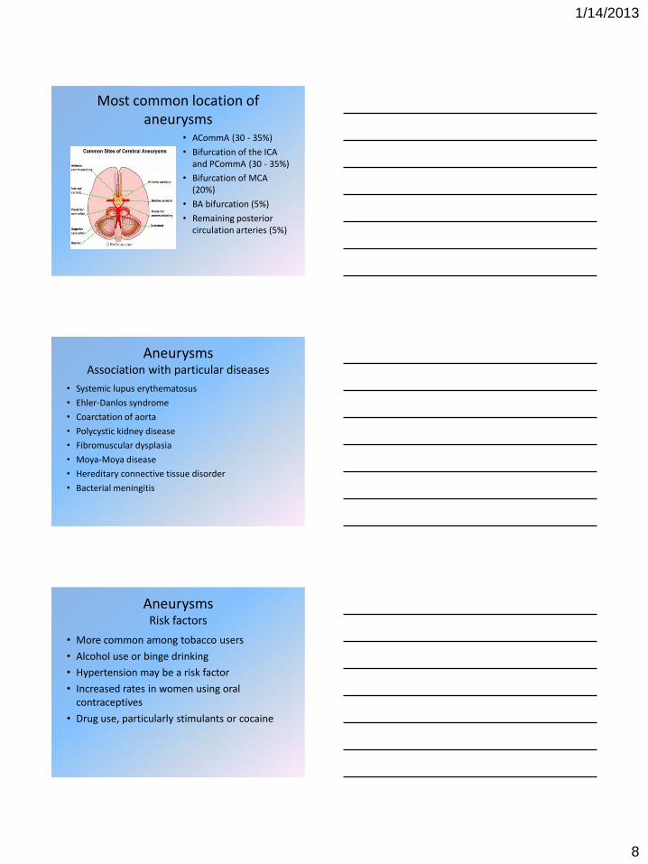
1142013
8
Most common location of aneurysms
bull ACommA (30 - 35)
bull Bifurcation of the ICA and PCommA (30 - 35)
bull Bifurcation of MCA (20)
bull BA bifurcation (5)
bull Remaining posterior circulation arteries (5)
Aneurysms Association with particular diseases
bull Systemic lupus erythematosus
bull Ehler-Danlos syndrome
bull Coarctation of aorta
bull Polycystic kidney disease
bull Fibromuscular dysplasia
bull Moya-Moya disease
bull Hereditary connective tissue disorder
bull Bacterial meningitis
Aneurysms Risk factors
bull More common among tobacco users
bull Alcohol use or binge drinking
bull Hypertension may be a risk factor
bull Increased rates in women using oral contraceptives
bull Drug use particularly stimulants or cocaine
1142013
9
Aneurysms Risk factors
bull Gender
In most series of aneurysmal SAH there is a striking female preponderance Adult women are affected by aneurysmal SAH more than men by ratio of 60 to 40
bull Genetics Family history (up to 11)
bull Geographic factors The incidence of SAH higher in Finland Japan and low in New Zealand and Middle East It varies significantly from 22-23100000 in Finland and Japan to 8-12100000 in other regions
Aneurysms Incidence
bull Incidence of brain aneurysms in general population is 1 - 5
bull Frequency of brain aneurysms ranged from 02 - 99
bull As many as 400000 people in the USA may have a brain aneurysm
SAH
1142013
10
SAH Etiology
bull Aneurysm 51
bull Hypertension 15
bull AVM 6
bull Other 28
Aneurysmal SAH Epidemiology
bull Approximately 5 - 10 of all strokes
bull Incidence averages 11 per 100000 (range 6-16
per 100000)
bull Adults of all ages
bull Incidence not declining
Aneurysmal SAH Epidemiology
bull 30000 Americans suffer non-traumatic SAH
each year
bull Overall mortality rates are 25
bull Morbidity among survivors is 50
1142013
11
Aneurysmal SAH ndash Presentation
bull Sudden severe headache
bull The hallmark of aneurysmal SAH-headache is that it develops within seconds
bull It is critical to inquire on how quickly the headache developed
bull Even a careful history cannot distinguish between headache secondary to aneurysm rupture and other more common and benign forms of headache
Congenital aneurysms
bull The circle of Willis has been dissected and three berry aneurysms are seen Multiple aneurysms are seen in about 20-30 of cases of berry aneurysm Such aneurysms are congenital in the sense that the defect in the arterial wall is present from birth but the actual aneurysm takes years to develop so that rupture is most likely to occur in young to middle age adults
Ruptured berry aneurysm in the circle of Willis
1142013
12
SAH Diagnosis
bull Non-contrast CT scan confirms subarachnoid blood in 98 of patients if performed within the first 12 hours after onset
bull Distribution of blood on CT provides information on the origin of the SAH
The Fisher Scale
bull The Fischer scale is a way of grouping subarachnoid
haemorrhage CT scans into four goups according to the amount of blood and is useful in predicting cerebral vasospasm
bull Group 1 No blood detected
bull Group 2 Diffuse thin (lt1mm) SAH with no clots
bull Group 3 Localised clots and or layers of blood gt1 mm in thickness
bull Group 4 Intracerebral or intraventricular blood (+- SAH)
SAH CT-Scan
bull Fisher Grade 3
1142013
13
Hunt amp Hess Scale describes the severity of subarachnoid
haemorrhage and is used as a predictor of survival bull Grade 1
ndash asymptomatic or minimal headache and slight neck stiffness
ndash 70 survival
bull Grade 2
ndash moderate to severe headache neck stiffness no neurologic deficit except cranial nerve palsy
ndash 60 survival
bull Grade 3
ndash drowsy minimal neurologic deficit
ndash 50 survival
bull Grade 4
ndash stuporous moderate to severe hemiparesis possibly early decerebrate rigidity and vegetative disturbances
ndash 20 survival
bull Grade 5
ndash deep coma decerebrate rigidity moribund
ndash 10 survival
The WFNS grading system uses the Glasgow Coma Scale and presence of focal neurological deficits
bull GCS 15 No deficit
bull GCS 13-14 No deficit
bull GCS 13-14 No deficit
bull GCS 7-12 may be a neurological deficit
bull GCS lt7 may be a neurological deficit
SAH Diagnosis - MRI
bull In the acute phase MRI with FLAIR demonstrates SAH as reliably as CT
bull MRI is superior to CT in evaluating extravasated blood after a few days (up to 6 weeks) and it is a useful tool in patients referred in a delayed fashion if the original diagnosis is in question
1142013
14
SAH Diagnosis ndash Lumbar puncture
bull Necessary in patients with suggestive history but negative CT
bull Preferably done after 6 or better 12 hours to allow for xanthochromia
bull Xanthochromia persists for at least 2 weeks after SAH
CIRCULATORY PATHOPHYSIOLOGY
Pathophysiological Alterations following Aneurysmal SAH
bull Intracerebral amp Intraventricular Hemorrhage
bull Hydrocephalus
bull Cerebral edema
bull Seizures amp Seizure-like Activity
bull Alteration in Respiratory Function
bull Effect on the Heart
bull Fluid and Electrolyte Disturbance
bull Cerebral ischemia
1142013
15
Vasospasm and Cerebral Ischemia
bull Although there are medical therapies for vasospasm after SAH early detection of vasospasm and initiation of aggressive medical therapy is of utmost importance to avoid delayed neurological ischemia morbidity and mortality
CEREBRAL VASOSPASM AND ITS CLINICAL SIGNIFICANCE
Cerebral Vasospasm
bull Some substances (numerous neurotransmitters blood constituents or breakdown products and autocoids) released at the time of SAH acts on smooth muscle wall to cause vasoconstriction
bull Morphological changes of the arterial wall consistent with vasonecrosis or vasculopathy
bull Mechanical compression may also result in vessels constriction
1142013
16
Vasospasm mechanisms Leung Stroke 2002
Macdonald NeurosurgRev 2006
Blood breakdown products
bull ApolipoproteinE genotype Immunomodulatory neurotoxic oxidative effects APOE4 less effective than APOE3 in suppressing
neurotoxicity
bull Endothelin1 release from CSF leukocytes Potent vasoconsrictor Synergistic effect in vasoconstriction between
bull APOE and Endothelin1
Double-hit model of delayed ischemic neurological deficits after SAH based on Dreier et al The two hits on the brain parenchyma consist of acutely triggered microvascular spasm in response to spreading depolarizations
superimposed on chronic vasospasm
Early Brain Injury Sehba F 2011
1142013
17
Early Brain Injury and Potential Therapy Sehba F 2011
Clinical Features of Symptomatic Vasospasm
bull Variable clinical course
bull Usually peaks at 7-10 days following SAH
bull Usually gradually evolves with waxing and waning symptoms
bull New HA seizures or decreased alertness
bull New focal neurological signs - MCAACAborder zone
Cerebral Vasospasm Clinical Significance
bull Cerebral vasospasm constitutes a major complication of SAH
bull The presence of vasospasm has been correlated with a 15 to 3-fold increase in mortality in the first 2 weeks after SAH
bull DID occur during a period ranging from 4 to 12 days but early (3d) or late manifestations (le 3 wk) may be observed
1142013
18
SAH Outcome
bull 30-day mortality rate still high
bull Many patients die before or shortly after reaching medical attention
bull Large percentage of survivors suffers from cognitive sequelae even after ldquosuccessfulrdquo treatment
Cerebral Vasospasm and Delayed Ischemic Deficit Dorsch et al 1994
bull Literature review of more than 30 000 cases
bull Angiographic vasospasm occurred in 433 (range 19 - 97)
bull DID occurred in 323 (range 5-90)
bull Outcome of DID - Death in 303 - Permanent deficit in 34 - Good outcome in 357
A clinical review of cerebral vasospasm and delayed ischaemia following aneurysm
rupture Dorsch N
2011
bull Online and physical searches have been made of the relevant literature
bull The incidence of delayed ischemic deficit (DID) or symptomatic vasospasm reported in 1994 was 325 in over 30000 reported cases In recent years 1994-2009 it was 677523806 or 285
bull Many of the recent reports did not specify whether a calcium antagonist was used routinely and when this was stated (usually nimodipine or nicardipine) DID was noted in 220 of 10739 reported patients
bull The outcome of DID in the earlier survey was a death rate of 316 with favorable outcomes in 362 In recent reports though with fewer than 1000 patients the outcome is possibly better with death in 256 and good outcome in 541
bull It thus appears likely that delayed vasospasm is still common but less so and that the overall outcome has improved This may be due to the more widespread use of calcium antagonists and more effective fluid management
1142013
19
SAH Management Macdonald Neurosurg Rev 2006
Anderson et al Am J Neuroradiol 2000
bull Vasospasm Diagnosis Change in clinical status 4-vessel angiography bull CTA Sensitivity 100 Specificity 92 Proximal arteries Internal carotid basilar and first
segments of ACA and MCA Less useful for distal vessels and for differentiating mild and
moderate spasm bull MRA Sensitivity 46 Specificity 70 Related to movement and time required for study bull TCD Sensitivity 50 ndash60 Specificity 90 () bull PbtO2 CBF microdialysis bull Glutamate glycerol lactate pyruvate bull CBF lt 15 mL 100 gm min
Management of comlications
bull Angiographic VSP is more common (occurring in about two thirds of patients) than is symptomatic vasospasm (with clinical evidence of cerebral ischemia)
bull TCD is performed daily to monitor for VSP which is defined as a mean CBFV of more than 100 cmsec in a major vessel TCD has a sensitivity that is similar to that of cerebral angiography for the detection of narrowed vessels particularly in the MCA and ICA
bull Once symptomatic VSP is evident (with focal neurologic signs) patients are treated with hypervolemia and induced hypertension
bull Patients whose condition does not improve with medical therapy undergo emergency cerebral angiography and transluminal angioplasty or vasodilator infusion when focal vessel narrowing is evident
Prevention of Vasospasm and DID Naval CCM 2006
HHH Therapy 1 Hypertensive ndashMAP = 100 to 120 mm Hg
2 Hypervolemic ndashCVP = 8 to 12 mm Hg
3 Hemodilution ndashHct = 30
bull Prevention is controversialhelliphelliphelliphelliphellip
bull Statins-protection
bull Mg++ -need further studies
1142013
20
VasoSpasm 2011 11th International Conference
bull Dr L MacDonald presented (CONSCIOUSS 1 2 and 3) C-2 led to a non-significant reduction in mortalityVSP related morbidity but was not associated with an improvement in GOSE (Glasgow Outcome Score Extended) C-3 trial was stopped due to the lack of efficacy data from the Phase 3 clinical study CONSCIOUS-2
bull Effect of intracisternal magnesium therapy Dr Mori et al from Japan Experimental work in dogs the reversible effect of intracisternal magnesium MgSO4 therapy required CSF Mg2+ concentration of more than 3 mEql effect was evident for 3 to 6 hrs therefore continuous or intermittent infusion probably needed to ameliorate VSP
bull Prolonged release of nicardipine from pellets (NP) that are placed around vessels during surgical clipping presented by Dr Kasuya (Japan) initially treated 100 pts provided very impressive results later 136 multicenter trial in Japan was similar to first results Now the randomized double-blind trial of 32 pts with severe SAH done in Germany and the incidence of angiographic vasospasm was significantly reduced 73 control vs 7 in patients with NPs
VasoSpasm 2011 11th International Conference
bull Current medical therapy (nimodipinenicardipine) or aggressive 3H-therapy will not prevent patients after aSAH to have vasospasm
bull Close to 100 of patients after aSAH would have vasospasm demonstrated by cerebral angiography andor TCD
bull No clear predictive value for patients who will have symptomatic vasospasm based on angio or TCD data developed
bull Early brain injury after a-SAH emerges as a new recent concept with emphasis on complex pathophysiological mechanisms that are linked to initial bleed However it remains unknown whether global ischemia itself or subsequent events are responsible for the detected cell death and neurodegeneration
bull Numerous experimental work going on trying to identify therapies for vasospasm preventiontreatment
Effects of a Single Dose of Dantrolene in Patients With Cerebral Vasospasm After Subarachnoid Hemorrhage
A Prospective Pilot Study Muechschlegel et al Stroke 2011
bull Dantrolene is a known ryanodine receptor inhibitor and is already approved by the
US FDA for other indications There is evidence that dantrolene is neuroprotective Furthermore in an ex vivo rat model dantrolene has been shown to inhibit cerebral vasoconstriction alone as well as in combination with nimodipine
bull In a prospective open-label single-dose ascending safety trial 5 patients received iv dantrolene 125 mgkg and the next 5 patients received 25 mgkg over the course of 60 minutes TCD was performed at 0 45 90 and 135 minutes relative to infusion start
bull Peak systolic CBFV decreased significantly (minus26 cms) with a borderline change in mean CBFV in the low-dose group (minus16 cmsec) and peak systolic CBFV in the high-dose group (minus26 cms)
bull In this pilot study a single dose of iv dantrolene in cerebral vasospasm after SAH appears feasible while inhibiting vasoconstriction in the low-dose group but it may lower MAP
1142013
21
TCD DIAGNOSIS OF VASOSPASM
CBF Measurements
Xe 133 SPECT
CBF vs CBFV
bull 133Xe
bull Stable xenon-enhanced CT
bull MRI
bull PET
bull SPECT
bull The time expense and complexity of these techniques still limits its use in routine clinical practice
1142013
22
TCD Diagnosis of Vasospasm Newell et al 1993
Diagnosis and Monitoring of Vasospasm ANGIOGRAPHY
bull Degree of angiographic vasospasm does not always correlate with the clinical condition Some patients remain asymptomatic with severe vasospasm demonstrated by angiography
bull Incidence of angiographic vasospasm is nearly twice that of DID
Diagnosis and Monitoring of Vasospasm TCD
bull High CBFV can identify patients at higher risk for developing DID but also may occur in asymptomatic patients
bull NeurologistNeurointensivist must determine whether the severity and location of the vessel narrowinghigh CBFV are appropriate to cause the clinical deficit
1142013
23
Is routine TCD monitoring useful in the management of SAH
Warlaw et al Neurosurgery 1998
bull 186 patients after SAH
bull Routine TCD examinations made an important positive contribution to the diagnosis of DID in 72 and led to altered management for the benefit of the patient in 43
bull In 9 of pts with recent SAH it was believed that outcome might have better if the TCD results had been acted upon appropriately
Is routine TCD monitoring useful in the management of SAH
Warlaw et al Neurosurgery 1998
bull The TCD results appeared to alter management beneficially in 37 of emergency and 48 of elective patients
bull In the patients admitted on an emergency basis this was often because TCD monitoring showed elevated CBFVrsquos and influenced (delaying or advancing) the timing of surgery or discharge or even led to simple actions such as continuing bet rest or administration of IV fluids for an additional 24 hrs
Transcranial Doppler in cerebral vasospasm Newell et al 1990
MCA CBFV ge 120 cms 25 narrowing
MCA CBFV ge 140 cms 25-50 narrowing
MCA CBFV ge 200 cms 50 narrowing
1142013
24
TCD Criteria for diagnosis of MCA vasospasm
Mean CBFV MCAICA ratio Interpretation (cms) (Lindegaard Ratio)
lt100 lt 3 Nonspecific
100-140 3-6 Mild
140-200 3-6 Moderate
gt200 gt6 Severe
TCD and anterior circulation vasospasm
TCD amp Angiography
bull MCA Sensitivity 94 Specificity 90 PP value 100
bull ACA Sensitivity 71 Specificity 100 PP value 100
TCD and posterior circulation vasospasm TCD and angiography
Sloan et al 1994
bull VA Sensitivity 44 Specificity 88 PP Value 54
bull BA Sensitivity 77 Specificity 79 PP Value 62
1142013
25
Basilar artery vasospasm and delayed posterior circulation ischemia after aneurysmal subarachnoid hemorrhage
Sviri et al Stroke 2004
bull Patients with very high BA CBFV (gt115 cms) had a 50 chance of developing delayed BS ischemia BA-VS was found at a higher rate in patients who experienced reduced rCBF in the cerebellum (563) thalamic nuclei (684) and occipital lobe (818) Although patients with delayed BS hypoperfusion did not present with a higher clinical grade their clinical outcome was significantly worse (Glasgow Outcome Score after 30 days 248+-116 versus 33+-127 P=0001)
bull These findings suggest for the first time that BA-VS after aneurysmal SAH is associated with hypoperfusion to BS and other posterior circulation territories
bull The risk for delayed BS ischemia increased significantly when TCD BA-FVs were gt115 cms
TCD Grading Criteria for BA vasospasm
Sviri et al 2006
bull 123 pts SAH angiography BA and extracranial VA TCD within 4 hrs before the DSA
bull CBFV ratio between the BA and extracranial VA strongly correlated with the degree of BA narrowing (r=086 plt00001)
bull A ratio higher than 2 associated with 73 sensitivity and 80 sensitivity for BA vasospasm
bull A ratio higher than 25 with BA CBFV 85 cmsec was associated with 86 sensitivity and 96 specificity for BA narrowing more than 25
bull A ratio higher than 30 with BA CBFV higher than 85 cmsec was associated with 92 sentivity and 97 specificity for BA narrowing more than 50
Prediction of symptomatic vasospasm after SAH with TCD
bull An early CBFV increase (Seiler et al 1988)
bull A rapid CBFV increase in the first 6 days (Grote et al 1988)
bull A CBFV increase of at least 50 cmsec during 24 hours (Grosset et al 1993)
bull A CBFV increase of 50 cmsec during 48 hours (Wardlow et al 1998)
bull Relative changes in CBFVrsquos (two or threefold CBFV increase) in patients with aneurysmal SAH correlated better with clinically significant vasospasm than absolute CBFVrsquos (Naval et al 2005)
1142013
26
TCD for diagnosis of cerebral vasospasm after SAH mean blood flow velocity ratio of the ipsilateral and contralateral MCA
Nakae et al
2011
bull Retrospective study 142 pts 1262 TCD tests DID defined as neurological deficit or CT evidence of infarct cause by vasospasm ()
bull The threshold value that best discriminated between pts with and wo DID was IC mCBFV of 15
bull IC mCBFV ratio demonstrated a more significant correlation to DID than the absolute m CBFV
Low Pulsatility Index on TCD Predicts Symptomatic Large-Vessel Vasospasm After Aneurysmal SAH
Rajajee et al Neurosurgery 2012
bull Medical records of patients admitted with aSAH between January 2007
and April 2009 were reviewed TCCD sonography was performed daily between days 2 and 14 Patients with unexplained acute neurological decline underwent catheter- or CT-angiography The lowest recorded PI and the highest mCBFV on TCCD were correlated to the occurrence of symptomatic large vessel vasospasm angiographic vasospasm and delayed cerebral infraction in multivariate analysis by use of logistic regression
bull Low PI on TCCD is an independent predictor of symptomatic vasospasm after aSAH whereas mCBFV is a better predictor of angiographic vasospasm
TCD changes after vasospasm
200
140
100
200
140
100
Day 1 Day 2 Day 4 Day 6 Day 7 Day 8
Day 10 Day 12 Day 14 Day 16 Day 18
RIGHTLEFT
RIGHTLEFT
1142013
27
Early Onset Abnormal TCD changes after vasospasm
200
140
100
200
140
100
Day 1 Day 2 Day 4 Day 6 Day 7 Day 8
Day 10 Day 12 Day 14 Day 16 Day 18
RIGHTLEFT
RIGHTLEFT
Unilateral TCD changes after vasospasm
200
140
100
200
140
100
Day 1 Day 3 Day 5 Day 6 Day 7 Day 8
Day 1 Day 3 Day 5 Day 6 Day 7 Day 8
RIGHT
LEFT
High ICP
Unilateral TCD changes after vasospasm
200
140
100
200
140
100
Day 1 Day 3 Day 5 Day 6 Day 7 Day 8
Day 1 Day 3 Day 5 Day 6 Day 7 Day 8
RIGHT
LEFT
More than 50 cms
1142013
28
TCD Wave-Form
Low-Resistant High-Resistant
High ICP ICP treatmentnormalization
1142013
29
Cerebral vasospasm evaluated by TCD at different intracranial pressures
Klingelhofer et al 1991
bull 76 patients with SAH
bull MAP
bull ICP
bull PaCO2
bull TCD
- CBFV - RI
bull Evaluation of the interdependence of the patientrsquos clinical grade VSP ICP and TCD parameters
Cerebral vasospasm evaluated by TCD at different
intracranial pressures
Klingehofer et al 1991
Cerebral vasospasm evaluated by TCD at different intracranial pressures
Klingelhofer et al 1991
bull There was no case in which both high ICP and high CBFV were observed simultaneously
bull During the time course of vasospasm an increase in the resistance index above values of 06 with a simultaneous CBFV decrease indicates a rise in ICP rather than a reduction in VSP
bull With a pronounced increase in ICP evaluation of severity and time course of VSP by TCD based solely upon the mean CBFV can lead to a false-negative results
1142013
30
SAH Pt 1
Day 1
Rt MCA (M1 segm) 60 cms Lt MCA (M1 segm) 64 cms
Day 3
Rt MCA 100 cms Lt MCA 110 cms
SAH Pt 1
Day 8
Rt MCA (M1 segm) 212 cms Lt MCA (M1 segm) 230 cms
Day 15
Rt MCA 308 cms Lt MCA 121 cms
SAH Pt 2
Day 5
Lt MCA 126 cms Rt MCA 88 cms
Day 6
Lt MCA 38 cms Rt MCA 118 cms
1142013
31
SAH Pt 2
Day 7
Lt MCA 139 cms Rt MCA 148 cms
SAH Pt 3
Day 3
Rt MCA 126 cms Lt MCA 88 cms
Day 4
Rt MCA 38 cms Lt MCA 43 cms
SAH Pt 3
Day 5
Rt MCA 131 cms Lt MCA 158 cms
1142013
32
SAH amp ICP
bull We are judging qualitative TCD wave form morphology changes
bull These changes usually will be obvious after ICP will be more 30 mm Hg
bull However one condition must be full filled if you would be using TCD wave from changes to predict intracranial hypertension MAP cardiac output and PaCO2 are normal and not different significantly compared to the previous day
Effect of age on MCA and ICA CBFV Torbey at el Stroke 2011
lt6 8(n=4 7 )
68
(n=3 4 )
p-v a lue
Ma x MC A CBFVMedian (c ms)Range
11435-254
7633-190
0002
Ti me to Max MC A C BFVMedian (days)Range
60-18
72-15
07
MC A pul satil ity i ndexMedianRange
088029-278
109031-283
lt00001
Ma x ICA C BFVMedian (c ms)Range
12640-211
76530-202
0003
Ti me to Max IC A CBFVMedian (days)Range
72-16
553-15
04
IC A pul sati l ity indexMedianRange
088035-55
11804-371
lt00001
lt68 (n=47)
68 (n=34)
p-value
Symptomatic Vasospasm n()
31(66) 15(44) 005
Time to symptomatic vasospasm Median (cms) Range
7
(2-16)
5
(2-10)
006
MCA CBFV 1 day at VSP Median (cms) Range
92
52-243
55
24-190
0009
ICA CBFV 1 day at VSP Median (cms) Range
92
33-196
605
30-171
0079
TCD VSP 42 23 lt00001 TCD confirmed VSP n() 10(53) 5(33) 02 Radiological VSP n() 14(74) 8(47) 01
Effect of age on MCA and ICA CBFV Torbey at el Stroke
2011
1142013
33
Kaplan-Meier Curve for VSP onset time Torbey at al Stroke 2001
Cumulative VSP Probability curve
Time to VSP symptoms
30 20 10 0
Cu
mu
lative
V
SP
11
10
9
8
7
6
5
4
3
AGE GROUP
Age lt68
Age 68
Role of TCD SAH
bull Elevated CBFVrsquos in asymptomatic patients warrant meticulous observation in some closely supervised setting until CBFVrsquos begin trend downward
bull Elevated CBFVrsquos in a particular vascular territory can focus subsequent neurologic examinations to detect subtle changes earlier in their clinical course
Role of TCD SAH
bull In symptomatic patients elevated CBFVrsquos most likely represent significant vessel narrowing and may obviate the need for cerebral angiography At this point triple-H therapy can be initiated or advanced
bull Asymptomatic patients without elevated CBFVrsquos probably can avoid additional angiography However we need to consider patientrsquos age because elderly patientrsquos could develop vasospasm in normal or slightly abnormal CBFV range
1142013
34
Factors influencing interpretation
bull Patient age
bull The presence of moderate to severe anemia (Hct lt27)
bull Impaired CBF autoregulation (passive CBFV variation with MAP changes)
bull Hyperemia induced by triple-H therapy
Guidelines for the Management of Aneurysmal SAH Stroke Council AHA 1994
bull Summary and Recommendations 1 SAH is a medical emergencyhellip 2 CT scanning for suspected SAH is strongly recommendedhellip 3 Selective cerebral angiography to document 4 TCD is recommended for the diagnosis and monitoring of vasospasm although the cerebral angiography may be required for definitive diagnosis
TCD MONITORING OF VASOSPASM
1142013
35
Transcranial Doppler monitoring and clinical decision-making after subarachnoid hemorrhage
McGirt et al 2003
bull Objective was to examine the impact of TCD vasospasm monitoring on clinical decision-making following SAH
bull The records of 50 randomly selected patients undergoing serial TCD monitoring following SAH were reviewed Dates and results of TCDs and cerebral angiograms the use of hypertensive hemodilution (HH) therapy and the development of new neurological deficits were recorded The independent effects of TCD-defined vasospasm and new neurological deficits on patient management were determined with multiple logistical regression Results were validated in a second randomly selected 50 patient cohort
Transcranial Doppler monitoring and clinical decision-making after subarachnoid hemorrhage
McGirt et al 2003
bull Mild or moderate TCD-defined vasospasm developed in 76 of patients 58 +- 05 days after SAH 38 developed severe TCD-defined vasospasm after 79 +- 07 days Focal neurological deficits occurred in 50 after 57 +- 06 days with TCD abnormalities preceding the deficit by 25 +- 07 days in 64
bull TCD-defined vasospasm did not independently influence the use of HH therapy or angiography with both decisions associated with the development of new neurological deficits
bull As TCD-defined vasospasm preceded the neurological deficit in 64 earlier intervention might reduce the incidence of vasospasm-related stroke in institutions with similar practice patterns
MCA and ICA CBFVrsquos Dynamics
1142013
36
Biphasic CBFVrsquos profile after SAH Luft et al Neurocritical Care 2004
bull 18 (99) patients demonstrated biphasic CBFVrsquos profile
bull 1st CBFVrsquos peak (134 + 11 cms) occurred on post-SAH day 6 + 1 2nd CBFVrsquos peak (148 + 12 cms) occurred on post-SAH day 13 + 1
bull Although the 2nd CBFVrsquos peak is usually not associated with a worsening of symptoms these patients were more likely to exhibit clinical symptoms during the 1st CBFV peak
Biphasic CBFVrsquos profile after SAH Luft et al Neurocritical Care 2004
Angiography examination in patient with
ACommA aneurysm before and after aneurysm clipping
1142013
37
Angiography examination after transluminal angioplasty
TCD Monitoring
0
50
100
150
200
250
1 2 3 4 5 6 7 8 9 10 11 12 13 14 15 16 17 18 19 1 2 3 4 5 6 7 8 9 10 11 12 13 14 15 16 17 18 19
MCA M1
MCA M2
ACA A1
ICA C1
VA
BA
MAP
Hct
ICP
Temp
PaCO2
RIGHT LEFT
Days after SAH
1st PEAK
2ND PEAK
Elevated Transcranial Doppler Ultrasound Velocities Following Therapeutic Arterial Dilation
Giller et al 1995
bull Elevated TCD CBFVrsquos seen after cerebral angioplasty are commonly interpreted as evidence of residual or recurrent stenosis but may conceivably arise from hyperemia and require different clinical management
bull Four cases of abnormally elevated mean CBFVrsquos obtained after therapeutic arterial dilation with either balloon angioplasty or intra-arterial administration of papaverine are described In each case cerebral angiography revealed a dilated vessel suggesting that hyperemia and impaired autoregulation were the causes of the high CBFVrsquos
bull These examples suggest that high TCD CBFVrsquos after vessel dilation may be produced by unpredictable amounts of vessel narrowing and flow alteration Although a normalizing CBFV after angioplasty suggests effective vessel dilation high CBFVrsquos may be due partly to hyperemia and cannot be interpreted as arising solely from recurrent stenosis
1142013
38
2011 AHAASA Metrics for Measuring Quality of Care in Comprehensive Stroke Centers
bull Among different measures for Comprehensive Stroke Centers is
Median frequency of noninvasive monitoring for surveillance for vasospasm in patients with aneurysmal SAH during the period between three and 14 days after SAH
POSTTRAUMATIC VASOSPASM
TBI
bull Every 21 seconds one person in the US sustains traumatic brain injury (TBI)
bull An estimated 53 million Americans ndash little more than 2 of the US population ndash currently live with disabilities resulting from brain injury
bull Each year 80000 Americans experience the onset of long-term disability following TBI
bull Okie NEJM 2005 Among surviving soldiers wounded in combat in Iraq and Afghanistan TBI appears to account for a larger proportion of casualties than it has in other recent US wars According to the Joint Theater Trauma Registry 22 had injuries to the head face or neck
Armonda et al Neurosurgery 2006 474 had traumatic cerebral vasospasm Majority were blast related injury
Civilian
Battlefield
1142013
39
Blast TBI
bull Blast injuries historically seen by providers as limited to military concern
Peacetime terrorism over the last decade has reminded us otherwise
bull
Why we need talk about blast TBI
bull Mortality
ndash Oklahoma City 167
ndash US Embassy 223
ndash World Trade Center 2801
ndash Madrid train bombings 191
ndash London 56
ndash Domodedovo 37
ndash Minsk metro 12
Large number of injuredhellip
TBI PATHOPHYSIOLOGY
1142013
40
Balancing Multisystem Interactions
TBI
TBI Pathophysiology
Primary Injury - ContusionsHemorrhages
- Diffuse Axonal Injury (DAI)
Secondary Injury (Intracranial) occurs hours to weeks after injury
- Blood Flow and Metabolic Changes
- Traumatic Hematomas
- Cerebral Edema
- Hydrocephalus
- Increased Intracranial Pressure
Decrease in CBF
BRAIN
EDEMA
More Brain Ischemic Blood Vessels
Edema brain cells Dilate
Increased ICP
Cell Injury
1142013
41
But where is the place for TCD monitoring
Multimodal Monitoring VasospasmIschemiahigh ICP Detection
bull MAP
bull SaO2
bull ECG
bull Et-CO2
bull CVP
bull Urine output
bull ICP
bull CBFVTCD PbO2
cEEG
CTMR Perfusion
The primary goal of
management for
TBI is the
prevention of
secondary damage
due to neuronal
hypoxia and
hypoperfusion
Daily TCD
Continuous EEG
ICP monitoring
Pb02
CBF
1142013
42
Traumatic Brain Injury amp TCD
bull Cerebral ischemia due to the post-traumatic vasospasm
bull Increased Intracranial Pressure
bull Brain Death
Post-Traumatic VSP
bull Ischemic symptoms caused by cerebral arterial spasm following traumatic SAH are comparable to those found following aneurysmal SAH
- appearance of symptoms between Days 4 and 16 after injury with the peak incidence on Days 9 and 10
- close correlation between the main site of the subarachnoid blood and the location of severe vasospasm responsible for the symptoms and a higher incidence of symptoms in patients with massive SAH than those with slight SAH
bull Subarachnoid blood plays an important role in the later development of vasospasm not only following aneurysm rupture but also after head injury Nevertheless there is no general agreement that subarachnoid blood in head injury is an important risk factor in the development of vasospasm and ischemic brain damage
TBI and VSP
bull Cerebral posttraumatic VSP (PTV) was first described by Lorn in 1936
bull The incidence of CT documented traumatic SAH has been identified in 4 to 63 of pts after TBI
bull Study from the University of Mississippi Medical Center indicated that traumatic SAH complicate course of TBI in 69 of the patients due to the presence of PTV
1142013
43
Clinical Significance
bull 41 of patients who died from TBI had PTV (MacPherson et al 1973)
bull 24 with massive tSAH developed ischemic symptoms in contrast to 3 of patients with mild tSAH (Taneda et al 1996)
bull Ischemic symptoms accompanying arterial VSP following tSAH are comparable to those found following aneurysmal SAH
WARTIME TRAUMATIC CEREBRAL VASOSPASM RECENT REVIEW OF COMBAT CASUALTIES
Armonda et al Neurosurgery 2006
bull The first study to analyze the effects of blast-related injury on the cerebral vasculature
bull This study showed that TCV occurred in a substantial number of patients with severe neurotrauma and clinical outcomes were worse for those with this condition
25 yo severe concussion no SAH (Courtesy of Dr Armonda)
CBFV 120 cms CBFV 57 cms
1142013
44
25 yo severe concussion no SAH (Courtesy of Dr Armonda)
CBFV 112 cms CBFV 89 cms
25 yo severe concussion no SAH (Courtesy of Dr Armonda)
CBFV
(cmsec)
Calendar
0
50
100
150
200
250
300
MCA M1
MCA M2
ACA A1
VA
BA
MAP
Hct
ICP
Temp
PaCO2
RIGHT LEFT
Ant circ TLA Post circ TLA Ant circ TLA
SEVERE VSP
MODERATE VSP
MILD VSP
EFFECT OF TLA ON PROLONG (SEVEN DAYS) VASOSPASM INVOLVING ANTERIOR AND POSTERIOR CIRCULATION
1142013
45
The CBFVrsquos and systemic hemodynamic trends TCD signs of severe vasospasm involving posterior and
anterior circulation lasting seven days before TLA
0
50
100
150
200
250
300
MCA M1
ACA A1
ICA C1
VA
BA
MAP
Hct
ICP
PaCO2
RIGHT LEFT
Severe VSP
Moderate VSP
MIld VSP
TLA TLA
Effect of TLA after prolong severe vasospasm
Left MCA CBFV 69 cmsec PI 17 Right MCA CBFV 66 cmsec PI 14
Left MCA 205 cmsec PI 08 Right MCA CBFV 195 cmsec PI 05
CBFVrsquos and PIrsquos Trends (PI x 100)
0
50
100
150
200
250
300
MCA M1
M1 PI
ACA A1
A1 PI
ICA C1
C1 PI
VA
VA PI
BA
BA PI
RIGHT LEFT
1142013
46
The CBFV amp PI Trends
bull MCA (M1 segm)
bull ICA (C1 segm)
0
50
100
150
200
250
300
MCA M1
M1 PI
RIGHT LEFT
Severe VSP
Moderate VSP
Mild VSP
0
50
100
150
200
250
300
ICA C1
C1 PI
RIGHT LEFT
Severe VSP
Moderate VSP
Mild VSP
Clinical Material
bull Ninety patients (2 females) aged 18 to 50 years (mean 259 years) who had suffered wartime TBI injuries (with Glasgow Coma Scale scores ranging from 3 to 15) were investigated with daily TCD studies
bull A total of 567 TCD studies (mean 64 testspatient ranged from 1 to 30) were made after admission
TCD signs of Vasospasm (in ) by type of TBI
7500
3680
6840
2900
3570
520
3150
2350
1430
520
1570 1760
0
10
20
30
40
50
60
70
80
PHI PHIIED CHI CHIIED
Mild ()
Mod ()
Severe ()
1142013
47
Presence of ICH ()
5700
5200
5800
5000
PHI PHIIED CHI CHIIED
TCD signs of VSP amp high ICP
TBI Type Post TBI
VSP
High
ICP
CHI 13144 12133
CHIIED 12133 910
PHI 21233 16177
PHIIED 11122 12133
Total 57633 49544
TCD AND TUMOR RESECTION
1142013
48
Tumor Resection and Vasospasm
bull The occurrence of vasospasm and delayed cerebral ischemia after resection of intracranial tumor has not received extensive attention clinically and is often misdiagnosed and improperly treated as surgical brain damage or brain swelling
bull However DID from vasospasm after tumor resection is a complication that is being reported in increasing numbers
Tumor Resection and Vasospasm Literature review
bull Reports are sparse and mainly are case series
bull Vasospasm was found in 2 to 49 patients No significant difference among age sex surgical approaches pathological diagnosis duration of surgery amount of blood loss and transfusion during surgery were found but significant difference was seen in cisternal hemorrhage on CT scan and the amount of blood in cerebrospinal fluid
Tumor Resection and Vasospasm Probable Mechanism
bull It is suggested that accumulation of blood in the basal cisterns may have been responsible for this unusual condition and it is therefore important to consider vasospasm as a probable etiological cause of clinical deterioration in patients undergoing the surgical removal of a cerebral tumor
bull For this reason whenever any neurological deterioration occurs in such patients it is advisable to perform TCD in order to verify the presence of any vasospasm and promptly commence suitable treatment
1142013
49
Tumor Resection and Vasospasm Management
bull Surgery To decrease the amount of blood in basal cistern by microsurgery
bull ICU TCD utilization in the diagnosis of vasospasm and its monitoring
bull ICU Differentiation of vasospasm from brain swelling are helpful to confirm the coexistent or causal relation based on neurological assessment CT TCD and ICP monitoring both in deciding therapeutic strategy and successfully controlling vasospasm
bull ICU Nimotop played a key role in preventing brain damage from vasospasm and cerebral swelling
Suprasellar tumor resection The CBFVs and Trends of the Systemic
Hemodynamic
RIGHT LEFT
0
20
40
60
80
100
120
140
160
180
103010311111121131141151161171181191110 103010311111121131141151161171181191110
Calendar
CB
FV
s a
nd
Vari
ab
les
0
20
40
60
80
100
120
140
160
180
CB
FV
s a
nd
Vari
ab
les
MCA M1
MCA M2
ACA
ICA C1
VA
BA
MAP
ICP
Hct
PaCO2
Temp
Patient wit tumor
bull 58 yo female sp trans-sphenoidal resection of tuberculum sella meningioma complicated by A-comm complex pseudoaneurysm formation vasospasm and ACA territory infarct
1142013
50
DWI demonstrating ACA territory infarct
DWI demonstrating ACA territory infarct
ICA injections demonstrating vasospasm in A1-A2 bilaterally
A-comm complex pseudoaneurysm and coiling
1142013
51
A-comm complex pseudoaneurysm before and after coiling
TCD Trends
Left Right
Role of TCD Tumor Resection
bull It is useful to perform TCD test after surgery and perform daily TCD studies when patient is in the ICU
bull The frequency with which TCD should be performed may be guided by patient clinical presentation knowledge of risk factors for VSP early clinical course
bull TCD studies should be performed after endovascular treatment to identify patients with recurrent VSP
1142013
52
Role of TCD VSP monitoring
bull It is useful to perform TCD test on admission (or ASAP after surgery) and perform daily TCD studies when patient is in the ICU
bull The frequency with which TCD should be performed may be guided by patient clinical presentation knowledge of risk factors for vasospasm early clinical course
bull TCD studies should be performed after endovascular treatment to identify patients with recurrent vasospasm
Role of TCD VSP Monitoring
bull The presence and temporal profile of CBFVrsquos in all available vessels must be detected and serially monitored
bull The pattern of CBFVrsquos elevation may indicate the need to follow patient carefully for evidence of deficits related to specific vascular territory
bull Waveform appearance either regionally or globally may be clinically significant
TCD and VSP
bull Currently the gold standard for vasospasm diagnosis is cerebral angiography replaceable by CTA only when angiography is not available Obviously it is not feasible to perform such investigation as frequently as bedside clinical assessment
bull Repeated clinical assessments of a patients neurological status carry the problem of detecting the clinical signs and symptoms of vasospasm which occur only after vasospasm has already manifested its deleterious effects on the cerebral parenchyma
bull TCD is a relatively new non-invasive tool allowing for bedside monitoring to determine CBFVrsquos indicative of changes in vascular diameter
1142013
53
TCD and VSP
bull TCD can be useful pre- intra- and post-operatively while helping to recognize the development of cerebral vasospasm before the onset of its clinical effects
bull Vasospasm following SAH is a very important source of morbidity and mortality Too often the first sign is a neurologic deficit which may be too late to reverse
bull TCD assists in the clinical decision-making regarding further diagnostic evaluation and therapeutic interventions When performed in isolation the contribution of TCD to improving patient outcome has not been established Nevertheless TCD has become a regularly employed tool in neurocritical care and perioperative settings
TCD is a Critical Tool in Critical Care
bull The value of TCD in clinical practice is well established especially to measure and grade vasospasm following SAH and TBI
bull Based on AHA Guidelines and many years of clinical practice TCD is a tool employed by the Neurosurgeon Neurointensivist and Neurologist in the management of vasospasm
bull Based on high frequency of posttraumatic vasospasm and intracranial hypertension TCD testing must be utilized for management of patients after SAH (aneurysm rupture wartime or civilian TBI tumor resection)
bull The use of TCD at hospital admission allows identification of patients with brain hypoperfusion due to the vasospasm andor intracranial hypertension In such high-risk patients early TCD goal-directed therapy can restore normal cerebral perfusion and might then potentially help in reducing the extent of secondary brain injury
bull TCD could provide information about abnormally high ICPbrain death
bull In the future incorporation of TCD data may facilitate more injury- and time-specific therapies for patients after SAH (aneurysm rupture wartime or civilian TBI tumor resection)
TCD is a Critical Tool in Critical Care
1142013
54
Do we know everything about TCD and vasospasm
bull TCD criteria for vasospasm for the young (less than 20-30 yo) and old (more than 68-70 yo) patients
bull CBFV calculation formula that will take into account Hct values
bull Cerebral angiography could be negative but TCD could be positive for vasospasm
bull TCD prediction for clinical vasospasm
Future

1142013
9
Aneurysms Risk factors
bull Gender
In most series of aneurysmal SAH there is a striking female preponderance Adult women are affected by aneurysmal SAH more than men by ratio of 60 to 40
bull Genetics Family history (up to 11)
bull Geographic factors The incidence of SAH higher in Finland Japan and low in New Zealand and Middle East It varies significantly from 22-23100000 in Finland and Japan to 8-12100000 in other regions
Aneurysms Incidence
bull Incidence of brain aneurysms in general population is 1 - 5
bull Frequency of brain aneurysms ranged from 02 - 99
bull As many as 400000 people in the USA may have a brain aneurysm
SAH
1142013
10
SAH Etiology
bull Aneurysm 51
bull Hypertension 15
bull AVM 6
bull Other 28
Aneurysmal SAH Epidemiology
bull Approximately 5 - 10 of all strokes
bull Incidence averages 11 per 100000 (range 6-16
per 100000)
bull Adults of all ages
bull Incidence not declining
Aneurysmal SAH Epidemiology
bull 30000 Americans suffer non-traumatic SAH
each year
bull Overall mortality rates are 25
bull Morbidity among survivors is 50
1142013
11
Aneurysmal SAH ndash Presentation
bull Sudden severe headache
bull The hallmark of aneurysmal SAH-headache is that it develops within seconds
bull It is critical to inquire on how quickly the headache developed
bull Even a careful history cannot distinguish between headache secondary to aneurysm rupture and other more common and benign forms of headache
Congenital aneurysms
bull The circle of Willis has been dissected and three berry aneurysms are seen Multiple aneurysms are seen in about 20-30 of cases of berry aneurysm Such aneurysms are congenital in the sense that the defect in the arterial wall is present from birth but the actual aneurysm takes years to develop so that rupture is most likely to occur in young to middle age adults
Ruptured berry aneurysm in the circle of Willis
1142013
12
SAH Diagnosis
bull Non-contrast CT scan confirms subarachnoid blood in 98 of patients if performed within the first 12 hours after onset
bull Distribution of blood on CT provides information on the origin of the SAH
The Fisher Scale
bull The Fischer scale is a way of grouping subarachnoid
haemorrhage CT scans into four goups according to the amount of blood and is useful in predicting cerebral vasospasm
bull Group 1 No blood detected
bull Group 2 Diffuse thin (lt1mm) SAH with no clots
bull Group 3 Localised clots and or layers of blood gt1 mm in thickness
bull Group 4 Intracerebral or intraventricular blood (+- SAH)
SAH CT-Scan
bull Fisher Grade 3
1142013
13
Hunt amp Hess Scale describes the severity of subarachnoid
haemorrhage and is used as a predictor of survival bull Grade 1
ndash asymptomatic or minimal headache and slight neck stiffness
ndash 70 survival
bull Grade 2
ndash moderate to severe headache neck stiffness no neurologic deficit except cranial nerve palsy
ndash 60 survival
bull Grade 3
ndash drowsy minimal neurologic deficit
ndash 50 survival
bull Grade 4
ndash stuporous moderate to severe hemiparesis possibly early decerebrate rigidity and vegetative disturbances
ndash 20 survival
bull Grade 5
ndash deep coma decerebrate rigidity moribund
ndash 10 survival
The WFNS grading system uses the Glasgow Coma Scale and presence of focal neurological deficits
bull GCS 15 No deficit
bull GCS 13-14 No deficit
bull GCS 13-14 No deficit
bull GCS 7-12 may be a neurological deficit
bull GCS lt7 may be a neurological deficit
SAH Diagnosis - MRI
bull In the acute phase MRI with FLAIR demonstrates SAH as reliably as CT
bull MRI is superior to CT in evaluating extravasated blood after a few days (up to 6 weeks) and it is a useful tool in patients referred in a delayed fashion if the original diagnosis is in question
1142013
14
SAH Diagnosis ndash Lumbar puncture
bull Necessary in patients with suggestive history but negative CT
bull Preferably done after 6 or better 12 hours to allow for xanthochromia
bull Xanthochromia persists for at least 2 weeks after SAH
CIRCULATORY PATHOPHYSIOLOGY
Pathophysiological Alterations following Aneurysmal SAH
bull Intracerebral amp Intraventricular Hemorrhage
bull Hydrocephalus
bull Cerebral edema
bull Seizures amp Seizure-like Activity
bull Alteration in Respiratory Function
bull Effect on the Heart
bull Fluid and Electrolyte Disturbance
bull Cerebral ischemia
1142013
15
Vasospasm and Cerebral Ischemia
bull Although there are medical therapies for vasospasm after SAH early detection of vasospasm and initiation of aggressive medical therapy is of utmost importance to avoid delayed neurological ischemia morbidity and mortality
CEREBRAL VASOSPASM AND ITS CLINICAL SIGNIFICANCE
Cerebral Vasospasm
bull Some substances (numerous neurotransmitters blood constituents or breakdown products and autocoids) released at the time of SAH acts on smooth muscle wall to cause vasoconstriction
bull Morphological changes of the arterial wall consistent with vasonecrosis or vasculopathy
bull Mechanical compression may also result in vessels constriction
1142013
16
Vasospasm mechanisms Leung Stroke 2002
Macdonald NeurosurgRev 2006
Blood breakdown products
bull ApolipoproteinE genotype Immunomodulatory neurotoxic oxidative effects APOE4 less effective than APOE3 in suppressing
neurotoxicity
bull Endothelin1 release from CSF leukocytes Potent vasoconsrictor Synergistic effect in vasoconstriction between
bull APOE and Endothelin1
Double-hit model of delayed ischemic neurological deficits after SAH based on Dreier et al The two hits on the brain parenchyma consist of acutely triggered microvascular spasm in response to spreading depolarizations
superimposed on chronic vasospasm
Early Brain Injury Sehba F 2011
1142013
17
Early Brain Injury and Potential Therapy Sehba F 2011
Clinical Features of Symptomatic Vasospasm
bull Variable clinical course
bull Usually peaks at 7-10 days following SAH
bull Usually gradually evolves with waxing and waning symptoms
bull New HA seizures or decreased alertness
bull New focal neurological signs - MCAACAborder zone
Cerebral Vasospasm Clinical Significance
bull Cerebral vasospasm constitutes a major complication of SAH
bull The presence of vasospasm has been correlated with a 15 to 3-fold increase in mortality in the first 2 weeks after SAH
bull DID occur during a period ranging from 4 to 12 days but early (3d) or late manifestations (le 3 wk) may be observed
1142013
18
SAH Outcome
bull 30-day mortality rate still high
bull Many patients die before or shortly after reaching medical attention
bull Large percentage of survivors suffers from cognitive sequelae even after ldquosuccessfulrdquo treatment
Cerebral Vasospasm and Delayed Ischemic Deficit Dorsch et al 1994
bull Literature review of more than 30 000 cases
bull Angiographic vasospasm occurred in 433 (range 19 - 97)
bull DID occurred in 323 (range 5-90)
bull Outcome of DID - Death in 303 - Permanent deficit in 34 - Good outcome in 357
A clinical review of cerebral vasospasm and delayed ischaemia following aneurysm
rupture Dorsch N
2011
bull Online and physical searches have been made of the relevant literature
bull The incidence of delayed ischemic deficit (DID) or symptomatic vasospasm reported in 1994 was 325 in over 30000 reported cases In recent years 1994-2009 it was 677523806 or 285
bull Many of the recent reports did not specify whether a calcium antagonist was used routinely and when this was stated (usually nimodipine or nicardipine) DID was noted in 220 of 10739 reported patients
bull The outcome of DID in the earlier survey was a death rate of 316 with favorable outcomes in 362 In recent reports though with fewer than 1000 patients the outcome is possibly better with death in 256 and good outcome in 541
bull It thus appears likely that delayed vasospasm is still common but less so and that the overall outcome has improved This may be due to the more widespread use of calcium antagonists and more effective fluid management
1142013
19
SAH Management Macdonald Neurosurg Rev 2006
Anderson et al Am J Neuroradiol 2000
bull Vasospasm Diagnosis Change in clinical status 4-vessel angiography bull CTA Sensitivity 100 Specificity 92 Proximal arteries Internal carotid basilar and first
segments of ACA and MCA Less useful for distal vessels and for differentiating mild and
moderate spasm bull MRA Sensitivity 46 Specificity 70 Related to movement and time required for study bull TCD Sensitivity 50 ndash60 Specificity 90 () bull PbtO2 CBF microdialysis bull Glutamate glycerol lactate pyruvate bull CBF lt 15 mL 100 gm min
Management of comlications
bull Angiographic VSP is more common (occurring in about two thirds of patients) than is symptomatic vasospasm (with clinical evidence of cerebral ischemia)
bull TCD is performed daily to monitor for VSP which is defined as a mean CBFV of more than 100 cmsec in a major vessel TCD has a sensitivity that is similar to that of cerebral angiography for the detection of narrowed vessels particularly in the MCA and ICA
bull Once symptomatic VSP is evident (with focal neurologic signs) patients are treated with hypervolemia and induced hypertension
bull Patients whose condition does not improve with medical therapy undergo emergency cerebral angiography and transluminal angioplasty or vasodilator infusion when focal vessel narrowing is evident
Prevention of Vasospasm and DID Naval CCM 2006
HHH Therapy 1 Hypertensive ndashMAP = 100 to 120 mm Hg
2 Hypervolemic ndashCVP = 8 to 12 mm Hg
3 Hemodilution ndashHct = 30
bull Prevention is controversialhelliphelliphelliphelliphellip
bull Statins-protection
bull Mg++ -need further studies
1142013
20
VasoSpasm 2011 11th International Conference
bull Dr L MacDonald presented (CONSCIOUSS 1 2 and 3) C-2 led to a non-significant reduction in mortalityVSP related morbidity but was not associated with an improvement in GOSE (Glasgow Outcome Score Extended) C-3 trial was stopped due to the lack of efficacy data from the Phase 3 clinical study CONSCIOUS-2
bull Effect of intracisternal magnesium therapy Dr Mori et al from Japan Experimental work in dogs the reversible effect of intracisternal magnesium MgSO4 therapy required CSF Mg2+ concentration of more than 3 mEql effect was evident for 3 to 6 hrs therefore continuous or intermittent infusion probably needed to ameliorate VSP
bull Prolonged release of nicardipine from pellets (NP) that are placed around vessels during surgical clipping presented by Dr Kasuya (Japan) initially treated 100 pts provided very impressive results later 136 multicenter trial in Japan was similar to first results Now the randomized double-blind trial of 32 pts with severe SAH done in Germany and the incidence of angiographic vasospasm was significantly reduced 73 control vs 7 in patients with NPs
VasoSpasm 2011 11th International Conference
bull Current medical therapy (nimodipinenicardipine) or aggressive 3H-therapy will not prevent patients after aSAH to have vasospasm
bull Close to 100 of patients after aSAH would have vasospasm demonstrated by cerebral angiography andor TCD
bull No clear predictive value for patients who will have symptomatic vasospasm based on angio or TCD data developed
bull Early brain injury after a-SAH emerges as a new recent concept with emphasis on complex pathophysiological mechanisms that are linked to initial bleed However it remains unknown whether global ischemia itself or subsequent events are responsible for the detected cell death and neurodegeneration
bull Numerous experimental work going on trying to identify therapies for vasospasm preventiontreatment
Effects of a Single Dose of Dantrolene in Patients With Cerebral Vasospasm After Subarachnoid Hemorrhage
A Prospective Pilot Study Muechschlegel et al Stroke 2011
bull Dantrolene is a known ryanodine receptor inhibitor and is already approved by the
US FDA for other indications There is evidence that dantrolene is neuroprotective Furthermore in an ex vivo rat model dantrolene has been shown to inhibit cerebral vasoconstriction alone as well as in combination with nimodipine
bull In a prospective open-label single-dose ascending safety trial 5 patients received iv dantrolene 125 mgkg and the next 5 patients received 25 mgkg over the course of 60 minutes TCD was performed at 0 45 90 and 135 minutes relative to infusion start
bull Peak systolic CBFV decreased significantly (minus26 cms) with a borderline change in mean CBFV in the low-dose group (minus16 cmsec) and peak systolic CBFV in the high-dose group (minus26 cms)
bull In this pilot study a single dose of iv dantrolene in cerebral vasospasm after SAH appears feasible while inhibiting vasoconstriction in the low-dose group but it may lower MAP
1142013
21
TCD DIAGNOSIS OF VASOSPASM
CBF Measurements
Xe 133 SPECT
CBF vs CBFV
bull 133Xe
bull Stable xenon-enhanced CT
bull MRI
bull PET
bull SPECT
bull The time expense and complexity of these techniques still limits its use in routine clinical practice
1142013
22
TCD Diagnosis of Vasospasm Newell et al 1993
Diagnosis and Monitoring of Vasospasm ANGIOGRAPHY
bull Degree of angiographic vasospasm does not always correlate with the clinical condition Some patients remain asymptomatic with severe vasospasm demonstrated by angiography
bull Incidence of angiographic vasospasm is nearly twice that of DID
Diagnosis and Monitoring of Vasospasm TCD
bull High CBFV can identify patients at higher risk for developing DID but also may occur in asymptomatic patients
bull NeurologistNeurointensivist must determine whether the severity and location of the vessel narrowinghigh CBFV are appropriate to cause the clinical deficit
1142013
23
Is routine TCD monitoring useful in the management of SAH
Warlaw et al Neurosurgery 1998
bull 186 patients after SAH
bull Routine TCD examinations made an important positive contribution to the diagnosis of DID in 72 and led to altered management for the benefit of the patient in 43
bull In 9 of pts with recent SAH it was believed that outcome might have better if the TCD results had been acted upon appropriately
Is routine TCD monitoring useful in the management of SAH
Warlaw et al Neurosurgery 1998
bull The TCD results appeared to alter management beneficially in 37 of emergency and 48 of elective patients
bull In the patients admitted on an emergency basis this was often because TCD monitoring showed elevated CBFVrsquos and influenced (delaying or advancing) the timing of surgery or discharge or even led to simple actions such as continuing bet rest or administration of IV fluids for an additional 24 hrs
Transcranial Doppler in cerebral vasospasm Newell et al 1990
MCA CBFV ge 120 cms 25 narrowing
MCA CBFV ge 140 cms 25-50 narrowing
MCA CBFV ge 200 cms 50 narrowing
1142013
24
TCD Criteria for diagnosis of MCA vasospasm
Mean CBFV MCAICA ratio Interpretation (cms) (Lindegaard Ratio)
lt100 lt 3 Nonspecific
100-140 3-6 Mild
140-200 3-6 Moderate
gt200 gt6 Severe
TCD and anterior circulation vasospasm
TCD amp Angiography
bull MCA Sensitivity 94 Specificity 90 PP value 100
bull ACA Sensitivity 71 Specificity 100 PP value 100
TCD and posterior circulation vasospasm TCD and angiography
Sloan et al 1994
bull VA Sensitivity 44 Specificity 88 PP Value 54
bull BA Sensitivity 77 Specificity 79 PP Value 62
1142013
25
Basilar artery vasospasm and delayed posterior circulation ischemia after aneurysmal subarachnoid hemorrhage
Sviri et al Stroke 2004
bull Patients with very high BA CBFV (gt115 cms) had a 50 chance of developing delayed BS ischemia BA-VS was found at a higher rate in patients who experienced reduced rCBF in the cerebellum (563) thalamic nuclei (684) and occipital lobe (818) Although patients with delayed BS hypoperfusion did not present with a higher clinical grade their clinical outcome was significantly worse (Glasgow Outcome Score after 30 days 248+-116 versus 33+-127 P=0001)
bull These findings suggest for the first time that BA-VS after aneurysmal SAH is associated with hypoperfusion to BS and other posterior circulation territories
bull The risk for delayed BS ischemia increased significantly when TCD BA-FVs were gt115 cms
TCD Grading Criteria for BA vasospasm
Sviri et al 2006
bull 123 pts SAH angiography BA and extracranial VA TCD within 4 hrs before the DSA
bull CBFV ratio between the BA and extracranial VA strongly correlated with the degree of BA narrowing (r=086 plt00001)
bull A ratio higher than 2 associated with 73 sensitivity and 80 sensitivity for BA vasospasm
bull A ratio higher than 25 with BA CBFV 85 cmsec was associated with 86 sensitivity and 96 specificity for BA narrowing more than 25
bull A ratio higher than 30 with BA CBFV higher than 85 cmsec was associated with 92 sentivity and 97 specificity for BA narrowing more than 50
Prediction of symptomatic vasospasm after SAH with TCD
bull An early CBFV increase (Seiler et al 1988)
bull A rapid CBFV increase in the first 6 days (Grote et al 1988)
bull A CBFV increase of at least 50 cmsec during 24 hours (Grosset et al 1993)
bull A CBFV increase of 50 cmsec during 48 hours (Wardlow et al 1998)
bull Relative changes in CBFVrsquos (two or threefold CBFV increase) in patients with aneurysmal SAH correlated better with clinically significant vasospasm than absolute CBFVrsquos (Naval et al 2005)
1142013
26
TCD for diagnosis of cerebral vasospasm after SAH mean blood flow velocity ratio of the ipsilateral and contralateral MCA
Nakae et al
2011
bull Retrospective study 142 pts 1262 TCD tests DID defined as neurological deficit or CT evidence of infarct cause by vasospasm ()
bull The threshold value that best discriminated between pts with and wo DID was IC mCBFV of 15
bull IC mCBFV ratio demonstrated a more significant correlation to DID than the absolute m CBFV
Low Pulsatility Index on TCD Predicts Symptomatic Large-Vessel Vasospasm After Aneurysmal SAH
Rajajee et al Neurosurgery 2012
bull Medical records of patients admitted with aSAH between January 2007
and April 2009 were reviewed TCCD sonography was performed daily between days 2 and 14 Patients with unexplained acute neurological decline underwent catheter- or CT-angiography The lowest recorded PI and the highest mCBFV on TCCD were correlated to the occurrence of symptomatic large vessel vasospasm angiographic vasospasm and delayed cerebral infraction in multivariate analysis by use of logistic regression
bull Low PI on TCCD is an independent predictor of symptomatic vasospasm after aSAH whereas mCBFV is a better predictor of angiographic vasospasm
TCD changes after vasospasm
200
140
100
200
140
100
Day 1 Day 2 Day 4 Day 6 Day 7 Day 8
Day 10 Day 12 Day 14 Day 16 Day 18
RIGHTLEFT
RIGHTLEFT
1142013
27
Early Onset Abnormal TCD changes after vasospasm
200
140
100
200
140
100
Day 1 Day 2 Day 4 Day 6 Day 7 Day 8
Day 10 Day 12 Day 14 Day 16 Day 18
RIGHTLEFT
RIGHTLEFT
Unilateral TCD changes after vasospasm
200
140
100
200
140
100
Day 1 Day 3 Day 5 Day 6 Day 7 Day 8
Day 1 Day 3 Day 5 Day 6 Day 7 Day 8
RIGHT
LEFT
High ICP
Unilateral TCD changes after vasospasm
200
140
100
200
140
100
Day 1 Day 3 Day 5 Day 6 Day 7 Day 8
Day 1 Day 3 Day 5 Day 6 Day 7 Day 8
RIGHT
LEFT
More than 50 cms
1142013
28
TCD Wave-Form
Low-Resistant High-Resistant
High ICP ICP treatmentnormalization
1142013
29
Cerebral vasospasm evaluated by TCD at different intracranial pressures
Klingelhofer et al 1991
bull 76 patients with SAH
bull MAP
bull ICP
bull PaCO2
bull TCD
- CBFV - RI
bull Evaluation of the interdependence of the patientrsquos clinical grade VSP ICP and TCD parameters
Cerebral vasospasm evaluated by TCD at different
intracranial pressures
Klingehofer et al 1991
Cerebral vasospasm evaluated by TCD at different intracranial pressures
Klingelhofer et al 1991
bull There was no case in which both high ICP and high CBFV were observed simultaneously
bull During the time course of vasospasm an increase in the resistance index above values of 06 with a simultaneous CBFV decrease indicates a rise in ICP rather than a reduction in VSP
bull With a pronounced increase in ICP evaluation of severity and time course of VSP by TCD based solely upon the mean CBFV can lead to a false-negative results
1142013
30
SAH Pt 1
Day 1
Rt MCA (M1 segm) 60 cms Lt MCA (M1 segm) 64 cms
Day 3
Rt MCA 100 cms Lt MCA 110 cms
SAH Pt 1
Day 8
Rt MCA (M1 segm) 212 cms Lt MCA (M1 segm) 230 cms
Day 15
Rt MCA 308 cms Lt MCA 121 cms
SAH Pt 2
Day 5
Lt MCA 126 cms Rt MCA 88 cms
Day 6
Lt MCA 38 cms Rt MCA 118 cms
1142013
31
SAH Pt 2
Day 7
Lt MCA 139 cms Rt MCA 148 cms
SAH Pt 3
Day 3
Rt MCA 126 cms Lt MCA 88 cms
Day 4
Rt MCA 38 cms Lt MCA 43 cms
SAH Pt 3
Day 5
Rt MCA 131 cms Lt MCA 158 cms
1142013
32
SAH amp ICP
bull We are judging qualitative TCD wave form morphology changes
bull These changes usually will be obvious after ICP will be more 30 mm Hg
bull However one condition must be full filled if you would be using TCD wave from changes to predict intracranial hypertension MAP cardiac output and PaCO2 are normal and not different significantly compared to the previous day
Effect of age on MCA and ICA CBFV Torbey at el Stroke 2011
lt6 8(n=4 7 )
68
(n=3 4 )
p-v a lue
Ma x MC A CBFVMedian (c ms)Range
11435-254
7633-190
0002
Ti me to Max MC A C BFVMedian (days)Range
60-18
72-15
07
MC A pul satil ity i ndexMedianRange
088029-278
109031-283
lt00001
Ma x ICA C BFVMedian (c ms)Range
12640-211
76530-202
0003
Ti me to Max IC A CBFVMedian (days)Range
72-16
553-15
04
IC A pul sati l ity indexMedianRange
088035-55
11804-371
lt00001
lt68 (n=47)
68 (n=34)
p-value
Symptomatic Vasospasm n()
31(66) 15(44) 005
Time to symptomatic vasospasm Median (cms) Range
7
(2-16)
5
(2-10)
006
MCA CBFV 1 day at VSP Median (cms) Range
92
52-243
55
24-190
0009
ICA CBFV 1 day at VSP Median (cms) Range
92
33-196
605
30-171
0079
TCD VSP 42 23 lt00001 TCD confirmed VSP n() 10(53) 5(33) 02 Radiological VSP n() 14(74) 8(47) 01
Effect of age on MCA and ICA CBFV Torbey at el Stroke
2011
1142013
33
Kaplan-Meier Curve for VSP onset time Torbey at al Stroke 2001
Cumulative VSP Probability curve
Time to VSP symptoms
30 20 10 0
Cu
mu
lative
V
SP
11
10
9
8
7
6
5
4
3
AGE GROUP
Age lt68
Age 68
Role of TCD SAH
bull Elevated CBFVrsquos in asymptomatic patients warrant meticulous observation in some closely supervised setting until CBFVrsquos begin trend downward
bull Elevated CBFVrsquos in a particular vascular territory can focus subsequent neurologic examinations to detect subtle changes earlier in their clinical course
Role of TCD SAH
bull In symptomatic patients elevated CBFVrsquos most likely represent significant vessel narrowing and may obviate the need for cerebral angiography At this point triple-H therapy can be initiated or advanced
bull Asymptomatic patients without elevated CBFVrsquos probably can avoid additional angiography However we need to consider patientrsquos age because elderly patientrsquos could develop vasospasm in normal or slightly abnormal CBFV range
1142013
34
Factors influencing interpretation
bull Patient age
bull The presence of moderate to severe anemia (Hct lt27)
bull Impaired CBF autoregulation (passive CBFV variation with MAP changes)
bull Hyperemia induced by triple-H therapy
Guidelines for the Management of Aneurysmal SAH Stroke Council AHA 1994
bull Summary and Recommendations 1 SAH is a medical emergencyhellip 2 CT scanning for suspected SAH is strongly recommendedhellip 3 Selective cerebral angiography to document 4 TCD is recommended for the diagnosis and monitoring of vasospasm although the cerebral angiography may be required for definitive diagnosis
TCD MONITORING OF VASOSPASM
1142013
35
Transcranial Doppler monitoring and clinical decision-making after subarachnoid hemorrhage
McGirt et al 2003
bull Objective was to examine the impact of TCD vasospasm monitoring on clinical decision-making following SAH
bull The records of 50 randomly selected patients undergoing serial TCD monitoring following SAH were reviewed Dates and results of TCDs and cerebral angiograms the use of hypertensive hemodilution (HH) therapy and the development of new neurological deficits were recorded The independent effects of TCD-defined vasospasm and new neurological deficits on patient management were determined with multiple logistical regression Results were validated in a second randomly selected 50 patient cohort
Transcranial Doppler monitoring and clinical decision-making after subarachnoid hemorrhage
McGirt et al 2003
bull Mild or moderate TCD-defined vasospasm developed in 76 of patients 58 +- 05 days after SAH 38 developed severe TCD-defined vasospasm after 79 +- 07 days Focal neurological deficits occurred in 50 after 57 +- 06 days with TCD abnormalities preceding the deficit by 25 +- 07 days in 64
bull TCD-defined vasospasm did not independently influence the use of HH therapy or angiography with both decisions associated with the development of new neurological deficits
bull As TCD-defined vasospasm preceded the neurological deficit in 64 earlier intervention might reduce the incidence of vasospasm-related stroke in institutions with similar practice patterns
MCA and ICA CBFVrsquos Dynamics
1142013
36
Biphasic CBFVrsquos profile after SAH Luft et al Neurocritical Care 2004
bull 18 (99) patients demonstrated biphasic CBFVrsquos profile
bull 1st CBFVrsquos peak (134 + 11 cms) occurred on post-SAH day 6 + 1 2nd CBFVrsquos peak (148 + 12 cms) occurred on post-SAH day 13 + 1
bull Although the 2nd CBFVrsquos peak is usually not associated with a worsening of symptoms these patients were more likely to exhibit clinical symptoms during the 1st CBFV peak
Biphasic CBFVrsquos profile after SAH Luft et al Neurocritical Care 2004
Angiography examination in patient with
ACommA aneurysm before and after aneurysm clipping
1142013
37
Angiography examination after transluminal angioplasty
TCD Monitoring
0
50
100
150
200
250
1 2 3 4 5 6 7 8 9 10 11 12 13 14 15 16 17 18 19 1 2 3 4 5 6 7 8 9 10 11 12 13 14 15 16 17 18 19
MCA M1
MCA M2
ACA A1
ICA C1
VA
BA
MAP
Hct
ICP
Temp
PaCO2
RIGHT LEFT
Days after SAH
1st PEAK
2ND PEAK
Elevated Transcranial Doppler Ultrasound Velocities Following Therapeutic Arterial Dilation
Giller et al 1995
bull Elevated TCD CBFVrsquos seen after cerebral angioplasty are commonly interpreted as evidence of residual or recurrent stenosis but may conceivably arise from hyperemia and require different clinical management
bull Four cases of abnormally elevated mean CBFVrsquos obtained after therapeutic arterial dilation with either balloon angioplasty or intra-arterial administration of papaverine are described In each case cerebral angiography revealed a dilated vessel suggesting that hyperemia and impaired autoregulation were the causes of the high CBFVrsquos
bull These examples suggest that high TCD CBFVrsquos after vessel dilation may be produced by unpredictable amounts of vessel narrowing and flow alteration Although a normalizing CBFV after angioplasty suggests effective vessel dilation high CBFVrsquos may be due partly to hyperemia and cannot be interpreted as arising solely from recurrent stenosis
1142013
38
2011 AHAASA Metrics for Measuring Quality of Care in Comprehensive Stroke Centers
bull Among different measures for Comprehensive Stroke Centers is
Median frequency of noninvasive monitoring for surveillance for vasospasm in patients with aneurysmal SAH during the period between three and 14 days after SAH
POSTTRAUMATIC VASOSPASM
TBI
bull Every 21 seconds one person in the US sustains traumatic brain injury (TBI)
bull An estimated 53 million Americans ndash little more than 2 of the US population ndash currently live with disabilities resulting from brain injury
bull Each year 80000 Americans experience the onset of long-term disability following TBI
bull Okie NEJM 2005 Among surviving soldiers wounded in combat in Iraq and Afghanistan TBI appears to account for a larger proportion of casualties than it has in other recent US wars According to the Joint Theater Trauma Registry 22 had injuries to the head face or neck
Armonda et al Neurosurgery 2006 474 had traumatic cerebral vasospasm Majority were blast related injury
Civilian
Battlefield
1142013
39
Blast TBI
bull Blast injuries historically seen by providers as limited to military concern
Peacetime terrorism over the last decade has reminded us otherwise
bull
Why we need talk about blast TBI
bull Mortality
ndash Oklahoma City 167
ndash US Embassy 223
ndash World Trade Center 2801
ndash Madrid train bombings 191
ndash London 56
ndash Domodedovo 37
ndash Minsk metro 12
Large number of injuredhellip
TBI PATHOPHYSIOLOGY
1142013
40
Balancing Multisystem Interactions
TBI
TBI Pathophysiology
Primary Injury - ContusionsHemorrhages
- Diffuse Axonal Injury (DAI)
Secondary Injury (Intracranial) occurs hours to weeks after injury
- Blood Flow and Metabolic Changes
- Traumatic Hematomas
- Cerebral Edema
- Hydrocephalus
- Increased Intracranial Pressure
Decrease in CBF
BRAIN
EDEMA
More Brain Ischemic Blood Vessels
Edema brain cells Dilate
Increased ICP
Cell Injury
1142013
41
But where is the place for TCD monitoring
Multimodal Monitoring VasospasmIschemiahigh ICP Detection
bull MAP
bull SaO2
bull ECG
bull Et-CO2
bull CVP
bull Urine output
bull ICP
bull CBFVTCD PbO2
cEEG
CTMR Perfusion
The primary goal of
management for
TBI is the
prevention of
secondary damage
due to neuronal
hypoxia and
hypoperfusion
Daily TCD
Continuous EEG
ICP monitoring
Pb02
CBF
1142013
42
Traumatic Brain Injury amp TCD
bull Cerebral ischemia due to the post-traumatic vasospasm
bull Increased Intracranial Pressure
bull Brain Death
Post-Traumatic VSP
bull Ischemic symptoms caused by cerebral arterial spasm following traumatic SAH are comparable to those found following aneurysmal SAH
- appearance of symptoms between Days 4 and 16 after injury with the peak incidence on Days 9 and 10
- close correlation between the main site of the subarachnoid blood and the location of severe vasospasm responsible for the symptoms and a higher incidence of symptoms in patients with massive SAH than those with slight SAH
bull Subarachnoid blood plays an important role in the later development of vasospasm not only following aneurysm rupture but also after head injury Nevertheless there is no general agreement that subarachnoid blood in head injury is an important risk factor in the development of vasospasm and ischemic brain damage
TBI and VSP
bull Cerebral posttraumatic VSP (PTV) was first described by Lorn in 1936
bull The incidence of CT documented traumatic SAH has been identified in 4 to 63 of pts after TBI
bull Study from the University of Mississippi Medical Center indicated that traumatic SAH complicate course of TBI in 69 of the patients due to the presence of PTV
1142013
43
Clinical Significance
bull 41 of patients who died from TBI had PTV (MacPherson et al 1973)
bull 24 with massive tSAH developed ischemic symptoms in contrast to 3 of patients with mild tSAH (Taneda et al 1996)
bull Ischemic symptoms accompanying arterial VSP following tSAH are comparable to those found following aneurysmal SAH
WARTIME TRAUMATIC CEREBRAL VASOSPASM RECENT REVIEW OF COMBAT CASUALTIES
Armonda et al Neurosurgery 2006
bull The first study to analyze the effects of blast-related injury on the cerebral vasculature
bull This study showed that TCV occurred in a substantial number of patients with severe neurotrauma and clinical outcomes were worse for those with this condition
25 yo severe concussion no SAH (Courtesy of Dr Armonda)
CBFV 120 cms CBFV 57 cms
1142013
44
25 yo severe concussion no SAH (Courtesy of Dr Armonda)
CBFV 112 cms CBFV 89 cms
25 yo severe concussion no SAH (Courtesy of Dr Armonda)
CBFV
(cmsec)
Calendar
0
50
100
150
200
250
300
MCA M1
MCA M2
ACA A1
VA
BA
MAP
Hct
ICP
Temp
PaCO2
RIGHT LEFT
Ant circ TLA Post circ TLA Ant circ TLA
SEVERE VSP
MODERATE VSP
MILD VSP
EFFECT OF TLA ON PROLONG (SEVEN DAYS) VASOSPASM INVOLVING ANTERIOR AND POSTERIOR CIRCULATION
1142013
45
The CBFVrsquos and systemic hemodynamic trends TCD signs of severe vasospasm involving posterior and
anterior circulation lasting seven days before TLA
0
50
100
150
200
250
300
MCA M1
ACA A1
ICA C1
VA
BA
MAP
Hct
ICP
PaCO2
RIGHT LEFT
Severe VSP
Moderate VSP
MIld VSP
TLA TLA
Effect of TLA after prolong severe vasospasm
Left MCA CBFV 69 cmsec PI 17 Right MCA CBFV 66 cmsec PI 14
Left MCA 205 cmsec PI 08 Right MCA CBFV 195 cmsec PI 05
CBFVrsquos and PIrsquos Trends (PI x 100)
0
50
100
150
200
250
300
MCA M1
M1 PI
ACA A1
A1 PI
ICA C1
C1 PI
VA
VA PI
BA
BA PI
RIGHT LEFT
1142013
46
The CBFV amp PI Trends
bull MCA (M1 segm)
bull ICA (C1 segm)
0
50
100
150
200
250
300
MCA M1
M1 PI
RIGHT LEFT
Severe VSP
Moderate VSP
Mild VSP
0
50
100
150
200
250
300
ICA C1
C1 PI
RIGHT LEFT
Severe VSP
Moderate VSP
Mild VSP
Clinical Material
bull Ninety patients (2 females) aged 18 to 50 years (mean 259 years) who had suffered wartime TBI injuries (with Glasgow Coma Scale scores ranging from 3 to 15) were investigated with daily TCD studies
bull A total of 567 TCD studies (mean 64 testspatient ranged from 1 to 30) were made after admission
TCD signs of Vasospasm (in ) by type of TBI
7500
3680
6840
2900
3570
520
3150
2350
1430
520
1570 1760
0
10
20
30
40
50
60
70
80
PHI PHIIED CHI CHIIED
Mild ()
Mod ()
Severe ()
1142013
47
Presence of ICH ()
5700
5200
5800
5000
PHI PHIIED CHI CHIIED
TCD signs of VSP amp high ICP
TBI Type Post TBI
VSP
High
ICP
CHI 13144 12133
CHIIED 12133 910
PHI 21233 16177
PHIIED 11122 12133
Total 57633 49544
TCD AND TUMOR RESECTION
1142013
48
Tumor Resection and Vasospasm
bull The occurrence of vasospasm and delayed cerebral ischemia after resection of intracranial tumor has not received extensive attention clinically and is often misdiagnosed and improperly treated as surgical brain damage or brain swelling
bull However DID from vasospasm after tumor resection is a complication that is being reported in increasing numbers
Tumor Resection and Vasospasm Literature review
bull Reports are sparse and mainly are case series
bull Vasospasm was found in 2 to 49 patients No significant difference among age sex surgical approaches pathological diagnosis duration of surgery amount of blood loss and transfusion during surgery were found but significant difference was seen in cisternal hemorrhage on CT scan and the amount of blood in cerebrospinal fluid
Tumor Resection and Vasospasm Probable Mechanism
bull It is suggested that accumulation of blood in the basal cisterns may have been responsible for this unusual condition and it is therefore important to consider vasospasm as a probable etiological cause of clinical deterioration in patients undergoing the surgical removal of a cerebral tumor
bull For this reason whenever any neurological deterioration occurs in such patients it is advisable to perform TCD in order to verify the presence of any vasospasm and promptly commence suitable treatment
1142013
49
Tumor Resection and Vasospasm Management
bull Surgery To decrease the amount of blood in basal cistern by microsurgery
bull ICU TCD utilization in the diagnosis of vasospasm and its monitoring
bull ICU Differentiation of vasospasm from brain swelling are helpful to confirm the coexistent or causal relation based on neurological assessment CT TCD and ICP monitoring both in deciding therapeutic strategy and successfully controlling vasospasm
bull ICU Nimotop played a key role in preventing brain damage from vasospasm and cerebral swelling
Suprasellar tumor resection The CBFVs and Trends of the Systemic
Hemodynamic
RIGHT LEFT
0
20
40
60
80
100
120
140
160
180
103010311111121131141151161171181191110 103010311111121131141151161171181191110
Calendar
CB
FV
s a
nd
Vari
ab
les
0
20
40
60
80
100
120
140
160
180
CB
FV
s a
nd
Vari
ab
les
MCA M1
MCA M2
ACA
ICA C1
VA
BA
MAP
ICP
Hct
PaCO2
Temp
Patient wit tumor
bull 58 yo female sp trans-sphenoidal resection of tuberculum sella meningioma complicated by A-comm complex pseudoaneurysm formation vasospasm and ACA territory infarct
1142013
50
DWI demonstrating ACA territory infarct
DWI demonstrating ACA territory infarct
ICA injections demonstrating vasospasm in A1-A2 bilaterally
A-comm complex pseudoaneurysm and coiling
1142013
51
A-comm complex pseudoaneurysm before and after coiling
TCD Trends
Left Right
Role of TCD Tumor Resection
bull It is useful to perform TCD test after surgery and perform daily TCD studies when patient is in the ICU
bull The frequency with which TCD should be performed may be guided by patient clinical presentation knowledge of risk factors for VSP early clinical course
bull TCD studies should be performed after endovascular treatment to identify patients with recurrent VSP
1142013
52
Role of TCD VSP monitoring
bull It is useful to perform TCD test on admission (or ASAP after surgery) and perform daily TCD studies when patient is in the ICU
bull The frequency with which TCD should be performed may be guided by patient clinical presentation knowledge of risk factors for vasospasm early clinical course
bull TCD studies should be performed after endovascular treatment to identify patients with recurrent vasospasm
Role of TCD VSP Monitoring
bull The presence and temporal profile of CBFVrsquos in all available vessels must be detected and serially monitored
bull The pattern of CBFVrsquos elevation may indicate the need to follow patient carefully for evidence of deficits related to specific vascular territory
bull Waveform appearance either regionally or globally may be clinically significant
TCD and VSP
bull Currently the gold standard for vasospasm diagnosis is cerebral angiography replaceable by CTA only when angiography is not available Obviously it is not feasible to perform such investigation as frequently as bedside clinical assessment
bull Repeated clinical assessments of a patients neurological status carry the problem of detecting the clinical signs and symptoms of vasospasm which occur only after vasospasm has already manifested its deleterious effects on the cerebral parenchyma
bull TCD is a relatively new non-invasive tool allowing for bedside monitoring to determine CBFVrsquos indicative of changes in vascular diameter
1142013
53
TCD and VSP
bull TCD can be useful pre- intra- and post-operatively while helping to recognize the development of cerebral vasospasm before the onset of its clinical effects
bull Vasospasm following SAH is a very important source of morbidity and mortality Too often the first sign is a neurologic deficit which may be too late to reverse
bull TCD assists in the clinical decision-making regarding further diagnostic evaluation and therapeutic interventions When performed in isolation the contribution of TCD to improving patient outcome has not been established Nevertheless TCD has become a regularly employed tool in neurocritical care and perioperative settings
TCD is a Critical Tool in Critical Care
bull The value of TCD in clinical practice is well established especially to measure and grade vasospasm following SAH and TBI
bull Based on AHA Guidelines and many years of clinical practice TCD is a tool employed by the Neurosurgeon Neurointensivist and Neurologist in the management of vasospasm
bull Based on high frequency of posttraumatic vasospasm and intracranial hypertension TCD testing must be utilized for management of patients after SAH (aneurysm rupture wartime or civilian TBI tumor resection)
bull The use of TCD at hospital admission allows identification of patients with brain hypoperfusion due to the vasospasm andor intracranial hypertension In such high-risk patients early TCD goal-directed therapy can restore normal cerebral perfusion and might then potentially help in reducing the extent of secondary brain injury
bull TCD could provide information about abnormally high ICPbrain death
bull In the future incorporation of TCD data may facilitate more injury- and time-specific therapies for patients after SAH (aneurysm rupture wartime or civilian TBI tumor resection)
TCD is a Critical Tool in Critical Care
1142013
54
Do we know everything about TCD and vasospasm
bull TCD criteria for vasospasm for the young (less than 20-30 yo) and old (more than 68-70 yo) patients
bull CBFV calculation formula that will take into account Hct values
bull Cerebral angiography could be negative but TCD could be positive for vasospasm
bull TCD prediction for clinical vasospasm
Future

1142013
10
SAH Etiology
bull Aneurysm 51
bull Hypertension 15
bull AVM 6
bull Other 28
Aneurysmal SAH Epidemiology
bull Approximately 5 - 10 of all strokes
bull Incidence averages 11 per 100000 (range 6-16
per 100000)
bull Adults of all ages
bull Incidence not declining
Aneurysmal SAH Epidemiology
bull 30000 Americans suffer non-traumatic SAH
each year
bull Overall mortality rates are 25
bull Morbidity among survivors is 50
1142013
11
Aneurysmal SAH ndash Presentation
bull Sudden severe headache
bull The hallmark of aneurysmal SAH-headache is that it develops within seconds
bull It is critical to inquire on how quickly the headache developed
bull Even a careful history cannot distinguish between headache secondary to aneurysm rupture and other more common and benign forms of headache
Congenital aneurysms
bull The circle of Willis has been dissected and three berry aneurysms are seen Multiple aneurysms are seen in about 20-30 of cases of berry aneurysm Such aneurysms are congenital in the sense that the defect in the arterial wall is present from birth but the actual aneurysm takes years to develop so that rupture is most likely to occur in young to middle age adults
Ruptured berry aneurysm in the circle of Willis
1142013
12
SAH Diagnosis
bull Non-contrast CT scan confirms subarachnoid blood in 98 of patients if performed within the first 12 hours after onset
bull Distribution of blood on CT provides information on the origin of the SAH
The Fisher Scale
bull The Fischer scale is a way of grouping subarachnoid
haemorrhage CT scans into four goups according to the amount of blood and is useful in predicting cerebral vasospasm
bull Group 1 No blood detected
bull Group 2 Diffuse thin (lt1mm) SAH with no clots
bull Group 3 Localised clots and or layers of blood gt1 mm in thickness
bull Group 4 Intracerebral or intraventricular blood (+- SAH)
SAH CT-Scan
bull Fisher Grade 3
1142013
13
Hunt amp Hess Scale describes the severity of subarachnoid
haemorrhage and is used as a predictor of survival bull Grade 1
ndash asymptomatic or minimal headache and slight neck stiffness
ndash 70 survival
bull Grade 2
ndash moderate to severe headache neck stiffness no neurologic deficit except cranial nerve palsy
ndash 60 survival
bull Grade 3
ndash drowsy minimal neurologic deficit
ndash 50 survival
bull Grade 4
ndash stuporous moderate to severe hemiparesis possibly early decerebrate rigidity and vegetative disturbances
ndash 20 survival
bull Grade 5
ndash deep coma decerebrate rigidity moribund
ndash 10 survival
The WFNS grading system uses the Glasgow Coma Scale and presence of focal neurological deficits
bull GCS 15 No deficit
bull GCS 13-14 No deficit
bull GCS 13-14 No deficit
bull GCS 7-12 may be a neurological deficit
bull GCS lt7 may be a neurological deficit
SAH Diagnosis - MRI
bull In the acute phase MRI with FLAIR demonstrates SAH as reliably as CT
bull MRI is superior to CT in evaluating extravasated blood after a few days (up to 6 weeks) and it is a useful tool in patients referred in a delayed fashion if the original diagnosis is in question
1142013
14
SAH Diagnosis ndash Lumbar puncture
bull Necessary in patients with suggestive history but negative CT
bull Preferably done after 6 or better 12 hours to allow for xanthochromia
bull Xanthochromia persists for at least 2 weeks after SAH
CIRCULATORY PATHOPHYSIOLOGY
Pathophysiological Alterations following Aneurysmal SAH
bull Intracerebral amp Intraventricular Hemorrhage
bull Hydrocephalus
bull Cerebral edema
bull Seizures amp Seizure-like Activity
bull Alteration in Respiratory Function
bull Effect on the Heart
bull Fluid and Electrolyte Disturbance
bull Cerebral ischemia
1142013
15
Vasospasm and Cerebral Ischemia
bull Although there are medical therapies for vasospasm after SAH early detection of vasospasm and initiation of aggressive medical therapy is of utmost importance to avoid delayed neurological ischemia morbidity and mortality
CEREBRAL VASOSPASM AND ITS CLINICAL SIGNIFICANCE
Cerebral Vasospasm
bull Some substances (numerous neurotransmitters blood constituents or breakdown products and autocoids) released at the time of SAH acts on smooth muscle wall to cause vasoconstriction
bull Morphological changes of the arterial wall consistent with vasonecrosis or vasculopathy
bull Mechanical compression may also result in vessels constriction
1142013
16
Vasospasm mechanisms Leung Stroke 2002
Macdonald NeurosurgRev 2006
Blood breakdown products
bull ApolipoproteinE genotype Immunomodulatory neurotoxic oxidative effects APOE4 less effective than APOE3 in suppressing
neurotoxicity
bull Endothelin1 release from CSF leukocytes Potent vasoconsrictor Synergistic effect in vasoconstriction between
bull APOE and Endothelin1
Double-hit model of delayed ischemic neurological deficits after SAH based on Dreier et al The two hits on the brain parenchyma consist of acutely triggered microvascular spasm in response to spreading depolarizations
superimposed on chronic vasospasm
Early Brain Injury Sehba F 2011
1142013
17
Early Brain Injury and Potential Therapy Sehba F 2011
Clinical Features of Symptomatic Vasospasm
bull Variable clinical course
bull Usually peaks at 7-10 days following SAH
bull Usually gradually evolves with waxing and waning symptoms
bull New HA seizures or decreased alertness
bull New focal neurological signs - MCAACAborder zone
Cerebral Vasospasm Clinical Significance
bull Cerebral vasospasm constitutes a major complication of SAH
bull The presence of vasospasm has been correlated with a 15 to 3-fold increase in mortality in the first 2 weeks after SAH
bull DID occur during a period ranging from 4 to 12 days but early (3d) or late manifestations (le 3 wk) may be observed
1142013
18
SAH Outcome
bull 30-day mortality rate still high
bull Many patients die before or shortly after reaching medical attention
bull Large percentage of survivors suffers from cognitive sequelae even after ldquosuccessfulrdquo treatment
Cerebral Vasospasm and Delayed Ischemic Deficit Dorsch et al 1994
bull Literature review of more than 30 000 cases
bull Angiographic vasospasm occurred in 433 (range 19 - 97)
bull DID occurred in 323 (range 5-90)
bull Outcome of DID - Death in 303 - Permanent deficit in 34 - Good outcome in 357
A clinical review of cerebral vasospasm and delayed ischaemia following aneurysm
rupture Dorsch N
2011
bull Online and physical searches have been made of the relevant literature
bull The incidence of delayed ischemic deficit (DID) or symptomatic vasospasm reported in 1994 was 325 in over 30000 reported cases In recent years 1994-2009 it was 677523806 or 285
bull Many of the recent reports did not specify whether a calcium antagonist was used routinely and when this was stated (usually nimodipine or nicardipine) DID was noted in 220 of 10739 reported patients
bull The outcome of DID in the earlier survey was a death rate of 316 with favorable outcomes in 362 In recent reports though with fewer than 1000 patients the outcome is possibly better with death in 256 and good outcome in 541
bull It thus appears likely that delayed vasospasm is still common but less so and that the overall outcome has improved This may be due to the more widespread use of calcium antagonists and more effective fluid management
1142013
19
SAH Management Macdonald Neurosurg Rev 2006
Anderson et al Am J Neuroradiol 2000
bull Vasospasm Diagnosis Change in clinical status 4-vessel angiography bull CTA Sensitivity 100 Specificity 92 Proximal arteries Internal carotid basilar and first
segments of ACA and MCA Less useful for distal vessels and for differentiating mild and
moderate spasm bull MRA Sensitivity 46 Specificity 70 Related to movement and time required for study bull TCD Sensitivity 50 ndash60 Specificity 90 () bull PbtO2 CBF microdialysis bull Glutamate glycerol lactate pyruvate bull CBF lt 15 mL 100 gm min
Management of comlications
bull Angiographic VSP is more common (occurring in about two thirds of patients) than is symptomatic vasospasm (with clinical evidence of cerebral ischemia)
bull TCD is performed daily to monitor for VSP which is defined as a mean CBFV of more than 100 cmsec in a major vessel TCD has a sensitivity that is similar to that of cerebral angiography for the detection of narrowed vessels particularly in the MCA and ICA
bull Once symptomatic VSP is evident (with focal neurologic signs) patients are treated with hypervolemia and induced hypertension
bull Patients whose condition does not improve with medical therapy undergo emergency cerebral angiography and transluminal angioplasty or vasodilator infusion when focal vessel narrowing is evident
Prevention of Vasospasm and DID Naval CCM 2006
HHH Therapy 1 Hypertensive ndashMAP = 100 to 120 mm Hg
2 Hypervolemic ndashCVP = 8 to 12 mm Hg
3 Hemodilution ndashHct = 30
bull Prevention is controversialhelliphelliphelliphelliphellip
bull Statins-protection
bull Mg++ -need further studies
1142013
20
VasoSpasm 2011 11th International Conference
bull Dr L MacDonald presented (CONSCIOUSS 1 2 and 3) C-2 led to a non-significant reduction in mortalityVSP related morbidity but was not associated with an improvement in GOSE (Glasgow Outcome Score Extended) C-3 trial was stopped due to the lack of efficacy data from the Phase 3 clinical study CONSCIOUS-2
bull Effect of intracisternal magnesium therapy Dr Mori et al from Japan Experimental work in dogs the reversible effect of intracisternal magnesium MgSO4 therapy required CSF Mg2+ concentration of more than 3 mEql effect was evident for 3 to 6 hrs therefore continuous or intermittent infusion probably needed to ameliorate VSP
bull Prolonged release of nicardipine from pellets (NP) that are placed around vessels during surgical clipping presented by Dr Kasuya (Japan) initially treated 100 pts provided very impressive results later 136 multicenter trial in Japan was similar to first results Now the randomized double-blind trial of 32 pts with severe SAH done in Germany and the incidence of angiographic vasospasm was significantly reduced 73 control vs 7 in patients with NPs
VasoSpasm 2011 11th International Conference
bull Current medical therapy (nimodipinenicardipine) or aggressive 3H-therapy will not prevent patients after aSAH to have vasospasm
bull Close to 100 of patients after aSAH would have vasospasm demonstrated by cerebral angiography andor TCD
bull No clear predictive value for patients who will have symptomatic vasospasm based on angio or TCD data developed
bull Early brain injury after a-SAH emerges as a new recent concept with emphasis on complex pathophysiological mechanisms that are linked to initial bleed However it remains unknown whether global ischemia itself or subsequent events are responsible for the detected cell death and neurodegeneration
bull Numerous experimental work going on trying to identify therapies for vasospasm preventiontreatment
Effects of a Single Dose of Dantrolene in Patients With Cerebral Vasospasm After Subarachnoid Hemorrhage
A Prospective Pilot Study Muechschlegel et al Stroke 2011
bull Dantrolene is a known ryanodine receptor inhibitor and is already approved by the
US FDA for other indications There is evidence that dantrolene is neuroprotective Furthermore in an ex vivo rat model dantrolene has been shown to inhibit cerebral vasoconstriction alone as well as in combination with nimodipine
bull In a prospective open-label single-dose ascending safety trial 5 patients received iv dantrolene 125 mgkg and the next 5 patients received 25 mgkg over the course of 60 minutes TCD was performed at 0 45 90 and 135 minutes relative to infusion start
bull Peak systolic CBFV decreased significantly (minus26 cms) with a borderline change in mean CBFV in the low-dose group (minus16 cmsec) and peak systolic CBFV in the high-dose group (minus26 cms)
bull In this pilot study a single dose of iv dantrolene in cerebral vasospasm after SAH appears feasible while inhibiting vasoconstriction in the low-dose group but it may lower MAP
1142013
21
TCD DIAGNOSIS OF VASOSPASM
CBF Measurements
Xe 133 SPECT
CBF vs CBFV
bull 133Xe
bull Stable xenon-enhanced CT
bull MRI
bull PET
bull SPECT
bull The time expense and complexity of these techniques still limits its use in routine clinical practice
1142013
22
TCD Diagnosis of Vasospasm Newell et al 1993
Diagnosis and Monitoring of Vasospasm ANGIOGRAPHY
bull Degree of angiographic vasospasm does not always correlate with the clinical condition Some patients remain asymptomatic with severe vasospasm demonstrated by angiography
bull Incidence of angiographic vasospasm is nearly twice that of DID
Diagnosis and Monitoring of Vasospasm TCD
bull High CBFV can identify patients at higher risk for developing DID but also may occur in asymptomatic patients
bull NeurologistNeurointensivist must determine whether the severity and location of the vessel narrowinghigh CBFV are appropriate to cause the clinical deficit
1142013
23
Is routine TCD monitoring useful in the management of SAH
Warlaw et al Neurosurgery 1998
bull 186 patients after SAH
bull Routine TCD examinations made an important positive contribution to the diagnosis of DID in 72 and led to altered management for the benefit of the patient in 43
bull In 9 of pts with recent SAH it was believed that outcome might have better if the TCD results had been acted upon appropriately
Is routine TCD monitoring useful in the management of SAH
Warlaw et al Neurosurgery 1998
bull The TCD results appeared to alter management beneficially in 37 of emergency and 48 of elective patients
bull In the patients admitted on an emergency basis this was often because TCD monitoring showed elevated CBFVrsquos and influenced (delaying or advancing) the timing of surgery or discharge or even led to simple actions such as continuing bet rest or administration of IV fluids for an additional 24 hrs
Transcranial Doppler in cerebral vasospasm Newell et al 1990
MCA CBFV ge 120 cms 25 narrowing
MCA CBFV ge 140 cms 25-50 narrowing
MCA CBFV ge 200 cms 50 narrowing
1142013
24
TCD Criteria for diagnosis of MCA vasospasm
Mean CBFV MCAICA ratio Interpretation (cms) (Lindegaard Ratio)
lt100 lt 3 Nonspecific
100-140 3-6 Mild
140-200 3-6 Moderate
gt200 gt6 Severe
TCD and anterior circulation vasospasm
TCD amp Angiography
bull MCA Sensitivity 94 Specificity 90 PP value 100
bull ACA Sensitivity 71 Specificity 100 PP value 100
TCD and posterior circulation vasospasm TCD and angiography
Sloan et al 1994
bull VA Sensitivity 44 Specificity 88 PP Value 54
bull BA Sensitivity 77 Specificity 79 PP Value 62
1142013
25
Basilar artery vasospasm and delayed posterior circulation ischemia after aneurysmal subarachnoid hemorrhage
Sviri et al Stroke 2004
bull Patients with very high BA CBFV (gt115 cms) had a 50 chance of developing delayed BS ischemia BA-VS was found at a higher rate in patients who experienced reduced rCBF in the cerebellum (563) thalamic nuclei (684) and occipital lobe (818) Although patients with delayed BS hypoperfusion did not present with a higher clinical grade their clinical outcome was significantly worse (Glasgow Outcome Score after 30 days 248+-116 versus 33+-127 P=0001)
bull These findings suggest for the first time that BA-VS after aneurysmal SAH is associated with hypoperfusion to BS and other posterior circulation territories
bull The risk for delayed BS ischemia increased significantly when TCD BA-FVs were gt115 cms
TCD Grading Criteria for BA vasospasm
Sviri et al 2006
bull 123 pts SAH angiography BA and extracranial VA TCD within 4 hrs before the DSA
bull CBFV ratio between the BA and extracranial VA strongly correlated with the degree of BA narrowing (r=086 plt00001)
bull A ratio higher than 2 associated with 73 sensitivity and 80 sensitivity for BA vasospasm
bull A ratio higher than 25 with BA CBFV 85 cmsec was associated with 86 sensitivity and 96 specificity for BA narrowing more than 25
bull A ratio higher than 30 with BA CBFV higher than 85 cmsec was associated with 92 sentivity and 97 specificity for BA narrowing more than 50
Prediction of symptomatic vasospasm after SAH with TCD
bull An early CBFV increase (Seiler et al 1988)
bull A rapid CBFV increase in the first 6 days (Grote et al 1988)
bull A CBFV increase of at least 50 cmsec during 24 hours (Grosset et al 1993)
bull A CBFV increase of 50 cmsec during 48 hours (Wardlow et al 1998)
bull Relative changes in CBFVrsquos (two or threefold CBFV increase) in patients with aneurysmal SAH correlated better with clinically significant vasospasm than absolute CBFVrsquos (Naval et al 2005)
1142013
26
TCD for diagnosis of cerebral vasospasm after SAH mean blood flow velocity ratio of the ipsilateral and contralateral MCA
Nakae et al
2011
bull Retrospective study 142 pts 1262 TCD tests DID defined as neurological deficit or CT evidence of infarct cause by vasospasm ()
bull The threshold value that best discriminated between pts with and wo DID was IC mCBFV of 15
bull IC mCBFV ratio demonstrated a more significant correlation to DID than the absolute m CBFV
Low Pulsatility Index on TCD Predicts Symptomatic Large-Vessel Vasospasm After Aneurysmal SAH
Rajajee et al Neurosurgery 2012
bull Medical records of patients admitted with aSAH between January 2007
and April 2009 were reviewed TCCD sonography was performed daily between days 2 and 14 Patients with unexplained acute neurological decline underwent catheter- or CT-angiography The lowest recorded PI and the highest mCBFV on TCCD were correlated to the occurrence of symptomatic large vessel vasospasm angiographic vasospasm and delayed cerebral infraction in multivariate analysis by use of logistic regression
bull Low PI on TCCD is an independent predictor of symptomatic vasospasm after aSAH whereas mCBFV is a better predictor of angiographic vasospasm
TCD changes after vasospasm
200
140
100
200
140
100
Day 1 Day 2 Day 4 Day 6 Day 7 Day 8
Day 10 Day 12 Day 14 Day 16 Day 18
RIGHTLEFT
RIGHTLEFT
1142013
27
Early Onset Abnormal TCD changes after vasospasm
200
140
100
200
140
100
Day 1 Day 2 Day 4 Day 6 Day 7 Day 8
Day 10 Day 12 Day 14 Day 16 Day 18
RIGHTLEFT
RIGHTLEFT
Unilateral TCD changes after vasospasm
200
140
100
200
140
100
Day 1 Day 3 Day 5 Day 6 Day 7 Day 8
Day 1 Day 3 Day 5 Day 6 Day 7 Day 8
RIGHT
LEFT
High ICP
Unilateral TCD changes after vasospasm
200
140
100
200
140
100
Day 1 Day 3 Day 5 Day 6 Day 7 Day 8
Day 1 Day 3 Day 5 Day 6 Day 7 Day 8
RIGHT
LEFT
More than 50 cms
1142013
28
TCD Wave-Form
Low-Resistant High-Resistant
High ICP ICP treatmentnormalization
1142013
29
Cerebral vasospasm evaluated by TCD at different intracranial pressures
Klingelhofer et al 1991
bull 76 patients with SAH
bull MAP
bull ICP
bull PaCO2
bull TCD
- CBFV - RI
bull Evaluation of the interdependence of the patientrsquos clinical grade VSP ICP and TCD parameters
Cerebral vasospasm evaluated by TCD at different
intracranial pressures
Klingehofer et al 1991
Cerebral vasospasm evaluated by TCD at different intracranial pressures
Klingelhofer et al 1991
bull There was no case in which both high ICP and high CBFV were observed simultaneously
bull During the time course of vasospasm an increase in the resistance index above values of 06 with a simultaneous CBFV decrease indicates a rise in ICP rather than a reduction in VSP
bull With a pronounced increase in ICP evaluation of severity and time course of VSP by TCD based solely upon the mean CBFV can lead to a false-negative results
1142013
30
SAH Pt 1
Day 1
Rt MCA (M1 segm) 60 cms Lt MCA (M1 segm) 64 cms
Day 3
Rt MCA 100 cms Lt MCA 110 cms
SAH Pt 1
Day 8
Rt MCA (M1 segm) 212 cms Lt MCA (M1 segm) 230 cms
Day 15
Rt MCA 308 cms Lt MCA 121 cms
SAH Pt 2
Day 5
Lt MCA 126 cms Rt MCA 88 cms
Day 6
Lt MCA 38 cms Rt MCA 118 cms
1142013
31
SAH Pt 2
Day 7
Lt MCA 139 cms Rt MCA 148 cms
SAH Pt 3
Day 3
Rt MCA 126 cms Lt MCA 88 cms
Day 4
Rt MCA 38 cms Lt MCA 43 cms
SAH Pt 3
Day 5
Rt MCA 131 cms Lt MCA 158 cms
1142013
32
SAH amp ICP
bull We are judging qualitative TCD wave form morphology changes
bull These changes usually will be obvious after ICP will be more 30 mm Hg
bull However one condition must be full filled if you would be using TCD wave from changes to predict intracranial hypertension MAP cardiac output and PaCO2 are normal and not different significantly compared to the previous day
Effect of age on MCA and ICA CBFV Torbey at el Stroke 2011
lt6 8(n=4 7 )
68
(n=3 4 )
p-v a lue
Ma x MC A CBFVMedian (c ms)Range
11435-254
7633-190
0002
Ti me to Max MC A C BFVMedian (days)Range
60-18
72-15
07
MC A pul satil ity i ndexMedianRange
088029-278
109031-283
lt00001
Ma x ICA C BFVMedian (c ms)Range
12640-211
76530-202
0003
Ti me to Max IC A CBFVMedian (days)Range
72-16
553-15
04
IC A pul sati l ity indexMedianRange
088035-55
11804-371
lt00001
lt68 (n=47)
68 (n=34)
p-value
Symptomatic Vasospasm n()
31(66) 15(44) 005
Time to symptomatic vasospasm Median (cms) Range
7
(2-16)
5
(2-10)
006
MCA CBFV 1 day at VSP Median (cms) Range
92
52-243
55
24-190
0009
ICA CBFV 1 day at VSP Median (cms) Range
92
33-196
605
30-171
0079
TCD VSP 42 23 lt00001 TCD confirmed VSP n() 10(53) 5(33) 02 Radiological VSP n() 14(74) 8(47) 01
Effect of age on MCA and ICA CBFV Torbey at el Stroke
2011
1142013
33
Kaplan-Meier Curve for VSP onset time Torbey at al Stroke 2001
Cumulative VSP Probability curve
Time to VSP symptoms
30 20 10 0
Cu
mu
lative
V
SP
11
10
9
8
7
6
5
4
3
AGE GROUP
Age lt68
Age 68
Role of TCD SAH
bull Elevated CBFVrsquos in asymptomatic patients warrant meticulous observation in some closely supervised setting until CBFVrsquos begin trend downward
bull Elevated CBFVrsquos in a particular vascular territory can focus subsequent neurologic examinations to detect subtle changes earlier in their clinical course
Role of TCD SAH
bull In symptomatic patients elevated CBFVrsquos most likely represent significant vessel narrowing and may obviate the need for cerebral angiography At this point triple-H therapy can be initiated or advanced
bull Asymptomatic patients without elevated CBFVrsquos probably can avoid additional angiography However we need to consider patientrsquos age because elderly patientrsquos could develop vasospasm in normal or slightly abnormal CBFV range
1142013
34
Factors influencing interpretation
bull Patient age
bull The presence of moderate to severe anemia (Hct lt27)
bull Impaired CBF autoregulation (passive CBFV variation with MAP changes)
bull Hyperemia induced by triple-H therapy
Guidelines for the Management of Aneurysmal SAH Stroke Council AHA 1994
bull Summary and Recommendations 1 SAH is a medical emergencyhellip 2 CT scanning for suspected SAH is strongly recommendedhellip 3 Selective cerebral angiography to document 4 TCD is recommended for the diagnosis and monitoring of vasospasm although the cerebral angiography may be required for definitive diagnosis
TCD MONITORING OF VASOSPASM
1142013
35
Transcranial Doppler monitoring and clinical decision-making after subarachnoid hemorrhage
McGirt et al 2003
bull Objective was to examine the impact of TCD vasospasm monitoring on clinical decision-making following SAH
bull The records of 50 randomly selected patients undergoing serial TCD monitoring following SAH were reviewed Dates and results of TCDs and cerebral angiograms the use of hypertensive hemodilution (HH) therapy and the development of new neurological deficits were recorded The independent effects of TCD-defined vasospasm and new neurological deficits on patient management were determined with multiple logistical regression Results were validated in a second randomly selected 50 patient cohort
Transcranial Doppler monitoring and clinical decision-making after subarachnoid hemorrhage
McGirt et al 2003
bull Mild or moderate TCD-defined vasospasm developed in 76 of patients 58 +- 05 days after SAH 38 developed severe TCD-defined vasospasm after 79 +- 07 days Focal neurological deficits occurred in 50 after 57 +- 06 days with TCD abnormalities preceding the deficit by 25 +- 07 days in 64
bull TCD-defined vasospasm did not independently influence the use of HH therapy or angiography with both decisions associated with the development of new neurological deficits
bull As TCD-defined vasospasm preceded the neurological deficit in 64 earlier intervention might reduce the incidence of vasospasm-related stroke in institutions with similar practice patterns
MCA and ICA CBFVrsquos Dynamics
1142013
36
Biphasic CBFVrsquos profile after SAH Luft et al Neurocritical Care 2004
bull 18 (99) patients demonstrated biphasic CBFVrsquos profile
bull 1st CBFVrsquos peak (134 + 11 cms) occurred on post-SAH day 6 + 1 2nd CBFVrsquos peak (148 + 12 cms) occurred on post-SAH day 13 + 1
bull Although the 2nd CBFVrsquos peak is usually not associated with a worsening of symptoms these patients were more likely to exhibit clinical symptoms during the 1st CBFV peak
Biphasic CBFVrsquos profile after SAH Luft et al Neurocritical Care 2004
Angiography examination in patient with
ACommA aneurysm before and after aneurysm clipping
1142013
37
Angiography examination after transluminal angioplasty
TCD Monitoring
0
50
100
150
200
250
1 2 3 4 5 6 7 8 9 10 11 12 13 14 15 16 17 18 19 1 2 3 4 5 6 7 8 9 10 11 12 13 14 15 16 17 18 19
MCA M1
MCA M2
ACA A1
ICA C1
VA
BA
MAP
Hct
ICP
Temp
PaCO2
RIGHT LEFT
Days after SAH
1st PEAK
2ND PEAK
Elevated Transcranial Doppler Ultrasound Velocities Following Therapeutic Arterial Dilation
Giller et al 1995
bull Elevated TCD CBFVrsquos seen after cerebral angioplasty are commonly interpreted as evidence of residual or recurrent stenosis but may conceivably arise from hyperemia and require different clinical management
bull Four cases of abnormally elevated mean CBFVrsquos obtained after therapeutic arterial dilation with either balloon angioplasty or intra-arterial administration of papaverine are described In each case cerebral angiography revealed a dilated vessel suggesting that hyperemia and impaired autoregulation were the causes of the high CBFVrsquos
bull These examples suggest that high TCD CBFVrsquos after vessel dilation may be produced by unpredictable amounts of vessel narrowing and flow alteration Although a normalizing CBFV after angioplasty suggests effective vessel dilation high CBFVrsquos may be due partly to hyperemia and cannot be interpreted as arising solely from recurrent stenosis
1142013
38
2011 AHAASA Metrics for Measuring Quality of Care in Comprehensive Stroke Centers
bull Among different measures for Comprehensive Stroke Centers is
Median frequency of noninvasive monitoring for surveillance for vasospasm in patients with aneurysmal SAH during the period between three and 14 days after SAH
POSTTRAUMATIC VASOSPASM
TBI
bull Every 21 seconds one person in the US sustains traumatic brain injury (TBI)
bull An estimated 53 million Americans ndash little more than 2 of the US population ndash currently live with disabilities resulting from brain injury
bull Each year 80000 Americans experience the onset of long-term disability following TBI
bull Okie NEJM 2005 Among surviving soldiers wounded in combat in Iraq and Afghanistan TBI appears to account for a larger proportion of casualties than it has in other recent US wars According to the Joint Theater Trauma Registry 22 had injuries to the head face or neck
Armonda et al Neurosurgery 2006 474 had traumatic cerebral vasospasm Majority were blast related injury
Civilian
Battlefield
1142013
39
Blast TBI
bull Blast injuries historically seen by providers as limited to military concern
Peacetime terrorism over the last decade has reminded us otherwise
bull
Why we need talk about blast TBI
bull Mortality
ndash Oklahoma City 167
ndash US Embassy 223
ndash World Trade Center 2801
ndash Madrid train bombings 191
ndash London 56
ndash Domodedovo 37
ndash Minsk metro 12
Large number of injuredhellip
TBI PATHOPHYSIOLOGY
1142013
40
Balancing Multisystem Interactions
TBI
TBI Pathophysiology
Primary Injury - ContusionsHemorrhages
- Diffuse Axonal Injury (DAI)
Secondary Injury (Intracranial) occurs hours to weeks after injury
- Blood Flow and Metabolic Changes
- Traumatic Hematomas
- Cerebral Edema
- Hydrocephalus
- Increased Intracranial Pressure
Decrease in CBF
BRAIN
EDEMA
More Brain Ischemic Blood Vessels
Edema brain cells Dilate
Increased ICP
Cell Injury
1142013
41
But where is the place for TCD monitoring
Multimodal Monitoring VasospasmIschemiahigh ICP Detection
bull MAP
bull SaO2
bull ECG
bull Et-CO2
bull CVP
bull Urine output
bull ICP
bull CBFVTCD PbO2
cEEG
CTMR Perfusion
The primary goal of
management for
TBI is the
prevention of
secondary damage
due to neuronal
hypoxia and
hypoperfusion
Daily TCD
Continuous EEG
ICP monitoring
Pb02
CBF
1142013
42
Traumatic Brain Injury amp TCD
bull Cerebral ischemia due to the post-traumatic vasospasm
bull Increased Intracranial Pressure
bull Brain Death
Post-Traumatic VSP
bull Ischemic symptoms caused by cerebral arterial spasm following traumatic SAH are comparable to those found following aneurysmal SAH
- appearance of symptoms between Days 4 and 16 after injury with the peak incidence on Days 9 and 10
- close correlation between the main site of the subarachnoid blood and the location of severe vasospasm responsible for the symptoms and a higher incidence of symptoms in patients with massive SAH than those with slight SAH
bull Subarachnoid blood plays an important role in the later development of vasospasm not only following aneurysm rupture but also after head injury Nevertheless there is no general agreement that subarachnoid blood in head injury is an important risk factor in the development of vasospasm and ischemic brain damage
TBI and VSP
bull Cerebral posttraumatic VSP (PTV) was first described by Lorn in 1936
bull The incidence of CT documented traumatic SAH has been identified in 4 to 63 of pts after TBI
bull Study from the University of Mississippi Medical Center indicated that traumatic SAH complicate course of TBI in 69 of the patients due to the presence of PTV
1142013
43
Clinical Significance
bull 41 of patients who died from TBI had PTV (MacPherson et al 1973)
bull 24 with massive tSAH developed ischemic symptoms in contrast to 3 of patients with mild tSAH (Taneda et al 1996)
bull Ischemic symptoms accompanying arterial VSP following tSAH are comparable to those found following aneurysmal SAH
WARTIME TRAUMATIC CEREBRAL VASOSPASM RECENT REVIEW OF COMBAT CASUALTIES
Armonda et al Neurosurgery 2006
bull The first study to analyze the effects of blast-related injury on the cerebral vasculature
bull This study showed that TCV occurred in a substantial number of patients with severe neurotrauma and clinical outcomes were worse for those with this condition
25 yo severe concussion no SAH (Courtesy of Dr Armonda)
CBFV 120 cms CBFV 57 cms
1142013
44
25 yo severe concussion no SAH (Courtesy of Dr Armonda)
CBFV 112 cms CBFV 89 cms
25 yo severe concussion no SAH (Courtesy of Dr Armonda)
CBFV
(cmsec)
Calendar
0
50
100
150
200
250
300
MCA M1
MCA M2
ACA A1
VA
BA
MAP
Hct
ICP
Temp
PaCO2
RIGHT LEFT
Ant circ TLA Post circ TLA Ant circ TLA
SEVERE VSP
MODERATE VSP
MILD VSP
EFFECT OF TLA ON PROLONG (SEVEN DAYS) VASOSPASM INVOLVING ANTERIOR AND POSTERIOR CIRCULATION
1142013
45
The CBFVrsquos and systemic hemodynamic trends TCD signs of severe vasospasm involving posterior and
anterior circulation lasting seven days before TLA
0
50
100
150
200
250
300
MCA M1
ACA A1
ICA C1
VA
BA
MAP
Hct
ICP
PaCO2
RIGHT LEFT
Severe VSP
Moderate VSP
MIld VSP
TLA TLA
Effect of TLA after prolong severe vasospasm
Left MCA CBFV 69 cmsec PI 17 Right MCA CBFV 66 cmsec PI 14
Left MCA 205 cmsec PI 08 Right MCA CBFV 195 cmsec PI 05
CBFVrsquos and PIrsquos Trends (PI x 100)
0
50
100
150
200
250
300
MCA M1
M1 PI
ACA A1
A1 PI
ICA C1
C1 PI
VA
VA PI
BA
BA PI
RIGHT LEFT
1142013
46
The CBFV amp PI Trends
bull MCA (M1 segm)
bull ICA (C1 segm)
0
50
100
150
200
250
300
MCA M1
M1 PI
RIGHT LEFT
Severe VSP
Moderate VSP
Mild VSP
0
50
100
150
200
250
300
ICA C1
C1 PI
RIGHT LEFT
Severe VSP
Moderate VSP
Mild VSP
Clinical Material
bull Ninety patients (2 females) aged 18 to 50 years (mean 259 years) who had suffered wartime TBI injuries (with Glasgow Coma Scale scores ranging from 3 to 15) were investigated with daily TCD studies
bull A total of 567 TCD studies (mean 64 testspatient ranged from 1 to 30) were made after admission
TCD signs of Vasospasm (in ) by type of TBI
7500
3680
6840
2900
3570
520
3150
2350
1430
520
1570 1760
0
10
20
30
40
50
60
70
80
PHI PHIIED CHI CHIIED
Mild ()
Mod ()
Severe ()
1142013
47
Presence of ICH ()
5700
5200
5800
5000
PHI PHIIED CHI CHIIED
TCD signs of VSP amp high ICP
TBI Type Post TBI
VSP
High
ICP
CHI 13144 12133
CHIIED 12133 910
PHI 21233 16177
PHIIED 11122 12133
Total 57633 49544
TCD AND TUMOR RESECTION
1142013
48
Tumor Resection and Vasospasm
bull The occurrence of vasospasm and delayed cerebral ischemia after resection of intracranial tumor has not received extensive attention clinically and is often misdiagnosed and improperly treated as surgical brain damage or brain swelling
bull However DID from vasospasm after tumor resection is a complication that is being reported in increasing numbers
Tumor Resection and Vasospasm Literature review
bull Reports are sparse and mainly are case series
bull Vasospasm was found in 2 to 49 patients No significant difference among age sex surgical approaches pathological diagnosis duration of surgery amount of blood loss and transfusion during surgery were found but significant difference was seen in cisternal hemorrhage on CT scan and the amount of blood in cerebrospinal fluid
Tumor Resection and Vasospasm Probable Mechanism
bull It is suggested that accumulation of blood in the basal cisterns may have been responsible for this unusual condition and it is therefore important to consider vasospasm as a probable etiological cause of clinical deterioration in patients undergoing the surgical removal of a cerebral tumor
bull For this reason whenever any neurological deterioration occurs in such patients it is advisable to perform TCD in order to verify the presence of any vasospasm and promptly commence suitable treatment
1142013
49
Tumor Resection and Vasospasm Management
bull Surgery To decrease the amount of blood in basal cistern by microsurgery
bull ICU TCD utilization in the diagnosis of vasospasm and its monitoring
bull ICU Differentiation of vasospasm from brain swelling are helpful to confirm the coexistent or causal relation based on neurological assessment CT TCD and ICP monitoring both in deciding therapeutic strategy and successfully controlling vasospasm
bull ICU Nimotop played a key role in preventing brain damage from vasospasm and cerebral swelling
Suprasellar tumor resection The CBFVs and Trends of the Systemic
Hemodynamic
RIGHT LEFT
0
20
40
60
80
100
120
140
160
180
103010311111121131141151161171181191110 103010311111121131141151161171181191110
Calendar
CB
FV
s a
nd
Vari
ab
les
0
20
40
60
80
100
120
140
160
180
CB
FV
s a
nd
Vari
ab
les
MCA M1
MCA M2
ACA
ICA C1
VA
BA
MAP
ICP
Hct
PaCO2
Temp
Patient wit tumor
bull 58 yo female sp trans-sphenoidal resection of tuberculum sella meningioma complicated by A-comm complex pseudoaneurysm formation vasospasm and ACA territory infarct
1142013
50
DWI demonstrating ACA territory infarct
DWI demonstrating ACA territory infarct
ICA injections demonstrating vasospasm in A1-A2 bilaterally
A-comm complex pseudoaneurysm and coiling
1142013
51
A-comm complex pseudoaneurysm before and after coiling
TCD Trends
Left Right
Role of TCD Tumor Resection
bull It is useful to perform TCD test after surgery and perform daily TCD studies when patient is in the ICU
bull The frequency with which TCD should be performed may be guided by patient clinical presentation knowledge of risk factors for VSP early clinical course
bull TCD studies should be performed after endovascular treatment to identify patients with recurrent VSP
1142013
52
Role of TCD VSP monitoring
bull It is useful to perform TCD test on admission (or ASAP after surgery) and perform daily TCD studies when patient is in the ICU
bull The frequency with which TCD should be performed may be guided by patient clinical presentation knowledge of risk factors for vasospasm early clinical course
bull TCD studies should be performed after endovascular treatment to identify patients with recurrent vasospasm
Role of TCD VSP Monitoring
bull The presence and temporal profile of CBFVrsquos in all available vessels must be detected and serially monitored
bull The pattern of CBFVrsquos elevation may indicate the need to follow patient carefully for evidence of deficits related to specific vascular territory
bull Waveform appearance either regionally or globally may be clinically significant
TCD and VSP
bull Currently the gold standard for vasospasm diagnosis is cerebral angiography replaceable by CTA only when angiography is not available Obviously it is not feasible to perform such investigation as frequently as bedside clinical assessment
bull Repeated clinical assessments of a patients neurological status carry the problem of detecting the clinical signs and symptoms of vasospasm which occur only after vasospasm has already manifested its deleterious effects on the cerebral parenchyma
bull TCD is a relatively new non-invasive tool allowing for bedside monitoring to determine CBFVrsquos indicative of changes in vascular diameter
1142013
53
TCD and VSP
bull TCD can be useful pre- intra- and post-operatively while helping to recognize the development of cerebral vasospasm before the onset of its clinical effects
bull Vasospasm following SAH is a very important source of morbidity and mortality Too often the first sign is a neurologic deficit which may be too late to reverse
bull TCD assists in the clinical decision-making regarding further diagnostic evaluation and therapeutic interventions When performed in isolation the contribution of TCD to improving patient outcome has not been established Nevertheless TCD has become a regularly employed tool in neurocritical care and perioperative settings
TCD is a Critical Tool in Critical Care
bull The value of TCD in clinical practice is well established especially to measure and grade vasospasm following SAH and TBI
bull Based on AHA Guidelines and many years of clinical practice TCD is a tool employed by the Neurosurgeon Neurointensivist and Neurologist in the management of vasospasm
bull Based on high frequency of posttraumatic vasospasm and intracranial hypertension TCD testing must be utilized for management of patients after SAH (aneurysm rupture wartime or civilian TBI tumor resection)
bull The use of TCD at hospital admission allows identification of patients with brain hypoperfusion due to the vasospasm andor intracranial hypertension In such high-risk patients early TCD goal-directed therapy can restore normal cerebral perfusion and might then potentially help in reducing the extent of secondary brain injury
bull TCD could provide information about abnormally high ICPbrain death
bull In the future incorporation of TCD data may facilitate more injury- and time-specific therapies for patients after SAH (aneurysm rupture wartime or civilian TBI tumor resection)
TCD is a Critical Tool in Critical Care
1142013
54
Do we know everything about TCD and vasospasm
bull TCD criteria for vasospasm for the young (less than 20-30 yo) and old (more than 68-70 yo) patients
bull CBFV calculation formula that will take into account Hct values
bull Cerebral angiography could be negative but TCD could be positive for vasospasm
bull TCD prediction for clinical vasospasm
Future

1142013
11
Aneurysmal SAH ndash Presentation
bull Sudden severe headache
bull The hallmark of aneurysmal SAH-headache is that it develops within seconds
bull It is critical to inquire on how quickly the headache developed
bull Even a careful history cannot distinguish between headache secondary to aneurysm rupture and other more common and benign forms of headache
Congenital aneurysms
bull The circle of Willis has been dissected and three berry aneurysms are seen Multiple aneurysms are seen in about 20-30 of cases of berry aneurysm Such aneurysms are congenital in the sense that the defect in the arterial wall is present from birth but the actual aneurysm takes years to develop so that rupture is most likely to occur in young to middle age adults
Ruptured berry aneurysm in the circle of Willis
1142013
12
SAH Diagnosis
bull Non-contrast CT scan confirms subarachnoid blood in 98 of patients if performed within the first 12 hours after onset
bull Distribution of blood on CT provides information on the origin of the SAH
The Fisher Scale
bull The Fischer scale is a way of grouping subarachnoid
haemorrhage CT scans into four goups according to the amount of blood and is useful in predicting cerebral vasospasm
bull Group 1 No blood detected
bull Group 2 Diffuse thin (lt1mm) SAH with no clots
bull Group 3 Localised clots and or layers of blood gt1 mm in thickness
bull Group 4 Intracerebral or intraventricular blood (+- SAH)
SAH CT-Scan
bull Fisher Grade 3
1142013
13
Hunt amp Hess Scale describes the severity of subarachnoid
haemorrhage and is used as a predictor of survival bull Grade 1
ndash asymptomatic or minimal headache and slight neck stiffness
ndash 70 survival
bull Grade 2
ndash moderate to severe headache neck stiffness no neurologic deficit except cranial nerve palsy
ndash 60 survival
bull Grade 3
ndash drowsy minimal neurologic deficit
ndash 50 survival
bull Grade 4
ndash stuporous moderate to severe hemiparesis possibly early decerebrate rigidity and vegetative disturbances
ndash 20 survival
bull Grade 5
ndash deep coma decerebrate rigidity moribund
ndash 10 survival
The WFNS grading system uses the Glasgow Coma Scale and presence of focal neurological deficits
bull GCS 15 No deficit
bull GCS 13-14 No deficit
bull GCS 13-14 No deficit
bull GCS 7-12 may be a neurological deficit
bull GCS lt7 may be a neurological deficit
SAH Diagnosis - MRI
bull In the acute phase MRI with FLAIR demonstrates SAH as reliably as CT
bull MRI is superior to CT in evaluating extravasated blood after a few days (up to 6 weeks) and it is a useful tool in patients referred in a delayed fashion if the original diagnosis is in question
1142013
14
SAH Diagnosis ndash Lumbar puncture
bull Necessary in patients with suggestive history but negative CT
bull Preferably done after 6 or better 12 hours to allow for xanthochromia
bull Xanthochromia persists for at least 2 weeks after SAH
CIRCULATORY PATHOPHYSIOLOGY
Pathophysiological Alterations following Aneurysmal SAH
bull Intracerebral amp Intraventricular Hemorrhage
bull Hydrocephalus
bull Cerebral edema
bull Seizures amp Seizure-like Activity
bull Alteration in Respiratory Function
bull Effect on the Heart
bull Fluid and Electrolyte Disturbance
bull Cerebral ischemia
1142013
15
Vasospasm and Cerebral Ischemia
bull Although there are medical therapies for vasospasm after SAH early detection of vasospasm and initiation of aggressive medical therapy is of utmost importance to avoid delayed neurological ischemia morbidity and mortality
CEREBRAL VASOSPASM AND ITS CLINICAL SIGNIFICANCE
Cerebral Vasospasm
bull Some substances (numerous neurotransmitters blood constituents or breakdown products and autocoids) released at the time of SAH acts on smooth muscle wall to cause vasoconstriction
bull Morphological changes of the arterial wall consistent with vasonecrosis or vasculopathy
bull Mechanical compression may also result in vessels constriction
1142013
16
Vasospasm mechanisms Leung Stroke 2002
Macdonald NeurosurgRev 2006
Blood breakdown products
bull ApolipoproteinE genotype Immunomodulatory neurotoxic oxidative effects APOE4 less effective than APOE3 in suppressing
neurotoxicity
bull Endothelin1 release from CSF leukocytes Potent vasoconsrictor Synergistic effect in vasoconstriction between
bull APOE and Endothelin1
Double-hit model of delayed ischemic neurological deficits after SAH based on Dreier et al The two hits on the brain parenchyma consist of acutely triggered microvascular spasm in response to spreading depolarizations
superimposed on chronic vasospasm
Early Brain Injury Sehba F 2011
1142013
17
Early Brain Injury and Potential Therapy Sehba F 2011
Clinical Features of Symptomatic Vasospasm
bull Variable clinical course
bull Usually peaks at 7-10 days following SAH
bull Usually gradually evolves with waxing and waning symptoms
bull New HA seizures or decreased alertness
bull New focal neurological signs - MCAACAborder zone
Cerebral Vasospasm Clinical Significance
bull Cerebral vasospasm constitutes a major complication of SAH
bull The presence of vasospasm has been correlated with a 15 to 3-fold increase in mortality in the first 2 weeks after SAH
bull DID occur during a period ranging from 4 to 12 days but early (3d) or late manifestations (le 3 wk) may be observed
1142013
18
SAH Outcome
bull 30-day mortality rate still high
bull Many patients die before or shortly after reaching medical attention
bull Large percentage of survivors suffers from cognitive sequelae even after ldquosuccessfulrdquo treatment
Cerebral Vasospasm and Delayed Ischemic Deficit Dorsch et al 1994
bull Literature review of more than 30 000 cases
bull Angiographic vasospasm occurred in 433 (range 19 - 97)
bull DID occurred in 323 (range 5-90)
bull Outcome of DID - Death in 303 - Permanent deficit in 34 - Good outcome in 357
A clinical review of cerebral vasospasm and delayed ischaemia following aneurysm
rupture Dorsch N
2011
bull Online and physical searches have been made of the relevant literature
bull The incidence of delayed ischemic deficit (DID) or symptomatic vasospasm reported in 1994 was 325 in over 30000 reported cases In recent years 1994-2009 it was 677523806 or 285
bull Many of the recent reports did not specify whether a calcium antagonist was used routinely and when this was stated (usually nimodipine or nicardipine) DID was noted in 220 of 10739 reported patients
bull The outcome of DID in the earlier survey was a death rate of 316 with favorable outcomes in 362 In recent reports though with fewer than 1000 patients the outcome is possibly better with death in 256 and good outcome in 541
bull It thus appears likely that delayed vasospasm is still common but less so and that the overall outcome has improved This may be due to the more widespread use of calcium antagonists and more effective fluid management
1142013
19
SAH Management Macdonald Neurosurg Rev 2006
Anderson et al Am J Neuroradiol 2000
bull Vasospasm Diagnosis Change in clinical status 4-vessel angiography bull CTA Sensitivity 100 Specificity 92 Proximal arteries Internal carotid basilar and first
segments of ACA and MCA Less useful for distal vessels and for differentiating mild and
moderate spasm bull MRA Sensitivity 46 Specificity 70 Related to movement and time required for study bull TCD Sensitivity 50 ndash60 Specificity 90 () bull PbtO2 CBF microdialysis bull Glutamate glycerol lactate pyruvate bull CBF lt 15 mL 100 gm min
Management of comlications
bull Angiographic VSP is more common (occurring in about two thirds of patients) than is symptomatic vasospasm (with clinical evidence of cerebral ischemia)
bull TCD is performed daily to monitor for VSP which is defined as a mean CBFV of more than 100 cmsec in a major vessel TCD has a sensitivity that is similar to that of cerebral angiography for the detection of narrowed vessels particularly in the MCA and ICA
bull Once symptomatic VSP is evident (with focal neurologic signs) patients are treated with hypervolemia and induced hypertension
bull Patients whose condition does not improve with medical therapy undergo emergency cerebral angiography and transluminal angioplasty or vasodilator infusion when focal vessel narrowing is evident
Prevention of Vasospasm and DID Naval CCM 2006
HHH Therapy 1 Hypertensive ndashMAP = 100 to 120 mm Hg
2 Hypervolemic ndashCVP = 8 to 12 mm Hg
3 Hemodilution ndashHct = 30
bull Prevention is controversialhelliphelliphelliphelliphellip
bull Statins-protection
bull Mg++ -need further studies
1142013
20
VasoSpasm 2011 11th International Conference
bull Dr L MacDonald presented (CONSCIOUSS 1 2 and 3) C-2 led to a non-significant reduction in mortalityVSP related morbidity but was not associated with an improvement in GOSE (Glasgow Outcome Score Extended) C-3 trial was stopped due to the lack of efficacy data from the Phase 3 clinical study CONSCIOUS-2
bull Effect of intracisternal magnesium therapy Dr Mori et al from Japan Experimental work in dogs the reversible effect of intracisternal magnesium MgSO4 therapy required CSF Mg2+ concentration of more than 3 mEql effect was evident for 3 to 6 hrs therefore continuous or intermittent infusion probably needed to ameliorate VSP
bull Prolonged release of nicardipine from pellets (NP) that are placed around vessels during surgical clipping presented by Dr Kasuya (Japan) initially treated 100 pts provided very impressive results later 136 multicenter trial in Japan was similar to first results Now the randomized double-blind trial of 32 pts with severe SAH done in Germany and the incidence of angiographic vasospasm was significantly reduced 73 control vs 7 in patients with NPs
VasoSpasm 2011 11th International Conference
bull Current medical therapy (nimodipinenicardipine) or aggressive 3H-therapy will not prevent patients after aSAH to have vasospasm
bull Close to 100 of patients after aSAH would have vasospasm demonstrated by cerebral angiography andor TCD
bull No clear predictive value for patients who will have symptomatic vasospasm based on angio or TCD data developed
bull Early brain injury after a-SAH emerges as a new recent concept with emphasis on complex pathophysiological mechanisms that are linked to initial bleed However it remains unknown whether global ischemia itself or subsequent events are responsible for the detected cell death and neurodegeneration
bull Numerous experimental work going on trying to identify therapies for vasospasm preventiontreatment
Effects of a Single Dose of Dantrolene in Patients With Cerebral Vasospasm After Subarachnoid Hemorrhage
A Prospective Pilot Study Muechschlegel et al Stroke 2011
bull Dantrolene is a known ryanodine receptor inhibitor and is already approved by the
US FDA for other indications There is evidence that dantrolene is neuroprotective Furthermore in an ex vivo rat model dantrolene has been shown to inhibit cerebral vasoconstriction alone as well as in combination with nimodipine
bull In a prospective open-label single-dose ascending safety trial 5 patients received iv dantrolene 125 mgkg and the next 5 patients received 25 mgkg over the course of 60 minutes TCD was performed at 0 45 90 and 135 minutes relative to infusion start
bull Peak systolic CBFV decreased significantly (minus26 cms) with a borderline change in mean CBFV in the low-dose group (minus16 cmsec) and peak systolic CBFV in the high-dose group (minus26 cms)
bull In this pilot study a single dose of iv dantrolene in cerebral vasospasm after SAH appears feasible while inhibiting vasoconstriction in the low-dose group but it may lower MAP
1142013
21
TCD DIAGNOSIS OF VASOSPASM
CBF Measurements
Xe 133 SPECT
CBF vs CBFV
bull 133Xe
bull Stable xenon-enhanced CT
bull MRI
bull PET
bull SPECT
bull The time expense and complexity of these techniques still limits its use in routine clinical practice
1142013
22
TCD Diagnosis of Vasospasm Newell et al 1993
Diagnosis and Monitoring of Vasospasm ANGIOGRAPHY
bull Degree of angiographic vasospasm does not always correlate with the clinical condition Some patients remain asymptomatic with severe vasospasm demonstrated by angiography
bull Incidence of angiographic vasospasm is nearly twice that of DID
Diagnosis and Monitoring of Vasospasm TCD
bull High CBFV can identify patients at higher risk for developing DID but also may occur in asymptomatic patients
bull NeurologistNeurointensivist must determine whether the severity and location of the vessel narrowinghigh CBFV are appropriate to cause the clinical deficit
1142013
23
Is routine TCD monitoring useful in the management of SAH
Warlaw et al Neurosurgery 1998
bull 186 patients after SAH
bull Routine TCD examinations made an important positive contribution to the diagnosis of DID in 72 and led to altered management for the benefit of the patient in 43
bull In 9 of pts with recent SAH it was believed that outcome might have better if the TCD results had been acted upon appropriately
Is routine TCD monitoring useful in the management of SAH
Warlaw et al Neurosurgery 1998
bull The TCD results appeared to alter management beneficially in 37 of emergency and 48 of elective patients
bull In the patients admitted on an emergency basis this was often because TCD monitoring showed elevated CBFVrsquos and influenced (delaying or advancing) the timing of surgery or discharge or even led to simple actions such as continuing bet rest or administration of IV fluids for an additional 24 hrs
Transcranial Doppler in cerebral vasospasm Newell et al 1990
MCA CBFV ge 120 cms 25 narrowing
MCA CBFV ge 140 cms 25-50 narrowing
MCA CBFV ge 200 cms 50 narrowing
1142013
24
TCD Criteria for diagnosis of MCA vasospasm
Mean CBFV MCAICA ratio Interpretation (cms) (Lindegaard Ratio)
lt100 lt 3 Nonspecific
100-140 3-6 Mild
140-200 3-6 Moderate
gt200 gt6 Severe
TCD and anterior circulation vasospasm
TCD amp Angiography
bull MCA Sensitivity 94 Specificity 90 PP value 100
bull ACA Sensitivity 71 Specificity 100 PP value 100
TCD and posterior circulation vasospasm TCD and angiography
Sloan et al 1994
bull VA Sensitivity 44 Specificity 88 PP Value 54
bull BA Sensitivity 77 Specificity 79 PP Value 62
1142013
25
Basilar artery vasospasm and delayed posterior circulation ischemia after aneurysmal subarachnoid hemorrhage
Sviri et al Stroke 2004
bull Patients with very high BA CBFV (gt115 cms) had a 50 chance of developing delayed BS ischemia BA-VS was found at a higher rate in patients who experienced reduced rCBF in the cerebellum (563) thalamic nuclei (684) and occipital lobe (818) Although patients with delayed BS hypoperfusion did not present with a higher clinical grade their clinical outcome was significantly worse (Glasgow Outcome Score after 30 days 248+-116 versus 33+-127 P=0001)
bull These findings suggest for the first time that BA-VS after aneurysmal SAH is associated with hypoperfusion to BS and other posterior circulation territories
bull The risk for delayed BS ischemia increased significantly when TCD BA-FVs were gt115 cms
TCD Grading Criteria for BA vasospasm
Sviri et al 2006
bull 123 pts SAH angiography BA and extracranial VA TCD within 4 hrs before the DSA
bull CBFV ratio between the BA and extracranial VA strongly correlated with the degree of BA narrowing (r=086 plt00001)
bull A ratio higher than 2 associated with 73 sensitivity and 80 sensitivity for BA vasospasm
bull A ratio higher than 25 with BA CBFV 85 cmsec was associated with 86 sensitivity and 96 specificity for BA narrowing more than 25
bull A ratio higher than 30 with BA CBFV higher than 85 cmsec was associated with 92 sentivity and 97 specificity for BA narrowing more than 50
Prediction of symptomatic vasospasm after SAH with TCD
bull An early CBFV increase (Seiler et al 1988)
bull A rapid CBFV increase in the first 6 days (Grote et al 1988)
bull A CBFV increase of at least 50 cmsec during 24 hours (Grosset et al 1993)
bull A CBFV increase of 50 cmsec during 48 hours (Wardlow et al 1998)
bull Relative changes in CBFVrsquos (two or threefold CBFV increase) in patients with aneurysmal SAH correlated better with clinically significant vasospasm than absolute CBFVrsquos (Naval et al 2005)
1142013
26
TCD for diagnosis of cerebral vasospasm after SAH mean blood flow velocity ratio of the ipsilateral and contralateral MCA
Nakae et al
2011
bull Retrospective study 142 pts 1262 TCD tests DID defined as neurological deficit or CT evidence of infarct cause by vasospasm ()
bull The threshold value that best discriminated between pts with and wo DID was IC mCBFV of 15
bull IC mCBFV ratio demonstrated a more significant correlation to DID than the absolute m CBFV
Low Pulsatility Index on TCD Predicts Symptomatic Large-Vessel Vasospasm After Aneurysmal SAH
Rajajee et al Neurosurgery 2012
bull Medical records of patients admitted with aSAH between January 2007
and April 2009 were reviewed TCCD sonography was performed daily between days 2 and 14 Patients with unexplained acute neurological decline underwent catheter- or CT-angiography The lowest recorded PI and the highest mCBFV on TCCD were correlated to the occurrence of symptomatic large vessel vasospasm angiographic vasospasm and delayed cerebral infraction in multivariate analysis by use of logistic regression
bull Low PI on TCCD is an independent predictor of symptomatic vasospasm after aSAH whereas mCBFV is a better predictor of angiographic vasospasm
TCD changes after vasospasm
200
140
100
200
140
100
Day 1 Day 2 Day 4 Day 6 Day 7 Day 8
Day 10 Day 12 Day 14 Day 16 Day 18
RIGHTLEFT
RIGHTLEFT
1142013
27
Early Onset Abnormal TCD changes after vasospasm
200
140
100
200
140
100
Day 1 Day 2 Day 4 Day 6 Day 7 Day 8
Day 10 Day 12 Day 14 Day 16 Day 18
RIGHTLEFT
RIGHTLEFT
Unilateral TCD changes after vasospasm
200
140
100
200
140
100
Day 1 Day 3 Day 5 Day 6 Day 7 Day 8
Day 1 Day 3 Day 5 Day 6 Day 7 Day 8
RIGHT
LEFT
High ICP
Unilateral TCD changes after vasospasm
200
140
100
200
140
100
Day 1 Day 3 Day 5 Day 6 Day 7 Day 8
Day 1 Day 3 Day 5 Day 6 Day 7 Day 8
RIGHT
LEFT
More than 50 cms
1142013
28
TCD Wave-Form
Low-Resistant High-Resistant
High ICP ICP treatmentnormalization
1142013
29
Cerebral vasospasm evaluated by TCD at different intracranial pressures
Klingelhofer et al 1991
bull 76 patients with SAH
bull MAP
bull ICP
bull PaCO2
bull TCD
- CBFV - RI
bull Evaluation of the interdependence of the patientrsquos clinical grade VSP ICP and TCD parameters
Cerebral vasospasm evaluated by TCD at different
intracranial pressures
Klingehofer et al 1991
Cerebral vasospasm evaluated by TCD at different intracranial pressures
Klingelhofer et al 1991
bull There was no case in which both high ICP and high CBFV were observed simultaneously
bull During the time course of vasospasm an increase in the resistance index above values of 06 with a simultaneous CBFV decrease indicates a rise in ICP rather than a reduction in VSP
bull With a pronounced increase in ICP evaluation of severity and time course of VSP by TCD based solely upon the mean CBFV can lead to a false-negative results
1142013
30
SAH Pt 1
Day 1
Rt MCA (M1 segm) 60 cms Lt MCA (M1 segm) 64 cms
Day 3
Rt MCA 100 cms Lt MCA 110 cms
SAH Pt 1
Day 8
Rt MCA (M1 segm) 212 cms Lt MCA (M1 segm) 230 cms
Day 15
Rt MCA 308 cms Lt MCA 121 cms
SAH Pt 2
Day 5
Lt MCA 126 cms Rt MCA 88 cms
Day 6
Lt MCA 38 cms Rt MCA 118 cms
1142013
31
SAH Pt 2
Day 7
Lt MCA 139 cms Rt MCA 148 cms
SAH Pt 3
Day 3
Rt MCA 126 cms Lt MCA 88 cms
Day 4
Rt MCA 38 cms Lt MCA 43 cms
SAH Pt 3
Day 5
Rt MCA 131 cms Lt MCA 158 cms
1142013
32
SAH amp ICP
bull We are judging qualitative TCD wave form morphology changes
bull These changes usually will be obvious after ICP will be more 30 mm Hg
bull However one condition must be full filled if you would be using TCD wave from changes to predict intracranial hypertension MAP cardiac output and PaCO2 are normal and not different significantly compared to the previous day
Effect of age on MCA and ICA CBFV Torbey at el Stroke 2011
lt6 8(n=4 7 )
68
(n=3 4 )
p-v a lue
Ma x MC A CBFVMedian (c ms)Range
11435-254
7633-190
0002
Ti me to Max MC A C BFVMedian (days)Range
60-18
72-15
07
MC A pul satil ity i ndexMedianRange
088029-278
109031-283
lt00001
Ma x ICA C BFVMedian (c ms)Range
12640-211
76530-202
0003
Ti me to Max IC A CBFVMedian (days)Range
72-16
553-15
04
IC A pul sati l ity indexMedianRange
088035-55
11804-371
lt00001
lt68 (n=47)
68 (n=34)
p-value
Symptomatic Vasospasm n()
31(66) 15(44) 005
Time to symptomatic vasospasm Median (cms) Range
7
(2-16)
5
(2-10)
006
MCA CBFV 1 day at VSP Median (cms) Range
92
52-243
55
24-190
0009
ICA CBFV 1 day at VSP Median (cms) Range
92
33-196
605
30-171
0079
TCD VSP 42 23 lt00001 TCD confirmed VSP n() 10(53) 5(33) 02 Radiological VSP n() 14(74) 8(47) 01
Effect of age on MCA and ICA CBFV Torbey at el Stroke
2011
1142013
33
Kaplan-Meier Curve for VSP onset time Torbey at al Stroke 2001
Cumulative VSP Probability curve
Time to VSP symptoms
30 20 10 0
Cu
mu
lative
V
SP
11
10
9
8
7
6
5
4
3
AGE GROUP
Age lt68
Age 68
Role of TCD SAH
bull Elevated CBFVrsquos in asymptomatic patients warrant meticulous observation in some closely supervised setting until CBFVrsquos begin trend downward
bull Elevated CBFVrsquos in a particular vascular territory can focus subsequent neurologic examinations to detect subtle changes earlier in their clinical course
Role of TCD SAH
bull In symptomatic patients elevated CBFVrsquos most likely represent significant vessel narrowing and may obviate the need for cerebral angiography At this point triple-H therapy can be initiated or advanced
bull Asymptomatic patients without elevated CBFVrsquos probably can avoid additional angiography However we need to consider patientrsquos age because elderly patientrsquos could develop vasospasm in normal or slightly abnormal CBFV range
1142013
34
Factors influencing interpretation
bull Patient age
bull The presence of moderate to severe anemia (Hct lt27)
bull Impaired CBF autoregulation (passive CBFV variation with MAP changes)
bull Hyperemia induced by triple-H therapy
Guidelines for the Management of Aneurysmal SAH Stroke Council AHA 1994
bull Summary and Recommendations 1 SAH is a medical emergencyhellip 2 CT scanning for suspected SAH is strongly recommendedhellip 3 Selective cerebral angiography to document 4 TCD is recommended for the diagnosis and monitoring of vasospasm although the cerebral angiography may be required for definitive diagnosis
TCD MONITORING OF VASOSPASM
1142013
35
Transcranial Doppler monitoring and clinical decision-making after subarachnoid hemorrhage
McGirt et al 2003
bull Objective was to examine the impact of TCD vasospasm monitoring on clinical decision-making following SAH
bull The records of 50 randomly selected patients undergoing serial TCD monitoring following SAH were reviewed Dates and results of TCDs and cerebral angiograms the use of hypertensive hemodilution (HH) therapy and the development of new neurological deficits were recorded The independent effects of TCD-defined vasospasm and new neurological deficits on patient management were determined with multiple logistical regression Results were validated in a second randomly selected 50 patient cohort
Transcranial Doppler monitoring and clinical decision-making after subarachnoid hemorrhage
McGirt et al 2003
bull Mild or moderate TCD-defined vasospasm developed in 76 of patients 58 +- 05 days after SAH 38 developed severe TCD-defined vasospasm after 79 +- 07 days Focal neurological deficits occurred in 50 after 57 +- 06 days with TCD abnormalities preceding the deficit by 25 +- 07 days in 64
bull TCD-defined vasospasm did not independently influence the use of HH therapy or angiography with both decisions associated with the development of new neurological deficits
bull As TCD-defined vasospasm preceded the neurological deficit in 64 earlier intervention might reduce the incidence of vasospasm-related stroke in institutions with similar practice patterns
MCA and ICA CBFVrsquos Dynamics
1142013
36
Biphasic CBFVrsquos profile after SAH Luft et al Neurocritical Care 2004
bull 18 (99) patients demonstrated biphasic CBFVrsquos profile
bull 1st CBFVrsquos peak (134 + 11 cms) occurred on post-SAH day 6 + 1 2nd CBFVrsquos peak (148 + 12 cms) occurred on post-SAH day 13 + 1
bull Although the 2nd CBFVrsquos peak is usually not associated with a worsening of symptoms these patients were more likely to exhibit clinical symptoms during the 1st CBFV peak
Biphasic CBFVrsquos profile after SAH Luft et al Neurocritical Care 2004
Angiography examination in patient with
ACommA aneurysm before and after aneurysm clipping
1142013
37
Angiography examination after transluminal angioplasty
TCD Monitoring
0
50
100
150
200
250
1 2 3 4 5 6 7 8 9 10 11 12 13 14 15 16 17 18 19 1 2 3 4 5 6 7 8 9 10 11 12 13 14 15 16 17 18 19
MCA M1
MCA M2
ACA A1
ICA C1
VA
BA
MAP
Hct
ICP
Temp
PaCO2
RIGHT LEFT
Days after SAH
1st PEAK
2ND PEAK
Elevated Transcranial Doppler Ultrasound Velocities Following Therapeutic Arterial Dilation
Giller et al 1995
bull Elevated TCD CBFVrsquos seen after cerebral angioplasty are commonly interpreted as evidence of residual or recurrent stenosis but may conceivably arise from hyperemia and require different clinical management
bull Four cases of abnormally elevated mean CBFVrsquos obtained after therapeutic arterial dilation with either balloon angioplasty or intra-arterial administration of papaverine are described In each case cerebral angiography revealed a dilated vessel suggesting that hyperemia and impaired autoregulation were the causes of the high CBFVrsquos
bull These examples suggest that high TCD CBFVrsquos after vessel dilation may be produced by unpredictable amounts of vessel narrowing and flow alteration Although a normalizing CBFV after angioplasty suggests effective vessel dilation high CBFVrsquos may be due partly to hyperemia and cannot be interpreted as arising solely from recurrent stenosis
1142013
38
2011 AHAASA Metrics for Measuring Quality of Care in Comprehensive Stroke Centers
bull Among different measures for Comprehensive Stroke Centers is
Median frequency of noninvasive monitoring for surveillance for vasospasm in patients with aneurysmal SAH during the period between three and 14 days after SAH
POSTTRAUMATIC VASOSPASM
TBI
bull Every 21 seconds one person in the US sustains traumatic brain injury (TBI)
bull An estimated 53 million Americans ndash little more than 2 of the US population ndash currently live with disabilities resulting from brain injury
bull Each year 80000 Americans experience the onset of long-term disability following TBI
bull Okie NEJM 2005 Among surviving soldiers wounded in combat in Iraq and Afghanistan TBI appears to account for a larger proportion of casualties than it has in other recent US wars According to the Joint Theater Trauma Registry 22 had injuries to the head face or neck
Armonda et al Neurosurgery 2006 474 had traumatic cerebral vasospasm Majority were blast related injury
Civilian
Battlefield
1142013
39
Blast TBI
bull Blast injuries historically seen by providers as limited to military concern
Peacetime terrorism over the last decade has reminded us otherwise
bull
Why we need talk about blast TBI
bull Mortality
ndash Oklahoma City 167
ndash US Embassy 223
ndash World Trade Center 2801
ndash Madrid train bombings 191
ndash London 56
ndash Domodedovo 37
ndash Minsk metro 12
Large number of injuredhellip
TBI PATHOPHYSIOLOGY
1142013
40
Balancing Multisystem Interactions
TBI
TBI Pathophysiology
Primary Injury - ContusionsHemorrhages
- Diffuse Axonal Injury (DAI)
Secondary Injury (Intracranial) occurs hours to weeks after injury
- Blood Flow and Metabolic Changes
- Traumatic Hematomas
- Cerebral Edema
- Hydrocephalus
- Increased Intracranial Pressure
Decrease in CBF
BRAIN
EDEMA
More Brain Ischemic Blood Vessels
Edema brain cells Dilate
Increased ICP
Cell Injury
1142013
41
But where is the place for TCD monitoring
Multimodal Monitoring VasospasmIschemiahigh ICP Detection
bull MAP
bull SaO2
bull ECG
bull Et-CO2
bull CVP
bull Urine output
bull ICP
bull CBFVTCD PbO2
cEEG
CTMR Perfusion
The primary goal of
management for
TBI is the
prevention of
secondary damage
due to neuronal
hypoxia and
hypoperfusion
Daily TCD
Continuous EEG
ICP monitoring
Pb02
CBF
1142013
42
Traumatic Brain Injury amp TCD
bull Cerebral ischemia due to the post-traumatic vasospasm
bull Increased Intracranial Pressure
bull Brain Death
Post-Traumatic VSP
bull Ischemic symptoms caused by cerebral arterial spasm following traumatic SAH are comparable to those found following aneurysmal SAH
- appearance of symptoms between Days 4 and 16 after injury with the peak incidence on Days 9 and 10
- close correlation between the main site of the subarachnoid blood and the location of severe vasospasm responsible for the symptoms and a higher incidence of symptoms in patients with massive SAH than those with slight SAH
bull Subarachnoid blood plays an important role in the later development of vasospasm not only following aneurysm rupture but also after head injury Nevertheless there is no general agreement that subarachnoid blood in head injury is an important risk factor in the development of vasospasm and ischemic brain damage
TBI and VSP
bull Cerebral posttraumatic VSP (PTV) was first described by Lorn in 1936
bull The incidence of CT documented traumatic SAH has been identified in 4 to 63 of pts after TBI
bull Study from the University of Mississippi Medical Center indicated that traumatic SAH complicate course of TBI in 69 of the patients due to the presence of PTV
1142013
43
Clinical Significance
bull 41 of patients who died from TBI had PTV (MacPherson et al 1973)
bull 24 with massive tSAH developed ischemic symptoms in contrast to 3 of patients with mild tSAH (Taneda et al 1996)
bull Ischemic symptoms accompanying arterial VSP following tSAH are comparable to those found following aneurysmal SAH
WARTIME TRAUMATIC CEREBRAL VASOSPASM RECENT REVIEW OF COMBAT CASUALTIES
Armonda et al Neurosurgery 2006
bull The first study to analyze the effects of blast-related injury on the cerebral vasculature
bull This study showed that TCV occurred in a substantial number of patients with severe neurotrauma and clinical outcomes were worse for those with this condition
25 yo severe concussion no SAH (Courtesy of Dr Armonda)
CBFV 120 cms CBFV 57 cms
1142013
44
25 yo severe concussion no SAH (Courtesy of Dr Armonda)
CBFV 112 cms CBFV 89 cms
25 yo severe concussion no SAH (Courtesy of Dr Armonda)
CBFV
(cmsec)
Calendar
0
50
100
150
200
250
300
MCA M1
MCA M2
ACA A1
VA
BA
MAP
Hct
ICP
Temp
PaCO2
RIGHT LEFT
Ant circ TLA Post circ TLA Ant circ TLA
SEVERE VSP
MODERATE VSP
MILD VSP
EFFECT OF TLA ON PROLONG (SEVEN DAYS) VASOSPASM INVOLVING ANTERIOR AND POSTERIOR CIRCULATION
1142013
45
The CBFVrsquos and systemic hemodynamic trends TCD signs of severe vasospasm involving posterior and
anterior circulation lasting seven days before TLA
0
50
100
150
200
250
300
MCA M1
ACA A1
ICA C1
VA
BA
MAP
Hct
ICP
PaCO2
RIGHT LEFT
Severe VSP
Moderate VSP
MIld VSP
TLA TLA
Effect of TLA after prolong severe vasospasm
Left MCA CBFV 69 cmsec PI 17 Right MCA CBFV 66 cmsec PI 14
Left MCA 205 cmsec PI 08 Right MCA CBFV 195 cmsec PI 05
CBFVrsquos and PIrsquos Trends (PI x 100)
0
50
100
150
200
250
300
MCA M1
M1 PI
ACA A1
A1 PI
ICA C1
C1 PI
VA
VA PI
BA
BA PI
RIGHT LEFT
1142013
46
The CBFV amp PI Trends
bull MCA (M1 segm)
bull ICA (C1 segm)
0
50
100
150
200
250
300
MCA M1
M1 PI
RIGHT LEFT
Severe VSP
Moderate VSP
Mild VSP
0
50
100
150
200
250
300
ICA C1
C1 PI
RIGHT LEFT
Severe VSP
Moderate VSP
Mild VSP
Clinical Material
bull Ninety patients (2 females) aged 18 to 50 years (mean 259 years) who had suffered wartime TBI injuries (with Glasgow Coma Scale scores ranging from 3 to 15) were investigated with daily TCD studies
bull A total of 567 TCD studies (mean 64 testspatient ranged from 1 to 30) were made after admission
TCD signs of Vasospasm (in ) by type of TBI
7500
3680
6840
2900
3570
520
3150
2350
1430
520
1570 1760
0
10
20
30
40
50
60
70
80
PHI PHIIED CHI CHIIED
Mild ()
Mod ()
Severe ()
1142013
47
Presence of ICH ()
5700
5200
5800
5000
PHI PHIIED CHI CHIIED
TCD signs of VSP amp high ICP
TBI Type Post TBI
VSP
High
ICP
CHI 13144 12133
CHIIED 12133 910
PHI 21233 16177
PHIIED 11122 12133
Total 57633 49544
TCD AND TUMOR RESECTION
1142013
48
Tumor Resection and Vasospasm
bull The occurrence of vasospasm and delayed cerebral ischemia after resection of intracranial tumor has not received extensive attention clinically and is often misdiagnosed and improperly treated as surgical brain damage or brain swelling
bull However DID from vasospasm after tumor resection is a complication that is being reported in increasing numbers
Tumor Resection and Vasospasm Literature review
bull Reports are sparse and mainly are case series
bull Vasospasm was found in 2 to 49 patients No significant difference among age sex surgical approaches pathological diagnosis duration of surgery amount of blood loss and transfusion during surgery were found but significant difference was seen in cisternal hemorrhage on CT scan and the amount of blood in cerebrospinal fluid
Tumor Resection and Vasospasm Probable Mechanism
bull It is suggested that accumulation of blood in the basal cisterns may have been responsible for this unusual condition and it is therefore important to consider vasospasm as a probable etiological cause of clinical deterioration in patients undergoing the surgical removal of a cerebral tumor
bull For this reason whenever any neurological deterioration occurs in such patients it is advisable to perform TCD in order to verify the presence of any vasospasm and promptly commence suitable treatment
1142013
49
Tumor Resection and Vasospasm Management
bull Surgery To decrease the amount of blood in basal cistern by microsurgery
bull ICU TCD utilization in the diagnosis of vasospasm and its monitoring
bull ICU Differentiation of vasospasm from brain swelling are helpful to confirm the coexistent or causal relation based on neurological assessment CT TCD and ICP monitoring both in deciding therapeutic strategy and successfully controlling vasospasm
bull ICU Nimotop played a key role in preventing brain damage from vasospasm and cerebral swelling
Suprasellar tumor resection The CBFVs and Trends of the Systemic
Hemodynamic
RIGHT LEFT
0
20
40
60
80
100
120
140
160
180
103010311111121131141151161171181191110 103010311111121131141151161171181191110
Calendar
CB
FV
s a
nd
Vari
ab
les
0
20
40
60
80
100
120
140
160
180
CB
FV
s a
nd
Vari
ab
les
MCA M1
MCA M2
ACA
ICA C1
VA
BA
MAP
ICP
Hct
PaCO2
Temp
Patient wit tumor
bull 58 yo female sp trans-sphenoidal resection of tuberculum sella meningioma complicated by A-comm complex pseudoaneurysm formation vasospasm and ACA territory infarct
1142013
50
DWI demonstrating ACA territory infarct
DWI demonstrating ACA territory infarct
ICA injections demonstrating vasospasm in A1-A2 bilaterally
A-comm complex pseudoaneurysm and coiling
1142013
51
A-comm complex pseudoaneurysm before and after coiling
TCD Trends
Left Right
Role of TCD Tumor Resection
bull It is useful to perform TCD test after surgery and perform daily TCD studies when patient is in the ICU
bull The frequency with which TCD should be performed may be guided by patient clinical presentation knowledge of risk factors for VSP early clinical course
bull TCD studies should be performed after endovascular treatment to identify patients with recurrent VSP
1142013
52
Role of TCD VSP monitoring
bull It is useful to perform TCD test on admission (or ASAP after surgery) and perform daily TCD studies when patient is in the ICU
bull The frequency with which TCD should be performed may be guided by patient clinical presentation knowledge of risk factors for vasospasm early clinical course
bull TCD studies should be performed after endovascular treatment to identify patients with recurrent vasospasm
Role of TCD VSP Monitoring
bull The presence and temporal profile of CBFVrsquos in all available vessels must be detected and serially monitored
bull The pattern of CBFVrsquos elevation may indicate the need to follow patient carefully for evidence of deficits related to specific vascular territory
bull Waveform appearance either regionally or globally may be clinically significant
TCD and VSP
bull Currently the gold standard for vasospasm diagnosis is cerebral angiography replaceable by CTA only when angiography is not available Obviously it is not feasible to perform such investigation as frequently as bedside clinical assessment
bull Repeated clinical assessments of a patients neurological status carry the problem of detecting the clinical signs and symptoms of vasospasm which occur only after vasospasm has already manifested its deleterious effects on the cerebral parenchyma
bull TCD is a relatively new non-invasive tool allowing for bedside monitoring to determine CBFVrsquos indicative of changes in vascular diameter
1142013
53
TCD and VSP
bull TCD can be useful pre- intra- and post-operatively while helping to recognize the development of cerebral vasospasm before the onset of its clinical effects
bull Vasospasm following SAH is a very important source of morbidity and mortality Too often the first sign is a neurologic deficit which may be too late to reverse
bull TCD assists in the clinical decision-making regarding further diagnostic evaluation and therapeutic interventions When performed in isolation the contribution of TCD to improving patient outcome has not been established Nevertheless TCD has become a regularly employed tool in neurocritical care and perioperative settings
TCD is a Critical Tool in Critical Care
bull The value of TCD in clinical practice is well established especially to measure and grade vasospasm following SAH and TBI
bull Based on AHA Guidelines and many years of clinical practice TCD is a tool employed by the Neurosurgeon Neurointensivist and Neurologist in the management of vasospasm
bull Based on high frequency of posttraumatic vasospasm and intracranial hypertension TCD testing must be utilized for management of patients after SAH (aneurysm rupture wartime or civilian TBI tumor resection)
bull The use of TCD at hospital admission allows identification of patients with brain hypoperfusion due to the vasospasm andor intracranial hypertension In such high-risk patients early TCD goal-directed therapy can restore normal cerebral perfusion and might then potentially help in reducing the extent of secondary brain injury
bull TCD could provide information about abnormally high ICPbrain death
bull In the future incorporation of TCD data may facilitate more injury- and time-specific therapies for patients after SAH (aneurysm rupture wartime or civilian TBI tumor resection)
TCD is a Critical Tool in Critical Care
1142013
54
Do we know everything about TCD and vasospasm
bull TCD criteria for vasospasm for the young (less than 20-30 yo) and old (more than 68-70 yo) patients
bull CBFV calculation formula that will take into account Hct values
bull Cerebral angiography could be negative but TCD could be positive for vasospasm
bull TCD prediction for clinical vasospasm
Future

1142013
13
Hunt amp Hess Scale describes the severity of subarachnoid
haemorrhage and is used as a predictor of survival bull Grade 1
ndash asymptomatic or minimal headache and slight neck stiffness
ndash 70 survival
bull Grade 2
ndash moderate to severe headache neck stiffness no neurologic deficit except cranial nerve palsy
ndash 60 survival
bull Grade 3
ndash drowsy minimal neurologic deficit
ndash 50 survival
bull Grade 4
ndash stuporous moderate to severe hemiparesis possibly early decerebrate rigidity and vegetative disturbances
ndash 20 survival
bull Grade 5
ndash deep coma decerebrate rigidity moribund
ndash 10 survival
The WFNS grading system uses the Glasgow Coma Scale and presence of focal neurological deficits
bull GCS 15 No deficit
bull GCS 13-14 No deficit
bull GCS 13-14 No deficit
bull GCS 7-12 may be a neurological deficit
bull GCS lt7 may be a neurological deficit
SAH Diagnosis - MRI
bull In the acute phase MRI with FLAIR demonstrates SAH as reliably as CT
bull MRI is superior to CT in evaluating extravasated blood after a few days (up to 6 weeks) and it is a useful tool in patients referred in a delayed fashion if the original diagnosis is in question
1142013
14
SAH Diagnosis ndash Lumbar puncture
bull Necessary in patients with suggestive history but negative CT
bull Preferably done after 6 or better 12 hours to allow for xanthochromia
bull Xanthochromia persists for at least 2 weeks after SAH
CIRCULATORY PATHOPHYSIOLOGY
Pathophysiological Alterations following Aneurysmal SAH
bull Intracerebral amp Intraventricular Hemorrhage
bull Hydrocephalus
bull Cerebral edema
bull Seizures amp Seizure-like Activity
bull Alteration in Respiratory Function
bull Effect on the Heart
bull Fluid and Electrolyte Disturbance
bull Cerebral ischemia
1142013
15
Vasospasm and Cerebral Ischemia
bull Although there are medical therapies for vasospasm after SAH early detection of vasospasm and initiation of aggressive medical therapy is of utmost importance to avoid delayed neurological ischemia morbidity and mortality
CEREBRAL VASOSPASM AND ITS CLINICAL SIGNIFICANCE
Cerebral Vasospasm
bull Some substances (numerous neurotransmitters blood constituents or breakdown products and autocoids) released at the time of SAH acts on smooth muscle wall to cause vasoconstriction
bull Morphological changes of the arterial wall consistent with vasonecrosis or vasculopathy
bull Mechanical compression may also result in vessels constriction
1142013
16
Vasospasm mechanisms Leung Stroke 2002
Macdonald NeurosurgRev 2006
Blood breakdown products
bull ApolipoproteinE genotype Immunomodulatory neurotoxic oxidative effects APOE4 less effective than APOE3 in suppressing
neurotoxicity
bull Endothelin1 release from CSF leukocytes Potent vasoconsrictor Synergistic effect in vasoconstriction between
bull APOE and Endothelin1
Double-hit model of delayed ischemic neurological deficits after SAH based on Dreier et al The two hits on the brain parenchyma consist of acutely triggered microvascular spasm in response to spreading depolarizations
superimposed on chronic vasospasm
Early Brain Injury Sehba F 2011
1142013
17
Early Brain Injury and Potential Therapy Sehba F 2011
Clinical Features of Symptomatic Vasospasm
bull Variable clinical course
bull Usually peaks at 7-10 days following SAH
bull Usually gradually evolves with waxing and waning symptoms
bull New HA seizures or decreased alertness
bull New focal neurological signs - MCAACAborder zone
Cerebral Vasospasm Clinical Significance
bull Cerebral vasospasm constitutes a major complication of SAH
bull The presence of vasospasm has been correlated with a 15 to 3-fold increase in mortality in the first 2 weeks after SAH
bull DID occur during a period ranging from 4 to 12 days but early (3d) or late manifestations (le 3 wk) may be observed
1142013
18
SAH Outcome
bull 30-day mortality rate still high
bull Many patients die before or shortly after reaching medical attention
bull Large percentage of survivors suffers from cognitive sequelae even after ldquosuccessfulrdquo treatment
Cerebral Vasospasm and Delayed Ischemic Deficit Dorsch et al 1994
bull Literature review of more than 30 000 cases
bull Angiographic vasospasm occurred in 433 (range 19 - 97)
bull DID occurred in 323 (range 5-90)
bull Outcome of DID - Death in 303 - Permanent deficit in 34 - Good outcome in 357
A clinical review of cerebral vasospasm and delayed ischaemia following aneurysm
rupture Dorsch N
2011
bull Online and physical searches have been made of the relevant literature
bull The incidence of delayed ischemic deficit (DID) or symptomatic vasospasm reported in 1994 was 325 in over 30000 reported cases In recent years 1994-2009 it was 677523806 or 285
bull Many of the recent reports did not specify whether a calcium antagonist was used routinely and when this was stated (usually nimodipine or nicardipine) DID was noted in 220 of 10739 reported patients
bull The outcome of DID in the earlier survey was a death rate of 316 with favorable outcomes in 362 In recent reports though with fewer than 1000 patients the outcome is possibly better with death in 256 and good outcome in 541
bull It thus appears likely that delayed vasospasm is still common but less so and that the overall outcome has improved This may be due to the more widespread use of calcium antagonists and more effective fluid management
1142013
19
SAH Management Macdonald Neurosurg Rev 2006
Anderson et al Am J Neuroradiol 2000
bull Vasospasm Diagnosis Change in clinical status 4-vessel angiography bull CTA Sensitivity 100 Specificity 92 Proximal arteries Internal carotid basilar and first
segments of ACA and MCA Less useful for distal vessels and for differentiating mild and
moderate spasm bull MRA Sensitivity 46 Specificity 70 Related to movement and time required for study bull TCD Sensitivity 50 ndash60 Specificity 90 () bull PbtO2 CBF microdialysis bull Glutamate glycerol lactate pyruvate bull CBF lt 15 mL 100 gm min
Management of comlications
bull Angiographic VSP is more common (occurring in about two thirds of patients) than is symptomatic vasospasm (with clinical evidence of cerebral ischemia)
bull TCD is performed daily to monitor for VSP which is defined as a mean CBFV of more than 100 cmsec in a major vessel TCD has a sensitivity that is similar to that of cerebral angiography for the detection of narrowed vessels particularly in the MCA and ICA
bull Once symptomatic VSP is evident (with focal neurologic signs) patients are treated with hypervolemia and induced hypertension
bull Patients whose condition does not improve with medical therapy undergo emergency cerebral angiography and transluminal angioplasty or vasodilator infusion when focal vessel narrowing is evident
Prevention of Vasospasm and DID Naval CCM 2006
HHH Therapy 1 Hypertensive ndashMAP = 100 to 120 mm Hg
2 Hypervolemic ndashCVP = 8 to 12 mm Hg
3 Hemodilution ndashHct = 30
bull Prevention is controversialhelliphelliphelliphelliphellip
bull Statins-protection
bull Mg++ -need further studies
1142013
20
VasoSpasm 2011 11th International Conference
bull Dr L MacDonald presented (CONSCIOUSS 1 2 and 3) C-2 led to a non-significant reduction in mortalityVSP related morbidity but was not associated with an improvement in GOSE (Glasgow Outcome Score Extended) C-3 trial was stopped due to the lack of efficacy data from the Phase 3 clinical study CONSCIOUS-2
bull Effect of intracisternal magnesium therapy Dr Mori et al from Japan Experimental work in dogs the reversible effect of intracisternal magnesium MgSO4 therapy required CSF Mg2+ concentration of more than 3 mEql effect was evident for 3 to 6 hrs therefore continuous or intermittent infusion probably needed to ameliorate VSP
bull Prolonged release of nicardipine from pellets (NP) that are placed around vessels during surgical clipping presented by Dr Kasuya (Japan) initially treated 100 pts provided very impressive results later 136 multicenter trial in Japan was similar to first results Now the randomized double-blind trial of 32 pts with severe SAH done in Germany and the incidence of angiographic vasospasm was significantly reduced 73 control vs 7 in patients with NPs
VasoSpasm 2011 11th International Conference
bull Current medical therapy (nimodipinenicardipine) or aggressive 3H-therapy will not prevent patients after aSAH to have vasospasm
bull Close to 100 of patients after aSAH would have vasospasm demonstrated by cerebral angiography andor TCD
bull No clear predictive value for patients who will have symptomatic vasospasm based on angio or TCD data developed
bull Early brain injury after a-SAH emerges as a new recent concept with emphasis on complex pathophysiological mechanisms that are linked to initial bleed However it remains unknown whether global ischemia itself or subsequent events are responsible for the detected cell death and neurodegeneration
bull Numerous experimental work going on trying to identify therapies for vasospasm preventiontreatment
Effects of a Single Dose of Dantrolene in Patients With Cerebral Vasospasm After Subarachnoid Hemorrhage
A Prospective Pilot Study Muechschlegel et al Stroke 2011
bull Dantrolene is a known ryanodine receptor inhibitor and is already approved by the
US FDA for other indications There is evidence that dantrolene is neuroprotective Furthermore in an ex vivo rat model dantrolene has been shown to inhibit cerebral vasoconstriction alone as well as in combination with nimodipine
bull In a prospective open-label single-dose ascending safety trial 5 patients received iv dantrolene 125 mgkg and the next 5 patients received 25 mgkg over the course of 60 minutes TCD was performed at 0 45 90 and 135 minutes relative to infusion start
bull Peak systolic CBFV decreased significantly (minus26 cms) with a borderline change in mean CBFV in the low-dose group (minus16 cmsec) and peak systolic CBFV in the high-dose group (minus26 cms)
bull In this pilot study a single dose of iv dantrolene in cerebral vasospasm after SAH appears feasible while inhibiting vasoconstriction in the low-dose group but it may lower MAP
1142013
21
TCD DIAGNOSIS OF VASOSPASM
CBF Measurements
Xe 133 SPECT
CBF vs CBFV
bull 133Xe
bull Stable xenon-enhanced CT
bull MRI
bull PET
bull SPECT
bull The time expense and complexity of these techniques still limits its use in routine clinical practice
1142013
22
TCD Diagnosis of Vasospasm Newell et al 1993
Diagnosis and Monitoring of Vasospasm ANGIOGRAPHY
bull Degree of angiographic vasospasm does not always correlate with the clinical condition Some patients remain asymptomatic with severe vasospasm demonstrated by angiography
bull Incidence of angiographic vasospasm is nearly twice that of DID
Diagnosis and Monitoring of Vasospasm TCD
bull High CBFV can identify patients at higher risk for developing DID but also may occur in asymptomatic patients
bull NeurologistNeurointensivist must determine whether the severity and location of the vessel narrowinghigh CBFV are appropriate to cause the clinical deficit
1142013
23
Is routine TCD monitoring useful in the management of SAH
Warlaw et al Neurosurgery 1998
bull 186 patients after SAH
bull Routine TCD examinations made an important positive contribution to the diagnosis of DID in 72 and led to altered management for the benefit of the patient in 43
bull In 9 of pts with recent SAH it was believed that outcome might have better if the TCD results had been acted upon appropriately
Is routine TCD monitoring useful in the management of SAH
Warlaw et al Neurosurgery 1998
bull The TCD results appeared to alter management beneficially in 37 of emergency and 48 of elective patients
bull In the patients admitted on an emergency basis this was often because TCD monitoring showed elevated CBFVrsquos and influenced (delaying or advancing) the timing of surgery or discharge or even led to simple actions such as continuing bet rest or administration of IV fluids for an additional 24 hrs
Transcranial Doppler in cerebral vasospasm Newell et al 1990
MCA CBFV ge 120 cms 25 narrowing
MCA CBFV ge 140 cms 25-50 narrowing
MCA CBFV ge 200 cms 50 narrowing
1142013
24
TCD Criteria for diagnosis of MCA vasospasm
Mean CBFV MCAICA ratio Interpretation (cms) (Lindegaard Ratio)
lt100 lt 3 Nonspecific
100-140 3-6 Mild
140-200 3-6 Moderate
gt200 gt6 Severe
TCD and anterior circulation vasospasm
TCD amp Angiography
bull MCA Sensitivity 94 Specificity 90 PP value 100
bull ACA Sensitivity 71 Specificity 100 PP value 100
TCD and posterior circulation vasospasm TCD and angiography
Sloan et al 1994
bull VA Sensitivity 44 Specificity 88 PP Value 54
bull BA Sensitivity 77 Specificity 79 PP Value 62
1142013
25
Basilar artery vasospasm and delayed posterior circulation ischemia after aneurysmal subarachnoid hemorrhage
Sviri et al Stroke 2004
bull Patients with very high BA CBFV (gt115 cms) had a 50 chance of developing delayed BS ischemia BA-VS was found at a higher rate in patients who experienced reduced rCBF in the cerebellum (563) thalamic nuclei (684) and occipital lobe (818) Although patients with delayed BS hypoperfusion did not present with a higher clinical grade their clinical outcome was significantly worse (Glasgow Outcome Score after 30 days 248+-116 versus 33+-127 P=0001)
bull These findings suggest for the first time that BA-VS after aneurysmal SAH is associated with hypoperfusion to BS and other posterior circulation territories
bull The risk for delayed BS ischemia increased significantly when TCD BA-FVs were gt115 cms
TCD Grading Criteria for BA vasospasm
Sviri et al 2006
bull 123 pts SAH angiography BA and extracranial VA TCD within 4 hrs before the DSA
bull CBFV ratio between the BA and extracranial VA strongly correlated with the degree of BA narrowing (r=086 plt00001)
bull A ratio higher than 2 associated with 73 sensitivity and 80 sensitivity for BA vasospasm
bull A ratio higher than 25 with BA CBFV 85 cmsec was associated with 86 sensitivity and 96 specificity for BA narrowing more than 25
bull A ratio higher than 30 with BA CBFV higher than 85 cmsec was associated with 92 sentivity and 97 specificity for BA narrowing more than 50
Prediction of symptomatic vasospasm after SAH with TCD
bull An early CBFV increase (Seiler et al 1988)
bull A rapid CBFV increase in the first 6 days (Grote et al 1988)
bull A CBFV increase of at least 50 cmsec during 24 hours (Grosset et al 1993)
bull A CBFV increase of 50 cmsec during 48 hours (Wardlow et al 1998)
bull Relative changes in CBFVrsquos (two or threefold CBFV increase) in patients with aneurysmal SAH correlated better with clinically significant vasospasm than absolute CBFVrsquos (Naval et al 2005)
1142013
26
TCD for diagnosis of cerebral vasospasm after SAH mean blood flow velocity ratio of the ipsilateral and contralateral MCA
Nakae et al
2011
bull Retrospective study 142 pts 1262 TCD tests DID defined as neurological deficit or CT evidence of infarct cause by vasospasm ()
bull The threshold value that best discriminated between pts with and wo DID was IC mCBFV of 15
bull IC mCBFV ratio demonstrated a more significant correlation to DID than the absolute m CBFV
Low Pulsatility Index on TCD Predicts Symptomatic Large-Vessel Vasospasm After Aneurysmal SAH
Rajajee et al Neurosurgery 2012
bull Medical records of patients admitted with aSAH between January 2007
and April 2009 were reviewed TCCD sonography was performed daily between days 2 and 14 Patients with unexplained acute neurological decline underwent catheter- or CT-angiography The lowest recorded PI and the highest mCBFV on TCCD were correlated to the occurrence of symptomatic large vessel vasospasm angiographic vasospasm and delayed cerebral infraction in multivariate analysis by use of logistic regression
bull Low PI on TCCD is an independent predictor of symptomatic vasospasm after aSAH whereas mCBFV is a better predictor of angiographic vasospasm
TCD changes after vasospasm
200
140
100
200
140
100
Day 1 Day 2 Day 4 Day 6 Day 7 Day 8
Day 10 Day 12 Day 14 Day 16 Day 18
RIGHTLEFT
RIGHTLEFT
1142013
27
Early Onset Abnormal TCD changes after vasospasm
200
140
100
200
140
100
Day 1 Day 2 Day 4 Day 6 Day 7 Day 8
Day 10 Day 12 Day 14 Day 16 Day 18
RIGHTLEFT
RIGHTLEFT
Unilateral TCD changes after vasospasm
200
140
100
200
140
100
Day 1 Day 3 Day 5 Day 6 Day 7 Day 8
Day 1 Day 3 Day 5 Day 6 Day 7 Day 8
RIGHT
LEFT
High ICP
Unilateral TCD changes after vasospasm
200
140
100
200
140
100
Day 1 Day 3 Day 5 Day 6 Day 7 Day 8
Day 1 Day 3 Day 5 Day 6 Day 7 Day 8
RIGHT
LEFT
More than 50 cms
1142013
28
TCD Wave-Form
Low-Resistant High-Resistant
High ICP ICP treatmentnormalization
1142013
29
Cerebral vasospasm evaluated by TCD at different intracranial pressures
Klingelhofer et al 1991
bull 76 patients with SAH
bull MAP
bull ICP
bull PaCO2
bull TCD
- CBFV - RI
bull Evaluation of the interdependence of the patientrsquos clinical grade VSP ICP and TCD parameters
Cerebral vasospasm evaluated by TCD at different
intracranial pressures
Klingehofer et al 1991
Cerebral vasospasm evaluated by TCD at different intracranial pressures
Klingelhofer et al 1991
bull There was no case in which both high ICP and high CBFV were observed simultaneously
bull During the time course of vasospasm an increase in the resistance index above values of 06 with a simultaneous CBFV decrease indicates a rise in ICP rather than a reduction in VSP
bull With a pronounced increase in ICP evaluation of severity and time course of VSP by TCD based solely upon the mean CBFV can lead to a false-negative results
1142013
30
SAH Pt 1
Day 1
Rt MCA (M1 segm) 60 cms Lt MCA (M1 segm) 64 cms
Day 3
Rt MCA 100 cms Lt MCA 110 cms
SAH Pt 1
Day 8
Rt MCA (M1 segm) 212 cms Lt MCA (M1 segm) 230 cms
Day 15
Rt MCA 308 cms Lt MCA 121 cms
SAH Pt 2
Day 5
Lt MCA 126 cms Rt MCA 88 cms
Day 6
Lt MCA 38 cms Rt MCA 118 cms
1142013
31
SAH Pt 2
Day 7
Lt MCA 139 cms Rt MCA 148 cms
SAH Pt 3
Day 3
Rt MCA 126 cms Lt MCA 88 cms
Day 4
Rt MCA 38 cms Lt MCA 43 cms
SAH Pt 3
Day 5
Rt MCA 131 cms Lt MCA 158 cms
1142013
32
SAH amp ICP
bull We are judging qualitative TCD wave form morphology changes
bull These changes usually will be obvious after ICP will be more 30 mm Hg
bull However one condition must be full filled if you would be using TCD wave from changes to predict intracranial hypertension MAP cardiac output and PaCO2 are normal and not different significantly compared to the previous day
Effect of age on MCA and ICA CBFV Torbey at el Stroke 2011
lt6 8(n=4 7 )
68
(n=3 4 )
p-v a lue
Ma x MC A CBFVMedian (c ms)Range
11435-254
7633-190
0002
Ti me to Max MC A C BFVMedian (days)Range
60-18
72-15
07
MC A pul satil ity i ndexMedianRange
088029-278
109031-283
lt00001
Ma x ICA C BFVMedian (c ms)Range
12640-211
76530-202
0003
Ti me to Max IC A CBFVMedian (days)Range
72-16
553-15
04
IC A pul sati l ity indexMedianRange
088035-55
11804-371
lt00001
lt68 (n=47)
68 (n=34)
p-value
Symptomatic Vasospasm n()
31(66) 15(44) 005
Time to symptomatic vasospasm Median (cms) Range
7
(2-16)
5
(2-10)
006
MCA CBFV 1 day at VSP Median (cms) Range
92
52-243
55
24-190
0009
ICA CBFV 1 day at VSP Median (cms) Range
92
33-196
605
30-171
0079
TCD VSP 42 23 lt00001 TCD confirmed VSP n() 10(53) 5(33) 02 Radiological VSP n() 14(74) 8(47) 01
Effect of age on MCA and ICA CBFV Torbey at el Stroke
2011
1142013
33
Kaplan-Meier Curve for VSP onset time Torbey at al Stroke 2001
Cumulative VSP Probability curve
Time to VSP symptoms
30 20 10 0
Cu
mu
lative
V
SP
11
10
9
8
7
6
5
4
3
AGE GROUP
Age lt68
Age 68
Role of TCD SAH
bull Elevated CBFVrsquos in asymptomatic patients warrant meticulous observation in some closely supervised setting until CBFVrsquos begin trend downward
bull Elevated CBFVrsquos in a particular vascular territory can focus subsequent neurologic examinations to detect subtle changes earlier in their clinical course
Role of TCD SAH
bull In symptomatic patients elevated CBFVrsquos most likely represent significant vessel narrowing and may obviate the need for cerebral angiography At this point triple-H therapy can be initiated or advanced
bull Asymptomatic patients without elevated CBFVrsquos probably can avoid additional angiography However we need to consider patientrsquos age because elderly patientrsquos could develop vasospasm in normal or slightly abnormal CBFV range
1142013
34
Factors influencing interpretation
bull Patient age
bull The presence of moderate to severe anemia (Hct lt27)
bull Impaired CBF autoregulation (passive CBFV variation with MAP changes)
bull Hyperemia induced by triple-H therapy
Guidelines for the Management of Aneurysmal SAH Stroke Council AHA 1994
bull Summary and Recommendations 1 SAH is a medical emergencyhellip 2 CT scanning for suspected SAH is strongly recommendedhellip 3 Selective cerebral angiography to document 4 TCD is recommended for the diagnosis and monitoring of vasospasm although the cerebral angiography may be required for definitive diagnosis
TCD MONITORING OF VASOSPASM
1142013
35
Transcranial Doppler monitoring and clinical decision-making after subarachnoid hemorrhage
McGirt et al 2003
bull Objective was to examine the impact of TCD vasospasm monitoring on clinical decision-making following SAH
bull The records of 50 randomly selected patients undergoing serial TCD monitoring following SAH were reviewed Dates and results of TCDs and cerebral angiograms the use of hypertensive hemodilution (HH) therapy and the development of new neurological deficits were recorded The independent effects of TCD-defined vasospasm and new neurological deficits on patient management were determined with multiple logistical regression Results were validated in a second randomly selected 50 patient cohort
Transcranial Doppler monitoring and clinical decision-making after subarachnoid hemorrhage
McGirt et al 2003
bull Mild or moderate TCD-defined vasospasm developed in 76 of patients 58 +- 05 days after SAH 38 developed severe TCD-defined vasospasm after 79 +- 07 days Focal neurological deficits occurred in 50 after 57 +- 06 days with TCD abnormalities preceding the deficit by 25 +- 07 days in 64
bull TCD-defined vasospasm did not independently influence the use of HH therapy or angiography with both decisions associated with the development of new neurological deficits
bull As TCD-defined vasospasm preceded the neurological deficit in 64 earlier intervention might reduce the incidence of vasospasm-related stroke in institutions with similar practice patterns
MCA and ICA CBFVrsquos Dynamics
1142013
36
Biphasic CBFVrsquos profile after SAH Luft et al Neurocritical Care 2004
bull 18 (99) patients demonstrated biphasic CBFVrsquos profile
bull 1st CBFVrsquos peak (134 + 11 cms) occurred on post-SAH day 6 + 1 2nd CBFVrsquos peak (148 + 12 cms) occurred on post-SAH day 13 + 1
bull Although the 2nd CBFVrsquos peak is usually not associated with a worsening of symptoms these patients were more likely to exhibit clinical symptoms during the 1st CBFV peak
Biphasic CBFVrsquos profile after SAH Luft et al Neurocritical Care 2004
Angiography examination in patient with
ACommA aneurysm before and after aneurysm clipping
1142013
37
Angiography examination after transluminal angioplasty
TCD Monitoring
0
50
100
150
200
250
1 2 3 4 5 6 7 8 9 10 11 12 13 14 15 16 17 18 19 1 2 3 4 5 6 7 8 9 10 11 12 13 14 15 16 17 18 19
MCA M1
MCA M2
ACA A1
ICA C1
VA
BA
MAP
Hct
ICP
Temp
PaCO2
RIGHT LEFT
Days after SAH
1st PEAK
2ND PEAK
Elevated Transcranial Doppler Ultrasound Velocities Following Therapeutic Arterial Dilation
Giller et al 1995
bull Elevated TCD CBFVrsquos seen after cerebral angioplasty are commonly interpreted as evidence of residual or recurrent stenosis but may conceivably arise from hyperemia and require different clinical management
bull Four cases of abnormally elevated mean CBFVrsquos obtained after therapeutic arterial dilation with either balloon angioplasty or intra-arterial administration of papaverine are described In each case cerebral angiography revealed a dilated vessel suggesting that hyperemia and impaired autoregulation were the causes of the high CBFVrsquos
bull These examples suggest that high TCD CBFVrsquos after vessel dilation may be produced by unpredictable amounts of vessel narrowing and flow alteration Although a normalizing CBFV after angioplasty suggests effective vessel dilation high CBFVrsquos may be due partly to hyperemia and cannot be interpreted as arising solely from recurrent stenosis
1142013
38
2011 AHAASA Metrics for Measuring Quality of Care in Comprehensive Stroke Centers
bull Among different measures for Comprehensive Stroke Centers is
Median frequency of noninvasive monitoring for surveillance for vasospasm in patients with aneurysmal SAH during the period between three and 14 days after SAH
POSTTRAUMATIC VASOSPASM
TBI
bull Every 21 seconds one person in the US sustains traumatic brain injury (TBI)
bull An estimated 53 million Americans ndash little more than 2 of the US population ndash currently live with disabilities resulting from brain injury
bull Each year 80000 Americans experience the onset of long-term disability following TBI
bull Okie NEJM 2005 Among surviving soldiers wounded in combat in Iraq and Afghanistan TBI appears to account for a larger proportion of casualties than it has in other recent US wars According to the Joint Theater Trauma Registry 22 had injuries to the head face or neck
Armonda et al Neurosurgery 2006 474 had traumatic cerebral vasospasm Majority were blast related injury
Civilian
Battlefield
1142013
39
Blast TBI
bull Blast injuries historically seen by providers as limited to military concern
Peacetime terrorism over the last decade has reminded us otherwise
bull
Why we need talk about blast TBI
bull Mortality
ndash Oklahoma City 167
ndash US Embassy 223
ndash World Trade Center 2801
ndash Madrid train bombings 191
ndash London 56
ndash Domodedovo 37
ndash Minsk metro 12
Large number of injuredhellip
TBI PATHOPHYSIOLOGY
1142013
40
Balancing Multisystem Interactions
TBI
TBI Pathophysiology
Primary Injury - ContusionsHemorrhages
- Diffuse Axonal Injury (DAI)
Secondary Injury (Intracranial) occurs hours to weeks after injury
- Blood Flow and Metabolic Changes
- Traumatic Hematomas
- Cerebral Edema
- Hydrocephalus
- Increased Intracranial Pressure
Decrease in CBF
BRAIN
EDEMA
More Brain Ischemic Blood Vessels
Edema brain cells Dilate
Increased ICP
Cell Injury
1142013
41
But where is the place for TCD monitoring
Multimodal Monitoring VasospasmIschemiahigh ICP Detection
bull MAP
bull SaO2
bull ECG
bull Et-CO2
bull CVP
bull Urine output
bull ICP
bull CBFVTCD PbO2
cEEG
CTMR Perfusion
The primary goal of
management for
TBI is the
prevention of
secondary damage
due to neuronal
hypoxia and
hypoperfusion
Daily TCD
Continuous EEG
ICP monitoring
Pb02
CBF
1142013
42
Traumatic Brain Injury amp TCD
bull Cerebral ischemia due to the post-traumatic vasospasm
bull Increased Intracranial Pressure
bull Brain Death
Post-Traumatic VSP
bull Ischemic symptoms caused by cerebral arterial spasm following traumatic SAH are comparable to those found following aneurysmal SAH
- appearance of symptoms between Days 4 and 16 after injury with the peak incidence on Days 9 and 10
- close correlation between the main site of the subarachnoid blood and the location of severe vasospasm responsible for the symptoms and a higher incidence of symptoms in patients with massive SAH than those with slight SAH
bull Subarachnoid blood plays an important role in the later development of vasospasm not only following aneurysm rupture but also after head injury Nevertheless there is no general agreement that subarachnoid blood in head injury is an important risk factor in the development of vasospasm and ischemic brain damage
TBI and VSP
bull Cerebral posttraumatic VSP (PTV) was first described by Lorn in 1936
bull The incidence of CT documented traumatic SAH has been identified in 4 to 63 of pts after TBI
bull Study from the University of Mississippi Medical Center indicated that traumatic SAH complicate course of TBI in 69 of the patients due to the presence of PTV
1142013
43
Clinical Significance
bull 41 of patients who died from TBI had PTV (MacPherson et al 1973)
bull 24 with massive tSAH developed ischemic symptoms in contrast to 3 of patients with mild tSAH (Taneda et al 1996)
bull Ischemic symptoms accompanying arterial VSP following tSAH are comparable to those found following aneurysmal SAH
WARTIME TRAUMATIC CEREBRAL VASOSPASM RECENT REVIEW OF COMBAT CASUALTIES
Armonda et al Neurosurgery 2006
bull The first study to analyze the effects of blast-related injury on the cerebral vasculature
bull This study showed that TCV occurred in a substantial number of patients with severe neurotrauma and clinical outcomes were worse for those with this condition
25 yo severe concussion no SAH (Courtesy of Dr Armonda)
CBFV 120 cms CBFV 57 cms
1142013
44
25 yo severe concussion no SAH (Courtesy of Dr Armonda)
CBFV 112 cms CBFV 89 cms
25 yo severe concussion no SAH (Courtesy of Dr Armonda)
CBFV
(cmsec)
Calendar
0
50
100
150
200
250
300
MCA M1
MCA M2
ACA A1
VA
BA
MAP
Hct
ICP
Temp
PaCO2
RIGHT LEFT
Ant circ TLA Post circ TLA Ant circ TLA
SEVERE VSP
MODERATE VSP
MILD VSP
EFFECT OF TLA ON PROLONG (SEVEN DAYS) VASOSPASM INVOLVING ANTERIOR AND POSTERIOR CIRCULATION
1142013
45
The CBFVrsquos and systemic hemodynamic trends TCD signs of severe vasospasm involving posterior and
anterior circulation lasting seven days before TLA
0
50
100
150
200
250
300
MCA M1
ACA A1
ICA C1
VA
BA
MAP
Hct
ICP
PaCO2
RIGHT LEFT
Severe VSP
Moderate VSP
MIld VSP
TLA TLA
Effect of TLA after prolong severe vasospasm
Left MCA CBFV 69 cmsec PI 17 Right MCA CBFV 66 cmsec PI 14
Left MCA 205 cmsec PI 08 Right MCA CBFV 195 cmsec PI 05
CBFVrsquos and PIrsquos Trends (PI x 100)
0
50
100
150
200
250
300
MCA M1
M1 PI
ACA A1
A1 PI
ICA C1
C1 PI
VA
VA PI
BA
BA PI
RIGHT LEFT
1142013
46
The CBFV amp PI Trends
bull MCA (M1 segm)
bull ICA (C1 segm)
0
50
100
150
200
250
300
MCA M1
M1 PI
RIGHT LEFT
Severe VSP
Moderate VSP
Mild VSP
0
50
100
150
200
250
300
ICA C1
C1 PI
RIGHT LEFT
Severe VSP
Moderate VSP
Mild VSP
Clinical Material
bull Ninety patients (2 females) aged 18 to 50 years (mean 259 years) who had suffered wartime TBI injuries (with Glasgow Coma Scale scores ranging from 3 to 15) were investigated with daily TCD studies
bull A total of 567 TCD studies (mean 64 testspatient ranged from 1 to 30) were made after admission
TCD signs of Vasospasm (in ) by type of TBI
7500
3680
6840
2900
3570
520
3150
2350
1430
520
1570 1760
0
10
20
30
40
50
60
70
80
PHI PHIIED CHI CHIIED
Mild ()
Mod ()
Severe ()
1142013
47
Presence of ICH ()
5700
5200
5800
5000
PHI PHIIED CHI CHIIED
TCD signs of VSP amp high ICP
TBI Type Post TBI
VSP
High
ICP
CHI 13144 12133
CHIIED 12133 910
PHI 21233 16177
PHIIED 11122 12133
Total 57633 49544
TCD AND TUMOR RESECTION
1142013
48
Tumor Resection and Vasospasm
bull The occurrence of vasospasm and delayed cerebral ischemia after resection of intracranial tumor has not received extensive attention clinically and is often misdiagnosed and improperly treated as surgical brain damage or brain swelling
bull However DID from vasospasm after tumor resection is a complication that is being reported in increasing numbers
Tumor Resection and Vasospasm Literature review
bull Reports are sparse and mainly are case series
bull Vasospasm was found in 2 to 49 patients No significant difference among age sex surgical approaches pathological diagnosis duration of surgery amount of blood loss and transfusion during surgery were found but significant difference was seen in cisternal hemorrhage on CT scan and the amount of blood in cerebrospinal fluid
Tumor Resection and Vasospasm Probable Mechanism
bull It is suggested that accumulation of blood in the basal cisterns may have been responsible for this unusual condition and it is therefore important to consider vasospasm as a probable etiological cause of clinical deterioration in patients undergoing the surgical removal of a cerebral tumor
bull For this reason whenever any neurological deterioration occurs in such patients it is advisable to perform TCD in order to verify the presence of any vasospasm and promptly commence suitable treatment
1142013
49
Tumor Resection and Vasospasm Management
bull Surgery To decrease the amount of blood in basal cistern by microsurgery
bull ICU TCD utilization in the diagnosis of vasospasm and its monitoring
bull ICU Differentiation of vasospasm from brain swelling are helpful to confirm the coexistent or causal relation based on neurological assessment CT TCD and ICP monitoring both in deciding therapeutic strategy and successfully controlling vasospasm
bull ICU Nimotop played a key role in preventing brain damage from vasospasm and cerebral swelling
Suprasellar tumor resection The CBFVs and Trends of the Systemic
Hemodynamic
RIGHT LEFT
0
20
40
60
80
100
120
140
160
180
103010311111121131141151161171181191110 103010311111121131141151161171181191110
Calendar
CB
FV
s a
nd
Vari
ab
les
0
20
40
60
80
100
120
140
160
180
CB
FV
s a
nd
Vari
ab
les
MCA M1
MCA M2
ACA
ICA C1
VA
BA
MAP
ICP
Hct
PaCO2
Temp
Patient wit tumor
bull 58 yo female sp trans-sphenoidal resection of tuberculum sella meningioma complicated by A-comm complex pseudoaneurysm formation vasospasm and ACA territory infarct
1142013
50
DWI demonstrating ACA territory infarct
DWI demonstrating ACA territory infarct
ICA injections demonstrating vasospasm in A1-A2 bilaterally
A-comm complex pseudoaneurysm and coiling
1142013
51
A-comm complex pseudoaneurysm before and after coiling
TCD Trends
Left Right
Role of TCD Tumor Resection
bull It is useful to perform TCD test after surgery and perform daily TCD studies when patient is in the ICU
bull The frequency with which TCD should be performed may be guided by patient clinical presentation knowledge of risk factors for VSP early clinical course
bull TCD studies should be performed after endovascular treatment to identify patients with recurrent VSP
1142013
52
Role of TCD VSP monitoring
bull It is useful to perform TCD test on admission (or ASAP after surgery) and perform daily TCD studies when patient is in the ICU
bull The frequency with which TCD should be performed may be guided by patient clinical presentation knowledge of risk factors for vasospasm early clinical course
bull TCD studies should be performed after endovascular treatment to identify patients with recurrent vasospasm
Role of TCD VSP Monitoring
bull The presence and temporal profile of CBFVrsquos in all available vessels must be detected and serially monitored
bull The pattern of CBFVrsquos elevation may indicate the need to follow patient carefully for evidence of deficits related to specific vascular territory
bull Waveform appearance either regionally or globally may be clinically significant
TCD and VSP
bull Currently the gold standard for vasospasm diagnosis is cerebral angiography replaceable by CTA only when angiography is not available Obviously it is not feasible to perform such investigation as frequently as bedside clinical assessment
bull Repeated clinical assessments of a patients neurological status carry the problem of detecting the clinical signs and symptoms of vasospasm which occur only after vasospasm has already manifested its deleterious effects on the cerebral parenchyma
bull TCD is a relatively new non-invasive tool allowing for bedside monitoring to determine CBFVrsquos indicative of changes in vascular diameter
1142013
53
TCD and VSP
bull TCD can be useful pre- intra- and post-operatively while helping to recognize the development of cerebral vasospasm before the onset of its clinical effects
bull Vasospasm following SAH is a very important source of morbidity and mortality Too often the first sign is a neurologic deficit which may be too late to reverse
bull TCD assists in the clinical decision-making regarding further diagnostic evaluation and therapeutic interventions When performed in isolation the contribution of TCD to improving patient outcome has not been established Nevertheless TCD has become a regularly employed tool in neurocritical care and perioperative settings
TCD is a Critical Tool in Critical Care
bull The value of TCD in clinical practice is well established especially to measure and grade vasospasm following SAH and TBI
bull Based on AHA Guidelines and many years of clinical practice TCD is a tool employed by the Neurosurgeon Neurointensivist and Neurologist in the management of vasospasm
bull Based on high frequency of posttraumatic vasospasm and intracranial hypertension TCD testing must be utilized for management of patients after SAH (aneurysm rupture wartime or civilian TBI tumor resection)
bull The use of TCD at hospital admission allows identification of patients with brain hypoperfusion due to the vasospasm andor intracranial hypertension In such high-risk patients early TCD goal-directed therapy can restore normal cerebral perfusion and might then potentially help in reducing the extent of secondary brain injury
bull TCD could provide information about abnormally high ICPbrain death
bull In the future incorporation of TCD data may facilitate more injury- and time-specific therapies for patients after SAH (aneurysm rupture wartime or civilian TBI tumor resection)
TCD is a Critical Tool in Critical Care
1142013
54
Do we know everything about TCD and vasospasm
bull TCD criteria for vasospasm for the young (less than 20-30 yo) and old (more than 68-70 yo) patients
bull CBFV calculation formula that will take into account Hct values
bull Cerebral angiography could be negative but TCD could be positive for vasospasm
bull TCD prediction for clinical vasospasm
Future
1142013
14
SAH Diagnosis ndash Lumbar puncture
bull Necessary in patients with suggestive history but negative CT
bull Preferably done after 6 or better 12 hours to allow for xanthochromia
bull Xanthochromia persists for at least 2 weeks after SAH
CIRCULATORY PATHOPHYSIOLOGY
Pathophysiological Alterations following Aneurysmal SAH
bull Intracerebral amp Intraventricular Hemorrhage
bull Hydrocephalus
bull Cerebral edema
bull Seizures amp Seizure-like Activity
bull Alteration in Respiratory Function
bull Effect on the Heart
bull Fluid and Electrolyte Disturbance
bull Cerebral ischemia
1142013
15
Vasospasm and Cerebral Ischemia
bull Although there are medical therapies for vasospasm after SAH early detection of vasospasm and initiation of aggressive medical therapy is of utmost importance to avoid delayed neurological ischemia morbidity and mortality
CEREBRAL VASOSPASM AND ITS CLINICAL SIGNIFICANCE
Cerebral Vasospasm
bull Some substances (numerous neurotransmitters blood constituents or breakdown products and autocoids) released at the time of SAH acts on smooth muscle wall to cause vasoconstriction
bull Morphological changes of the arterial wall consistent with vasonecrosis or vasculopathy
bull Mechanical compression may also result in vessels constriction
1142013
16
Vasospasm mechanisms Leung Stroke 2002
Macdonald NeurosurgRev 2006
Blood breakdown products
bull ApolipoproteinE genotype Immunomodulatory neurotoxic oxidative effects APOE4 less effective than APOE3 in suppressing
neurotoxicity
bull Endothelin1 release from CSF leukocytes Potent vasoconsrictor Synergistic effect in vasoconstriction between
bull APOE and Endothelin1
Double-hit model of delayed ischemic neurological deficits after SAH based on Dreier et al The two hits on the brain parenchyma consist of acutely triggered microvascular spasm in response to spreading depolarizations
superimposed on chronic vasospasm
Early Brain Injury Sehba F 2011
1142013
17
Early Brain Injury and Potential Therapy Sehba F 2011
Clinical Features of Symptomatic Vasospasm
bull Variable clinical course
bull Usually peaks at 7-10 days following SAH
bull Usually gradually evolves with waxing and waning symptoms
bull New HA seizures or decreased alertness
bull New focal neurological signs - MCAACAborder zone
Cerebral Vasospasm Clinical Significance
bull Cerebral vasospasm constitutes a major complication of SAH
bull The presence of vasospasm has been correlated with a 15 to 3-fold increase in mortality in the first 2 weeks after SAH
bull DID occur during a period ranging from 4 to 12 days but early (3d) or late manifestations (le 3 wk) may be observed
1142013
18
SAH Outcome
bull 30-day mortality rate still high
bull Many patients die before or shortly after reaching medical attention
bull Large percentage of survivors suffers from cognitive sequelae even after ldquosuccessfulrdquo treatment
Cerebral Vasospasm and Delayed Ischemic Deficit Dorsch et al 1994
bull Literature review of more than 30 000 cases
bull Angiographic vasospasm occurred in 433 (range 19 - 97)
bull DID occurred in 323 (range 5-90)
bull Outcome of DID - Death in 303 - Permanent deficit in 34 - Good outcome in 357
A clinical review of cerebral vasospasm and delayed ischaemia following aneurysm
rupture Dorsch N
2011
bull Online and physical searches have been made of the relevant literature
bull The incidence of delayed ischemic deficit (DID) or symptomatic vasospasm reported in 1994 was 325 in over 30000 reported cases In recent years 1994-2009 it was 677523806 or 285
bull Many of the recent reports did not specify whether a calcium antagonist was used routinely and when this was stated (usually nimodipine or nicardipine) DID was noted in 220 of 10739 reported patients
bull The outcome of DID in the earlier survey was a death rate of 316 with favorable outcomes in 362 In recent reports though with fewer than 1000 patients the outcome is possibly better with death in 256 and good outcome in 541
bull It thus appears likely that delayed vasospasm is still common but less so and that the overall outcome has improved This may be due to the more widespread use of calcium antagonists and more effective fluid management
1142013
19
SAH Management Macdonald Neurosurg Rev 2006
Anderson et al Am J Neuroradiol 2000
bull Vasospasm Diagnosis Change in clinical status 4-vessel angiography bull CTA Sensitivity 100 Specificity 92 Proximal arteries Internal carotid basilar and first
segments of ACA and MCA Less useful for distal vessels and for differentiating mild and
moderate spasm bull MRA Sensitivity 46 Specificity 70 Related to movement and time required for study bull TCD Sensitivity 50 ndash60 Specificity 90 () bull PbtO2 CBF microdialysis bull Glutamate glycerol lactate pyruvate bull CBF lt 15 mL 100 gm min
Management of comlications
bull Angiographic VSP is more common (occurring in about two thirds of patients) than is symptomatic vasospasm (with clinical evidence of cerebral ischemia)
bull TCD is performed daily to monitor for VSP which is defined as a mean CBFV of more than 100 cmsec in a major vessel TCD has a sensitivity that is similar to that of cerebral angiography for the detection of narrowed vessels particularly in the MCA and ICA
bull Once symptomatic VSP is evident (with focal neurologic signs) patients are treated with hypervolemia and induced hypertension
bull Patients whose condition does not improve with medical therapy undergo emergency cerebral angiography and transluminal angioplasty or vasodilator infusion when focal vessel narrowing is evident
Prevention of Vasospasm and DID Naval CCM 2006
HHH Therapy 1 Hypertensive ndashMAP = 100 to 120 mm Hg
2 Hypervolemic ndashCVP = 8 to 12 mm Hg
3 Hemodilution ndashHct = 30
bull Prevention is controversialhelliphelliphelliphelliphellip
bull Statins-protection
bull Mg++ -need further studies
1142013
20
VasoSpasm 2011 11th International Conference
bull Dr L MacDonald presented (CONSCIOUSS 1 2 and 3) C-2 led to a non-significant reduction in mortalityVSP related morbidity but was not associated with an improvement in GOSE (Glasgow Outcome Score Extended) C-3 trial was stopped due to the lack of efficacy data from the Phase 3 clinical study CONSCIOUS-2
bull Effect of intracisternal magnesium therapy Dr Mori et al from Japan Experimental work in dogs the reversible effect of intracisternal magnesium MgSO4 therapy required CSF Mg2+ concentration of more than 3 mEql effect was evident for 3 to 6 hrs therefore continuous or intermittent infusion probably needed to ameliorate VSP
bull Prolonged release of nicardipine from pellets (NP) that are placed around vessels during surgical clipping presented by Dr Kasuya (Japan) initially treated 100 pts provided very impressive results later 136 multicenter trial in Japan was similar to first results Now the randomized double-blind trial of 32 pts with severe SAH done in Germany and the incidence of angiographic vasospasm was significantly reduced 73 control vs 7 in patients with NPs
VasoSpasm 2011 11th International Conference
bull Current medical therapy (nimodipinenicardipine) or aggressive 3H-therapy will not prevent patients after aSAH to have vasospasm
bull Close to 100 of patients after aSAH would have vasospasm demonstrated by cerebral angiography andor TCD
bull No clear predictive value for patients who will have symptomatic vasospasm based on angio or TCD data developed
bull Early brain injury after a-SAH emerges as a new recent concept with emphasis on complex pathophysiological mechanisms that are linked to initial bleed However it remains unknown whether global ischemia itself or subsequent events are responsible for the detected cell death and neurodegeneration
bull Numerous experimental work going on trying to identify therapies for vasospasm preventiontreatment
Effects of a Single Dose of Dantrolene in Patients With Cerebral Vasospasm After Subarachnoid Hemorrhage
A Prospective Pilot Study Muechschlegel et al Stroke 2011
bull Dantrolene is a known ryanodine receptor inhibitor and is already approved by the
US FDA for other indications There is evidence that dantrolene is neuroprotective Furthermore in an ex vivo rat model dantrolene has been shown to inhibit cerebral vasoconstriction alone as well as in combination with nimodipine
bull In a prospective open-label single-dose ascending safety trial 5 patients received iv dantrolene 125 mgkg and the next 5 patients received 25 mgkg over the course of 60 minutes TCD was performed at 0 45 90 and 135 minutes relative to infusion start
bull Peak systolic CBFV decreased significantly (minus26 cms) with a borderline change in mean CBFV in the low-dose group (minus16 cmsec) and peak systolic CBFV in the high-dose group (minus26 cms)
bull In this pilot study a single dose of iv dantrolene in cerebral vasospasm after SAH appears feasible while inhibiting vasoconstriction in the low-dose group but it may lower MAP
1142013
21
TCD DIAGNOSIS OF VASOSPASM
CBF Measurements
Xe 133 SPECT
CBF vs CBFV
bull 133Xe
bull Stable xenon-enhanced CT
bull MRI
bull PET
bull SPECT
bull The time expense and complexity of these techniques still limits its use in routine clinical practice
1142013
22
TCD Diagnosis of Vasospasm Newell et al 1993
Diagnosis and Monitoring of Vasospasm ANGIOGRAPHY
bull Degree of angiographic vasospasm does not always correlate with the clinical condition Some patients remain asymptomatic with severe vasospasm demonstrated by angiography
bull Incidence of angiographic vasospasm is nearly twice that of DID
Diagnosis and Monitoring of Vasospasm TCD
bull High CBFV can identify patients at higher risk for developing DID but also may occur in asymptomatic patients
bull NeurologistNeurointensivist must determine whether the severity and location of the vessel narrowinghigh CBFV are appropriate to cause the clinical deficit
1142013
23
Is routine TCD monitoring useful in the management of SAH
Warlaw et al Neurosurgery 1998
bull 186 patients after SAH
bull Routine TCD examinations made an important positive contribution to the diagnosis of DID in 72 and led to altered management for the benefit of the patient in 43
bull In 9 of pts with recent SAH it was believed that outcome might have better if the TCD results had been acted upon appropriately
Is routine TCD monitoring useful in the management of SAH
Warlaw et al Neurosurgery 1998
bull The TCD results appeared to alter management beneficially in 37 of emergency and 48 of elective patients
bull In the patients admitted on an emergency basis this was often because TCD monitoring showed elevated CBFVrsquos and influenced (delaying or advancing) the timing of surgery or discharge or even led to simple actions such as continuing bet rest or administration of IV fluids for an additional 24 hrs
Transcranial Doppler in cerebral vasospasm Newell et al 1990
MCA CBFV ge 120 cms 25 narrowing
MCA CBFV ge 140 cms 25-50 narrowing
MCA CBFV ge 200 cms 50 narrowing
1142013
24
TCD Criteria for diagnosis of MCA vasospasm
Mean CBFV MCAICA ratio Interpretation (cms) (Lindegaard Ratio)
lt100 lt 3 Nonspecific
100-140 3-6 Mild
140-200 3-6 Moderate
gt200 gt6 Severe
TCD and anterior circulation vasospasm
TCD amp Angiography
bull MCA Sensitivity 94 Specificity 90 PP value 100
bull ACA Sensitivity 71 Specificity 100 PP value 100
TCD and posterior circulation vasospasm TCD and angiography
Sloan et al 1994
bull VA Sensitivity 44 Specificity 88 PP Value 54
bull BA Sensitivity 77 Specificity 79 PP Value 62
1142013
25
Basilar artery vasospasm and delayed posterior circulation ischemia after aneurysmal subarachnoid hemorrhage
Sviri et al Stroke 2004
bull Patients with very high BA CBFV (gt115 cms) had a 50 chance of developing delayed BS ischemia BA-VS was found at a higher rate in patients who experienced reduced rCBF in the cerebellum (563) thalamic nuclei (684) and occipital lobe (818) Although patients with delayed BS hypoperfusion did not present with a higher clinical grade their clinical outcome was significantly worse (Glasgow Outcome Score after 30 days 248+-116 versus 33+-127 P=0001)
bull These findings suggest for the first time that BA-VS after aneurysmal SAH is associated with hypoperfusion to BS and other posterior circulation territories
bull The risk for delayed BS ischemia increased significantly when TCD BA-FVs were gt115 cms
TCD Grading Criteria for BA vasospasm
Sviri et al 2006
bull 123 pts SAH angiography BA and extracranial VA TCD within 4 hrs before the DSA
bull CBFV ratio between the BA and extracranial VA strongly correlated with the degree of BA narrowing (r=086 plt00001)
bull A ratio higher than 2 associated with 73 sensitivity and 80 sensitivity for BA vasospasm
bull A ratio higher than 25 with BA CBFV 85 cmsec was associated with 86 sensitivity and 96 specificity for BA narrowing more than 25
bull A ratio higher than 30 with BA CBFV higher than 85 cmsec was associated with 92 sentivity and 97 specificity for BA narrowing more than 50
Prediction of symptomatic vasospasm after SAH with TCD
bull An early CBFV increase (Seiler et al 1988)
bull A rapid CBFV increase in the first 6 days (Grote et al 1988)
bull A CBFV increase of at least 50 cmsec during 24 hours (Grosset et al 1993)
bull A CBFV increase of 50 cmsec during 48 hours (Wardlow et al 1998)
bull Relative changes in CBFVrsquos (two or threefold CBFV increase) in patients with aneurysmal SAH correlated better with clinically significant vasospasm than absolute CBFVrsquos (Naval et al 2005)
1142013
26
TCD for diagnosis of cerebral vasospasm after SAH mean blood flow velocity ratio of the ipsilateral and contralateral MCA
Nakae et al
2011
bull Retrospective study 142 pts 1262 TCD tests DID defined as neurological deficit or CT evidence of infarct cause by vasospasm ()
bull The threshold value that best discriminated between pts with and wo DID was IC mCBFV of 15
bull IC mCBFV ratio demonstrated a more significant correlation to DID than the absolute m CBFV
Low Pulsatility Index on TCD Predicts Symptomatic Large-Vessel Vasospasm After Aneurysmal SAH
Rajajee et al Neurosurgery 2012
bull Medical records of patients admitted with aSAH between January 2007
and April 2009 were reviewed TCCD sonography was performed daily between days 2 and 14 Patients with unexplained acute neurological decline underwent catheter- or CT-angiography The lowest recorded PI and the highest mCBFV on TCCD were correlated to the occurrence of symptomatic large vessel vasospasm angiographic vasospasm and delayed cerebral infraction in multivariate analysis by use of logistic regression
bull Low PI on TCCD is an independent predictor of symptomatic vasospasm after aSAH whereas mCBFV is a better predictor of angiographic vasospasm
TCD changes after vasospasm
200
140
100
200
140
100
Day 1 Day 2 Day 4 Day 6 Day 7 Day 8
Day 10 Day 12 Day 14 Day 16 Day 18
RIGHTLEFT
RIGHTLEFT
1142013
27
Early Onset Abnormal TCD changes after vasospasm
200
140
100
200
140
100
Day 1 Day 2 Day 4 Day 6 Day 7 Day 8
Day 10 Day 12 Day 14 Day 16 Day 18
RIGHTLEFT
RIGHTLEFT
Unilateral TCD changes after vasospasm
200
140
100
200
140
100
Day 1 Day 3 Day 5 Day 6 Day 7 Day 8
Day 1 Day 3 Day 5 Day 6 Day 7 Day 8
RIGHT
LEFT
High ICP
Unilateral TCD changes after vasospasm
200
140
100
200
140
100
Day 1 Day 3 Day 5 Day 6 Day 7 Day 8
Day 1 Day 3 Day 5 Day 6 Day 7 Day 8
RIGHT
LEFT
More than 50 cms
1142013
28
TCD Wave-Form
Low-Resistant High-Resistant
High ICP ICP treatmentnormalization
1142013
29
Cerebral vasospasm evaluated by TCD at different intracranial pressures
Klingelhofer et al 1991
bull 76 patients with SAH
bull MAP
bull ICP
bull PaCO2
bull TCD
- CBFV - RI
bull Evaluation of the interdependence of the patientrsquos clinical grade VSP ICP and TCD parameters
Cerebral vasospasm evaluated by TCD at different
intracranial pressures
Klingehofer et al 1991
Cerebral vasospasm evaluated by TCD at different intracranial pressures
Klingelhofer et al 1991
bull There was no case in which both high ICP and high CBFV were observed simultaneously
bull During the time course of vasospasm an increase in the resistance index above values of 06 with a simultaneous CBFV decrease indicates a rise in ICP rather than a reduction in VSP
bull With a pronounced increase in ICP evaluation of severity and time course of VSP by TCD based solely upon the mean CBFV can lead to a false-negative results
1142013
30
SAH Pt 1
Day 1
Rt MCA (M1 segm) 60 cms Lt MCA (M1 segm) 64 cms
Day 3
Rt MCA 100 cms Lt MCA 110 cms
SAH Pt 1
Day 8
Rt MCA (M1 segm) 212 cms Lt MCA (M1 segm) 230 cms
Day 15
Rt MCA 308 cms Lt MCA 121 cms
SAH Pt 2
Day 5
Lt MCA 126 cms Rt MCA 88 cms
Day 6
Lt MCA 38 cms Rt MCA 118 cms
1142013
31
SAH Pt 2
Day 7
Lt MCA 139 cms Rt MCA 148 cms
SAH Pt 3
Day 3
Rt MCA 126 cms Lt MCA 88 cms
Day 4
Rt MCA 38 cms Lt MCA 43 cms
SAH Pt 3
Day 5
Rt MCA 131 cms Lt MCA 158 cms
1142013
32
SAH amp ICP
bull We are judging qualitative TCD wave form morphology changes
bull These changes usually will be obvious after ICP will be more 30 mm Hg
bull However one condition must be full filled if you would be using TCD wave from changes to predict intracranial hypertension MAP cardiac output and PaCO2 are normal and not different significantly compared to the previous day
Effect of age on MCA and ICA CBFV Torbey at el Stroke 2011
lt6 8(n=4 7 )
68
(n=3 4 )
p-v a lue
Ma x MC A CBFVMedian (c ms)Range
11435-254
7633-190
0002
Ti me to Max MC A C BFVMedian (days)Range
60-18
72-15
07
MC A pul satil ity i ndexMedianRange
088029-278
109031-283
lt00001
Ma x ICA C BFVMedian (c ms)Range
12640-211
76530-202
0003
Ti me to Max IC A CBFVMedian (days)Range
72-16
553-15
04
IC A pul sati l ity indexMedianRange
088035-55
11804-371
lt00001
lt68 (n=47)
68 (n=34)
p-value
Symptomatic Vasospasm n()
31(66) 15(44) 005
Time to symptomatic vasospasm Median (cms) Range
7
(2-16)
5
(2-10)
006
MCA CBFV 1 day at VSP Median (cms) Range
92
52-243
55
24-190
0009
ICA CBFV 1 day at VSP Median (cms) Range
92
33-196
605
30-171
0079
TCD VSP 42 23 lt00001 TCD confirmed VSP n() 10(53) 5(33) 02 Radiological VSP n() 14(74) 8(47) 01
Effect of age on MCA and ICA CBFV Torbey at el Stroke
2011
1142013
33
Kaplan-Meier Curve for VSP onset time Torbey at al Stroke 2001
Cumulative VSP Probability curve
Time to VSP symptoms
30 20 10 0
Cu
mu
lative
V
SP
11
10
9
8
7
6
5
4
3
AGE GROUP
Age lt68
Age 68
Role of TCD SAH
bull Elevated CBFVrsquos in asymptomatic patients warrant meticulous observation in some closely supervised setting until CBFVrsquos begin trend downward
bull Elevated CBFVrsquos in a particular vascular territory can focus subsequent neurologic examinations to detect subtle changes earlier in their clinical course
Role of TCD SAH
bull In symptomatic patients elevated CBFVrsquos most likely represent significant vessel narrowing and may obviate the need for cerebral angiography At this point triple-H therapy can be initiated or advanced
bull Asymptomatic patients without elevated CBFVrsquos probably can avoid additional angiography However we need to consider patientrsquos age because elderly patientrsquos could develop vasospasm in normal or slightly abnormal CBFV range
1142013
34
Factors influencing interpretation
bull Patient age
bull The presence of moderate to severe anemia (Hct lt27)
bull Impaired CBF autoregulation (passive CBFV variation with MAP changes)
bull Hyperemia induced by triple-H therapy
Guidelines for the Management of Aneurysmal SAH Stroke Council AHA 1994
bull Summary and Recommendations 1 SAH is a medical emergencyhellip 2 CT scanning for suspected SAH is strongly recommendedhellip 3 Selective cerebral angiography to document 4 TCD is recommended for the diagnosis and monitoring of vasospasm although the cerebral angiography may be required for definitive diagnosis
TCD MONITORING OF VASOSPASM
1142013
35
Transcranial Doppler monitoring and clinical decision-making after subarachnoid hemorrhage
McGirt et al 2003
bull Objective was to examine the impact of TCD vasospasm monitoring on clinical decision-making following SAH
bull The records of 50 randomly selected patients undergoing serial TCD monitoring following SAH were reviewed Dates and results of TCDs and cerebral angiograms the use of hypertensive hemodilution (HH) therapy and the development of new neurological deficits were recorded The independent effects of TCD-defined vasospasm and new neurological deficits on patient management were determined with multiple logistical regression Results were validated in a second randomly selected 50 patient cohort
Transcranial Doppler monitoring and clinical decision-making after subarachnoid hemorrhage
McGirt et al 2003
bull Mild or moderate TCD-defined vasospasm developed in 76 of patients 58 +- 05 days after SAH 38 developed severe TCD-defined vasospasm after 79 +- 07 days Focal neurological deficits occurred in 50 after 57 +- 06 days with TCD abnormalities preceding the deficit by 25 +- 07 days in 64
bull TCD-defined vasospasm did not independently influence the use of HH therapy or angiography with both decisions associated with the development of new neurological deficits
bull As TCD-defined vasospasm preceded the neurological deficit in 64 earlier intervention might reduce the incidence of vasospasm-related stroke in institutions with similar practice patterns
MCA and ICA CBFVrsquos Dynamics
1142013
36
Biphasic CBFVrsquos profile after SAH Luft et al Neurocritical Care 2004
bull 18 (99) patients demonstrated biphasic CBFVrsquos profile
bull 1st CBFVrsquos peak (134 + 11 cms) occurred on post-SAH day 6 + 1 2nd CBFVrsquos peak (148 + 12 cms) occurred on post-SAH day 13 + 1
bull Although the 2nd CBFVrsquos peak is usually not associated with a worsening of symptoms these patients were more likely to exhibit clinical symptoms during the 1st CBFV peak
Biphasic CBFVrsquos profile after SAH Luft et al Neurocritical Care 2004
Angiography examination in patient with
ACommA aneurysm before and after aneurysm clipping
1142013
37
Angiography examination after transluminal angioplasty
TCD Monitoring
0
50
100
150
200
250
1 2 3 4 5 6 7 8 9 10 11 12 13 14 15 16 17 18 19 1 2 3 4 5 6 7 8 9 10 11 12 13 14 15 16 17 18 19
MCA M1
MCA M2
ACA A1
ICA C1
VA
BA
MAP
Hct
ICP
Temp
PaCO2
RIGHT LEFT
Days after SAH
1st PEAK
2ND PEAK
Elevated Transcranial Doppler Ultrasound Velocities Following Therapeutic Arterial Dilation
Giller et al 1995
bull Elevated TCD CBFVrsquos seen after cerebral angioplasty are commonly interpreted as evidence of residual or recurrent stenosis but may conceivably arise from hyperemia and require different clinical management
bull Four cases of abnormally elevated mean CBFVrsquos obtained after therapeutic arterial dilation with either balloon angioplasty or intra-arterial administration of papaverine are described In each case cerebral angiography revealed a dilated vessel suggesting that hyperemia and impaired autoregulation were the causes of the high CBFVrsquos
bull These examples suggest that high TCD CBFVrsquos after vessel dilation may be produced by unpredictable amounts of vessel narrowing and flow alteration Although a normalizing CBFV after angioplasty suggests effective vessel dilation high CBFVrsquos may be due partly to hyperemia and cannot be interpreted as arising solely from recurrent stenosis
1142013
38
2011 AHAASA Metrics for Measuring Quality of Care in Comprehensive Stroke Centers
bull Among different measures for Comprehensive Stroke Centers is
Median frequency of noninvasive monitoring for surveillance for vasospasm in patients with aneurysmal SAH during the period between three and 14 days after SAH
POSTTRAUMATIC VASOSPASM
TBI
bull Every 21 seconds one person in the US sustains traumatic brain injury (TBI)
bull An estimated 53 million Americans ndash little more than 2 of the US population ndash currently live with disabilities resulting from brain injury
bull Each year 80000 Americans experience the onset of long-term disability following TBI
bull Okie NEJM 2005 Among surviving soldiers wounded in combat in Iraq and Afghanistan TBI appears to account for a larger proportion of casualties than it has in other recent US wars According to the Joint Theater Trauma Registry 22 had injuries to the head face or neck
Armonda et al Neurosurgery 2006 474 had traumatic cerebral vasospasm Majority were blast related injury
Civilian
Battlefield
1142013
39
Blast TBI
bull Blast injuries historically seen by providers as limited to military concern
Peacetime terrorism over the last decade has reminded us otherwise
bull
Why we need talk about blast TBI
bull Mortality
ndash Oklahoma City 167
ndash US Embassy 223
ndash World Trade Center 2801
ndash Madrid train bombings 191
ndash London 56
ndash Domodedovo 37
ndash Minsk metro 12
Large number of injuredhellip
TBI PATHOPHYSIOLOGY
1142013
40
Balancing Multisystem Interactions
TBI
TBI Pathophysiology
Primary Injury - ContusionsHemorrhages
- Diffuse Axonal Injury (DAI)
Secondary Injury (Intracranial) occurs hours to weeks after injury
- Blood Flow and Metabolic Changes
- Traumatic Hematomas
- Cerebral Edema
- Hydrocephalus
- Increased Intracranial Pressure
Decrease in CBF
BRAIN
EDEMA
More Brain Ischemic Blood Vessels
Edema brain cells Dilate
Increased ICP
Cell Injury
1142013
41
But where is the place for TCD monitoring
Multimodal Monitoring VasospasmIschemiahigh ICP Detection
bull MAP
bull SaO2
bull ECG
bull Et-CO2
bull CVP
bull Urine output
bull ICP
bull CBFVTCD PbO2
cEEG
CTMR Perfusion
The primary goal of
management for
TBI is the
prevention of
secondary damage
due to neuronal
hypoxia and
hypoperfusion
Daily TCD
Continuous EEG
ICP monitoring
Pb02
CBF
1142013
42
Traumatic Brain Injury amp TCD
bull Cerebral ischemia due to the post-traumatic vasospasm
bull Increased Intracranial Pressure
bull Brain Death
Post-Traumatic VSP
bull Ischemic symptoms caused by cerebral arterial spasm following traumatic SAH are comparable to those found following aneurysmal SAH
- appearance of symptoms between Days 4 and 16 after injury with the peak incidence on Days 9 and 10
- close correlation between the main site of the subarachnoid blood and the location of severe vasospasm responsible for the symptoms and a higher incidence of symptoms in patients with massive SAH than those with slight SAH
bull Subarachnoid blood plays an important role in the later development of vasospasm not only following aneurysm rupture but also after head injury Nevertheless there is no general agreement that subarachnoid blood in head injury is an important risk factor in the development of vasospasm and ischemic brain damage
TBI and VSP
bull Cerebral posttraumatic VSP (PTV) was first described by Lorn in 1936
bull The incidence of CT documented traumatic SAH has been identified in 4 to 63 of pts after TBI
bull Study from the University of Mississippi Medical Center indicated that traumatic SAH complicate course of TBI in 69 of the patients due to the presence of PTV
1142013
43
Clinical Significance
bull 41 of patients who died from TBI had PTV (MacPherson et al 1973)
bull 24 with massive tSAH developed ischemic symptoms in contrast to 3 of patients with mild tSAH (Taneda et al 1996)
bull Ischemic symptoms accompanying arterial VSP following tSAH are comparable to those found following aneurysmal SAH
WARTIME TRAUMATIC CEREBRAL VASOSPASM RECENT REVIEW OF COMBAT CASUALTIES
Armonda et al Neurosurgery 2006
bull The first study to analyze the effects of blast-related injury on the cerebral vasculature
bull This study showed that TCV occurred in a substantial number of patients with severe neurotrauma and clinical outcomes were worse for those with this condition
25 yo severe concussion no SAH (Courtesy of Dr Armonda)
CBFV 120 cms CBFV 57 cms
1142013
44
25 yo severe concussion no SAH (Courtesy of Dr Armonda)
CBFV 112 cms CBFV 89 cms
25 yo severe concussion no SAH (Courtesy of Dr Armonda)
CBFV
(cmsec)
Calendar
0
50
100
150
200
250
300
MCA M1
MCA M2
ACA A1
VA
BA
MAP
Hct
ICP
Temp
PaCO2
RIGHT LEFT
Ant circ TLA Post circ TLA Ant circ TLA
SEVERE VSP
MODERATE VSP
MILD VSP
EFFECT OF TLA ON PROLONG (SEVEN DAYS) VASOSPASM INVOLVING ANTERIOR AND POSTERIOR CIRCULATION
1142013
45
The CBFVrsquos and systemic hemodynamic trends TCD signs of severe vasospasm involving posterior and
anterior circulation lasting seven days before TLA
0
50
100
150
200
250
300
MCA M1
ACA A1
ICA C1
VA
BA
MAP
Hct
ICP
PaCO2
RIGHT LEFT
Severe VSP
Moderate VSP
MIld VSP
TLA TLA
Effect of TLA after prolong severe vasospasm
Left MCA CBFV 69 cmsec PI 17 Right MCA CBFV 66 cmsec PI 14
Left MCA 205 cmsec PI 08 Right MCA CBFV 195 cmsec PI 05
CBFVrsquos and PIrsquos Trends (PI x 100)
0
50
100
150
200
250
300
MCA M1
M1 PI
ACA A1
A1 PI
ICA C1
C1 PI
VA
VA PI
BA
BA PI
RIGHT LEFT
1142013
46
The CBFV amp PI Trends
bull MCA (M1 segm)
bull ICA (C1 segm)
0
50
100
150
200
250
300
MCA M1
M1 PI
RIGHT LEFT
Severe VSP
Moderate VSP
Mild VSP
0
50
100
150
200
250
300
ICA C1
C1 PI
RIGHT LEFT
Severe VSP
Moderate VSP
Mild VSP
Clinical Material
bull Ninety patients (2 females) aged 18 to 50 years (mean 259 years) who had suffered wartime TBI injuries (with Glasgow Coma Scale scores ranging from 3 to 15) were investigated with daily TCD studies
bull A total of 567 TCD studies (mean 64 testspatient ranged from 1 to 30) were made after admission
TCD signs of Vasospasm (in ) by type of TBI
7500
3680
6840
2900
3570
520
3150
2350
1430
520
1570 1760
0
10
20
30
40
50
60
70
80
PHI PHIIED CHI CHIIED
Mild ()
Mod ()
Severe ()
1142013
47
Presence of ICH ()
5700
5200
5800
5000
PHI PHIIED CHI CHIIED
TCD signs of VSP amp high ICP
TBI Type Post TBI
VSP
High
ICP
CHI 13144 12133
CHIIED 12133 910
PHI 21233 16177
PHIIED 11122 12133
Total 57633 49544
TCD AND TUMOR RESECTION
1142013
48
Tumor Resection and Vasospasm
bull The occurrence of vasospasm and delayed cerebral ischemia after resection of intracranial tumor has not received extensive attention clinically and is often misdiagnosed and improperly treated as surgical brain damage or brain swelling
bull However DID from vasospasm after tumor resection is a complication that is being reported in increasing numbers
Tumor Resection and Vasospasm Literature review
bull Reports are sparse and mainly are case series
bull Vasospasm was found in 2 to 49 patients No significant difference among age sex surgical approaches pathological diagnosis duration of surgery amount of blood loss and transfusion during surgery were found but significant difference was seen in cisternal hemorrhage on CT scan and the amount of blood in cerebrospinal fluid
Tumor Resection and Vasospasm Probable Mechanism
bull It is suggested that accumulation of blood in the basal cisterns may have been responsible for this unusual condition and it is therefore important to consider vasospasm as a probable etiological cause of clinical deterioration in patients undergoing the surgical removal of a cerebral tumor
bull For this reason whenever any neurological deterioration occurs in such patients it is advisable to perform TCD in order to verify the presence of any vasospasm and promptly commence suitable treatment
1142013
49
Tumor Resection and Vasospasm Management
bull Surgery To decrease the amount of blood in basal cistern by microsurgery
bull ICU TCD utilization in the diagnosis of vasospasm and its monitoring
bull ICU Differentiation of vasospasm from brain swelling are helpful to confirm the coexistent or causal relation based on neurological assessment CT TCD and ICP monitoring both in deciding therapeutic strategy and successfully controlling vasospasm
bull ICU Nimotop played a key role in preventing brain damage from vasospasm and cerebral swelling
Suprasellar tumor resection The CBFVs and Trends of the Systemic
Hemodynamic
RIGHT LEFT
0
20
40
60
80
100
120
140
160
180
103010311111121131141151161171181191110 103010311111121131141151161171181191110
Calendar
CB
FV
s a
nd
Vari
ab
les
0
20
40
60
80
100
120
140
160
180
CB
FV
s a
nd
Vari
ab
les
MCA M1
MCA M2
ACA
ICA C1
VA
BA
MAP
ICP
Hct
PaCO2
Temp
Patient wit tumor
bull 58 yo female sp trans-sphenoidal resection of tuberculum sella meningioma complicated by A-comm complex pseudoaneurysm formation vasospasm and ACA territory infarct
1142013
50
DWI demonstrating ACA territory infarct
DWI demonstrating ACA territory infarct
ICA injections demonstrating vasospasm in A1-A2 bilaterally
A-comm complex pseudoaneurysm and coiling
1142013
51
A-comm complex pseudoaneurysm before and after coiling
TCD Trends
Left Right
Role of TCD Tumor Resection
bull It is useful to perform TCD test after surgery and perform daily TCD studies when patient is in the ICU
bull The frequency with which TCD should be performed may be guided by patient clinical presentation knowledge of risk factors for VSP early clinical course
bull TCD studies should be performed after endovascular treatment to identify patients with recurrent VSP
1142013
52
Role of TCD VSP monitoring
bull It is useful to perform TCD test on admission (or ASAP after surgery) and perform daily TCD studies when patient is in the ICU
bull The frequency with which TCD should be performed may be guided by patient clinical presentation knowledge of risk factors for vasospasm early clinical course
bull TCD studies should be performed after endovascular treatment to identify patients with recurrent vasospasm
Role of TCD VSP Monitoring
bull The presence and temporal profile of CBFVrsquos in all available vessels must be detected and serially monitored
bull The pattern of CBFVrsquos elevation may indicate the need to follow patient carefully for evidence of deficits related to specific vascular territory
bull Waveform appearance either regionally or globally may be clinically significant
TCD and VSP
bull Currently the gold standard for vasospasm diagnosis is cerebral angiography replaceable by CTA only when angiography is not available Obviously it is not feasible to perform such investigation as frequently as bedside clinical assessment
bull Repeated clinical assessments of a patients neurological status carry the problem of detecting the clinical signs and symptoms of vasospasm which occur only after vasospasm has already manifested its deleterious effects on the cerebral parenchyma
bull TCD is a relatively new non-invasive tool allowing for bedside monitoring to determine CBFVrsquos indicative of changes in vascular diameter
1142013
53
TCD and VSP
bull TCD can be useful pre- intra- and post-operatively while helping to recognize the development of cerebral vasospasm before the onset of its clinical effects
bull Vasospasm following SAH is a very important source of morbidity and mortality Too often the first sign is a neurologic deficit which may be too late to reverse
bull TCD assists in the clinical decision-making regarding further diagnostic evaluation and therapeutic interventions When performed in isolation the contribution of TCD to improving patient outcome has not been established Nevertheless TCD has become a regularly employed tool in neurocritical care and perioperative settings
TCD is a Critical Tool in Critical Care
bull The value of TCD in clinical practice is well established especially to measure and grade vasospasm following SAH and TBI
bull Based on AHA Guidelines and many years of clinical practice TCD is a tool employed by the Neurosurgeon Neurointensivist and Neurologist in the management of vasospasm
bull Based on high frequency of posttraumatic vasospasm and intracranial hypertension TCD testing must be utilized for management of patients after SAH (aneurysm rupture wartime or civilian TBI tumor resection)
bull The use of TCD at hospital admission allows identification of patients with brain hypoperfusion due to the vasospasm andor intracranial hypertension In such high-risk patients early TCD goal-directed therapy can restore normal cerebral perfusion and might then potentially help in reducing the extent of secondary brain injury
bull TCD could provide information about abnormally high ICPbrain death
bull In the future incorporation of TCD data may facilitate more injury- and time-specific therapies for patients after SAH (aneurysm rupture wartime or civilian TBI tumor resection)
TCD is a Critical Tool in Critical Care
1142013
54
Do we know everything about TCD and vasospasm
bull TCD criteria for vasospasm for the young (less than 20-30 yo) and old (more than 68-70 yo) patients
bull CBFV calculation formula that will take into account Hct values
bull Cerebral angiography could be negative but TCD could be positive for vasospasm
bull TCD prediction for clinical vasospasm
Future

1142013
20
VasoSpasm 2011 11th International Conference
bull Dr L MacDonald presented (CONSCIOUSS 1 2 and 3) C-2 led to a non-significant reduction in mortalityVSP related morbidity but was not associated with an improvement in GOSE (Glasgow Outcome Score Extended) C-3 trial was stopped due to the lack of efficacy data from the Phase 3 clinical study CONSCIOUS-2
bull Effect of intracisternal magnesium therapy Dr Mori et al from Japan Experimental work in dogs the reversible effect of intracisternal magnesium MgSO4 therapy required CSF Mg2+ concentration of more than 3 mEql effect was evident for 3 to 6 hrs therefore continuous or intermittent infusion probably needed to ameliorate VSP
bull Prolonged release of nicardipine from pellets (NP) that are placed around vessels during surgical clipping presented by Dr Kasuya (Japan) initially treated 100 pts provided very impressive results later 136 multicenter trial in Japan was similar to first results Now the randomized double-blind trial of 32 pts with severe SAH done in Germany and the incidence of angiographic vasospasm was significantly reduced 73 control vs 7 in patients with NPs
VasoSpasm 2011 11th International Conference
bull Current medical therapy (nimodipinenicardipine) or aggressive 3H-therapy will not prevent patients after aSAH to have vasospasm
bull Close to 100 of patients after aSAH would have vasospasm demonstrated by cerebral angiography andor TCD
bull No clear predictive value for patients who will have symptomatic vasospasm based on angio or TCD data developed
bull Early brain injury after a-SAH emerges as a new recent concept with emphasis on complex pathophysiological mechanisms that are linked to initial bleed However it remains unknown whether global ischemia itself or subsequent events are responsible for the detected cell death and neurodegeneration
bull Numerous experimental work going on trying to identify therapies for vasospasm preventiontreatment
Effects of a Single Dose of Dantrolene in Patients With Cerebral Vasospasm After Subarachnoid Hemorrhage
A Prospective Pilot Study Muechschlegel et al Stroke 2011
bull Dantrolene is a known ryanodine receptor inhibitor and is already approved by the
US FDA for other indications There is evidence that dantrolene is neuroprotective Furthermore in an ex vivo rat model dantrolene has been shown to inhibit cerebral vasoconstriction alone as well as in combination with nimodipine
bull In a prospective open-label single-dose ascending safety trial 5 patients received iv dantrolene 125 mgkg and the next 5 patients received 25 mgkg over the course of 60 minutes TCD was performed at 0 45 90 and 135 minutes relative to infusion start
bull Peak systolic CBFV decreased significantly (minus26 cms) with a borderline change in mean CBFV in the low-dose group (minus16 cmsec) and peak systolic CBFV in the high-dose group (minus26 cms)
bull In this pilot study a single dose of iv dantrolene in cerebral vasospasm after SAH appears feasible while inhibiting vasoconstriction in the low-dose group but it may lower MAP
1142013
21
TCD DIAGNOSIS OF VASOSPASM
CBF Measurements
Xe 133 SPECT
CBF vs CBFV
bull 133Xe
bull Stable xenon-enhanced CT
bull MRI
bull PET
bull SPECT
bull The time expense and complexity of these techniques still limits its use in routine clinical practice
1142013
22
TCD Diagnosis of Vasospasm Newell et al 1993
Diagnosis and Monitoring of Vasospasm ANGIOGRAPHY
bull Degree of angiographic vasospasm does not always correlate with the clinical condition Some patients remain asymptomatic with severe vasospasm demonstrated by angiography
bull Incidence of angiographic vasospasm is nearly twice that of DID
Diagnosis and Monitoring of Vasospasm TCD
bull High CBFV can identify patients at higher risk for developing DID but also may occur in asymptomatic patients
bull NeurologistNeurointensivist must determine whether the severity and location of the vessel narrowinghigh CBFV are appropriate to cause the clinical deficit
1142013
23
Is routine TCD monitoring useful in the management of SAH
Warlaw et al Neurosurgery 1998
bull 186 patients after SAH
bull Routine TCD examinations made an important positive contribution to the diagnosis of DID in 72 and led to altered management for the benefit of the patient in 43
bull In 9 of pts with recent SAH it was believed that outcome might have better if the TCD results had been acted upon appropriately
Is routine TCD monitoring useful in the management of SAH
Warlaw et al Neurosurgery 1998
bull The TCD results appeared to alter management beneficially in 37 of emergency and 48 of elective patients
bull In the patients admitted on an emergency basis this was often because TCD monitoring showed elevated CBFVrsquos and influenced (delaying or advancing) the timing of surgery or discharge or even led to simple actions such as continuing bet rest or administration of IV fluids for an additional 24 hrs
Transcranial Doppler in cerebral vasospasm Newell et al 1990
MCA CBFV ge 120 cms 25 narrowing
MCA CBFV ge 140 cms 25-50 narrowing
MCA CBFV ge 200 cms 50 narrowing
1142013
24
TCD Criteria for diagnosis of MCA vasospasm
Mean CBFV MCAICA ratio Interpretation (cms) (Lindegaard Ratio)
lt100 lt 3 Nonspecific
100-140 3-6 Mild
140-200 3-6 Moderate
gt200 gt6 Severe
TCD and anterior circulation vasospasm
TCD amp Angiography
bull MCA Sensitivity 94 Specificity 90 PP value 100
bull ACA Sensitivity 71 Specificity 100 PP value 100
TCD and posterior circulation vasospasm TCD and angiography
Sloan et al 1994
bull VA Sensitivity 44 Specificity 88 PP Value 54
bull BA Sensitivity 77 Specificity 79 PP Value 62
1142013
25
Basilar artery vasospasm and delayed posterior circulation ischemia after aneurysmal subarachnoid hemorrhage
Sviri et al Stroke 2004
bull Patients with very high BA CBFV (gt115 cms) had a 50 chance of developing delayed BS ischemia BA-VS was found at a higher rate in patients who experienced reduced rCBF in the cerebellum (563) thalamic nuclei (684) and occipital lobe (818) Although patients with delayed BS hypoperfusion did not present with a higher clinical grade their clinical outcome was significantly worse (Glasgow Outcome Score after 30 days 248+-116 versus 33+-127 P=0001)
bull These findings suggest for the first time that BA-VS after aneurysmal SAH is associated with hypoperfusion to BS and other posterior circulation territories
bull The risk for delayed BS ischemia increased significantly when TCD BA-FVs were gt115 cms
TCD Grading Criteria for BA vasospasm
Sviri et al 2006
bull 123 pts SAH angiography BA and extracranial VA TCD within 4 hrs before the DSA
bull CBFV ratio between the BA and extracranial VA strongly correlated with the degree of BA narrowing (r=086 plt00001)
bull A ratio higher than 2 associated with 73 sensitivity and 80 sensitivity for BA vasospasm
bull A ratio higher than 25 with BA CBFV 85 cmsec was associated with 86 sensitivity and 96 specificity for BA narrowing more than 25
bull A ratio higher than 30 with BA CBFV higher than 85 cmsec was associated with 92 sentivity and 97 specificity for BA narrowing more than 50
Prediction of symptomatic vasospasm after SAH with TCD
bull An early CBFV increase (Seiler et al 1988)
bull A rapid CBFV increase in the first 6 days (Grote et al 1988)
bull A CBFV increase of at least 50 cmsec during 24 hours (Grosset et al 1993)
bull A CBFV increase of 50 cmsec during 48 hours (Wardlow et al 1998)
bull Relative changes in CBFVrsquos (two or threefold CBFV increase) in patients with aneurysmal SAH correlated better with clinically significant vasospasm than absolute CBFVrsquos (Naval et al 2005)
1142013
26
TCD for diagnosis of cerebral vasospasm after SAH mean blood flow velocity ratio of the ipsilateral and contralateral MCA
Nakae et al
2011
bull Retrospective study 142 pts 1262 TCD tests DID defined as neurological deficit or CT evidence of infarct cause by vasospasm ()
bull The threshold value that best discriminated between pts with and wo DID was IC mCBFV of 15
bull IC mCBFV ratio demonstrated a more significant correlation to DID than the absolute m CBFV
Low Pulsatility Index on TCD Predicts Symptomatic Large-Vessel Vasospasm After Aneurysmal SAH
Rajajee et al Neurosurgery 2012
bull Medical records of patients admitted with aSAH between January 2007
and April 2009 were reviewed TCCD sonography was performed daily between days 2 and 14 Patients with unexplained acute neurological decline underwent catheter- or CT-angiography The lowest recorded PI and the highest mCBFV on TCCD were correlated to the occurrence of symptomatic large vessel vasospasm angiographic vasospasm and delayed cerebral infraction in multivariate analysis by use of logistic regression
bull Low PI on TCCD is an independent predictor of symptomatic vasospasm after aSAH whereas mCBFV is a better predictor of angiographic vasospasm
TCD changes after vasospasm
200
140
100
200
140
100
Day 1 Day 2 Day 4 Day 6 Day 7 Day 8
Day 10 Day 12 Day 14 Day 16 Day 18
RIGHTLEFT
RIGHTLEFT
1142013
27
Early Onset Abnormal TCD changes after vasospasm
200
140
100
200
140
100
Day 1 Day 2 Day 4 Day 6 Day 7 Day 8
Day 10 Day 12 Day 14 Day 16 Day 18
RIGHTLEFT
RIGHTLEFT
Unilateral TCD changes after vasospasm
200
140
100
200
140
100
Day 1 Day 3 Day 5 Day 6 Day 7 Day 8
Day 1 Day 3 Day 5 Day 6 Day 7 Day 8
RIGHT
LEFT
High ICP
Unilateral TCD changes after vasospasm
200
140
100
200
140
100
Day 1 Day 3 Day 5 Day 6 Day 7 Day 8
Day 1 Day 3 Day 5 Day 6 Day 7 Day 8
RIGHT
LEFT
More than 50 cms
1142013
28
TCD Wave-Form
Low-Resistant High-Resistant
High ICP ICP treatmentnormalization
1142013
29
Cerebral vasospasm evaluated by TCD at different intracranial pressures
Klingelhofer et al 1991
bull 76 patients with SAH
bull MAP
bull ICP
bull PaCO2
bull TCD
- CBFV - RI
bull Evaluation of the interdependence of the patientrsquos clinical grade VSP ICP and TCD parameters
Cerebral vasospasm evaluated by TCD at different
intracranial pressures
Klingehofer et al 1991
Cerebral vasospasm evaluated by TCD at different intracranial pressures
Klingelhofer et al 1991
bull There was no case in which both high ICP and high CBFV were observed simultaneously
bull During the time course of vasospasm an increase in the resistance index above values of 06 with a simultaneous CBFV decrease indicates a rise in ICP rather than a reduction in VSP
bull With a pronounced increase in ICP evaluation of severity and time course of VSP by TCD based solely upon the mean CBFV can lead to a false-negative results
1142013
30
SAH Pt 1
Day 1
Rt MCA (M1 segm) 60 cms Lt MCA (M1 segm) 64 cms
Day 3
Rt MCA 100 cms Lt MCA 110 cms
SAH Pt 1
Day 8
Rt MCA (M1 segm) 212 cms Lt MCA (M1 segm) 230 cms
Day 15
Rt MCA 308 cms Lt MCA 121 cms
SAH Pt 2
Day 5
Lt MCA 126 cms Rt MCA 88 cms
Day 6
Lt MCA 38 cms Rt MCA 118 cms
1142013
31
SAH Pt 2
Day 7
Lt MCA 139 cms Rt MCA 148 cms
SAH Pt 3
Day 3
Rt MCA 126 cms Lt MCA 88 cms
Day 4
Rt MCA 38 cms Lt MCA 43 cms
SAH Pt 3
Day 5
Rt MCA 131 cms Lt MCA 158 cms
1142013
32
SAH amp ICP
bull We are judging qualitative TCD wave form morphology changes
bull These changes usually will be obvious after ICP will be more 30 mm Hg
bull However one condition must be full filled if you would be using TCD wave from changes to predict intracranial hypertension MAP cardiac output and PaCO2 are normal and not different significantly compared to the previous day
Effect of age on MCA and ICA CBFV Torbey at el Stroke 2011
lt6 8(n=4 7 )
68
(n=3 4 )
p-v a lue
Ma x MC A CBFVMedian (c ms)Range
11435-254
7633-190
0002
Ti me to Max MC A C BFVMedian (days)Range
60-18
72-15
07
MC A pul satil ity i ndexMedianRange
088029-278
109031-283
lt00001
Ma x ICA C BFVMedian (c ms)Range
12640-211
76530-202
0003
Ti me to Max IC A CBFVMedian (days)Range
72-16
553-15
04
IC A pul sati l ity indexMedianRange
088035-55
11804-371
lt00001
lt68 (n=47)
68 (n=34)
p-value
Symptomatic Vasospasm n()
31(66) 15(44) 005
Time to symptomatic vasospasm Median (cms) Range
7
(2-16)
5
(2-10)
006
MCA CBFV 1 day at VSP Median (cms) Range
92
52-243
55
24-190
0009
ICA CBFV 1 day at VSP Median (cms) Range
92
33-196
605
30-171
0079
TCD VSP 42 23 lt00001 TCD confirmed VSP n() 10(53) 5(33) 02 Radiological VSP n() 14(74) 8(47) 01
Effect of age on MCA and ICA CBFV Torbey at el Stroke
2011
1142013
33
Kaplan-Meier Curve for VSP onset time Torbey at al Stroke 2001
Cumulative VSP Probability curve
Time to VSP symptoms
30 20 10 0
Cu
mu
lative
V
SP
11
10
9
8
7
6
5
4
3
AGE GROUP
Age lt68
Age 68
Role of TCD SAH
bull Elevated CBFVrsquos in asymptomatic patients warrant meticulous observation in some closely supervised setting until CBFVrsquos begin trend downward
bull Elevated CBFVrsquos in a particular vascular territory can focus subsequent neurologic examinations to detect subtle changes earlier in their clinical course
Role of TCD SAH
bull In symptomatic patients elevated CBFVrsquos most likely represent significant vessel narrowing and may obviate the need for cerebral angiography At this point triple-H therapy can be initiated or advanced
bull Asymptomatic patients without elevated CBFVrsquos probably can avoid additional angiography However we need to consider patientrsquos age because elderly patientrsquos could develop vasospasm in normal or slightly abnormal CBFV range
1142013
34
Factors influencing interpretation
bull Patient age
bull The presence of moderate to severe anemia (Hct lt27)
bull Impaired CBF autoregulation (passive CBFV variation with MAP changes)
bull Hyperemia induced by triple-H therapy
Guidelines for the Management of Aneurysmal SAH Stroke Council AHA 1994
bull Summary and Recommendations 1 SAH is a medical emergencyhellip 2 CT scanning for suspected SAH is strongly recommendedhellip 3 Selective cerebral angiography to document 4 TCD is recommended for the diagnosis and monitoring of vasospasm although the cerebral angiography may be required for definitive diagnosis
TCD MONITORING OF VASOSPASM
1142013
35
Transcranial Doppler monitoring and clinical decision-making after subarachnoid hemorrhage
McGirt et al 2003
bull Objective was to examine the impact of TCD vasospasm monitoring on clinical decision-making following SAH
bull The records of 50 randomly selected patients undergoing serial TCD monitoring following SAH were reviewed Dates and results of TCDs and cerebral angiograms the use of hypertensive hemodilution (HH) therapy and the development of new neurological deficits were recorded The independent effects of TCD-defined vasospasm and new neurological deficits on patient management were determined with multiple logistical regression Results were validated in a second randomly selected 50 patient cohort
Transcranial Doppler monitoring and clinical decision-making after subarachnoid hemorrhage
McGirt et al 2003
bull Mild or moderate TCD-defined vasospasm developed in 76 of patients 58 +- 05 days after SAH 38 developed severe TCD-defined vasospasm after 79 +- 07 days Focal neurological deficits occurred in 50 after 57 +- 06 days with TCD abnormalities preceding the deficit by 25 +- 07 days in 64
bull TCD-defined vasospasm did not independently influence the use of HH therapy or angiography with both decisions associated with the development of new neurological deficits
bull As TCD-defined vasospasm preceded the neurological deficit in 64 earlier intervention might reduce the incidence of vasospasm-related stroke in institutions with similar practice patterns
MCA and ICA CBFVrsquos Dynamics
1142013
36
Biphasic CBFVrsquos profile after SAH Luft et al Neurocritical Care 2004
bull 18 (99) patients demonstrated biphasic CBFVrsquos profile
bull 1st CBFVrsquos peak (134 + 11 cms) occurred on post-SAH day 6 + 1 2nd CBFVrsquos peak (148 + 12 cms) occurred on post-SAH day 13 + 1
bull Although the 2nd CBFVrsquos peak is usually not associated with a worsening of symptoms these patients were more likely to exhibit clinical symptoms during the 1st CBFV peak
Biphasic CBFVrsquos profile after SAH Luft et al Neurocritical Care 2004
Angiography examination in patient with
ACommA aneurysm before and after aneurysm clipping
1142013
37
Angiography examination after transluminal angioplasty
TCD Monitoring
0
50
100
150
200
250
1 2 3 4 5 6 7 8 9 10 11 12 13 14 15 16 17 18 19 1 2 3 4 5 6 7 8 9 10 11 12 13 14 15 16 17 18 19
MCA M1
MCA M2
ACA A1
ICA C1
VA
BA
MAP
Hct
ICP
Temp
PaCO2
RIGHT LEFT
Days after SAH
1st PEAK
2ND PEAK
Elevated Transcranial Doppler Ultrasound Velocities Following Therapeutic Arterial Dilation
Giller et al 1995
bull Elevated TCD CBFVrsquos seen after cerebral angioplasty are commonly interpreted as evidence of residual or recurrent stenosis but may conceivably arise from hyperemia and require different clinical management
bull Four cases of abnormally elevated mean CBFVrsquos obtained after therapeutic arterial dilation with either balloon angioplasty or intra-arterial administration of papaverine are described In each case cerebral angiography revealed a dilated vessel suggesting that hyperemia and impaired autoregulation were the causes of the high CBFVrsquos
bull These examples suggest that high TCD CBFVrsquos after vessel dilation may be produced by unpredictable amounts of vessel narrowing and flow alteration Although a normalizing CBFV after angioplasty suggests effective vessel dilation high CBFVrsquos may be due partly to hyperemia and cannot be interpreted as arising solely from recurrent stenosis
1142013
38
2011 AHAASA Metrics for Measuring Quality of Care in Comprehensive Stroke Centers
bull Among different measures for Comprehensive Stroke Centers is
Median frequency of noninvasive monitoring for surveillance for vasospasm in patients with aneurysmal SAH during the period between three and 14 days after SAH
POSTTRAUMATIC VASOSPASM
TBI
bull Every 21 seconds one person in the US sustains traumatic brain injury (TBI)
bull An estimated 53 million Americans ndash little more than 2 of the US population ndash currently live with disabilities resulting from brain injury
bull Each year 80000 Americans experience the onset of long-term disability following TBI
bull Okie NEJM 2005 Among surviving soldiers wounded in combat in Iraq and Afghanistan TBI appears to account for a larger proportion of casualties than it has in other recent US wars According to the Joint Theater Trauma Registry 22 had injuries to the head face or neck
Armonda et al Neurosurgery 2006 474 had traumatic cerebral vasospasm Majority were blast related injury
Civilian
Battlefield
1142013
39
Blast TBI
bull Blast injuries historically seen by providers as limited to military concern
Peacetime terrorism over the last decade has reminded us otherwise
bull
Why we need talk about blast TBI
bull Mortality
ndash Oklahoma City 167
ndash US Embassy 223
ndash World Trade Center 2801
ndash Madrid train bombings 191
ndash London 56
ndash Domodedovo 37
ndash Minsk metro 12
Large number of injuredhellip
TBI PATHOPHYSIOLOGY
1142013
40
Balancing Multisystem Interactions
TBI
TBI Pathophysiology
Primary Injury - ContusionsHemorrhages
- Diffuse Axonal Injury (DAI)
Secondary Injury (Intracranial) occurs hours to weeks after injury
- Blood Flow and Metabolic Changes
- Traumatic Hematomas
- Cerebral Edema
- Hydrocephalus
- Increased Intracranial Pressure
Decrease in CBF
BRAIN
EDEMA
More Brain Ischemic Blood Vessels
Edema brain cells Dilate
Increased ICP
Cell Injury
1142013
41
But where is the place for TCD monitoring
Multimodal Monitoring VasospasmIschemiahigh ICP Detection
bull MAP
bull SaO2
bull ECG
bull Et-CO2
bull CVP
bull Urine output
bull ICP
bull CBFVTCD PbO2
cEEG
CTMR Perfusion
The primary goal of
management for
TBI is the
prevention of
secondary damage
due to neuronal
hypoxia and
hypoperfusion
Daily TCD
Continuous EEG
ICP monitoring
Pb02
CBF
1142013
42
Traumatic Brain Injury amp TCD
bull Cerebral ischemia due to the post-traumatic vasospasm
bull Increased Intracranial Pressure
bull Brain Death
Post-Traumatic VSP
bull Ischemic symptoms caused by cerebral arterial spasm following traumatic SAH are comparable to those found following aneurysmal SAH
- appearance of symptoms between Days 4 and 16 after injury with the peak incidence on Days 9 and 10
- close correlation between the main site of the subarachnoid blood and the location of severe vasospasm responsible for the symptoms and a higher incidence of symptoms in patients with massive SAH than those with slight SAH
bull Subarachnoid blood plays an important role in the later development of vasospasm not only following aneurysm rupture but also after head injury Nevertheless there is no general agreement that subarachnoid blood in head injury is an important risk factor in the development of vasospasm and ischemic brain damage
TBI and VSP
bull Cerebral posttraumatic VSP (PTV) was first described by Lorn in 1936
bull The incidence of CT documented traumatic SAH has been identified in 4 to 63 of pts after TBI
bull Study from the University of Mississippi Medical Center indicated that traumatic SAH complicate course of TBI in 69 of the patients due to the presence of PTV
1142013
43
Clinical Significance
bull 41 of patients who died from TBI had PTV (MacPherson et al 1973)
bull 24 with massive tSAH developed ischemic symptoms in contrast to 3 of patients with mild tSAH (Taneda et al 1996)
bull Ischemic symptoms accompanying arterial VSP following tSAH are comparable to those found following aneurysmal SAH
WARTIME TRAUMATIC CEREBRAL VASOSPASM RECENT REVIEW OF COMBAT CASUALTIES
Armonda et al Neurosurgery 2006
bull The first study to analyze the effects of blast-related injury on the cerebral vasculature
bull This study showed that TCV occurred in a substantial number of patients with severe neurotrauma and clinical outcomes were worse for those with this condition
25 yo severe concussion no SAH (Courtesy of Dr Armonda)
CBFV 120 cms CBFV 57 cms
1142013
44
25 yo severe concussion no SAH (Courtesy of Dr Armonda)
CBFV 112 cms CBFV 89 cms
25 yo severe concussion no SAH (Courtesy of Dr Armonda)
CBFV
(cmsec)
Calendar
0
50
100
150
200
250
300
MCA M1
MCA M2
ACA A1
VA
BA
MAP
Hct
ICP
Temp
PaCO2
RIGHT LEFT
Ant circ TLA Post circ TLA Ant circ TLA
SEVERE VSP
MODERATE VSP
MILD VSP
EFFECT OF TLA ON PROLONG (SEVEN DAYS) VASOSPASM INVOLVING ANTERIOR AND POSTERIOR CIRCULATION
1142013
45
The CBFVrsquos and systemic hemodynamic trends TCD signs of severe vasospasm involving posterior and
anterior circulation lasting seven days before TLA
0
50
100
150
200
250
300
MCA M1
ACA A1
ICA C1
VA
BA
MAP
Hct
ICP
PaCO2
RIGHT LEFT
Severe VSP
Moderate VSP
MIld VSP
TLA TLA
Effect of TLA after prolong severe vasospasm
Left MCA CBFV 69 cmsec PI 17 Right MCA CBFV 66 cmsec PI 14
Left MCA 205 cmsec PI 08 Right MCA CBFV 195 cmsec PI 05
CBFVrsquos and PIrsquos Trends (PI x 100)
0
50
100
150
200
250
300
MCA M1
M1 PI
ACA A1
A1 PI
ICA C1
C1 PI
VA
VA PI
BA
BA PI
RIGHT LEFT
1142013
46
The CBFV amp PI Trends
bull MCA (M1 segm)
bull ICA (C1 segm)
0
50
100
150
200
250
300
MCA M1
M1 PI
RIGHT LEFT
Severe VSP
Moderate VSP
Mild VSP
0
50
100
150
200
250
300
ICA C1
C1 PI
RIGHT LEFT
Severe VSP
Moderate VSP
Mild VSP
Clinical Material
bull Ninety patients (2 females) aged 18 to 50 years (mean 259 years) who had suffered wartime TBI injuries (with Glasgow Coma Scale scores ranging from 3 to 15) were investigated with daily TCD studies
bull A total of 567 TCD studies (mean 64 testspatient ranged from 1 to 30) were made after admission
TCD signs of Vasospasm (in ) by type of TBI
7500
3680
6840
2900
3570
520
3150
2350
1430
520
1570 1760
0
10
20
30
40
50
60
70
80
PHI PHIIED CHI CHIIED
Mild ()
Mod ()
Severe ()
1142013
47
Presence of ICH ()
5700
5200
5800
5000
PHI PHIIED CHI CHIIED
TCD signs of VSP amp high ICP
TBI Type Post TBI
VSP
High
ICP
CHI 13144 12133
CHIIED 12133 910
PHI 21233 16177
PHIIED 11122 12133
Total 57633 49544
TCD AND TUMOR RESECTION
1142013
48
Tumor Resection and Vasospasm
bull The occurrence of vasospasm and delayed cerebral ischemia after resection of intracranial tumor has not received extensive attention clinically and is often misdiagnosed and improperly treated as surgical brain damage or brain swelling
bull However DID from vasospasm after tumor resection is a complication that is being reported in increasing numbers
Tumor Resection and Vasospasm Literature review
bull Reports are sparse and mainly are case series
bull Vasospasm was found in 2 to 49 patients No significant difference among age sex surgical approaches pathological diagnosis duration of surgery amount of blood loss and transfusion during surgery were found but significant difference was seen in cisternal hemorrhage on CT scan and the amount of blood in cerebrospinal fluid
Tumor Resection and Vasospasm Probable Mechanism
bull It is suggested that accumulation of blood in the basal cisterns may have been responsible for this unusual condition and it is therefore important to consider vasospasm as a probable etiological cause of clinical deterioration in patients undergoing the surgical removal of a cerebral tumor
bull For this reason whenever any neurological deterioration occurs in such patients it is advisable to perform TCD in order to verify the presence of any vasospasm and promptly commence suitable treatment
1142013
49
Tumor Resection and Vasospasm Management
bull Surgery To decrease the amount of blood in basal cistern by microsurgery
bull ICU TCD utilization in the diagnosis of vasospasm and its monitoring
bull ICU Differentiation of vasospasm from brain swelling are helpful to confirm the coexistent or causal relation based on neurological assessment CT TCD and ICP monitoring both in deciding therapeutic strategy and successfully controlling vasospasm
bull ICU Nimotop played a key role in preventing brain damage from vasospasm and cerebral swelling
Suprasellar tumor resection The CBFVs and Trends of the Systemic
Hemodynamic
RIGHT LEFT
0
20
40
60
80
100
120
140
160
180
103010311111121131141151161171181191110 103010311111121131141151161171181191110
Calendar
CB
FV
s a
nd
Vari
ab
les
0
20
40
60
80
100
120
140
160
180
CB
FV
s a
nd
Vari
ab
les
MCA M1
MCA M2
ACA
ICA C1
VA
BA
MAP
ICP
Hct
PaCO2
Temp
Patient wit tumor
bull 58 yo female sp trans-sphenoidal resection of tuberculum sella meningioma complicated by A-comm complex pseudoaneurysm formation vasospasm and ACA territory infarct
1142013
50
DWI demonstrating ACA territory infarct
DWI demonstrating ACA territory infarct
ICA injections demonstrating vasospasm in A1-A2 bilaterally
A-comm complex pseudoaneurysm and coiling
1142013
51
A-comm complex pseudoaneurysm before and after coiling
TCD Trends
Left Right
Role of TCD Tumor Resection
bull It is useful to perform TCD test after surgery and perform daily TCD studies when patient is in the ICU
bull The frequency with which TCD should be performed may be guided by patient clinical presentation knowledge of risk factors for VSP early clinical course
bull TCD studies should be performed after endovascular treatment to identify patients with recurrent VSP
1142013
52
Role of TCD VSP monitoring
bull It is useful to perform TCD test on admission (or ASAP after surgery) and perform daily TCD studies when patient is in the ICU
bull The frequency with which TCD should be performed may be guided by patient clinical presentation knowledge of risk factors for vasospasm early clinical course
bull TCD studies should be performed after endovascular treatment to identify patients with recurrent vasospasm
Role of TCD VSP Monitoring
bull The presence and temporal profile of CBFVrsquos in all available vessels must be detected and serially monitored
bull The pattern of CBFVrsquos elevation may indicate the need to follow patient carefully for evidence of deficits related to specific vascular territory
bull Waveform appearance either regionally or globally may be clinically significant
TCD and VSP
bull Currently the gold standard for vasospasm diagnosis is cerebral angiography replaceable by CTA only when angiography is not available Obviously it is not feasible to perform such investigation as frequently as bedside clinical assessment
bull Repeated clinical assessments of a patients neurological status carry the problem of detecting the clinical signs and symptoms of vasospasm which occur only after vasospasm has already manifested its deleterious effects on the cerebral parenchyma
bull TCD is a relatively new non-invasive tool allowing for bedside monitoring to determine CBFVrsquos indicative of changes in vascular diameter
1142013
53
TCD and VSP
bull TCD can be useful pre- intra- and post-operatively while helping to recognize the development of cerebral vasospasm before the onset of its clinical effects
bull Vasospasm following SAH is a very important source of morbidity and mortality Too often the first sign is a neurologic deficit which may be too late to reverse
bull TCD assists in the clinical decision-making regarding further diagnostic evaluation and therapeutic interventions When performed in isolation the contribution of TCD to improving patient outcome has not been established Nevertheless TCD has become a regularly employed tool in neurocritical care and perioperative settings
TCD is a Critical Tool in Critical Care
bull The value of TCD in clinical practice is well established especially to measure and grade vasospasm following SAH and TBI
bull Based on AHA Guidelines and many years of clinical practice TCD is a tool employed by the Neurosurgeon Neurointensivist and Neurologist in the management of vasospasm
bull Based on high frequency of posttraumatic vasospasm and intracranial hypertension TCD testing must be utilized for management of patients after SAH (aneurysm rupture wartime or civilian TBI tumor resection)
bull The use of TCD at hospital admission allows identification of patients with brain hypoperfusion due to the vasospasm andor intracranial hypertension In such high-risk patients early TCD goal-directed therapy can restore normal cerebral perfusion and might then potentially help in reducing the extent of secondary brain injury
bull TCD could provide information about abnormally high ICPbrain death
bull In the future incorporation of TCD data may facilitate more injury- and time-specific therapies for patients after SAH (aneurysm rupture wartime or civilian TBI tumor resection)
TCD is a Critical Tool in Critical Care
1142013
54
Do we know everything about TCD and vasospasm
bull TCD criteria for vasospasm for the young (less than 20-30 yo) and old (more than 68-70 yo) patients
bull CBFV calculation formula that will take into account Hct values
bull Cerebral angiography could be negative but TCD could be positive for vasospasm
bull TCD prediction for clinical vasospasm
Future
1142013
21
TCD DIAGNOSIS OF VASOSPASM
CBF Measurements
Xe 133 SPECT
CBF vs CBFV
bull 133Xe
bull Stable xenon-enhanced CT
bull MRI
bull PET
bull SPECT
bull The time expense and complexity of these techniques still limits its use in routine clinical practice
1142013
22
TCD Diagnosis of Vasospasm Newell et al 1993
Diagnosis and Monitoring of Vasospasm ANGIOGRAPHY
bull Degree of angiographic vasospasm does not always correlate with the clinical condition Some patients remain asymptomatic with severe vasospasm demonstrated by angiography
bull Incidence of angiographic vasospasm is nearly twice that of DID
Diagnosis and Monitoring of Vasospasm TCD
bull High CBFV can identify patients at higher risk for developing DID but also may occur in asymptomatic patients
bull NeurologistNeurointensivist must determine whether the severity and location of the vessel narrowinghigh CBFV are appropriate to cause the clinical deficit
1142013
23
Is routine TCD monitoring useful in the management of SAH
Warlaw et al Neurosurgery 1998
bull 186 patients after SAH
bull Routine TCD examinations made an important positive contribution to the diagnosis of DID in 72 and led to altered management for the benefit of the patient in 43
bull In 9 of pts with recent SAH it was believed that outcome might have better if the TCD results had been acted upon appropriately
Is routine TCD monitoring useful in the management of SAH
Warlaw et al Neurosurgery 1998
bull The TCD results appeared to alter management beneficially in 37 of emergency and 48 of elective patients
bull In the patients admitted on an emergency basis this was often because TCD monitoring showed elevated CBFVrsquos and influenced (delaying or advancing) the timing of surgery or discharge or even led to simple actions such as continuing bet rest or administration of IV fluids for an additional 24 hrs
Transcranial Doppler in cerebral vasospasm Newell et al 1990
MCA CBFV ge 120 cms 25 narrowing
MCA CBFV ge 140 cms 25-50 narrowing
MCA CBFV ge 200 cms 50 narrowing
1142013
24
TCD Criteria for diagnosis of MCA vasospasm
Mean CBFV MCAICA ratio Interpretation (cms) (Lindegaard Ratio)
lt100 lt 3 Nonspecific
100-140 3-6 Mild
140-200 3-6 Moderate
gt200 gt6 Severe
TCD and anterior circulation vasospasm
TCD amp Angiography
bull MCA Sensitivity 94 Specificity 90 PP value 100
bull ACA Sensitivity 71 Specificity 100 PP value 100
TCD and posterior circulation vasospasm TCD and angiography
Sloan et al 1994
bull VA Sensitivity 44 Specificity 88 PP Value 54
bull BA Sensitivity 77 Specificity 79 PP Value 62
1142013
25
Basilar artery vasospasm and delayed posterior circulation ischemia after aneurysmal subarachnoid hemorrhage
Sviri et al Stroke 2004
bull Patients with very high BA CBFV (gt115 cms) had a 50 chance of developing delayed BS ischemia BA-VS was found at a higher rate in patients who experienced reduced rCBF in the cerebellum (563) thalamic nuclei (684) and occipital lobe (818) Although patients with delayed BS hypoperfusion did not present with a higher clinical grade their clinical outcome was significantly worse (Glasgow Outcome Score after 30 days 248+-116 versus 33+-127 P=0001)
bull These findings suggest for the first time that BA-VS after aneurysmal SAH is associated with hypoperfusion to BS and other posterior circulation territories
bull The risk for delayed BS ischemia increased significantly when TCD BA-FVs were gt115 cms
TCD Grading Criteria for BA vasospasm
Sviri et al 2006
bull 123 pts SAH angiography BA and extracranial VA TCD within 4 hrs before the DSA
bull CBFV ratio between the BA and extracranial VA strongly correlated with the degree of BA narrowing (r=086 plt00001)
bull A ratio higher than 2 associated with 73 sensitivity and 80 sensitivity for BA vasospasm
bull A ratio higher than 25 with BA CBFV 85 cmsec was associated with 86 sensitivity and 96 specificity for BA narrowing more than 25
bull A ratio higher than 30 with BA CBFV higher than 85 cmsec was associated with 92 sentivity and 97 specificity for BA narrowing more than 50
Prediction of symptomatic vasospasm after SAH with TCD
bull An early CBFV increase (Seiler et al 1988)
bull A rapid CBFV increase in the first 6 days (Grote et al 1988)
bull A CBFV increase of at least 50 cmsec during 24 hours (Grosset et al 1993)
bull A CBFV increase of 50 cmsec during 48 hours (Wardlow et al 1998)
bull Relative changes in CBFVrsquos (two or threefold CBFV increase) in patients with aneurysmal SAH correlated better with clinically significant vasospasm than absolute CBFVrsquos (Naval et al 2005)
1142013
26
TCD for diagnosis of cerebral vasospasm after SAH mean blood flow velocity ratio of the ipsilateral and contralateral MCA
Nakae et al
2011
bull Retrospective study 142 pts 1262 TCD tests DID defined as neurological deficit or CT evidence of infarct cause by vasospasm ()
bull The threshold value that best discriminated between pts with and wo DID was IC mCBFV of 15
bull IC mCBFV ratio demonstrated a more significant correlation to DID than the absolute m CBFV
Low Pulsatility Index on TCD Predicts Symptomatic Large-Vessel Vasospasm After Aneurysmal SAH
Rajajee et al Neurosurgery 2012
bull Medical records of patients admitted with aSAH between January 2007
and April 2009 were reviewed TCCD sonography was performed daily between days 2 and 14 Patients with unexplained acute neurological decline underwent catheter- or CT-angiography The lowest recorded PI and the highest mCBFV on TCCD were correlated to the occurrence of symptomatic large vessel vasospasm angiographic vasospasm and delayed cerebral infraction in multivariate analysis by use of logistic regression
bull Low PI on TCCD is an independent predictor of symptomatic vasospasm after aSAH whereas mCBFV is a better predictor of angiographic vasospasm
TCD changes after vasospasm
200
140
100
200
140
100
Day 1 Day 2 Day 4 Day 6 Day 7 Day 8
Day 10 Day 12 Day 14 Day 16 Day 18
RIGHTLEFT
RIGHTLEFT
1142013
27
Early Onset Abnormal TCD changes after vasospasm
200
140
100
200
140
100
Day 1 Day 2 Day 4 Day 6 Day 7 Day 8
Day 10 Day 12 Day 14 Day 16 Day 18
RIGHTLEFT
RIGHTLEFT
Unilateral TCD changes after vasospasm
200
140
100
200
140
100
Day 1 Day 3 Day 5 Day 6 Day 7 Day 8
Day 1 Day 3 Day 5 Day 6 Day 7 Day 8
RIGHT
LEFT
High ICP
Unilateral TCD changes after vasospasm
200
140
100
200
140
100
Day 1 Day 3 Day 5 Day 6 Day 7 Day 8
Day 1 Day 3 Day 5 Day 6 Day 7 Day 8
RIGHT
LEFT
More than 50 cms
1142013
28
TCD Wave-Form
Low-Resistant High-Resistant
High ICP ICP treatmentnormalization
1142013
29
Cerebral vasospasm evaluated by TCD at different intracranial pressures
Klingelhofer et al 1991
bull 76 patients with SAH
bull MAP
bull ICP
bull PaCO2
bull TCD
- CBFV - RI
bull Evaluation of the interdependence of the patientrsquos clinical grade VSP ICP and TCD parameters
Cerebral vasospasm evaluated by TCD at different
intracranial pressures
Klingehofer et al 1991
Cerebral vasospasm evaluated by TCD at different intracranial pressures
Klingelhofer et al 1991
bull There was no case in which both high ICP and high CBFV were observed simultaneously
bull During the time course of vasospasm an increase in the resistance index above values of 06 with a simultaneous CBFV decrease indicates a rise in ICP rather than a reduction in VSP
bull With a pronounced increase in ICP evaluation of severity and time course of VSP by TCD based solely upon the mean CBFV can lead to a false-negative results
1142013
30
SAH Pt 1
Day 1
Rt MCA (M1 segm) 60 cms Lt MCA (M1 segm) 64 cms
Day 3
Rt MCA 100 cms Lt MCA 110 cms
SAH Pt 1
Day 8
Rt MCA (M1 segm) 212 cms Lt MCA (M1 segm) 230 cms
Day 15
Rt MCA 308 cms Lt MCA 121 cms
SAH Pt 2
Day 5
Lt MCA 126 cms Rt MCA 88 cms
Day 6
Lt MCA 38 cms Rt MCA 118 cms
1142013
31
SAH Pt 2
Day 7
Lt MCA 139 cms Rt MCA 148 cms
SAH Pt 3
Day 3
Rt MCA 126 cms Lt MCA 88 cms
Day 4
Rt MCA 38 cms Lt MCA 43 cms
SAH Pt 3
Day 5
Rt MCA 131 cms Lt MCA 158 cms
1142013
32
SAH amp ICP
bull We are judging qualitative TCD wave form morphology changes
bull These changes usually will be obvious after ICP will be more 30 mm Hg
bull However one condition must be full filled if you would be using TCD wave from changes to predict intracranial hypertension MAP cardiac output and PaCO2 are normal and not different significantly compared to the previous day
Effect of age on MCA and ICA CBFV Torbey at el Stroke 2011
lt6 8(n=4 7 )
68
(n=3 4 )
p-v a lue
Ma x MC A CBFVMedian (c ms)Range
11435-254
7633-190
0002
Ti me to Max MC A C BFVMedian (days)Range
60-18
72-15
07
MC A pul satil ity i ndexMedianRange
088029-278
109031-283
lt00001
Ma x ICA C BFVMedian (c ms)Range
12640-211
76530-202
0003
Ti me to Max IC A CBFVMedian (days)Range
72-16
553-15
04
IC A pul sati l ity indexMedianRange
088035-55
11804-371
lt00001
lt68 (n=47)
68 (n=34)
p-value
Symptomatic Vasospasm n()
31(66) 15(44) 005
Time to symptomatic vasospasm Median (cms) Range
7
(2-16)
5
(2-10)
006
MCA CBFV 1 day at VSP Median (cms) Range
92
52-243
55
24-190
0009
ICA CBFV 1 day at VSP Median (cms) Range
92
33-196
605
30-171
0079
TCD VSP 42 23 lt00001 TCD confirmed VSP n() 10(53) 5(33) 02 Radiological VSP n() 14(74) 8(47) 01
Effect of age on MCA and ICA CBFV Torbey at el Stroke
2011
1142013
33
Kaplan-Meier Curve for VSP onset time Torbey at al Stroke 2001
Cumulative VSP Probability curve
Time to VSP symptoms
30 20 10 0
Cu
mu
lative
V
SP
11
10
9
8
7
6
5
4
3
AGE GROUP
Age lt68
Age 68
Role of TCD SAH
bull Elevated CBFVrsquos in asymptomatic patients warrant meticulous observation in some closely supervised setting until CBFVrsquos begin trend downward
bull Elevated CBFVrsquos in a particular vascular territory can focus subsequent neurologic examinations to detect subtle changes earlier in their clinical course
Role of TCD SAH
bull In symptomatic patients elevated CBFVrsquos most likely represent significant vessel narrowing and may obviate the need for cerebral angiography At this point triple-H therapy can be initiated or advanced
bull Asymptomatic patients without elevated CBFVrsquos probably can avoid additional angiography However we need to consider patientrsquos age because elderly patientrsquos could develop vasospasm in normal or slightly abnormal CBFV range
1142013
34
Factors influencing interpretation
bull Patient age
bull The presence of moderate to severe anemia (Hct lt27)
bull Impaired CBF autoregulation (passive CBFV variation with MAP changes)
bull Hyperemia induced by triple-H therapy
Guidelines for the Management of Aneurysmal SAH Stroke Council AHA 1994
bull Summary and Recommendations 1 SAH is a medical emergencyhellip 2 CT scanning for suspected SAH is strongly recommendedhellip 3 Selective cerebral angiography to document 4 TCD is recommended for the diagnosis and monitoring of vasospasm although the cerebral angiography may be required for definitive diagnosis
TCD MONITORING OF VASOSPASM
1142013
35
Transcranial Doppler monitoring and clinical decision-making after subarachnoid hemorrhage
McGirt et al 2003
bull Objective was to examine the impact of TCD vasospasm monitoring on clinical decision-making following SAH
bull The records of 50 randomly selected patients undergoing serial TCD monitoring following SAH were reviewed Dates and results of TCDs and cerebral angiograms the use of hypertensive hemodilution (HH) therapy and the development of new neurological deficits were recorded The independent effects of TCD-defined vasospasm and new neurological deficits on patient management were determined with multiple logistical regression Results were validated in a second randomly selected 50 patient cohort
Transcranial Doppler monitoring and clinical decision-making after subarachnoid hemorrhage
McGirt et al 2003
bull Mild or moderate TCD-defined vasospasm developed in 76 of patients 58 +- 05 days after SAH 38 developed severe TCD-defined vasospasm after 79 +- 07 days Focal neurological deficits occurred in 50 after 57 +- 06 days with TCD abnormalities preceding the deficit by 25 +- 07 days in 64
bull TCD-defined vasospasm did not independently influence the use of HH therapy or angiography with both decisions associated with the development of new neurological deficits
bull As TCD-defined vasospasm preceded the neurological deficit in 64 earlier intervention might reduce the incidence of vasospasm-related stroke in institutions with similar practice patterns
MCA and ICA CBFVrsquos Dynamics
1142013
36
Biphasic CBFVrsquos profile after SAH Luft et al Neurocritical Care 2004
bull 18 (99) patients demonstrated biphasic CBFVrsquos profile
bull 1st CBFVrsquos peak (134 + 11 cms) occurred on post-SAH day 6 + 1 2nd CBFVrsquos peak (148 + 12 cms) occurred on post-SAH day 13 + 1
bull Although the 2nd CBFVrsquos peak is usually not associated with a worsening of symptoms these patients were more likely to exhibit clinical symptoms during the 1st CBFV peak
Biphasic CBFVrsquos profile after SAH Luft et al Neurocritical Care 2004
Angiography examination in patient with
ACommA aneurysm before and after aneurysm clipping
1142013
37
Angiography examination after transluminal angioplasty
TCD Monitoring
0
50
100
150
200
250
1 2 3 4 5 6 7 8 9 10 11 12 13 14 15 16 17 18 19 1 2 3 4 5 6 7 8 9 10 11 12 13 14 15 16 17 18 19
MCA M1
MCA M2
ACA A1
ICA C1
VA
BA
MAP
Hct
ICP
Temp
PaCO2
RIGHT LEFT
Days after SAH
1st PEAK
2ND PEAK
Elevated Transcranial Doppler Ultrasound Velocities Following Therapeutic Arterial Dilation
Giller et al 1995
bull Elevated TCD CBFVrsquos seen after cerebral angioplasty are commonly interpreted as evidence of residual or recurrent stenosis but may conceivably arise from hyperemia and require different clinical management
bull Four cases of abnormally elevated mean CBFVrsquos obtained after therapeutic arterial dilation with either balloon angioplasty or intra-arterial administration of papaverine are described In each case cerebral angiography revealed a dilated vessel suggesting that hyperemia and impaired autoregulation were the causes of the high CBFVrsquos
bull These examples suggest that high TCD CBFVrsquos after vessel dilation may be produced by unpredictable amounts of vessel narrowing and flow alteration Although a normalizing CBFV after angioplasty suggests effective vessel dilation high CBFVrsquos may be due partly to hyperemia and cannot be interpreted as arising solely from recurrent stenosis
1142013
38
2011 AHAASA Metrics for Measuring Quality of Care in Comprehensive Stroke Centers
bull Among different measures for Comprehensive Stroke Centers is
Median frequency of noninvasive monitoring for surveillance for vasospasm in patients with aneurysmal SAH during the period between three and 14 days after SAH
POSTTRAUMATIC VASOSPASM
TBI
bull Every 21 seconds one person in the US sustains traumatic brain injury (TBI)
bull An estimated 53 million Americans ndash little more than 2 of the US population ndash currently live with disabilities resulting from brain injury
bull Each year 80000 Americans experience the onset of long-term disability following TBI
bull Okie NEJM 2005 Among surviving soldiers wounded in combat in Iraq and Afghanistan TBI appears to account for a larger proportion of casualties than it has in other recent US wars According to the Joint Theater Trauma Registry 22 had injuries to the head face or neck
Armonda et al Neurosurgery 2006 474 had traumatic cerebral vasospasm Majority were blast related injury
Civilian
Battlefield
1142013
39
Blast TBI
bull Blast injuries historically seen by providers as limited to military concern
Peacetime terrorism over the last decade has reminded us otherwise
bull
Why we need talk about blast TBI
bull Mortality
ndash Oklahoma City 167
ndash US Embassy 223
ndash World Trade Center 2801
ndash Madrid train bombings 191
ndash London 56
ndash Domodedovo 37
ndash Minsk metro 12
Large number of injuredhellip
TBI PATHOPHYSIOLOGY
1142013
40
Balancing Multisystem Interactions
TBI
TBI Pathophysiology
Primary Injury - ContusionsHemorrhages
- Diffuse Axonal Injury (DAI)
Secondary Injury (Intracranial) occurs hours to weeks after injury
- Blood Flow and Metabolic Changes
- Traumatic Hematomas
- Cerebral Edema
- Hydrocephalus
- Increased Intracranial Pressure
Decrease in CBF
BRAIN
EDEMA
More Brain Ischemic Blood Vessels
Edema brain cells Dilate
Increased ICP
Cell Injury
1142013
41
But where is the place for TCD monitoring
Multimodal Monitoring VasospasmIschemiahigh ICP Detection
bull MAP
bull SaO2
bull ECG
bull Et-CO2
bull CVP
bull Urine output
bull ICP
bull CBFVTCD PbO2
cEEG
CTMR Perfusion
The primary goal of
management for
TBI is the
prevention of
secondary damage
due to neuronal
hypoxia and
hypoperfusion
Daily TCD
Continuous EEG
ICP monitoring
Pb02
CBF
1142013
42
Traumatic Brain Injury amp TCD
bull Cerebral ischemia due to the post-traumatic vasospasm
bull Increased Intracranial Pressure
bull Brain Death
Post-Traumatic VSP
bull Ischemic symptoms caused by cerebral arterial spasm following traumatic SAH are comparable to those found following aneurysmal SAH
- appearance of symptoms between Days 4 and 16 after injury with the peak incidence on Days 9 and 10
- close correlation between the main site of the subarachnoid blood and the location of severe vasospasm responsible for the symptoms and a higher incidence of symptoms in patients with massive SAH than those with slight SAH
bull Subarachnoid blood plays an important role in the later development of vasospasm not only following aneurysm rupture but also after head injury Nevertheless there is no general agreement that subarachnoid blood in head injury is an important risk factor in the development of vasospasm and ischemic brain damage
TBI and VSP
bull Cerebral posttraumatic VSP (PTV) was first described by Lorn in 1936
bull The incidence of CT documented traumatic SAH has been identified in 4 to 63 of pts after TBI
bull Study from the University of Mississippi Medical Center indicated that traumatic SAH complicate course of TBI in 69 of the patients due to the presence of PTV
1142013
43
Clinical Significance
bull 41 of patients who died from TBI had PTV (MacPherson et al 1973)
bull 24 with massive tSAH developed ischemic symptoms in contrast to 3 of patients with mild tSAH (Taneda et al 1996)
bull Ischemic symptoms accompanying arterial VSP following tSAH are comparable to those found following aneurysmal SAH
WARTIME TRAUMATIC CEREBRAL VASOSPASM RECENT REVIEW OF COMBAT CASUALTIES
Armonda et al Neurosurgery 2006
bull The first study to analyze the effects of blast-related injury on the cerebral vasculature
bull This study showed that TCV occurred in a substantial number of patients with severe neurotrauma and clinical outcomes were worse for those with this condition
25 yo severe concussion no SAH (Courtesy of Dr Armonda)
CBFV 120 cms CBFV 57 cms
1142013
44
25 yo severe concussion no SAH (Courtesy of Dr Armonda)
CBFV 112 cms CBFV 89 cms
25 yo severe concussion no SAH (Courtesy of Dr Armonda)
CBFV
(cmsec)
Calendar
0
50
100
150
200
250
300
MCA M1
MCA M2
ACA A1
VA
BA
MAP
Hct
ICP
Temp
PaCO2
RIGHT LEFT
Ant circ TLA Post circ TLA Ant circ TLA
SEVERE VSP
MODERATE VSP
MILD VSP
EFFECT OF TLA ON PROLONG (SEVEN DAYS) VASOSPASM INVOLVING ANTERIOR AND POSTERIOR CIRCULATION
1142013
45
The CBFVrsquos and systemic hemodynamic trends TCD signs of severe vasospasm involving posterior and
anterior circulation lasting seven days before TLA
0
50
100
150
200
250
300
MCA M1
ACA A1
ICA C1
VA
BA
MAP
Hct
ICP
PaCO2
RIGHT LEFT
Severe VSP
Moderate VSP
MIld VSP
TLA TLA
Effect of TLA after prolong severe vasospasm
Left MCA CBFV 69 cmsec PI 17 Right MCA CBFV 66 cmsec PI 14
Left MCA 205 cmsec PI 08 Right MCA CBFV 195 cmsec PI 05
CBFVrsquos and PIrsquos Trends (PI x 100)
0
50
100
150
200
250
300
MCA M1
M1 PI
ACA A1
A1 PI
ICA C1
C1 PI
VA
VA PI
BA
BA PI
RIGHT LEFT
1142013
46
The CBFV amp PI Trends
bull MCA (M1 segm)
bull ICA (C1 segm)
0
50
100
150
200
250
300
MCA M1
M1 PI
RIGHT LEFT
Severe VSP
Moderate VSP
Mild VSP
0
50
100
150
200
250
300
ICA C1
C1 PI
RIGHT LEFT
Severe VSP
Moderate VSP
Mild VSP
Clinical Material
bull Ninety patients (2 females) aged 18 to 50 years (mean 259 years) who had suffered wartime TBI injuries (with Glasgow Coma Scale scores ranging from 3 to 15) were investigated with daily TCD studies
bull A total of 567 TCD studies (mean 64 testspatient ranged from 1 to 30) were made after admission
TCD signs of Vasospasm (in ) by type of TBI
7500
3680
6840
2900
3570
520
3150
2350
1430
520
1570 1760
0
10
20
30
40
50
60
70
80
PHI PHIIED CHI CHIIED
Mild ()
Mod ()
Severe ()
1142013
47
Presence of ICH ()
5700
5200
5800
5000
PHI PHIIED CHI CHIIED
TCD signs of VSP amp high ICP
TBI Type Post TBI
VSP
High
ICP
CHI 13144 12133
CHIIED 12133 910
PHI 21233 16177
PHIIED 11122 12133
Total 57633 49544
TCD AND TUMOR RESECTION
1142013
48
Tumor Resection and Vasospasm
bull The occurrence of vasospasm and delayed cerebral ischemia after resection of intracranial tumor has not received extensive attention clinically and is often misdiagnosed and improperly treated as surgical brain damage or brain swelling
bull However DID from vasospasm after tumor resection is a complication that is being reported in increasing numbers
Tumor Resection and Vasospasm Literature review
bull Reports are sparse and mainly are case series
bull Vasospasm was found in 2 to 49 patients No significant difference among age sex surgical approaches pathological diagnosis duration of surgery amount of blood loss and transfusion during surgery were found but significant difference was seen in cisternal hemorrhage on CT scan and the amount of blood in cerebrospinal fluid
Tumor Resection and Vasospasm Probable Mechanism
bull It is suggested that accumulation of blood in the basal cisterns may have been responsible for this unusual condition and it is therefore important to consider vasospasm as a probable etiological cause of clinical deterioration in patients undergoing the surgical removal of a cerebral tumor
bull For this reason whenever any neurological deterioration occurs in such patients it is advisable to perform TCD in order to verify the presence of any vasospasm and promptly commence suitable treatment
1142013
49
Tumor Resection and Vasospasm Management
bull Surgery To decrease the amount of blood in basal cistern by microsurgery
bull ICU TCD utilization in the diagnosis of vasospasm and its monitoring
bull ICU Differentiation of vasospasm from brain swelling are helpful to confirm the coexistent or causal relation based on neurological assessment CT TCD and ICP monitoring both in deciding therapeutic strategy and successfully controlling vasospasm
bull ICU Nimotop played a key role in preventing brain damage from vasospasm and cerebral swelling
Suprasellar tumor resection The CBFVs and Trends of the Systemic
Hemodynamic
RIGHT LEFT
0
20
40
60
80
100
120
140
160
180
103010311111121131141151161171181191110 103010311111121131141151161171181191110
Calendar
CB
FV
s a
nd
Vari
ab
les
0
20
40
60
80
100
120
140
160
180
CB
FV
s a
nd
Vari
ab
les
MCA M1
MCA M2
ACA
ICA C1
VA
BA
MAP
ICP
Hct
PaCO2
Temp
Patient wit tumor
bull 58 yo female sp trans-sphenoidal resection of tuberculum sella meningioma complicated by A-comm complex pseudoaneurysm formation vasospasm and ACA territory infarct
1142013
50
DWI demonstrating ACA territory infarct
DWI demonstrating ACA territory infarct
ICA injections demonstrating vasospasm in A1-A2 bilaterally
A-comm complex pseudoaneurysm and coiling
1142013
51
A-comm complex pseudoaneurysm before and after coiling
TCD Trends
Left Right
Role of TCD Tumor Resection
bull It is useful to perform TCD test after surgery and perform daily TCD studies when patient is in the ICU
bull The frequency with which TCD should be performed may be guided by patient clinical presentation knowledge of risk factors for VSP early clinical course
bull TCD studies should be performed after endovascular treatment to identify patients with recurrent VSP
1142013
52
Role of TCD VSP monitoring
bull It is useful to perform TCD test on admission (or ASAP after surgery) and perform daily TCD studies when patient is in the ICU
bull The frequency with which TCD should be performed may be guided by patient clinical presentation knowledge of risk factors for vasospasm early clinical course
bull TCD studies should be performed after endovascular treatment to identify patients with recurrent vasospasm
Role of TCD VSP Monitoring
bull The presence and temporal profile of CBFVrsquos in all available vessels must be detected and serially monitored
bull The pattern of CBFVrsquos elevation may indicate the need to follow patient carefully for evidence of deficits related to specific vascular territory
bull Waveform appearance either regionally or globally may be clinically significant
TCD and VSP
bull Currently the gold standard for vasospasm diagnosis is cerebral angiography replaceable by CTA only when angiography is not available Obviously it is not feasible to perform such investigation as frequently as bedside clinical assessment
bull Repeated clinical assessments of a patients neurological status carry the problem of detecting the clinical signs and symptoms of vasospasm which occur only after vasospasm has already manifested its deleterious effects on the cerebral parenchyma
bull TCD is a relatively new non-invasive tool allowing for bedside monitoring to determine CBFVrsquos indicative of changes in vascular diameter
1142013
53
TCD and VSP
bull TCD can be useful pre- intra- and post-operatively while helping to recognize the development of cerebral vasospasm before the onset of its clinical effects
bull Vasospasm following SAH is a very important source of morbidity and mortality Too often the first sign is a neurologic deficit which may be too late to reverse
bull TCD assists in the clinical decision-making regarding further diagnostic evaluation and therapeutic interventions When performed in isolation the contribution of TCD to improving patient outcome has not been established Nevertheless TCD has become a regularly employed tool in neurocritical care and perioperative settings
TCD is a Critical Tool in Critical Care
bull The value of TCD in clinical practice is well established especially to measure and grade vasospasm following SAH and TBI
bull Based on AHA Guidelines and many years of clinical practice TCD is a tool employed by the Neurosurgeon Neurointensivist and Neurologist in the management of vasospasm
bull Based on high frequency of posttraumatic vasospasm and intracranial hypertension TCD testing must be utilized for management of patients after SAH (aneurysm rupture wartime or civilian TBI tumor resection)
bull The use of TCD at hospital admission allows identification of patients with brain hypoperfusion due to the vasospasm andor intracranial hypertension In such high-risk patients early TCD goal-directed therapy can restore normal cerebral perfusion and might then potentially help in reducing the extent of secondary brain injury
bull TCD could provide information about abnormally high ICPbrain death
bull In the future incorporation of TCD data may facilitate more injury- and time-specific therapies for patients after SAH (aneurysm rupture wartime or civilian TBI tumor resection)
TCD is a Critical Tool in Critical Care
1142013
54
Do we know everything about TCD and vasospasm
bull TCD criteria for vasospasm for the young (less than 20-30 yo) and old (more than 68-70 yo) patients
bull CBFV calculation formula that will take into account Hct values
bull Cerebral angiography could be negative but TCD could be positive for vasospasm
bull TCD prediction for clinical vasospasm
Future

1142013
22
TCD Diagnosis of Vasospasm Newell et al 1993
Diagnosis and Monitoring of Vasospasm ANGIOGRAPHY
bull Degree of angiographic vasospasm does not always correlate with the clinical condition Some patients remain asymptomatic with severe vasospasm demonstrated by angiography
bull Incidence of angiographic vasospasm is nearly twice that of DID
Diagnosis and Monitoring of Vasospasm TCD
bull High CBFV can identify patients at higher risk for developing DID but also may occur in asymptomatic patients
bull NeurologistNeurointensivist must determine whether the severity and location of the vessel narrowinghigh CBFV are appropriate to cause the clinical deficit
1142013
23
Is routine TCD monitoring useful in the management of SAH
Warlaw et al Neurosurgery 1998
bull 186 patients after SAH
bull Routine TCD examinations made an important positive contribution to the diagnosis of DID in 72 and led to altered management for the benefit of the patient in 43
bull In 9 of pts with recent SAH it was believed that outcome might have better if the TCD results had been acted upon appropriately
Is routine TCD monitoring useful in the management of SAH
Warlaw et al Neurosurgery 1998
bull The TCD results appeared to alter management beneficially in 37 of emergency and 48 of elective patients
bull In the patients admitted on an emergency basis this was often because TCD monitoring showed elevated CBFVrsquos and influenced (delaying or advancing) the timing of surgery or discharge or even led to simple actions such as continuing bet rest or administration of IV fluids for an additional 24 hrs
Transcranial Doppler in cerebral vasospasm Newell et al 1990
MCA CBFV ge 120 cms 25 narrowing
MCA CBFV ge 140 cms 25-50 narrowing
MCA CBFV ge 200 cms 50 narrowing
1142013
24
TCD Criteria for diagnosis of MCA vasospasm
Mean CBFV MCAICA ratio Interpretation (cms) (Lindegaard Ratio)
lt100 lt 3 Nonspecific
100-140 3-6 Mild
140-200 3-6 Moderate
gt200 gt6 Severe
TCD and anterior circulation vasospasm
TCD amp Angiography
bull MCA Sensitivity 94 Specificity 90 PP value 100
bull ACA Sensitivity 71 Specificity 100 PP value 100
TCD and posterior circulation vasospasm TCD and angiography
Sloan et al 1994
bull VA Sensitivity 44 Specificity 88 PP Value 54
bull BA Sensitivity 77 Specificity 79 PP Value 62
1142013
25
Basilar artery vasospasm and delayed posterior circulation ischemia after aneurysmal subarachnoid hemorrhage
Sviri et al Stroke 2004
bull Patients with very high BA CBFV (gt115 cms) had a 50 chance of developing delayed BS ischemia BA-VS was found at a higher rate in patients who experienced reduced rCBF in the cerebellum (563) thalamic nuclei (684) and occipital lobe (818) Although patients with delayed BS hypoperfusion did not present with a higher clinical grade their clinical outcome was significantly worse (Glasgow Outcome Score after 30 days 248+-116 versus 33+-127 P=0001)
bull These findings suggest for the first time that BA-VS after aneurysmal SAH is associated with hypoperfusion to BS and other posterior circulation territories
bull The risk for delayed BS ischemia increased significantly when TCD BA-FVs were gt115 cms
TCD Grading Criteria for BA vasospasm
Sviri et al 2006
bull 123 pts SAH angiography BA and extracranial VA TCD within 4 hrs before the DSA
bull CBFV ratio between the BA and extracranial VA strongly correlated with the degree of BA narrowing (r=086 plt00001)
bull A ratio higher than 2 associated with 73 sensitivity and 80 sensitivity for BA vasospasm
bull A ratio higher than 25 with BA CBFV 85 cmsec was associated with 86 sensitivity and 96 specificity for BA narrowing more than 25
bull A ratio higher than 30 with BA CBFV higher than 85 cmsec was associated with 92 sentivity and 97 specificity for BA narrowing more than 50
Prediction of symptomatic vasospasm after SAH with TCD
bull An early CBFV increase (Seiler et al 1988)
bull A rapid CBFV increase in the first 6 days (Grote et al 1988)
bull A CBFV increase of at least 50 cmsec during 24 hours (Grosset et al 1993)
bull A CBFV increase of 50 cmsec during 48 hours (Wardlow et al 1998)
bull Relative changes in CBFVrsquos (two or threefold CBFV increase) in patients with aneurysmal SAH correlated better with clinically significant vasospasm than absolute CBFVrsquos (Naval et al 2005)
1142013
26
TCD for diagnosis of cerebral vasospasm after SAH mean blood flow velocity ratio of the ipsilateral and contralateral MCA
Nakae et al
2011
bull Retrospective study 142 pts 1262 TCD tests DID defined as neurological deficit or CT evidence of infarct cause by vasospasm ()
bull The threshold value that best discriminated between pts with and wo DID was IC mCBFV of 15
bull IC mCBFV ratio demonstrated a more significant correlation to DID than the absolute m CBFV
Low Pulsatility Index on TCD Predicts Symptomatic Large-Vessel Vasospasm After Aneurysmal SAH
Rajajee et al Neurosurgery 2012
bull Medical records of patients admitted with aSAH between January 2007
and April 2009 were reviewed TCCD sonography was performed daily between days 2 and 14 Patients with unexplained acute neurological decline underwent catheter- or CT-angiography The lowest recorded PI and the highest mCBFV on TCCD were correlated to the occurrence of symptomatic large vessel vasospasm angiographic vasospasm and delayed cerebral infraction in multivariate analysis by use of logistic regression
bull Low PI on TCCD is an independent predictor of symptomatic vasospasm after aSAH whereas mCBFV is a better predictor of angiographic vasospasm
TCD changes after vasospasm
200
140
100
200
140
100
Day 1 Day 2 Day 4 Day 6 Day 7 Day 8
Day 10 Day 12 Day 14 Day 16 Day 18
RIGHTLEFT
RIGHTLEFT
1142013
27
Early Onset Abnormal TCD changes after vasospasm
200
140
100
200
140
100
Day 1 Day 2 Day 4 Day 6 Day 7 Day 8
Day 10 Day 12 Day 14 Day 16 Day 18
RIGHTLEFT
RIGHTLEFT
Unilateral TCD changes after vasospasm
200
140
100
200
140
100
Day 1 Day 3 Day 5 Day 6 Day 7 Day 8
Day 1 Day 3 Day 5 Day 6 Day 7 Day 8
RIGHT
LEFT
High ICP
Unilateral TCD changes after vasospasm
200
140
100
200
140
100
Day 1 Day 3 Day 5 Day 6 Day 7 Day 8
Day 1 Day 3 Day 5 Day 6 Day 7 Day 8
RIGHT
LEFT
More than 50 cms
1142013
28
TCD Wave-Form
Low-Resistant High-Resistant
High ICP ICP treatmentnormalization
1142013
29
Cerebral vasospasm evaluated by TCD at different intracranial pressures
Klingelhofer et al 1991
bull 76 patients with SAH
bull MAP
bull ICP
bull PaCO2
bull TCD
- CBFV - RI
bull Evaluation of the interdependence of the patientrsquos clinical grade VSP ICP and TCD parameters
Cerebral vasospasm evaluated by TCD at different
intracranial pressures
Klingehofer et al 1991
Cerebral vasospasm evaluated by TCD at different intracranial pressures
Klingelhofer et al 1991
bull There was no case in which both high ICP and high CBFV were observed simultaneously
bull During the time course of vasospasm an increase in the resistance index above values of 06 with a simultaneous CBFV decrease indicates a rise in ICP rather than a reduction in VSP
bull With a pronounced increase in ICP evaluation of severity and time course of VSP by TCD based solely upon the mean CBFV can lead to a false-negative results
1142013
30
SAH Pt 1
Day 1
Rt MCA (M1 segm) 60 cms Lt MCA (M1 segm) 64 cms
Day 3
Rt MCA 100 cms Lt MCA 110 cms
SAH Pt 1
Day 8
Rt MCA (M1 segm) 212 cms Lt MCA (M1 segm) 230 cms
Day 15
Rt MCA 308 cms Lt MCA 121 cms
SAH Pt 2
Day 5
Lt MCA 126 cms Rt MCA 88 cms
Day 6
Lt MCA 38 cms Rt MCA 118 cms
1142013
31
SAH Pt 2
Day 7
Lt MCA 139 cms Rt MCA 148 cms
SAH Pt 3
Day 3
Rt MCA 126 cms Lt MCA 88 cms
Day 4
Rt MCA 38 cms Lt MCA 43 cms
SAH Pt 3
Day 5
Rt MCA 131 cms Lt MCA 158 cms
1142013
32
SAH amp ICP
bull We are judging qualitative TCD wave form morphology changes
bull These changes usually will be obvious after ICP will be more 30 mm Hg
bull However one condition must be full filled if you would be using TCD wave from changes to predict intracranial hypertension MAP cardiac output and PaCO2 are normal and not different significantly compared to the previous day
Effect of age on MCA and ICA CBFV Torbey at el Stroke 2011
lt6 8(n=4 7 )
68
(n=3 4 )
p-v a lue
Ma x MC A CBFVMedian (c ms)Range
11435-254
7633-190
0002
Ti me to Max MC A C BFVMedian (days)Range
60-18
72-15
07
MC A pul satil ity i ndexMedianRange
088029-278
109031-283
lt00001
Ma x ICA C BFVMedian (c ms)Range
12640-211
76530-202
0003
Ti me to Max IC A CBFVMedian (days)Range
72-16
553-15
04
IC A pul sati l ity indexMedianRange
088035-55
11804-371
lt00001
lt68 (n=47)
68 (n=34)
p-value
Symptomatic Vasospasm n()
31(66) 15(44) 005
Time to symptomatic vasospasm Median (cms) Range
7
(2-16)
5
(2-10)
006
MCA CBFV 1 day at VSP Median (cms) Range
92
52-243
55
24-190
0009
ICA CBFV 1 day at VSP Median (cms) Range
92
33-196
605
30-171
0079
TCD VSP 42 23 lt00001 TCD confirmed VSP n() 10(53) 5(33) 02 Radiological VSP n() 14(74) 8(47) 01
Effect of age on MCA and ICA CBFV Torbey at el Stroke
2011
1142013
33
Kaplan-Meier Curve for VSP onset time Torbey at al Stroke 2001
Cumulative VSP Probability curve
Time to VSP symptoms
30 20 10 0
Cu
mu
lative
V
SP
11
10
9
8
7
6
5
4
3
AGE GROUP
Age lt68
Age 68
Role of TCD SAH
bull Elevated CBFVrsquos in asymptomatic patients warrant meticulous observation in some closely supervised setting until CBFVrsquos begin trend downward
bull Elevated CBFVrsquos in a particular vascular territory can focus subsequent neurologic examinations to detect subtle changes earlier in their clinical course
Role of TCD SAH
bull In symptomatic patients elevated CBFVrsquos most likely represent significant vessel narrowing and may obviate the need for cerebral angiography At this point triple-H therapy can be initiated or advanced
bull Asymptomatic patients without elevated CBFVrsquos probably can avoid additional angiography However we need to consider patientrsquos age because elderly patientrsquos could develop vasospasm in normal or slightly abnormal CBFV range
1142013
34
Factors influencing interpretation
bull Patient age
bull The presence of moderate to severe anemia (Hct lt27)
bull Impaired CBF autoregulation (passive CBFV variation with MAP changes)
bull Hyperemia induced by triple-H therapy
Guidelines for the Management of Aneurysmal SAH Stroke Council AHA 1994
bull Summary and Recommendations 1 SAH is a medical emergencyhellip 2 CT scanning for suspected SAH is strongly recommendedhellip 3 Selective cerebral angiography to document 4 TCD is recommended for the diagnosis and monitoring of vasospasm although the cerebral angiography may be required for definitive diagnosis
TCD MONITORING OF VASOSPASM
1142013
35
Transcranial Doppler monitoring and clinical decision-making after subarachnoid hemorrhage
McGirt et al 2003
bull Objective was to examine the impact of TCD vasospasm monitoring on clinical decision-making following SAH
bull The records of 50 randomly selected patients undergoing serial TCD monitoring following SAH were reviewed Dates and results of TCDs and cerebral angiograms the use of hypertensive hemodilution (HH) therapy and the development of new neurological deficits were recorded The independent effects of TCD-defined vasospasm and new neurological deficits on patient management were determined with multiple logistical regression Results were validated in a second randomly selected 50 patient cohort
Transcranial Doppler monitoring and clinical decision-making after subarachnoid hemorrhage
McGirt et al 2003
bull Mild or moderate TCD-defined vasospasm developed in 76 of patients 58 +- 05 days after SAH 38 developed severe TCD-defined vasospasm after 79 +- 07 days Focal neurological deficits occurred in 50 after 57 +- 06 days with TCD abnormalities preceding the deficit by 25 +- 07 days in 64
bull TCD-defined vasospasm did not independently influence the use of HH therapy or angiography with both decisions associated with the development of new neurological deficits
bull As TCD-defined vasospasm preceded the neurological deficit in 64 earlier intervention might reduce the incidence of vasospasm-related stroke in institutions with similar practice patterns
MCA and ICA CBFVrsquos Dynamics
1142013
36
Biphasic CBFVrsquos profile after SAH Luft et al Neurocritical Care 2004
bull 18 (99) patients demonstrated biphasic CBFVrsquos profile
bull 1st CBFVrsquos peak (134 + 11 cms) occurred on post-SAH day 6 + 1 2nd CBFVrsquos peak (148 + 12 cms) occurred on post-SAH day 13 + 1
bull Although the 2nd CBFVrsquos peak is usually not associated with a worsening of symptoms these patients were more likely to exhibit clinical symptoms during the 1st CBFV peak
Biphasic CBFVrsquos profile after SAH Luft et al Neurocritical Care 2004
Angiography examination in patient with
ACommA aneurysm before and after aneurysm clipping
1142013
37
Angiography examination after transluminal angioplasty
TCD Monitoring
0
50
100
150
200
250
1 2 3 4 5 6 7 8 9 10 11 12 13 14 15 16 17 18 19 1 2 3 4 5 6 7 8 9 10 11 12 13 14 15 16 17 18 19
MCA M1
MCA M2
ACA A1
ICA C1
VA
BA
MAP
Hct
ICP
Temp
PaCO2
RIGHT LEFT
Days after SAH
1st PEAK
2ND PEAK
Elevated Transcranial Doppler Ultrasound Velocities Following Therapeutic Arterial Dilation
Giller et al 1995
bull Elevated TCD CBFVrsquos seen after cerebral angioplasty are commonly interpreted as evidence of residual or recurrent stenosis but may conceivably arise from hyperemia and require different clinical management
bull Four cases of abnormally elevated mean CBFVrsquos obtained after therapeutic arterial dilation with either balloon angioplasty or intra-arterial administration of papaverine are described In each case cerebral angiography revealed a dilated vessel suggesting that hyperemia and impaired autoregulation were the causes of the high CBFVrsquos
bull These examples suggest that high TCD CBFVrsquos after vessel dilation may be produced by unpredictable amounts of vessel narrowing and flow alteration Although a normalizing CBFV after angioplasty suggests effective vessel dilation high CBFVrsquos may be due partly to hyperemia and cannot be interpreted as arising solely from recurrent stenosis
1142013
38
2011 AHAASA Metrics for Measuring Quality of Care in Comprehensive Stroke Centers
bull Among different measures for Comprehensive Stroke Centers is
Median frequency of noninvasive monitoring for surveillance for vasospasm in patients with aneurysmal SAH during the period between three and 14 days after SAH
POSTTRAUMATIC VASOSPASM
TBI
bull Every 21 seconds one person in the US sustains traumatic brain injury (TBI)
bull An estimated 53 million Americans ndash little more than 2 of the US population ndash currently live with disabilities resulting from brain injury
bull Each year 80000 Americans experience the onset of long-term disability following TBI
bull Okie NEJM 2005 Among surviving soldiers wounded in combat in Iraq and Afghanistan TBI appears to account for a larger proportion of casualties than it has in other recent US wars According to the Joint Theater Trauma Registry 22 had injuries to the head face or neck
Armonda et al Neurosurgery 2006 474 had traumatic cerebral vasospasm Majority were blast related injury
Civilian
Battlefield
1142013
39
Blast TBI
bull Blast injuries historically seen by providers as limited to military concern
Peacetime terrorism over the last decade has reminded us otherwise
bull
Why we need talk about blast TBI
bull Mortality
ndash Oklahoma City 167
ndash US Embassy 223
ndash World Trade Center 2801
ndash Madrid train bombings 191
ndash London 56
ndash Domodedovo 37
ndash Minsk metro 12
Large number of injuredhellip
TBI PATHOPHYSIOLOGY
1142013
40
Balancing Multisystem Interactions
TBI
TBI Pathophysiology
Primary Injury - ContusionsHemorrhages
- Diffuse Axonal Injury (DAI)
Secondary Injury (Intracranial) occurs hours to weeks after injury
- Blood Flow and Metabolic Changes
- Traumatic Hematomas
- Cerebral Edema
- Hydrocephalus
- Increased Intracranial Pressure
Decrease in CBF
BRAIN
EDEMA
More Brain Ischemic Blood Vessels
Edema brain cells Dilate
Increased ICP
Cell Injury
1142013
41
But where is the place for TCD monitoring
Multimodal Monitoring VasospasmIschemiahigh ICP Detection
bull MAP
bull SaO2
bull ECG
bull Et-CO2
bull CVP
bull Urine output
bull ICP
bull CBFVTCD PbO2
cEEG
CTMR Perfusion
The primary goal of
management for
TBI is the
prevention of
secondary damage
due to neuronal
hypoxia and
hypoperfusion
Daily TCD
Continuous EEG
ICP monitoring
Pb02
CBF
1142013
42
Traumatic Brain Injury amp TCD
bull Cerebral ischemia due to the post-traumatic vasospasm
bull Increased Intracranial Pressure
bull Brain Death
Post-Traumatic VSP
bull Ischemic symptoms caused by cerebral arterial spasm following traumatic SAH are comparable to those found following aneurysmal SAH
- appearance of symptoms between Days 4 and 16 after injury with the peak incidence on Days 9 and 10
- close correlation between the main site of the subarachnoid blood and the location of severe vasospasm responsible for the symptoms and a higher incidence of symptoms in patients with massive SAH than those with slight SAH
bull Subarachnoid blood plays an important role in the later development of vasospasm not only following aneurysm rupture but also after head injury Nevertheless there is no general agreement that subarachnoid blood in head injury is an important risk factor in the development of vasospasm and ischemic brain damage
TBI and VSP
bull Cerebral posttraumatic VSP (PTV) was first described by Lorn in 1936
bull The incidence of CT documented traumatic SAH has been identified in 4 to 63 of pts after TBI
bull Study from the University of Mississippi Medical Center indicated that traumatic SAH complicate course of TBI in 69 of the patients due to the presence of PTV
1142013
43
Clinical Significance
bull 41 of patients who died from TBI had PTV (MacPherson et al 1973)
bull 24 with massive tSAH developed ischemic symptoms in contrast to 3 of patients with mild tSAH (Taneda et al 1996)
bull Ischemic symptoms accompanying arterial VSP following tSAH are comparable to those found following aneurysmal SAH
WARTIME TRAUMATIC CEREBRAL VASOSPASM RECENT REVIEW OF COMBAT CASUALTIES
Armonda et al Neurosurgery 2006
bull The first study to analyze the effects of blast-related injury on the cerebral vasculature
bull This study showed that TCV occurred in a substantial number of patients with severe neurotrauma and clinical outcomes were worse for those with this condition
25 yo severe concussion no SAH (Courtesy of Dr Armonda)
CBFV 120 cms CBFV 57 cms
1142013
44
25 yo severe concussion no SAH (Courtesy of Dr Armonda)
CBFV 112 cms CBFV 89 cms
25 yo severe concussion no SAH (Courtesy of Dr Armonda)
CBFV
(cmsec)
Calendar
0
50
100
150
200
250
300
MCA M1
MCA M2
ACA A1
VA
BA
MAP
Hct
ICP
Temp
PaCO2
RIGHT LEFT
Ant circ TLA Post circ TLA Ant circ TLA
SEVERE VSP
MODERATE VSP
MILD VSP
EFFECT OF TLA ON PROLONG (SEVEN DAYS) VASOSPASM INVOLVING ANTERIOR AND POSTERIOR CIRCULATION
1142013
45
The CBFVrsquos and systemic hemodynamic trends TCD signs of severe vasospasm involving posterior and
anterior circulation lasting seven days before TLA
0
50
100
150
200
250
300
MCA M1
ACA A1
ICA C1
VA
BA
MAP
Hct
ICP
PaCO2
RIGHT LEFT
Severe VSP
Moderate VSP
MIld VSP
TLA TLA
Effect of TLA after prolong severe vasospasm
Left MCA CBFV 69 cmsec PI 17 Right MCA CBFV 66 cmsec PI 14
Left MCA 205 cmsec PI 08 Right MCA CBFV 195 cmsec PI 05
CBFVrsquos and PIrsquos Trends (PI x 100)
0
50
100
150
200
250
300
MCA M1
M1 PI
ACA A1
A1 PI
ICA C1
C1 PI
VA
VA PI
BA
BA PI
RIGHT LEFT
1142013
46
The CBFV amp PI Trends
bull MCA (M1 segm)
bull ICA (C1 segm)
0
50
100
150
200
250
300
MCA M1
M1 PI
RIGHT LEFT
Severe VSP
Moderate VSP
Mild VSP
0
50
100
150
200
250
300
ICA C1
C1 PI
RIGHT LEFT
Severe VSP
Moderate VSP
Mild VSP
Clinical Material
bull Ninety patients (2 females) aged 18 to 50 years (mean 259 years) who had suffered wartime TBI injuries (with Glasgow Coma Scale scores ranging from 3 to 15) were investigated with daily TCD studies
bull A total of 567 TCD studies (mean 64 testspatient ranged from 1 to 30) were made after admission
TCD signs of Vasospasm (in ) by type of TBI
7500
3680
6840
2900
3570
520
3150
2350
1430
520
1570 1760
0
10
20
30
40
50
60
70
80
PHI PHIIED CHI CHIIED
Mild ()
Mod ()
Severe ()
1142013
47
Presence of ICH ()
5700
5200
5800
5000
PHI PHIIED CHI CHIIED
TCD signs of VSP amp high ICP
TBI Type Post TBI
VSP
High
ICP
CHI 13144 12133
CHIIED 12133 910
PHI 21233 16177
PHIIED 11122 12133
Total 57633 49544
TCD AND TUMOR RESECTION
1142013
48
Tumor Resection and Vasospasm
bull The occurrence of vasospasm and delayed cerebral ischemia after resection of intracranial tumor has not received extensive attention clinically and is often misdiagnosed and improperly treated as surgical brain damage or brain swelling
bull However DID from vasospasm after tumor resection is a complication that is being reported in increasing numbers
Tumor Resection and Vasospasm Literature review
bull Reports are sparse and mainly are case series
bull Vasospasm was found in 2 to 49 patients No significant difference among age sex surgical approaches pathological diagnosis duration of surgery amount of blood loss and transfusion during surgery were found but significant difference was seen in cisternal hemorrhage on CT scan and the amount of blood in cerebrospinal fluid
Tumor Resection and Vasospasm Probable Mechanism
bull It is suggested that accumulation of blood in the basal cisterns may have been responsible for this unusual condition and it is therefore important to consider vasospasm as a probable etiological cause of clinical deterioration in patients undergoing the surgical removal of a cerebral tumor
bull For this reason whenever any neurological deterioration occurs in such patients it is advisable to perform TCD in order to verify the presence of any vasospasm and promptly commence suitable treatment
1142013
49
Tumor Resection and Vasospasm Management
bull Surgery To decrease the amount of blood in basal cistern by microsurgery
bull ICU TCD utilization in the diagnosis of vasospasm and its monitoring
bull ICU Differentiation of vasospasm from brain swelling are helpful to confirm the coexistent or causal relation based on neurological assessment CT TCD and ICP monitoring both in deciding therapeutic strategy and successfully controlling vasospasm
bull ICU Nimotop played a key role in preventing brain damage from vasospasm and cerebral swelling
Suprasellar tumor resection The CBFVs and Trends of the Systemic
Hemodynamic
RIGHT LEFT
0
20
40
60
80
100
120
140
160
180
103010311111121131141151161171181191110 103010311111121131141151161171181191110
Calendar
CB
FV
s a
nd
Vari
ab
les
0
20
40
60
80
100
120
140
160
180
CB
FV
s a
nd
Vari
ab
les
MCA M1
MCA M2
ACA
ICA C1
VA
BA
MAP
ICP
Hct
PaCO2
Temp
Patient wit tumor
bull 58 yo female sp trans-sphenoidal resection of tuberculum sella meningioma complicated by A-comm complex pseudoaneurysm formation vasospasm and ACA territory infarct
1142013
50
DWI demonstrating ACA territory infarct
DWI demonstrating ACA territory infarct
ICA injections demonstrating vasospasm in A1-A2 bilaterally
A-comm complex pseudoaneurysm and coiling
1142013
51
A-comm complex pseudoaneurysm before and after coiling
TCD Trends
Left Right
Role of TCD Tumor Resection
bull It is useful to perform TCD test after surgery and perform daily TCD studies when patient is in the ICU
bull The frequency with which TCD should be performed may be guided by patient clinical presentation knowledge of risk factors for VSP early clinical course
bull TCD studies should be performed after endovascular treatment to identify patients with recurrent VSP
1142013
52
Role of TCD VSP monitoring
bull It is useful to perform TCD test on admission (or ASAP after surgery) and perform daily TCD studies when patient is in the ICU
bull The frequency with which TCD should be performed may be guided by patient clinical presentation knowledge of risk factors for vasospasm early clinical course
bull TCD studies should be performed after endovascular treatment to identify patients with recurrent vasospasm
Role of TCD VSP Monitoring
bull The presence and temporal profile of CBFVrsquos in all available vessels must be detected and serially monitored
bull The pattern of CBFVrsquos elevation may indicate the need to follow patient carefully for evidence of deficits related to specific vascular territory
bull Waveform appearance either regionally or globally may be clinically significant
TCD and VSP
bull Currently the gold standard for vasospasm diagnosis is cerebral angiography replaceable by CTA only when angiography is not available Obviously it is not feasible to perform such investigation as frequently as bedside clinical assessment
bull Repeated clinical assessments of a patients neurological status carry the problem of detecting the clinical signs and symptoms of vasospasm which occur only after vasospasm has already manifested its deleterious effects on the cerebral parenchyma
bull TCD is a relatively new non-invasive tool allowing for bedside monitoring to determine CBFVrsquos indicative of changes in vascular diameter
1142013
53
TCD and VSP
bull TCD can be useful pre- intra- and post-operatively while helping to recognize the development of cerebral vasospasm before the onset of its clinical effects
bull Vasospasm following SAH is a very important source of morbidity and mortality Too often the first sign is a neurologic deficit which may be too late to reverse
bull TCD assists in the clinical decision-making regarding further diagnostic evaluation and therapeutic interventions When performed in isolation the contribution of TCD to improving patient outcome has not been established Nevertheless TCD has become a regularly employed tool in neurocritical care and perioperative settings
TCD is a Critical Tool in Critical Care
bull The value of TCD in clinical practice is well established especially to measure and grade vasospasm following SAH and TBI
bull Based on AHA Guidelines and many years of clinical practice TCD is a tool employed by the Neurosurgeon Neurointensivist and Neurologist in the management of vasospasm
bull Based on high frequency of posttraumatic vasospasm and intracranial hypertension TCD testing must be utilized for management of patients after SAH (aneurysm rupture wartime or civilian TBI tumor resection)
bull The use of TCD at hospital admission allows identification of patients with brain hypoperfusion due to the vasospasm andor intracranial hypertension In such high-risk patients early TCD goal-directed therapy can restore normal cerebral perfusion and might then potentially help in reducing the extent of secondary brain injury
bull TCD could provide information about abnormally high ICPbrain death
bull In the future incorporation of TCD data may facilitate more injury- and time-specific therapies for patients after SAH (aneurysm rupture wartime or civilian TBI tumor resection)
TCD is a Critical Tool in Critical Care
1142013
54
Do we know everything about TCD and vasospasm
bull TCD criteria for vasospasm for the young (less than 20-30 yo) and old (more than 68-70 yo) patients
bull CBFV calculation formula that will take into account Hct values
bull Cerebral angiography could be negative but TCD could be positive for vasospasm
bull TCD prediction for clinical vasospasm
Future
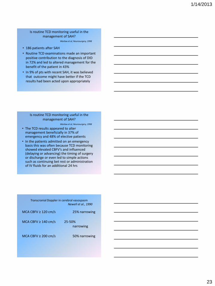
1142013
30
SAH Pt 1
Day 1
Rt MCA (M1 segm) 60 cms Lt MCA (M1 segm) 64 cms
Day 3
Rt MCA 100 cms Lt MCA 110 cms
SAH Pt 1
Day 8
Rt MCA (M1 segm) 212 cms Lt MCA (M1 segm) 230 cms
Day 15
Rt MCA 308 cms Lt MCA 121 cms
SAH Pt 2
Day 5
Lt MCA 126 cms Rt MCA 88 cms
Day 6
Lt MCA 38 cms Rt MCA 118 cms
1142013
31
SAH Pt 2
Day 7
Lt MCA 139 cms Rt MCA 148 cms
SAH Pt 3
Day 3
Rt MCA 126 cms Lt MCA 88 cms
Day 4
Rt MCA 38 cms Lt MCA 43 cms
SAH Pt 3
Day 5
Rt MCA 131 cms Lt MCA 158 cms
1142013
32
SAH amp ICP
bull We are judging qualitative TCD wave form morphology changes
bull These changes usually will be obvious after ICP will be more 30 mm Hg
bull However one condition must be full filled if you would be using TCD wave from changes to predict intracranial hypertension MAP cardiac output and PaCO2 are normal and not different significantly compared to the previous day
Effect of age on MCA and ICA CBFV Torbey at el Stroke 2011
lt6 8(n=4 7 )
68
(n=3 4 )
p-v a lue
Ma x MC A CBFVMedian (c ms)Range
11435-254
7633-190
0002
Ti me to Max MC A C BFVMedian (days)Range
60-18
72-15
07
MC A pul satil ity i ndexMedianRange
088029-278
109031-283
lt00001
Ma x ICA C BFVMedian (c ms)Range
12640-211
76530-202
0003
Ti me to Max IC A CBFVMedian (days)Range
72-16
553-15
04
IC A pul sati l ity indexMedianRange
088035-55
11804-371
lt00001
lt68 (n=47)
68 (n=34)
p-value
Symptomatic Vasospasm n()
31(66) 15(44) 005
Time to symptomatic vasospasm Median (cms) Range
7
(2-16)
5
(2-10)
006
MCA CBFV 1 day at VSP Median (cms) Range
92
52-243
55
24-190
0009
ICA CBFV 1 day at VSP Median (cms) Range
92
33-196
605
30-171
0079
TCD VSP 42 23 lt00001 TCD confirmed VSP n() 10(53) 5(33) 02 Radiological VSP n() 14(74) 8(47) 01
Effect of age on MCA and ICA CBFV Torbey at el Stroke
2011
1142013
33
Kaplan-Meier Curve for VSP onset time Torbey at al Stroke 2001
Cumulative VSP Probability curve
Time to VSP symptoms
30 20 10 0
Cu
mu
lative
V
SP
11
10
9
8
7
6
5
4
3
AGE GROUP
Age lt68
Age 68
Role of TCD SAH
bull Elevated CBFVrsquos in asymptomatic patients warrant meticulous observation in some closely supervised setting until CBFVrsquos begin trend downward
bull Elevated CBFVrsquos in a particular vascular territory can focus subsequent neurologic examinations to detect subtle changes earlier in their clinical course
Role of TCD SAH
bull In symptomatic patients elevated CBFVrsquos most likely represent significant vessel narrowing and may obviate the need for cerebral angiography At this point triple-H therapy can be initiated or advanced
bull Asymptomatic patients without elevated CBFVrsquos probably can avoid additional angiography However we need to consider patientrsquos age because elderly patientrsquos could develop vasospasm in normal or slightly abnormal CBFV range
1142013
34
Factors influencing interpretation
bull Patient age
bull The presence of moderate to severe anemia (Hct lt27)
bull Impaired CBF autoregulation (passive CBFV variation with MAP changes)
bull Hyperemia induced by triple-H therapy
Guidelines for the Management of Aneurysmal SAH Stroke Council AHA 1994
bull Summary and Recommendations 1 SAH is a medical emergencyhellip 2 CT scanning for suspected SAH is strongly recommendedhellip 3 Selective cerebral angiography to document 4 TCD is recommended for the diagnosis and monitoring of vasospasm although the cerebral angiography may be required for definitive diagnosis
TCD MONITORING OF VASOSPASM
1142013
35
Transcranial Doppler monitoring and clinical decision-making after subarachnoid hemorrhage
McGirt et al 2003
bull Objective was to examine the impact of TCD vasospasm monitoring on clinical decision-making following SAH
bull The records of 50 randomly selected patients undergoing serial TCD monitoring following SAH were reviewed Dates and results of TCDs and cerebral angiograms the use of hypertensive hemodilution (HH) therapy and the development of new neurological deficits were recorded The independent effects of TCD-defined vasospasm and new neurological deficits on patient management were determined with multiple logistical regression Results were validated in a second randomly selected 50 patient cohort
Transcranial Doppler monitoring and clinical decision-making after subarachnoid hemorrhage
McGirt et al 2003
bull Mild or moderate TCD-defined vasospasm developed in 76 of patients 58 +- 05 days after SAH 38 developed severe TCD-defined vasospasm after 79 +- 07 days Focal neurological deficits occurred in 50 after 57 +- 06 days with TCD abnormalities preceding the deficit by 25 +- 07 days in 64
bull TCD-defined vasospasm did not independently influence the use of HH therapy or angiography with both decisions associated with the development of new neurological deficits
bull As TCD-defined vasospasm preceded the neurological deficit in 64 earlier intervention might reduce the incidence of vasospasm-related stroke in institutions with similar practice patterns
MCA and ICA CBFVrsquos Dynamics
1142013
36
Biphasic CBFVrsquos profile after SAH Luft et al Neurocritical Care 2004
bull 18 (99) patients demonstrated biphasic CBFVrsquos profile
bull 1st CBFVrsquos peak (134 + 11 cms) occurred on post-SAH day 6 + 1 2nd CBFVrsquos peak (148 + 12 cms) occurred on post-SAH day 13 + 1
bull Although the 2nd CBFVrsquos peak is usually not associated with a worsening of symptoms these patients were more likely to exhibit clinical symptoms during the 1st CBFV peak
Biphasic CBFVrsquos profile after SAH Luft et al Neurocritical Care 2004
Angiography examination in patient with
ACommA aneurysm before and after aneurysm clipping
1142013
37
Angiography examination after transluminal angioplasty
TCD Monitoring
0
50
100
150
200
250
1 2 3 4 5 6 7 8 9 10 11 12 13 14 15 16 17 18 19 1 2 3 4 5 6 7 8 9 10 11 12 13 14 15 16 17 18 19
MCA M1
MCA M2
ACA A1
ICA C1
VA
BA
MAP
Hct
ICP
Temp
PaCO2
RIGHT LEFT
Days after SAH
1st PEAK
2ND PEAK
Elevated Transcranial Doppler Ultrasound Velocities Following Therapeutic Arterial Dilation
Giller et al 1995
bull Elevated TCD CBFVrsquos seen after cerebral angioplasty are commonly interpreted as evidence of residual or recurrent stenosis but may conceivably arise from hyperemia and require different clinical management
bull Four cases of abnormally elevated mean CBFVrsquos obtained after therapeutic arterial dilation with either balloon angioplasty or intra-arterial administration of papaverine are described In each case cerebral angiography revealed a dilated vessel suggesting that hyperemia and impaired autoregulation were the causes of the high CBFVrsquos
bull These examples suggest that high TCD CBFVrsquos after vessel dilation may be produced by unpredictable amounts of vessel narrowing and flow alteration Although a normalizing CBFV after angioplasty suggests effective vessel dilation high CBFVrsquos may be due partly to hyperemia and cannot be interpreted as arising solely from recurrent stenosis
1142013
38
2011 AHAASA Metrics for Measuring Quality of Care in Comprehensive Stroke Centers
bull Among different measures for Comprehensive Stroke Centers is
Median frequency of noninvasive monitoring for surveillance for vasospasm in patients with aneurysmal SAH during the period between three and 14 days after SAH
POSTTRAUMATIC VASOSPASM
TBI
bull Every 21 seconds one person in the US sustains traumatic brain injury (TBI)
bull An estimated 53 million Americans ndash little more than 2 of the US population ndash currently live with disabilities resulting from brain injury
bull Each year 80000 Americans experience the onset of long-term disability following TBI
bull Okie NEJM 2005 Among surviving soldiers wounded in combat in Iraq and Afghanistan TBI appears to account for a larger proportion of casualties than it has in other recent US wars According to the Joint Theater Trauma Registry 22 had injuries to the head face or neck
Armonda et al Neurosurgery 2006 474 had traumatic cerebral vasospasm Majority were blast related injury
Civilian
Battlefield
1142013
39
Blast TBI
bull Blast injuries historically seen by providers as limited to military concern
Peacetime terrorism over the last decade has reminded us otherwise
bull
Why we need talk about blast TBI
bull Mortality
ndash Oklahoma City 167
ndash US Embassy 223
ndash World Trade Center 2801
ndash Madrid train bombings 191
ndash London 56
ndash Domodedovo 37
ndash Minsk metro 12
Large number of injuredhellip
TBI PATHOPHYSIOLOGY
1142013
40
Balancing Multisystem Interactions
TBI
TBI Pathophysiology
Primary Injury - ContusionsHemorrhages
- Diffuse Axonal Injury (DAI)
Secondary Injury (Intracranial) occurs hours to weeks after injury
- Blood Flow and Metabolic Changes
- Traumatic Hematomas
- Cerebral Edema
- Hydrocephalus
- Increased Intracranial Pressure
Decrease in CBF
BRAIN
EDEMA
More Brain Ischemic Blood Vessels
Edema brain cells Dilate
Increased ICP
Cell Injury
1142013
41
But where is the place for TCD monitoring
Multimodal Monitoring VasospasmIschemiahigh ICP Detection
bull MAP
bull SaO2
bull ECG
bull Et-CO2
bull CVP
bull Urine output
bull ICP
bull CBFVTCD PbO2
cEEG
CTMR Perfusion
The primary goal of
management for
TBI is the
prevention of
secondary damage
due to neuronal
hypoxia and
hypoperfusion
Daily TCD
Continuous EEG
ICP monitoring
Pb02
CBF
1142013
42
Traumatic Brain Injury amp TCD
bull Cerebral ischemia due to the post-traumatic vasospasm
bull Increased Intracranial Pressure
bull Brain Death
Post-Traumatic VSP
bull Ischemic symptoms caused by cerebral arterial spasm following traumatic SAH are comparable to those found following aneurysmal SAH
- appearance of symptoms between Days 4 and 16 after injury with the peak incidence on Days 9 and 10
- close correlation between the main site of the subarachnoid blood and the location of severe vasospasm responsible for the symptoms and a higher incidence of symptoms in patients with massive SAH than those with slight SAH
bull Subarachnoid blood plays an important role in the later development of vasospasm not only following aneurysm rupture but also after head injury Nevertheless there is no general agreement that subarachnoid blood in head injury is an important risk factor in the development of vasospasm and ischemic brain damage
TBI and VSP
bull Cerebral posttraumatic VSP (PTV) was first described by Lorn in 1936
bull The incidence of CT documented traumatic SAH has been identified in 4 to 63 of pts after TBI
bull Study from the University of Mississippi Medical Center indicated that traumatic SAH complicate course of TBI in 69 of the patients due to the presence of PTV
1142013
43
Clinical Significance
bull 41 of patients who died from TBI had PTV (MacPherson et al 1973)
bull 24 with massive tSAH developed ischemic symptoms in contrast to 3 of patients with mild tSAH (Taneda et al 1996)
bull Ischemic symptoms accompanying arterial VSP following tSAH are comparable to those found following aneurysmal SAH
WARTIME TRAUMATIC CEREBRAL VASOSPASM RECENT REVIEW OF COMBAT CASUALTIES
Armonda et al Neurosurgery 2006
bull The first study to analyze the effects of blast-related injury on the cerebral vasculature
bull This study showed that TCV occurred in a substantial number of patients with severe neurotrauma and clinical outcomes were worse for those with this condition
25 yo severe concussion no SAH (Courtesy of Dr Armonda)
CBFV 120 cms CBFV 57 cms
1142013
44
25 yo severe concussion no SAH (Courtesy of Dr Armonda)
CBFV 112 cms CBFV 89 cms
25 yo severe concussion no SAH (Courtesy of Dr Armonda)
CBFV
(cmsec)
Calendar
0
50
100
150
200
250
300
MCA M1
MCA M2
ACA A1
VA
BA
MAP
Hct
ICP
Temp
PaCO2
RIGHT LEFT
Ant circ TLA Post circ TLA Ant circ TLA
SEVERE VSP
MODERATE VSP
MILD VSP
EFFECT OF TLA ON PROLONG (SEVEN DAYS) VASOSPASM INVOLVING ANTERIOR AND POSTERIOR CIRCULATION
1142013
45
The CBFVrsquos and systemic hemodynamic trends TCD signs of severe vasospasm involving posterior and
anterior circulation lasting seven days before TLA
0
50
100
150
200
250
300
MCA M1
ACA A1
ICA C1
VA
BA
MAP
Hct
ICP
PaCO2
RIGHT LEFT
Severe VSP
Moderate VSP
MIld VSP
TLA TLA
Effect of TLA after prolong severe vasospasm
Left MCA CBFV 69 cmsec PI 17 Right MCA CBFV 66 cmsec PI 14
Left MCA 205 cmsec PI 08 Right MCA CBFV 195 cmsec PI 05
CBFVrsquos and PIrsquos Trends (PI x 100)
0
50
100
150
200
250
300
MCA M1
M1 PI
ACA A1
A1 PI
ICA C1
C1 PI
VA
VA PI
BA
BA PI
RIGHT LEFT
1142013
46
The CBFV amp PI Trends
bull MCA (M1 segm)
bull ICA (C1 segm)
0
50
100
150
200
250
300
MCA M1
M1 PI
RIGHT LEFT
Severe VSP
Moderate VSP
Mild VSP
0
50
100
150
200
250
300
ICA C1
C1 PI
RIGHT LEFT
Severe VSP
Moderate VSP
Mild VSP
Clinical Material
bull Ninety patients (2 females) aged 18 to 50 years (mean 259 years) who had suffered wartime TBI injuries (with Glasgow Coma Scale scores ranging from 3 to 15) were investigated with daily TCD studies
bull A total of 567 TCD studies (mean 64 testspatient ranged from 1 to 30) were made after admission
TCD signs of Vasospasm (in ) by type of TBI
7500
3680
6840
2900
3570
520
3150
2350
1430
520
1570 1760
0
10
20
30
40
50
60
70
80
PHI PHIIED CHI CHIIED
Mild ()
Mod ()
Severe ()
1142013
47
Presence of ICH ()
5700
5200
5800
5000
PHI PHIIED CHI CHIIED
TCD signs of VSP amp high ICP
TBI Type Post TBI
VSP
High
ICP
CHI 13144 12133
CHIIED 12133 910
PHI 21233 16177
PHIIED 11122 12133
Total 57633 49544
TCD AND TUMOR RESECTION
1142013
48
Tumor Resection and Vasospasm
bull The occurrence of vasospasm and delayed cerebral ischemia after resection of intracranial tumor has not received extensive attention clinically and is often misdiagnosed and improperly treated as surgical brain damage or brain swelling
bull However DID from vasospasm after tumor resection is a complication that is being reported in increasing numbers
Tumor Resection and Vasospasm Literature review
bull Reports are sparse and mainly are case series
bull Vasospasm was found in 2 to 49 patients No significant difference among age sex surgical approaches pathological diagnosis duration of surgery amount of blood loss and transfusion during surgery were found but significant difference was seen in cisternal hemorrhage on CT scan and the amount of blood in cerebrospinal fluid
Tumor Resection and Vasospasm Probable Mechanism
bull It is suggested that accumulation of blood in the basal cisterns may have been responsible for this unusual condition and it is therefore important to consider vasospasm as a probable etiological cause of clinical deterioration in patients undergoing the surgical removal of a cerebral tumor
bull For this reason whenever any neurological deterioration occurs in such patients it is advisable to perform TCD in order to verify the presence of any vasospasm and promptly commence suitable treatment
1142013
49
Tumor Resection and Vasospasm Management
bull Surgery To decrease the amount of blood in basal cistern by microsurgery
bull ICU TCD utilization in the diagnosis of vasospasm and its monitoring
bull ICU Differentiation of vasospasm from brain swelling are helpful to confirm the coexistent or causal relation based on neurological assessment CT TCD and ICP monitoring both in deciding therapeutic strategy and successfully controlling vasospasm
bull ICU Nimotop played a key role in preventing brain damage from vasospasm and cerebral swelling
Suprasellar tumor resection The CBFVs and Trends of the Systemic
Hemodynamic
RIGHT LEFT
0
20
40
60
80
100
120
140
160
180
103010311111121131141151161171181191110 103010311111121131141151161171181191110
Calendar
CB
FV
s a
nd
Vari
ab
les
0
20
40
60
80
100
120
140
160
180
CB
FV
s a
nd
Vari
ab
les
MCA M1
MCA M2
ACA
ICA C1
VA
BA
MAP
ICP
Hct
PaCO2
Temp
Patient wit tumor
bull 58 yo female sp trans-sphenoidal resection of tuberculum sella meningioma complicated by A-comm complex pseudoaneurysm formation vasospasm and ACA territory infarct
1142013
50
DWI demonstrating ACA territory infarct
DWI demonstrating ACA territory infarct
ICA injections demonstrating vasospasm in A1-A2 bilaterally
A-comm complex pseudoaneurysm and coiling
1142013
51
A-comm complex pseudoaneurysm before and after coiling
TCD Trends
Left Right
Role of TCD Tumor Resection
bull It is useful to perform TCD test after surgery and perform daily TCD studies when patient is in the ICU
bull The frequency with which TCD should be performed may be guided by patient clinical presentation knowledge of risk factors for VSP early clinical course
bull TCD studies should be performed after endovascular treatment to identify patients with recurrent VSP
1142013
52
Role of TCD VSP monitoring
bull It is useful to perform TCD test on admission (or ASAP after surgery) and perform daily TCD studies when patient is in the ICU
bull The frequency with which TCD should be performed may be guided by patient clinical presentation knowledge of risk factors for vasospasm early clinical course
bull TCD studies should be performed after endovascular treatment to identify patients with recurrent vasospasm
Role of TCD VSP Monitoring
bull The presence and temporal profile of CBFVrsquos in all available vessels must be detected and serially monitored
bull The pattern of CBFVrsquos elevation may indicate the need to follow patient carefully for evidence of deficits related to specific vascular territory
bull Waveform appearance either regionally or globally may be clinically significant
TCD and VSP
bull Currently the gold standard for vasospasm diagnosis is cerebral angiography replaceable by CTA only when angiography is not available Obviously it is not feasible to perform such investigation as frequently as bedside clinical assessment
bull Repeated clinical assessments of a patients neurological status carry the problem of detecting the clinical signs and symptoms of vasospasm which occur only after vasospasm has already manifested its deleterious effects on the cerebral parenchyma
bull TCD is a relatively new non-invasive tool allowing for bedside monitoring to determine CBFVrsquos indicative of changes in vascular diameter
1142013
53
TCD and VSP
bull TCD can be useful pre- intra- and post-operatively while helping to recognize the development of cerebral vasospasm before the onset of its clinical effects
bull Vasospasm following SAH is a very important source of morbidity and mortality Too often the first sign is a neurologic deficit which may be too late to reverse
bull TCD assists in the clinical decision-making regarding further diagnostic evaluation and therapeutic interventions When performed in isolation the contribution of TCD to improving patient outcome has not been established Nevertheless TCD has become a regularly employed tool in neurocritical care and perioperative settings
TCD is a Critical Tool in Critical Care
bull The value of TCD in clinical practice is well established especially to measure and grade vasospasm following SAH and TBI
bull Based on AHA Guidelines and many years of clinical practice TCD is a tool employed by the Neurosurgeon Neurointensivist and Neurologist in the management of vasospasm
bull Based on high frequency of posttraumatic vasospasm and intracranial hypertension TCD testing must be utilized for management of patients after SAH (aneurysm rupture wartime or civilian TBI tumor resection)
bull The use of TCD at hospital admission allows identification of patients with brain hypoperfusion due to the vasospasm andor intracranial hypertension In such high-risk patients early TCD goal-directed therapy can restore normal cerebral perfusion and might then potentially help in reducing the extent of secondary brain injury
bull TCD could provide information about abnormally high ICPbrain death
bull In the future incorporation of TCD data may facilitate more injury- and time-specific therapies for patients after SAH (aneurysm rupture wartime or civilian TBI tumor resection)
TCD is a Critical Tool in Critical Care
1142013
54
Do we know everything about TCD and vasospasm
bull TCD criteria for vasospasm for the young (less than 20-30 yo) and old (more than 68-70 yo) patients
bull CBFV calculation formula that will take into account Hct values
bull Cerebral angiography could be negative but TCD could be positive for vasospasm
bull TCD prediction for clinical vasospasm
Future
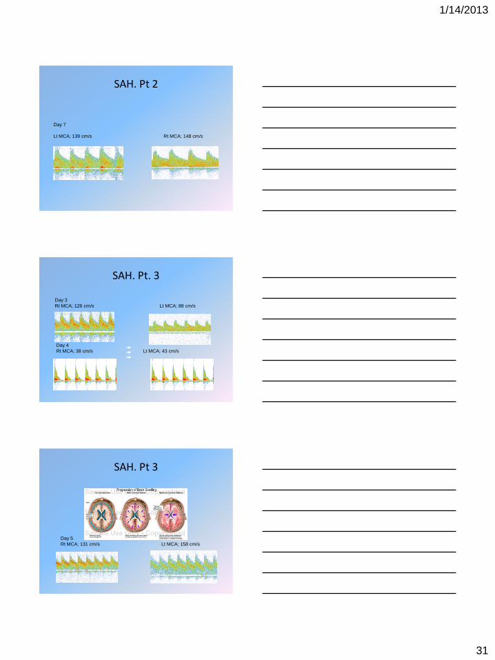
1142013
31
SAH Pt 2
Day 7
Lt MCA 139 cms Rt MCA 148 cms
SAH Pt 3
Day 3
Rt MCA 126 cms Lt MCA 88 cms
Day 4
Rt MCA 38 cms Lt MCA 43 cms
SAH Pt 3
Day 5
Rt MCA 131 cms Lt MCA 158 cms
1142013
32
SAH amp ICP
bull We are judging qualitative TCD wave form morphology changes
bull These changes usually will be obvious after ICP will be more 30 mm Hg
bull However one condition must be full filled if you would be using TCD wave from changes to predict intracranial hypertension MAP cardiac output and PaCO2 are normal and not different significantly compared to the previous day
Effect of age on MCA and ICA CBFV Torbey at el Stroke 2011
lt6 8(n=4 7 )
68
(n=3 4 )
p-v a lue
Ma x MC A CBFVMedian (c ms)Range
11435-254
7633-190
0002
Ti me to Max MC A C BFVMedian (days)Range
60-18
72-15
07
MC A pul satil ity i ndexMedianRange
088029-278
109031-283
lt00001
Ma x ICA C BFVMedian (c ms)Range
12640-211
76530-202
0003
Ti me to Max IC A CBFVMedian (days)Range
72-16
553-15
04
IC A pul sati l ity indexMedianRange
088035-55
11804-371
lt00001
lt68 (n=47)
68 (n=34)
p-value
Symptomatic Vasospasm n()
31(66) 15(44) 005
Time to symptomatic vasospasm Median (cms) Range
7
(2-16)
5
(2-10)
006
MCA CBFV 1 day at VSP Median (cms) Range
92
52-243
55
24-190
0009
ICA CBFV 1 day at VSP Median (cms) Range
92
33-196
605
30-171
0079
TCD VSP 42 23 lt00001 TCD confirmed VSP n() 10(53) 5(33) 02 Radiological VSP n() 14(74) 8(47) 01
Effect of age on MCA and ICA CBFV Torbey at el Stroke
2011
1142013
33
Kaplan-Meier Curve for VSP onset time Torbey at al Stroke 2001
Cumulative VSP Probability curve
Time to VSP symptoms
30 20 10 0
Cu
mu
lative
V
SP
11
10
9
8
7
6
5
4
3
AGE GROUP
Age lt68
Age 68
Role of TCD SAH
bull Elevated CBFVrsquos in asymptomatic patients warrant meticulous observation in some closely supervised setting until CBFVrsquos begin trend downward
bull Elevated CBFVrsquos in a particular vascular territory can focus subsequent neurologic examinations to detect subtle changes earlier in their clinical course
Role of TCD SAH
bull In symptomatic patients elevated CBFVrsquos most likely represent significant vessel narrowing and may obviate the need for cerebral angiography At this point triple-H therapy can be initiated or advanced
bull Asymptomatic patients without elevated CBFVrsquos probably can avoid additional angiography However we need to consider patientrsquos age because elderly patientrsquos could develop vasospasm in normal or slightly abnormal CBFV range
1142013
34
Factors influencing interpretation
bull Patient age
bull The presence of moderate to severe anemia (Hct lt27)
bull Impaired CBF autoregulation (passive CBFV variation with MAP changes)
bull Hyperemia induced by triple-H therapy
Guidelines for the Management of Aneurysmal SAH Stroke Council AHA 1994
bull Summary and Recommendations 1 SAH is a medical emergencyhellip 2 CT scanning for suspected SAH is strongly recommendedhellip 3 Selective cerebral angiography to document 4 TCD is recommended for the diagnosis and monitoring of vasospasm although the cerebral angiography may be required for definitive diagnosis
TCD MONITORING OF VASOSPASM
1142013
35
Transcranial Doppler monitoring and clinical decision-making after subarachnoid hemorrhage
McGirt et al 2003
bull Objective was to examine the impact of TCD vasospasm monitoring on clinical decision-making following SAH
bull The records of 50 randomly selected patients undergoing serial TCD monitoring following SAH were reviewed Dates and results of TCDs and cerebral angiograms the use of hypertensive hemodilution (HH) therapy and the development of new neurological deficits were recorded The independent effects of TCD-defined vasospasm and new neurological deficits on patient management were determined with multiple logistical regression Results were validated in a second randomly selected 50 patient cohort
Transcranial Doppler monitoring and clinical decision-making after subarachnoid hemorrhage
McGirt et al 2003
bull Mild or moderate TCD-defined vasospasm developed in 76 of patients 58 +- 05 days after SAH 38 developed severe TCD-defined vasospasm after 79 +- 07 days Focal neurological deficits occurred in 50 after 57 +- 06 days with TCD abnormalities preceding the deficit by 25 +- 07 days in 64
bull TCD-defined vasospasm did not independently influence the use of HH therapy or angiography with both decisions associated with the development of new neurological deficits
bull As TCD-defined vasospasm preceded the neurological deficit in 64 earlier intervention might reduce the incidence of vasospasm-related stroke in institutions with similar practice patterns
MCA and ICA CBFVrsquos Dynamics
1142013
36
Biphasic CBFVrsquos profile after SAH Luft et al Neurocritical Care 2004
bull 18 (99) patients demonstrated biphasic CBFVrsquos profile
bull 1st CBFVrsquos peak (134 + 11 cms) occurred on post-SAH day 6 + 1 2nd CBFVrsquos peak (148 + 12 cms) occurred on post-SAH day 13 + 1
bull Although the 2nd CBFVrsquos peak is usually not associated with a worsening of symptoms these patients were more likely to exhibit clinical symptoms during the 1st CBFV peak
Biphasic CBFVrsquos profile after SAH Luft et al Neurocritical Care 2004
Angiography examination in patient with
ACommA aneurysm before and after aneurysm clipping
1142013
37
Angiography examination after transluminal angioplasty
TCD Monitoring
0
50
100
150
200
250
1 2 3 4 5 6 7 8 9 10 11 12 13 14 15 16 17 18 19 1 2 3 4 5 6 7 8 9 10 11 12 13 14 15 16 17 18 19
MCA M1
MCA M2
ACA A1
ICA C1
VA
BA
MAP
Hct
ICP
Temp
PaCO2
RIGHT LEFT
Days after SAH
1st PEAK
2ND PEAK
Elevated Transcranial Doppler Ultrasound Velocities Following Therapeutic Arterial Dilation
Giller et al 1995
bull Elevated TCD CBFVrsquos seen after cerebral angioplasty are commonly interpreted as evidence of residual or recurrent stenosis but may conceivably arise from hyperemia and require different clinical management
bull Four cases of abnormally elevated mean CBFVrsquos obtained after therapeutic arterial dilation with either balloon angioplasty or intra-arterial administration of papaverine are described In each case cerebral angiography revealed a dilated vessel suggesting that hyperemia and impaired autoregulation were the causes of the high CBFVrsquos
bull These examples suggest that high TCD CBFVrsquos after vessel dilation may be produced by unpredictable amounts of vessel narrowing and flow alteration Although a normalizing CBFV after angioplasty suggests effective vessel dilation high CBFVrsquos may be due partly to hyperemia and cannot be interpreted as arising solely from recurrent stenosis
1142013
38
2011 AHAASA Metrics for Measuring Quality of Care in Comprehensive Stroke Centers
bull Among different measures for Comprehensive Stroke Centers is
Median frequency of noninvasive monitoring for surveillance for vasospasm in patients with aneurysmal SAH during the period between three and 14 days after SAH
POSTTRAUMATIC VASOSPASM
TBI
bull Every 21 seconds one person in the US sustains traumatic brain injury (TBI)
bull An estimated 53 million Americans ndash little more than 2 of the US population ndash currently live with disabilities resulting from brain injury
bull Each year 80000 Americans experience the onset of long-term disability following TBI
bull Okie NEJM 2005 Among surviving soldiers wounded in combat in Iraq and Afghanistan TBI appears to account for a larger proportion of casualties than it has in other recent US wars According to the Joint Theater Trauma Registry 22 had injuries to the head face or neck
Armonda et al Neurosurgery 2006 474 had traumatic cerebral vasospasm Majority were blast related injury
Civilian
Battlefield
1142013
39
Blast TBI
bull Blast injuries historically seen by providers as limited to military concern
Peacetime terrorism over the last decade has reminded us otherwise
bull
Why we need talk about blast TBI
bull Mortality
ndash Oklahoma City 167
ndash US Embassy 223
ndash World Trade Center 2801
ndash Madrid train bombings 191
ndash London 56
ndash Domodedovo 37
ndash Minsk metro 12
Large number of injuredhellip
TBI PATHOPHYSIOLOGY
1142013
40
Balancing Multisystem Interactions
TBI
TBI Pathophysiology
Primary Injury - ContusionsHemorrhages
- Diffuse Axonal Injury (DAI)
Secondary Injury (Intracranial) occurs hours to weeks after injury
- Blood Flow and Metabolic Changes
- Traumatic Hematomas
- Cerebral Edema
- Hydrocephalus
- Increased Intracranial Pressure
Decrease in CBF
BRAIN
EDEMA
More Brain Ischemic Blood Vessels
Edema brain cells Dilate
Increased ICP
Cell Injury
1142013
41
But where is the place for TCD monitoring
Multimodal Monitoring VasospasmIschemiahigh ICP Detection
bull MAP
bull SaO2
bull ECG
bull Et-CO2
bull CVP
bull Urine output
bull ICP
bull CBFVTCD PbO2
cEEG
CTMR Perfusion
The primary goal of
management for
TBI is the
prevention of
secondary damage
due to neuronal
hypoxia and
hypoperfusion
Daily TCD
Continuous EEG
ICP monitoring
Pb02
CBF
1142013
42
Traumatic Brain Injury amp TCD
bull Cerebral ischemia due to the post-traumatic vasospasm
bull Increased Intracranial Pressure
bull Brain Death
Post-Traumatic VSP
bull Ischemic symptoms caused by cerebral arterial spasm following traumatic SAH are comparable to those found following aneurysmal SAH
- appearance of symptoms between Days 4 and 16 after injury with the peak incidence on Days 9 and 10
- close correlation between the main site of the subarachnoid blood and the location of severe vasospasm responsible for the symptoms and a higher incidence of symptoms in patients with massive SAH than those with slight SAH
bull Subarachnoid blood plays an important role in the later development of vasospasm not only following aneurysm rupture but also after head injury Nevertheless there is no general agreement that subarachnoid blood in head injury is an important risk factor in the development of vasospasm and ischemic brain damage
TBI and VSP
bull Cerebral posttraumatic VSP (PTV) was first described by Lorn in 1936
bull The incidence of CT documented traumatic SAH has been identified in 4 to 63 of pts after TBI
bull Study from the University of Mississippi Medical Center indicated that traumatic SAH complicate course of TBI in 69 of the patients due to the presence of PTV
1142013
43
Clinical Significance
bull 41 of patients who died from TBI had PTV (MacPherson et al 1973)
bull 24 with massive tSAH developed ischemic symptoms in contrast to 3 of patients with mild tSAH (Taneda et al 1996)
bull Ischemic symptoms accompanying arterial VSP following tSAH are comparable to those found following aneurysmal SAH
WARTIME TRAUMATIC CEREBRAL VASOSPASM RECENT REVIEW OF COMBAT CASUALTIES
Armonda et al Neurosurgery 2006
bull The first study to analyze the effects of blast-related injury on the cerebral vasculature
bull This study showed that TCV occurred in a substantial number of patients with severe neurotrauma and clinical outcomes were worse for those with this condition
25 yo severe concussion no SAH (Courtesy of Dr Armonda)
CBFV 120 cms CBFV 57 cms
1142013
44
25 yo severe concussion no SAH (Courtesy of Dr Armonda)
CBFV 112 cms CBFV 89 cms
25 yo severe concussion no SAH (Courtesy of Dr Armonda)
CBFV
(cmsec)
Calendar
0
50
100
150
200
250
300
MCA M1
MCA M2
ACA A1
VA
BA
MAP
Hct
ICP
Temp
PaCO2
RIGHT LEFT
Ant circ TLA Post circ TLA Ant circ TLA
SEVERE VSP
MODERATE VSP
MILD VSP
EFFECT OF TLA ON PROLONG (SEVEN DAYS) VASOSPASM INVOLVING ANTERIOR AND POSTERIOR CIRCULATION
1142013
45
The CBFVrsquos and systemic hemodynamic trends TCD signs of severe vasospasm involving posterior and
anterior circulation lasting seven days before TLA
0
50
100
150
200
250
300
MCA M1
ACA A1
ICA C1
VA
BA
MAP
Hct
ICP
PaCO2
RIGHT LEFT
Severe VSP
Moderate VSP
MIld VSP
TLA TLA
Effect of TLA after prolong severe vasospasm
Left MCA CBFV 69 cmsec PI 17 Right MCA CBFV 66 cmsec PI 14
Left MCA 205 cmsec PI 08 Right MCA CBFV 195 cmsec PI 05
CBFVrsquos and PIrsquos Trends (PI x 100)
0
50
100
150
200
250
300
MCA M1
M1 PI
ACA A1
A1 PI
ICA C1
C1 PI
VA
VA PI
BA
BA PI
RIGHT LEFT
1142013
46
The CBFV amp PI Trends
bull MCA (M1 segm)
bull ICA (C1 segm)
0
50
100
150
200
250
300
MCA M1
M1 PI
RIGHT LEFT
Severe VSP
Moderate VSP
Mild VSP
0
50
100
150
200
250
300
ICA C1
C1 PI
RIGHT LEFT
Severe VSP
Moderate VSP
Mild VSP
Clinical Material
bull Ninety patients (2 females) aged 18 to 50 years (mean 259 years) who had suffered wartime TBI injuries (with Glasgow Coma Scale scores ranging from 3 to 15) were investigated with daily TCD studies
bull A total of 567 TCD studies (mean 64 testspatient ranged from 1 to 30) were made after admission
TCD signs of Vasospasm (in ) by type of TBI
7500
3680
6840
2900
3570
520
3150
2350
1430
520
1570 1760
0
10
20
30
40
50
60
70
80
PHI PHIIED CHI CHIIED
Mild ()
Mod ()
Severe ()
1142013
47
Presence of ICH ()
5700
5200
5800
5000
PHI PHIIED CHI CHIIED
TCD signs of VSP amp high ICP
TBI Type Post TBI
VSP
High
ICP
CHI 13144 12133
CHIIED 12133 910
PHI 21233 16177
PHIIED 11122 12133
Total 57633 49544
TCD AND TUMOR RESECTION
1142013
48
Tumor Resection and Vasospasm
bull The occurrence of vasospasm and delayed cerebral ischemia after resection of intracranial tumor has not received extensive attention clinically and is often misdiagnosed and improperly treated as surgical brain damage or brain swelling
bull However DID from vasospasm after tumor resection is a complication that is being reported in increasing numbers
Tumor Resection and Vasospasm Literature review
bull Reports are sparse and mainly are case series
bull Vasospasm was found in 2 to 49 patients No significant difference among age sex surgical approaches pathological diagnosis duration of surgery amount of blood loss and transfusion during surgery were found but significant difference was seen in cisternal hemorrhage on CT scan and the amount of blood in cerebrospinal fluid
Tumor Resection and Vasospasm Probable Mechanism
bull It is suggested that accumulation of blood in the basal cisterns may have been responsible for this unusual condition and it is therefore important to consider vasospasm as a probable etiological cause of clinical deterioration in patients undergoing the surgical removal of a cerebral tumor
bull For this reason whenever any neurological deterioration occurs in such patients it is advisable to perform TCD in order to verify the presence of any vasospasm and promptly commence suitable treatment
1142013
49
Tumor Resection and Vasospasm Management
bull Surgery To decrease the amount of blood in basal cistern by microsurgery
bull ICU TCD utilization in the diagnosis of vasospasm and its monitoring
bull ICU Differentiation of vasospasm from brain swelling are helpful to confirm the coexistent or causal relation based on neurological assessment CT TCD and ICP monitoring both in deciding therapeutic strategy and successfully controlling vasospasm
bull ICU Nimotop played a key role in preventing brain damage from vasospasm and cerebral swelling
Suprasellar tumor resection The CBFVs and Trends of the Systemic
Hemodynamic
RIGHT LEFT
0
20
40
60
80
100
120
140
160
180
103010311111121131141151161171181191110 103010311111121131141151161171181191110
Calendar
CB
FV
s a
nd
Vari
ab
les
0
20
40
60
80
100
120
140
160
180
CB
FV
s a
nd
Vari
ab
les
MCA M1
MCA M2
ACA
ICA C1
VA
BA
MAP
ICP
Hct
PaCO2
Temp
Patient wit tumor
bull 58 yo female sp trans-sphenoidal resection of tuberculum sella meningioma complicated by A-comm complex pseudoaneurysm formation vasospasm and ACA territory infarct
1142013
50
DWI demonstrating ACA territory infarct
DWI demonstrating ACA territory infarct
ICA injections demonstrating vasospasm in A1-A2 bilaterally
A-comm complex pseudoaneurysm and coiling
1142013
51
A-comm complex pseudoaneurysm before and after coiling
TCD Trends
Left Right
Role of TCD Tumor Resection
bull It is useful to perform TCD test after surgery and perform daily TCD studies when patient is in the ICU
bull The frequency with which TCD should be performed may be guided by patient clinical presentation knowledge of risk factors for VSP early clinical course
bull TCD studies should be performed after endovascular treatment to identify patients with recurrent VSP
1142013
52
Role of TCD VSP monitoring
bull It is useful to perform TCD test on admission (or ASAP after surgery) and perform daily TCD studies when patient is in the ICU
bull The frequency with which TCD should be performed may be guided by patient clinical presentation knowledge of risk factors for vasospasm early clinical course
bull TCD studies should be performed after endovascular treatment to identify patients with recurrent vasospasm
Role of TCD VSP Monitoring
bull The presence and temporal profile of CBFVrsquos in all available vessels must be detected and serially monitored
bull The pattern of CBFVrsquos elevation may indicate the need to follow patient carefully for evidence of deficits related to specific vascular territory
bull Waveform appearance either regionally or globally may be clinically significant
TCD and VSP
bull Currently the gold standard for vasospasm diagnosis is cerebral angiography replaceable by CTA only when angiography is not available Obviously it is not feasible to perform such investigation as frequently as bedside clinical assessment
bull Repeated clinical assessments of a patients neurological status carry the problem of detecting the clinical signs and symptoms of vasospasm which occur only after vasospasm has already manifested its deleterious effects on the cerebral parenchyma
bull TCD is a relatively new non-invasive tool allowing for bedside monitoring to determine CBFVrsquos indicative of changes in vascular diameter
1142013
53
TCD and VSP
bull TCD can be useful pre- intra- and post-operatively while helping to recognize the development of cerebral vasospasm before the onset of its clinical effects
bull Vasospasm following SAH is a very important source of morbidity and mortality Too often the first sign is a neurologic deficit which may be too late to reverse
bull TCD assists in the clinical decision-making regarding further diagnostic evaluation and therapeutic interventions When performed in isolation the contribution of TCD to improving patient outcome has not been established Nevertheless TCD has become a regularly employed tool in neurocritical care and perioperative settings
TCD is a Critical Tool in Critical Care
bull The value of TCD in clinical practice is well established especially to measure and grade vasospasm following SAH and TBI
bull Based on AHA Guidelines and many years of clinical practice TCD is a tool employed by the Neurosurgeon Neurointensivist and Neurologist in the management of vasospasm
bull Based on high frequency of posttraumatic vasospasm and intracranial hypertension TCD testing must be utilized for management of patients after SAH (aneurysm rupture wartime or civilian TBI tumor resection)
bull The use of TCD at hospital admission allows identification of patients with brain hypoperfusion due to the vasospasm andor intracranial hypertension In such high-risk patients early TCD goal-directed therapy can restore normal cerebral perfusion and might then potentially help in reducing the extent of secondary brain injury
bull TCD could provide information about abnormally high ICPbrain death
bull In the future incorporation of TCD data may facilitate more injury- and time-specific therapies for patients after SAH (aneurysm rupture wartime or civilian TBI tumor resection)
TCD is a Critical Tool in Critical Care
1142013
54
Do we know everything about TCD and vasospasm
bull TCD criteria for vasospasm for the young (less than 20-30 yo) and old (more than 68-70 yo) patients
bull CBFV calculation formula that will take into account Hct values
bull Cerebral angiography could be negative but TCD could be positive for vasospasm
bull TCD prediction for clinical vasospasm
Future
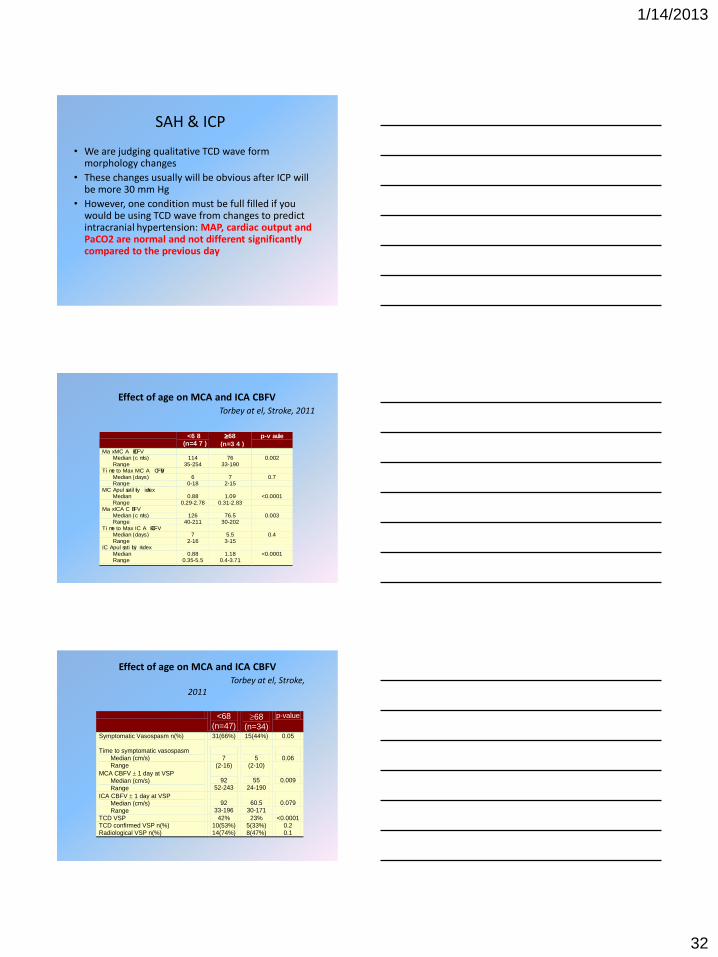
1142013
40
Balancing Multisystem Interactions
TBI
TBI Pathophysiology
Primary Injury - ContusionsHemorrhages
- Diffuse Axonal Injury (DAI)
Secondary Injury (Intracranial) occurs hours to weeks after injury
- Blood Flow and Metabolic Changes
- Traumatic Hematomas
- Cerebral Edema
- Hydrocephalus
- Increased Intracranial Pressure
Decrease in CBF
BRAIN
EDEMA
More Brain Ischemic Blood Vessels
Edema brain cells Dilate
Increased ICP
Cell Injury
1142013
41
But where is the place for TCD monitoring
Multimodal Monitoring VasospasmIschemiahigh ICP Detection
bull MAP
bull SaO2
bull ECG
bull Et-CO2
bull CVP
bull Urine output
bull ICP
bull CBFVTCD PbO2
cEEG
CTMR Perfusion
The primary goal of
management for
TBI is the
prevention of
secondary damage
due to neuronal
hypoxia and
hypoperfusion
Daily TCD
Continuous EEG
ICP monitoring
Pb02
CBF
1142013
42
Traumatic Brain Injury amp TCD
bull Cerebral ischemia due to the post-traumatic vasospasm
bull Increased Intracranial Pressure
bull Brain Death
Post-Traumatic VSP
bull Ischemic symptoms caused by cerebral arterial spasm following traumatic SAH are comparable to those found following aneurysmal SAH
- appearance of symptoms between Days 4 and 16 after injury with the peak incidence on Days 9 and 10
- close correlation between the main site of the subarachnoid blood and the location of severe vasospasm responsible for the symptoms and a higher incidence of symptoms in patients with massive SAH than those with slight SAH
bull Subarachnoid blood plays an important role in the later development of vasospasm not only following aneurysm rupture but also after head injury Nevertheless there is no general agreement that subarachnoid blood in head injury is an important risk factor in the development of vasospasm and ischemic brain damage
TBI and VSP
bull Cerebral posttraumatic VSP (PTV) was first described by Lorn in 1936
bull The incidence of CT documented traumatic SAH has been identified in 4 to 63 of pts after TBI
bull Study from the University of Mississippi Medical Center indicated that traumatic SAH complicate course of TBI in 69 of the patients due to the presence of PTV
1142013
43
Clinical Significance
bull 41 of patients who died from TBI had PTV (MacPherson et al 1973)
bull 24 with massive tSAH developed ischemic symptoms in contrast to 3 of patients with mild tSAH (Taneda et al 1996)
bull Ischemic symptoms accompanying arterial VSP following tSAH are comparable to those found following aneurysmal SAH
WARTIME TRAUMATIC CEREBRAL VASOSPASM RECENT REVIEW OF COMBAT CASUALTIES
Armonda et al Neurosurgery 2006
bull The first study to analyze the effects of blast-related injury on the cerebral vasculature
bull This study showed that TCV occurred in a substantial number of patients with severe neurotrauma and clinical outcomes were worse for those with this condition
25 yo severe concussion no SAH (Courtesy of Dr Armonda)
CBFV 120 cms CBFV 57 cms
1142013
44
25 yo severe concussion no SAH (Courtesy of Dr Armonda)
CBFV 112 cms CBFV 89 cms
25 yo severe concussion no SAH (Courtesy of Dr Armonda)
CBFV
(cmsec)
Calendar
0
50
100
150
200
250
300
MCA M1
MCA M2
ACA A1
VA
BA
MAP
Hct
ICP
Temp
PaCO2
RIGHT LEFT
Ant circ TLA Post circ TLA Ant circ TLA
SEVERE VSP
MODERATE VSP
MILD VSP
EFFECT OF TLA ON PROLONG (SEVEN DAYS) VASOSPASM INVOLVING ANTERIOR AND POSTERIOR CIRCULATION
1142013
45
The CBFVrsquos and systemic hemodynamic trends TCD signs of severe vasospasm involving posterior and
anterior circulation lasting seven days before TLA
0
50
100
150
200
250
300
MCA M1
ACA A1
ICA C1
VA
BA
MAP
Hct
ICP
PaCO2
RIGHT LEFT
Severe VSP
Moderate VSP
MIld VSP
TLA TLA
Effect of TLA after prolong severe vasospasm
Left MCA CBFV 69 cmsec PI 17 Right MCA CBFV 66 cmsec PI 14
Left MCA 205 cmsec PI 08 Right MCA CBFV 195 cmsec PI 05
CBFVrsquos and PIrsquos Trends (PI x 100)
0
50
100
150
200
250
300
MCA M1
M1 PI
ACA A1
A1 PI
ICA C1
C1 PI
VA
VA PI
BA
BA PI
RIGHT LEFT
1142013
46
The CBFV amp PI Trends
bull MCA (M1 segm)
bull ICA (C1 segm)
0
50
100
150
200
250
300
MCA M1
M1 PI
RIGHT LEFT
Severe VSP
Moderate VSP
Mild VSP
0
50
100
150
200
250
300
ICA C1
C1 PI
RIGHT LEFT
Severe VSP
Moderate VSP
Mild VSP
Clinical Material
bull Ninety patients (2 females) aged 18 to 50 years (mean 259 years) who had suffered wartime TBI injuries (with Glasgow Coma Scale scores ranging from 3 to 15) were investigated with daily TCD studies
bull A total of 567 TCD studies (mean 64 testspatient ranged from 1 to 30) were made after admission
TCD signs of Vasospasm (in ) by type of TBI
7500
3680
6840
2900
3570
520
3150
2350
1430
520
1570 1760
0
10
20
30
40
50
60
70
80
PHI PHIIED CHI CHIIED
Mild ()
Mod ()
Severe ()
1142013
47
Presence of ICH ()
5700
5200
5800
5000
PHI PHIIED CHI CHIIED
TCD signs of VSP amp high ICP
TBI Type Post TBI
VSP
High
ICP
CHI 13144 12133
CHIIED 12133 910
PHI 21233 16177
PHIIED 11122 12133
Total 57633 49544
TCD AND TUMOR RESECTION
1142013
48
Tumor Resection and Vasospasm
bull The occurrence of vasospasm and delayed cerebral ischemia after resection of intracranial tumor has not received extensive attention clinically and is often misdiagnosed and improperly treated as surgical brain damage or brain swelling
bull However DID from vasospasm after tumor resection is a complication that is being reported in increasing numbers
Tumor Resection and Vasospasm Literature review
bull Reports are sparse and mainly are case series
bull Vasospasm was found in 2 to 49 patients No significant difference among age sex surgical approaches pathological diagnosis duration of surgery amount of blood loss and transfusion during surgery were found but significant difference was seen in cisternal hemorrhage on CT scan and the amount of blood in cerebrospinal fluid
Tumor Resection and Vasospasm Probable Mechanism
bull It is suggested that accumulation of blood in the basal cisterns may have been responsible for this unusual condition and it is therefore important to consider vasospasm as a probable etiological cause of clinical deterioration in patients undergoing the surgical removal of a cerebral tumor
bull For this reason whenever any neurological deterioration occurs in such patients it is advisable to perform TCD in order to verify the presence of any vasospasm and promptly commence suitable treatment
1142013
49
Tumor Resection and Vasospasm Management
bull Surgery To decrease the amount of blood in basal cistern by microsurgery
bull ICU TCD utilization in the diagnosis of vasospasm and its monitoring
bull ICU Differentiation of vasospasm from brain swelling are helpful to confirm the coexistent or causal relation based on neurological assessment CT TCD and ICP monitoring both in deciding therapeutic strategy and successfully controlling vasospasm
bull ICU Nimotop played a key role in preventing brain damage from vasospasm and cerebral swelling
Suprasellar tumor resection The CBFVs and Trends of the Systemic
Hemodynamic
RIGHT LEFT
0
20
40
60
80
100
120
140
160
180
103010311111121131141151161171181191110 103010311111121131141151161171181191110
Calendar
CB
FV
s a
nd
Vari
ab
les
0
20
40
60
80
100
120
140
160
180
CB
FV
s a
nd
Vari
ab
les
MCA M1
MCA M2
ACA
ICA C1
VA
BA
MAP
ICP
Hct
PaCO2
Temp
Patient wit tumor
bull 58 yo female sp trans-sphenoidal resection of tuberculum sella meningioma complicated by A-comm complex pseudoaneurysm formation vasospasm and ACA territory infarct
1142013
50
DWI demonstrating ACA territory infarct
DWI demonstrating ACA territory infarct
ICA injections demonstrating vasospasm in A1-A2 bilaterally
A-comm complex pseudoaneurysm and coiling
1142013
51
A-comm complex pseudoaneurysm before and after coiling
TCD Trends
Left Right
Role of TCD Tumor Resection
bull It is useful to perform TCD test after surgery and perform daily TCD studies when patient is in the ICU
bull The frequency with which TCD should be performed may be guided by patient clinical presentation knowledge of risk factors for VSP early clinical course
bull TCD studies should be performed after endovascular treatment to identify patients with recurrent VSP
1142013
52
Role of TCD VSP monitoring
bull It is useful to perform TCD test on admission (or ASAP after surgery) and perform daily TCD studies when patient is in the ICU
bull The frequency with which TCD should be performed may be guided by patient clinical presentation knowledge of risk factors for vasospasm early clinical course
bull TCD studies should be performed after endovascular treatment to identify patients with recurrent vasospasm
Role of TCD VSP Monitoring
bull The presence and temporal profile of CBFVrsquos in all available vessels must be detected and serially monitored
bull The pattern of CBFVrsquos elevation may indicate the need to follow patient carefully for evidence of deficits related to specific vascular territory
bull Waveform appearance either regionally or globally may be clinically significant
TCD and VSP
bull Currently the gold standard for vasospasm diagnosis is cerebral angiography replaceable by CTA only when angiography is not available Obviously it is not feasible to perform such investigation as frequently as bedside clinical assessment
bull Repeated clinical assessments of a patients neurological status carry the problem of detecting the clinical signs and symptoms of vasospasm which occur only after vasospasm has already manifested its deleterious effects on the cerebral parenchyma
bull TCD is a relatively new non-invasive tool allowing for bedside monitoring to determine CBFVrsquos indicative of changes in vascular diameter
1142013
53
TCD and VSP
bull TCD can be useful pre- intra- and post-operatively while helping to recognize the development of cerebral vasospasm before the onset of its clinical effects
bull Vasospasm following SAH is a very important source of morbidity and mortality Too often the first sign is a neurologic deficit which may be too late to reverse
bull TCD assists in the clinical decision-making regarding further diagnostic evaluation and therapeutic interventions When performed in isolation the contribution of TCD to improving patient outcome has not been established Nevertheless TCD has become a regularly employed tool in neurocritical care and perioperative settings
TCD is a Critical Tool in Critical Care
bull The value of TCD in clinical practice is well established especially to measure and grade vasospasm following SAH and TBI
bull Based on AHA Guidelines and many years of clinical practice TCD is a tool employed by the Neurosurgeon Neurointensivist and Neurologist in the management of vasospasm
bull Based on high frequency of posttraumatic vasospasm and intracranial hypertension TCD testing must be utilized for management of patients after SAH (aneurysm rupture wartime or civilian TBI tumor resection)
bull The use of TCD at hospital admission allows identification of patients with brain hypoperfusion due to the vasospasm andor intracranial hypertension In such high-risk patients early TCD goal-directed therapy can restore normal cerebral perfusion and might then potentially help in reducing the extent of secondary brain injury
bull TCD could provide information about abnormally high ICPbrain death
bull In the future incorporation of TCD data may facilitate more injury- and time-specific therapies for patients after SAH (aneurysm rupture wartime or civilian TBI tumor resection)
TCD is a Critical Tool in Critical Care
1142013
54
Do we know everything about TCD and vasospasm
bull TCD criteria for vasospasm for the young (less than 20-30 yo) and old (more than 68-70 yo) patients
bull CBFV calculation formula that will take into account Hct values
bull Cerebral angiography could be negative but TCD could be positive for vasospasm
bull TCD prediction for clinical vasospasm
Future

1142013
54
Do we know everything about TCD and vasospasm
bull TCD criteria for vasospasm for the young (less than 20-30 yo) and old (more than 68-70 yo) patients
bull CBFV calculation formula that will take into account Hct values
bull Cerebral angiography could be negative but TCD could be positive for vasospasm
bull TCD prediction for clinical vasospasm
Future