amelioration of arsenic toxicity by l-ascorbic acid in laboratory · pdf filejournal of...
TRANSCRIPT
Journal of Environmental Biology �April, 2007�
Amelioration of arsenic toxicity by L-Ascorbic acid in laboratory rat
Sohini Singh and S.V.S. Rana*
Department of Zoology, Toxicology Laboratory, Ch. Charan Singh University, Meerut-250 004, India
(Received: October 28, 2005 ; Revised received: July 25, 2006 ; Accepted: August 30, 2006)
Abstract: A study, so as to confirm the protective effects of L-ascorbic acid against inorganic arsenic (As2O
3) toxicity was made in male Wistar rats.
Multiphase observations made on iAs concentration in target organs viz. liver and kidney, liver function, histopathological changes, ultrastructural
alterations, lipid peroxidation, oxidative stress and iAs-DNA interaction strongly favoured its ameliorative effects. These effects could mainly be attributed
to its antioxidative property. It offers help in regeneration of GSH and α-tocopherol. The chelation of iAs by ascorbic acid has also been hypothesized.
Inhibition of DNA damage by ascorbic acid in liver and kidney appears to be the most significant part of this study. On the basis of these results, we
conclude that administration of L-ascorbic acid to arsenic affected population may prevent the occurrence of fatal human diseases.
Key words: Arsenic, L-ascorbic acid, Liver, Kidney, Lipid peroxidation and Oxidative stress
*Corresponding author: E-Mail: [email protected], Tel.: 91-121-2774569, Fax: 91-121-2772930
Introduction
Arsenic is a naturally occurring metalloid (atomic number
33), located on group V of the periodic table. Exposure to high
levels of arsenic through drinking water has been recognized for
many decades in some regions of the world, i.e. China, India,
and some countries in Central and South America. Millions of
people are at risk of cancer and other diseases because of chronic
arsenic exposure (NRC, 1999, 2001). Environmental exposure
to arsenic can cause a variety of cancers, most commonly
nonmelanoma skin cancers, and chronic toxicity may manifest
as diffuse symptoms not easily recognizable as chronic heavy
metal toxicity. General adverse health effects associated with
human exposure to arsenicals include cardiovascular diseases,
developmental abnormalities, neurologic and neurobehavioral
disorders, diabetes, hearing loss, fibrosis of the liver and lung,
hematological disorders and blackfoot disease (Abernathy et al.,
1999; Sordo et al., 2001 and Tchounwou et al., 1999). In humans,
arsenic is known to cause cancer of the skin (Rossman et al.,
2004) lung, bladder, liver and kidney (Abernathy et al., 1999;
Kitchin, 2001 and Tchounwou et al., 1999).
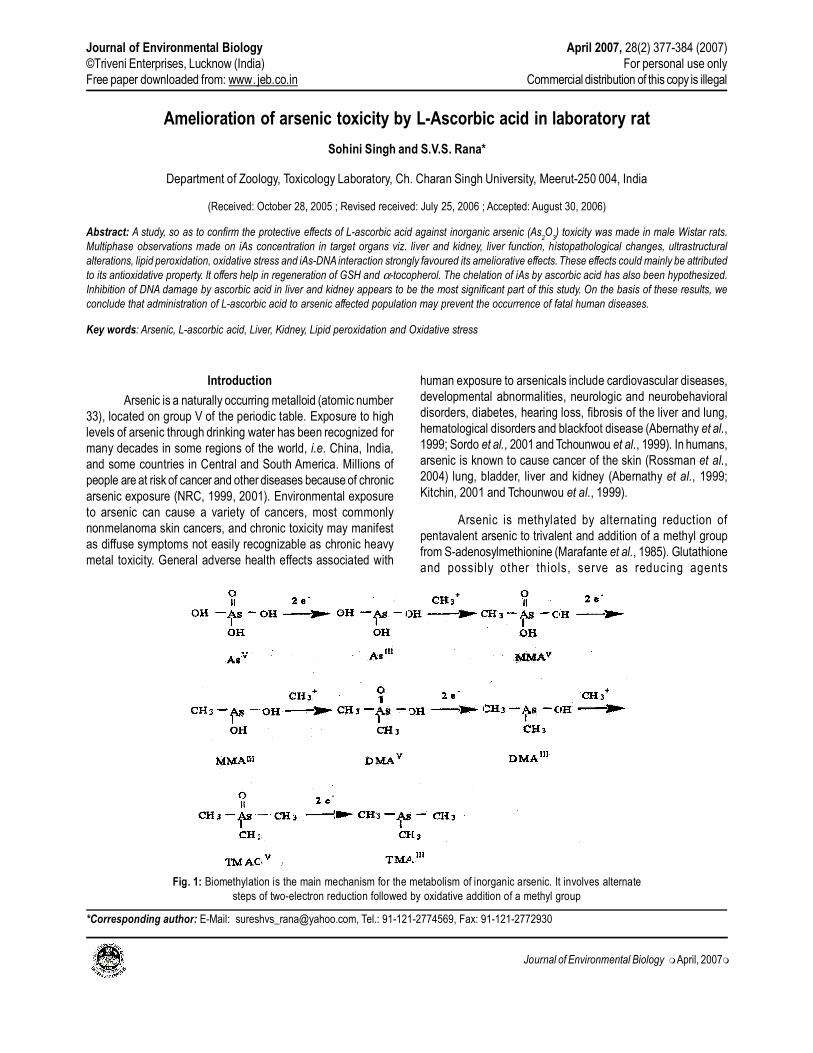
Arsenic is methylated by alternating reduction of
pentavalent arsenic to trivalent and addition of a methyl group
from S-adenosylmethionine (Marafante et al., 1985). Glutathione
and possibly other thiols, serve as reducing agents
Fig. 1: Biomethylation is the main mechanism for the metabolism of inorganic arsenic. It involves alternate
steps of two-electron reduction followed by oxidative addition of a methyl group
Journal of Environmental Biology April 2007, 28(2) 377-384 (2007)
©Triveni Enterprises, Lucknow (India) For personal use only
Free paper downloaded from: www. jeb.co.in Commercial distribution of this copy is illegal
Journal of Environmental Biology �April, 2007�
Sohini Singh and S.V.S. Rana
(Delnomdedieu et al., 1994; Styblo et al., 1995). Liver is the most
important site of arsenic methylation (Marafante et al., 1985;
Geubel et al., 1988) but most organs show methylating activity.
The end metabolites are methylarsenic acid (MMA) and dimethyl
arsenic acid (DMAA) (Fig. 1). These compounds are readily
excreted in urine. However, reactive intermediates may be
formed.
Arsenite is known to bind to cellular sulfhydryl, particularly
vicinal ones, accounting for its ability to interfere with energy
generation (Aposhian, 1989). Once in the tissues, arsenic exerts
its toxic effects through several mechanisms, the most significant
of which is, the reversible combination with sulfhydryls groups.
Arsenic also inhibits numerous other cellular enzymes, especially
those involved in cellular glucose uptake, gluconeogenesis, fatty
acid oxidation and product-ion of glutathione through sulfhydryl
group binding
A second major form of toxicity is termed “arsenolysis”.
Pentavalent arsenate can substitute competitively for phosphate
in biochemical reactions, where ADP would normally
phosphorylate into ATP. In the presence of arsenic, ADP arsenate
is the end product and high energy phosphate bonds are not
formed. The unstable ADP-arsenate decomposes spontaneously
and irreversibly resulting in loss of energy by the cell. ROSs are
capable of damaging a wide variety of cellular macromolecules
including DNA, lipids and proteins. Finally, cellular signal
transduction can be altered (e.g. activation of trans factors,
changes of gene expression), cell growth, proliferation and
differentiation can be promoted and apoptosis leading to cell
death or cancer developments can be induced (Yang and Frenkel,
2002; Qian et al., 2003).
The idea that arsenical induced toxicity could be modified
by nutrients was initially proposed in the early 1930’s by Mayer
and Sulzberger (1931), who suggested that adequate levels of
ascorbic acid, in the diet prevented or reduced occurrence of
arsenic induced anaphylaxis. L-ascorbic acid is a primary
defensive nutrient by virtue of its function as a free radical
scavenger. It can react in aqueous media against in vivo
peroxidants, including quenching of singlet oxygen species. It
increases the turn over rate of toxic metals and reduces damage
by scavenging free radicals generated by their metabolism (Hume
et al., 1991). During acute response to different stressors such
as metals and heat shock etc. ascorbic acid is depleted (Parihar
and Dubey, 1995 and Lackner, 1998). Therefore, a study on the
plausible ameliorative effects of L-ascorbic acid against iAs
toxicity in rat was proposed.
Materials and Methods
Chemicals: Arsenic trioxide was purchased from Loba Chemie
(Mumbai). L-ascorbic acid, reduced glutathione (GSH),
nicotinamide adenine dinucleotide (NAD), 5-5, dithio-bis-2-
nitrobenzoic acid, 1-chloro-2, 4-dinitrobenzene, bovine serum
albumin, triton X-100, sodium lauryl sulphate, proteinase-K,
RNAase, were obtained from Sigma Chemical Company (St
Louis, Mo., USA). Thiobarbituric acid (TBA) was purchased from
Wako Chemical Company (Japan). NADPH was obtained from
SRL (Mumbai). All other chemicals or reagents of highest purity
were procured from S. Merck, S. D. Fine Chemicals and
Qualigens (Mumbai).
Model: Male Wistar rats (150 ± 20 g) were procured from the
animal facility of Jamia Hamdard, New Delhi. They were housed
individually in polypropylene cages under standard laboratory
conditions (room temperature 25 ± 5oC; RH = 50 ± 10%). Each
rat was offered food pellets (Golden Feeds, New Delhi) and tap
water ad libitum. All animal treatments and protocols employed
in this study received prior approval of the Institutional Ethical
Committee and met the standards laid down by Govt. of India.
Treatments: Twenty healthy male rats weighing 200 ± 30 g were
selected for present study. After acclimatization to laboratory
conditions, rats were divided into four groups, each containing
five rats. Rats of group A, in addition to food and drinking water
were administered 1.0 ml saline by gavage each alternate day
and treated as controls. Rats of group B were administered
predetermined sublethal dose (4 mg/100 g body weight) (LD50
10 mg/100g body weight) of arsenic trioxide dissolved in saline
through gavage on each alternate day for 30 days as described
earlier (Allen and Rana, 2003). Rats of group C were administered
the same dose of arsenic trioxide in the same concentration and
manner similar to the rats of group B. Moreover, they were
administered with 25 mg/100 g body weight of L-ascorbic acid
simultaneously on each alternate day for thirty days. Rats of group
D were administered L-ascorbic acid only (25 mg/100 g body
weight) as the rats of group C and treated as controls.
Sample preparations: On 31st day rats were starved overnight
and sacrificed next morning by light ether anesthesia. Blood was
collected through cardiac puncture. Serum was separated by
centrifugation at 5000 rpm for 20 min. and processed for the
estimation of serum transaminases and bilirubin. Small pieces
of liver and kidney were carefully removed from each rat and
processed suitably for the estimation of arsenic, glutathione,
microsomal lipid peroxidation, glutathione-S-transferase, isolation
and quantification of DNA, histopathological and EM studies.
Estimation of arsenic in soft tissue, serum and urine: 1.0
gm of wet tissue/1.0 ml serum or urine sample was digested in
10.0 ml of concentrated nitric acid at 80oC for 16 hr. A 2.0 ml aliquot
of the digest was analyzed for inorganic As by hydride generation
at pH 6, using sodium borohydride as the reducing agent. The
analyses were done using atomic absorption spectrophotometer
(Electronic Corporation, India). Absorbance was recorded at 193.7
nm, using a hollow cathode lamp for arsenic.
Determination of serum transaminases: Alanine amino
transaminase (ALT) and aspartate amino transferase (AST)
378

Journal of Environmental Biology �April, 2007�
Arsenic toxicity in laboratory rat
activity in serum were determined following the method of
Reitman and Frankel (1957) using a kit procured from Span
Diagnostics (Surat, Gujrat).
Determination of total bilirubin in serum: Total bilirubin in
serum was determined by a commercial kit obtained from Ozone
Biomedicals Pvt. Ltd. (Hyderabad) using the method of Burtis
and Ashwood (1996).
Histopathological study: Small pieces of liver and kidney were
carefully removed from experimental animals and fixed in 10%
buffered neutral formalin. 5 micron thick paraffin sections thus
prepared were stained with hematoxylin and eosin and examined
under research microscopes (Nikon, Japan).
Electron microscopic studies (Transmission electron
microscopy): Very small cubes (1 mm3) of liver and kidney were
immersed in 2.5% glutraldehyde, post fixed in 1.0% osmium
tetraoxide, dehydrated through a graded series of ethanol and
embedded in Epon 812 after several changes of propylene oxide.
Ultra thin sections stained with uranyl acetate and lead citrate
were examined under a Phillips, CMIO Transmission electron
microscope, at AIIMS, New Delhi.
Estimation of reduced glutathione: Glutathione was measured
as acid soluble sulfhydryl levels assayed by the method described
by Ellman (1959). The acid soluble sulfhydryl group generated a
yellow colored complex (5-thio-2-nitrobenzene) with DTNB. Its
absorbance was recorded at 412 nm.
Oxidized glutathione: GSSG was estimated in the liver and kidney
following the method suggested by Ohmori et al. (1986). Peptides
having a c-terminal glycine exhibit a color reaction similar to that
of glycine with acetic anhydride, p-dimethylamino-benzaldehyde
and pyridine. Absorbance was recorded at 458 nm.
Glutathione-S-transferase (E.C.2.5.1.8): Glutathione-S-
transferase was assayed by the method of Habig et al. (1974).
Its activity was determined by the rate of formation of conjugate
between reduced glutathione and 1-chloro 2,4-dinitrobenzene.
The conjugate absorbs strongly at 340 nm wavelength.
Determination of lipid peroxidation: Peroxidized membrane
lipids were estimated by method described by Jordan and
Schenkman (1982). Microsomal pellets were precipitated by
calcium according to the method suggested by Schenkman and
Cinti (1978).
Malondialdehyde and other reactive oxidized products
of membrane lipids, under acidic conditions, react with
thiobarbituric acid and form a pink colored chromogen which is
strongly absorbed at 532 nm wavelangth.
Protein content was determined by the method described
by Lowry and coworkers using bovine serum albumin (Sigma,
USA) as the standard Lowry et al. (1951).
AsIII-DNA interaction: DNA from liver and kidney samples were
eluted using Genelute mammalian genomic DNA miniprep kit
procured from Sigma. The concentration was determined by
spectrophotometric analysis and the absorbance was measured
at 260 nm and 280 nm using a quartz microcuvette (Sambrook
et al., 1989).
Estimation of fragmented DNA: Cells were lysed using tris-
EDTA buffer and Tritox-100. Lysate was centrifuged at 13000xg
and intact and fragmented DNA were separated. The respective
samples were precipitated by trichloroacetic acid. DNA in each
fraction was quantified using diphenylamine reaction. Absorbance
was recorded at 600 nm against a blank (Sellins and Cohen,
1987).
Statistical analyses: Statistical evaluation of results was
simultaneously made employing SPSS software.
Results and Discussion
Urinary excretion of arsenic increased several fold in
arsenic fed male and female rats. Arsenic concentration
decreased in liver, kidney, urine and serum on simultaneous
treatments with ascorbic acid (Table 1, 2). Ascorbic acid has
earlier been reported as a possible chelator of lead with similar
potency as that of EDTA (Bratton et al., 1981). One possibility of
ascorbate protection might be the mobilization of arsenic from
Table - 1: Arsenic concentration (µg/ml) in the serum and urine of arsenic
and ascorbic acid treated rats
Group Treatment Serum Urine
A Control 0.011 ± 0.003 ND
B Arsenic 0.599 ± 0.076* 0.100 ± 0.01*
C Arsenic + 0.136 ± 0.014*� 0.071 ± 0.013*�
ascorbic acid
D Ascorbic acid 0.0126 ± 0.003 NS� 0.020 ± 0.016*�
Results are expressed as mean ± SE (n = 5)NS denotes non significant
* denotes significantly different values from control rats (p<0.05) “t” test� denotes significantly different results from arsenic treated rats (p< 0.05)
‘t’ test
Table -2: Arsenic concentration (µg/gm) in the liver and kidney of arsenic
and ascorbic acid treated rats
Group Treatment Liver Kidney
A Control 0.014 ± 0.002 0.032 ± 0.006
B Arsenic 105 ± 13.07* 127 ± 5.41*
C Arsenic + 64.4 ± 7.28*� 71.4 ± 7.39*�
ascorbic acid
D Ascorbic acid 0.008 ± 0.028NS� 0.018 ± 0.006NS�
Results are expressed as mean ± SE (n = 5)
NS denotes non significant
* denotes significantly different values from control rats (p<0.05) “t” test� denotes significantly different results from arsenic treated rats (p< 0.05)
“t” test
379

Journal of Environmental Biology �April, 2007�
soft tissues. Another reason might be the reduced
absorption of arsenic from intestine. Ingested arsenic that
accumulates in tissues, binds with sulfhydryl groups (Aposhian
and Aposhian, 1989) and the remaining is excreted in the urine.
Thus it could be envisaged that ascorbic acid might play a
therapeutic role against general arsenic toxicity.
Arsenic is known to produce disturbances in liver
function (Fowler et al., 1977). AST and ALT are reliable
determinants of liver parenchymal injury (Moss et al., 1987).
Activities of both ALT and AST significantly increased in arsenic
treated rats indicating liver dysfunction. In arsenic and ascorbic
acid treated rat, values of ALT and AST declined significantly.
Similarly serum bilirubin increased in arsenic treated rats but
declined in ascorbic acid and arsenic treated rats (Table 3). These
results suggest that ascorbic acid protects against hepato toxicity
of arsenic by improving liver function.
In the present study low levels of GSH were observed in
the liver and kidney of arsenic treated male and female rats. A
moderate increase in oxidized glutathione (GSSG) was, however,
recorded (Table 4 and 5). The intracellular level of GSH has been
inversely correlated with cytotoxicity of arsenic (Ochi et al., 1996).
GSH status improved on ascorbic acid co-treatment. The
improved levels of GSH might protected the sulfydryl groups from
binding with arsenic and promoted the detoxification of arsenic
by modulating arsenic methylation reactions (Buchet and
Lauwerys, 1988). When GSH levels are reduced, inorganic
arsenic becomes more toxic (Shimizu et al., 1998). GSH depletion
causes essentially nontoxic MMAsV to become toxic (Sakurai,
2003). DMAsV induces apoptosis after GSH depletion (Sakurai,
2002), which could allow the survival of damaged cells.
Glutathione-S-transferase (GST) consists of a large family
of GSH utilizing enzymes that play an important role in
detoxication of xenobiotics in mammalian systems. Arsenic
trioxide was found to inhibit glutathione-S-transferase activity
significantly in liver and kidney both (Habig et al., 1974; Lee et
al., 1989 and Wendel, 1980). However, ascorbate co-treatment
restored enzyme activity in male rats. This was considered as an
Table - 4: Reduced glutathione (GSH) (µg/g wet weight) in the liver and kidney of arsenic treated rats
Group Treatment Gender Liver Kidney
A Control Male 0.154 ± 0.060 0.169 ± 0.039
B Arsenic Male 0.126 ± 0.006* 0.105 ± 0.013*
C Arsenic + ascorbic acid Male 0.130 ± 0.010*NS 0.170 ± 0.004NS�
D Ascorbic acid Male 0.139 ± 0.002NS� 0.136 ± 0.006*�
Results are expressed as mean ± SE (n = 5)NS denotes non significant
* denotes significantly different values from control rats (p<0.05) “t” test� denotes significantly different results from arsenic treated rats (p< 0.05) “t” test
Table - 5: Oxidized glutathione (GSSG) (µ moles/gm wet weight) in the
liver and kidney of arsenic treated rats
Group Treatment Liver Kidney
A Control 0.535 ± 0.030 0.429 ± 0.077
B Arsenic 3.168 ± 0.053* 3.644 ± 0.018*
C Arsenic + 1.627 ± 0.114*+ 0.956 ± 0.060*+
ascorbic acid
D Ascorbic acid 0.560 ± 0.023NS+ 0.392 ± 0.033*+
Results are expressed as mean ± SE (n = 5)NS denotes non significant
* denotes significantly different values from control rats (p<0.05) “t” test+denotes significantly different results from arsenic treated rats (p< 0.05)
“t” test
Table - 6: Glutathione-S-transferases (GST) (n moles/NADPH/min/mg
protein) in the liver and kidney of arsenic treated rats
Group Treatment Liver Kidney
A Control 0.865 ± 0.035 0.727 ± 0.015
B Arsenic 0.426 ± 0.021* 0.355 ± 0.008*
C Arsenic +
ascorbic acid 0.677 ± 0.007*� 0.570 ± 0.013*�
D Ascorbic acid 0.806 ± 0.013*� 0.689 ± 0.031*�
Results are expressed as mean ± SE (n = 5)NS denotes non significant
* denotes significantly different values from control rats (p<0.05) “t” test� denotes significantly different results from arsenic treated rats (p< 0.05)
“t” test
Table - 3: AST (Karmen Units), ALT (Karmen Units) and bilirubin (mg/dl) in the serum of arsenic treated rats
Group Treatments AST (Karmen Units) ALT (Karmen Units) Bilirubin (mg/dl)
A Control 42.25 ± 1.29 42.0 ± 1.15 17.98 ± 0.53
B Arsenic 120.51 ± 1.48* 136.96 ± 2.19* 40.58 ± 0.921*
C Arsenic + Ascorbic acid 89.71 ± 1.39*+ 98.99 ± 1.057*+ 28.22 ± 1.00*+
D Ascorbic acid only 42.90 ± 0.81NS+ 41.95 ± 0.623NS+ 15.63 ± 0.66NS+
Values are expressed as mean ± SE (n = 5) p<0.05
* denotes values significantly different from control rats.+ Denotes values significantly different from arsenic treated rats
Sohini Singh and S.V.S. Rana380

Journal of Environmental Biology �April, 2007�
adaptive response facilitated by ascorbic acid against arsenic
induced stress (Table 6).
Lipid peroxidation has been largely considered as a
molecular mechanism involved in deleterious effects of a variety
of xenobiotics including heavy metals (Sunderman et al., 1985).
Biomembranes and subcellular organelles are the major site of
lipid peroxidation (Halliwell and Gutteridge, 1989). Co-treatments
of ascorbic acid and arsenic inhibited lipid peroxidation in liver
and kidney both. This effect could be attributed to its antioxidative
property. Ascorbic acid is well known to inhibit oxidative damage
to membranes (Li et al., 2001; Nandi et al., 2005) (Table 7).
The induction of oxidative DNA damage by arsenic in
mammalian cells has been paid considerable attention by earlier
workers. (Kessel et al., 2002). There is evidence that both DMAIII
and MMA III exert DNA damaging effect through an intermediate
involving ROS production (Nesnow et al., 2002). Arsenic perturbs
cells in numerous ways i.e. inducing chromosomal abnormalities,
altering DNA repair or DNA methylation patterns and producing
oxidative stress (Hamadeh et al., 2002; Liu and Jan, 2000; Mass
et al., 2001). Present observations on total amount of DNA in
liver and kidney showed that arsenic trioxide significantly
damaged DNA. Results also indicated higher percentage of
fragmented DNA in liver and kidney both after arsenic treatment.
Further, it was found that cotreatments with ascorbic acid reduced
the percentage of fragmented DNA and increased the amount of
total DNA in liver and kidney both (Table 8, 9 and 10). Thus a
protective effect of ascorbic acid on DNA damage could be
envisaged. An earlier study suggested that ascorbic acid inhibited
DNA damage caused by an antitumor drug (Blasiak and Kowalik,
2001).
To gather more evidence to support the protective
behavior of ascorbic acid against arsenic toxicity, histopathological
studies, using both light and electron microscopy were
undertaken. Light microscopical observations showed that arsenic
caused hepatic parenchymal degeneration, hyperplasia and
vascular lesions in male rats. Sinusoids were dilated and found
to be filled with foam cells. Significant protective effects on
different lesions were observed in the liver of rats treated with
arsenic and ascorbic acid. There was no neoplastic formation,
however, mild inflammation of hepatic cells still persisted (Fig. 1,
2). While the ultrastructural study of liver, of arsenic treated rats,
showed inflammed nuclei and increased number of mitochondria
in hepatic cells treated with arsenic and ascorbic acid. The
mitochondria of different shapes and sizes were observed and
several small vacuoles were observed in the matrix of the cell
(Fig. 3 and 4).
Light microscopical observations on kidney of arsenic
treated rats showed glomerulonephritis, proximal tubular necrosis,
epithelial damage and loss of nuclei. In cortex focal tubular
necrosis was observed along with pycnosis and appearance of
Table - 7: Microsomal malondialdehyde (n moles/mg protein) in the
liver and kidney of arsenic treated rats
Group Treatment Liver Kidney
A Control 0.120 ± 0.004 0.135 ± 0.005
B Arsenic 0.325 ± 0.009* 0.460 ± 0.059*
C Arsenic + 0.211 ± 0.003*� 0.235 ± 0.007*�
ascorbic acid
D Ascorbic acid 0.113 ± 0.012NS� 0.125 ± 0.017NS�
Results are expressed as mean ± SE (n = 5)NS denotes non significant
* denotes significantly different values from control rats (p<0.05) “t” test� denotes significantly different results from arsenic treated rats (p< 0.05)
“t” test
Table - 8: Intact and fragmented DNA (µg/ml) in the liver of arsenic and
ascorbic acid treated rats
Group Treatment Total DNA % Fragmentation
A Control 31.59 ± 0.332 23.46 ± 0.478
B Arsenic 12.56 ± 0.253* 34.69 ± 0.445*
C Arsenic + 29.58 ± 0.389*� 25.84 ± 0.345*�
ascorbic acid
D Ascorbic acid 33.22 ± 0.482*� 21.65 ± 0.779*�
Results are expressed as mean ± SE (n = 5)NS denotes non significant
* denotes significantly different values from control rats (p<0.05) “t” test� denotes significantly different results from arsenic treated rats (p< 0.05)
“t” test
Table - 9: Intact and fragmented DNA (µg/ml) in the kidney of arsenic
and ascorbic acid treated rats
Group Treatment Total DNA % Fragmentation
A Control 26.39±.299 20.76±0.492
B Arsenic 15.48±0.322* 38.20±0.257*
C Arsenic + 31.95±0.425* 26.10±0.697*�
ascorbic acid
D Ascorbic acid 30.71±0.399*� 21.13±0.898NS�
Results are expressed as mean ± SE (n = 5)NS denotes non significant
* denotes significantly different values from control rats (p<0.05) “t” test� denotes significantly different results from arsenic treated rats (p< 0.05)
“t” test
Table - 10: DNA (µg/ml) in the liver and kidney of arsenic treated rats
Group Treatment Liver Kidney
A Control 39.6 ± 0.452 39.2 ± 0.717
B Arsenic 33.4 ± 0.358* 45.60 ± 0.457*
C Arsenic +
ascorbic acid 152.6 ± 5.850*� 55.20 ± 3.23*�
D Ascorbic acid 81.6 ± 4.73*� 35.6 ± 1.807*�
Results are expressed as mean ± SE (n = 5)NS denotes non significant
* denotes significantly different values from control rats (p<0.05) “t” test� denotes significantly different results from arsenic treated rats (p< 0.05)
“t” test
Arsenic toxicity in laboratory rat 381

Journal of Environmental Biology �April, 2007�
Fig. 2: T.S. of liver of an arsenic fed rat shows parenchymal
degeneration, hyperplasia and vascular lesions (H/E X 400)
Fig. 3: T.S. of liver of rat treated with arsenic and ascorbic acid
shows binucleated cells and focal pycnosis (H/E X400)
Fig. 4: T.E.M. study of liver of arsenic treated rat shows swollen
nuclei and increased number of mitochondria (M) (880X)
Fig. 5: T.E.M. study of liver of rat treated with arsenic and ascorbic acid
shows normal nucleus (N). Several vacuoles are also observed (880X)
Fig. 6: T.S. of kidney of arsenic treated rat shows inflammed
glomerulus, and proximal tubular necrosis (H/E X 400)
Fig. 7: T.S. of kidney of rat treated with arsenic and ascorbic acid
shows mild glomerulonephritis and tubular necrosis (H/E X 400)
Sohini Singh and S.V.S. Rana382

Journal of Environmental Biology �April, 2007�
Fig. 8: T.E.M. study of kidney of arsenic treated rat shows
mitochondria (M) of different shapes and sizes (880X)
Fig. 9: T.E.M. study of kidney of rat treated with arsenic and
ascorbic acid shows basolateral membrance infoldimgs
tightly associated with mitochondria (M) (1400X)
hyaline like deposits in medulla. Kidney of rat treated with
arsenic and ascorbic acid, showed little improvement in terms of
proximal tubular necrosis and glomerular injury (Fig. 5 and 6).
Ultrastructure of kidney of arsenic treated rats showed, basolateral
infoldings. Number of lysosomes was also found to be increased.
Brush border, the proximal tubule and distal tubules were damaged
(Fig. 7). Ultrastructure of kidney of male rats, treated with arsenic
and ascorbic acid, showed improvements in renal structure.
Proximal tubule was found to be lined with cuboidal cells and
apical brush border. Basolateral membrane infoldings were found
to be tightly associated with mitochondria (Fig. 8).
Ascorbic acid has a few therapeutic characters. It is a
water soluble antioxidant. It predominantly works as a radical
chain terminator. One ascorbate molecle reacts with a peroxyl
radical to yield a hydroperoxide and ascorbyl radical,
subsequently the ascorbyl radical can react with another peroxy-
radical and produce the oxidized ascorbic acid i.e., dihydroscorbic
acid (Combs and Gray, 1998). Thus one molecule of ascorbate
can trap two molecules of peroxyl radicals. Another hypothesis
suggests that ascorbic acid may be involved in the regeneration
or restoration of antioxidant properties of α-tocopherol. The
tocopherol is converted to α-tocopherol quinone (Chow, 1985).
Ascorbate can both chelate and reduce transition metal
ions and the reduced metal ions in turn can reduce oxygen or
H2O
2 to superoxide and hydroxyl radicals, respectively.
AsCH- + Me(n+1)+ → AsC*- + H+ + M n+ (Eq. 1)
H2O
2 + M n+ → OH*. + OH- + M (n+ 1)+ (Eq. 2)
Superoxide and hydroxyl radicals are scavenged by
ascorbate with second order rate constant of 1x10⁵ and 1.1x10¹⁰
M-1S-1 respectively. (Carr and Frei, 1999). Therefore, ascorbic
acid forms the first line of antioxidant defense (Frei, 1999; Carr
and Frei, 2000). These properties of ascorbic acid make it a
suitable antidote for arsenic toxicity in rodents and possibly in
human subjects.
References
Abernathy, C.O., Y.P. Liu, D. Longfellow, H.V. Aposhian, B. Beck, B. Fowler, R.
Goyer, R. Menzer, T. Rossman, C. Thompson and W. Michael: Arsenic:
health effects, mechanisms of actions, and research issues. Environ.
Hlth. Perspect., 107, 593-597 (1999).
Allen, T. and S.V.S. Rana: Oxidative stress by inorganic arsenic:modulation by
thyroid hormones in rat. Comp. Biochem. Physiol., 135, 157-162 (2003).
Aposhian, H.V.: Biochemical toxicology of arsenic. In: Reviews of Biochemistry
and Toxicology (Eds: E. Hodgson, J.R. Bend and R.M. Philpot). Vol. 10,
Elsevier, New York. pp. 265-299 (1989).
Aposhian, H.V. and M.M. Aposhian: Newer developments in arsenic toxicity. J.
Am. Coll. Toxicol., 8, 1297-1305 (1989).
Blasiak, J. and J. Kowalik: Protective action of vitamin C against DNA damage
induced by selenium-cisplatin conjugate. Acta Biochimica. Polonica,
48(1), 233-240 (2001).
Bratton, G.R., J. Zmudzki, M.C. Bell and L.G. Warnoch: Thiamine (Vitamin B1)
effects on lead intoxication and deposition of lead in tissues. Therapeutic
potential. Toxicol. Appl. Pharmacol., 59, 164-172 (1981).
Buchet, J.P. and R. Lauwerys: Role of thiols in the in vitro methylation of inorganic
arsenic by rat liver cytosol. Biochem. Pharmacol., 37, 3149-3153 (1988).
Burtis, C. A. and R.A. Edward: Liver Function in Tietz fundamentals of clinical
chemistry W.B. Saunders and Company, Philadelphia. PA. pp. 539-568
(1996).
Carr, A. and B. Frei: Towards a new recommended dietary allowance for vitamin
C based on antioxidant and health effects in humans. Am. J. Clin. Nutr.,
69, 1086-1107 (1999b).
Carr, A. and B. Frei: The role of natural antioxidants in preserving the biological
activity of endothelium derived nitric oxide. Free Radic. Biol. Med., 28,
1806-1814 (2000).
Chow, C.K.: Vit. E and blood. World Rev. Nutr. Dieta., 45, 133-66 (1985).
Combs, G.F. and W.P. Gray: Chemopreventive agents: Selenium. Pharmacol.
Ther., 79, 179-192 (1998).
Delnomdedieu, M., M.M. Basti, M. Styblo, J.D. Otvos and D.J. Thomas:
Complexation of arsenic species in rabbit erythrocytes. Chem. Res.
Toxicol., 7, 621-627 (1994).
Ellman, G.L.: Tissue sulfhydryl groups. Arch. Biochem., 82, 70-77 (1959).
Fowler, B.A., J.S. Woods and C.M. Schiller: Ultrastructural and biochemical
effects of prolonged oral arsenic exposure on liver mitochondria of rats.
Environ. Hlth. Perspect., 19, 197-204 (1977).
Arsenic toxicity in laboratory rat 383

Journal of Environmental Biology �April, 2007�
Frei, B.: On the role of Vitamin C and other antioxidants in atherogenesis and
vascular dysfunction. Proc. Soc. Exp. Biol. Med., 222, 196-204 (1999).
Geubel, A.P., M.C. Mairlot, J.P. Buchet, C. Dive and R. Lauwerys: Abnormal
methylation capacity in human liver cirrhosis. Int. J. Clin. Pharmacol.
Res., 8(2), 117-122 (1988).
Habig, W.H., M.J. Pabst and W.B. Jakoby: Glutathione-S-transferases. The
first enzymatic step in mercapturic acid formation. J. Biol. Chem., 249,
7130-7139 (1974).
Halliwell, B. and J.M.C. Gutteridge: In: Free radicals in biology and medicine.
2nd Edn., Oxford University Press (Clrendon) Oxford (1989).
Hamadeh, H.H., K.J. Trouba, R.P. Amin, C.A. Afshari and D. Germolec:
Coordination of altered DNA repair and damage pathways in arsenite
exposed Keratinocytes. Toxicol. Sci., 69, 306-16 (2002).
Hume, R. I., R. Dingledine and S.F. Heinemann: Identification of a site in
glutamate receptor subunits that controls calcium permeability. Science,
253, 1028-1031 (1991).
Jordan, R.A. and J.B. Schankman: Relationship between malon-dialdehyde
production and arachidonate consumption during NADPH supported
microsomal lipid peroxidation. Biochem. Pharmacol., 31, 1393 (1982).
Kessel, M., S.X. Liu, A. Xu, R. Santella and T.K. Hei: Arsenic induces oxidative
DNA damage in mammalian cells. Mol. Cell. Biochem., 234, 301-308
(2002).
Kitchin, K.T.: Recent advances in carcinogenesis: modes of action, animal model
systems and methylated arsenic metabolites. Toxicol. Appl. Pharmacol.,
172, 249-261 (2001).
Lackner, R.: Oxidative stress in fish by environmental pollutants. In: Fish
Exotoxicology (Eds.: T. Braunbeck, D.E. Hinton and B. Streit). Brikhauser
Verlag Basel, Switzerland. pp. 203-224 (1998).
Lee, T.C., M.L. Wei, W.J. Chang, I.C. Ho,J.F. Lo, K.Y. Jan and H. Huang:
Elevation of glutathione levels and glutathione S-transferase activity in
arsenic-resistant Chinese hamster ovary cells. In vitro Cell Dev. Biol.,
25, 442-448 (1989).
Li, X., C.E. Cobb, K.E. Hill, R.F. Burk and J.M. May: Mitochondrial uptake and
recycling of ascorbic acid. Arch. Bicohem. Biophys., 387, 143-153 (2001).
Liu, F. and K.Y. Jan: DNA damage in arsenite and cadmium treated bovine
aortic endothelial cells. Free Radic. Biol. Med., 28, 55-63 (2000).
Locke, J.: The determination of eight elements in human liver tissue by flame
atomic absorption spectrometry in sulfuric acid solution. Annl. Chem.
Acta, 104, 225-231 (1982).
Lowry, O.H., N.J. Rosenbrough, A.L. Farr and R.J. Randall: Protein measurement
with folin phenol reagent. J. Biol. Chem., 193, 265-275 (1951).
Marafante, E., M. Vahter and J. Envall: The role of methylation in the detoxication
of arsenate in the rabbit. Chem. Biol. Interact., 56, 225-238 (1985).
Mass, M.J., A. Tennant, B.C. Roop, W.R. Cullen, M. Styblo, D.J. Thomas and
A.D. Kligerman: Methylated trivalent arsenic species are genotoxic.
Chem. Res. Toxicol., 14, 355-61 (2001).
Mayer, R.L. and M.B. Sulzberger: Zur. Frug der yahreszeit lichen schwanKungen
der Krannei ten. Der E ing luss der Kos t auf. Experimental le
sensipilisierungen. Arshiv. Fuer. Dermat und Syph., 163, 245 (1931).
Moss, D.W., A.R. Henderson and J.F. Kachmar: In: Fundamentals of Clinical
Chemistry. 3rd Edn. (Ed: N.W. Tietz). W.B. Saurders, Philadelphia. pp.
346-421 (1987).
Nandi, D., R.C. Patra and D. Swarup: Effect of cysteine, methionine, ascorbic
acid and thiamine on arsenic- induced oxidative stress and biochemical
alteration in rats. Toxicol., 211, 26-35 (2005).
National Research Council: Arsenic in Drinking Water. National Academy Press,
Washington, D.C. (1999).
National Research Council: Arsenic in Drinking Water: Update. National
Academy Press, Washington, D.C. (2001).
Nesnow, S., B.C. Roop, G. Lambert, M. Kadiiska, R.P. Mason and W.R. Cullen:
DNA damage induced by methylated trivalent arsenicals is mediated by
reactive oxygen species. Chem. Res. Toxicol., 15, 1627-1634 (2002).
Ochi, T., F. Nakajima, T. Sakurai, T. Kaise and Y. Oya-Ohta: Dimethylarsinic
acid causes apoptosis in HL-60 cells via interaction with glutathione.
Arch. Toxicol., 70, 815-821 (1996).
Ohmori, S., M. Ikeda, E. Kasahra, H. Hyodoh and K.A. Hirota: Colorimetric
determination of total glutathione based on its c-terminal glycine residue
and its application to blood, liver and yeast. Chem. Pharm. Bull., 29(5),
1355-1360 (1981).
Parihar, M.S. and A.K. Dubey: Lipid peroxidation and ascorbic acid status in
respiratory organs of male and female freshwater catfish Heteropneustes
fossilis exposed to temprature increase. Comp. Biochem. Physiol., 112,
309-313 (1995).
Qian, Y., V. Castranova and X.J. Shi: New perspectives in arsenic-induced cell
signal transduction. Inorg. Biochem., 96, 271-278 (2003).
Reitman, S. and S. Frankel: A colorimetric method for determination of serum
glutamic-oxaloacitic and glutamic-pyruvic transaminase. Am. J. Clin.
Pathol. 28, 56-68 (1957).
Rossman, T.G., A.N. Uddin and F.J. Burns: Evidence that arsenite acts as a
cocarcinogen in skin cancer. Toxicol. Appl. Pharmacol., 198, 394-404
(2004).
Sakurai, T.: Molecular mechanisms of dimethyl arsenic acid induced apoptosis,
Biomed. Res. Trace Elem., 13, 167-176 (2002).
Sakurai, T.: Biomethylation of arsenic is essentially detoxicating event, J. Hlth.
Sci., 49, 171-178 (2003).
Sambrook, J., E.F. Fritsch and T. Manniatis: Molecular cloning: A laboratory
manual. 2nd Edn. Cold spring Harbor Laboratory Press, Plainview, New
York (1989).
Schenkman, J.B. and D.L. Cinti: Preparation of microsomes with calcium.
Methods in Enzymol., 52, 83-88 (1978).
Sellins, K.S. and J.J. Cohen: Gene induction by α-irradiation leads to DNA
fragmentation in lymphocytes. J. Immunol., 139, 3199-3206 (1987).
Shimizu, M., J.F. Hochadel, B.A. Fulmer and M.P. Waalkes: Effect of glutathione
depletion and metallothionein gene expression on arsenic induced
cytotoxicity and c-myc expression in vitro. Toxicol. Sci., 45, 204-211
(1998).
Sordo, M., L.A. Herrera, P. Ostrosky-Wegman and E. Rojas: Cytotoxic and
genotoxic effects of As, MMA, and DMA on leukocytes and stimulated
human lymphocytes. Teratog. Carcinog. Mutagen., 21, 249-260 (2001).
Styblo, M., H. Yamauchi and D.J. Thomas: Comparative in vitro methylation of
trivalent and pentavalent arsenicals. Toxicol. Appl. Pharmacol., 135,
172-178 (1995).
Sunderman, F.W. Jr., A. Marzouk, S.M. Hopfer, O. Zaharia and M.C. Reid:
Increased lipid peroxidation in tissues of nickel chloride treated rats. Annl.
Clin. Lab. Sci., 15, 229-236 (1985).
Tchounwou, P.B., B. Wilson and A. Ishaque: Important considerations in the
development of public health advisories for arsenic and arsenic-
containing compounds in drinking water. Rev. Environ. Hlth., 14,
211-229 (1999).
Wendel, A.: Glutathione Peroxidase. In: Enzymatic basis of detoxicaiton (Ed:
W.B. Jacoby). Vol.1, Academic Press, New York. pp. 333-353 (1980).
Yang, C. and K. Frenkel: Arsenic-mediated cellular signal transduction,
transcription factor activation, and aberrant gene expression: implications
in carcinogenesis. J. Environ. Pathol. Toxicol. Oncol., 21, 331-342 (2002).
Sohini Singh and S.V.S. Rana384