alveolar rhabdomyosarcoma the molecular drivers of pax3/7
TRANSCRIPT

Skeletal MuscleMarshall and Grosveld Skeletal Muscle 2012, 2:25http://www.skeletalmusclejournal.com/content/2/1/25
REVIEW Open Access
Alveolar rhabdomyosarcoma – The moleculardrivers of PAX3/7-FOXO1-induced tumorigenesisAmy D Marshall1,2 and Gerard C Grosveld1*
Abstract
Rhabdomyosarcoma is a soft tissue sarcoma arising from cells of a mesenchymal or skeletal muscle lineage. Alveolarrhabdomyosarcoma (ARMS) is more aggressive than the more common embryonal (ERMS) subtype. ARMS is moreprone to metastasis and carries a poorer prognosis. In contrast to ERMS, the majority of ARMS tumors carry one ofseveral characteristic chromosomal translocations, such as t(2;13)(q35;q14), which results in the expression of aPAX3-FOXO1 fusion transcription factor. In this review we discuss the genes that cooperate with PAX3-FOXO1, as wellas the target genes of the fusion transcription factor that contribute to various aspects of ARMS tumorigenesis. Thecharacterization of these pathways will lead to a better understanding of ARMS tumorigenesis and will allow thedesign of novel targeted therapies that will lead to better treatment for this aggressive pediatric tumor.
Keywords: Alveolar rhabdomyosarcoma, PAX3-FOXO1, PAX7-FOXO1, FGFR4, CNR1, IRIZIO, N-MYC, IGF2, MET,CXCR4, p53, MDM2, P-Cadherin, TFAP2B, miR17-92
IntroductionAccording to the American Cancer Society, rhabdomyo-sarcoma (RMS) comprises about three percent of child-hood cancers, with about 350 new cases occurringannually in the US [1], and it affects slightly more malesthan females [2]. RMS is a small, round, blue cell tumorusually arising in skeletal muscle tissue, and it is thoughtto originate from mesenchymal cells likely committed tothe skeletal muscle lineage. Consistent with a myogenicorigin, RMS tumors express skeletal muscle markers suchas skeletal muscle actin and myosin, desmin, myoglobin,Z-band protein, MYOD and often myogenin [3-7]. RMSconsists of two major histological subtypes, embryonaland alveolar RMS. The embryonal subtype (ERMS) isthought to be histologically reminiscent of embryonicdeveloping skeletal muscle [7]. ERMS is the most preva-lent of the subtypes, accounting for about 60% of RMScases [2]. It occurs mainly in children younger than 10years and is usually associated with a favorable prognosis,with a failure-free survival rate of 81% [8,9]. Tumors usu-ally localize to the head and neck (including the extraocu-lar muscles of the eye), the genitourinary tract and the
* Correspondence: [email protected] of Genetics, St Jude Children’s Research Hospital, Memphis, TN38105, USAFull list of author information is available at the end of the article
© 2012 Marshall and Grosveld; licensee BioMeCreative Commons Attribution License (http:/distribution, and reproduction in any medium
retroperitoneum [7,8]. The alveolar subtype (ARMS) con-stitutes approximately another 20% of RMS cases [10] andoccurs predominantly in adolescents. Histologically,ARMS tumors typically contain alveoli structures similarin appearance to those seen in the lung [7], though solid-variant ARMS does occur [11]. Primary ARMS tumorstypically arise in the extremities and trunk [7-9,12], andthey are more aggressive than their ERMS counterparts.ARMS is associated with a poorer prognosis, with a 5-yearfailure-free survival of 65% [8].A characteristic of the ARMS subtype is the occurrence
of recurrent chromosomal translocations. The most com-mon of these is t(2;13)(q35;q14), which results in theexpression of an oncogenic fusion protein. This fusionprotein consists of the paired and homeodomains of thePAX3 transcription factor with the potent transcriptionalactivation domain of FOXO1 (FKHR), a member of theforkhead (FOX) family of transcription factors [13-15].The PAX3 homeodomain is required to recapitulatePAX3-FOXO1-induced tumorigenesis, though the paireddomain may play a minor role [16,17]. The PAX3-FOXO1fusion protein can be detected in about 55% of ARMScases [18]. A similar translocation of t(1;13)(p36;q14) fusesthe PAX7 DNA-binding domains, the closest homolog ofPAX3, to FOXO1 [19]. This translocation occurs in a fur-ther 22% of ARMS cases [18]. Recently, further similar
d Central Ltd. This is an Open Access article distributed under the terms of the/creativecommons.org/licenses/by/2.0), which permits unrestricted use,, provided the original work is properly cited.

Marshall and Grosveld Skeletal Muscle 2012, 2:25 Page 2 of 14http://www.skeletalmusclejournal.com/content/2/1/25
translocations have been found in individual ARMS cases:t(2;X)(q35;q13), which results in PAX3-AFX fusion [20],and t(2;2)(q35;p23) and t(2;8)(q35;q13), which generate afusion protein of PAX3-NCOA1 and PAX3-NCOA2,respectively [21,22]. These “cryptic” rare fusion variantsare thought to be present in up to another 10% of ARMStumors [7]. During normal development, PAX3 expressionoccurs in the neural tube and dermomyotome [23], and itis required for the normal migration of skeletal muscleprecursors to the limb bud [24]. PAX7 expression is amarker of satellite cells in adult skeletal muscle [25] and isrequired for normal self-renewal [26]. Unlike skeletalmuscle-specific PAX3 and PAX7, FOXO1A, AFX,NCOA1 and NCOA2 are widely expressed and mediategene transcription downstream of cell signaling pathways[27-31]. All of the ARMS fusion proteins consist of thePAX3/7 DNA-binding domains fused to the transcrip-tional activation domains of more potent transcription fac-tors (see Figure 1) [14,15,22]. Genome-wide transcriptionfactor-binding studies have not yet been performed to de-termine whether wild-type PAX3 and PAX7-binding sitesdiffer from these PAX3/7 fusion transcription factor-binding sites.Expression of these ARMS fusion transcription factors is
thought to abrogate normal skeletal muscle differentiation,allowing aberrant cell division and tumor development.PAX3 expression can inhibit myogenic differentiation ofcultured myoblasts [35]. Although PAX3 protein is rapidlydegraded during early myogenic differentiation, PAX3-FOXO1 has a significantly longer half-life than wild-typePAX3 [36]. PAX3/7-FOXO1 is capable of suppressingMyoD expression and activity [37,38]. PAX7-FOXO1 ex-pression induces NFκB signaling, which inhibits myogen-esis via activation of cyclin D1/CDK4 complexes. Thesecomplexes sequester MyoD, which would normally drivecell cycle withdrawal and myogenic differentiation [39]. Inaddition transcriptionally inactive MyoD can enhance
Figure 1 Gene translocation in alveolar rhabdomyosarcoma. Scale diaarising from chromosomal translocations occurring in ARMS. Green or yelloare indicated in like colors. DNA-binding domains are indicated as: paired dand basic helix-loop-helix domain (bHLH). Regions of the proteins known tdomains include the octapeptide domain (O), PAS domains (PAS A/B), LXXderived from the following references: [14,19-22,32-34].
PAX3-FOXO1 transcriptional activity [37]. There is alsoevidence that MyoD transcriptional activity is abrogated inERMS tumors [40].The remaining ARMS tumors are classed as fusion-
negative ARMS. However, fusion-negative ARMS are in-distinguishable on the levels of gene expression and inclinical outcome from ERMS tumors, leading some toargue that translocation status should be the definingfactor of ARMS [41-44]. Within the ARMS subtype,prognosis can vary by disease stage at diagnosis as wellas translocation status. For example, patients presentingwith metastatic disease have an estimated 4-year overallsurvival rate of 75% for PAX7-FOXO1, while those withthe PAX3-FOXO1 translocation have only 8% estimated4-year overall survival [18]. Indeed, there is evidencethat the PAX3-FOXO1 is a more potent oncogene thanPAX7-FOXO1. Barr et al. [45] found that only 1/24PAX3-FOXO1-positive ARMS tumors had amplificationof the PAX3-FOXO1 gene, while PAX7-FOXO1 wasamplified in 10/11 PAX7-FOXO1 ARMS, implying thatgenomic amplification of PAX7-FOXO1 is required fortumorigenesis, while a single copy of PAX3-FOXO1 issufficient. However the gene expression profiles ofPAX3-FOXO1- and PAX7-FOXO1-expressing tumorshave not, to the author’s knowledge, been specificallycompared to identify the gene set responsible for thisdifference in prognosis between ARMS tumors withthese two fusion genes.
ReviewPAX3-FOXO1 is the most common fusion gene inARMS. This fusion transcription factor is thought todrive the gene expression that causes the worse progno-sis in ARMS tumors. Many studies have sought to iden-tify the differences in gene expression between ERMSand ARMS, as well as the genes aberrantly regulated byPAX3-FOXO1. In this review, we summarize these gene
gram showing the parent proteins and the resulting fusion proteinsw indicates the protein fusion sites [14,19-22]. Homologous domainsomain (PD), homeodomain (HD), fork head DNA-binding domain (FH)o act as transcriptional activation domains are indicated (TAD). OtherLL motifs (L1-L7) and glutamine-rich region (Q-rich). Maps were

Marshall and Grosveld Skeletal Muscle 2012, 2:25 Page 3 of 14http://www.skeletalmusclejournal.com/content/2/1/25
expression changes specific to PAX3-FOXO1 expressionand/or ARMS. Moreover, we consolidate these data intoa list of genes that may well represent the means bywhich ARMS tumors obtain a more aggressive pheno-type than ERMS.
Cooperating mutations in ARMS tumorsIt is likely that PAX3/7-FOXO1 translocation is one ofthe earliest events in ARMS tumorigenesis as it occursin the majority of ARMS cases, more often than anyother genetic lesion characterized in the disease. How-ever, PAX3-FOXO1 expression in normal cells is notsufficient to induce transformation, and other geneticalterations are required [46-48]. Genomic amplificationis common in ARMS tumors. The three most commonamplifications seen in ARMS involve regions of chromo-somes 2, 12 and 13 [49].The region of chromosome 12 amplification spans
12q13-15 and is reported in 28% to 56% of ARMStumors [49-52]. This 12q13-15 region includes genessuch as C/EBP-homolog and transcription factor CHOP/DDIT3/GADD153, sarcoma-amplified sequence andtransmembrane 4 superfamily member SAS/TSPAN31,alpha 2-macroglobulin receptor A2MR/LRP1, Sonichedgehog (SHH) pathway effector and zinc finger tran-scription factor GLI1, cyclin-dependent kinase cell cycleregulator CDK4 and p53 pathway modulator MDM2. Inmost cases, gene amplification accompanies an increasein gene expression [50,53].Though GLI1 is amplified genetically, the expression
of this gene is not always associated with its geneticamplification. When GLI1 is overexpressed in RMS, ithas been associated with an undifferentiated subtype ra-ther than ERMS or ARMS, indicating that GLI1 mayplay a role in tumors that show primitive histopatho-logical features [54]. Thus, GLI1 overexpression cannotbe well associated with the ARMS pathology.MDM2 is perhaps the best candidate oncogene in this re-
gion because of its inhibitory effect on p53 function [55].However, MDM2 is not always included in this 12q13-15amplification. RH30, an ARMS cell line, lacks amplificationof MDM2 but shows amplification and overexpression ofSAS, CHOP, GLI1 and A2MR [53]. In addition, the fre-quency of MDM2 gene amplification specifically may be aslow as 10% in ARMS tumors [56]. One study found only 2of 34 ARMS samples to be highly immunoreactive forMDM2 [12]. Moreover, MDM2 expression shows no asso-ciation with patient prognosis or other clinicopathologicparameters [12]. Thus, it may be amplification of one of theother genes at this chromosome 12 locus that is the import-ant cooperating mutation with PAX3-FOXO1.Other alterations in the p53 pathway have been found
in ARMS. In ARMS tumor samples mutated p53 was
reported in 0 to 22% of cases [12,56,57]. RMS cell linesshow a significantly higher rate of p53 abnormalitieswith 60%, indicating establishment of these cell linesthrough xenograft and cell culture increases the propor-tion of cell lines with p53 alterations [56]. Looking atp53 and MDM2 expression levels, both are low inARMS and ERMS. Metastatic ERMS tumors show sig-nificantly higher p53 protein expression, indicating thatp53 gene alterations are a late event in rhabdomyosarco-magenesis. Again, p53 status did not show any correl-ation to prognosis [58].Chromosome 2 has been shown to be amplified at
2p24 in 32 to 60% of ARMS tumors [49,51,59,60]. Thisregion is known to contain the proto-oncogene N-MYC.Two independent studies have shown that a gain in thegenomic copy number of the N-MYC gene is associatedwith an unfavorable disease outcome [59,61]. Inaddition, N-MYC is more highly expressed in ARMScells lines than ERMS lines, despite the fact that it wasfound to only be genomically amplified in one of the fivelines, indicating more than one mechanism of N-MYCoverexpression in ARMS. However, in this study no clearrelationship in N-MYC expression was seen with regardto primary tumor samples [62].Another chromosomal region frequently amplified in
ARMS is 13q31-32, showing amplification in between 14and 19% of ARMS tumors [49,51]. Presence of this amp-lification is significantly associated with poorer failure-free survival in ARMS [63]. The minimum overlappingregion of amplification at this region was originallydefined as only containing two genes: GPC5 andC13ORF25. The C12ORF25 gene encodes the micro-RNA cluster miR-17-92 (MIR17HG) in an intron [64].GPC5 overexpression can increase cell proliferationthrough the modulation of the growth factor activity ofFGF2, HGF and WNT1a [64]. However, more thoroughmapping of the genetic amplification showed that theentire GPC5 locus was only amplified in 12.5% of 13q31amplified ARMS tumors, while the minimally amplifiedregion contains only the peptidylprolyl isomerasepseudogene (LOC390419) and MIR17HG. This amplifi-cation is particularly prevalent in PAX7-FOXO1-positiveARMS tumors. The miR-17-92 cluster of micro-RNAshas been shown to play a role in a variety of cancer types(for review, see [65]). In PAX7-FOXO1, but not PAX3-FOXO1 expressing ARMS, overexpression of miR-17,-19a, -19b, 20a and 92a is specifically associated with anincreased rate of 2-year treatment failure. This indicatesa possible pro-tumorigenic interaction between PAX7-FOXO1 and miR-17-92 locus overexpression [63].Rhabdomyosarcoma can also be associated with a loss
of heterozygosity (LOH) or loss of imprinting (LOI) at11p15.5 [66,67]. This region contains several imprintedgenes such as IGF2, which is maternally imprinted
Marshall and Grosveld Skeletal Muscle 2012, 2:25 Page 4 of 14http://www.skeletalmusclejournal.com/content/2/1/25
(paternal allele is expressed), and H19 and p57/Kip2,which are paternally imprinted (maternal allele isexpressed) [68-70]. IGF2 expression appears to be spe-cifically upregulated by changes in imprinting or LOH atthis locus in RMS. ERMS tumors are associated predom-inantly with a LOH at the IGF2 locus, though there issome discrepancy in the proportion of ERMS tumorsshowing this change: 23% according to Anderson et al.[66] and 72% according to Visser et al. [67]. Conversely,IGF2 is upregulated by LOI in 46% of fusion-positiveARMS tumors, while imprinting of H19 is conserved in93% [66]. This indicates that an increase in IGF2 expres-sion in RMS is important for tumorigenesis, though themechanism of this upregulation, either LOH or LOI,varies by subtype.A screen for PAX3-FOXO1-interacting proteins using
ARF−/− primary mouse myoblasts expressing PAX3-FOXO1 and an RH30 cDNA expression library identi-fied a gene that could induce tumor formation whereARF−/− myoblasts expressing PAX3-FOXO1 alone didnot. The RH30 gene expression library expressed a trun-cated fragment of this novel gene dubbed IRIZIO, andexpression of either this truncated form or the full-length wild-type IRIZIO were protumorigenic in thismodel [71]. Due to the nature of the screen, and giventhat abrogation of the p53 and pRb pathways arerequired for PAX3-FOXO1-driven cell transformation[46,47], this screen was expected to identify proteins thatcould abrogate the pRb pathway [71]. The mechanism ofthe interaction between IRIZIO and pRb, however, hasyet to be identified.
PAX3-FOXO1 target genesMany gene expression studies have been performed byvarious groups to try to identify genes that are eitherdownstream of PAX3-FOXO1 gene expression in vari-ous cell types or are indicative of ARMS tumor geneexpression profiles (see Table 1). Only a small propor-tion of these studies have gone on to further investigatethe mechanism of PAX3-FOXO1 regulation of thesegenes and/or what role these genes may play in ARMStumorigenesis.Two of these genes have already been mentioned as
cooperating mutations seen in ARMS tumors, N-MYCand IGF2. The N-MYC locus is known to be amplifiedin a proportion of ARMS tumors, and the IGF2 locus isknown to show LOI in ARMS tumors (see cooperatingmutations in ARMS tumors). However, these studiesalso indicate that PAX3-FOXO1 may regulate the geneexpression from these loci.N-MYC expression has been shown to be upregulated
in four independent studies using PAX3-FOXO1 target-ing siRNA in the ARMS cell line, RH4 [72], PAX3-FOXO1 overexpression in the RD (ERMS) cell line
[73,81] and in ARMS versus non ARMS tumor cell lines[78]. A tamoxifen (4-OHT)-inducible PAX3-FOXO1-ERconstruct induced upregulation of both N-MYC mRNAand protein in the transduced ERMS cell line, RD, andthis was not sensitive to cycloheximide treatment, indi-cating that N-MYC is a direct transcriptional target ofPAX3-FOXO1. However the PAX3-FOXO1 regulatoryregion for N-MYC did not appear to be contained within−1871 to +1058 of the N-MYC gene. Consistent with atransforming role for both PAX3-FOXO1 and N-MYC,the two genes synergized in soft agar colony-formingassays [81]. In addition, knockdown of N-MYC expres-sion identified a positive feedback loop between N-MYCand PAX3-FOXO1 [93].IGF2 was shown to be specifically overexpressed in
ARMS compared to Ewing’s sarcoma cell lines [75],which is perhaps not surprising given that LOI of theIGF2 is seen in almost half of ARMS tumors [66]. How-ever, Khan et al. [5] have shown that, in NIH3T3 cells,PAX3-FOXO1 expression can induce the upregulationof IGF2 mRNA. Interestingly, H19 expression was alsofound to be upregulated in response to PAX3-FOXO1expression in these cells. Khan et al. [5] did not investi-gate whether PAX3-FOXO1 can regulate imprinting ofthis locus, and the mechanism of PAX3-FOXO1 regula-tion of IGF2 mRNA expression remains unknown.Two factors shown to be upregulated in PAX3-
FOXO1-expressing cells, MET [73,81,90,91] and CXCR4[85,86], are thought to play a role in the increased pro-pensity for metastasis seen with ARMS.MET is the receptor for hepatocyte growth factor/scatter
factor (HGF/SF) [94]. HGF-MET signaling has been shownto play an important role in both normal skeletal muscledevelopment and regeneration, and it is involved in regu-lating myogenic cell migration, survival, proliferation anddifferentiation [95,96]. MET appears to be a downstreamtarget of PAX3. Splotch mice, which express a mutantPAX3 and fail to form limb muscles because of an inhib-ition of myogenic precursor cell migration, show adecreased expression of MET [24,97]. MET has also beenfound in five independent studies to be downstream of thePAX3-FOXO1 fusion protein in ARMS [73,81,90-92].However, in the case of ARMS, it appears that HGF mayplay a role in chemoattraction of tumor cells to the bonemarrow, which is a common site of metastasis in ARMScases [98,99]. ARMS cell line CW9019 shows chemotaxistoward bone marrow-derived-fibroblast-conditioned mediain vitro, and this migration is inhibited by the MET-blocking agent, K-252a. Moreover, both RH30 andCW9019 ARMS lines home to the bone marrow in lethallyirradiated mice, while the ERMS cell lines RD, SMS-CTR,and RH18 do not [99]. Conversely, siRNA against PAX3-FOXO1 prevents migration of cells in wound-healingassays of RH30 cells in response to HGF. In addition to a
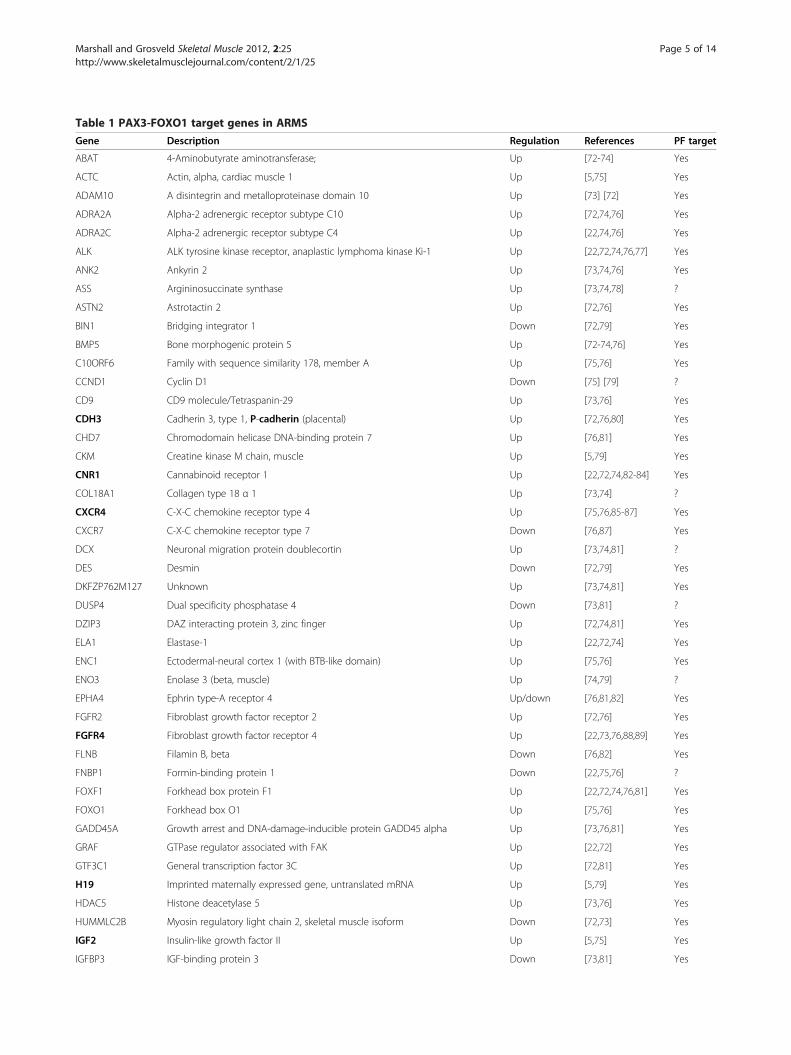
Table 1 PAX3-FOXO1 target genes in ARMS
Gene Description Regulation References PF target
ABAT 4-Aminobutyrate aminotransferase; Up [72-74] Yes
ACTC Actin, alpha, cardiac muscle 1 Up [5,75] Yes
ADAM10 A disintegrin and metalloproteinase domain 10 Up [73] [72] Yes
ADRA2A Alpha-2 adrenergic receptor subtype C10 Up [72,74,76] Yes
ADRA2C Alpha-2 adrenergic receptor subtype C4 Up [22,74,76] Yes
ALK ALK tyrosine kinase receptor, anaplastic lymphoma kinase Ki-1 Up [22,72,74,76,77] Yes
ANK2 Ankyrin 2 Up [73,74,76] Yes
ASS Argininosuccinate synthase Up [73,74,78] ?
ASTN2 Astrotactin 2 Up [72,76] Yes
BIN1 Bridging integrator 1 Down [72,79] Yes
BMP5 Bone morphogenic protein 5 Up [72-74,76] Yes
C10ORF6 Family with sequence similarity 178, member A Up [75,76] Yes
CCND1 Cyclin D1 Down [75] [79] ?
CD9 CD9 molecule/Tetraspanin-29 Up [73,76] Yes
CDH3 Cadherin 3, type 1, P-cadherin (placental) Up [72,76,80] Yes
CHD7 Chromodomain helicase DNA-binding protein 7 Up [76,81] Yes
CKM Creatine kinase M chain, muscle Up [5,79] Yes
CNR1 Cannabinoid receptor 1 Up [22,72,74,82-84] Yes
COL18A1 Collagen type 18 α 1 Up [73,74] ?
CXCR4 C-X-C chemokine receptor type 4 Up [75,76,85-87] Yes
CXCR7 C-X-C chemokine receptor type 7 Down [76,87] Yes
DCX Neuronal migration protein doublecortin Up [73,74,81] ?
DES Desmin Down [72,79] Yes
DKFZP762M127 Unknown Up [73,74,81] Yes
DUSP4 Dual specificity phosphatase 4 Down [73,81] ?
DZIP3 DAZ interacting protein 3, zinc finger Up [72,74,81] Yes
ELA1 Elastase-1 Up [22,72,74] Yes
ENC1 Ectodermal-neural cortex 1 (with BTB-like domain) Up [75,76] Yes
ENO3 Enolase 3 (beta, muscle) Up [74,79] ?
EPHA4 Ephrin type-A receptor 4 Up/down [76,81,82] Yes
FGFR2 Fibroblast growth factor receptor 2 Up [72,76] Yes
FGFR4 Fibroblast growth factor receptor 4 Up [22,73,76,88,89] Yes
FLNB Filamin B, beta Down [76,82] Yes
FNBP1 Formin-binding protein 1 Down [22,75,76] ?
FOXF1 Forkhead box protein F1 Up [22,72,74,76,81] Yes
FOXO1 Forkhead box O1 Up [75,76] Yes
GADD45A Growth arrest and DNA-damage-inducible protein GADD45 alpha Up [73,76,81] Yes
GRAF GTPase regulator associated with FAK Up [22,72] Yes
GTF3C1 General transcription factor 3C Up [72,81] Yes
H19 Imprinted maternally expressed gene, untranslated mRNA Up [5,79] Yes
HDAC5 Histone deacetylase 5 Up [73,76] Yes
HUMMLC2B Myosin regulatory light chain 2, skeletal muscle isoform Down [72,73] Yes
IGF2 Insulin-like growth factor II Up [5,75] Yes
IGFBP3 IGF-binding protein 3 Down [73,81] Yes
Marshall and Grosveld Skeletal Muscle 2012, 2:25 Page 5 of 14http://www.skeletalmusclejournal.com/content/2/1/25

Table 1 PAX3-FOXO1 target genes in ARMS (Continued)
IGFBP5 IGF-binding protein 5 Up [5] Yes
IL4R Interleukin 4 receptor Up [72,73,75,76] Yes
JAKMIP2 Janus kinase and microtubule-interacting protein 2 Up [72-74,76] Yes
KCNN3 Small conductance calcium-activated potassium channel protein 3 Up [73,74,81] Yes
KCNS3 Potassium voltage-gated channel subfamily S member 3 Up [73,74] ?
LRRFIP2 Leucine-rich repeat (in FLII) interacting protein 2 Up [72,74] Yes
MARCH3 Membrane-associated RING finger protein 3 Up [73,81] Yes
MCAM Melanoma cell adhesion molecule Up [73,81] Yes
MEG3 Maternally expressed 3 Up [73-75] ?
MN1 Meningioma (disrupted in balanced translocation) 1 Up [73,76] Yes
MET Hepatocyte growth factor receptor Up [73,76,81,90-92] Yes
MTUS2 Microtubule associated tumor suppressor candidate 2 Up [72,76] Yes
MYBPH Myosin-binding protein H Up/down [5,72,75] Yes
MYCN N-MYC proto-oncogene protein Up [72,73,76,78,81] Yes
MYH8 Myosin, heavy chain 8, skeletal muscle, perinatal Up/down [5,72] Yes
MYL1 Myosin, light chain 1, alkali; skeletal, fast Up/down [5,79] Yes
MYL4 Myosin, light chain 4, alkali; atrial, embryonic Up/down [5,72] Yes
MYOD Myoblast determination protein 1 Up [5,73,76,81] Yes
MYOG Myogenin (myogenic factor 4) Up [5,90] Yes
NEBL Nebulette Up/down [72,73,76,81] Yes
NELL1 NEL-like protein 1 Up [22,72-74,76] Yes
NHLH1 Nescient helix loop helix 1 Up [22,76] Yes
NPTX2 Neuronal pentraxin II Down [76,81] Yes
NRCAM Neuronal cell adhesion molecule Up [72-74] Yes
OLIG2 Oligodendrocyte transcription factor 2 Up [22,74] ?
PALMD Palmdelphin Down [72,76] Yes
PBK PDZ-binding kinase Up [73,76] Yes
PCDH7 Protocadherin 7 Up [76,82] Yes
PDZRN3 PDZ domain containing ring finger 3 Up [74,76] Yes
PGBD5 PiggyBac transposable element-derived protein 5 Up [22,72,74] Yes
PHF17 PHD finger protein 17 Up [74,76] Yes
PIPOX Pipecolic acid oxidase Up [22,72,74,76] Yes
PKP1 Plakophilin 1 (ectodermal dysplasia/skin fragility syndrome) Up [72,76] Yes
PLAG1 Pleiomorphic adenoma gene 1 protein Down [75,81] Yes
PLK2 Polo-like kinase 2 Down [73,76] Yes
PODXL Podocalyxin-like protein 1 Up [22,74,76] Yes
POU4F1 Brain-specific homeobox/POU domain protein 3A Up [72,73,76] Yes
PPARGC1A Peroxisome proliferator-activated receptor gamma, coactivator 1 alpha Up [22,76] Yes
PRKAR2B Protein kinase, cAMP-dependent, regulatory, type II, beta Up [73,81,82] Yes
PRKCA Protein kinase C, alpha Up [73,76] Yes
PSEN2 Presenilin 2 (Alzheimer’s disease 4) Up [73,74] ?
PTHLT Parathyroid hormone-like hormone Up [76,82] Yes
QDPR Quinoid dihydropteridine reductase Up [74,81] Yes
RASSF4 Ras association (RalGDS/AF-6) domain family 4 Up [72,74,76] Yes
RRP22 Ras-like protein family member 10A, on chm 22 Up [73,74] ?
Marshall and Grosveld Skeletal Muscle 2012, 2:25 Page 6 of 14http://www.skeletalmusclejournal.com/content/2/1/25

Table 1 PAX3-FOXO1 target genes in ARMS (Continued)
RYR1 Skeletal muscle-type ryanodine receptor Up [5,74] Yes
RYR3 Brain-type ryanodine receptor Up [74,76,81] Yes
SLC24A3 Solute carrier family 24 (sodium/potassium/calcium exchanger), member 3 Up [74,76] Yes
SIX1 SIX homeobox 1 Up [5,75] Yes
SOX14 SRY (sex determining region Y)-box 14 Up [22,76] Yes
STX11 Syntaxin 11 Up [76,82] Yes
SULF1 Sulfatase 1 Up [73,76] Yes
SVIL Supervillin Down [72,76] Yes
TCF712 Transcription factor 7-like 2 (T-cell specific, HMG-box) Up [73,81] Yes
TGFB1 Transforming growth factor, beta 1 Up [5,76] Yes
TFAP2B Transcription factor AP-2 beta Up [22,72] Yes
TIAF1 TGF-beta-1-induced antiapoptotic factor 1 Up [73,74] ?
TM4SF10 Transmembrane 4 superfamily member 10 Up [73,81] Yes
TNFAIP3 Tumor necrosis factor, alpha-induced protein 3 Up [73,76] Yes
TNNC2 Troponin C type 2 (fast) Up/down [5,72,73,79] Yes
TNNI2 Troponin I type 2 (skeletal, fast) Up/down [5,72] Yes
TNNT2 Troponin T type 2 (cardiac) Up [5,79] Yes
TNNT3 Troponin T type 3 (skeletal, fast) Down [72,79] Yes
TRAM2 Translocation-associated membrane protein 2 Up [73,76] Yes
TSC22D2 TSC22 domain family, member 2 Up [74,76] Yes
UBE2G2 Ubiquitin-conjugating enzyme E2G 2 (UBC7 homolog, yeast) Up [22,76] Yes
WSCD1 WSC domain-containing protein 1 Up [22,74] ?
WVA5A Von Willebrand factor A domain containing 5A Up [72-74] Yes
Summary of genes found to be differentially regulated in more than one reference in ARMS tumors and/or cell lines, or by PAX3-FOXO1 overexpression, in variouscell types. Gene name and description are indicated and also the relative expression in ARMS or PAX3-FOXO1. Also indicated is whether the study indicates thatthese genes are downstream of PAX3-FOXO1 expression. Genes indicated in bold will be further discussed.
Marshall and Grosveld Skeletal Muscle 2012, 2:25 Page 7 of 14http://www.skeletalmusclejournal.com/content/2/1/25
migratory function for MET in ARMS, knockdown ofMET by shRNA in ERMS and ARMS inhibits cell prolifera-tion and induces apoptosis. Moreover, shRNA-mediatedknockdown of MET inhibits anchorage-independentgrowth of ARMS and ERMS, and mutant MET-expressingMEFS prevent PAX3-FKHR transformation [91]. Consist-ent with METas a PAX3-FOXO1 target, high MET expres-sion in RMS correlates with ARMS histology, advanceddisease at diagnosis and bone marrow involvement [100].CXCR4 is normally expressed in satellite cells within
skeletal muscle and is used as a marker of mononucleatedcells capable of differentiating into myofibers [101].CXCR4 is a cell surface receptor; it binds and mediatesthe signaling of stromal-derived factor-1 (SDF-1) andinduces cell chemotaxis [102]. SDF-1 can induce migra-tion and chemotactic invasion in ARMS cell lines, but notERMS cell lines [85], and this migration can be inhibitedby SDF-1-neutralizing antibody or the CXCR4 inhibitorAMD3100 [103]. Moreover, expression of CXCR4 andMET in ARMS lines appears to synergize to induce themigration of cells toward bone marrow-derived fibroblastconditioned media in vitro. Inhibition of each receptor
reduces migration, and combined inhibition reveals syner-gism between these receptors [99]. SDF-1 can also induceproliferation of the ARMS cell line, RH30 [103]. Consist-ent with CXCR4 expression being downstream of PAX3-FOXO1 transgene expression, CXCR4 expression in RMScorrelates with the ARMS histology, unfavorable primarysite, advanced disease at diagnosis and bone marrow in-volvement [100].Other genes have been further confirmed as down-
stream genes of PAX3-FOXO1. Cannabinoid receptor 1(CNR1) is specifically upregulated at both the mRNAand protein level in fusion-positive ARMS cells [74].CNR1 is normally highly expressed in brain [104] but isalso expressed in skeletal muscle at levels detectable byRT-PCR [105]. CNR1 has been confirmed, using ChIPanalysis, as a direct target of both PAX3 and PAX3-FOXO1 transcriptional activity [82]. Furthermore, thehomeodomain of PAX3-FOXO1 appears to be the im-portant domain for the regulation of CNR1 expression[72]. CNR1 has been proposed to be a potential drugtarget in ARMS. Treatment with CNR1 agonists can in-duce apoptosis in some ARMS cell lines [83]. In addition,

Marshall and Grosveld Skeletal Muscle 2012, 2:25 Page 8 of 14http://www.skeletalmusclejournal.com/content/2/1/25
CNR1 expression has been linked with an increase propen-sity for PAX3-FOXO1 expressing mouse myoblast invasive-ness and lung metastasis formation. Moreover, treatmentwith an inverse agonist to CNR1 can abrogate in vitro inva-sion and in vivo lung metastasis formation [84]. Thus,CNR1 may represent a viable therapeutic target specific forthe increased metastatic capacity of PAX3-FOXO1 expres-sing ARMS.Transcription factor AP2β (TFAP2B) has been shown to
be a downstream target of PAX3-FOXO1 and appears torequire the paired domain of PAX3-FOXO1 to be induced.TFAP2B promoter expression is induced by PAX3, whichhas been shown to bind to the TFAP2B promoter by ChIPanalysis. siRNA targeting TFAP2B introduced into PAX3-FKHR-positive ARMS induces apoptosis, indicating thatTFAP2B can mediate cell survival in ARMS, downstreamof PAX3-FOXO1 [72].FGFR4 has been identified as a direct transcriptional tar-
get of PAX3 and PAX3-FOXO1, which bind to a down-stream enhancer region [76,106]. Accordingly, FGFR4 issignificantly upregulated by PAX3-FOXO1 expression[22,73,76,88,89,107]. However, upregulation of FGFR4downstream of PAX3-FOXO1 in primary myoblasts doesnot appear to act as an effector of PAX3-FOXO1-mediatedmyoblast transformation given that wild-type FGFR4 upre-gulation is not required for PAX3-FOXO1-induced prolif-eration, transformation, invasion or inhibition of myogenicdifferentiation [89]. However, knockdown of FGFR4 inRMS cell lines does show a reduction in cell proliferationand an increase in apoptosis, suggesting that at later stagesof ARMS tumorigenesis FGFR4 overexpression may inter-act with other unknown genetic lesions within these celllines to induce pro-survival and proliferation effects [107].It is interesting to note however that kinase domain-activating mutations in FGFR4 have been identified in 7.5%of RMS, including fusion-positive ARMS [108], and cancontribute to myoblast growth advantage and transform-ation [89]. Thus, FGFR4-activating mutations likely repre-sent cooperating mutations in RMS and upregulation ofFGFR4 in fusion-positive ARMS would enhance this effect.The CDH3/P-cadherin gene has been identified as a dir-
ect transcriptional target of PAX3/7-FOXO1 [72,76,80]. P-cadherin expression in the C2C12 myoblast cell line inhi-bits myogenic differentiation and maintains a proliferativestate through maintaining cyclin D1 expression. This inturn results in transformation of C2C12 cells, allowing col-ony formation in soft agar. Additionally, P-cadherin expres-sion resulted in enhanced cell motility, as well as cadherinswitching, a hallmark of epithelial to mesenchymal transi-tion and metastatic progression [80].
In vitro models of ARMSMany different cell lines have been derived from humanARMS tumors; these are regularly used to investigate
the biology of ARMS. These include ARMS cell linesderived from human tumors in the laboratory of Dr. PeterHoughton: RH3, RH4, RH10, RH28, RH30 and RH41, all ofwhich express the PAX3-FOXO1 fusion protein[13,109,110] and have been widely used in the field. Inaddition, the NCI-supported Pediatric Preclinical TestingProgram (PPTP) uses RH10, RH28, RH30, RH30R, RH41and RH65 subcutaneous xenograft tumors to test drug effi-cacy in a well-characterized preclinical model of manypediatric cancers [111-113].Other in vitro models that have been used involve the
introduction of the PAX3/7-FOXO1 fusion proteins intoboth myogenic and non-myogenic cell lines including fibro-blast cell lines, ERMS cell lines, MEFs, mesenchymal stemcells (MSC), normal or immortalized human or mousemyogenic cells and even an osteosarcoma cell line[5,81,82,91,114-119]. Given that the cell of origin for ARMShas yet to be identified, perhaps this variation in model celllines is prudent. However, it is likely that ARMS and ERMStumors are derived from a mesenchymal cell likely of thethe myogenic lineage because of skeletal muscle lineage-specific gene expression seen in these tumors [7].Conversely, some studies have used endogenous
PAX3-FOXO1 in ARMS cell lines to determine the tran-scriptional targets of this fusion protein within theARMS tumor cell context. Both Kikuchi et al. [90] andEbauer et al. [72] used siRNA specifically targetingPAX3-FOXO1 or both PAX3 and PAX3-FOXO1sequences, respectively. Inhibition of PAX3-FOXO1expression reduced cell proliferation and motility andallowed some myogenic differentiation [90]. In addition,comparative gene expression studies were performedidentifying over 100 PAX3-FOXO1 gene targets. Caoet al. [76] performed ChIP sequencing studies using aPAX3-FOXO1-specific antibody and were able to iden-tify 1,463 putative PAX3-FOXO1-binding sites in thehuman genome. Furthermore, PAX3-FOXO1-bindingsites adjacent to MyoD, FGFR4 and IGF1R were verifiedas transcriptionally regulated by PAX3-FOXO1.
In vivo models of ARMSMany different transgenic and knock-in animal modelshave been attempted to recapitulate ARMS tumor for-mation in vivo. Several of these models have attemptedto constitutively express PAX3/7-FOXO1 fusion proteinsin the skeletal muscle lineage during development, onlyto result in developmental defects and not tumor forma-tion [120-123]. Transgenic mice expressing PAX3-FOXO1 under the control of the PAX3 promoter andenhancer regions resulted in expression of PAX3-FOXO1 in the dorsal neural tube and lateral dermomyo-tome. PAX3-FOXO1 expression in this context appearedto interfere with normal PAX3 developmental functionsincluding neural tube and neural crest abnormalities
Marshall and Grosveld Skeletal Muscle 2012, 2:25 Page 9 of 14http://www.skeletalmusclejournal.com/content/2/1/25
similar to those seen in PAX3 mutant Splotch mice. Themajority of defects appeared to be in neural develop-ment, though defects were seen in hind limb skeletalmuscle; however, no tumors developed [121,122].Lagutina et al. [120] developed a model where PAX3-
FOXO1 was knocked into the PAX3 locus. This knock-inlocus expressed low amounts of PAX3-FOXO1, which inheterozygous pups was sufficient to result in developmentaldefects in the heart and diaphragm, leading to congestiveheart failure and perinatal death, as well as malformationsof some hypaxial muscles. However, neither chimeric adultsnor their newborn heterozygous pups developed malignan-cies. It was hypothesized that PAX3-FOXO1 expressionfrom the PAX3 control sequences was insufficient to causeARMS formation, and downstream regions of the FOXO1locus may be required to induce sufficient PAX3-FOXO1expression to induce tumor development.A PAX7-FOXO1 model of ARMS was also attempted
in Drosophila [123]. Expression of UAS-hPAX7-FOXO1,
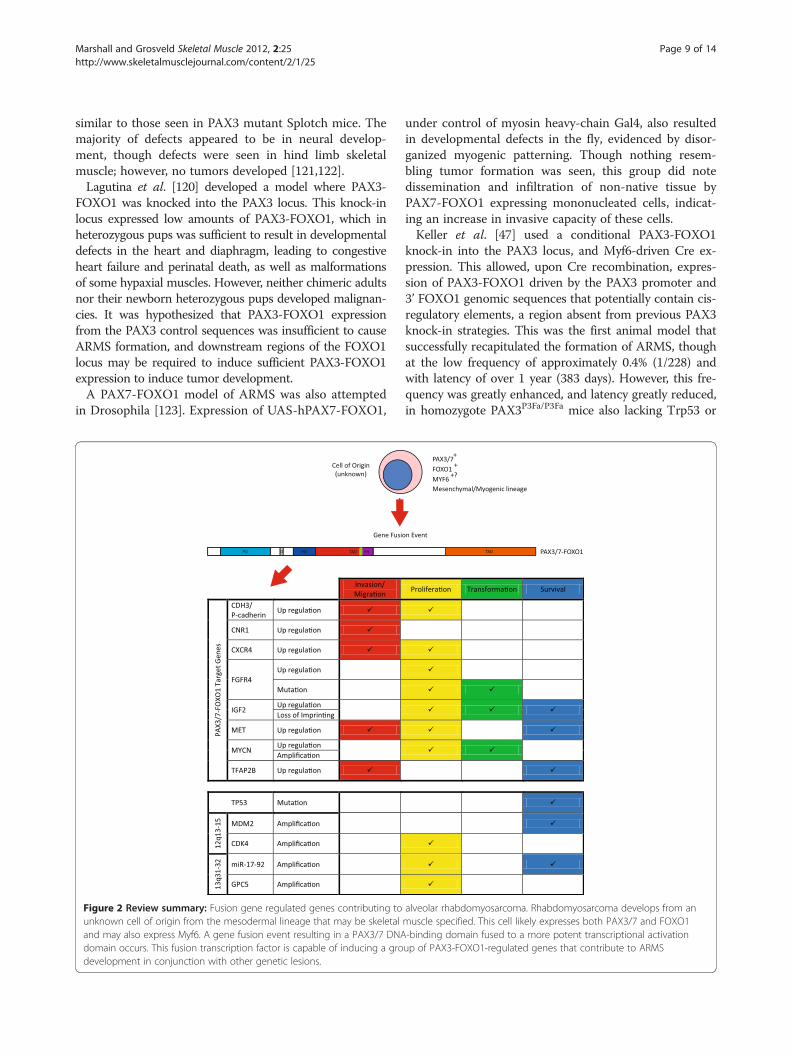
Figure 2 Review summary: Fusion gene regulated genes contributing tounknown cell of origin from the mesodermal lineage that may be skeletaland may also express Myf6. A gene fusion event resulting in a PAX3/7 DNAdomain occurs. This fusion transcription factor is capable of inducing a grodevelopment in conjunction with other genetic lesions.
under control of myosin heavy-chain Gal4, also resultedin developmental defects in the fly, evidenced by disor-ganized myogenic patterning. Though nothing resem-bling tumor formation was seen, this group did notedissemination and infiltration of non-native tissue byPAX7-FOXO1 expressing mononucleated cells, indicat-ing an increase in invasive capacity of these cells.Keller et al. [47] used a conditional PAX3-FOXO1
knock-in into the PAX3 locus, and Myf6-driven Cre ex-pression. This allowed, upon Cre recombination, expres-sion of PAX3-FOXO1 driven by the PAX3 promoter and3’ FOXO1 genomic sequences that potentially contain cis-regulatory elements, a region absent from previous PAX3knock-in strategies. This was the first animal model thatsuccessfully recapitulated the formation of ARMS, thoughat the low frequency of approximately 0.4% (1/228) andwith latency of over 1 year (383 days). However, this fre-quency was greatly enhanced, and latency greatly reduced,in homozygote PAX3P3Fa/P3Fa mice also lacking Trp53 or
alveolar rhabdomyosarcoma. Rhabdomyosarcoma develops from anmuscle specified. This cell likely expresses both PAX3/7 and FOXO1-binding domain fused to a more potent transcriptional activationup of PAX3-FOXO1-regulated genes that contribute to ARMS

Marshall and Grosveld Skeletal Muscle 2012, 2:25 Page 10 of 14http://www.skeletalmusclejournal.com/content/2/1/25
Ink4a/Arf. Subsequently, ARMS tumors have devel-oped in this conditional PAX3-FOXO1 knock-inmodel with a Pax7-CreER and M-Cre (Pax3 hypaxialmuscle enhancer) also lacking functional Trp53[124]. Moreover, histologically diagnosed fusion-negative ARMS tumors have been found to developin conditional Ptch1+/− Trp53−/− mice when Cre isexpressed from Pax7-CreER. The latency and incidence ofARMS tumor development in these different models haveyet to be compared.Clearly the problems that have arisen during the devel-
opment of an animal model for ARMS indicate that thetiming and the cell lineage targeted for PAX3-FOXO1expression are very important for the development ofARMS tumor formation and for avoiding developmentaldefects. In a review [125] following the publication ofthe animal model [47], Keller et al. discuss the possibil-ities for the cell of origin for ARMS; because Kelleret al. achieved the formation of ARMS tumors in theirmouse model using Myf6-Cre-driven conditional PAX3-FOXO1, and Myf6 is usually expressed in differentiatingskeletal muscle myotubes, they propose a potential de-differentiation mechanism for ARMS development.However, the formation of a fusion gene such as PAX3-FOXO1 suggests that the cell of origin for ARMS shouldexpress both PAX3 and FOXO1 at the time that thetranslocation occurs, given that open chromatin is likelyrequired for these two genomically distinct regions totranslocate. Anecdotal evidence for this includes that thegenome translocations that occur in many different can-cer types occur between genes that are expressed in thecell type of origin. A recent study by Osborne et al.[126] showed that the MYC and IGH genes, which areinvolved in a chromosomal translocation common inBurkitt lymphoma, are colocalized at the same transcrip-tion factories more often in activated B-cells, the origin-ating cell of Burkitt lymphoma, than resting B cells. Thiscolocalization at the same transcription factory allowsfor close proximity of these gene loci in euchromatin,providing the circumstances where these genes would bein close association, facilitating the specific translocationevent. PAX3 is rapidly downregulated upon myoblastdifferentiation, so it would be unlikely that the PAX3loci would be expressed in a nascent myotube expressingMyf6, making it difficult to understand how transloca-tion could occur in nascent myotubes and therefore castsome doubt on whether the dedifferentiation model isfeasible. However, it is possible that Myf6 expressiondoes rarely occur in a small subset of undifferentiatedmyogenic cells in conjunction with PAX3. This couldallow for this model to produce ARMS tumors and ac-count for the low frequency at which these tumors areseen as well as the requirement for homozygous PAX3-FOXO1 knock-in alleles [47].
From these animal models it is apparent that the tim-ing of PAX3-FOXO1 expression is critical for ARMSdevelopment. Too early and widespread expression ofPAX3-FOXO1 expression can result in developmentaldefects and no apparent tumor development [120-123],whereas later expression of PAX3-FOXO1, via a Myf6-driven Cre recombinase, does cause disease, though at alow frequency [47]. Perhaps inducible expression, drivenby various myogenic genes with carefully characterizedexpression profiling, would result in an increased fre-quency of disease and help to narrow down the exactstage in which PAX3-FOXO1 expression drives ARMStumorigenesis. Nevertheless, the cell of origin forARMS is yet to be identified, and animal models ofARMS will no doubt play an important role in itsidentification.
ConclusionTo date, numerous factors (outlined in Figure 2) havebeen identified that contribute to ARMS tumor develop-ment and its aggressive clinical phenotype. These consistof both PAX3/7-FOXO1 target genes, such as N-MYC,IGF2, MET, CXCR4, CNR1, TFAP2B, FGFR4 and P-cad-herin, and PAX3/7-FOXO1 cooperating factors, such asthe abrogation of the p53 pathway, IGF2 deregulation,N-MYC and miR17-92 amplification, and IRIZIOexpression. Future ARMS research will continue to dis-cover the mechanisms by which ARMS tumorigenesisoccurs. This will involve the identification of more PAX3-FOXO1 target and cooperating genes; more importantly,the mechanisms by which these genes contribute totumorigenesis will be elucidated. It is critical that wedevelop a mechanistic understanding of how these factorscontribute and interact to perpetrate ARMS tumo-rigenesis. This will allow new opportunities to developspecifically targeted therapies for this aggressive pediatricdisease.
AbbreviationsARMS: Alveolar rhabdomyosarcoma; bHLH: Basic helix loop helix domain;CDK: Cyclin-dependent kinase; CNR1: Cannabinoid receptor 1;ERMS: Embryonal rhabdomyosarcoma; FH: Forkhead DNA-binding domain;FKHR: Forkhead in rhabdomyosarcoma (now known as FOXO1);HD: Homeodomain DNA-binding domain; HGF/SF: Hepatocyte growthfactor/scatter factor; LOH: Loss of heterozygosity; LOI: Loss of imprinting;MEF: Mouse embryonic fibroblast; miR: Micro RNA; MSC: Mesenchymal stemcells; PD: Paired box DNA-binding domain; PPTP: Pediatric Preclinical TestingProgram; RMS: Rhabdomyosarcoma; SDF-1: Stromal-derived factor-1;SHH: Sonic hedgehog; siRNA: Short interfering RNA; TFAP2B: Transcriptionfactor AP2 b.
Competing interestsThe authors have no competing interests to declare.
Authors’ contributionsAM was responsible for the drafting of the manuscript. GG was responsiblefor critical revision of the content and approved the final version of themanuscript. All authors read and approved the final manuscript.

Marshall and Grosveld Skeletal Muscle 2012, 2:25 Page 11 of 14http://www.skeletalmusclejournal.com/content/2/1/25
Author details1Department of Genetics, St Jude Children’s Research Hospital, Memphis, TN38105, USA. 2Gene and Stem Cell Therapy Laboratory, Centenary Institute,University of Sydney, Missenden Road, Camperdown, NSW 2050, Australia.
Received: 13 July 2012 Accepted: 18 October 2012Published: 3 December 2012
References1. American Cancer Society: Rhabdomyosarcoma. http://www.cancer.org/
Cancer/Rhabdomyosarcoma/DetailedGuide/rhabdomyosarcoma.2. Punyko JA, Mertens AC, Baker KS, Ness KK, Robison LL, Gurney JG:
Long-term survival probabilities for childhood rhabdomyosarcoma. Apopulation-based evaluation. Cancer 2005, 103:1475–1483.
3. Tonin PN, Scrable H, Shimada H, Cavenee WK: Muscle-specific geneexpression in rhabdomyosarcomas and stages of human fetal skeletalmuscle development. Cancer Res 1991, 51:5100–5106.
4. Dias P, Chen B, Dilday B, Palmer H, Hosoi H, Singh S, Wu C, Li X, ThompsonJ, Parham D, et al: Strong immunostaining for myogenin inrhabdomyosarcoma is significantly associated with tumors of thealveolar subclass. Am J Pathol 2000, 156:399–408.
5. Khan J, Bittner ML, Saal LH, Teichmann U, Azorsa DO, Gooden GC, PavanWJ, Trent JM, Meltzer PS: cDNA microarrays detect activation of amyogenic transcription program by the PAX3-FKHR fusion oncogene.Proc Natl Acad Sci USA 1999, 96:13264–13269.
6. Heerema-McKenney A, Wijnaendts LC, Pulliam JF, Lopez-Terrada D,McKenney JK, Zhu S, Montgomery K, Mitchell J, Marinelli RJ, Hart AA, et al:Diffuse myogenin expression by immunohistochemistry is anindependent marker of poor survival in pediatric rhabdomyosarcoma: atissue microarray study of 71 primary tumors including correlation withmolecular phenotype. Am J Surg Pathol 2008, 32:1513–1522.
7. Wexler L, Meyer W, Helman L: Rhabdomyosarcoma and theundifferentiated sarcomas. In Principles and Practice of Pediatric Oncology.Fifthth editionth edition. Edited by Pizzo PA, Poplack D. Philidelphia:Lippincott Williams and Wilkins; 2006:971–1001.
8. Meza JL, Anderson J, Pappo AS, Meyer WH: Analysis of prognostic factorsin patients with nonmetastatic rhabdomyosarcoma treated onintergroup rhabdomyosarcoma studies III and IV: the Children'sOncology Group. J Clin Oncol 2006, 24:3844–3851.
9. Xia SJ, Pressey JG, Barr FG: Molecular pathogenesis ofrhabdomyosarcoma. Cancer Biol Ther 2002, 1:97–104.
10. Punyko JA, Mertens AC, Gurney JG, Yasui Y, Donaldson SS, Rodeberg DA,Raney RB, Stovall M, Sklar CA, Robison LL, Baker KS: Long-term medicaleffects of childhood and adolescent rhabdomyosarcoma: a reportfrom the childhood cancer survivor study. Pediatr Blood Cancer 2005,44:643–653.
11. Cerveira N, Torres L, Ribeiro FR, Henrique R, Pinto A, Bizarro S, Ferreira AM,Lopes C, Teixeira MR: Multimodal genetic diagnosis of solid variantalveolar rhabdomyosarcoma. Cancer Genet Cytogenet 2005, 163:138–143.
12. Takahashi Y, Oda Y, Kawaguchi K, Tamiya S, Yamamoto H, Suita S,Tsuneyoshi M: Altered expression and molecular abnormalities of cell-cycle-regulatory proteins in rhabdomyosarcoma. Mod Pathol 2004,17:660–669.
13. Douglass EC, Valentine M, Etcubanas E, Parham D, Webber BL, Houghton PJ,Houghton JA, Green AA: A specific chromosomal abnormality inrhabdomyosarcoma. Cytogenet Cell Genet 1987, 45:148–155.
14. Galili N, Davis RJ, Fredericks WJ, Mukhopadhyay S, Rauscher FJ 3rd, EmanuelBS, Rovera G, Barr FG: Fusion of a fork head domain gene to PAX3 in thesolid tumour alveolar rhabdomyosarcoma. Nat Genet 1993, 5:230–235.
15. Fredericks WJ, Galili N, Mukhopadhyay S, Rovera G, Bennicelli J, Barr FG,Rauscher FJ 3rd: The PAX3-FKHR fusion protein created by the t(2;13)translocation in alveolar rhabdomyosarcomas is a more potenttranscriptional activator than PAX3. Mol Cell Biol 1995, 15:1522–1535.
16. Lam PY, Sublett JE, Hollenbach AD, Roussel MF: The oncogenic potentialof the Pax3-FKHR fusion protein requires the Pax3 homeodomainrecognition helix but not the Pax3 paired-box DNA binding domain. MolCell Biol 1999, 19:594–601.
17. Zhang Y, Schwartz J, Wang C: Comparative analysis of paired- andhomeodomain-specific roles in PAX3-FKHR oncogenesis. Int J Clin ExpPathol 2009, 2:370–383.
18. Sorensen PH, Lynch JC, Qualman SJ, Tirabosco R, Lim JF, Maurer HM, BridgeJA, Crist WM, Triche TJ, Barr FG: PAX3-FKHR and PAX7-FKHR gene fusionsare prognostic indicators in alveolar rhabdomyosarcoma: a report fromthe children's oncology group. J Clin Oncol 2002, 20:2672–2679.
19. Davis RJ, D'Cruz CM, Lovell MA, Biegel JA, Barr FG: Fusion of PAX7 to FKHR bythe variant t(1;13)(p36;q14) translocation in alveolar rhabdomyosarcoma.Cancer Res 1994, 54:2869–2872.
20. Barr FG, Qualman SJ, Macris MH, Melnyk N, Lawlor ER, Strzelecki DM, TricheTJ, Bridge JA, Sorensen PH: Genetic heterogeneity in the alveolarrhabdomyosarcoma subset without typical gene fusions. Cancer Res 2002,62:4704–4710.
21. Sumegi J, Streblow R, Frayer RW, Dal Cin P, Rosenberg A, Meloni-Ehrig A,Bridge JA: Recurrent t(2;2) and t(2;8) translocations in rhabdomyosarcomawithout the canonical PAX-FOXO1 fuse PAX3 to members of the nuclearreceptor transcriptional coactivator family. Genes Chromosomes Cancer 2010,49:224–236.
22. Wachtel M, Dettling M, Koscielniak E, Stegmaier S, Treuner J, Simon-Klingenstein K, Buhlmann P, Niggli FK, Schafer BW: Gene expressionsignatures identify rhabdomyosarcoma subtypes and detect a novel t(2;2)(q35;p23) translocation fusing PAX3 to NCOA1. Cancer Res 2004,64:5539–5545.
23. Goulding MD, Chalepakis G, Deutsch U, Erselius JR, Gruss P: Pax-3, a novelmurine DNA binding protein expressed during early neurogenesis. EMBOJ 1991, 10:1135–1147.
24. Daston G, Lamar E, Olivier M, Goulding M: Pax-3 is necessary for migrationbut not differentiation of limb muscle precursors in the mouse.Development 1996, 122:1017–1027.
25. Seale P, Sabourin LA, Girgis-Gabardo A, Mansouri A, Gruss P, Rudnicki MA:Pax7 is required for the specification of myogenic satellite cells. Cell2000, 102:777–786.
26. Oustanina S, Hause G, Braun T: Pax7 directs postnatal renewal andpropagation of myogenic satellite cells but not their specification. EMBOJ 2004, 23:3430–3439.
27. Furuyama T, Nakazawa T, Nakano I, Mori N: Identification of the differentialdistribution patterns of mRNAs and consensus binding sequences formouse DAF-16 homologues. Biochem J 2000, 349:629–634.
28. Misiti S, Koibuchi N, Bei M, Farsetti A, Chin WW: Expression of steroidreceptor coactivator-1 mRNA in the developing mouse embryo: apossible role in olfactory epithelium development. Endocrinology 1999,140:1957–1960.
29. Voegel JJ, Heine MJ, Zechel C, Chambon P, Gronemeyer H: TIF2, a 160 kDatranscriptional mediator for the ligand-dependent activation functionAF-2 of nuclear receptors. EMBO J 1996, 15:3667–3675.
30. Burgering BM, Kops GJ: Cell cycle and death control: long live Forkheads.Trends Biochem Sci 2002, 27:352–360.
31. Xu J, Li Q: Review of the in vivo functions of the p160 steroid receptorcoactivator family. Mol Endocrinol 2003, 17:1681–1692.
32. Kempf BE, Vogt PK: A genetic analysis of PAX3-FKHR, the oncogene ofalveolar rhabdomyosarcoma. Cell Growth Differ 1999, 10:813–818.
33. Li H, Gomes PJ, Chen JD: RAC3, a steroid/nuclear receptor-associatedcoactivator that is related to SRC-1 and TIF2. Proc Natl Acad Sci USA 1997,94:8479–8484.
34. Belandia B, Parker MG: Functional interaction between the p160coactivator proteins and the transcriptional enhancer factor family oftranscription factors. J Biol Chem 2000, 275:30801–30805.
35. Epstein JA, Lam P, Jepeal L, Maas RL, Shapiro DN: Pax3 inhibits myogenicdifferentiation of cultured myoblast cells. J Biol Chem 1995, 270:11719–11722.
36. Miller PJ, Hollenbach AD: The oncogenic fusion protein Pax3-FKHR has agreater post-translational stability relative to Pax3 during earlymyogenesis. Biochim Biophys Acta 2007, 1770:1450–1458.
37. Olguin HC, Patzlaff NE, Olwin BB: Pax7-FKHR transcriptional activity isenhanced by transcriptionally repressed MyoD. J Cell Biochem 2011,112:1410–1417.
38. Calhabeu F, Hayashi S, Morgan JE, Relaix F, Zammit PS: Alveolarrhabdomyosarcoma-associated proteins PAX3/FOXO1A and PAX7/FOXO1A suppress the transcriptional activity of MyoD-target genes inmuscle stem cells. Oncogene 2012, Epub ahead of print.
39. Charytonowicz E, Matushansky I, Domenech JD, Castillo-Martin M, LadanyiM, Cordon-Cardo C, Ziman M: PAX7-FKHR fusion gene inhibits myogenicdifferentiation via NF-kappaB upregulation. Clin Transl Oncol 2012,14:197–206.

Marshall and Grosveld Skeletal Muscle 2012, 2:25 Page 12 of 14http://www.skeletalmusclejournal.com/content/2/1/25
40. Tapscott SJ, Thayer MJ, Weintraub H: Deficiency in rhabdomyosarcomas ofa factor required for MyoD activity and myogenesis. Science 1993,259:1450–1453.
41. Wexler LH, Ladanyi M: Diagnosing alveolar rhabdomyosarcoma: morphologymust be coupled with fusion confirmation. J Clin Oncol 2010, 28:2126–2128.
42. Anderson JR, Barr FG, Hawkins DS, Parham DM, Skapek SX, Triche TJ:Fusion-negative alveolar rhabdomyosarcoma: modification of riskstratification is premature. J Clin Oncol 2010, 28:e587–588. author replye589-590.
43. Davicioni E, Anderson MJ, Finckenstein FG, Lynch JC, Qualman SJ, ShimadaH, Schofield DE, Buckley JD, Meyer WH, Sorensen PH, Triche TJ: Molecularclassification of rhabdomyosarcoma–genotypic and phenotypicdeterminants of diagnosis: a report from the Children's Oncology Group.Am J Pathol 2009, 174:550–564.
44. Williamson D, Missiaglia E, de Reynies A, Pierron G, Thuille B, Palenzuela G,Thway K, Orbach D, Lae M, Freneaux P, et al: Fusion gene-negative alveolarrhabdomyosarcoma is clinically and molecularly indistinguishable fromembryonal rhabdomyosarcoma. J Clin Oncol 2010, 28:2151–2158.
45. Barr FG, Nauta LE, Davis RJ, Schafer BW, Nycum LM, Biegel JA: In vivoamplification of the PAX3-FKHR and PAX7-FKHR fusion genes in alveolarrhabdomyosarcoma. Hum Mol Genet 1996, 5:15–21.
46. Naini S, Etheridge KT, Adam SJ, Qualman SJ, Bentley RC, Counter CM,Linardic CM: Defining the cooperative genetic changes that temporallydrive alveolar rhabdomyosarcoma. Cancer Res 2008, 68:9583–9588.
47. Keller C, Arenkiel BR, Coffin CM, El-Bardeesy N, DePinho RA, Capecchi MR:Alveolar rhabdomyosarcomas in conditional Pax3:Fkhr mice: cooperativityof Ink4a/ARF and Trp53 loss of function. Genes Dev 2004, 18:2614–2626.
48. Sharp R, Recio JA, Jhappan C, Otsuka T, Liu S, Yu Y, Liu W, Anver M, Navid F,Helman LJ, et al: Synergism between INK4a/ARF inactivation and aberrantHGF/SF signaling in rhabdomyosarcomagenesis. Nat Med 2002, 8:1276–1280.
49. Weber-Hall S, Anderson J, McManus A, Abe S, Nojima T, Pinkerton R,Pritchard-Jones K, Shipley J: Gains, losses, and amplification of genomicmaterial in rhabdomyosarcoma analyzed by comparative genomichybridization. Cancer Res 1996, 56:3220–3224.
50. Ragazzini P, Gamberi G, Pazzaglia L, Serra M, Magagnoli G, Ponticelli F,Ferrari C, Ghinelli C, Alberghini M, Bertoni F, et al: Amplification of CDK4,MDM2, SAS and GLI genes in leiomyosarcoma, alveolar and embryonalrhabdomyosarcoma. Histol Histopathol 2004, 19:401–411.
51. Gordon AT, Brinkschmidt C, Anderson J, Coleman N, Dockhorn-DworniczakB, Pritchard-Jones K, Shipley J: A novel and consistent amplicon at 13q31associated with alveolar rhabdomyosarcoma. Genes Chromosomes Cancer2000, 28:220–226.
52. Bridge JA, Liu J, Qualman SJ, Suijkerbuijk R, Wenger G, Zhang J, Wan X,Baker KS, Sorensen P, Barr FG: Genomic gains and losses are similar ingenetic and histologic subsets of rhabdomyosarcoma, whereasamplification predominates in embryonal with anaplasia and alveolarsubtypes. Genes Chromosomes Cancer 2002, 33:310–321.
53. Forus A, Florenes VA, Maelandsmo GM, Meltzer PS, Fodstad O, Myklebost O:Mapping of amplification units in the q13-14 region of chromosome 12in human sarcomas: some amplica do not include MDM2. Cell GrowthDiffer 1993, 4:1065–1070.
54. Roberts WM, Douglass EC, Peiper SC, Houghton PJ, Look AT: Amplificationof the gli gene in childhood sarcomas. Cancer Res 1989, 49:5407–5413.
55. Momand J, Zambetti GP, Olson DC, George D, Levine AJ: The mdm-2oncogene product forms a complex with the p53 protein and inhibitsp53-mediated transactivation. Cell 1992, 69:1237–1245.
56. Taylor AC, Shu L, Danks MK, Poquette CA, Shetty S, Thayer MJ, Houghton PJ,Harris LC: P53 mutation and MDM2 amplification frequency in pediatricrhabdomyosarcoma tumors and cell lines. Med Pediatr Oncol 2000, 35:96–103.
57. Felix CA, Kappel CC, Mitsudomi T, Nau MM, Tsokos M, Crouch GD, Nisen PD,Winick NJ, Helman LJ: Frequency and diversity of p53 mutations inchildhood rhabdomyosarcoma. Cancer Res 1992, 52:2243–2247.
58. Leuschner I, Langhans I, Schmitz R, Harms D, Mattke A, Treuner J: p53 andmdm-2 expression in Rhabdomyosarcoma of childhood andadolescence: clinicopathologic study by the Kiel Pediatric TumorRegistry and the German Cooperative Soft Tissue Sarcoma Study. PediatrDev Pathol 2003, 6:128–136.
59. Hachitanda Y, Toyoshima S, Akazawa K, Tsuneyoshi M: N-myc geneamplification in rhabdomyosarcoma detected by fluorescence in situhybridization: its correlation with histologic features. Mod Pathol 1998,11:1222–1227.
60. Driman D, Thorner PS, Greenberg ML, Chilton-MacNeill S, Squire J: MYCNgene amplification in rhabdomyosarcoma. Cancer 1994, 73:2231–2237.
61. Williamson D, Lu YJ, Gordon T, Sciot R, Kelsey A, Fisher C, Poremba C,Anderson J, Pritchard-Jones K, Shipley J: Relationship between MYCN copynumber and expression in rhabdomyosarcomas and correlation withadverse prognosis in the alveolar subtype. J Clin Oncol 2005, 23:880–888.
62. Toffolatti L, Frascella E, Ninfo V, Gambini C, Forni M, Carli M, Rosolen A:MYCN expression in human rhabdomyosarcoma cell lines and tumoursamples. J Pathol 2002, 196:450–458.
63. Reichek JL, Duan F, Smith LM, Gustafson DM, O'Connor RS, Zhang C,Dunlevy MJ, Gastier-Foster JM, Barr FG: Genomic and clinical analysis ofamplification of the 13q31 chromosomal region in alveolarrhabdomyosarcoma: a report from the Children's Oncology Group. ClinCancer Res 2011, 17:1463–1473.
64. Williamson D, Selfe J, Gordon T, Lu YJ, Pritchard-Jones K, Murai K, Jones P,Workman P, Shipley J: Role for amplification and expression of glypican-5in rhabdomyosarcoma. Cancer Res 2007, 67:57–65.
65. Cho WC: OncomiRs: the discovery and progress of microRNAs in cancers.Mol Cancer 2007, 6:60.
66. Anderson J, Gordon A, McManus A, Shipley J, Pritchard-Jones K: Disruptionof imprinted genes at chromosome region 11p15.5 in paediatricrhabdomyosarcoma. Neoplasia 1999, 1:340–348.
67. Visser M, Sijmons C, Bras J, Arceci RJ, Godfried M, Valentijn LJ, Voute PA,Baas F: Allelotype of pediatric rhabdomyosarcoma. Oncogene 1997,15:1309–1314.
68. Bartolomei MS, Zemel S, Tilghman SM: Parental imprinting of the mouseH19 gene. Nature 1991, 351:153–155.
69. Hatada I, Inazawa J, Abe T, Nakayama M, Kaneko Y, Jinno Y, Niikawa N,Ohashi H, Fukushima Y, Iida K, et al: Genomic imprinting of humanp57KIP2 and its reduced expression in Wilms' tumors. Hum Mol Genet1996, 5:783–788.
70. Rachmilewitz J, Goshen R, Ariel I, Schneider T, de Groot N, Hochberg A:Parental imprinting of the human H19 gene. FEBS Lett 1992, 309:25–28.
71. Picchione F, Pritchard C, Lagutina I, Janke L, Grosveld GC: IRIZIO: a novelgene cooperating with PAX3-FOXO1 in alveolar rhabdomyosarcoma(ARMS). Carcinogenesis 2011, 32:452–461.
72. Ebauer M, Wachtel M, Niggli FK, Schafer BW: Comparative expression profilingidentifies an in vivo target gene signature with TFAP2B as a mediator of thesurvival function of PAX3/FKHR. Oncogene 2007, 26:7267–7281.
73. Davicioni E, Finckenstein FG, Shahbazian V, Buckley JD, Triche TJ, AndersonMJ: Identification of a PAX-FKHR gene expression signature that definesmolecular classes and determines the prognosis of alveolarrhabdomyosarcomas. Cancer Res 2006, 66:6936–6946.
74. Lae M, Ahn EH, Mercado GE, Chuai S, Edgar M, Pawel BR, Olshen A, Barr FG,Ladanyi M: Global gene expression profiling of PAX-FKHR fusion-positivealveolar and PAX-FKHR fusion-negative embryonal rhabdomyosarcomas.J Pathol 2007, 212:143–151.
75. Baer C, Nees M, Breit S, Selle B, Kulozik AE, Schaefer KL, Braun Y, Wai D,Poremba C: Profiling and functional annotation of mRNA geneexpression in pediatric rhabdomyosarcoma and Ewing's sarcoma. Int JCancer 2004, 110:687–694.
76. Cao L, Yu Y, Bilke S, Walker RL, Mayeenuddin LH, Azorsa DO, Yang F, PinedaM, Helman LJ, Meltzer PS: Genome-wide identification of PAX3-FKHRbinding sites in rhabdomyosarcoma reveals candidate target genesimportant for development and cancer. Cancer Res 2010, 70:6497–6508.
77. Corao DA, Biegel JA, Coffin CM, Barr FG, Wainwright LM, Ernst LM, Choi JK,Zhang PJ, Pawel BR: ALK expression in rhabdomyosarcomas: correlationwith histologic subtype and fusion status. Pediatr Dev Pathol 2009,12:275–283.
78. Khan J, Simon R, Bittner M, Chen Y, Leighton SB, Pohida T, Smith PD, Jiang Y,Gooden GC, Trent JM, Meltzer PS: Gene expression profiling of alveolarrhabdomyosarcoma with cDNA microarrays. Cancer Res 1998, 58:5009–5013.
79. De Pitta C, Tombolan L, Albiero G, Sartori F, Romualdi C, Jurman G, Carli M,Furlanello C, Lanfranchi G, Rosolen A: Gene expression profiling identifiespotential relevant genes in alveolar rhabdomyosarcoma pathogenesisand discriminates PAX3-FKHR positive and negative tumors. Int J Cancer2006, 118:2772–2781.
80. Thuault S, Hayashi S, Lagirand-Cantaloube J, Plutoni C, Comunale F, DelattreO, Relaix F, Gauthier-Rouviere C: P-cadherin is a direct PAX3-FOXO1Atarget involved in alveolar rhabdomyosarcoma aggressiveness. Oncogene2012, Epub ahead of print.

Marshall and Grosveld Skeletal Muscle 2012, 2:25 Page 13 of 14http://www.skeletalmusclejournal.com/content/2/1/25
81. Mercado GE, Xia SJ, Zhang C, Ahn EH, Gustafson DM, Lae M, Ladanyi M, BarrFG: Identification of PAX3-FKHR-regulated genes differentially expressedbetween alveolar and embryonal rhabdomyosarcoma: focus on MYCN as abiologically relevant target. Genes Chromosomes Cancer 2008, 47:510–520.
82. Begum S, Emami N, Cheung A, Wilkins O, Der S, Hamel PA: Cell-type-specific regulation of distinct sets of gene targets by Pax3 and Pax3/FKHR. Oncogene 2005, 24:1860–1872.
83. Oesch S, Walter D, Wachtel M, Pretre K, Salazar M, Guzman M, Velasco G,Schafer BW: Cannabinoid receptor 1 is a potential drug target fortreatment of translocation-positive rhabdomyosarcoma. Mol Cancer Ther2009, 8:1838–1845.
84. Marshall AD, Lagutina I, Grosveld GC: PAX3-FOXO1 induces cannabinoid receptor1 to enhance cell invasion and metastasis. Cancer Res 2011, 71(24):7471–7480.
85. Libura J, Drukala J, Majka M, Tomescu O, Navenot JM, Kucia M, Marquez L,Peiper SC, Barr FG, Janowska-Wieczorek A, Ratajczak MZ: CXCR4-SDF-1signaling is active in rhabdomyosarcoma cells and regulates locomotion,chemotaxis, and adhesion. Blood 2002, 100:2597–2606.
86. Tomescu O, Xia SJ, Strezlecki D, Bennicelli JL, Ginsberg J, Pawel B, Barr FG:Inducible short-term and stable long-term cell culture systems revealthat the PAX3-FKHR fusion oncoprotein regulates CXCR4, PAX3, andPAX7 expression. Lab Invest 2004, 84:1060–1070.
87. Tarnowski M, Grymula K, Reca R, Jankowski K, Maksym R, Tarnowska J,Przybylski G, Barr FG, Kucia M, Ratajczak MZ: Regulation of expression ofstromal-derived factor-1 receptors: CXCR4 and CXCR7 in humanrhabdomyosarcomas. Mol Cancer Res 2010, 8:1–14.
88. Khan J, Wei JS, Ringner M, Saal LH, Ladanyi M, Westermann F, Berthold F,Schwab M, Antonescu CR, Peterson C, Meltzer PS: Classification anddiagnostic prediction of cancers using gene expression profiling andartificial neural networks. Nat Med 2001, 7:673–679.
89. Marshall AD, van der Ent MA, Grosveld GC: PAX3-FOXO1 and FGFR4 inalveolar rhabdomyosarcoma. Mol Carcinog 2012, 51(10):807–815.
90. Kikuchi K, Tsuchiya K, Otabe O, Gotoh T, Tamura S, Katsumi Y, Yagyu S,Tsubai-Shimizu S, Miyachi M, Iehara T, Hosoi H: Effects of PAX3-FKHR onmalignant phenotypes in alveolar rhabdomyosarcoma. Biochem BiophysRes Commun 2008, 365:568–574.
91. Taulli R, Scuoppo C, Bersani F, Accornero P, Forni PE, Miretti S, Grinza A,Allegra P, Schmitt-Ney M, Crepaldi T, Ponzetto C: Validation of met as atherapeutic target in alveolar and embryonal rhabdomyosarcoma. CancerRes 2006, 66:4742–4749.
92. Ginsberg JP, Davis RJ, Bennicelli JL, Nauta LE, Barr FG: Up-regulation of METbut not neural cell adhesion molecule expression by the PAX3-FKHR fusionprotein in alveolar rhabdomyosarcoma. Cancer Res 1998, 58:3542–3546.
93. Tonelli R, McIntyre A, Camerin C, Walters ZS, Di Leo K, Selfe J, Purgato S,Missiaglia E, Tortori A, Astolfi A, et al: Antitumor activity of sustainedN-Myc reduction in Rhabdomyosarcomas and transcriptional block byantigene therapy. Clin Cancer Res 2011, .
94. Bottaro DP, Rubin JS, Faletto DL, Chan AM, Kmiecik TE, Vande-Woude GF,Aaronson SA: Identification of the hepatocyte growth factor receptor asthe c-met proto-oncogene product. Science 1991, 251:802–804.
95. Brand-Saberi B, Muller TS, Wilting J, Christ B, Birchmeier C: Scatter factor/hepatocyte growth factor (SF/HGF) induces emigration of myogenic cellsat interlimb level in vivo. Dev Biol 1996, 179:303–308.
96. Bladt F, Riethmacher D, Isenmann S, Aguzzi A, Birchmeier C: Essential rolefor the c-met receptor in the migration of myogenic precursor cells intothe limb bud. Nature 1995, 376:768–771.
97. Epstein JA, Shapiro DN, Cheng J, Lam PY, Maas RL: Pax3 modulatesexpression of the c-Met receptor during limb muscle development. ProcNatl Acad Sci USA 1996, 93:4213–4218.
98. Lukasiewicz E, Miekus K, Kijowski J, Drabik G, Wilusz M, Bobis-Wozowicz S,Majka M: Inhibition of rhabdomyosarcoma's metastatic behavior throughdownregulation of MET receptor signaling. Folia Histochem Cytobiol 2009,47:485–489.
99. Jankowski K, Kucia M, Wysoczynski M, Reca R, Zhao D, Trzyna E, Trent J,Peiper S, Zembala M, Ratajczak J, et al: Both hepatocyte growth factor(HGF) and stromal-derived factor-1 regulate the metastatic behavior ofhuman rhabdomyosarcoma cells, but only HGF enhances theirresistance to radiochemotherapy. Cancer Res 2003, 63:7926–7935.
100. Diomedi-Camassei F, McDowell HP, De Ioris MA, Uccini S, Altavista P,Raschella G, Vitali R, Mannarino O, De Sio L, Cozzi DA, et al: Clinicalsignificance of CXC chemokine receptor-4 and c-Met in childhoodrhabdomyosarcoma. Clin Cancer Res 2008, 14:4119–4127.
101. Sherwood RI, Christensen JL, Conboy IM, Conboy MJ, Rando TA, WeissmanIL, Wagers AJ: Isolation of adult mouse myogenic progenitors: functionalheterogeneity of cells within and engrafting skeletal muscle. Cell 2004,119:543–554.
102. D'Apuzzo M, Rolink A, Loetscher M, Hoxie JA, Clark-Lewis I, Melchers F,Baggiolini M, Moser B: The chemokine SDF-1, stromal cell-derived factor1, attracts early stage B cell precursors via the chemokine receptorCXCR4. Eur J Immunol 1997, 27:1788–1793.
103. Strahm B, Durbin AD, Sexsmith E, Malkin D: The CXCR4-SDF1alpha axis is acritical mediator of rhabdomyosarcoma metastatic signaling induced bybone marrow stroma. Clin Exp Metastasis 2008, 25:1–10.
104. Marsicano G, Lutz B: Expression of the cannabinoid receptor CB1 indistinct neuronal subpopulations in the adult mouse forebrain. Eur JNeurosci 1999, 11:4213–4225.
105. Cavuoto P, McAinch AJ, Hatzinikolas G, Janovska A, Game P, Wittert GA: Theexpression of receptors for endocannabinoids in human and rodentskeletal muscle. Biochem Biophys Res Commun 2007, 364:105–110.
106. Lagha M, Sato T, Bajard L, Daubas P, Esner M, Montarras D, Relaix F,Buckingham M: Regulation of skeletal muscle stem cell behavior by Pax3and Pax7. Cold Spring Harb Symp Quant Biol 2008, 73:307–315.
107. Crose LE, Etheridge KT, Chen C, Belyea B, Talbot LJ, Bentley RC, Linardic CM:FGFR4 blockade exerts distinct antitumorigenic effects in human embryonalversus alveolar rhabdomyosarcoma. Clin Cancer Res 2012, 18(14):3780–3790.
108. Taylor JG, Cheuk AT, Tsang PS, Chung JY, Song YK, Desai K, Yu Y, Chen QR,Shah K, Youngblood V, et al: Identification of FGFR4-activating mutationsin human rhabdomyosarcomas that promote metastasis inxenotransplanted models. J Clin Invest 2009, 119:3395–3407.
109. Hazelton BJ, Houghton JA, Parham DM, Douglass EC, Torrance PM, Holt H,Houghton PJ: Characterization of cell lines derived from xenografts ofchildhood rhabdomyosarcoma. Cancer Res 1987, 47:4501–4507.
110. Croci S, Landuzzi L, Astolfi A, Nicoletti G, Rosolen A, Sartori F, Follo MY,Oliver N, De Giovanni C, Nanni P, Lollini PL: Inhibition of connective tissuegrowth factor (CTGF/CCN2) expression decreases the survival andmyogenic differentiation of human rhabdomyosarcoma cells. Cancer Res2004, 64:1730–1736.
111. Houghton PJ, Morton CL, Tucker C, Payne D, Favours E, Cole C, Gorlick R,Kolb EA, Zhang W, Lock R, et al: The pediatric preclinical testing program:description of models and early testing results. Pediatr Blood Cancer 2007,49:928–940.
112. Kang MH, Smith MA, Morton CL, Keshelava N, Houghton PJ, Reynolds CP: NationalCancer Institute pediatric preclinical testing program: model description forin vitro cytotoxicity testing. Pediatr Blood Cancer 2011, 56:239–249.
113. Neale G, Su X, Morton CL, Phelps D, Gorlick R, Lock RB, Reynolds CP, MarisJM, Friedman HS, Dome J, et al: Molecular characterization of thepediatric preclinical testing panel. Clin Cancer Res 2008, 14:4572–4583.
114. Xia SJ, Holder DD, Pawel BR, Zhang C, Barr FG: High expression of thePAX3-FKHR oncoprotein is required to promote tumorigenesis of humanmyoblasts. Am J Pathol 2009, 175:2600–2608.
115. Scuoppo C, Riess I, Schmitt-Ney M, Allegra P, Forni PE, Bersani F, Taulli R,Accornero P, Crepaldi T, Ponzetto C: The oncogenic transcription factorPAX3-FKHR can convert fibroblasts into contractile myotubes. Exp Cell Res2007, 313:2308–2317.
116. Xia SJ, Barr FG: Analysis of the transforming and growth suppressiveactivities of the PAX3-FKHR oncoprotein. Oncogene 2004, 23:6864–6871.
117. Ren YX, Finckenstein FG, Abdueva DA, Shahbazian V, Chung B, Weinberg KI,Triche TJ, Shimada H, Anderson MJ: Mouse mesenchymal stem cellsexpressing PAX-FKHR form alveolar rhabdomyosarcomas by cooperatingwith secondary mutations. Cancer Res 2008, 68:6587–6597.
118. Roeb W, Boyer A, Cavenee WK, Arden KC: PAX3-FOXO1 controlsexpression of the p57Kip2 cell-cycle regulator through degradation ofEGR1. Proc Natl Acad Sci USA 2007, 104:18085–18090.
119. Pressey JG, Pressey CS, Robinson G, Herring R, Wilson L, Kelly DR, Kim H:2D-difference gel electrophoretic proteomic analysis of a cell culturemodel of alveolar rhabdomyosarcoma. J Proteome Res 2011, 10:624–636.
120. Lagutina I, Conway SJ, Sublett J, Grosveld GC: Pax3-FKHR knock-in miceshow developmental aberrations but do not develop tumors. Mol CellBiol 2002, 22:7204–7216.
121. Finckenstein FG, Davicioni E, Osborn KG, Cavenee WK, Arden KC, AndersonMJ: Transgenic mice expressing PAX3-FKHR have multiple defects inmuscle development, including ectopic skeletal myogenesis in thedeveloping neural tube. Transgenic Res 2006, 15:595–614.

Marshall and Grosveld Skeletal Muscle 2012, 2:25 Page 14 of 14http://www.skeletalmusclejournal.com/content/2/1/25
122. Anderson MJ, Shelton GD, Cavenee WK, Arden KC: Embryonic expression ofthe tumor-associated PAX3-FKHR fusion protein interferes with thedevelopmental functions of Pax3. Proc Natl Acad Sci USA 2001, 98:1589–1594.
123. Galindo RL, Allport JA, Olson EN: A Drosophila model of therhabdomyosarcoma initiator PAX7-FKHR. Proc Natl Acad Sci USA 2006,103:13439–13444.
124. Nishijo K, Hosoyama T, Bjornson CR, Schaffer BS, Prajapati SI, Bahadur AN,Hansen MS, Blandford MC, McCleish AT, Rubin BP, et al: Biomarker systemfor studying muscle, stem cells, and cancer in vivo. FASEB J 2009,23:2681–2690.
125. Keller C, Capecchi MR: New genetic tactics to model alveolarrhabdomyosarcoma in the mouse. Cancer Res 2005, 65:7530–7532.
126. Osborne CS, Chakalova L, Mitchell JA, Horton A, Wood AL, Bolland DJ,Corcoran AE, Fraser P: Myc dynamically and preferentially relocates to atranscription factory occupied by Igh. PLoS Biol 2007, 5:e192.
doi:10.1186/2044-5040-2-25Cite this article as: Marshall and Grosveld: Alveolar rhabdomyosarcoma –The molecular drivers of PAX3/7-FOXO1-induced tumorigenesis. SkeletalMuscle 2012 2:25.
Submit your next manuscript to BioMed Centraland take full advantage of:
• Convenient online submission
• Thorough peer review
• No space constraints or color figure charges
• Immediate publication on acceptance
• Inclusion in PubMed, CAS, Scopus and Google Scholar
• Research which is freely available for redistribution
Submit your manuscript at www.biomedcentral.com/submit