alveolar bone healing in rats: micro- ct ...alveolar bone healing in rats: micro-ct,...
TRANSCRIPT

J Appl Oral Sci.
Abstract
Submitted: July 21, 2017Modification: December 5, 2017
Accepted: December 11, 2017
Alveolar bone healing in rats: micro-CT, immunohistochemical and molecular analysis
Alveolar bone healing after upper incisor extraction in rats is a classical model of preclinical studies. The underlying morphometric, cellular and molecular mechanism, however, remains imprecise in a unique study. Objectives: The aim of this study was therefore to characterize the alveolar bone healing after upper incisor extraction in rats by micro computed tomographic (Micro-CT), immunohistochemical and real-time polymerase chain reaction (RT-PCR) analysis. Material and Methods: Thirty animals (Rattus norvegicus, Albinus Wistar) were divided into three groups after upper incisors extraction at 7, 14, and 28 days. Micro-CT was evaluated based on the morphometric parameters. Subsequently, the histological analyses and immunostaining of osteoprotegerin (OPG), receptor activator of nuclear kappa B ligand (RANKL) and tartrate resistant acid phosphate (TRAP) was performed. In addition, RT-PCR analyses of OPG, RANKL, the runt-related transcription factor 2 (RUNX2), osteocalcin (OC), osteopontin (OPN), osterix (OST) and receptor activator of nuclear kappa B (RANK) were performed to determine the expression of these proteins in the alveolar bone healing. Results: Micro-CT: The morphometric parameters of bone volume and trabecular thickness progressively increased over time. Consequently, a gradual decrease in trabecular separation, trabecular space and total bone porosity was observed. Immunohistochemical: There were no differences statistically significant between the positive labeling for OPG, RANKL and TRAP in the different periods. RT-PCR: At 28 days, there was a significant increase in OPG expression, while RANKL expression and the RANKL/OPG ratio both decreased over time. Conclusion: Micro-CT showed the newly formed bone had favorable morphometric characteristics of quality and quantity. Beyond the RUNX2, OC, OPN, OST, and RANK proteins expressed in the alveolar bone healing, OPG and RANKL activity showed to be essential for activation of basic multicellular units during the alveolar bone healing.
Keywords: Rats. Gene expression. Osteoprotegerin. Bones and bone. Bone remodeling.
Jaqueline Suemi HASSUMI1
Gabriel MULINARI-SANTOS2
André Luis da Silva FABRIS2
Ricardo Garcia Mureb JACOB2
Alaíde GONÇALVES1
Ana Cláudia ROSSI3
Alexandre Rodrigues FREIRE3
Leonardo Pérez FAVERANI2
Roberta OKAMOTO1
Original Articlehttp://dx.doi.org/10.1590/1678-7757-2017-0326
1Univ. Estadual Paulista, Faculdade de Odontologia de Araçatuba, Departamento de Cirurgia e Clínica Integrada, Araçatuba, São Paulo, Brasil.2Univ. Estadual Paulista, Faculdade de Odontologia de Araçatuba, Departamento de Ciências Básicas, Araçatuba, São Paulo, Brasil.3Universidade Estadual de Campinas, Faculdade de Odontologia de Piracicaba, Departamento de Morfologia, Piracicaba, São Paulo, Brasil.
Corresponding address:Gabriel Mulinari dos Santos
Departamento de Cirurgia e Clínica Integrada - Faculdade de Odontologia de Araçatuba -
Univ. Estadual Paulista Júlio de Mesquita FilhoRua José Bonifácio, 1193 - Vila Mendonça -
16015-050 - Araçatuba - SP - Brasil.Phone: +55 18 3636-3270/3636-3237e-mail: [email protected]
2018;26:e201703261/12

J Appl Oral Sci. 2018;26:e201703262/12
Introduction
Given the search for a favorable bone and an ideal
support for dental implant placement, understanding
the alveolar bone healing in preclinical studies
is crucial. Bone is a dynamic tissue, where bone
cells drive the molecular and cellular mechanisms
involved in the bone healing1,12. These cells act
together signaling molecules to maintain the bone
turnover28. Also, the mechanisms of development
and maintenance of bone occurs constantly, since
local factors such as mechanical stimulation and
systemic factors can interfere in this process29. In ideal
conditions, bone turnover is balanced by formation and
resorption, allowing the maintenance of bone mass and
ensuring calcium and phosphate levels27. Therefore,
the characterization of the dynamic process of bone to
replace an extracted tooth is a topic of special interest
in Dentistry4,5.
Alveolar bone healing after tooth extraction has
been analyzed in many experimental and clinical
conditions. A classical model used to study bone healing
after upper incisor extraction in rats is to describe
the alveolar healing in three different phases22,23:
first, coagulum formation and cells proliferation from
connective tissue; second, connective tissue formation
and healing; finally, ossification phase completed with
28 days, and corresponding to 64 days in humans22,23.
Okamoto and de Russo22 (1973) defined the dynamic
of these events showing an important proteins activity
during the 14th day after tooth extraction. Another
similar histological study revealed that this process is
complete at 28th day with the alveolar socket almost
totally filled with bone23. In addition, described that
the alveolar socket has the crest remodelation and
the gingival epithelium regeneration in the end of
this process 23.
The advent of Molecular Biology raises questions
regarding the molecular aspects of bone healing
beyond the histological studies, in particular about
identifying genes responsible for proteins synthesis
involved in the mechanisms of bone healing after
tooth extraction. Moreover, the gene expression can
be related to the immunohistochemical staining in
different areas of the alveolar bone7,8, which also
results in the morphometric parameters evaluated
by micro-CT during the alveolar bone healing30. In
addition, physiologic conditions such as osteoporosis,
uncontrolled diabetes and hypertension have been
associated with impaired bone metabolism6,20. All
information analyzed in these compromised bone can
be compared with the normal condition, in order to
provide the morphometric and cellular alterations that
influence a favorable bone healing12.
Additional to the proteins involved in the bone
healing, such as RUNX2, OST, OC, and OPN, the OPG
and RANKL are members of the tumor necrosis factor
family that are signaled during the cellular responses
of bone remodeling2. The ratio of OPG to RANKL
expression provides an indication whether tissue
response tends to bone formation with a predominance
of OPG or bone resorption with increase of RANKL2,16.
Thus, a balanced bone remodeling occurs when levels
of OPG and RANKL are similar14,24. Describing how the
genes responsible for protein production are expressed
during the different steps of the bone healing is
important, specifically if, at 14th day after tooth
extraction, these genes are overexpressed during
the alveolar bone healing as previous suggested22,23.
Therefore, the aim of this study was to evaluate
the morphometric aspects using micro-CT, performing
a volumetric assessment of the newly formed bone
during the alveolar bone healing in rats, besides the
immunostaining of OPG, RANKL, and TRAP, and the
messenger RNA (mRNA) expression of OPG, RANKL,
RUNX2, OC, OPN, OST and RANK.
Material and methods
Study design and ethicsThis research was approved by the Ethics
Committee for Animal Use from Faculdade de
Odontologia de Araçatuba, UNESP – Univ. Estadual
Paulista, Brazil (number process 00123-2013).
We used 30 adult (6 months old) male rats (Rattus
norvegicus, Albinus Wistar) with an average body
weight of 275 g ±25 g. The animals were divided into
three groups according to their time of upper right
incisor extraction:
Group I - Analyzed 7 days after tooth extraction.
Group II - Analyzed 14 days after tooth extraction.
Group III - Analyzed 28 days after tooth extraction.
In each group with 10 animals, being 5 for micro-
CT and immunohistochemical analysis and others 5
for RT-PCR analysis.
The sample number was elected by power test
analysis in the website http://www.lee.dante.br. Level
Alveolar bone healing in rats: micro-CT, immunohistochemical and molecular analysis

J Appl Oral Sci. 2018;26:e201703263/12
of significance of 5% and power test of 95% were
adopted, and it was suggested four animals per group.
Thus, with a possible animal loss, it was used five per
period of analysis.
The animals were kept in cages in an environment
at a stable temperature (22±2°C) and a controlled
light cycle (12 h light and 12 h dark). The animals were
fed a ground solid diet and powder diet 14 days after
surgery (Anderson & Clayton SA - Abbot Laboratórios
do Brasil Ltda., São Paulo, SP, Brazil) with water ad
libitum, except during the 12 h prior to surgery.
Tooth extractionSurgery was performed under sedation via an
intramuscular injection of xylazine hydrochloride
(0.03 ml per 100 g body weight; Coopers Brasil Ltda,
Cotia, SP, Brazil), to promote muscle relaxation, and
ketamine hydrochloride (0.07 ml per 100 g body
weight; Fort Dodge Animal Health, IA, USA), to induce
anesthesia. The anterior portion of the right maxilla
was disinfected with iodized polyvinylpyrrolidone
(PVP Topic 10%, Riodeine – Indústria Farmacêutica
Rioquímica Ltda., São José do Rio Preto, SP, Brazil).
Animals had their gingival mucosa detached using
specific retractors dislocation; subsequently the upper
right incisor was extracted using specially adapted
forceps. The gingival mucosa was sutured with 4-0
polyglactin 910 suture thread (Johnson & Johnson,
São José dos Campos, SP, Brazil). The tooth extraction
was performed according to previous studies16,22,23.
Micro-CT analysisAt 7, 14, or 28 days after tooth extraction,
15 animals (n=5 per group) were euthanized by
anesthesia overdose (pentobarbital sodium, 100 mg/
kg). The right maxilla was removed and fixed in 4%
paraformaldehyde solution and 10% 0.1 M phosphate
buffer (pH 7.4), after 48 hours in fixation solution.
The middle third of the alveolar sockets were
scanned in the direction from apical to cervical in the
longitudinal plane using SkyScan Model 1172 (Bruker,
Kontich, Belgium) microtomography. The tube current
was 165 uA and peak voltage was 60 kV. Image Pixel
Size was 9.92 um. The filter to correct beam hardening
was Al 0.5 mm, and the frame averaging was 4 and
rotation step was 0.6 deg.
After scanning, the images were imported into
NRecon Reconstruction software (SkyScan, Leuven,
Belgium) for reconstruction in grayscale, presenting
x-ray attenuation coefficients with values related to
bone structure. Attenuation coefficients were obtained
using calibration values for the aqueous medium
(formaldehyde solution 10% and 0.1 M phosphate
buffer, pH 7.4).
After the bone three-dimensional reconstruction,
the morphometric parameters were measured using
CT-Analyzer software (SkyScan, Leuven, Belgium):
bone volume (BV; mm3), percentage of bone volume
(BV/TV; %) in relation to the total measured area,
trabecular thickness (Tb.Th; mm), trabecular
separation (Tb.Sp; mm), and percentage of total bone
porosity (Po-tot; %). The morphometric parameters
were three-dimensionally calculated.
The segmentation was standardized in the CT-
Analyzer software (SkyScan, Leuven, Belgium) after
the micro-CT analysis. The values used in segmentation
were selected to eliminate artifacts and to keep the
bone structure. The greyscale threshold ranged from
70 to 255 for all pieces, in an interval from 0 to 255.
Due to the irregular alveolar socket morphology,
the region of interest (ROI) was standardized using
the total number of slices along the middle third
of the socket. After determining the ROI, using
the interpolated ROI tool, images were converted
to grayscale for the three-dimensional calculation,
which was performed by the software. (Figures 1 and
2). The Materialise MIMICS Research v18 software
(Materialise NV, Leuven, Belgium) was used to create
a three-dimensional surface model from Micro-CT of
each group (Figures 3).
The Micro-CT analysis followed the guide for
evaluation of bone microarchitecture in rats using
computed microtomography3.
Sample preparation and histological analysisSubsequently, the same bones used in the micro-
CT analysis were washed for 24 h in running water
and decalcified in 10% EDTA for 6 weeks. They were
washed for 24 h, dehydrated through an alcohol
sequence, cleared in xylene and embedded in paraffin
(Merck, Kenilworth, NJ, USA). Sections (5-um thick)
were cut with a microtome and mounted on glass
slides. Microtome cuts were intended for histological
and immunohistochemical analysis. The hematoxylin
and eosin stained slides were captured using a Nikon
microscope (Eclipse 80i, Shinagawa, Tokyo, Japan).
The morphology of the bone tissues obtained were
qualitatively evaluated, establishing a comparison
between the groups. The slides were photomicrographs
HASSUMI JS, G, FABRIS ALS, JACOB RGM, GONÇALVES A, ROSSI AC, FREIRE AR, FAVERANI LP, OKAMOTO R

J Appl Oral Sci. 2018;26:e201703264/12
magnified from the originals by 6.3x.
Immunohistochemical analysisPreviously to the immunohistochemistry,
endogenous peroxidase activity was inhibited by the
sections incubating in hydrogen peroxide. Sections
were subjected to antigen retrieval with citrate
phosphate buffer (pH 6.0). In order to evaluate
cellular responses during bone remodeling, primary
antibodies were used against OPG, RANKL and TRAP,
polyclonal antibodies, produced in goat (Santa Cruz
Biotechnology, Dallas, TX, USA).
The signal was detected using the immunoperoxidase
method with a biotinylated anti-goat secondary antibody
raised in rabbit (Pierce Biotechnology, Life Technologies
Corporation, Grand Island, NY, USA), an avidin and
biotin amplifier (Vector Laboratories, Burlingame, CA,
USA) and diaminobenzidine (Dako, Carpinteria, CA,
USA) as a chromogen. After the diaminobenzidine color
reaction, sections were counter-stained with Harris
hematoxylin, a counterstaining that allows having
the cytoarchitecture reference of the alveolar socket
evaluated. Expression of OPG, RANKL, and TRAP
proteins was semi-quantitatively evaluated, through
a visual evaluation. The evaluations were made under
the same conditions and by the same evaluator. The
examination was performed in the middle third of
the alveolar socket, in a semi-quantitative way, by
assigning different “scores” according to the area
of positive immunostained cells. Therefore, score
0 represents absence of immunostaining; score 1
denotes mild immunostaining and represents less
than 25% of the area; score 2 represents moderate
immunostaining and up to 50% of the area; score
Micro-CT analysis using CT Analyzer software (SkyScan, Leuven, Belgium). Coronal plane. The area outlined in red indicates the region of interest (ROI), which was positioned within the alveolar socket undergoing bone healing
Figure 1- Micro-CT evaluation of alveolar socket
Region for analysis of middle third of the socket observed using the color filter in the CT Analyzer software (SkyScan, Leuven, Belgium). Sagittal plane. Black arrows delimit the contour of the alveolar socket. Red arrows delimit the area of bone formation
Figure 2- Micro-CT evaluation of alveolar socket
Alveolar bone healing in rats: micro-CT, immunohistochemical and molecular analysis

J Appl Oral Sci. 2018;26:e201703265/12
3 is intense immunostaining and more than 75%
of the area, according to previous studies8,18,19,25.
The representation of the immunolabeling data
was performed through the most frequency score
attributed to the animals of each period of evaluation.
Molecular analysisFifteen animals, 5 per group at 7, 14, and 28 days
after extraction, were submitted to sedation with
xylazine hydrochloride (0.03 ml per 100 g body weight;
Coopers Brasil Ltda., Cotia, SP, Brazil), to promote
muscle relaxation, and ketamine hydrochloride (0.07
ml per 100 g body weight; Fort Dodge Animal Health,
IA, USA), to induce anesthesia. The right maxilla was
collected; thereafter the alveolar bone of socket was
separated. Each fragment was washed in phosphate
buffer solution and frozen in liquid nitrogen. Total RNA
was extracted using Trizol reagent (Life Technologies
Invitrogen, Carlsbad, CA, USA).
Reverse transcription polymerase chain reaction
(RT-PCR) was performed to assess the expression
of osteoprotegerin (OPG), receptor activator of
nuclear factor kappa B ligant (RANKL), runt-related
transcription factor 2 (Runx2), osteocalcin (OC),
osteopontin (OPN), osterix (OST), and receptor
activator of nuclear factor kappa B (RANK). The
rat genes and the TaqMan Gene Expression Assays
(Applied Biosystems, Foster City, CA, USA) of the
primer/probe sets used were: OPG (Tnfrsf11b,
Rn00563499_m1), RANKL (Tnfrsf11, Rn00589289_
m1), RUNX2 (Runx2, Rn01512298_m1), OC (Bglap,
Rn0056386_g1), OPN (Spp1, Rn00681031_m1),
OST (Sp7, Rn02769744_s1) and RANK (Tnfrsf11a,
Rn00589289_m1).
After determining the integrity, purity, and
concentration of the RNA, cDNA was made using 1
μg of RNA in a reverse transcriptase reaction (M-MLV
reverse transcriptase; Promega Corporation, Madison,
WI, USA). RT-PCR was performed using a detection
system for RT-PCR CFX96 (Bio-Rad Laboratories,
Philadelphia, PA, USA) with the SybrGreen system
(Applied Biosystems, Warrington, UK) under the
conditions: 50°C (2 min), 95°C (10 min) and 40
cycles of 95°C (15 s), 60°C (1 min), followed by a
standard denaturation curve. Relative gene expression
was calculated in reference to the expression of
proteins ribosomal mitochondrial and normalized the
gene expression of the alveolar bone at the different
experimental periods (ΔΔCT method). Assays were
performed in quadruplicate.
Three-dimensional surface models of the socket observed using the Mimics software (Materialise, Leuven, Belgium). Sagittal plane. White color represents the bone formation in the alveolar socket healing
Figure 3- Three-dimensional images of alveolar socket
HASSUMI JS, G, FABRIS ALS, JACOB RGM, GONÇALVES A, ROSSI AC, FREIRE AR, FAVERANI LP, OKAMOTO R

J Appl Oral Sci. 2018;26:e201703266/12
Statistical analysisMicro-CT data were subjected to the Shapiro-Wilk
normality test, which showed homogeneity for some
parameters: BV, BV/TV, Tb.Th, and Po-tot, whichever
is the parametric test ANOVA-1 factor and those
who showed statistical significance, the Holm-Sidak
post-test was applied. The Tb.Sp parameter showed
heterogeneity, as indicated by the nonparametric
Kruskal-Wallis test and a post-test using the Dunn
method. RT-PCR data were compared using the
nonparametric Kruskal-Wallis test and the Shapiro-Wilk
post-test, considering statistical significance p<0.05.
Only RANK were subjected to parametric test ANOVA-1
factor and the Shapiro-Wilk post-test, considering
statistical significance p<0.05. Immunolabeling data
were compared using the nonparametric Kruskal-
Wallis test and the Shapiro-Wilk post-test, considering
statistical significance p<0.05. All tests considered a
significance level of 5%. The statistical program used
was SigmaPlot 13.0 (Scientific Data Analysis and
Graphing Software, San Jose, CA, USA).
Results
Micro-CT analysisBV and BV/TV showed a gradual increase
throughout the time analyzed (Figures 4A and 4B),
with an average of 0.08 mm, 0.12 mm and 0.17
mm in BV and 11.29%, 29.58%, and 64.57% in BV/
TV at 7, 14, and 28 days, respectively. Comparison
between the periods showed a significant increase
for BV (p=0.015, Holm-Sidak) and BV/TV (p<0.001,
Holm-Sidak). Tb.Th also progressively increased
Mean values and standard deviation of BV, BV/TV, Tb.Th, Tb.Sp and Po-tot. BV, BV/TB and Tb.Th increased progressively over time. Consequently, a gradual decrease of Tb.Sp and Po-tot was observed. The different letters (a, b and c) indicate significant statistical difference (p<0.05) between the groups
Figure 4- Morphometric results
Alveolar bone healing in rats: micro-CT, immunohistochemical and molecular analysis
J Appl Oral Sci. 2018;26:e201703267/12
over time (Figure 4C), with the highest values at 28
days (0.163 mm ±0.01) and the lowest values at 7
days (0.07 mm ±0.005) (p<0.001, Kruskal-Wallis).
Consequently, the opposite was observed in Tb.Sp and
Po(tot). For Tb.Sp, the highest values were seen at 7
days, and then they gradually decreased over time,
with the lowest values at 28 days (Figure 4D). For
Tb.Sp, median values were 0.46 mm at 7 days, 0.41
mm at 14 days, and 0.21 mm after 28 days (p<0.05,
Kruskal-Wallis). For Tb.N the highest values were at
28 days (3.9 per mm), followed by 14 days (2.86
per mm) and the lowest at 7 days (1.54 per mm) (p
<0.05, Kruskal-Wallis, Figure 4E). For Po-tot, values
were 88.69% ±3.1 at 7 days, 70.40% ±9.45 at 14
days, and 35.32% ±7.88 at 28 days (p<0.001, Holm-
Sidak, Figure 4F). Since 7 days until the 28 days, the
three-dimensional images revealed the bone volume
and trabecular thickness progressive increasing over
time in the alveolar socket healing.
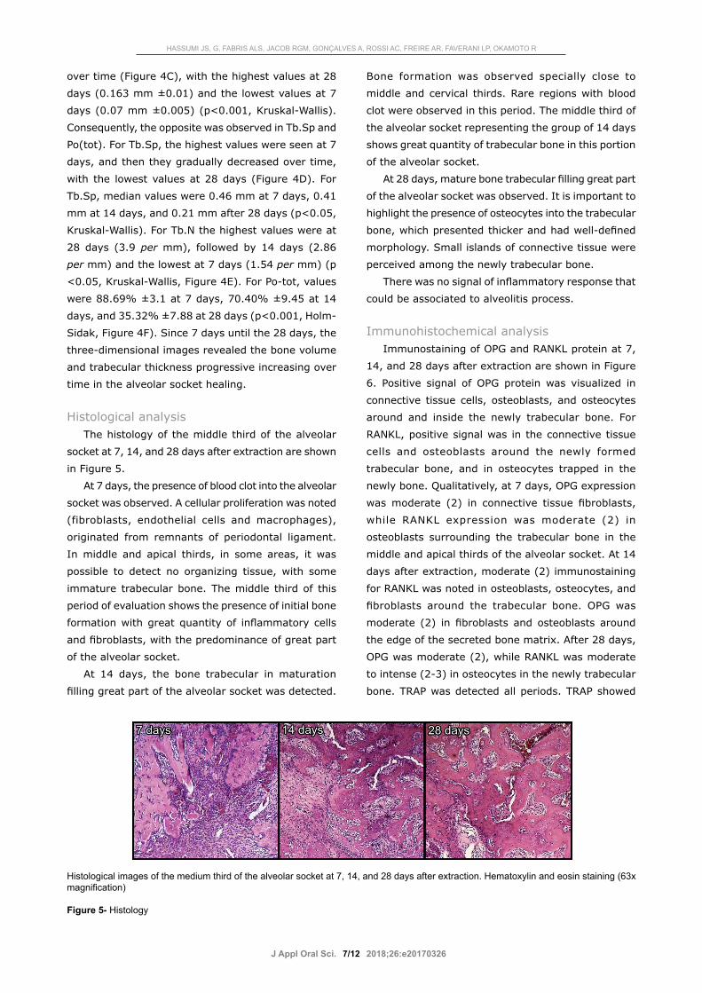
Histological analysisThe histology of the middle third of the alveolar
socket at 7, 14, and 28 days after extraction are shown
in Figure 5.
At 7 days, the presence of blood clot into the alveolar
socket was observed. A cellular proliferation was noted
(fibroblasts, endothelial cells and macrophages),
originated from remnants of periodontal ligament.
In middle and apical thirds, in some areas, it was
possible to detect no organizing tissue, with some
immature trabecular bone. The middle third of this
period of evaluation shows the presence of initial bone
formation with great quantity of inflammatory cells
and fibroblasts, with the predominance of great part
of the alveolar socket.
At 14 days, the bone trabecular in maturation
filling great part of the alveolar socket was detected.
Bone formation was observed specially close to
middle and cervical thirds. Rare regions with blood
clot were observed in this period. The middle third of
the alveolar socket representing the group of 14 days
shows great quantity of trabecular bone in this portion
of the alveolar socket.
At 28 days, mature bone trabecular filling great part
of the alveolar socket was observed. It is important to
highlight the presence of osteocytes into the trabecular
bone, which presented thicker and had well-defined
morphology. Small islands of connective tissue were
perceived among the newly trabecular bone.
There was no signal of inflammatory response that
could be associated to alveolitis process.
Immunohistochemical analysisImmunostaining of OPG and RANKL protein at 7,
14, and 28 days after extraction are shown in Figure
6. Positive signal of OPG protein was visualized in
connective tissue cells, osteoblasts, and osteocytes
around and inside the newly trabecular bone. For
RANKL, positive signal was in the connective tissue
cells and osteoblasts around the newly formed
trabecular bone, and in osteocytes trapped in the
newly bone. Qualitatively, at 7 days, OPG expression
was moderate (2) in connective tissue fibroblasts,
while RANKL expression was moderate (2) in
osteoblasts surrounding the trabecular bone in the
middle and apical thirds of the alveolar socket. At 14
days after extraction, moderate (2) immunostaining
for RANKL was noted in osteoblasts, osteocytes, and
fibroblasts around the trabecular bone. OPG was
moderate (2) in fibroblasts and osteoblasts around
the edge of the secreted bone matrix. After 28 days,
OPG was moderate (2), while RANKL was moderate
to intense (2-3) in osteocytes in the newly trabecular
bone. TRAP was detected all periods. TRAP showed
Histological images of the medium third of the alveolar socket at 7, 14, and 28 days after extraction. Hematoxylin and eosin staining (63x magnification)
Figure 5- Histology
HASSUMI JS, G, FABRIS ALS, JACOB RGM, GONÇALVES A, ROSSI AC, FREIRE AR, FAVERANI LP, OKAMOTO R

J Appl Oral Sci. 2018;26:e201703268/12
mild (1) at 7 days, moderate (2) at 14 days and mild
(1) at 28 days.
The scores were submitted to statistical analysis,
and there were no differences between the periods
for each protein (p>0.05 for comparisons of OPG,
RANKL and TRAP) (Figure 7). This result suggests an
equilibrium of bone remodeling, represented by OPG
(Bone formation) and RANKL (Bone resorption) in all
periods of this study.
Molecular analysisThe relative gene expression of OPG showed an
increase at 28 days compared with 7 days (p<0.05,
Shapiro-Wilk), (Figure 8A). RANKL increased at 14
days compared with 7 and 28 days (p<0.001, Shapiro-
Wilk) (Figure 8B). Runx2 expression increased at 28
days compared with 7 days (p<0.05, Shapiro-Wilk)
(Figure 8C). OC expression increased at 28 days
compared with 7 and 14 days (p<0.05, Shapiro-Wilk)
(Figure 8D). OPN expression increased at 28 days
compared with 7 and 14 days (p<0.05, Shapiro-Wilk)
Representative immunostaining of OPG, RANKL, and TRAP at 7, 14, and 28 days after right upper incisor extraction. OPG and RANKL showed moderate staining all periods analyzed. TRAP was moderate at 14 days and mild at 7 and 28 days. Red arrows indicate intensity of protein tags (63x magnification)
Figure 6- Immunohistochemical staining
Graph with the scores and p-value of OPG, RANK and TRAP
Figure 7- Immunohistochemical results
Alveolar bone healing in rats: micro-CT, immunohistochemical and molecular analysis

J Appl Oral Sci. 2018;26:e201703269/12
Graph showing the relative gene expression and standard deviation of OPG, RANKL, Runx2, OC, OPN, OST, RANK, RANKL/OPG. The * indicates significant statistical difference (p<0.05) between time periods
Figure 8- Molecular results
HASSUMI JS, G, FABRIS ALS, JACOB RGM, GONÇALVES A, ROSSI AC, FREIRE AR, FAVERANI LP, OKAMOTO R

J Appl Oral Sci. 2018;26:e2017032610/12
(Figure 8E). OST expression increased at 28 and 14
days compared with 7 days (p<0.05, Shapiro-Wilk)
(Figure 8F). RANK was not significantly increased (p
>0.05, ANOVA-1 factor) (Figure 8G). Bone turnover,
calculated using the RANKL/OPG ratio, was decreased
at 28 days (p<0.05, Tukey test) when compared with
7 and 14 days (Figure 8H).
Discussion
This study characterized the alveolar bone healing
after the upper right incisor extraction in rats. The
alveolar bone healing showed to be complete at 28
days after surgery, supporting previous studies15,16,22,23.
This finding was confirmed by micro-CT analysis,
which showed the alveolar socket filled with a thick
trabecular bone, smaller trabecular separation and
reduced bone porous at 28 days after extraction. Also
the morphologic data corroborated the qualitative
immunohistochemical and molecular results. In the
qualitative immunohistochemical, discrete changes
in the behavior of OPG, RANKL and TRAP expression
during the periods was observed, however, in the
quantitative data, after the nonparametric test, there
were no statistical differences. Therefore, there
were no significant differences in the pattern of the
expression of OPG, RANKL and TRAP proteins. It
demonstrates that during all of the periods that were
evaluated in this study, there was an equilibrium
between OPG and RANKL and, consequently, in the
remodeling process during the repairing process. In
consequence, osteoclast activity represented by TRAP
immunolabeling presented similar in all of the periods
evaluated in this study. Otherwise, discrete changes
observed in the qualitative analysis requires to be
considered, specially in association with the molecular
and morphometric data, since all methodology need
to be regarded together. Thus, this study should be
considered when thinking about alveolar bone healing
in rats, since this detailed data can support further
experimentations. Our morphometric results revealed
that, under normal conditions, the newly bone formed
displays quite favorable qualitative and quantitative
characteristics in this model, in line with the guide
for quantitative assessment of bone analysis and
supporting that the three-dimensional analyses allow
a better characterization of bone3.
Alveolar bone healing in rats can be divided
into three steps22,23. First, the formation of a stable
fibrin clot allowing cell proliferation that raises
the granulation tissue. Second, the granulation
tissue forms new connective tissue, which contains
mature collagen. Third, this new tissue drives the
intramembranous ossification, allowing the alveolar
socket to fill itself with the new bone. In this new
bone, the basic multicellular unit (BMU) can be well-
defined. The BMU is a balance of bone resorption and
formation, and osteoclasts and osteoblasts10,17. Thus,
during the alveolar healing, the bone should develop
aspects of a mature bone, increasing bone volume and
trabecular thickness, causing a decrease of porosity
and trabecular space, and it was confirmed in this
study. Therefore, these morphological data are an
indication of the performance of BMUs towards the
alveolar bone healing.
In support of our findings, another study of alveolar
socket healing in mice also showed bone volume
and trabecular thickness progressive increasing over
time by Micro-CT analysis30. In addition, the same
study confirmed the correspondence between these
morphometric aspects and the gene expression profile
and histological events30. Interestingly, a clinical
investigation used the Micro-CT to evaluate the
alveolar bone healing with induced laser phototherapy
after third molar extraction26. In this study was possible
to establish a correlation between the morphology by
Micro-CT and the histometrical parameters, supporting
the acceleration of alveolar bone healing by the laser
phototherapy26. Another study evaluated the alveolar
socket healing in a canine model and demonstrated
that periodontal and endodontic pathology can delay
the alveolar bone healing after tooth extraction11.
Thus, it is essential to determinate the alveolar
healing nature of a model in order to compare with
no physiological conditions. Therefore, the alveolar
healing characterization provides knowledge to further
researches for a favorable bone healing.
The OPG, RANK and RANKL are of particular interest
to bone healing. The dynamic actions of these proteins,
especially OPG and RANKL, must be balanced to have
bone homeostasis2,16,19. It will result in equilibrium of
osteoclasts and osteoblasts in the BMUs17. There is
strong evidence that suggest a role of OPG/RANK/
RANKL system in BMU activity9,28. Our findings revealed
different expressions of OPG and RANKL during the
alveolar bone healing. OPG expression increased,
and it was significantly elevated at 28 days. RANKL
Alveolar bone healing in rats: micro-CT, immunohistochemical and molecular analysis

J Appl Oral Sci. 2018;26:e2017032611/12
expression was higher at 14 days. These findings are
consistent with effective alveolar healing, since should
have an initial organization of granulation tissue,
following a gradual replacement by connective tissue
and bone. This process is proportional to an increase of
extracellular OPG, which blocks the bone resorption13.
Also RANKL that regulates osteoclast activity and bone
resorption9 had its expression increased after 14 days,
succeeding the bone formation. After 28 days, RANKL
expression was lower due to inhibition by OPG, which
took control of bone formation. TRAP activated by
RANKL, represents the osteoclast activity resorption25.
Consistent with our immunohistochemical data, TRAP
showed a higher expression at 14 days. This suggests
that a great BMU activity is observed in the middle of
the alveolar bone healing, at 14 days after extraction.
Moreover, in relation to the expression of the mRNA,
there were differences in the expression of the proteins
RUNX2, OST, OC, and OPN along the bone healing15,21.
As expected, the RUNX2 expression increased at 28
days after extraction, performing the differentiation of
the pre-osteoblasts into osteoblasts, responsible for
the bone formation21. For this reason, an increase of
the mineral deposition was observed, characterized by
the protein OST at 14 and 28 days. In the same way,
the bone maturation and organization occurred with
the elevation of OC and OPN, which increased with
28 days. This proteins expression detailed the bone
healing organization from the granulation tissue to
connective tissue until the bone formation.
The question arises about the correlation of mRNA
and protein expression in the developing bone. A direct
relationship of mRNA and protein is expected. However,
in the current study, it was true only for RANKL. The
OPG mRNA expression was increased after 28 days,
but it did not correspond to the protein expression
over the same period. It could be explained by the
fact that OPG is a soluble receptor14. Consequently,
after synthesized by osteoblasts, OPG is released into
the extracellular medium, making its detection more
difficult by immunohistochemical staining. Despite
this, our molecular findings indicate an increase of OPG
expression. These data demonstrate the significance of
multiple techniques to describe more completely the
behavior of a specific protein during the bone healing.
Taken together, these findings demonstrate a
significant function of the OPG/RANK/RANKL system
in the osteoclasts and osteoblasts response, and BMU
activation. Once activated, BMUs provide valuable
information of amount and quality to form the bone17.
It will allow the alveolar socket to fill a thicker and
slightly separated trabecular bone. These findings
complete previous studies that described the alveolar
bone healing in rat ended at 28 days22,23. Another
aspect evaluated was the metabolic activity inside
the alveolar socket. At 14 days after extraction,
immunostaining revealed high activity of proteins
involved in synthesis of the mineralized matrix.
Additionally, OPG and RANKL protein showed strong
activity at 14 days after extraction, which can be
related to higher BMU activity during this time.
In summary, a favorable alveolar bone healing is
expected at 28 days after tooth extraction in rats, with
equilibrium of RANKL and OPG expression or even the
predominance of OPG in the end. The gene expression
can be related to the morphometric parameters
obtained by Micro-CT analysis. Further studies will
be needed to better characterize extracellular matrix
formation of the alveolar bone. Moreover, identifying
other proteins that are involved in the bone formation
and resorption is required. Lastly, we would like to
highlight the significance of performing a Micro-CT
analysis of bone healing, parallel to protein expression
profile and molecular responses. We consider that
Micro-CT analysis must be performed to improve the
parameters that determine the quality and quantity of
the newly formed bone after tooth extraction.
Conclusions
1) Micro-CT revealed that the newly formed
alveolar bone after tooth extraction displays very
favorable morphometric characteristics of quality and
quantity on this experimental model.
2) Beyond other proteins expressed, the mutual
activity of OPG and RANKL is essential for BMUs
activation during the alveolar bone healing.
AcknowledgmentsThe authors would like to thank the Research
Pro-Rectory - UNESP (#145/004/13 PROPe CDC)
and the São Paulo Research Foundation (FAPESP,
#2004/1959-0) for financial support. We also thank
Fabíola Singaretti de Oliveira and Adriana Luísa
Gonçalves. We certify that do not have any commercial
or associate interest that represents a conflict of
interest in connection with the submitted manuscript.
HASSUMI JS, G, FABRIS ALS, JACOB RGM, GONÇALVES A, ROSSI AC, FREIRE AR, FAVERANI LP, OKAMOTO R

J Appl Oral Sci. 2018;26:e2017032612/12
References1- Amler MH. The time sequence of tissue regeneration in human extraction wounds. Oral Surg Oral Med Oral Pathol. 1969;27(3):309-18.2- Belibasakis GN, Bostanci N. The RANKL-OPG system in clinical periodontology. J Clin Periodontol. 2012;39(3):239-48.3- Bouxsein ML, Boyd SK, Christiansen BA, Guldberg RE, Jepsen KJ, Müller R. Guidelines for assessment of bone microstructure in rodents using micro-computed tomography. J Bone Miner Res. 2010;25(7):1468-86.4- Calixto RF, Teófilo JM, Brentegani LG, Lamano-Carvalho TL. Grafting of tooth extraction socket with inorganic bovine bone or bioactive glass particles: comparative histometric study in rats. Implant Dent. 2007;16(3):260-9.5- Carvalho TL, Bombonato KF, Brentegani LG. Histometric analysis of rat alveolar wound healing. Braz Dent J. 1997;8(1):9-12.6- Chen H, Liu N, Xu X, Qu X, Lu E. Smoking, radiotherapy, diabetes and osteoporosis as risk factors for dental implant failure: a meta-analysis. PLoS One. 2013;8(8):e71955.7- Faverani LP, Polo TO, Ramalho-Ferreira G, Momesso GA, Hassumi JS, Rossi AC, et al. Raloxifene but not alendronate can compensate the impaired osseointegration in osteoporotic rats. Clin Oral Investig. 2018;22(1):255-65.8- Gealh WC, Pereira CC, Luvizuto ER, Garcia-Júnior IR, Antoniali C, Okamoto R. Healing process of autogenous bone graft in spontaneously hypertensive rats treated with losartan: an immunohistochemical and histomorphometric study. J Oral Maxillofac Surg. 2014;72(12):2569-81.9- Honma M, Ikebuchi Y, Kariya Y, Suzuki H. Regulatory mechanisms of RANKL presentation to osteoclast precursors. Curr Osteoporos Rep. 2014;12(1):115-20.10- Katagiri T, Takahashi N. Regulatory mechanisms of osteoblast and osteoclast differentiation. Oral Dis. 2002;8(3):147-59.11- Kim JH, Koo KT, Capetillo J, Kim JJ, Yoo JM, Ben Amara H, et al. Periodontal and endodontic pathology delays extraction socket healing in a canine model. J Periodontal Implant Sci. 2017;47(3):143-53.12- Kim JH, Park YB, Li Z, Shim JS, Moon HS, Jung HS, et al. Effect of alendronate on healing of extraction sockets and healing around implants. Oral Dis. 2011;17(7):705-11.13- Kostenuik PJ, Shalhoub V. Osteoprotegerin: a physiological and pharmacological inhibitor of bone resorption. Curr Pharm Des. 2001;7(8):613-35.14- Lacey DL, Timms E, Tan HL, Kelley MJ, Dunstan CR, Burgess T, et al. Osteoprotegerin ligand is a cytokine that regulates osteoclast differentiation and activation. Cell. 1998;93(2):165-76.15- Luvizuto ER, Dias SM, Queiroz TP, Okamoto T, Garcia IR Jr, Okamoto R, et al. Osteocalcin immunolabeling during the alveolar healing process in ovariectomized rats treated with estrogen or raloxifene. Bone. 2010;46(4):1021-9.16- Luvizuto ER, Queiroz TP, Dias SM, Okamoto T, Dornelles RC, Garcia IR Jr, et al. Histomorphometric analysis and immunolocalization of RANKL and OPG during the alveolar healing process in female ovariectomized rats treated with oestrogen or raloxifene. Arch Oral Biol. 2010;55(1):52-9.
17- Manolagas SC. Birth and death of bone cells: basic regulatory mechanisms and implications for the pathogenesis and treatment of osteoporosis. Endocr Rev. 2000;21(2):115-37.18- Manrique N, Pereira CC, Garcia LM, Micaroni S, Carvalho AA, Perri SH, et al. Alveolar bone healing process in spontaneously hypertensive rats (SHR). A radiographic densitometry study. J Appl Oral Sci. 2012;20(2):222-7.19- Manrique N, Pereira CC, Luvizuto ER, Sánchez MP, Okamoto T, Okamoto R, et al. Hypertension modifies OPG, RANK, and RANKL expression during the dental socket bone healing process in spontaneously hypertensive rats. Clin Oral Investig. 2015;19(6):1319-27.20- McCarron DA, Yung NN, Ugoretz BA, Krutzik S. Disturbances of calcium metabolism in the spontaneously hypertensive rat. Hypertension. 1981;3(3 Pt 2):I162-7.21- Nanci A. Bone. In: ______. Ten Cate’s oral histology: development, structure and function. Rio de Janeiro: Elsevier; 2013. p.109-40.22- Okamoto T, de Russo MC. Wound healing following tooth extraction. Histochemical study in rats. Rev Fac Odontol Aracatuba. 1973;2(2):153-69.23- Okamoto T, Vasconcelos Fialho AC. Comparative histological study of two methods of obtaining alveolar sections in rats. Rev Odontol UNESP. 1990;19(1):63-74.24- Onyia JE, Galvin RJ, Ma YL, Halladay DL, Miles RR, Yang X, et al. Novel and selective small molecule stimulators of osteoprotegerin expression inhibit bone resorption. J Pharmacol Exp Ther. 2004;309(1):369-79.25- Pedrosa WF Jr, Okamoto R, Faria PE, Arnez MF, Xavier SP, Salata LA. Immunohistochemical, tomographic and histological study on onlay bone graft remodeling. Part II: calvarial bone. Clin Oral Implants Res. 2009;20(11):1254-64.26- Romão MM, Marques MM, Cortes AR, Horliana AC, Moreira MS, Lascala CA. Micro-computed tomography and histomorphometric analysis of human alveolar bone repair induced by laser phototherapy: a pilot study. Int J Oral Maxillofac Surg. 2015;44(12):1521-8.27- Schoppet M, Preissner KT, Hofbauer LC. RANK ligand and osteoprotegerin: paracrine regulators of bone metabolism and vascular function. Arterioscler Thromb Vasc Biol. 2002;22(4):549-53.28- Teitelbaum SL. Bone resorption by osteoclasts. Science. 2000;289(5484):1504-8.29- Turner CH. Biomechanics of bone: determinants of skeletal fragility and bone quality. Osteoporos Int. 2002;13(2):97-104.30- Vieira AE, Repeke CE, Ferreira Junior SB, Colavite PM, Biguetti CC, Oliveira RC, et al. Intramembranous bone healing process subsequent to tooth extraction in mice: micro-computed tomography, histomorphometric and molecular characterization. PLoS One. 2015;10(5):e0128021.
Alveolar bone healing in rats: micro-CT, immunohistochemical and molecular analysis